| ABC
ABC Family Arquivos Brasileiros de Cardiologia International Journal of Cardiovascular Sciences ABC Heart Failure & Cardiomyopathy ABC Imagem Cardiovascular
Português English
ABC Family | ABC
Português English
  • About
    • About the Journal
    • Internal Regulation
    • Editors Section
    • Editorial Board
    • History
    • SBC Award
  • Instructions to Authors
    • Guidelines for Publication
    • Editorial Policy
    • Ethics Policy
    • Policy on the Use of Artificial Intelligence
    • Mandatory Publication Form
    • Publication Fee
  • Issues
  • SBC Guidelines
  • Abstracts
    • How to Publish an Abstract Supplement?
    • Supplements from the Brazilian Congress of Cardiology
  • About
    • About the Journal
    • Internal Regulation
    • Editors Section
    • Editorial Board
    • History
    • SBC Award
  • Instructions to Authors
    • Guidelines for Publication
    • Editorial Policy
    • Ethics Policy
    • Policy on the Use of Artificial Intelligence
    • Mandatory Publication Form
    • Publication Fee
  • Issues
  • SBC Guidelines
  • Abstracts
    • How to Publish an Abstract Supplement?
    • Supplements from the Brazilian Congress of Cardiology

ABC Family

  • ABC Family
  • Arquivos Brasileiros de Cardiologia
  • International Journal of Cardiovascular Sciences
  • ABC Heart Failure & Cardiomyopathy
  • ABC Imagem Cardiovascular
Search in

No content found.

Journals
Ordering
Article types
Others

  1. Home
  2. ABC
  3. Issues
  4. 2019, Volume 112, Number 1
Erratum

Erratum

August issue of 2018, vol. 111(2), pages 230-289

Consider correct the following page numbering of contents for the items: for page 4, consider 231; respectively: 5, 234; 6, 235; 7, 236; 10, 239; 11, 240; 13, 242; 15, 244; 16, 245; 18, 247; 19, 248; 20, 249; 21, 250; 22, 251; 23, 252; 29, 258; 30, 259; 33, 262; 34, 263; 35, 264; 37, 266; 39, 268; 41, 272; 44, 273; 46, 275; 47, 276; 48, 277; 49, 278; 50, 50; 51, 280. For the items indicated with the pages 24-28 and 31-32, consider: 4.1.2, 4.1.2.1, 4.1.2.2, A) Ciclosporina – page 253; B) Tacrolimus, 4.1.2.3, A) Azatioprina, B) Micofenolato, 4.1.2.4, A) Sirolimus – page 255; B) Evrrolimus, 4.1.3, 4.2, 4.2.1, 4.2.2, 4.2.2.1 – page 256; 4.2.2.2 – page 257; 4.2.3 – page 258; 4.2.3.1 – page 258; 4.2.3.2 – page 258; 4.3.2, 4.3.3, 4.3.3.1 – page 260; 4.3.3.2 – page 261. On page 251, the correct numbering of the items “Gravidade e Biomarcadores”, respectively, is 3.4.4 and 3.4.5. On page 252, the correct numbering of the “Estratégias de Prevenção e Tratamento” item is 3.4.6.

[…]

Read Full Article
Creative Commons Attribution (CC BY) Creative Commons license
01/01/2019 DOI: 10.5935/abc.20190004
Editorial

Usefulness of Myocardial Deformation Indices in Preventing Cardiotoxicity in Breast Cancer Patients

Marcelo Dantas Tavares de Melo et al. Vera Maria Cury Salemi

The first description of chemotherapy-induced heart failure (Stage C) was published in 1967. There has been a therapeutic evolution in oncological treatment since then, as shown by the fact that, as of 2005, the survival rate exceeded that of mortality. This has resulted in a new epidemiological problem for these survivors, since at least 30% of them will show some degree of cardiotoxicity, which can occur up to decades after the end of the chemotherapy. Moreover, cardiovascular mortality is already considered the second most common cause of death, second only to cancer.–

The classically accepted definition for cardiotoxicity during treatment was proposed in 2014, which described it as an absolute decrease in left ventricular (LV) ejection fraction of 10 percentage points to values below 53%, with its re-evaluation being recommended after 2 to 3 weeks. Additionally, the subclinical lesion is based on the relative decrease in global LV longitudinal strain by 15% in relation to the baseline. The major concern is that systolic dysfunction can lead to a therapeutic dose adjustment, less effective alternative therapy regimens, or, in the worst-case scenario, to chemotherapy discontinuation.

[…]

  • Antineoplastic Agents
  • Breast Cancer
  • Cardiotoxicity
  • Drug Therapy
  • Ventricular Dysfunction
Read Full Article
Creative Commons Attribution (CC BY) Creative Commons license
01/01/2019 DOI: 10.5935/abc.20190009
Brief Communication

Diuretics are Similar to Losartan on Echocardiographic Target-Organ Damage in Stage I Hypertension. PREVER-Treatment Study

Carolina Bertoluci et al. Murilo Foppa Angela Barreto Santiago Santos Sandra C. Fuchs Flávio Danni Fuchs

Abstract

Blood pressure (BP)-lowering therapy improves left ventricular (LV) parameters of hypertensive target-organ damage in stage II hypertension, but whether there is a drug-class difference in echocardiographic parameters in stage I hypertension patients is less often studied. In the PREVER treatment study, where individuals with stage I hypertension were randomized for treatment with diuretics (chlorthalidone/amiloride) or losartan, 110 participants accepted to participate in a sub-study, where two-dimensional echocardiograms were performed at baseline and after 18 months of antihypertensive treatment. As in the general study, systolic BP reduction was similar with diuretics or with losartan. Echocardiographic parameters showed small but significant changes in both treatment groups, with a favorable LV remodeling with antihypertensive treatment for 18 months when target blood pressure was achieved either with chlorthalidone/amiloride or with losartan as the initial treatment strategy. In conclusion, even in stage I hypertension, blood pressure reduction is associated with improvement in echocardiographic parameters, either with diuretics or losartan as first-drug regimens.

  • Angiotensin-Converting Enzyme Inhibitors
  • Anihypertensive Agents
  • Antihypertensive Agents/therapy
  • Blood Pressure
  • Calcium Channel
  • Echocardiography
  • Hypertension/complications
  • Hypertension/therapy
Read Full Article
Creative Commons Attribution (CC BY) Creative Commons license
01/01/2019 DOI: 10.5935/abc.20180249 Views 3
Figure 1
Review Article

Arrhythmogenic Right Ventricular Cardiomyopathy/Dysplasia (ARVC/D) – What We Have Learned after 40 Years of the Diagnosis of This Clinical Entity

Jorge Elias Neto et al. Joelci Tonet Robert Frank Guy Fontaine

Abstract

Arrhythmogenic right ventricular cardiomyopathy/dysplasia (ARVC/D) was initially recognized as a clinical entity by Fontaine and Marcus, who evaluated a group of patients with ventricular tachyarrhythmia from a structurally impaired right ventricle (RV). Since then, there have been significant advances in the understanding of the pathophysiology, manifestation and clinical progression, and prognosis of the pathology. The identification of genetic mutations impairing cardiac desmosomes led to the inclusion of this entity in the classification of cardiomyopathies. Classically, ARVC/D is an inherited disease characterized by ventricular arrhythmias, right and / or left ventricular dysfunction; and fibro-fatty substitution of cardiomyocytes; its identification can often be challenging, due to heterogeneous clinical presentation, highly variable intra- and inter-family expressiveness, and incomplete penetrance.

In the absence of a gold standard that allows the diagnosis of ARVC/D, several diagnostic categories were combined and recently reviewed for a higher diagnostic sensitivity, without compromising the specificity. The finding that electrical abnormalities, particularly ventricular arrhythmias, usually precede structural abnormalities is extremely important for risk stratification in positive genetic members. Among the complementary exams, cardiac magnetic resonance imaging (CMR) allows the early diagnosis of left ventricular impairment, even before morpho-functional abnormalities. Risk stratification remains a major clinical challenge, and antiarrhythmic drugs, catheter ablation and implantable cardioverter defibrillator are the currently available therapeutic tools. The disqualification of the sport prevents cases of sudden death because the effort can trigger not only the electrical instability, but also the onset and progression of the disease.

  • Arrhythmias, Cardiac/diagnostic imaging
  • Arrhythmogenic Right Ventricular Dysplasia/physiopathology
  • Catheter Ablation
  • Defibrillators, Implantable
  • Magnetic Resonance Imaging
Read Full Article
Creative Commons Attribution (CC BY) Creative Commons license
01/01/2019 DOI: 10.5935/abc.20180266 Views 1
Figure 1
Image

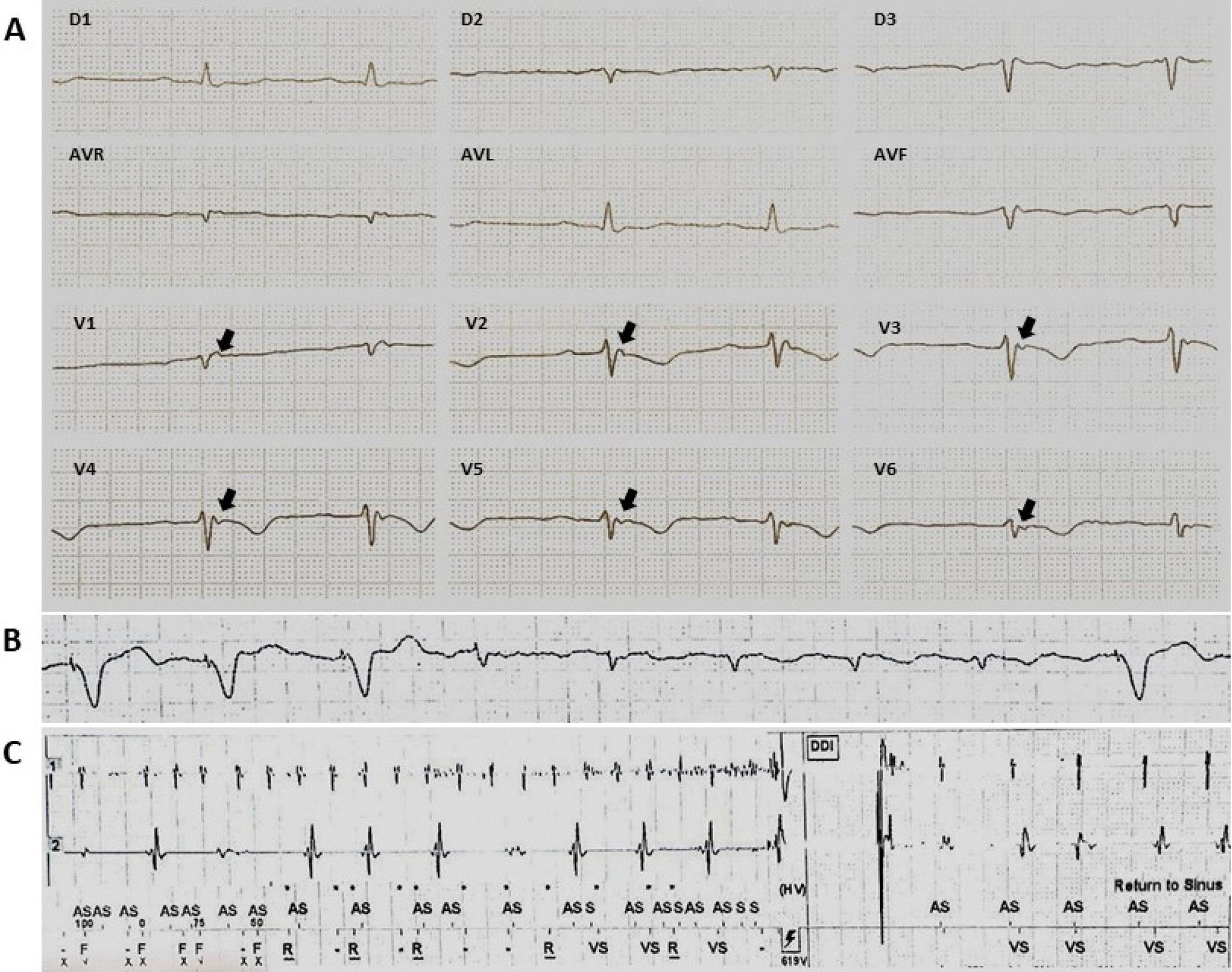
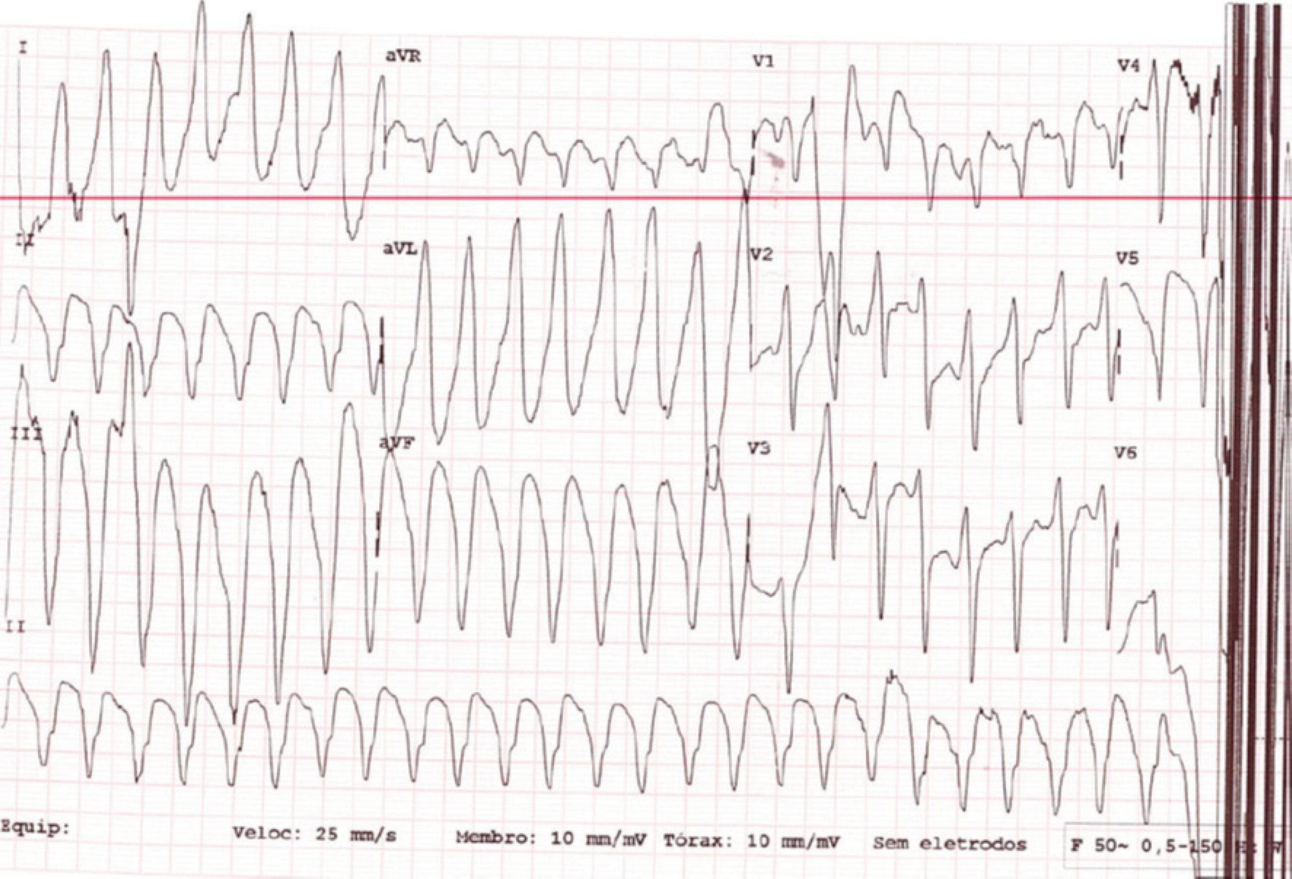
A Large Cardiac Metastasis of a Parathyroid Tumour Presenting with Ventricular Tachycardia

Rita Ilhão Moreira et al. Sílvia Aguiar Rosa Ana Galrinho Nuno Jalles Tavares Rui Cruz Ferreira

A-81-years old woman was admitted after an episode of ventricular tachycardia with hemodynamic instability converted after electrical cardioversion (). Past medical history was significant for poorly differentiated squamous cell carcinoma of left parathyroid, diabetes and hypertension.

[…]

  • Arrhythmias Cardiac
  • Carcinoma, Squamous Cell
  • Diagnosis Imaging
  • Echocardiography/methods
  • Neoplasms Metastasis
  • Neoplasms Metastasis/therapy
  • Parathyroid Neoplasms
Read Full Article
Creative Commons Attribution (CC BY) Creative Commons license
01/01/2019 DOI: 10.5935/abc.20180255 Views 3
Editorial

Analysis of FFR Measurement Clinical Impact and Cost-Effectiveness Compared to Angiography In Multi-Arterial Patients Undergoing PCI

Fernando Mendes Sant'Anna et al. Lucas Bonacossa Sant'Anna

The study by Quintella et al. published in this issue of the journal, brings us valuable information about the use of an important physiological evaluation tool in the hemodynamic laboratory. FFR-guided treatment (myocardial fractional flow reserve), used in the percutaneous coronary intervention (PCI) with bare-metal stent (BMS) implantation in multi-arterial patients treated in the Unified Health System (SUS) has been shown to be useful in decreasing the incidence of new revascularization of the target vessel (clinical restenosis), as well as being cost-effective when compared to the angiography-guided treatment.

The value of FFR to predict major adverse cardiovascular events (MCAEs) prior to PCIs has been established for many years. Its ability to detect ischemia and, with this, to guide the most appropriate treatment, has undergone the test of time, and passed. The 15-year follow-up of the DEFER study in single-vessel patients, and the 5-year studies, FAME 1, and FAME 2, in multiarterial patients, showed consistent and unquestionable results, with a better, or at least similar, clinical progression, in the FFR-guided groups, using less stents with fewer lesions and consequently lower costs, as well as evidenced the safety of leaving lesions whose FFR was not indicative of ischemia only on drug treatment.

[…]

  • Angioplasty, Balloon, Coronary
  • Coronary Artery Disease/economics
  • Cost-Benefit Analysis
  • fractional Flow Reserve,Myocardial
  • Stents
Read Full Article
Creative Commons Attribution (CC BY) Creative Commons license
01/01/2019 DOI: 10.5935/abc.20180261 Views 1
Figure 1
Clinicoradiological Correlation

Case 1 / 2019 – Natural Evolution of Double Outlet Right Ventricle with Noncommitted Ventricular Septal Defect and Pulmonary Stenosis in a 28-Year-Old Asymptomatic Woman

Edmar Atik et al. Alessandra Costa Barreto Maria Angélica Binotto

Clinical data

The patient evolved without symptoms from birth, when the diagnosis of heart disease was made, as evidenced by heart murmur. Two years ago, supraventricular extrasystoles caused the use of atenolol, propafenone, and magnesium, without improvement. Infective endocarditis was effectively treated 10 years ago. The patient uses levothyroxine 50 mcg for hypothyroidism.

Physical examination: good overall status, eupneic, acyanotic, normal pulses in the four limbs. Weight: 60 Kg, Height 160 cm, right upper extremity blood pressure: 120 x 70 mmHg, HR: 60 bpm.

[…]

  • Adult
  • Clinical Evolution
  • Double outlet Right Ventricle
  • Heart Septal Defects, Ventricular
  • Pulmonary Valve Stenosis
Read Full Article
Creative Commons Attribution (CC BY) Creative Commons license
01/01/2019 DOI: 10.5935/abc.20180260 Views 1
Editorial

Cardiovascular Rehabilitation in Coronary Artery Disease and Better Knowledge of Its Own Disease

Luiz Antonio Machado César

In the last ten to fifteen years, we are seen more and more concern with patients´ awareness about their diseases.– Campaigns have been brought to the public to teach them about symptoms and signs that might bring concern and make them ask for help and go to an emergency room or, in the United States to call 911. This is true especially for patients with coronary artery disease (CAD), given the possibility of better quality of life and in some cases even a better prognosis of the disease in respect to morbidity and mortality.

Beside this kind of campaigns, recently others started to aware people already with a diagnosis of CAD to the symptoms that may alert them of a problem and the consciousness about all medicines that modify the course of the disease and ways of life that are proved to contribute to this amelioration, as is physical activity.–

[…]

  • Cardiac Rehabilitation
  • Coronary Artery Disease/rehabilitation
  • Exercise
  • Health Promotion
  • Patient Health Questionnaire
  • Preventive Medicine
Read Full Article
Creative Commons Attribution (CC BY) Creative Commons license
01/01/2019 DOI: 10.5935/abc.20180258 Views 1
Editorial

Prenatal Stress: Molecular Mechanisms and Cardiovascular Disease

Marcelo Diarcadia Mariano Cezar et al. Mariana Janini Gomes Ricardo Luiz Damatto

Concern of the researchers in finding the causes of cardiovascular disorders is evident. Recently, scientists are focusing in intrauterine environment investigations in order to seek early causes of these diseases. Studies indicates that prenatal stress increases risk of cardiovascular diseases in adulthood., Among the risks, susceptibility to adult hypertension is a concern, and the sympathetic nervous system is one of the targets of interest, specifically the activity of beta-adrenergic receptors, which has subtype β1 cardiac predominance., These receptors modulates cardiac changes and may lead to ventricular dysfunction as well as severe conditions of heart failure, which increases mortality risk as showed in hypertensive rats studies.,

Another factor associated with the occurrence of heart failure is monoamine oxidase A (MAO-A). This enzyme has its activity increased in hypertension and is responsible for the degradation of catecholamine, which increases the reactive oxygen species generation leading to cardiotoxicity. Therefore, it is extremely important target the causes to prevent or attenuate the of alterations resulting from cardiovascular diseases.

[…]

  • Cardiovascular Diseases
  • Heart Failure
  • Hypertension
  • Rats
  • Stress, Psychological
Read Full Article
Creative Commons Attribution (CC BY) Creative Commons license
01/01/2019 DOI: 10.5935/abc.20180246 Views 1
Editorial

Reperfusion Criteria in Patients Submitted to Fibrinolysis: Is There Room for Improvement?

Milena Soriano Marcolino et al. Antonio Luiz Pinho Ribeiro

Many ST-elevation acute myocardial infarction (STEMI) patients seek care in hospitals without percutaneous coronary intervention (PCI) capability and cannot be submitted to PCI within the guideline-recommended timelines, and, instead, they are often submitted to fibrinolysis as the initial reperfusion therapy. Rapid, simple and readily available bedside measures are of utmost importance for timely assessment of the efficacy of reperfusion therapy early after fibrinolysis in acute STEMI, in order to immediately identify the ones who require rescue PCI.,

In an editorial for Circulation in 2001, Gibson stated “In a time of dizzying advances in diagnostic modalities, it is refreshing to see what a useful, simple, noninvasive, broadly accessible, easily repeatable/applied, and affordable tool the electrocardiography (ECG) is”. This is still up to date. Multiple studies have demonstrated improved outcomes among patients who achieve complete ST resolution at 60-90 minutes after fibrinolytic therapy, and it is recommended that the absence of > 50% reduction in ST elevation in the worst lead at 60-90 minutes should prompt strong consideration of coronary angiography and rescue PCI. , However, this measure, combined with the absence of reperfusion arrhythmias at 2 hours after treatment, has a positive predictive value of 87% and a negative predictive value of 83% to predict failure of reperfusion, , indicating that there is still room for improvement in accuracy.

[…]

  • Electrocardiography/methods
  • Fibrinolysis
  • Percutaneous Coronary Intervention/economics
  • ST Elevation Myocardial Infarction/mortality
  • Thrombolytic THerapy/methods
  • Time Factors
Read Full Article
Creative Commons Attribution (CC BY) Creative Commons license
01/01/2019 DOI: 10.5935/abc.20180245
Editorial

A New Marker of Myocardial Bridge?

Paolo Blanco Villela et al. Gláucia Maria Moraes de Oliveira

Myocardial bridges (MBs) have been associated with an increased incidence of cardiovascular events. Even though the pathophysiology of this association is still elusive, it seems to be related to the development of atherosclerosis. This hypothesis is based on changes in blood flow resulting from systolic compression of coronary arteries and leading to changes in arterial wall shear stress, which could act as a proatherogenic event affecting the endothelium of the arterial segment proximal to the MB.

Recently, Akishima-Fukasawa et al. assessed 150 autopsies of individuals without cardiovascular heart disease to verify the influence of MBs on the development of atherosclerosis. The authors found the occurrence of MBs in 93 hearts, and using computer-assisted histomorphometry, observed a higher frequency of luminal stenosis in segments proximal to the MB. Using a multiple comparison test, the authors showed a relation between the presence of risk factors like hypertension, diabetes, and dyslipidemia to a higher rate of stenosis affecting segments located 2.5 cm proximally to the MB. Despite the absence of MB flow evaluation, the authors documented an anatomical association between significant atherosclerotic lesions and the presence of MBs. We could speculate that this may be one of the explanations for the increased incidence of cardiovascular events in patients with MB.

[…]

  • Atherosclerosis
  • Inflammation
  • Lipoproteins, HDL
  • Monocytes
  • Myocardial Bridging/physiopathology
  • Risk Factors
Read Full Article
Creative Commons Attribution (CC BY) Creative Commons license
01/01/2019 DOI: 10.5935/abc.20180264
Editorial

Synergy of Rapamycin and Cyanoacrylate in Reducing Intimal Hyperplasia

Marcia Kiyomi Koike

Myocardial revascularization (CABG) surgery remains one of the main therapies for coronary disease. However, vascular disease that follows CABG remains a challenge to medicine. The mechanical, molecular and cellular changes undergone by the venous vascular graft when inserted into the coronary artery flow culminate in thrombosis, intimal hyperplasia, and atherosclerotic process, and end with restenosis.,

CABG experimental models were crucial to the understanding of mechanisms present in the course of vascular disease. The vascular graft model with anastomosis of the external jugular vein with the carotid artery represents an experimental model of vascular disease similar to that occurring after CABG. Pigs, rabbits, and rats may be used in this model because of protocol reproducibility, cost, and benefit.,

[…]

  • Cianoacrilates
  • Coronary Artery Diseases
  • Hyperplasia
  • Imunosupressive Agents
  • Myocardial Revascularization
  • Rats
  • Vascular Diseases
Read Full Article
Creative Commons Attribution (CC BY) Creative Commons license
01/01/2019 DOI: 10.5935/abc.20180269
Editorial

Cardiac Fibrosis Occurs before Arterial Hypertension Becomes Well Defined?

Claudio Pinho

Target organ damage (TOD) of systemic arterial hypertension (AH) in the heart modifies the cardiomyocyte, Interstice and its arteries. Alterations that occur in AH include cardiomyocyte hypertrophy, connective tissue hyperplasia and neovascularization stimulation, among others.– These alterations depend essentially on the stimulus of the renin-angiotensin-aldosterone system (RAAS) and the sympathetic nervous system (SNS)– which may not homogeneously impact Kidneys, heart, brain and blood vessels. The response of the connective tissue to AH induces collagen production by fibroblasts and consequently interstitial fibrosis.–

Since 2006 with the data published by Das MK et al., we have started to correlate the presence of notches that form the fragmented QRS (fQRS) with non-homogeneous electrical conduction resulting from myocardial fibrosis which can be restorative or reactive.

[…]

  • Blood Pressue Monitoring Ambulatory/methods
  • Hypertension
  • Myoblasts, Cardiac
  • Myocites, Cardiac
  • Prehypertension
  • Renin-Angiotensin System
Read Full Article
Creative Commons Attribution (CC BY) Creative Commons license
01/01/2019 DOI: 10.5935/abc.20180243 Views 1
Editorial

Is There a Role For Whole Body Vibration in Protecting Cardiovascular Disease?

Leonardo Zornoff et al. Marcos Ferreira Minicucci

Cardiovascular disease is the leading cause of death worldwide. In this context, it is well accepted that physical activity plays a critical role as a powerful therapeutic strategy for prevention and progression of cardiovascular disease, both in experimental models and in different clinical situations.,

Whole body vibration training (WBV) is a new intriguing training program. Importantly, if vibration-induced protection is an effective method of exercise, it could be an alternative to exercise training especially for those who are unable to perform the traditional exercise. Therefore, the effects of WVB on different systems, including the cardiovascular system, has been investigated in recent years.

[…]

  • Cardiovascular Diseases
  • Exercise Movement Techniques
  • Exercise/prevention and control
  • Mobility
  • Mortality
  • Stroke/rehabilitation
  • Vibration
Read Full Article
Creative Commons Attribution (CC BY) Creative Commons license
01/01/2019 DOI: 10.5935/abc.20180257
Figure 1
Case Report

Myocardial Infarction Due to an Anomalous Origin of the Left Coronary Artery with Unique Aggravating Features

Jorge Alberto Silva Estrada et al. Alejandra Domínguez Camacho Lorenzo Reyes de la Cruz Jesus Reyna Figueroa

Introduction

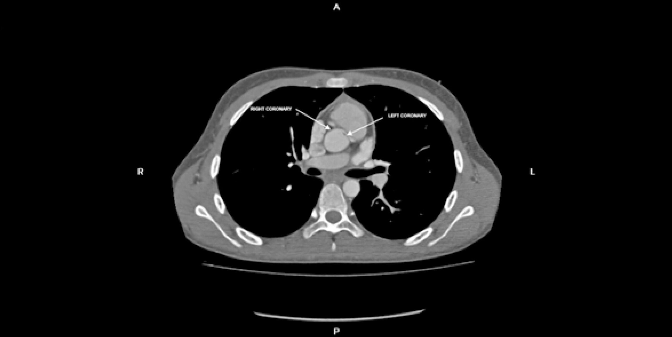
Anomalous left coronary artery arising at the right sinus of Valsalva is a relatively rare congenital cardiac anomaly that can cause myocardial ischemia. It can follow one out of five aberrant courses: interarterial, subpulmonic, pre-pulmonic, retroaortic, or retrocardiac.

We present the case of a young patient with an anomalous left coronary artery arising at the right sinus of Valsalva with severe stenosis and hypoplasia throughout the interarterial segment, which hampered corrective surgery. The timely detection and adequate treatment of this specific anomaly gains relevance because of its association with increased risk of sudden cardiac death. Although the true prevalence of interarterial anomalous left coronary artery is unknown, owing to the lack of population-wide screening studies and at times asymptomatic course, its frequency has been reported at 0.03%. Imaging techniques allow the characterization of the coronary anomalous origin, course, morphology and surrounding structures. Transthoracic echocardiography, magnetic resonance angiography, and computed tomography (CT) angiography are first line noninvasive tests available while invasive tests such as coronary angiography and intravascular ultrasound are second line diagnostic alternatives. Treatment approaches are still controversial and election of the optimal surgical procedure, whenever applicable, must be an individualized and patient-centered decision.

[…]

  • Echocardiography/methods
  • Heart Defects, Congenital
  • Magnetic Resonance Spectroscopy/ methods
  • Myocardial Infarction
  • Myocardial Revascularization
Read Full Article
Creative Commons Attribution (CC BY) Creative Commons license
01/01/2019 DOI: 10.5935/abc.20180217
Viewpoint

Coronary Computed Tomography Angiography Takes the Center Stage and Here is Why

Illan Gottlieb et al. Marcio Sommer Bittencourt Carlos Eduardo Rochitte João L. Cavalcante

Max Planck once said that “A new scientific truth does not triumph by convincing its opponents and making them see the light, but rather because its opponents eventually die, and a new generation grows up that is familiar with it.” In its beginnings, coronary computed tomography angiography (CCTA) was accused of having too low accuracy for the diagnosis of obstructive coronary artery disease (CAD) to be used in clinical practice. Over the last decade, major technical developments such as larger axial coverage (from 2 cm to 16 cm) and improved temporal resolution, have enabled CCTA to become by far the most accurate non-invasive imaging method for diagnosis of obstructive CAD, with sensitivity and specificity of approximately 95% and 90%, respectively.

Then CCTA was burdened with the accusation of exposing patients to radiation doses so high, that warranted some society guidelines to specifically point this out and limit its use. At that time, CCTA exposed patients to doses ranging from 20 to 25 mSV, while triphasic abdomen CT exposed patients to 30 to 40 mSv and scintigraphic myocardial perfusion studies with Thallium used up to 40 mSv. In 2018, radiation exposure from CCTA dropped to well below 5 mSv (most advanced clinical centers use much less), a fraction of the dose used in myocardial perfusion studies with MIBI tetrophosmin. Then the cost-effectiveness wave came with societies rightfully demanding proof that CCTA offered more value at an acceptable cost compared to other imaging modalities, and CCTA once again proved to be more cost-effective than other modalities. Although one hardly finds cost-effectiveness studies comparing nuclear scans with ECG treadmill tests, providing better diagnosis performance is not enough anymore. More recently, this strategy has even been put into challenge in large randomized clinical trials comparing CCTA with the standard of care in the investigation of suspected CAD both in the acute and in the outpatient settings.–

[…]

  • Computed Tomography Angiography
  • Coronary Artery Disease/diagnostic imaging
  • Coronary Artery Disease/physiopathology
  • Coronary Artery Disease/prevention and control
  • Tomography, X-Ray Computed
Read Full Article
Creative Commons Attribution (CC BY) Creative Commons license
01/01/2019 DOI: 10.5935/abc.20190003 Views 1
Editorial

Coronary Artery Calcium – From Screening to a Personalized Shared Decision-Making Tool: The New American Prevention Guidelines

Marcio Sommer Bittencourt et al. Michael J. Blaha Khurram Nasir

The United States (US) National Cholesterol Education Program (NCEP) formed the Adult Treatment Panel (ATP) in 1985 with the aim to educate clinician and provide guideline recommendations for the treatment of dyslipidemias. In its first 1998 recommendations, the approach to primary prevention of cardiovascular disease included LDL-cholesterol (LDL-C) reduction in individuals with more than two risk factors and LDL-C levels above 160mg/dL and optional treatment in those with “borderline” LDL-C levels between 130 – 159 mg/dL. In its second version, in 1994, a category of secondary prevention with a target LDL-C below 100mg/dL was introduced. In 2001 the third version of the document, ATP-III introduced the concept of “optimal” LDL-C < 100mg/dL and introduced the use of the 10-year Framingham risk score (FRS) for the estimation of risk to define the intensity of treatment and target LDL-C levels, and an update of this document introduced a more aggressive LDL-C < 70 mg/dL target for those at extremely high risk. The ATP-III also mentions coronary artery calcium (CAC) as an “emerging risk factor”, stating it could be of value for additional risk stratification, predominantly in intermediate risk groups. Interestingly, at this point the recommendations were that CAC could be of use in individuals with multiple risk factors or older individuals in whom “traditional risk factors lose some of their predictive power”. In both cases CAC was proposed as a tool to screen for individuals at an even higher than expected risk, though the ATP-III clearly advised against the widespread use of CAC as a screening tool.

After the update of ATP-III there was a considerable gap before the publication of the 2013 ACC/AHA Blood Cholesterol guidelines, and a completely new approach towards the selection of candidates for treatment of LDL-C was taken. First, this document updated the equations for calculating 10-year cardiovascular risk (the Framingham Risk Score only predicted risk of coronary heart disease). Second, it identified higher risk groups which should be treated irrespective of risk (LDL-C > 190 mg/dL, diabetics). Third, it proposed a much broader recommendation of statin use for primary prevention including all individuals with LDL-C > 70 mg/dL and a calculated atherosclerotic cardiovascular disease risk > 5% in 10 years. This broader approach has been criticized by many , as it resulted in a substantial increase in the number of individuals in whom statins would be recommended, including treatment recommendations of lower risk individuals due to overestimation of risk derived from the risk assessment tool. This document also gave CAC a class IIb indication for a selective use in individuals fitting the vague description “in whom the decision to start treatment was unclear”. In this document additional markers of risk included CAC, high sensitivity C-reactive protein, ankle braquial index, family history of premature cardiovascular disease or individuals with LDL-C > 160 mg/dL – and all were given similar IIb recommendations with little to differentiate their predictive power. As recommended in ATP-III, the use of those markers was as an additional screening tool to identify individuals with a higher risk for more aggressive treatment, though no clear recommendation on its use was provided.

[…]

  • Calcium Score
  • Cardiovascular Diseases/prevention and control
  • Cholesterol,LDL
  • Decision Making
  • Dyslipidemias/prevention and control
  • Health Care/Public Health)
  • Risk Factors
Read Full Article
Creative Commons Attribution (CC BY) Creative Commons license
01/01/2019 DOI: 10.5935/abc.20190008 Views 1
Figure 1
Original Article

Regional QT Interval Dispersion as an Early Predictor of Reperfusion in Patients with Acute Myocardial Infarction after Fibrinolytic Therapy

Gabriel Dotta et al. Francisco Antonio Helfenstein Fonseca Maria Cristina de Oliveira Izar Marco Tulio de Souza … Flavio Tocci Moreira Luiz Fernando Muniz Pinheiro Adriano Henrique Pereira Barbosa Adriano Mendes Caixeta Rui Manoel Santos Póvoa Antônio Carlos Carvalho Henrique Tria Bianco

Abstract

Background:

Patients with ST-elevation acute myocardial infarction attending primary care centers, treated with pharmaco-invasive strategy, are submitted to coronary angiography within 2-24 hours of fibrinolytic treatment. In this context, the knowledge about biomarkers of reperfusion, such as 50% ST-segment resolution is crucial.

Objective:

To evaluate the performance of QT interval dispersion in addition to other classical criteria, as an early marker of reperfusion after thrombolytic therapy.

Methods:

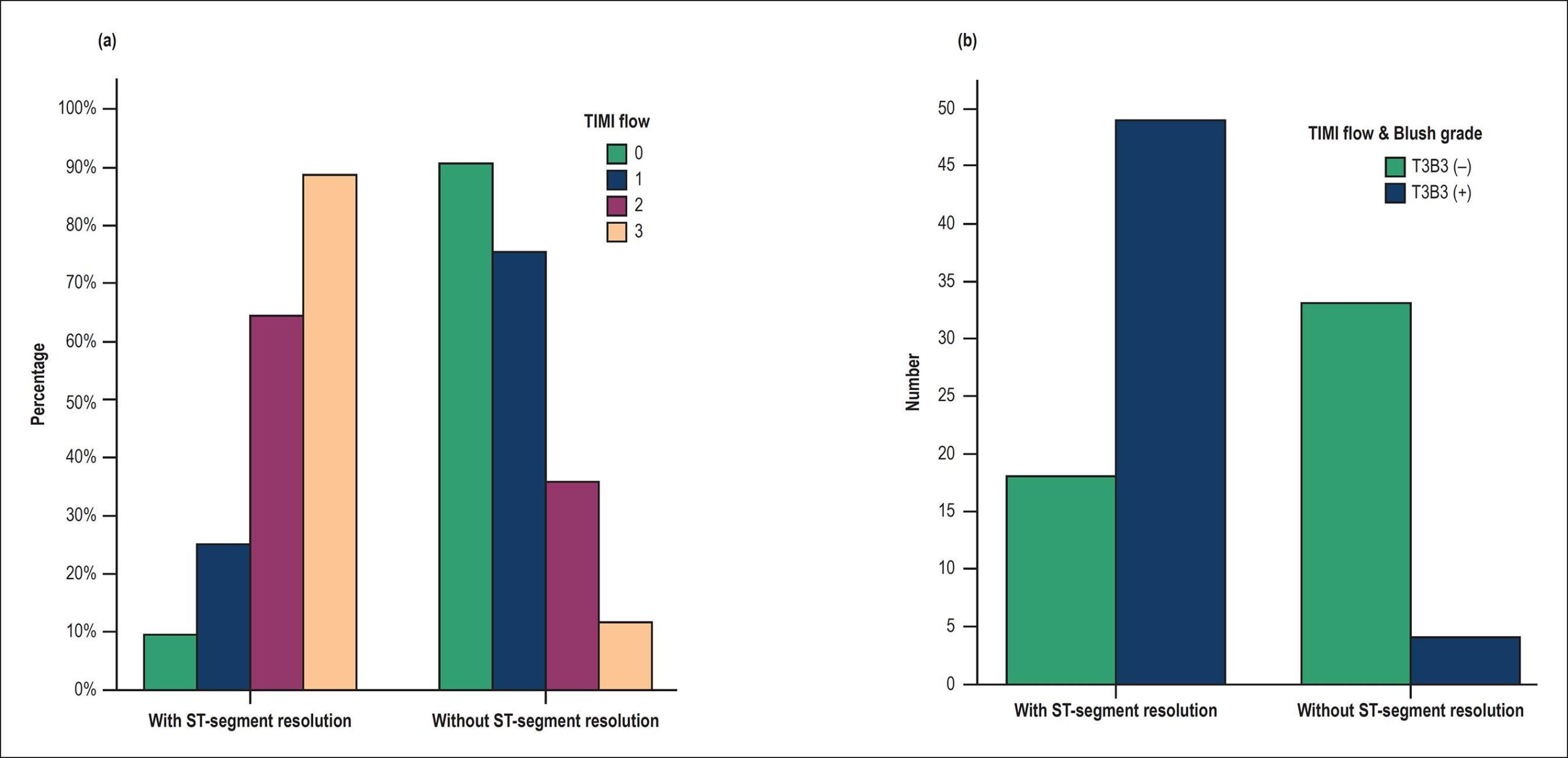
Observational study including 104 patients treated with tenecteplase (TNK), referred for a tertiary hospital. Electrocardiographic analysis consisted of measurements of the QT interval and QT dispersion in the 12 leads or in the ST-segment elevation area prior to and 60 minutes after TNK administration. All patients underwent angiography, with determination of TIMI flow and Blush grade in the culprit artery. P-values < 0.05 were considered statistically significant.

Results:

We found an increase in regional dispersion of the QT interval, corrected for heart rate (regional QTcD) 60 minutes after thrombolysis (p = 0.06) in anterior wall infarction in patients with TIMI flow 3 and Blush grade 3 [T3B3(+)]. When regional QTcD was added to the electrocardiographic criteria for reperfusion (i.e., > 50% ST-segment resolution), the area under the curve increased to 0.87 [(0.78-0.96). 95% IC. p 50% and regional QTcD > 13 ms, we found a 93% sensitivity and 71% specificity for reperfusion in T3B3(+), and 6% of patients with successful reperfusion were reclassified.

Conclusion:

Our data suggest that regional QTcD is a promising non-invasive instrument for detection of reperfusion in the culprit artery 60 minutes after thrombolysis.

  • Biomarkers
  • Electrocardiography
  • Myocardial Reperfusion
  • Percutaneous Coronary Intervention
  • ST Elevation Myocardial Infarction
Read Full Article
Creative Commons Attribution (CC BY) Creative Commons license
17/12/2018 DOI: 10.5935/abc.20180239 Views 2
Original Article

Validation of the Brazilian Version of CADE-Q II to Assess Knowledge of Coronary Artery Disease Patients

Rafaella Zulianello dos Santos et al. Gabriela Lima Melo Ghisi Christiani Decker Batista Bonin Gabriela Chaves … Clarissa Machado Haase Raquel Rodrigues Britto Magnus Benetti

Abstract

Background:

The Coronary Artery Disease Education Questionnaire (CADE-Q), an instrument aimed at assessing patients’ knowledge about coronary artery disease (CAD), was originally developed and psychometrically validated in Brazil. It was later translated, cross-culturally adapted, and validated to English. Although both versions demonstrated good reliability and validity, new studies in the area have pointed out the need of implementing the CADE-Q with other components of cardiac rehabilitation (CR) programs, such as psychologic factors, which had not been considered in previous version and were added in the subsequent, adapted version. Thus, a second version of CADE-Q was developed in English, the CADE-Q II.

Objective:

to translate, culturally adapt and psychometrically validate the CADE-Q II in Brazilian Portuguese.

Methods:

After translation and review by a Committee of specialists in CR, a version in Brazilian Portuguese was generated and tested in 307 patients in CR. Test-retest reliability was assessed by intraclass correlation coefficient (ICC) in 49 patients; internal consistency was assessed using Cronbach’s alpha (α); and, criterion validity was assessed regarding patients’ educational level and family income. The level of significance adopted for all tests was 5%.

Results:

After the ICC analysis, 4 items were excluded. The questionnaire was considered internally consistent (α > 0.7). Associations were found between the mean total scores and the variables schooling (p < 0.001) and income (p < 0.001). Median total score was 53 (14) points corresponding to 65.4% of the total possible score.

Conclusion:

The Portuguese version of the CADE-Q II showed sufficient reliability, consistency and validity, supporting its use in future studies.

  • Cardiac Rehabilitation
  • Cardiovascular Diseases/physiopathology
  • Coronary Artery Disease
  • Patient Education as Topic
  • Surveys and Questionnaires
  • Validation Studies
Read Full Article
Creative Commons Attribution (CC BY) Creative Commons license
17/12/2018 DOI: 10.5935/abc.20180244 Views 1
Figure 1
Original Article

Clinical Outcomes and Cost-Effectiveness Analysis of FFR Compared with Angiography in Multivessel Disease Patient

Edgard Freitas Quintella et al. Esmeralci Ferreira Vitor Manuel Pereira Azevedo Denizar V. Araujo … Fernando Mendes Sant'Anna Bernardo Amorim Denilson Campos de Albuquerque

Abstract

Background:

In multivessel disease patients with moderate stenosis, fractional flow reserve (FFR) allows the analysis of the lesions and guides treatment, and could contribute to the cost-effectiveness (CE) of non-pharmacological stents (NPS).

Objectives:

To evaluate CE and clinical impact of FFR-guided versus angiography-guided angioplasty (ANGIO) in multivessel patients using NPS.

Methods:

Multivessel disease patients were prospectively randomized to FFR or ANGIO groups during a 5 year-period and followed for < 12 months. Outcomes measures were major adverse cardiac events (MACE), restenosis and CE.

Results:

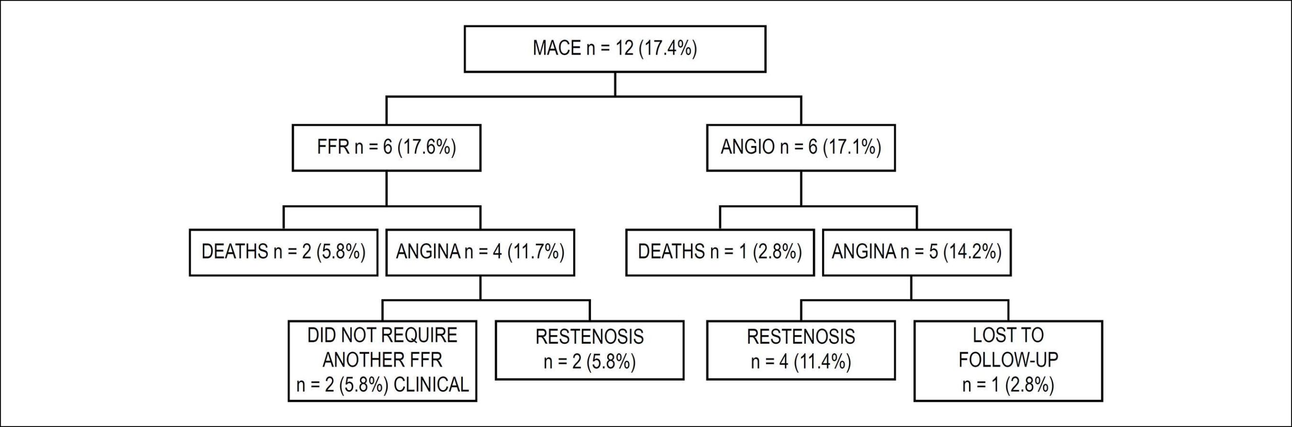
We studied 69 patients, 47 (68.1%) men, aged 62.0 ± 9.0 years, 34 (49.2%) in FFR group and 53 (50.7%) in ANGIO group, with stable angina or acute coronary syndrome. In FFR, there were 26 patients with biarterial disease (76.5%) and 8 (23.5%) with triarterial disease, and in ANGIO, 24 (68.6%) with biarterial and 11 (31.4%) with triarterial disease. Twelve MACEs were observed – 3 deaths: 2 (5.8%) in FFR and 1 (2.8%) in ANGIO, 9 (13.0%) angina: 4(11.7%) in FFR and 5(14.2%) in ANGIO, 6 restenosis: 2(5.8%) in FFR and 4 (11.4%) in ANGIO. Angiography detected 87(53.0%) lesions in FFR, 39(23.7%) with PCI and 48(29.3%) with medical treatment; and 77 (47.0%) lesions in ANGIO, all treated with angioplasty. Thirty-nine (33.3%) stents were registered in FFR (0.45 ± 0.50 stents/lesion) and 78 (1.05 ± 0.22 stents/lesion) in ANGIO (p = 0.0001), 51.4% greater in ANGIO than FFR. CE analysis revealed a cost of BRL 5,045.97 BRL 5,430.60 in ANGIO and FFR, respectively. The difference of effectiveness was of 1.82%.

Conclusion:

FFR reduced the number of lesions treated and stents, and the need for target-lesion revascularization, with a CE comparable with that of angiography.

  • Angioplasty, Balloon, Coronary
  • Coronary Artery Disease/economics
  • Cost-Benefit Analysis
  • Fractional Flow Reserve, Myocardial
  • Stents
Read Full Article
Creative Commons Attribution (CC BY) Creative Commons license
17/12/2018 DOI: 10.5935/abc.20180262
Figure 1
Original Article

Whole-Body Vibration Training Increases Myocardial Salvage Against Acute Ischemia in Adult Male Rats

Shahnaz Shekarforoush et al. Mohammad Reza Naghii

Abstract

Background:

Whole body vibration training (WBV) is a new training program, which is safe and effective. It can be followed by the public. However, data on the safety and efficacy of vibration on myocardial ischemia reperfusion (IR) injury are lacking.

Objective:

To examine the effect of WBV on the tolerance of the myocardium to acute IR injury in an experimental rat model.

Methods:

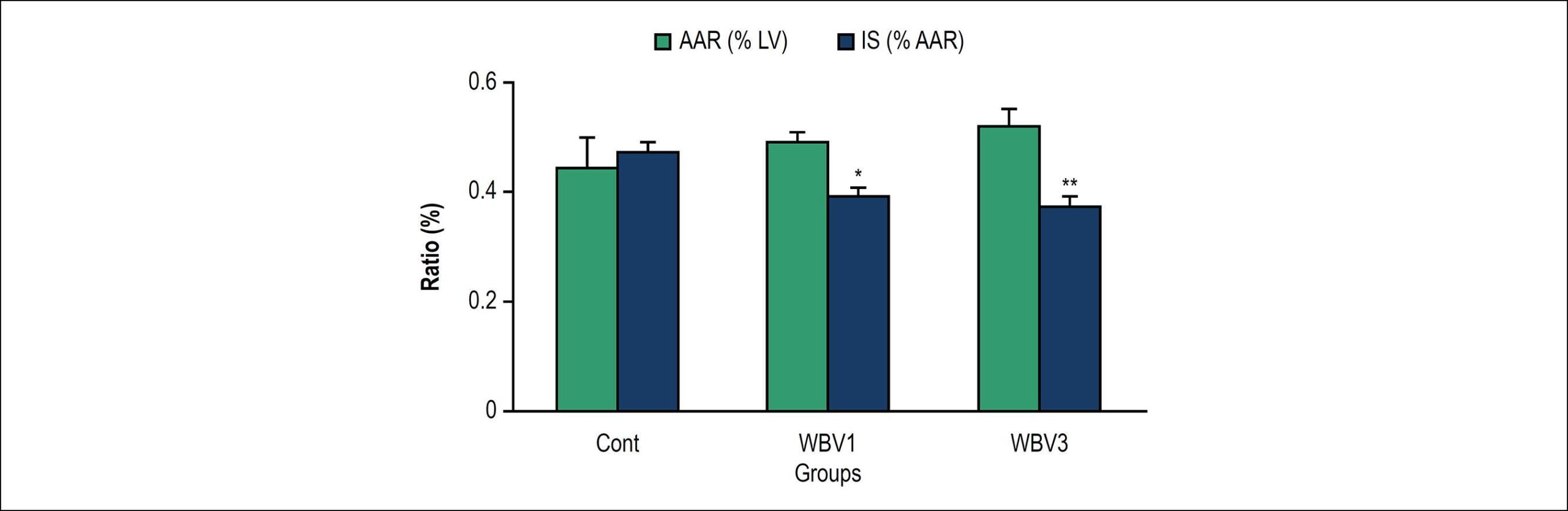
Twenty-four male Wistar rats were divided into control and vibration groups. Vibration training consisted of vertical sinusoidal whole body vibration for 30 min per day, 6 days per week, for 1 or 3 weeks (WBV1 and WBV3 groups, respectively). All the rats were submitted to myocardial IR injury. Myocardial infarct size and ischemia-induced arrhythmias were assessed. Differences between variables were considered significant when p < 0.05.

Results:

No differences were observed between the groups regarding the baseline hemodynamic parameters. Infarct size was smaller in the experimental group (control, 47 ± 2%; WBV1, 39 ± 2%; WBV3, 37 ± 2%; p < 0.05, vs. control). Vibration produced a significant decrease in the number and duration of ventricular tachycardia (VT) episodes compared to the control value. All ventricular fibrillation (VF) episodes in the vibration groups were self-limited, while 33% of the rats in the control group died due to irreversible VF (p = 0.02).

Conclusion:

The data showed that vibration training significantly increased cardiac tolerance to IR injury in rats, as evidenced by reduction in the infarct size and cardiac arrhythmias, and by facilitating spontaneous defibrillation.

  • Blood Viscosity
  • Body Composition
  • Cardiovascular Diseases
  • Ischemia
  • Ischemic Preconditioning
  • Osteoporosis/prevention and control
  • Rats Wistar
  • Vibration
Read Full Article
Creative Commons Attribution (CC BY) Creative Commons license
17/12/2018 DOI: 10.5935/abc.20180252 Views 2
Figure 1
Original Article

Assessment of the Relationship between Monocyte to High-Density Lipoprotein Ratio and Myocardial Bridge

Asim Enhos et al. Kahraman Cosansu Mustafa Ahmet Huyut Fahrettin Turna … Erdem Karacop Nijad Bakshaliyev Aydin Nadir Ramazan Ozdemir Mahmut Uluganyan

Abstract

Background:

Assessing the monocyte to high-density lipoprotein ratio (MHR) is a new tool for predicting inflamation, which plays a major role in atherosclerosis. Myocardial bridge (MB) is thought to be a benign condition with development of atherosclerosis, particularly at the proximal segment of the brigde.

Objective:

To evaluate the relationhip between MHR and the presence of MB.

Methods:

We consecutively scanned patients referred for coronary angiography between January 2013- December 2016, and a total of 160 patients who had a MB and normal coronary artery were enrolled in the study. The patients’ angiographic, demographic and clinic characteristics of the patients were reviewed from medical records. Monocytes and HDL-cholesterols were measured via complete blood count. MHR was calculated as the ratio of the absolute monocyte count to the HDL-cholesterol value. MHR values were divided into three tertiles as follows: lower (8.25 ± 1.61), moderate (13.11 ± 1.46), and higher (21.21 ± 4.30) tertile. A p-value of < 0.05 was considered significant.

Results:

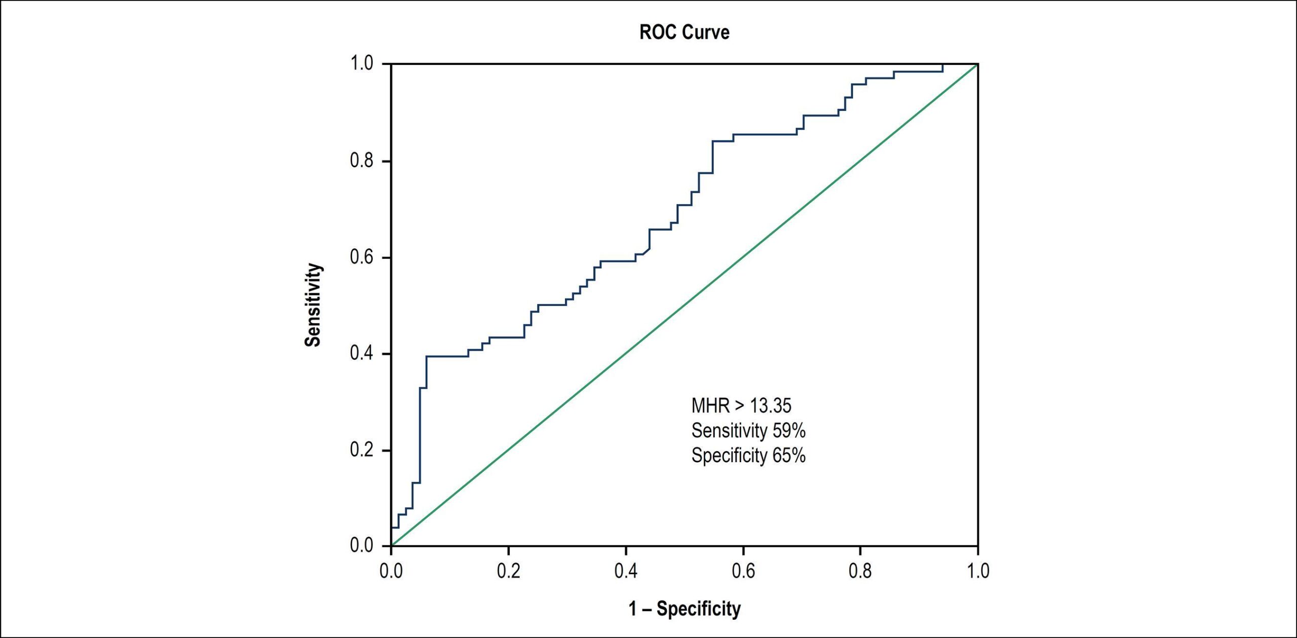
MHR was significantly higher in the MB group compared to the control group with normal coronary arteries. We found the frequency of MB (p = 0.002) to increase as the MHR tertiles rose. The Monocyte-HDL ratio with a cut-point of 13.35 had 59% sensitivity and 65.0% specificity (ROC area under curve: 0.687, 95% CI: 0.606-0.769, p < 0.001) in accurately predicting a MB diagnosis. In the multivariate analysis, MHR (p = 0.013) was found to be a significant independent predictor of the presence of MB, after adjusting for other risk factors.

Conclusion:

The present study revealed a significant correlation between MHR and MB.

  • Atherosclerosis
  • Biomarkers/blood
  • Cholesterol, HDL/blood
  • Inflammation
  • Monocytes/citology
  • Myocardial Bridging
Read Full Article
Creative Commons Attribution (CC BY) Creative Commons license
17/12/2018 DOI: 10.5935/abc.20180253
Figure 1
Original Article

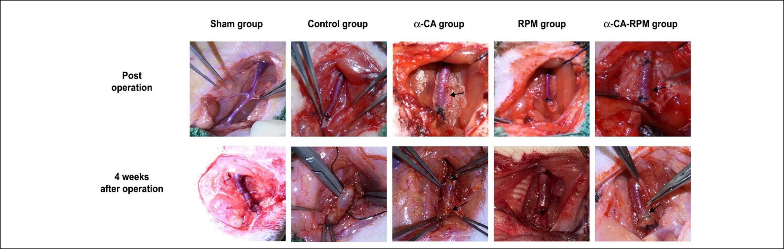
Rapamycin Combined with α-Cyanoacrylate Contributes to Inhibiting Intimal Hyperplasia in Rat Models

Jianjun Ge

Abstract

Background:

Vein graft restenosis has an adverse impact on bridge vessel circulation and patient prognosis after coronary artery bypass grafting.

Objectives:

We used the extravascular supporter α-cyanoacrylate (α-CA), the local application rapamycin/sirolimus (RPM), and a combination of the two (α-CA-RPM) in rat models of autogenous vein graft to stimulate vein graft change. The aim of our study was to observe the effect of α-CA, RPM, and α-CA-RPM on vein hyperplasia.

Methods:

Fifty healthy Sprague Dawley (SD) rats were randomized into the following 5 groups: sham, control, α-CA, RPM, and α-CA-RPM. Operating procedure as subsequently described was used to build models of grafted rat jugular vein on carotid artery on one side. The level of endothelin-1 (ET-1) was determined by enzyme-linked immunosorbent assay (ELISA). Grafted veins were observed via naked eye 4 weeks later; fresh veins were observed via microscope and image-processing software in hematoxylin-eosin (HE) staining and immunohistochemistry after having been fixed and stored” (i.e. First they were fixed and stored, and second they were observed); α-Smooth Muscle Actin (αSMA) and von Willebrand factor (vWF) were measured with reverse transcription-polymerase chain reaction (RT-PCR). Comparisons were made with single-factor analysis of variance and Fisher’s least significant difference test, with p < 0.05 considered significant.

Results:

We found that intimal thickness of the α-CA, RPM, and α-CA-RPM groups was lower than that of the control group (p < 0.01), and the thickness of the α-CA-RPM group was notably lower than that of the α-CA and RPM groups (p < 0.05).

Conclusion:

RPM combined with α-CA contributes to inhibiting intimal hyperplasia in rat models and is more effective for vascular patency than individual use of either α-CA or RPM.

  • Cyanocrylates
  • Graft Occlusion, Vascular
  • Hyperplasia
  • Myocardial Revascularization/surgery
  • Rats
  • Sirolimus
  • Vascular Patency
Read Full Article
Creative Commons Attribution (CC BY) Creative Commons license
17/12/2018 DOI: 10.5935/abc.20180247 Views 1
Figure 1
Original Article

Association Between Non-Dipping and Fragmented QRS Complexes in Prehypertensive Patients

Mehmet Eyuboglu et al. Bahri Akdeniz

Abstract

Background:

Fragmented QRS (fQRS) is a sign of adverse cardiovascular events in various cardiovascular diseases. It is also associated with increased blood pressure and non-dipping in hypertensive patients. However, no study has investigated the importance of fQRS in prehypertensive patients.

Objectives:

The aim of our study is to investigate the relationship between fQRS and non-dipper status in prehypertensive patients.

Methods:

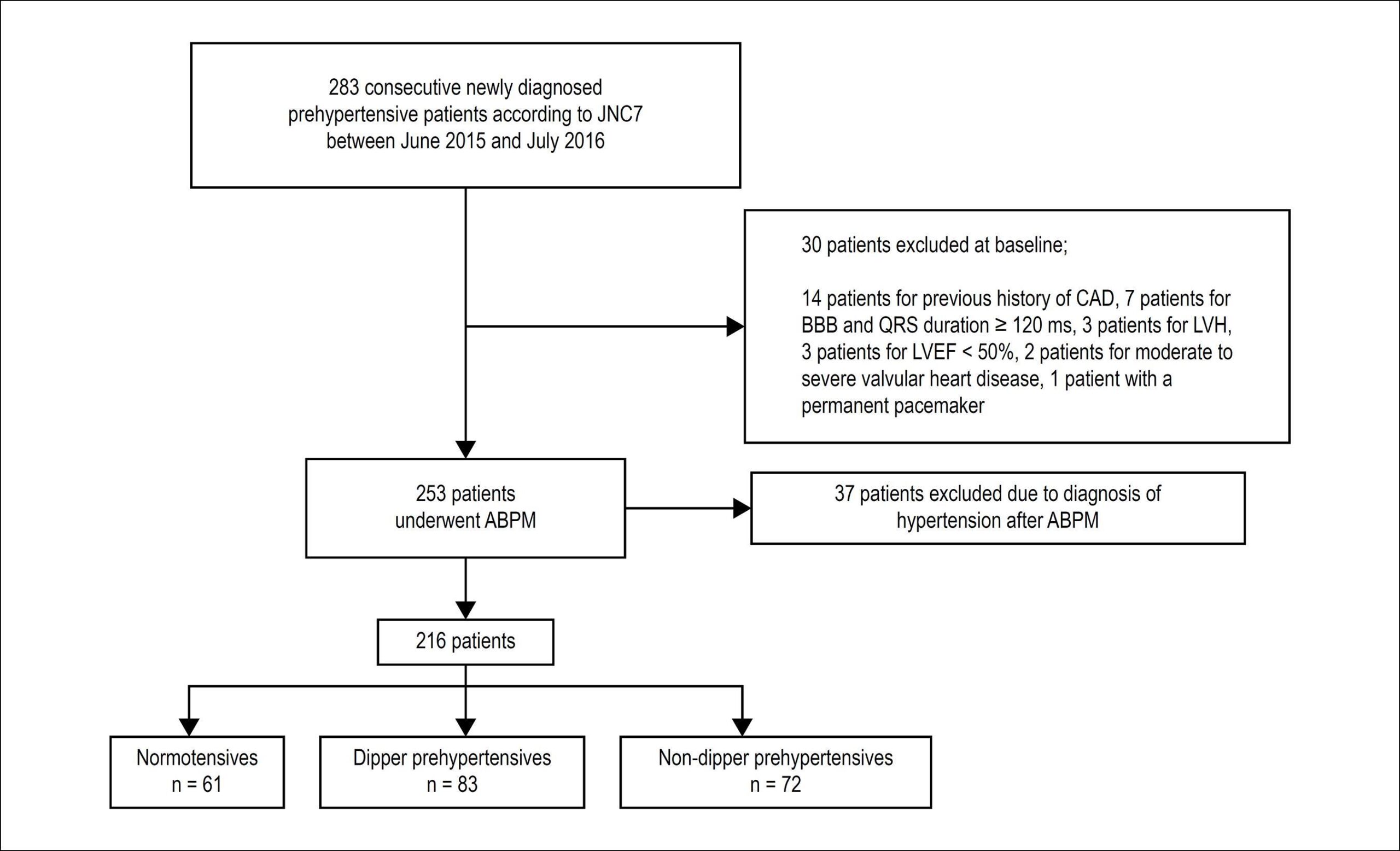
Two hundred and sixteen eligible, newly diagnosed prehypertensive patients who underwent 24-hour ambulatory blood pressure monitoring (ABPM) for further evaluation of blood pressure between June 2015 and July 2016 were included into the study. Patients were divided into three groups according to ABPM results: normotensives, dipper prehypertensives, and non-dipper prehypertensives. Groups were compared regarding presence of fQRS on electrocardiography. Additionally, multinomial logistic regression analysis was used to determine the relationship between fQRS and blood pressure pattern in prehypertensive patients.

Results:

According to ABPM recordings, 61 patients had normotensive blood pressure pattern (systolic blood pressure < 120 mmHg and diastolic blood pressure < 80 mmHg). Of the remaining 155 prehypertensive patients, 83 were dippers and 72 were non-dippers. Non-dipper prehypertensives had a significantly higher frequency of fQRS compared to normotensives (p = 0.048). Furthermore, multinomial logistic regression analysis revealed that fQRS is an independent predictor of non-dipping blood pressure pattern in prehypertensive patients (p = 0.017, OR: 4.071, 95% CI: 1.281-12.936).

Conclusions:

We found that fQRS is a predictor of non-dipping in prehypertensives. As a marker of fibrosis and higher fibrotic burden within myocardium, fQRS may be useful in identifying high-risk prehypertensive patients before the development of hypertension.

  • Ambulatory Blood Pressure Monitoring
  • Electrocardiography
  • Fragmented QRS
  • Hypertension
  • Non-dipping
  • Prehypertension
Read Full Article
Creative Commons Attribution (CC BY) Creative Commons license
17/12/2018 DOI: 10.5935/abc.20180242 Views 2

Could not load more articles. Please try again.

How to cite this article

EndNote RIS

Electronic Document Format (Vancouver)

. Erratum. Arquivos Brasileiros de Cardiologia 2019;112(1):116.

Electronic Document Format (ABNT)

. Erratum. Arquivos Brasileiros de Cardiologia, v. 112, n. 1, p. 116-116, Dec. 2018.

Electronic Document Format (APA)

(2019). Erratum. Arquivos Brasileiros de Cardiologia, 112(1), 116-116.

Electronic Document Format (ISO)

. Erratum. Arquivos Brasileiros de Cardiologia [online]. 2019, vol. 112, n. 1, [cited 2026-10-15], pp. 116-116. Available from: <https://journals.cardiol.br/en/article/erratum-12/>. ISSN 0066-782X

EndNote RIS

Electronic Document Format (Vancouver)

Melo MDT, Salemi VMC. Usefulness of Myocardial Deformation Indices in Preventing Cardiotoxicity in Breast Cancer Patients. Arquivos Brasileiros de Cardiologia 2019;112(1):57-8.

Electronic Document Format (ABNT)

Melo, Marcelo Dantas Tavares de; Salemi, Vera Maria Cury. Usefulness of Myocardial Deformation Indices in Preventing Cardiotoxicity in Breast Cancer Patients. Arquivos Brasileiros de Cardiologia, v. 112, n. 1, p. 57-58, Dec. 2018.

Electronic Document Format (APA)

Melo, M. D. T. , & Salemi, V. M. C. (2019). Usefulness of Myocardial Deformation Indices in Preventing Cardiotoxicity in Breast Cancer Patients. Arquivos Brasileiros de Cardiologia, 112(1), 57-58.

Electronic Document Format (ISO)

Melo, Marcelo Dantas Tavares de and Salemi, Vera Maria Cury. Usefulness of Myocardial Deformation Indices in Preventing Cardiotoxicity in Breast Cancer Patients. Arquivos Brasileiros de Cardiologia [online]. 2019, vol. 112, n. 1, [cited 2026-10-15], pp. 57-58. Available from: <https://journals.cardiol.br/en/article/usefulness-of-myocardial-deformation-indices-in-preventing-cardiotoxicity-in-breast-cancer-patients/>. ISSN 0066-782X

EndNote RIS

Electronic Document Format (Vancouver)

Bertoluci C, Foppa M, Santos ABS, Fuchs SC, Fuchs FD. Diuretics are Similar to Losartan on Echocardiographic Target-Organ Damage in Stage I Hypertension. PREVER-Treatment Study. Arquivos Brasileiros de Cardiologia 2019;112(1):87-90.

Electronic Document Format (ABNT)

Bertoluci, Carolina; Foppa, Murilo; Santos, Angela Barreto Santiago; Fuchs, Sandra C.; Fuchs, Flávio Danni. Diuretics are Similar to Losartan on Echocardiographic Target-Organ Damage in Stage I Hypertension. PREVER-Treatment Study. Arquivos Brasileiros de Cardiologia, v. 112, n. 1, p. 87-90, Dec. 2018.

Electronic Document Format (APA)

Bertoluci, C., Foppa, M., Santos, A. B. S., Fuchs, S. C., & Fuchs, F. D. (2019). Diuretics are Similar to Losartan on Echocardiographic Target-Organ Damage in Stage I Hypertension. PREVER-Treatment Study. Arquivos Brasileiros de Cardiologia, 112(1), 87-90.

Electronic Document Format (ISO)

Bertoluci, Carolina and Foppa, Murilo and Santos, Angela Barreto Santiago and Fuchs, Sandra C. and Fuchs, Flávio Danni. Diuretics are Similar to Losartan on Echocardiographic Target-Organ Damage in Stage I Hypertension. PREVER-Treatment Study. Arquivos Brasileiros de Cardiologia [online]. 2019, vol. 112, n. 1, [cited 2026-10-15], pp. 87-90. Available from: <https://journals.cardiol.br/en/article/diuretics-are-similar-to-losartan-on-echocardiographic-target-organ-damage-in-stage-i-hypertension-prever-treatment-study/>. ISSN 0066-782X

EndNote RIS

Electronic Document Format (Vancouver)

Elias Neto J, Tonet J, Frank R, Fontaine G. Arrhythmogenic Right Ventricular Cardiomyopathy/Dysplasia (ARVC/D) – What We Have Learned after 40 Years of the Diagnosis of This Clinical Entity. Arquivos Brasileiros de Cardiologia 2019;112(1):91-103.

Electronic Document Format (ABNT)

Elias Neto, Jorge; Tonet, Joelci; Frank, Robert; Fontaine, Guy. Arrhythmogenic Right Ventricular Cardiomyopathy/Dysplasia (ARVC/D) – What We Have Learned after 40 Years of the Diagnosis of This Clinical Entity. Arquivos Brasileiros de Cardiologia, v. 112, n. 1, p. 91-103, Dec. 2018.

Electronic Document Format (APA)

Elias Neto, J., Tonet, J., Frank, R., & Fontaine, G. (2019). Arrhythmogenic Right Ventricular Cardiomyopathy/Dysplasia (ARVC/D) – What We Have Learned after 40 Years of the Diagnosis of This Clinical Entity. Arquivos Brasileiros de Cardiologia, 112(1), 91-103.

Electronic Document Format (ISO)

Elias Neto, Jorge and Tonet, Joelci and Frank, Robert and Fontaine, Guy. Arrhythmogenic Right Ventricular Cardiomyopathy/Dysplasia (ARVC/D) – What We Have Learned after 40 Years of the Diagnosis of This Clinical Entity. Arquivos Brasileiros de Cardiologia [online]. 2019, vol. 112, n. 1, [cited 2026-10-15], pp. 91-103. Available from: <https://journals.cardiol.br/en/article/arrhythmogenic-right-ventricular-cardiomyopathy-dysplasia-arvc-d-what-we-have-learned-after-40-years-of-the-diagnosis-of-this-clinical-entity/>. ISSN 0066-782X

EndNote RIS

Electronic Document Format (Vancouver)

Moreira RI, Rosa SA, Galrinho A, Tavares NJ, Ferreira RC. A Large Cardiac Metastasis of a Parathyroid Tumour Presenting with Ventricular Tachycardia. Arquivos Brasileiros de Cardiologia 2019;112(1):113-5.

Electronic Document Format (ABNT)

Moreira, Rita Ilhão; Rosa, Sílvia Aguiar; Galrinho, Ana; Tavares, Nuno Jalles; Ferreira, Rui Cruz. A Large Cardiac Metastasis of a Parathyroid Tumour Presenting with Ventricular Tachycardia. Arquivos Brasileiros de Cardiologia, v. 112, n. 1, p. 113-115, Dec. 2018.

Electronic Document Format (APA)

Moreira, R. I., Rosa, S. A., Galrinho, A., Tavares, N. J., & Ferreira, R. C. (2019). A Large Cardiac Metastasis of a Parathyroid Tumour Presenting with Ventricular Tachycardia. Arquivos Brasileiros de Cardiologia, 112(1), 113-115.

Electronic Document Format (ISO)

Moreira, Rita Ilhão and Rosa, Sílvia Aguiar and Galrinho, Ana and Tavares, Nuno Jalles and Ferreira, Rui Cruz. A Large Cardiac Metastasis of a Parathyroid Tumour Presenting with Ventricular Tachycardia. Arquivos Brasileiros de Cardiologia [online]. 2019, vol. 112, n. 1, [cited 2026-10-15], pp. 113-115. Available from: <https://journals.cardiol.br/en/article/a-large-cardiac-metastasis-of-a-parathyroid-tumour-presenting-with-ventricular-tachycardia/>. ISSN 0066-782X

EndNote RIS

Electronic Document Format (Vancouver)

Sant’Anna FM, Sant’Anna LB. Analysis of FFR Measurement Clinical Impact and Cost-Effectiveness Compared to Angiography In Multi-Arterial Patients Undergoing PCI. Arquivos Brasileiros de Cardiologia 2019;112(1):48-9.

Electronic Document Format (ABNT)

Sant’Anna, Fernando Mendes; Sant’Anna, Lucas Bonacossa. Analysis of FFR Measurement Clinical Impact and Cost-Effectiveness Compared to Angiography In Multi-Arterial Patients Undergoing PCI. Arquivos Brasileiros de Cardiologia, v. 112, n. 1, p. 48-49, Dec. 2018.

Electronic Document Format (APA)

Sant’Anna, F. M., & Sant’Anna, L. B. (2019). Analysis of FFR Measurement Clinical Impact and Cost-Effectiveness Compared to Angiography In Multi-Arterial Patients Undergoing PCI. Arquivos Brasileiros de Cardiologia, 112(1), 48-49.

Electronic Document Format (ISO)

Sant’Anna, Fernando Mendes and Sant’Anna, Lucas Bonacossa. Analysis of FFR Measurement Clinical Impact and Cost-Effectiveness Compared to Angiography In Multi-Arterial Patients Undergoing PCI. Arquivos Brasileiros de Cardiologia [online]. 2019, vol. 112, n. 1, [cited 2026-10-15], pp. 48-49. Available from: <https://journals.cardiol.br/en/article/analysis-of-ffr-measurement-clinical-impact-and-cost-effectiveness-compared-to-angiography-in-multi-arterial-patients-undergoing-pci/>. ISSN 0066-782X

EndNote RIS

Electronic Document Format (Vancouver)

Atik E, Barreto AC, Binotto MA. Case 1 / 2019 – Natural Evolution of Double Outlet Right Ventricle with Noncommitted Ventricular Septal Defect and Pulmonary Stenosis in a 28-Year-Old Asymptomatic Woman. Arquivos Brasileiros de Cardiologia 2019;112(1):107-9.

Electronic Document Format (ABNT)

Atik, Edmar; Barreto, Alessandra Costa; Binotto, Maria Angélica. Case 1 / 2019 – Natural Evolution of Double Outlet Right Ventricle with Noncommitted Ventricular Septal Defect and Pulmonary Stenosis in a 28-Year-Old Asymptomatic Woman. Arquivos Brasileiros de Cardiologia, v. 112, n. 1, p. 107-109, Dec. 2018.

Electronic Document Format (APA)

Atik, E., Barreto, A. C., & Binotto, M. A. (2019). Case 1 / 2019 – Natural Evolution of Double Outlet Right Ventricle with Noncommitted Ventricular Septal Defect and Pulmonary Stenosis in a 28-Year-Old Asymptomatic Woman. Arquivos Brasileiros de Cardiologia, 112(1), 107-109.

Electronic Document Format (ISO)

Atik, Edmar and Barreto, Alessandra Costa and Binotto, Maria Angélica. Case 1 / 2019 – Natural Evolution of Double Outlet Right Ventricle with Noncommitted Ventricular Septal Defect and Pulmonary Stenosis in a 28-Year-Old Asymptomatic Woman. Arquivos Brasileiros de Cardiologia [online]. 2019, vol. 112, n. 1, [cited 2026-10-15], pp. 107-109. Available from: <https://journals.cardiol.br/en/article/case-1-2019-natural-evolution-of-double-outlet-right-ventricle-with-noncommitted-ventricular-septal-defect-and-pulmonary-stenosis-in-a-28-year-old-asymptomatic-woman/>. ISSN 0066-782X

EndNote RIS

Electronic Document Format (Vancouver)

César LAM. Cardiovascular Rehabilitation in Coronary Artery Disease and Better Knowledge of Its Own Disease. Arquivos Brasileiros de Cardiologia 2019;112(1):85-6.

Electronic Document Format (ABNT)

César, Luiz Antonio Machado. Cardiovascular Rehabilitation in Coronary Artery Disease and Better Knowledge of Its Own Disease. Arquivos Brasileiros de Cardiologia, v. 112, n. 1, p. 85-86, Dec. 2018.

Electronic Document Format (APA)

César, L. A. M. (2019). Cardiovascular Rehabilitation in Coronary Artery Disease and Better Knowledge of Its Own Disease. Arquivos Brasileiros de Cardiologia, 112(1), 85-86.

Electronic Document Format (ISO)

César, Luiz Antonio Machado. Cardiovascular Rehabilitation in Coronary Artery Disease and Better Knowledge of Its Own Disease. Arquivos Brasileiros de Cardiologia [online]. 2019, vol. 112, n. 1, [cited 2026-10-15], pp. 85-86. Available from: <https://journals.cardiol.br/en/article/cardiovascular-rehabilitation-in-coronary-artery-disease-and-better-knowledge-of-its-own-disease/>. ISSN 0066-782X

EndNote RIS

Electronic Document Format (Vancouver)

Cezar MDM, Gomes MJ, Damatto RL. Prenatal Stress: Molecular Mechanisms and Cardiovascular Disease. Arquivos Brasileiros de Cardiologia 2019;112(1):76-7.

Electronic Document Format (ABNT)

Cezar, Marcelo Diarcadia Mariano; Gomes, Mariana Janini; Damatto, Ricardo Luiz. Prenatal Stress: Molecular Mechanisms and Cardiovascular Disease. Arquivos Brasileiros de Cardiologia, v. 112, n. 1, p. 76-77, Dec. 2018.

Electronic Document Format (APA)

Cezar, M. D. M., Gomes, M. J., & Damatto, R. L. (2019). Prenatal Stress: Molecular Mechanisms and Cardiovascular Disease. Arquivos Brasileiros de Cardiologia, 112(1), 76-77.

Electronic Document Format (ISO)

Cezar, Marcelo Diarcadia Mariano and Gomes, Mariana Janini and Damatto, Ricardo Luiz. Prenatal Stress: Molecular Mechanisms and Cardiovascular Disease. Arquivos Brasileiros de Cardiologia [online]. 2019, vol. 112, n. 1, [cited 2026-10-15], pp. 76-77. Available from: <https://journals.cardiol.br/en/article/prenatal-stress-molecular-mechanisms-and-cardiovascular-disease/>. ISSN 0066-782X

EndNote RIS

Electronic Document Format (Vancouver)

Marcolino MS, Ribeiro ALP. Reperfusion Criteria in Patients Submitted to Fibrinolysis: Is There Room for Improvement?. Arquivos Brasileiros de Cardiologia 2019;112(1):30-1.

Electronic Document Format (ABNT)

Marcolino, Milena Soriano; Ribeiro, Antonio Luiz Pinho. Reperfusion Criteria in Patients Submitted to Fibrinolysis: Is There Room for Improvement?. Arquivos Brasileiros de Cardiologia, v. 112, n. 1, p. 30-31, Dec. 2018.

Electronic Document Format (APA)

Marcolino, M. S., & Ribeiro, A. L. P. (2019). Reperfusion Criteria in Patients Submitted to Fibrinolysis: Is There Room for Improvement?. Arquivos Brasileiros de Cardiologia, 112(1), 30-31.

Electronic Document Format (ISO)

Marcolino, Milena Soriano and Ribeiro, Antonio Luiz Pinho. Reperfusion Criteria in Patients Submitted to Fibrinolysis: Is There Room for Improvement?. Arquivos Brasileiros de Cardiologia [online]. 2019, vol. 112, n. 1, [cited 2026-10-15], pp. 30-31. Available from: <https://journals.cardiol.br/en/article/reperfusion-criteria-in-patients-submitted-to-fibrinolysis-is-there-room-for-improvement/>. ISSN 0066-782X

EndNote RIS

Electronic Document Format (Vancouver)

Villela PB, Oliveira GMM. A New Marker of Myocardial Bridge?. Arquivos Brasileiros de Cardiologia 2019;112(1):18-9.

Electronic Document Format (ABNT)

Villela, Paolo Blanco; Oliveira, Gláucia Maria Moraes de. A New Marker of Myocardial Bridge?. Arquivos Brasileiros de Cardiologia, v. 112, n. 1, p. 18-19, Dec. 2018.

Electronic Document Format (APA)

Villela, P. B., & Oliveira, G. M. M. (2019). A New Marker of Myocardial Bridge?. Arquivos Brasileiros de Cardiologia, 112(1), 18-19.

Electronic Document Format (ISO)

Villela, Paolo Blanco and Oliveira, Gláucia Maria Moraes de. A New Marker of Myocardial Bridge?. Arquivos Brasileiros de Cardiologia [online]. 2019, vol. 112, n. 1, [cited 2026-10-15], pp. 18-19. Available from: <https://journals.cardiol.br/en/article/a-new-marker-of-myocardial-bridge/>. ISSN 0066-782X

EndNote RIS

Electronic Document Format (Vancouver)

Koike MK. Synergy of Rapamycin and Cyanoacrylate in Reducing Intimal Hyperplasia. Arquivos Brasileiros de Cardiologia 2019;112(1):11.

Electronic Document Format (ABNT)

Koike, Marcia Kiyomi. Synergy of Rapamycin and Cyanoacrylate in Reducing Intimal Hyperplasia. Arquivos Brasileiros de Cardiologia, v. 112, n. 1, p. 11-11, Dec. 2018.

Electronic Document Format (APA)

Koike, M. K. (2019). Synergy of Rapamycin and Cyanoacrylate in Reducing Intimal Hyperplasia. Arquivos Brasileiros de Cardiologia, 112(1), 11-11.

Electronic Document Format (ISO)

Koike, Marcia Kiyomi. Synergy of Rapamycin and Cyanoacrylate in Reducing Intimal Hyperplasia. Arquivos Brasileiros de Cardiologia [online]. 2019, vol. 112, n. 1, [cited 2026-10-15], pp. 11-11. Available from: <https://journals.cardiol.br/en/article/synergy-of-rapamycin-and-cyanoacrylate-in-reducing-intimal-hyperplasia/>. ISSN 0066-782X

EndNote RIS

Electronic Document Format (Vancouver)

Pinho C. Cardiac Fibrosis Occurs before Arterial Hypertension Becomes Well Defined?. Arquivos Brasileiros de Cardiologia 2019;112(1):65-6.

Electronic Document Format (ABNT)

Pinho, Claudio. Cardiac Fibrosis Occurs before Arterial Hypertension Becomes Well Defined?. Arquivos Brasileiros de Cardiologia, v. 112, n. 1, p. 65-66, Dec. 2018.

Electronic Document Format (APA)

Pinho, C. (2019). Cardiac Fibrosis Occurs before Arterial Hypertension Becomes Well Defined?. Arquivos Brasileiros de Cardiologia, 112(1), 65-66.

Electronic Document Format (ISO)

Pinho, Claudio. Cardiac Fibrosis Occurs before Arterial Hypertension Becomes Well Defined?. Arquivos Brasileiros de Cardiologia [online]. 2019, vol. 112, n. 1, [cited 2026-10-15], pp. 65-66. Available from: <https://journals.cardiol.br/en/article/cardiac-fibrosis-occurs-before-arterial-hypertension-becomes-well-defined/>. ISSN 0066-782X

EndNote RIS

Electronic Document Format (Vancouver)

Zornoff L, Minicucci MF. Is There a Role For Whole Body Vibration in Protecting Cardiovascular Disease?. Arquivos Brasileiros de Cardiologia 2019;112(1):38-9.

Electronic Document Format (ABNT)

Zornoff, Leonardo; Minicucci, Marcos Ferreira. Is There a Role For Whole Body Vibration in Protecting Cardiovascular Disease?. Arquivos Brasileiros de Cardiologia, v. 112, n. 1, p. 38-39, Dec. 2018.

Electronic Document Format (APA)

Zornoff, L., & Minicucci, M. F. (2019). Is There a Role For Whole Body Vibration in Protecting Cardiovascular Disease?. Arquivos Brasileiros de Cardiologia, 112(1), 38-39.

Electronic Document Format (ISO)

Zornoff, Leonardo and Minicucci, Marcos Ferreira. Is There a Role For Whole Body Vibration in Protecting Cardiovascular Disease?. Arquivos Brasileiros de Cardiologia [online]. 2019, vol. 112, n. 1, [cited 2026-10-15], pp. 38-39. Available from: <https://journals.cardiol.br/en/article/is-there-a-role-for-whole-body-vibration-in-protecting-cardiovascular-disease/>. ISSN 0066-782X

EndNote RIS

Electronic Document Format (Vancouver)

Estrada JAS, Camacho AD, Cruz LR, Figueroa JR. Myocardial Infarction Due to an Anomalous Origin of the Left Coronary Artery with Unique Aggravating Features. Arquivos Brasileiros de Cardiologia 2019;112(1):110-2.

Electronic Document Format (ABNT)

Estrada, Jorge Alberto Silva; Camacho, Alejandra Domínguez; Cruz, Lorenzo Reyes de la; Figueroa, Jesus Reyna. Myocardial Infarction Due to an Anomalous Origin of the Left Coronary Artery with Unique Aggravating Features. Arquivos Brasileiros de Cardiologia, v. 112, n. 1, p. 110-112, Dec. 2018.

Electronic Document Format (APA)

Estrada, J. A. S., Camacho, A. D., Cruz, L. R. , & Figueroa, J. R. (2019). Myocardial Infarction Due to an Anomalous Origin of the Left Coronary Artery with Unique Aggravating Features. Arquivos Brasileiros de Cardiologia, 112(1), 110-112.

Electronic Document Format (ISO)

Estrada, Jorge Alberto Silva and Camacho, Alejandra Domínguez and Cruz, Lorenzo Reyes de la and Figueroa, Jesus Reyna. Myocardial Infarction Due to an Anomalous Origin of the Left Coronary Artery with Unique Aggravating Features. Arquivos Brasileiros de Cardiologia [online]. 2019, vol. 112, n. 1, [cited 2026-10-15], pp. 110-112. Available from: <https://journals.cardiol.br/en/article/myocardial-infarction-due-to-an-anomalous-origin-of-the-left-coronary-artery-with-unique-aggravating-features/>. ISSN 0066-782X

EndNote RIS

Electronic Document Format (Vancouver)

Gottlieb I, Bittencourt MS, Rochitte CE, Cavalcante JL. Coronary Computed Tomography Angiography Takes the Center Stage and Here is Why. Arquivos Brasileiros de Cardiologia 2019;112(1):104-6.

Electronic Document Format (ABNT)

Gottlieb, Illan; Bittencourt, Marcio Sommer; Rochitte, Carlos Eduardo; Cavalcante, João L.. Coronary Computed Tomography Angiography Takes the Center Stage and Here is Why. Arquivos Brasileiros de Cardiologia, v. 112, n. 1, p. 104-106, Dec. 2018.

Electronic Document Format (APA)

Gottlieb, I., Bittencourt, M. S., Rochitte, C. E., & Cavalcante, J. L. (2019). Coronary Computed Tomography Angiography Takes the Center Stage and Here is Why. Arquivos Brasileiros de Cardiologia, 112(1), 104-106.

Electronic Document Format (ISO)

Gottlieb, Illan and Bittencourt, Marcio Sommer and Rochitte, Carlos Eduardo and Cavalcante, João L.. Coronary Computed Tomography Angiography Takes the Center Stage and Here is Why. Arquivos Brasileiros de Cardiologia [online]. 2019, vol. 112, n. 1, [cited 2026-10-15], pp. 104-106. Available from: <https://journals.cardiol.br/en/article/coronary-computed-tomography-angiography-takes-the-center-stage-and-here-is-why/>. ISSN 0066-782X

EndNote RIS

Electronic Document Format (Vancouver)

Bittencourt MS, Blaha MJ, Nasir K. Coronary Artery Calcium – From Screening to a Personalized Shared Decision-Making Tool: The New American Prevention Guidelines. Arquivos Brasileiros de Cardiologia 2019;112(1):1-2.

Electronic Document Format (ABNT)

Bittencourt, Marcio Sommer; Blaha, Michael J.; Nasir, Khurram. Coronary Artery Calcium – From Screening to a Personalized Shared Decision-Making Tool: The New American Prevention Guidelines. Arquivos Brasileiros de Cardiologia, v. 112, n. 1, p. 1-2, Dec. 2018.

Electronic Document Format (APA)

Bittencourt, M. S., Blaha, M. J., & Nasir, K. (2019). Coronary Artery Calcium – From Screening to a Personalized Shared Decision-Making Tool: The New American Prevention Guidelines. Arquivos Brasileiros de Cardiologia, 112(1), 1-2.

Electronic Document Format (ISO)

Bittencourt, Marcio Sommer and Blaha, Michael J. and Nasir, Khurram. Coronary Artery Calcium – From Screening to a Personalized Shared Decision-Making Tool: The New American Prevention Guidelines. Arquivos Brasileiros de Cardiologia [online]. 2019, vol. 112, n. 1, [cited 2026-10-15], pp. 1-2. Available from: <https://journals.cardiol.br/en/article/coronary-artery-calcium-from-screening-to-a-personalized-shared-decision-making-tool-the-new-american-prevention-guidelines/>. ISSN 0066-782X

EndNote RIS

Electronic Document Format (Vancouver)

Dotta G, Fonseca FAH, Izar MCO, Souza MT, Moreira FT, Pinheiro LFM, Barbosa AHP, et al. Regional QT Interval Dispersion as an Early Predictor of Reperfusion in Patients with Acute Myocardial Infarction after Fibrinolytic Therapy. Arquivos Brasileiros de Cardiologia 2018;112(1):20-9.

Electronic Document Format (ABNT)

Dotta, Gabriel; Fonseca, Francisco Antonio Helfenstein; Izar, Maria Cristina de Oliveira; Souza, Marco Tulio de; Moreira, Flavio Tocci; Pinheiro, Luiz Fernando Muniz; Barbosa, Adriano Henrique Pereira; Caixeta, Adriano Mendes; Póvoa, Rui Manoel Santos; Carvalho, Antônio Carlos; Bianco, Henrique Tria. Regional QT Interval Dispersion as an Early Predictor of Reperfusion in Patients with Acute Myocardial Infarction after Fibrinolytic Therapy. Arquivos Brasileiros de Cardiologia, v. 112, n. 1, p. 20-29, Dec. 2018.

Electronic Document Format (APA)

Dotta, G., Fonseca, F. A. H., Izar, M. C. O., Souza, M. T. , Moreira, F. T., Pinheiro, L. F. M., Barbosa, A. H. P., Caixeta, A. M., Póvoa, R. M. S., Carvalho, A. C., & Bianco, H. T. (2018). Regional QT Interval Dispersion as an Early Predictor of Reperfusion in Patients with Acute Myocardial Infarction after Fibrinolytic Therapy. Arquivos Brasileiros de Cardiologia, 112(1), 20-29.

Electronic Document Format (ISO)

Dotta, Gabriel and Fonseca, Francisco Antonio Helfenstein and Izar, Maria Cristina de Oliveira and Souza, Marco Tulio de and Moreira, Flavio Tocci and Pinheiro, Luiz Fernando Muniz and Barbosa, Adriano Henrique Pereira and Caixeta, Adriano Mendes and Póvoa, Rui Manoel Santos and Carvalho, Antônio Carlos and Bianco, Henrique Tria. Regional QT Interval Dispersion as an Early Predictor of Reperfusion in Patients with Acute Myocardial Infarction after Fibrinolytic Therapy. Arquivos Brasileiros de Cardiologia [online]. 2018, vol. 112, n. 1, [cited 2026-10-15], pp. 20-29. Available from: <https://journals.cardiol.br/en/article/regional-qt-interval-dispersion-as-an-early-predictor-of-reperfusion-in-patients-with-acute-myocardial-infarction-after-fibrinolytic-therapy/>. ISSN 0066-782X

EndNote RIS

Electronic Document Format (Vancouver)

Santos RZ, Ghisi GLM, Bonin CDB, Chaves G, Haase CM, Britto RR, Benetti M, et al. Validation of the Brazilian Version of CADE-Q II to Assess Knowledge of Coronary Artery Disease Patients. Arquivos Brasileiros de Cardiologia 2018;112(1):78-84.

Electronic Document Format (ABNT)

Santos, Rafaella Zulianello dos; Ghisi, Gabriela Lima Melo; Bonin, Christiani Decker Batista; Chaves, Gabriela; Haase, Clarissa Machado; Britto, Raquel Rodrigues; Benetti, Magnus. Validation of the Brazilian Version of CADE-Q II to Assess Knowledge of Coronary Artery Disease Patients. Arquivos Brasileiros de Cardiologia, v. 112, n. 1, p. 78-84, Dec. 2018.

Electronic Document Format (APA)

Santos, R. Z. , Ghisi, G. L. M., Bonin, C. D. B., Chaves, G., Haase, C. M., Britto, R. R., & Benetti, M. (2018). Validation of the Brazilian Version of CADE-Q II to Assess Knowledge of Coronary Artery Disease Patients. Arquivos Brasileiros de Cardiologia, 112(1), 78-84.

Electronic Document Format (ISO)

Santos, Rafaella Zulianello dos and Ghisi, Gabriela Lima Melo and Bonin, Christiani Decker Batista and Chaves, Gabriela and Haase, Clarissa Machado and Britto, Raquel Rodrigues and Benetti, Magnus. Validation of the Brazilian Version of CADE-Q II to Assess Knowledge of Coronary Artery Disease Patients. Arquivos Brasileiros de Cardiologia [online]. 2018, vol. 112, n. 1, [cited 2026-10-15], pp. 78-84. Available from: <https://journals.cardiol.br/en/article/validation-of-the-brazilian-version-of-cade-q-ii-to-assess-knowledge-of-coronary-artery-disease-patients/>. ISSN 0066-782X

EndNote RIS

Electronic Document Format (Vancouver)

Quintella EF, Ferreira E, Azevedo VMP, Araujo DV, Sant’Anna FM, Amorim B, Albuquerque DC, et al. Clinical Outcomes and Cost-Effectiveness Analysis of FFR Compared with Angiography in Multivessel Disease Patient. Arquivos Brasileiros de Cardiologia 2018;112(1):40-7.

Electronic Document Format (ABNT)

Quintella, Edgard Freitas; Ferreira, Esmeralci; Azevedo, Vitor Manuel Pereira; Araujo, Denizar V.; Sant’Anna, Fernando Mendes; Amorim, Bernardo; Albuquerque, Denilson Campos de. Clinical Outcomes and Cost-Effectiveness Analysis of FFR Compared with Angiography in Multivessel Disease Patient. Arquivos Brasileiros de Cardiologia, v. 112, n. 1, p. 40-47, Dec. 2018.

Electronic Document Format (APA)

Quintella, E. F., Ferreira, E., Azevedo, V. M. P., Araujo, D. V., Sant’Anna, F. M., Amorim, B., & Albuquerque, D. C. (2018). Clinical Outcomes and Cost-Effectiveness Analysis of FFR Compared with Angiography in Multivessel Disease Patient. Arquivos Brasileiros de Cardiologia, 112(1), 40-47.

Electronic Document Format (ISO)

Quintella, Edgard Freitas and Ferreira, Esmeralci and Azevedo, Vitor Manuel Pereira and Araujo, Denizar V. and Sant’Anna, Fernando Mendes and Amorim, Bernardo and Albuquerque, Denilson Campos de. Clinical Outcomes and Cost-Effectiveness Analysis of FFR Compared with Angiography in Multivessel Disease Patient. Arquivos Brasileiros de Cardiologia [online]. 2018, vol. 112, n. 1, [cited 2026-10-15], pp. 40-47. Available from: <https://journals.cardiol.br/en/article/clinical-outcomes-and-cost-effectiveness-analysis-of-ffr-compared-with-angiography-in-multivessel-disease-patient/>. ISSN 0066-782X

EndNote RIS

Electronic Document Format (Vancouver)

Shekarforoush S, Naghii MR. Whole-Body Vibration Training Increases Myocardial Salvage Against Acute Ischemia in Adult Male Rats. Arquivos Brasileiros de Cardiologia 2018;112(1):32-7.

Electronic Document Format (ABNT)

Shekarforoush, Shahnaz; Naghii, Mohammad Reza. Whole-Body Vibration Training Increases Myocardial Salvage Against Acute Ischemia in Adult Male Rats. Arquivos Brasileiros de Cardiologia, v. 112, n. 1, p. 32-37, Dec. 2018.

Electronic Document Format (APA)

Shekarforoush, S., & Naghii, M. R. (2018). Whole-Body Vibration Training Increases Myocardial Salvage Against Acute Ischemia in Adult Male Rats. Arquivos Brasileiros de Cardiologia, 112(1), 32-37.

Electronic Document Format (ISO)

Shekarforoush, Shahnaz and Naghii, Mohammad Reza. Whole-Body Vibration Training Increases Myocardial Salvage Against Acute Ischemia in Adult Male Rats. Arquivos Brasileiros de Cardiologia [online]. 2018, vol. 112, n. 1, [cited 2026-10-15], pp. 32-37. Available from: <https://journals.cardiol.br/en/article/whole-body-vibration-training-increases-myocardial-salvage-against-acute-ischemia-in-adult-male-rats/>. ISSN 0066-782X

EndNote RIS

Electronic Document Format (Vancouver)

Enhos A, Cosansu K, Huyut MA, Turna F, Karacop E, Bakshaliyev N, Nadir A, et al. Assessment of the Relationship between Monocyte to High-Density Lipoprotein Ratio and Myocardial Bridge. Arquivos Brasileiros de Cardiologia 2018;112(1):12-7.

Electronic Document Format (ABNT)

Enhos, Asim; Cosansu, Kahraman; Huyut, Mustafa Ahmet; Turna, Fahrettin; Karacop, Erdem; Bakshaliyev, Nijad; Nadir, Aydin; Ozdemir, Ramazan; Uluganyan, Mahmut. Assessment of the Relationship between Monocyte to High-Density Lipoprotein Ratio and Myocardial Bridge. Arquivos Brasileiros de Cardiologia, v. 112, n. 1, p. 12-17, Dec. 2018.

Electronic Document Format (APA)

Enhos, A., Cosansu, K., Huyut, M. A., Turna, F., Karacop, E., Bakshaliyev, N., Nadir, A., Ozdemir, R., & Uluganyan, M. (2018). Assessment of the Relationship between Monocyte to High-Density Lipoprotein Ratio and Myocardial Bridge. Arquivos Brasileiros de Cardiologia, 112(1), 12-17.

Electronic Document Format (ISO)

Enhos, Asim and Cosansu, Kahraman and Huyut, Mustafa Ahmet and Turna, Fahrettin and Karacop, Erdem and Bakshaliyev, Nijad and Nadir, Aydin and Ozdemir, Ramazan and Uluganyan, Mahmut. Assessment of the Relationship between Monocyte to High-Density Lipoprotein Ratio and Myocardial Bridge. Arquivos Brasileiros de Cardiologia [online]. 2018, vol. 112, n. 1, [cited 2026-10-15], pp. 12-17. Available from: <https://journals.cardiol.br/en/article/assessment-of-the-relationship-between-monocyte-to-high-density-lipoprotein-ratio-and-myocardial-bridge/>. ISSN 0066-782X

EndNote RIS

Electronic Document Format (Vancouver)

Ge J. Rapamycin Combined with α-Cyanoacrylate Contributes to Inhibiting Intimal Hyperplasia in Rat Models. Arquivos Brasileiros de Cardiologia 2018;112(1):3-10.

Electronic Document Format (ABNT)

Ge, Jianjun. Rapamycin Combined with α-Cyanoacrylate Contributes to Inhibiting Intimal Hyperplasia in Rat Models. Arquivos Brasileiros de Cardiologia, v. 112, n. 1, p. 3-10, Dec. 2018.

Electronic Document Format (APA)

Ge, J. (2018). Rapamycin Combined with α-Cyanoacrylate Contributes to Inhibiting Intimal Hyperplasia in Rat Models. Arquivos Brasileiros de Cardiologia, 112(1), 3-10.

Electronic Document Format (ISO)

Ge, Jianjun. Rapamycin Combined with α-Cyanoacrylate Contributes to Inhibiting Intimal Hyperplasia in Rat Models. Arquivos Brasileiros de Cardiologia [online]. 2018, vol. 112, n. 1, [cited 2026-10-15], pp. 3-10. Available from: <https://journals.cardiol.br/en/article/rapamycin-combined-with-%ce%b1-cyanoacrylate-contributes-to-inhibiting-intimal-hyperplasia-in-rat-models/>. ISSN 0066-782X

EndNote RIS

Electronic Document Format (Vancouver)

Eyuboglu M, Akdeniz B. Association Between Non-Dipping and Fragmented QRS Complexes in Prehypertensive Patients. Arquivos Brasileiros de Cardiologia 2018;112(1):59-64.

Electronic Document Format (ABNT)

Eyuboglu, Mehmet; Akdeniz, Bahri. Association Between Non-Dipping and Fragmented QRS Complexes in Prehypertensive Patients. Arquivos Brasileiros de Cardiologia, v. 112, n. 1, p. 59-64, Dec. 2018.

Electronic Document Format (APA)

Eyuboglu, M., & Akdeniz, B. (2018). Association Between Non-Dipping and Fragmented QRS Complexes in Prehypertensive Patients. Arquivos Brasileiros de Cardiologia, 112(1), 59-64.

Electronic Document Format (ISO)

Eyuboglu, Mehmet and Akdeniz, Bahri. Association Between Non-Dipping and Fragmented QRS Complexes in Prehypertensive Patients. Arquivos Brasileiros de Cardiologia [online]. 2018, vol. 112, n. 1, [cited 2026-10-15], pp. 59-64. Available from: <https://journals.cardiol.br/en/article/association-between-non-dipping-and-fragmented-qrs-complexes-in-prehypertensive-patients/>. ISSN 0066-782X

Sede – Rio de Janeiro
Av. Marechal Câmara, 160
3º andar – Sala: 330 – Centro
Rio de Janeiro – CEP: 20020-907
Tel.: (55 +21) 3478-2700
ou (21) 99386-3961
E-mail: revista@cardiol.br

Sede – São Paulo
Alameda Santos, 705 – 11º andar
Cerqueira César | São Paulo
CEP: 01419-001
Tel.: (55 +11) 3411-5500
E-mail: revista@cardiol.br

  • About the Journal
  • Internal Regulation
  • Editors Section
  • Editorial Board
  • History
  • SBC Award

  • Guidelines for Publication
  • Editorial Policy
  • Ethics Policy
  • Policy on the Use of Artificial Intelligence
  • Mandatory Publication Form
  • Publication Fee

Issues

SBC Guidelines

  • How to Publish an Abstract Supplement?
  • Supplements from the Brazilian Congress of Cardiology