ABC Family Arquivos Brasileiros de Cardiologia International Journal of Cardiovascular Sciences ABC Heart Failure & Cardiomyopathy ABC Imagem Cardiovascular
Português English
ABC Family
Português English
  • About
    • About ABC Family
    • Internal Regulation
    • Editorial Board
    • Brazilian Society of Cardiology (SBC)
  • Issues
  • SBC Guidelines
  • Content
    • Videos
    • Podcasts
    • News
  • About
    • About ABC Family
    • Internal Regulation
    • Editorial Board
    • Brazilian Society of Cardiology (SBC)
  • Issues
  • SBC Guidelines
  • Content
    • Videos
    • Podcasts
    • News

ABC Family

  • ABC Family
  • Arquivos Brasileiros de Cardiologia
  • International Journal of Cardiovascular Sciences
  • ABC Heart Failure & Cardiomyopathy
  • ABC Imagem Cardiovascular
Search in

No content found.

Journals
Ordering
Article types
Others

  1. Home
  2. Keyword: Tomography, X-Ray Computed
Figure 1
ABCIC
Case Report

Giant Right Coronary Artery Aneurysm with Arteriovenous Fistula

Maria Eduarda Menezes de Siqueira et al. Mayra Isabel Dias Ana Claudia Vincezi Raudan Uski Luciano Figueiredo de Aguiar Filho

Introduction

Coronary artery aneurysm (CAA) is defined as a dilation that exceeds the normal diameter of the adjacent artery or the diameter of the patient’s largest coronary artery by 1.5 times. Some authors consider a giant coronary aneurysm when the diameter is > 20 mm, whereas the American Heart Association defines giant aneurysms as those > 8 mm. Giant CAA is a rare pathology with an approximate incidence of 0.02%. We report a case of giant right coronary artery aneurysm with fistula to the coronary sinus, appearing as a giant “mass”.

[…]

  • Coronary Aneurysm
  • Coronary Vessel Anomalies
  • Tomography, X-Ray Computed
  • Ventricular Dysfunction Right
Read Full Article
Creative Commons Attribution (CC BY) Creative Commons license
26/07/2023 DOI: 10.36660/abcimg.20230025i
Figure 1
ABCIC
Case Report

New Application of FFRCT in Clinical Practice: Evaluation of Interarterial Anomalous Coronary Course

Fábio Bordin Trindade et al. Thamara Carvalho Morais Roberto Nery Dantas Junior Roberto Vitor Almeida Torres … Clarice Santos Parreira Soares José Rodrigues Parga Filho

Discussion

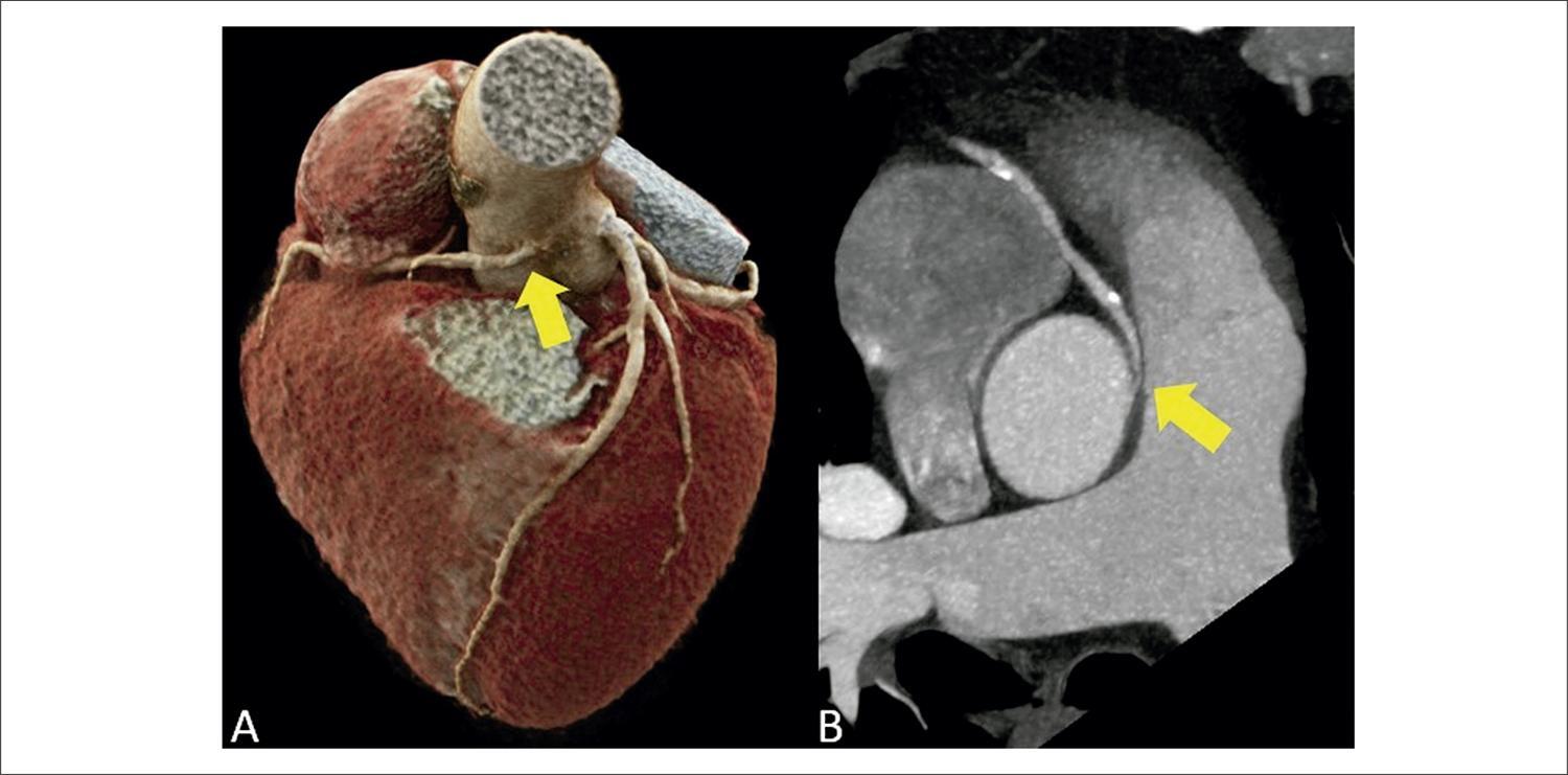
Anomalous origin and course of coronary arteries are rare congenital heart diseases, affecting less than 1% of the general population., Anomalous coronary origin with an interarterial (suprapulmonary) course is characterized by the course of the coronary artery between the ascending aorta and the pulmonary artery trunk, most commonly involving the right coronary artery., Most individuals are asymptomatic, but, among symptomatic patients, chest pain and dyspnea on exertion are the most prevalent complaints. There is also an increase in rates of arrhythmia, sudden death, and acute myocardial infarction., Sudden death is the main complication of this anatomical variant, occurring in approximately 30% of patients.,, In these cases, the narrowing and stretching of the anomalous ostium, mainly during physical exercise and in stressful situations, with consequent reduction in coronary flow, is the substrate for potentially fatal ischemic alterations.,, Since electrocardiogram, in most cases, does not reveal ischemic alterations, diagnosis is generally made through an incidental finding on imaging exams. These exams are of great importance, especially coronary CTA, which, in addition to being a non-invasive exam with high negative predictive value, allows detailed anatomical visualization (angle of the origin, presence of intramural trajectory in the aorta, degree of ostial/proximal luminal reduction) and the correct classification in relation to the pulmonary valve plane (suprapulmonary versus subpulmonary).,,

Recent studies have demonstrated that coronary CTA is an accurate test for identifying myocardial ischemia through FFRCT, when compared to invasive measurement (iFFR) by coronary angiography.,,, The detection of ischemia in coronary CTA is of great importance in decision-making, mainly in plaques considered moderate (50% to 69% luminal reduction) or when there is diagnostic doubt, reducing the number of unnecessary referrals to coronary angiography in cases without ischemia on FFRct.,, Due to the excellent accuracy between the methods in the analysis of a coronary tree without anomaly, the applicability of the method in the context of coronary anomaly has been extrapolated., Our group applied the most current version of a tool for calculating FFRCT currently available only on a research platform developed by Siemens Healthineers (cFRR, version 3.0.0) in the clinical case described. This research tool is available for installation on standard configuration computers, and it uses artificial intelligence tools, with reduced processing time. Currently, commercially available options require the step of sending images in DICOM format for processing in specific centers, with delivery of results at least 24 hours after the images have been sent., The research tool used in this clinical case has advantages, such as fast processing time on standard configuration computers in the analysis room and use of tomographic images from the standard routine protocol, without requiring the addition of a specific protocol or a higher dose of radiation, without the use of stressors., This tool, in general, has some limitations, such as difficulty in defining the coronary borders in the presence of excessive calcification, and the need for high-quality images, without movement artifacts, for adequate automatic detection of the central luminal line and the lines that delimit the coronary borders, allowing adequate calculation of FFRct. It is important to emphasize that this tool distinguishes the interarterial pathways associated with ischemia, considering only the flow obstacle during rest, without estimating the risk of ischemic events associated with dynamic changes secondary to intense exercise. Use of this tool is scarce in the literature, and it still does not have a robust body of evidence. Nevertheless, in the clinical case described, the FFRCT tool was applied in a context different than what is usual, where luminal reduction was not determined by coronary atheromatosis, but by ostial angulation and compression of the proximal segment of the anomalous interarterial pathway. The ischemic response was considered a parameter of poor prognosis; subsequently, the confirmatory invasive functional test (iFFR) was indicated to assist in the therapeutic decision.

[…]
  • Congenital Abnormalities
  • Fractional Flow Reserve, Myocardial
  • Machine Learning
  • Myocardial Ischemia
  • Tomography, X-Ray Computed
Read Full Article
Creative Commons Attribution (CC BY) Creative Commons license
10/05/2023 DOI: 10.36660/abcimg.2023361i
ABCIC
Original Article

Incidental Tomographic Findings of Coronary Artery Calcifications: A Prevalence Study in Southern Brazil

Karen Rafaela Okaseski Scopel et al. Tássia Machado Medeiros Bibiana Natalia Porto Maicá Maria Carbonari Velho … Mariana Motta Dias da Silva Juliane Nascimento Mattos Guilherme Galante Heuser Eliane Roseli Winkelmann

Abstract

Introduction

Coronary artery calcifications (CAC) are shown to be a predictive factor of cardiovascular diseases. Computed tomography (CT) of the chest with a low-dose acquisition protocol is accurate in identifying CAC and provides incidental findings of these calcifications, which are commonly overlooked. This study will analyze the prevalence of incidental findings of calcification in coronary arteries in non-cardiac individuals undergoing chest CT.

Methods

Consecutive cross-sectional study of an analytical and descriptive nature. Individuals of both genders who underwent chest CT by referral, over 18 years of age and without heart disease were included. Data collection was carried out using medical records and a self-applied anamnesis form. The variables referring to the CAC and the extension of the impairment were obtained from the reassessment of the chest CT images available in the institution’s system. The exams were anonymized and evaluated by two experienced radiologists. P≤0.05 was considered statistically significant.

Results

397 exams were analyzed. A prevalence of calcifications was found in 176 (44%) of the cases. The existence of these coronary calcifications is related to age (p<0.001). Calcifications are related to gender (p = 0.03) with a higher odds ratio of development in men (odds ratio [OR] = 1.55). Smoking (p<0.001), sedentary lifestyle (p<0.001), systemic arterial hypertension (p<0.001), Diabetes Mellitus (p = 0.04), and dyslipidemia (p<0.001) showed a positive association.

Conclusion

The prevalence of incidental CAC findings was 44%; vary in greater numbers between mild and severe; higher odds ratio in males and increased prevalence with age. Therefore, chest CT proves to be an effective method to assess CAC, and together with the patient’s clinical history, it can be used to measure risk factors for CVD and intervene in the outcome of the condition.

  • Coronary Vessels
  • Incidental Findings
  • Tomography, X-Ray Computed
  • Vascular Calcification
Read Full Article
Creative Commons Attribution (CC BY) Creative Commons license
24/03/2023 DOI: 10.36660/abcimg.2023368i Views 2
ABCIC
Image

Dual-Energy Computed Tomography with Material Decomposition: Before and after Thrombolysis in Massive PTE

Paula de Castro Carvalho Gorgulho et al. Bruno Soares da Silva Rangel Sicilia Pacheco e Silva Ilan Gottlieb Gustavo Luiz Gouvêa de Almeida Junior

[…]

  • Perfusion/instrumentation
  • Pulmonary Embolism/diagnostic imaging
  • Tomography, X-Ray Computed
Read Full Article
Creative Commons Attribution (CC BY) Creative Commons license
20/01/2022 DOI: 10.5935/2318-8219.20180009
ABCIC
Image

Neurovascular Lesion Caused by Non-Lethal Projectile in Cervical Region

Milton Sérgio Bohatch Júnior et al. Amanda Fernandes Vidal da Silva Agnaldo José Graciano

[…]

  • Jugular Veins/surgery
  • Sternotomy
  • Tomography, X-Ray Computed
  • Venous Thrombosis
  • Wounds, Gunshot/surgery
Read Full Article
Creative Commons Attribution (CC BY) Creative Commons license
20/01/2022 DOI: 10.5935/2318-8219.20180017 Views 1
ABCIC
Image

Extensive Renal Tumor Diagnosed from Echocardiography Findings

Morgahna Nathalie Wamser et al. James Alberton

[…]

  • Diagnostic Imaging
  • Echocardiography/methods
  • Kidney Neoplasms
  • Kidney/injuries
  • Tomography, X-Ray Computed
  • Vena Cava, Inferior/injuries
Read Full Article
Creative Commons Attribution (CC BY) Creative Commons license
20/01/2022 DOI: 10.5935/2318-8219.20170010 Views 1
ABCIC
Original Article

Correlation between Degrees of Pulmonary Hypertension and Findings in Complementary Tests

João Arthur Pazello et al. Ana Cristina Camarozano

Abstract

Introduction

Pulmonary hypertension is potentially fatal and courses with important day-to-day limitation. While the treatment is capable of slowing the disease’s progression, increase in life expectancy is directly linked to early diagnosis and treatment.

Objectives

To analyze the relation between alterations detectable on routine exams performed on pulmonary hypertension patients (echocardiography, electrocardiography and computerized tomography) and increases on pulmonary artery systolic pressure.

Methods

We analyzed the recordings of patients presented with pulmonary hypertension, and separated them in two groups based on their echocardiography-estimated pulmonary artery systolic pressure. Group 1 was composed of patients with pulmonary artery systolic pressure between 35 mmHg and 65 mmHg, and Group 2 of patients with pulmonary artery systolic pressure > 65 mmHg. We analyzed the prevalence of alterations suggestive of pulmonary hypertension on echocardiography, electrocardiography and computerized tomography. A descriptive analysis of the findings was conducted, followed by comparative analysis between the groups.

Results

We analyzed 101 patients (43 from Group 1 and 58 from Group 2). Most were women (82). Higher pulmonary artery systolic pressure values were correlated with right-heart dilation on echocardiography (p < 0.001). Electrocardiography findings revealed that right-heart hypertrophy and ventricular strain pattern were more common on Group 2 (p < 0.05). Computerized tomography analysis showed Group 2 patients had superior pulmonary artery diameters (p < 0.05). There was correlation between high pulmonary artery systolic pressure and clinical progression of the disease measured via the NYHA score (p < 0.05).

Conclusion

It is possible to make use of clinical examination and a simple electrocardiography to stratify the severity of a pulmonary artery patient. Computerized tomography and echocardiography should be used to confirm these findings. (Arq Bras Cardiol: Imagem cardiovasc. 2018;31(3):175-182)

  • Blood Pressure
  • Diagnostic Imaging
  • Echocardiography/methods
  • Electrocardiography
  • Hypertension Pulmonary
  • Pulmonary Arterial
  • Tomography, X-Ray Computed
Read Full Article
Creative Commons Attribution (CC BY) Creative Commons license
20/01/2022 DOI: 10.5935/2318-8219.20180026
ABC
Viewpoint

Coronary Computed Tomography Angiography Takes the Center Stage and Here is Why

Illan Gottlieb et al. Marcio Sommer Bittencourt Carlos Eduardo Rochitte João L. Cavalcante

Max Planck once said that “A new scientific truth does not triumph by convincing its opponents and making them see the light, but rather because its opponents eventually die, and a new generation grows up that is familiar with it.” In its beginnings, coronary computed tomography angiography (CCTA) was accused of having too low accuracy for the diagnosis of obstructive coronary artery disease (CAD) to be used in clinical practice. Over the last decade, major technical developments such as larger axial coverage (from 2 cm to 16 cm) and improved temporal resolution, have enabled CCTA to become by far the most accurate non-invasive imaging method for diagnosis of obstructive CAD, with sensitivity and specificity of approximately 95% and 90%, respectively.

Then CCTA was burdened with the accusation of exposing patients to radiation doses so high, that warranted some society guidelines to specifically point this out and limit its use. At that time, CCTA exposed patients to doses ranging from 20 to 25 mSV, while triphasic abdomen CT exposed patients to 30 to 40 mSv and scintigraphic myocardial perfusion studies with Thallium used up to 40 mSv. In 2018, radiation exposure from CCTA dropped to well below 5 mSv (most advanced clinical centers use much less), a fraction of the dose used in myocardial perfusion studies with MIBI tetrophosmin. Then the cost-effectiveness wave came with societies rightfully demanding proof that CCTA offered more value at an acceptable cost compared to other imaging modalities, and CCTA once again proved to be more cost-effective than other modalities. Although one hardly finds cost-effectiveness studies comparing nuclear scans with ECG treadmill tests, providing better diagnosis performance is not enough anymore. More recently, this strategy has even been put into challenge in large randomized clinical trials comparing CCTA with the standard of care in the investigation of suspected CAD both in the acute and in the outpatient settings.–

[…]

  • Computed Tomography Angiography
  • Coronary Artery Disease/diagnostic imaging
  • Coronary Artery Disease/physiopathology
  • Coronary Artery Disease/prevention and control
  • Tomography, X-Ray Computed
Read Full Article
Creative Commons Attribution (CC BY) Creative Commons license
01/01/2019 DOI: 10.5935/abc.20190003 Views 1
Figure 1
ABC
Image

Computed Tomography-Guided Core Needle Biopsy of Cardiac Angiosarcoma

Luis Gorospe et al. Alberto Cabañero-Sánchez Gemma María Muñoz-Molina Ana María Ayala-Carbonero María Ángeles Fernández-Méndez

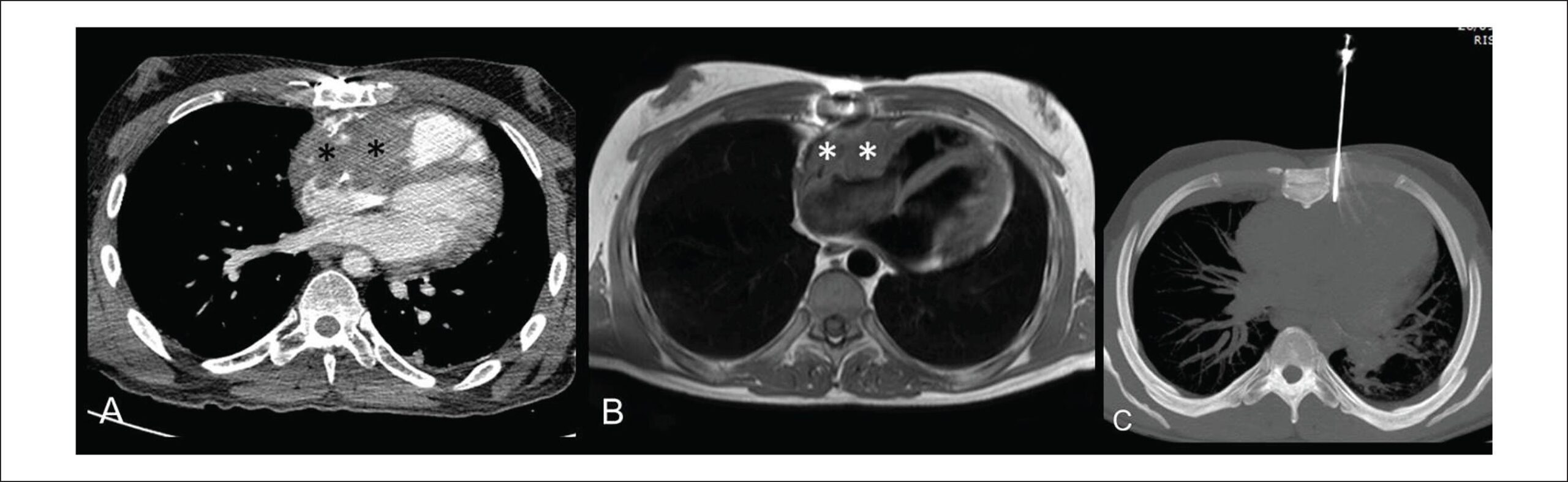
A 34-year-old man was referred to our institution after an echocardiography performed at another center because of tachycardia (atrial flutter), which showed heterogeneous pericardial mass infiltrating the right chambers. Thoracic computed tomography (CT) and cardiac magnetic resonance (MR) imaging were performed for more accurate assessment of the exact tumor location, size, and potential infiltration of other cardiac and mediastinal structures. CT () and MR imaging () confirmed an 8-cm ill-defined heterogeneous enhancing pericardial mass infiltrating the anterior and superior walls of the right atrium and extending to lateral and inferior walls of the right ventricle, consistent with cardiac angiosarcoma. The patient was deemed inoperable, as the mass also invaded the superior vena cava, aortic root, and epicardial fat. CT-guided core needle biopsy of the cardiac mass was the method of choice for histological verification of tentative diagnosis. Once the patient signed the informed consent and was put in supine position, an 18-gauge biopsy needle was driven between the left thoracic internal arteries and the left border of the sternal body () and a tissue specimen was safely obtained from the beating heart without adverse events. The procedure was performed by an experienced interventional thoracic radiologist under local anesthesia and in the presence of a thoracic surgeon. CT images obtained immediately after biopsy showed no post-procedural complications. Preliminary histological analysis performed on-site by a pathologist determined the adequacy of the tissue specimen. Final histopathologic diagnosis was high-grade cardiac angiosarcoma. To the best of our knowledge, only one case of a CT-guided core needle biopsy of a cardiac angiosarcoma involving the right chambers has been previously reported in English-language scientific literature.

[…]

  • Biopsy, Large-Core Needle
  • Hemangiosarcoma/diagnosis
  • Hemangiosarcoma/pathology
  • Neoplasm Staging
  • Tomography, X-Ray Computed
Read Full Article
Creative Commons Attribution (CC BY) Creative Commons license
01/05/2018 DOI: 10.5935/abc.20180064 Views 2
Figure 1
IJCS
Original Article

Overview with Meta-analysis of Systematic Reviews of the Diagnostic and Prognostic Value of Coronary Computed Tomography Angiography in the Emergency Department

Irlaneide da Silva Tavares et al. Carlos José Oliveira de Matos Marco Antonio Prado Nunes Antonio Carlos Sobral Sousa … Divaldo Pereira de Lyra Júnior Joselina Luzia Menezes Oliveira

Abstract

Background:

The high prevalence of CAD, as well as your impact on health expenditure and the various treatment options to reduce morbidity and mortality related to CAD, comes to develop a diagnostic tool precis and with important findings in the Emergency Department.

Objetive:

To conduct an overview with meta-analysis to compile evidence from multiple systematic reviews (SR) on the diagnostic and prognostic value of coronary computed tomography angiography (CCTA) to assess acute chest pain in the emergency department (ED).

Methods:

We included SR of primary studies that evaluated the diagnostic and prognostic value of CCTA ≥ 64 channels in the ED. The studies were conducted in patients at low and intermediate risk for coronary artery disease (CAD). Quality assessment was performed using PRISMA and approved reviews that scored ≥ 80%. Two authors independently extracted data using a standardized form. Spearman correlation test, Chi-square test, Cochran’s Q test or Higgins and Thompson statistical I2 were used. For meta-analysis, “mada” package statistical software R Core Team, 2015, was used. The significance level adopted was 95%.

Results:

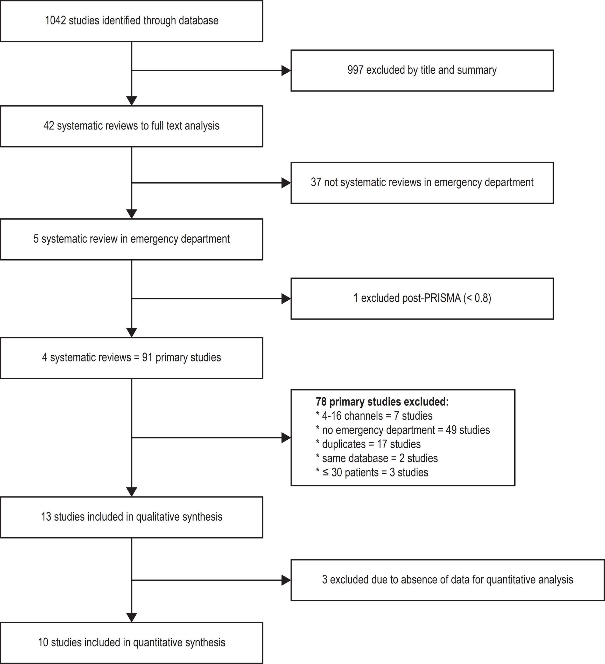
Four reviews were eligible for inclusion in this overview, resulting in 13 articles after applying the exclusion criteria, and only 10 of these were used for meta-analysis, adding up to a total of 4831 patients (mean age, 54 ± 6 years; 51% male), of whom 46% were hypertensive, 32% had dyslipidemia, 13% had diabetes and 26% had a family history of premature CAD. In the meta-analysis, 9 studies defined CCTA positive in the presence of luminal lesions ≥ 50%, while 1 study defined it as luminal lesions ≥ 70%. Sensitivity ranged from 77% to 98%, and specificity, from 73% to 100%. The univariate analysis showed homogeneity of diagnostic odds ratio (DOR) [Q = 8.5 (df = 9), p = 0.48 and I2 = 0%]. The pooled mean DOR for CCTA in primary analyses was 4.33 (95% CI: 3.47 – 5.18). The area under the curve (AUC) was 0.982 (95% CI: 0.967 – 0.999). There was no death, 29 (0.6%) infarcts, 92 (1.9%) revascularizations and 312 (6.4%) invasive coronary angiographies. The diagnosis of acute coronary syndrome occurred in 7.3% of the 1655 patients included in the meta-analysis.

Conclusions:

The use of CCTA as a tool for stratification of patients at low or intermediate cardiovascular risk, who are in the ED with chest pain, has high accuracy, safety, reduces length of hospital stay and probably the costs, producing an early diagnosis and more effective decision making. (Int J Cardiovasc Sci. 2017; [online].ahead print, PP.0-0)

  • Chest Pain
  • Coronary Artery Disease
  • emergency medicine
  • Meta-Analysis as Topic
  • Tomography, X-Ray Computed
Read Full Article
Creative Commons Attribution (CC BY) Creative Commons license
23/10/2017 DOI: 10.5935/2359-4802.20170086 Views 1
Figure 1
ABC
Original Article

Factors Associated With Coronary Artery Disease Progression Assessed By Serial Coronary Computed Tomography Angiography

Gabriel Cordeiro Camargo et al. Tamara Rothstein Maria Eduarda Derenne Leticia Sabioni … João A. C. Lima Ronaldo de Souza Leão Lima Ilan Gottlieb

Abstract

Background:

Coronary computed tomography angiography (CCTA) allows for noninvasive coronary artery disease (CAD) phenotyping. Factors related to CAD progression are epidemiologically valuable.

Objective:

To identify factors associated with CAD progression in patients undergoing sequential CCTA testing.

Methods:

We retrospectively analyzed 384 consecutive patients who had at least two CCTA studies between December 2005 and March 2013. Due to limitations in the quantification of CAD progression, we excluded patients who had undergone surgical revascularization previously or percutaneous coronary intervention (PCI) between studies. CAD progression was defined as any increase in the adapted segment stenosis score (calculated using the number of diseased segments and stenosis severity) in all coronary segments without stent (in-stent restenosis was excluded from the analysis). Stepwise logistic regression was used to assess variables associated with CAD progression.

Results:

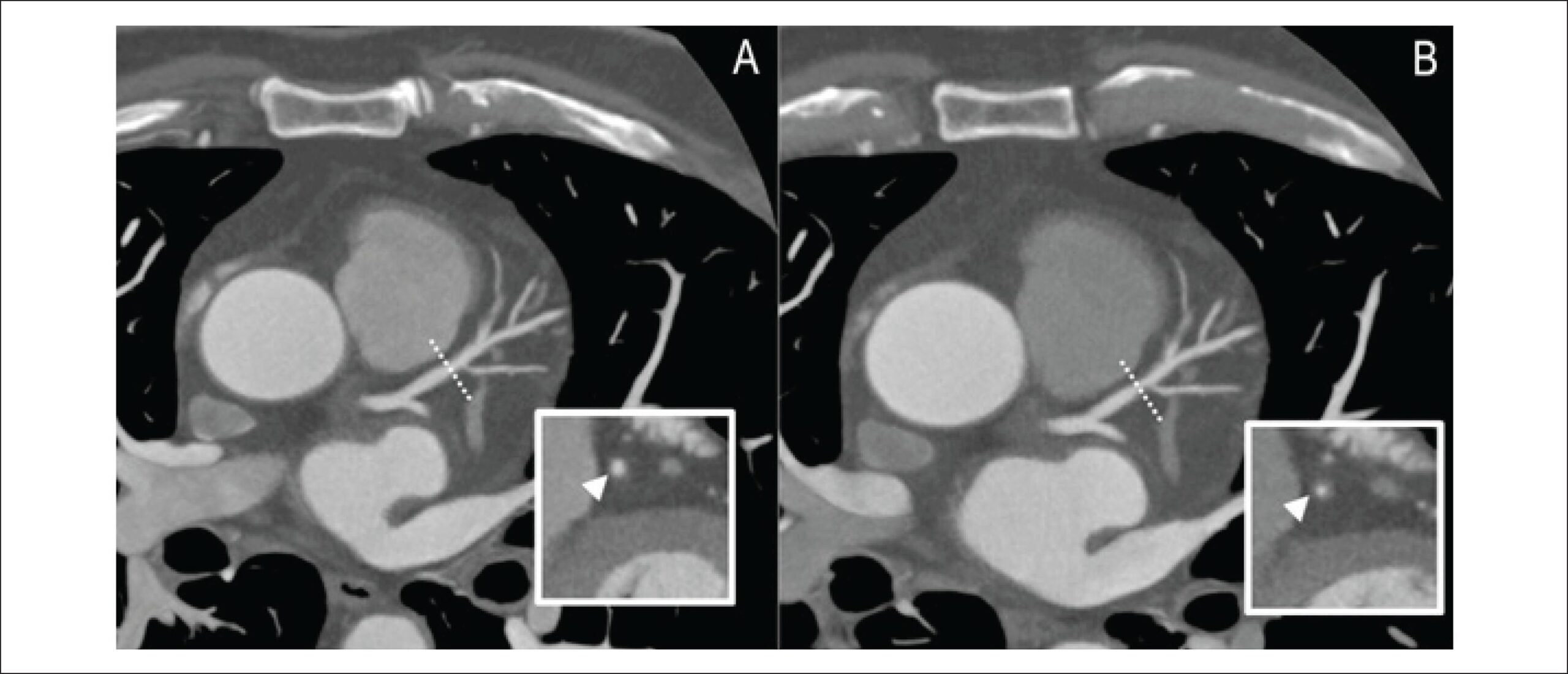
From a final population of 234 patients, a total of 117 (50%) had CAD progression. In a model accounting for major CAD risk factors and other baseline characteristics, only age (odds ratio [OR] 1.04, 95% confidence interval [95%CI] 1.01-1.07), interstudy interval (OR 1.03, 95%CI 1.01-1.04), and past PCI (OR 3.66, 95%CI 1.77-7.55) showed an independent relationship with CAD progression.

Conclusions:

A history of PCI with stent placement was independently associated with a 3.7-fold increase in the odds of CAD progression, excluding in-stent restenosis. Age and interstudy interval were also independent predictors of progression.

  • Coronary Amgiography
  • Coronary Artery Disease/physiopathology
  • Percutaneous Coronary Intervention
  • Tomography, X-Ray Computed
Read Full Article
Creative Commons Attribution (CC BY) Creative Commons license
04/05/2017 DOI: 10.5935/abc.20170049 Views 2
Figure 1
ABC
Image

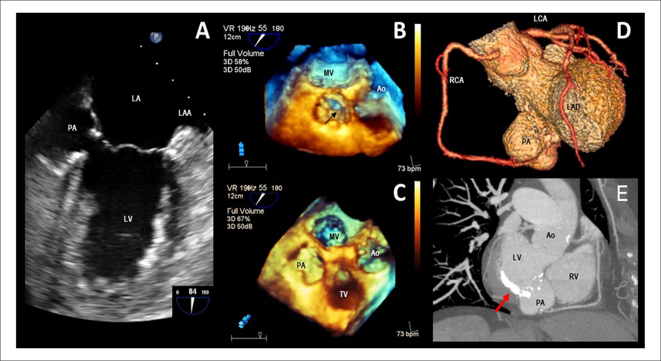
Left Ventricular Pseudoaneurysm Secondary to Mitral Valve Endocarditis

Bruno Bochard-Villanueva et al. Jordi Estornell-Erill

A

[…]

  • Echocardiography, Transesophageal
  • Endocarditis, bacterial
  • Heart Failure
  • Heart Valve Diseases
  • Tomography, X-Ray Computed
  • Ventricular Dysfunction, Left/Complications
Read Full Article
Creative Commons Attribution (CC BY) Creative Commons license
01/09/2015 DOI: 10.5935/abc.20150071

How to cite this article

EndNote RIS

Electronic Document Format (Vancouver)

Siqueira MEM, Dias MI, Uski ACVR, Aguiar Filho LF. Giant Right Coronary Artery Aneurysm with Arteriovenous Fistula. ABC Imagem Cardiovascular 2023;36(3):e20230025.

Electronic Document Format (ABNT)

Siqueira, Maria Eduarda Menezes de; Dias, Mayra Isabel; Uski, Ana Claudia Vincezi Raudan; Aguiar Filho, Luciano Figueiredo de. Giant Right Coronary Artery Aneurysm with Arteriovenous Fistula. ABC Imagem Cardiovascular, v. 36, n. 3, e20230025, Jul. 2023.

Electronic Document Format (APA)

Siqueira, M. E. M. , Dias, M. I., Uski, A. C. V. R., & Aguiar Filho, L. F. (2023). Giant Right Coronary Artery Aneurysm with Arteriovenous Fistula. ABC Imagem Cardiovascular, 36(3), e20230025.

Electronic Document Format (ISO)

Siqueira, Maria Eduarda Menezes de and Dias, Mayra Isabel and Uski, Ana Claudia Vincezi Raudan and Aguiar Filho, Luciano Figueiredo de. Giant Right Coronary Artery Aneurysm with Arteriovenous Fistula. ABC Imagem Cardiovascular [online]. 2023, vol. 36, n. 3, [cited 2026-10-14], e20230025. Available from: <https://journals.cardiol.br/en/article/giant-right-coronary-artery-aneurysm-with-arteriovenous-fistula/>. ISSN 2318-8219

EndNote RIS

Electronic Document Format (Vancouver)

Trindade FB, Morais TC, Dantas Junior RN, Torres RVA, Soares CSP, Parga Filho JR. New Application of FFRCT in Clinical Practice: Evaluation of Interarterial Anomalous Coronary Course. ABC Imagem Cardiovascular 2023;36(2):e361.

Electronic Document Format (ABNT)

Trindade, Fábio Bordin; Morais, Thamara Carvalho; Dantas Junior, Roberto Nery; Torres, Roberto Vitor Almeida; Soares, Clarice Santos Parreira; Parga Filho, José Rodrigues. New Application of FFRCT in Clinical Practice: Evaluation of Interarterial Anomalous Coronary Course. ABC Imagem Cardiovascular, v. 36, n. 2, e361, May. 2023.

Electronic Document Format (APA)

Trindade, F. B., Morais, T. C., Dantas Junior, R. N., Torres, R. V. A., Soares, C. S. P., & Parga Filho, J. R. (2023). New Application of FFRCT in Clinical Practice: Evaluation of Interarterial Anomalous Coronary Course. ABC Imagem Cardiovascular, 36(2), e361.

Electronic Document Format (ISO)

Trindade, Fábio Bordin and Morais, Thamara Carvalho and Dantas Junior, Roberto Nery and Torres, Roberto Vitor Almeida and Soares, Clarice Santos Parreira and Parga Filho, José Rodrigues. New Application of FFRCT in Clinical Practice: Evaluation of Interarterial Anomalous Coronary Course. ABC Imagem Cardiovascular [online]. 2023, vol. 36, n. 2, [cited 2026-10-14], e361. Available from: <https://journals.cardiol.br/en/article/new-application-of-ffrct-in-clinical-practice-evaluation-of-interarterial-anomalous-coronary-course/>. ISSN 2318-8219

EndNote RIS

Electronic Document Format (Vancouver)

Scopel KRO, Medeiros TM, Maicá BNP, Velho MC, Silva MMD, Mattos JN, Heuser GG, et al. Incidental Tomographic Findings of Coronary Artery Calcifications: A Prevalence Study in Southern Brazil. Arquivos Brasileiros de Cardiologia Imagem cardiovascular 2023;36(1):e368.

Electronic Document Format (ABNT)

Scopel, Karen Rafaela Okaseski; Medeiros, Tássia Machado; Maicá, Bibiana Natalia Porto; Velho, Maria Carbonari; Silva, Mariana Motta Dias da; Mattos, Juliane Nascimento; Heuser, Guilherme Galante; Winkelmann, Eliane Roseli. Incidental Tomographic Findings of Coronary Artery Calcifications: A Prevalence Study in Southern Brazil. Arquivos Brasileiros de Cardiologia Imagem cardiovascular, v. 36, n. 1, e368, Mar. 2023.

Electronic Document Format (APA)

Scopel, K. R. O., Medeiros, T. M., Maicá, B. N. P., Velho, M. C., Silva, M. M. D. , Mattos, J. N., Heuser, G. G., & Winkelmann, E. R. (2023). Incidental Tomographic Findings of Coronary Artery Calcifications: A Prevalence Study in Southern Brazil. Arquivos Brasileiros de Cardiologia Imagem cardiovascular, 36(1), e368.

Electronic Document Format (ISO)

Scopel, Karen Rafaela Okaseski and Medeiros, Tássia Machado and Maicá, Bibiana Natalia Porto and Velho, Maria Carbonari and Silva, Mariana Motta Dias da and Mattos, Juliane Nascimento and Heuser, Guilherme Galante and Winkelmann, Eliane Roseli. Incidental Tomographic Findings of Coronary Artery Calcifications: A Prevalence Study in Southern Brazil. Arquivos Brasileiros de Cardiologia Imagem cardiovascular [online]. 2023, vol. 36, n. 1, [cited 2026-10-14], e368. Available from: <https://journals.cardiol.br/en/article/incidental-tomographic-findings-of-coronary-artery-calcifications-a-prevalence-study-in-southern-brazil/>. ISSN 2318-8219

EndNote RIS

Electronic Document Format (Vancouver)

Gorgulho PCC, Rangel BSS, Silva SP, Gottlieb I, Almeida Junior GLG. Dual-Energy Computed Tomography with Material Decomposition: Before and after Thrombolysis in Massive PTE. Arquivos Brasileiros de Cardiologia Imagem cardiovascular 2022;31(1):24-5.

Electronic Document Format (ABNT)

Gorgulho, Paula de Castro Carvalho; Rangel, Bruno Soares da Silva; Silva, Sicilia Pacheco e; Gottlieb, Ilan; Almeida Junior, Gustavo Luiz Gouvêa de. Dual-Energy Computed Tomography with Material Decomposition: Before and after Thrombolysis in Massive PTE. Arquivos Brasileiros de Cardiologia Imagem cardiovascular, v. 31, n. 1, p. 24-25, Jan. 2022.

Electronic Document Format (APA)

Gorgulho, P. C. C., Rangel, B. S. S., Silva, S. P. , Gottlieb, I., & Almeida Junior, G. L. G. (2022). Dual-Energy Computed Tomography with Material Decomposition: Before and after Thrombolysis in Massive PTE. Arquivos Brasileiros de Cardiologia Imagem cardiovascular, 31(1), 24-25.

Electronic Document Format (ISO)

Gorgulho, Paula de Castro Carvalho and Rangel, Bruno Soares da Silva and Silva, Sicilia Pacheco e and Gottlieb, Ilan and Almeida Junior, Gustavo Luiz Gouvêa de. Dual-Energy Computed Tomography with Material Decomposition: Before and after Thrombolysis in Massive PTE. Arquivos Brasileiros de Cardiologia Imagem cardiovascular [online]. 2022, vol. 31, n. 1, [cited 2026-10-14], pp. 24-25. Available from: <https://journals.cardiol.br/article/dual-energy-computed-tomography-with-material-decomposition-before-and-after-thrombolysis-in-massive-pte/>. ISSN 2318-8219

EndNote RIS

Electronic Document Format (Vancouver)

Bohatch Júnior MS, Silva AFV, Graciano AJ. Neurovascular Lesion Caused by Non-Lethal Projectile in Cervical Region. Arquivos Brasileiros de Cardiologia Imagem cardiovascular 2022;31(2):110-1.

Electronic Document Format (ABNT)

Bohatch Júnior, Milton Sérgio; Silva, Amanda Fernandes Vidal da; Graciano, Agnaldo José. Neurovascular Lesion Caused by Non-Lethal Projectile in Cervical Region. Arquivos Brasileiros de Cardiologia Imagem cardiovascular, v. 31, n. 2, p. 110-111, Jan. 2022.

Electronic Document Format (APA)

Bohatch Júnior, M. S., Silva, A. F. V. , & Graciano, A. J. (2022). Neurovascular Lesion Caused by Non-Lethal Projectile in Cervical Region. Arquivos Brasileiros de Cardiologia Imagem cardiovascular, 31(2), 110-111.

Electronic Document Format (ISO)

Bohatch Júnior, Milton Sérgio and Silva, Amanda Fernandes Vidal da and Graciano, Agnaldo José. Neurovascular Lesion Caused by Non-Lethal Projectile in Cervical Region. Arquivos Brasileiros de Cardiologia Imagem cardiovascular [online]. 2022, vol. 31, n. 2, [cited 2026-10-14], pp. 110-111. Available from: <https://journals.cardiol.br/en/article/neurovascular-lesion-caused-by-non-lethal-projectile-in-cervical-region/>. ISSN 2318-8219

EndNote RIS

Electronic Document Format (Vancouver)

Wamser MN, Alberton J. Extensive Renal Tumor Diagnosed from Echocardiography Findings. Arquivos Brasileiros de Cardiologia Imagem cardiovascular 2022;30(1):37.

Electronic Document Format (ABNT)

Wamser, Morgahna Nathalie; Alberton, James. Extensive Renal Tumor Diagnosed from Echocardiography Findings. Arquivos Brasileiros de Cardiologia Imagem cardiovascular, v. 30, n. 1, p. 37-37, Jan. 2022.

Electronic Document Format (APA)

Wamser, M. N., & Alberton, J. (2022). Extensive Renal Tumor Diagnosed from Echocardiography Findings. Arquivos Brasileiros de Cardiologia Imagem cardiovascular, 30(1), 37-37.

Electronic Document Format (ISO)

Wamser, Morgahna Nathalie and Alberton, James. Extensive Renal Tumor Diagnosed from Echocardiography Findings. Arquivos Brasileiros de Cardiologia Imagem cardiovascular [online]. 2022, vol. 30, n. 1, [cited 2026-10-14], pp. 37-37. Available from: <https://journals.cardiol.br/en/article/extensive-renal-tumor-diagnosed-from-echocardiography-findings/>. ISSN 2318-8219

EndNote RIS

Electronic Document Format (Vancouver)

Pazello JA, Camarozano AC. Correlation between Degrees of Pulmonary Hypertension and Findings in Complementary Tests. Arquivos Brasileiros de Cardiologia Imagem cardiovascular 2022;31(3):175-82.

Electronic Document Format (ABNT)

Pazello, João Arthur; Camarozano, Ana Cristina. Correlation between Degrees of Pulmonary Hypertension and Findings in Complementary Tests. Arquivos Brasileiros de Cardiologia Imagem cardiovascular, v. 31, n. 3, p. 175-182, Jan. 2022.

Electronic Document Format (APA)

Pazello, J. A., & Camarozano, A. C. (2022). Correlation between Degrees of Pulmonary Hypertension and Findings in Complementary Tests. Arquivos Brasileiros de Cardiologia Imagem cardiovascular, 31(3), 175-182.

Electronic Document Format (ISO)

Pazello, João Arthur and Camarozano, Ana Cristina. Correlation between Degrees of Pulmonary Hypertension and Findings in Complementary Tests. Arquivos Brasileiros de Cardiologia Imagem cardiovascular [online]. 2022, vol. 31, n. 3, [cited 2026-10-14], pp. 175-182. Available from: <https://journals.cardiol.br/en/article/correlation-between-degrees-of-pulmonary-hypertension-and-findings-in-complementary-tests/>. ISSN 2318-8219

EndNote RIS

Electronic Document Format (Vancouver)

Gottlieb I, Bittencourt MS, Rochitte CE, Cavalcante JL. Coronary Computed Tomography Angiography Takes the Center Stage and Here is Why. Arquivos Brasileiros de Cardiologia 2019;112(1):104-6.

Electronic Document Format (ABNT)

Gottlieb, Illan; Bittencourt, Marcio Sommer; Rochitte, Carlos Eduardo; Cavalcante, João L.. Coronary Computed Tomography Angiography Takes the Center Stage and Here is Why. Arquivos Brasileiros de Cardiologia, v. 112, n. 1, p. 104-106, Dec. 2018.

Electronic Document Format (APA)

Gottlieb, I., Bittencourt, M. S., Rochitte, C. E., & Cavalcante, J. L. (2019). Coronary Computed Tomography Angiography Takes the Center Stage and Here is Why. Arquivos Brasileiros de Cardiologia, 112(1), 104-106.

Electronic Document Format (ISO)

Gottlieb, Illan and Bittencourt, Marcio Sommer and Rochitte, Carlos Eduardo and Cavalcante, João L.. Coronary Computed Tomography Angiography Takes the Center Stage and Here is Why. Arquivos Brasileiros de Cardiologia [online]. 2019, vol. 112, n. 1, [cited 2026-10-14], pp. 104-106. Available from: <https://journals.cardiol.br/en/article/coronary-computed-tomography-angiography-takes-the-center-stage-and-here-is-why/>. ISSN 0066-782X

EndNote RIS

Electronic Document Format (Vancouver)

Gorospe L, Cabañero-Sánchez A, Muñoz-Molina GM, Ayala-Carbonero AM, Fernández-Méndez M. Computed Tomography-Guided Core Needle Biopsy of Cardiac Angiosarcoma. Arquivos Brasileiros de Cardiologia 2018;110(5):493-4.

Electronic Document Format (ABNT)

Gorospe, Luis; Cabañero-Sánchez, Alberto; Muñoz-Molina, Gemma María; Ayala-Carbonero, Ana María; Fernández-Méndez, María Ángeles. Computed Tomography-Guided Core Needle Biopsy of Cardiac Angiosarcoma. Arquivos Brasileiros de Cardiologia, v. 110, n. 5, p. 493-494, Apr. 2018.

Electronic Document Format (APA)

Gorospe, L., Cabañero-Sánchez, A., Muñoz-Molina, G. M., Ayala-Carbonero, A. M., & Fernández-Méndez, M. (2018). Computed Tomography-Guided Core Needle Biopsy of Cardiac Angiosarcoma. Arquivos Brasileiros de Cardiologia, 110(5), 493-494.

Electronic Document Format (ISO)

Gorospe, Luis and Cabañero-Sánchez, Alberto and Muñoz-Molina, Gemma María and Ayala-Carbonero, Ana María and Fernández-Méndez, María Ángeles. Computed Tomography-Guided Core Needle Biopsy of Cardiac Angiosarcoma. Arquivos Brasileiros de Cardiologia [online]. 2018, vol. 110, n. 5, [cited 2026-10-14], pp. 493-494. Available from: <https://journals.cardiol.br/en/article/computed-tomography-guided-core-needle-biopsy-of-cardiac-angiosarcoma/>. ISSN 0066-782X

EndNote RIS

Electronic Document Format (Vancouver)

Tavares IS, Matos CJO, Nunes MAP, Sousa ACS, Lyra Júnior DP, Oliveira JLM. Overview with Meta-analysis of Systematic Reviews of the Diagnostic and Prognostic Value of Coronary Computed Tomography Angiography in the Emergency Department. International Journal of Cardiovascular Sciences 2017;31(1):33-46.

Electronic Document Format (ABNT)

Tavares, Irlaneide da Silva; Matos, Carlos José Oliveira de; Nunes, Marco Antonio Prado; Sousa, Antonio Carlos Sobral; Lyra Júnior, Divaldo Pereira de; Oliveira, Joselina Luzia Menezes. Overview with Meta-analysis of Systematic Reviews of the Diagnostic and Prognostic Value of Coronary Computed Tomography Angiography in the Emergency Department. International Journal of Cardiovascular Sciences, v. 31, n. 1, p. 33-46, Oct. 2017.

Electronic Document Format (APA)

Tavares, I. S., Matos, C. J. O. , Nunes, M. A. P., Sousa, A. C. S., Lyra Júnior, D. P. , & Oliveira, J. L. M. (2017). Overview with Meta-analysis of Systematic Reviews of the Diagnostic and Prognostic Value of Coronary Computed Tomography Angiography in the Emergency Department. International Journal of Cardiovascular Sciences, 31(1), 33-46.

Electronic Document Format (ISO)

Tavares, Irlaneide da Silva and Matos, Carlos José Oliveira de and Nunes, Marco Antonio Prado and Sousa, Antonio Carlos Sobral and Lyra Júnior, Divaldo Pereira de and Oliveira, Joselina Luzia Menezes. Overview with Meta-analysis of Systematic Reviews of the Diagnostic and Prognostic Value of Coronary Computed Tomography Angiography in the Emergency Department. International Journal of Cardiovascular Sciences [online]. 2017, vol. 31, n. 1, [cited 2026-10-14], pp. 33-46. Available from: <https://journals.cardiol.br/en/article/overview-with-meta-analysis-of-systematic-reviews-of-the-diagnostic-and-prognostic-value-of-coronary-computed-tomography-angiography-in-the-emergency-department/>. ISSN 2359-4802

EndNote RIS

Electronic Document Format (Vancouver)

Camargo GC, Rothstein T, Derenne ME, Sabioni L, Lima JAC, Lima RSL, Gottlieb I, et al. Factors Associated With Coronary Artery Disease Progression Assessed By Serial Coronary Computed Tomography Angiography. Arquivos Brasileiros de Cardiologia 2017;108(5):396-404.

Electronic Document Format (ABNT)

Camargo, Gabriel Cordeiro; Rothstein, Tamara; Derenne, Maria Eduarda; Sabioni, Leticia; Lima, João A. C.; Lima, Ronaldo de Souza Leão; Gottlieb, Ilan. Factors Associated With Coronary Artery Disease Progression Assessed By Serial Coronary Computed Tomography Angiography. Arquivos Brasileiros de Cardiologia, v. 108, n. 5, p. 396-404, May. 2017.

Electronic Document Format (APA)

Camargo, G. C., Rothstein, T., Derenne, M. E., Sabioni, L., Lima, J. A. C., Lima, R. S. L., & Gottlieb, I. (2017). Factors Associated With Coronary Artery Disease Progression Assessed By Serial Coronary Computed Tomography Angiography. Arquivos Brasileiros de Cardiologia, 108(5), 396-404.

Electronic Document Format (ISO)

Camargo, Gabriel Cordeiro and Rothstein, Tamara and Derenne, Maria Eduarda and Sabioni, Leticia and Lima, João A. C. and Lima, Ronaldo de Souza Leão and Gottlieb, Ilan. Factors Associated With Coronary Artery Disease Progression Assessed By Serial Coronary Computed Tomography Angiography. Arquivos Brasileiros de Cardiologia [online]. 2017, vol. 108, n. 5, [cited 2026-10-14], pp. 396-404. Available from: <https://journals.cardiol.br/en/article/factors-associated-with-coronary-artery-disease-progression-assessed-by-serial-coronary-computed-tomography-angiography/>. ISSN 0066-782X

EndNote RIS

Electronic Document Format (Vancouver)

Bochard-Villanueva B, Estornell-Erill J. Left Ventricular Pseudoaneurysm Secondary to Mitral Valve Endocarditis. Arquivos Brasileiros de Cardiologia 2015;105(3):320-1.

Electronic Document Format (ABNT)

Bochard-Villanueva, Bruno; Estornell-Erill, Jordi. Left Ventricular Pseudoaneurysm Secondary to Mitral Valve Endocarditis. Arquivos Brasileiros de Cardiologia, v. 105, n. 3, p. 320-321, Aug. 2015.

Electronic Document Format (APA)

Bochard-Villanueva, B., & Estornell-Erill, J. (2015). Left Ventricular Pseudoaneurysm Secondary to Mitral Valve Endocarditis. Arquivos Brasileiros de Cardiologia, 105(3), 320-321.

Electronic Document Format (ISO)

Bochard-Villanueva, Bruno and Estornell-Erill, Jordi. Left Ventricular Pseudoaneurysm Secondary to Mitral Valve Endocarditis. Arquivos Brasileiros de Cardiologia [online]. 2015, vol. 105, n. 3, [cited 2026-10-14], pp. 320-321. Available from: <https://journals.cardiol.br/en/article/left-ventricular-pseudoaneurysm-secondary-to-mitral-valve-endocarditis/>. ISSN 0066-782X

  • About ABC Family
  • Internal Regulation
  • Editorial Board
  • Brazilian Society of Cardiology (SBC)

Issues

SBC Guidelines

  • Videos
  • Podcasts
  • News