ABC Family Arquivos Brasileiros de Cardiologia International Journal of Cardiovascular Sciences ABC Heart Failure & Cardiomyopathy ABC Imagem Cardiovascular
Português English
ABC Family
Português English
  • About
    • About ABC Family
    • Internal Regulation
    • Editorial Board
    • Brazilian Society of Cardiology (SBC)
  • Issues
  • SBC Guidelines
  • Content
    • Videos
    • Podcasts
    • News
  • About
    • About ABC Family
    • Internal Regulation
    • Editorial Board
    • Brazilian Society of Cardiology (SBC)
  • Issues
  • SBC Guidelines
  • Content
    • Videos
    • Podcasts
    • News

ABC Family

  • ABC Family
  • Arquivos Brasileiros de Cardiologia
  • International Journal of Cardiovascular Sciences
  • ABC Heart Failure & Cardiomyopathy
  • ABC Imagem Cardiovascular
Search in

No content found.

Journals
Ordering
Article types
Others

  1. Home
  2. Keyword: Electrocardiography
Figure 1
ABCIC
Case Report

Apical Hypertrophic Cardiomyopathy with Microfistulas: Correlation Between Coronary Flow Steal, Positive Inotropic Effect, and Normalization of T-Wave Polarity

José Sebastiao de Abreu et al. Tereza Cristina Pinheiro Diógenes Marília Esther Benevides Abreu Isadora Sucupira Machado Chagas Mateus Paiva Marques Feitosa

Introduction

Apical hypertrophic cardiomyopathy (HCM) with microfistulas is a rare condition, and the incidence of multiple microfistulas draining into the left ventricle (LV) ranges from 0.07% to 0.09%., Diagnosis can be established via contrast ventriculography, coronary angiography, and echocardiography.–

Two-dimensional echocardiography with color and pulsed-wave Doppler allows visualization of microfistula flow and measurement of diastolic flow velocity (DFV) using the same approach as for the left anterior descending artery (LAD).

[…]

  • Electrocardiography
  • Hypertrophic Cardiomyopathy
  • Stress echocardiography
Read Full Article
Creative Commons Attribution (CC BY) Creative Commons license
21/09/2026 DOI: 10.36660/abcimg.20260090i Views 1
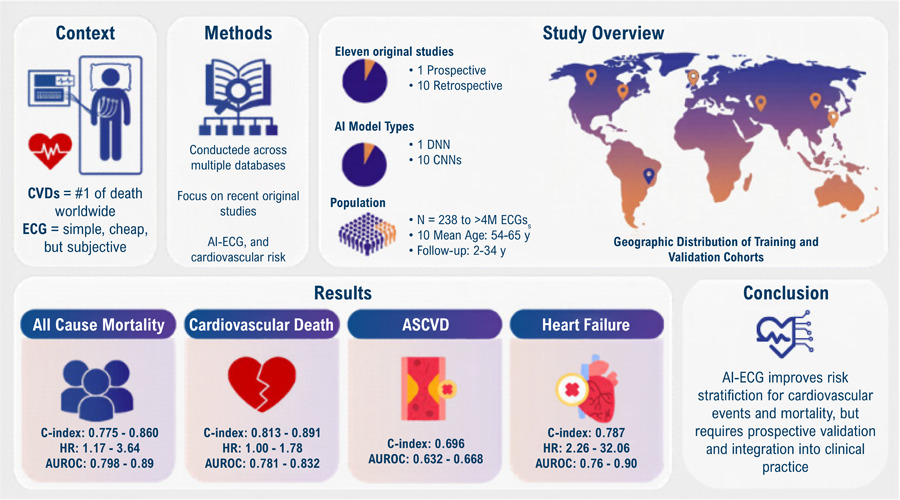
Artificial Intelligence Applied to Electrocardiogram-Based Cardiovascular Risk Assessment: A Systematic Review
ABC
Review Article

Artificial Intelligence Applied to Electrocardiogram-Based Cardiovascular Risk Assessment: A Systematic Review

Maria Clara Mantoan Pinheiro et al. Lívia Felberg Isadora Cristine Reis Sguizzato Bozzi Antônio Luiz Pinho Ribeiro Gabriela Miana de Mattos Paixão

Abstract

Cardiovascular diseases (CVDs) are the leading cause of mortality worldwide, underscoring the need for effective risk prediction and early detection. Although the electrocardiogram (ECG) is a widely available and low-cost diagnostic tool, its traditional interpretation is limited by subjectivity. Artificial intelligence (AI) has emerged as a promising approach, capable of extracting hidden prognostic information from ECG signals. This systematic review aimed to assess original studies applying AI techniques to ECGs for cardiovascular risk prediction and mortality. Original studies that used ECG signals as the sole input variable for AI models, focusing on cardiovascular risk outcomes, were included. A systematic search was conducted in different databases, and data were synthesized narratively. Eleven studies were included, predominantly retrospective cohorts applying convolutional neural networks (CNNs) to predict cardiovascular risk or mortality. The sample primarily consisted of adult populations in high-income countries. Primary outcomes included all-cause mortality, cardiovascular death, and major adverse cardiovascular events (MACE). Reported AUROC values ranged from 0.63 to 0.961 in training sets, with some models outperforming traditional risk scores. AI-ECG models demonstrated the potential to detect subclinical disease, enabling early risk stratification even in normal ECGs. However, challenges remain regarding population diversity, model interpretability, and prospective validation. The application of AI to ECG analysis represents a promising advancement in personalized cardiovascular risk assessment. Nonetheless, further research is needed to ensure the safety, effectiveness, and equitable clinical integration of these technologies.

  • Artificial Intelligence
  • Electrocardiography
  • Heart Disease Risk Factors
Read Full Article
Creative Commons Attribution (CC BY) Creative Commons license
03/08/2026 DOI: 10.36660/abc.20250790i
Figure 1
IJCS
Editorial

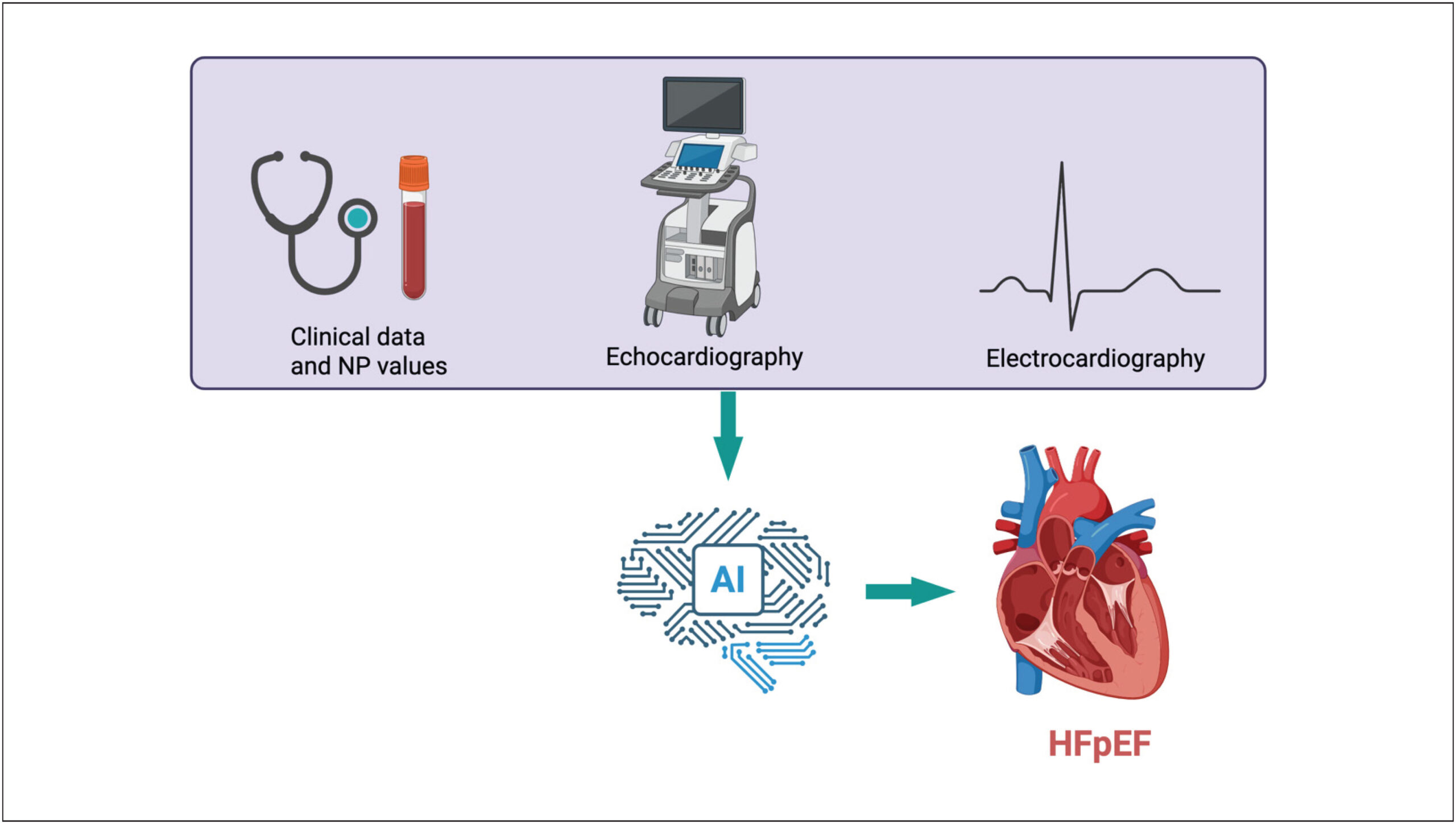
AI-Enhanced ECGs for the Diagnosis of HFpEF: A Promising Frontier, But Not Yet a Standalone Solution

Humberto Villacorta

Heart Failure with preserved Ejection Fraction (HFpEF) is among the most challenging syndromes in contemporary cardiology. Despite accounting for nearly half of all heart failure cases, HFpEF remains chronically underdiagnosed and is associated with significant morbidity and mortality., The diagnosis of HFpEF is challenging and relies on clinical history, natriuretic peptide measurements, and echocardiography. Diagnostic scores have been developed that integrate these three key elements., Echocardiography is indispensable for identifying structural and functional criteria, but its availability and cost make population-level screening impractical in many healthcare systems. In addition, in many places, a comprehensive echocardiogram is not performed. Likewise, natriuretic peptides are not always available. Therefore, new solutions to facilitate diagnosis are welcome.

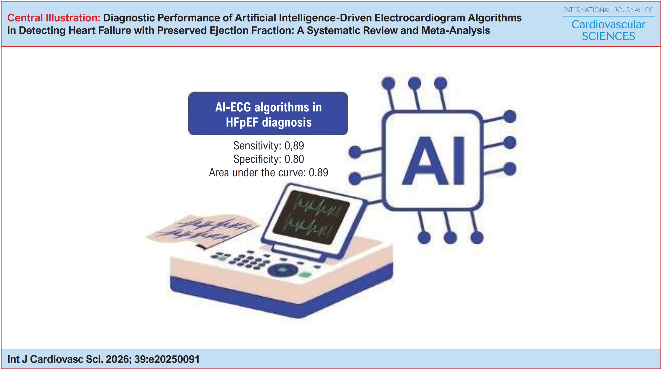
In this context, the systematic review and meta-analysis presented in this issue of International Journal of Cardiovascular Sciences, provide important insights into an increasingly discussed alternative: Artificial Intelligence-enhanced Electrocardiography (AI-ECG). Drawing from more than 170,000 patients across three observational studies, the authors report a pooled sensitivity of 0.89, specificity of 0.80, and an AUC of 0.89 for AI-ECG in detecting HFpEF. These findings suggest that deep learning models can extract subtle electrical signatures associated with diastolic dysfunction – signals that may be visually imperceptible on standard ECGs. The appeal is clear: the ECG is universal, inexpensive, rapid, and deeply embedded in clinical workflows. If Artificial Intelligence could reliably identify patients requiring echocardiographic confirmation, screening strategies might be revolutionized.

[…]

  • Artificial Intelligence
  • Diagnosis
  • Electrocardiography
  • Heart Failure
  • Insuficiência Cardíaca Diastólica
Read Full Article
Creative Commons Attribution (CC BY) Creative Commons license
27/07/2026 DOI: 10.36660/ijcs.20250272 Views 2
ABC
Editorial

Artificial Intelligence Attack! The Rising Importance of Chagas Disease

Roberto Coury Pedrosa

Chagas disease (Chd) is a chronic condition with primarily cardiac sequelae that can persist and worsen many years after the initial infection, with a serious impact on healthcare systems. The PAHO estimated that 90% of new infections go undiagnosed, and about 70% of those infected are unaware of their condition. Additionally, less than 1% of the estimated population at risk has been tested. As a result, this leads to a delay in diagnosis, which increases morbidity and mortality from its complications, highlighting a clear need to expand access to diagnosis from a public health perspective. To mitigate this impact, Chd was included in the Millennium Development Goals and the Sustainable Development Goals of the 2030 Agenda for Sustainable Development (UN, 2018).

The gold standard for diagnosing Chd is serological testing for T. cruzi antibodies. The biggest challenge in Chd remains predicting which patients will develop the cardiac form, a leading cause of heart failure, especially in rural areas of Brazil. Notably, electrocardiography is an accessible and low-cost tool that can detect early signs of cardiac involvement in Chd. In recent years, AI-ECG (Electrocardiogram with artificial intelligence) models have been shown to diagnose cardiac conditions accurately; many have been trained and tested primarily on data from high-income countries. Furthermore, these models have been evaluated retrospectively on large historical datasets, with relatively few prospective external validation studies. These gaps in AI-ECG research need to be addressed.

[…]

  • Artificial Intelligence
  • Chagas disease
  • Electrocardiography
  • Public Health
  • screening
Read Full Article
Creative Commons Attribution (CC BY) Creative Commons license
20/07/2026 DOI: 10.36660/abc.20260409i Views 4
ABC
Viewpoint

The Old Habits Need to be Abandoned and the Term "Overload" Replaced by "High Amplitude Criteria"

José Nunes de Alencar et al. Sandro Pinelli Felicioni Matheus Kiszka Scheffer Mariana Nogueira De Marchi Ljuba Bacharova

Left ventricular overload?

Brazilian electrocardiograms (ECG) reports have long labeled voltage-positive tracings as “left ventricular overload,” a legacy of historical teaching that linked hemodynamic loads to characteristic repolarization patterns. The problem is not the physiology but the nomenclature: overload is a hemodynamic claim, whereas the ECG measures electrical phenomena. Contemporary evidence reframes voltage positivity as an electrical phenotype with variable correlation to left ventricular mass (LVM) and geometry. In this article, we argue that Brazil should retire “overload,” adopt “high-amplitude criteria” as the report label, and explicitly teach the underlying concept of left ventricular electrical remodeling (LVER), so that our language matches what the ECG actually measures.

Phrasing “left ventricular overload” feels pragmatic — if surface ECG is “insensitive” to anatomic hypertrophy, perhaps what it detects is something else, namely “overload.” The evidence base argues otherwise. Electrocardiography records electric fields at the body surface; it does not directly measure mass, geometry, or loading conditions. This distinction is the core of the modern paradigm articulated by Ljuba Bacharova et al.: QRS voltage and morphology reflect an interplay of spatial determinants (source geometry, torso conductivity, electrode distance) and non-spatial determinants (conduction velocity, intercellular coupling, fibrosis, ionic remodeling), such that the same LVM can yield different voltages — and high voltage can arise with normal mass — depending on the myocardium’s electrical properties. The Working Group’s “4th Report” in the Journal of Electrocardiology integrates mechanistic simulation studies with population data and draws three conclusions that are decisive for our terminology. First, increased LVM and geometric type (concentric, eccentric, dilation) are not the main or only determinants of QRS patterns; combinations of anatomy and slowed activation propagation produce the observed spectrum from “pseudo-normal” tracings through high voltage and prolonged intrinsicoid deflection to left bundle branch block. Second, diffuse conduction slowing or midwall conduction abnormalities — without any increase in mass — can generate QRS changes that meet conventional electrocardiographic-left ventricular hypertrophy (LVH) criteria in a normal-sized ventricle. Third, in community cohorts the electrical and anatomic phenotypes frequently diverge, and each carries independent prognostic information.

[…]

  • Electrocardiography
  • Left Ventricular Hypertrophy
Read Full Article
Creative Commons Attribution (CC BY) Creative Commons license
17/07/2026 DOI: 10.36660/abc.20250709i
Artificial Intelligence-Enhanced Electrocardiography Analysis for Diagnosing Chagas Disease
ABC
Original Article

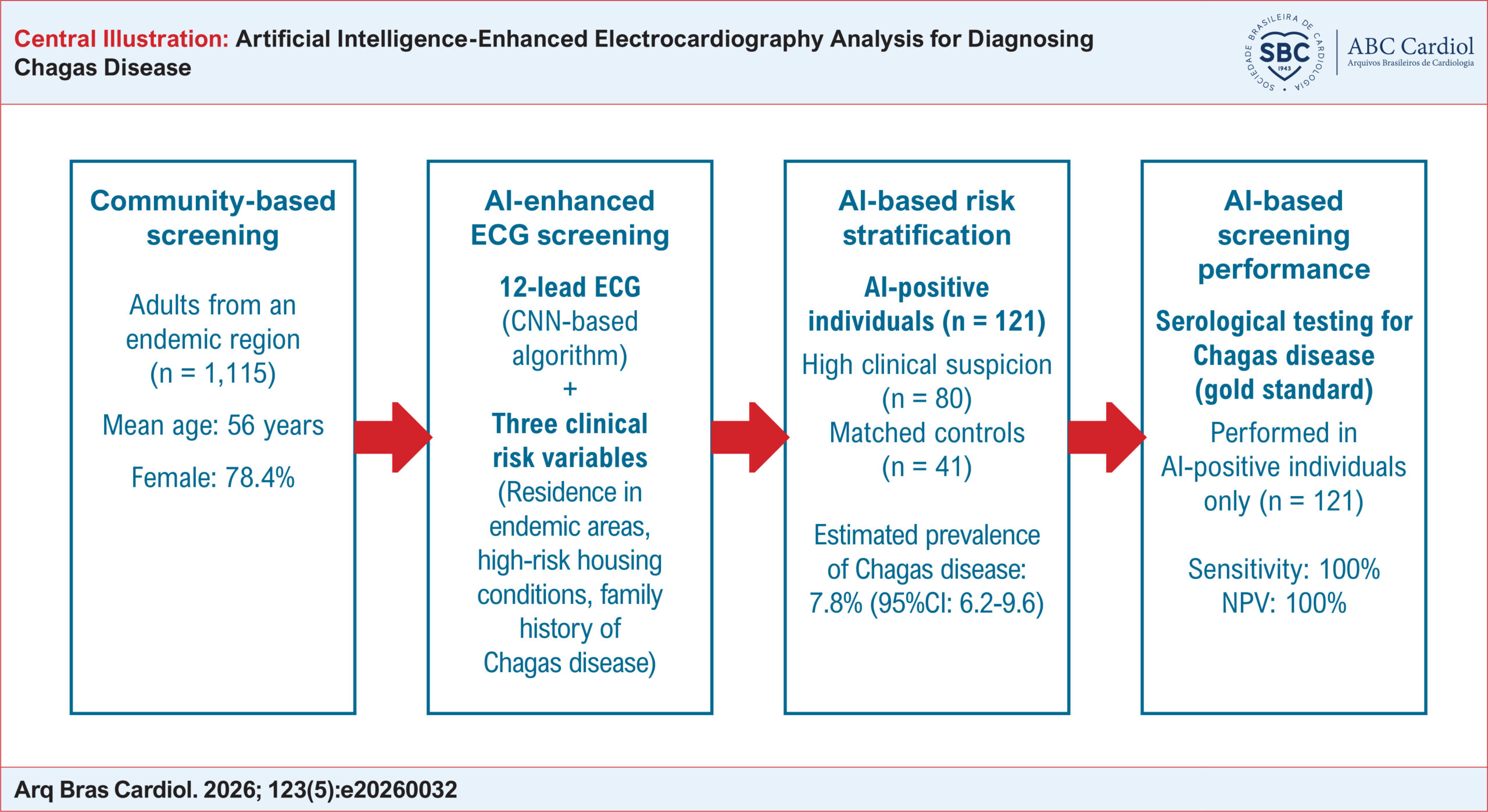
Artificial Intelligence-Enhanced Electrocardiography Analysis for Diagnosing Chagas Disease

Isabella Moreira Gonzalez Fonseca et al. Maria Carmo Pereira Nunes Craig Sable Renata Fonseca Mendoza … Fred Luciano Neves Santos Paulo Rodrigues Gomes Adson Patrik Vieira Carvalho Bernardo Fonseca Mendoza Noilson Lazaro Souza José Augusto de Almeida Barbosa Clareci Silva Cardoso Bruno Ramos Nascimento Antonio L. P. Ribeiro

Abstract

Background:

Chagas disease affects millions worldwide and remains a leading cause of cardiomyopathy in Latin America. Early diagnosis remains challenging in endemic regions. Artificial intelligence (AI)-based electrocardiography (ECG) analysis may offer a low-cost strategy for large-scale screening in resource-limited settings.

Objectives:

To evaluate the performance of an AI-ECG algorithm combined with clinical data for detecting Chagas disease in a community-based screening program conducted in a highly endemic region in Northeastern Brazil.

Methods:

In August 2024, 1,115 adults underwent standardized 12-lead ECG acquisition during a field campaign in Feira de Santana, Bahia, Northeast Brazil. A previously trained AI model analyzed ECG tracings and incorporated three clinical variables: i) prior residence in triatomine-infested areas, ii) poor housing conditions, and iii) family history of Chagas disease. Individuals flagged as AI-positive were classified as suspected cases. Suspected cases and matched controls (2:1) underwent point-of-care serological testing.

Results:

The algorithm flagged 121 individuals (10.9%), corresponding to an estimated AI-based prevalence of Chagas disease of 7.8% (95%CI: 6.2-9.6). Among the 112 individuals who completed serological testing, 13 tested positive, all within the AI-positive suspected group; no seropositive cases were identified among controls. Sensitivity was 100% (95%CI: 69-100), specificity 40% (95%CI: 30-50), negative predictive value 100% (95%CI: 91-100), and positive predictive value 12% (95%CI: 11-14), with a diagnostic odds ratio of 6.6.

Conclusions:

An AI-ECG algorithm combined with simple clinical variables demonstrated excellent sensitivity for the detection of Chagas disease and may represent a valuable triage tool in endemic, resource-constrained settings.

  • Artificial Intelligence
  • Chagas disease
  • Electrocardiography
  • Mass Screening
Read Full Article
Creative Commons Attribution (CC BY) Creative Commons license
17/07/2026 DOI: 10.36660/abc.20260032i Views 1
Artificial Intelligence-Derived ECG-Age as a Predictor of Mortality and Cardiovascular Events: A Systematic Review and Meta-Analysis
ABC
Original Article

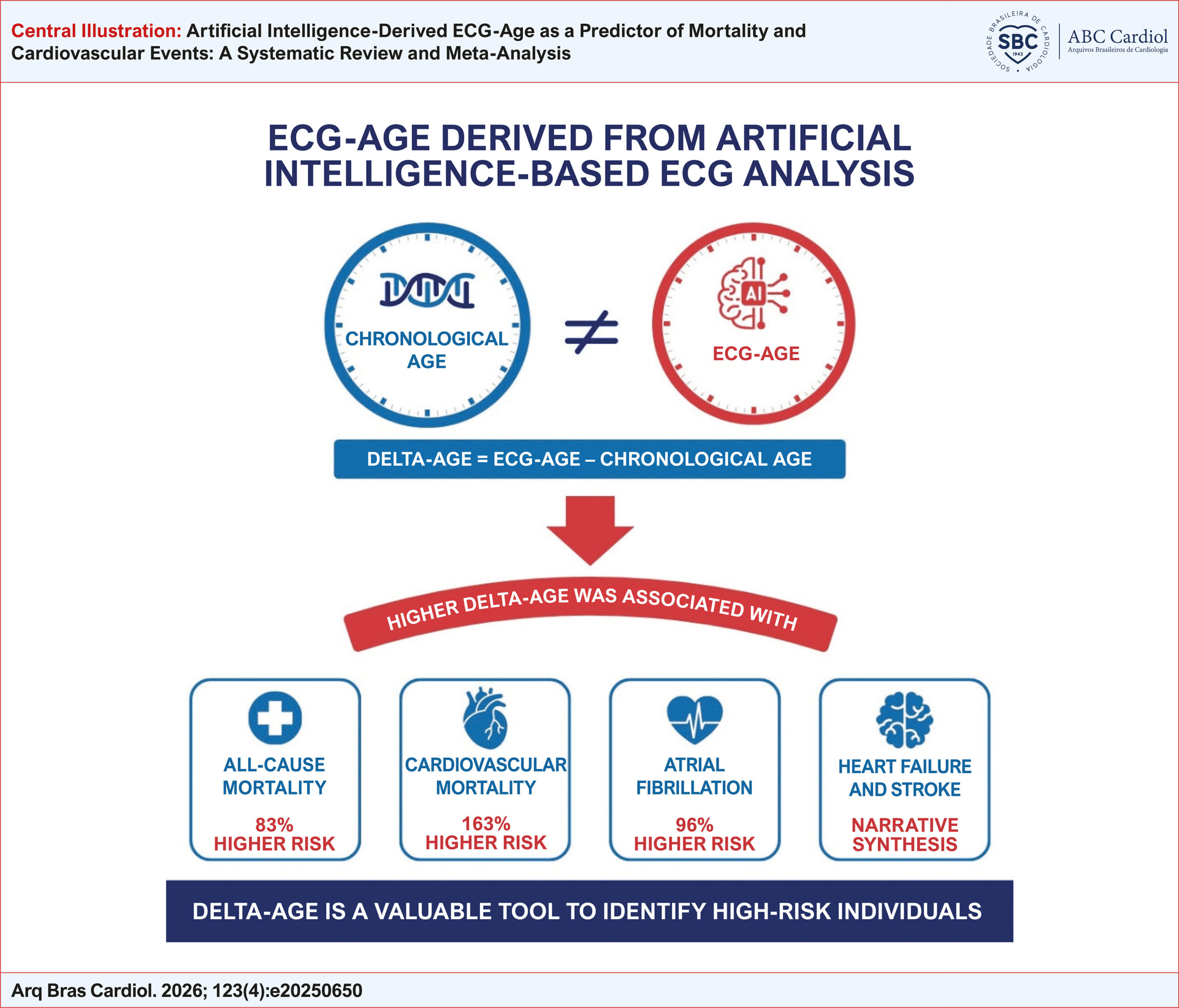
Artificial Intelligence-Derived ECG-Age as a Predictor of Mortality and Cardiovascular Events: A Systematic Review and Meta-Analysis

Isadora Cristine Reis Sguizzato Bozzi et al. Maria Clara de Araujo Gontijo Lima Antonio Luiz Pinho Ribeiro Gabriela Miana de Mattos Paixão

Abstract

Background

Artificial intelligence (AI)-derived electrocardiographic age (ECG-age) and the difference between ECG-age and chronological age (delta-age) are emerging biomarkers of cardiovascular aging and adverse outcomes, but their prognostic value remains unclear.

Objectives

We performed a systematic review and meta-analysis to evaluate associations between AI-derived ECG-age or delta-age and clinical outcomes.

Methods

Nine databases were searched through May 1, 2025. Eligible studies evaluated mortality or cardiovascular events and reported measures of association. Pooled hazard ratios (pHRs) with 95% confidence intervals (CIs) were calculated via fixed- or random-effects models, with statistical significance set at p < 0.05. The protocol was registered in PROSPERO (CRD420251042467).

Results

Ten studies (2021–2025) with over 550,000 participants from East Asia, the Americas, and the UK were included. Most used convolutional neural networks to estimate ECG-age; delta-age was calculated as ECG-age minus chronological age. Elevated delta-age was associated with increased all-cause mortality (pHR = 1.83, 95% CI: 1.45–2.32) and cardiovascular mortality (pHR = 2.63, 95% CI: 1.93–3.58). Three studies reported increased risk of atrial fibrillation (pHR = 1.96, 95% CI: 1.43–2.69), although data were limited and heterogeneous. Descriptive analyses suggested that greater delta-age predicts incident heart failure and stroke.

Conclusions

AI-derived ECG-age, particularly delta-age, is associated with all-cause and cardiovascular mortality, supporting its role as a noninvasive biomarker of cardiovascular aging. Evidence for atrial fibrillation is suggestive but limited. Standardized algorithms, robust external validation, and prospective multicenter studies are needed to confirm clinical utility and integrate ECG-age into risk stratification, even in asymptomatic individuals.

  • Artificial Intelligence
  • Deep Learning
  • Electrocardiography
  • Machine Learning
  • Prognosis
Read Full Article
Creative Commons Attribution (CC BY) Creative Commons license
12/06/2026 DOI: 10.36660/abc.20250650i
Accuracy and Prognosis of Electrocardiographic and Echocardiographic Left Ventricular Hypertrophy in Brazilian Adults - ELSA-Brasil Study
ABC
Original Article

Accuracy and Prognosis of Electrocardiographic and Echocardiographic Left Ventricular Hypertrophy in Brazilian Adults – ELSA-Brasil Study

Marcelo M. Pinto-Filho et al. Lillian Guimarães de Faria Murilo Foppa Angela Barreto Santiago Santos … Sandhi Maria Barreto Antonio Luiz Pinho Ribeiro

Abstract

Background

Left ventricular hypertrophy (LVH), defined by increased left ventricular mass, is an independent predictor of cardiovascular mortality. Evidence suggests that LVH identified by electrocardiogram (ECG) and echocardiography represents distinct entities with potentially different prognostic implications.

Objectives

Assess the diagnostic accuracy of electrocardiographic criteria for LVH compared with transthoracic echocardiogram (TTE) and to evaluate their predictive value for fatal cardiovascular outcomes.

Methods

We conducted a cross-sectional diagnostic accuracy analysis and a prospective follow-up within the Longitudinal Study of Adult Health. LVH on ECG was defined by Minnesota Code criteria, and LVH on TTE by increased calculated left ventricular mass. Significance level was defined at 0,05.

Results

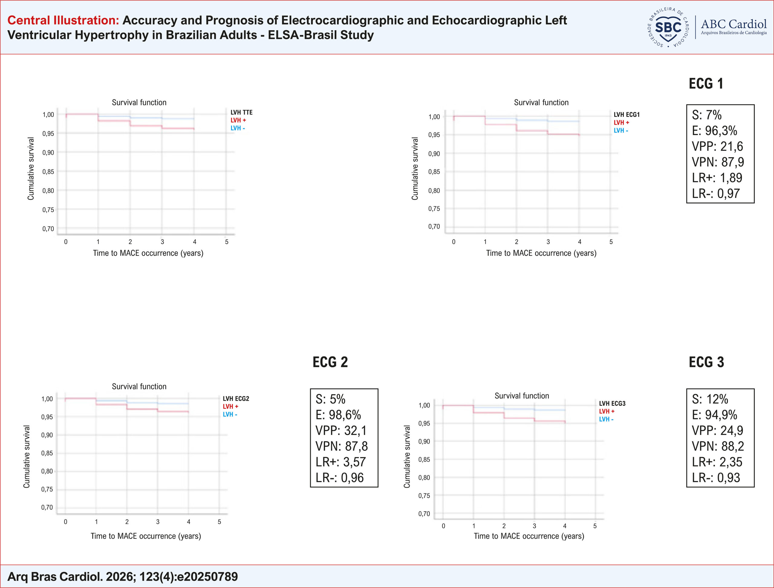
Among 2,849 participants (43.6% male), median age 62 years, 12.5% had LVH by TTE and 5.9% by ECG criteria. ECG-defined LVH showed, from different criteria, very low sensitivity (5–12%) but high specificity (95–99%) compared with TTE. In prognostic analyses, TTE-defined LVH was associated with significant fatal adverse cardiovascular events (FMACE) (HR 3.2, 95% CI 1.7–5.9). ECG-defined LVH also predicted FMACE, with HR close to 3.5 across the different MC criteria in multivariate regression analysis. The predictive capacity remained significant after further adjustment for hypertrophy defined by either of the methods.

Conclusions

Although electrocardiographic criteria demonstrated poor diagnostic accuracy for LVH compared with TTE, both modalities were independently associated with adverse cardiovascular outcomes. These findings suggest that ECG-defined and TTE-defined LVH provide complementary prognostic information and may reflect distinct pathophysiological processes.

  • Electrocardiography
  • Left Ventricular Hypertrophy
  • Longitudinal Studies
  • Prognosis
Read Full Article
Creative Commons Attribution (CC BY) Creative Commons license
12/06/2026 DOI: 10.36660/abc.20250789i Views 1
Figure 1
ABC
Letter to the Editor

The End of "Left Atrial Overload"

José Nunes de Alencar et al. Daniel Teixeira de Campos e Barreiro Adrian Baranchuk

To the Editor,

We read with great interest the article by Oliveira et al. The authors are to be commended for conducting a study of this magnitude and rigor. While previous literature relied on smaller sample sizes or non-standard/obsolete echocardiographic cutoffs for left atrial enlargement (LAE),– the ELSA-Brasil cohort provides the statistical power necessary to settle a longstanding debate in clinical cardiology. We believe this research is destined to become a reference point in standard electrocardiography textbooks.

[…]

  • Diagnosis
  • Electrocardiography
  • Heart Atria
Read Full Article
Creative Commons Attribution (CC BY) Creative Commons license
01/06/2026 DOI: 10.36660/abc.20250814i Views 1
Diagnostic Performance of Artificial Intelligence-Driven Electrocardiogram Algorithms in Detecting Heart Failure with Preserved Ejection Fraction: A Systematic Review and Meta-Analysis
IJCS
Original Article

Diagnostic Performance of Artificial Intelligence-Driven Electrocardiogram Algorithms in Detecting Heart Failure with Preserved Ejection Fraction: A Systematic Review and Meta-Analysis

Gustavo Barbosa David et al. Nicolas Ramos Ramon Huntermann Juan Peres de Oliveira … Maria Eduarda Molinari Caroline de Oliveira Fischer Bacca

Abstract

Background:

Heart failure with preserved ejection fraction (HFpEF) is a highly prevalent syndrome associated with substantial morbidity and mortality. Accurate diagnosis is often challenging and costly. Recently, artificial intelligence-based electrocardiogram (AI-ECG) algorithms have emerged to make the diagnosis of HFpEF less expensive and more accessible. However, there is a lack of data regarding their accuracy.

Objective:

We aimed to conduct a systematic review and meta-analysis to evaluate the diagnostic performance of AI-ECG algorithms in detecting HFpEF.

Methods:

We searched PubMed, Embase, and Cochrane databases for studies evaluating AI-ECG algorithms in diagnosing HFpEF. We extracted true-positive, true-negative, false-positive, and false-negative events to estimate pooled sensitivity, specificity with 95% confidence intervals (CI), and area under the curve. Statistical analysis was performed using R under a random-effects model. Heterogeneity was assessed with I² statistics.

Results:

We included three studies comprising 171,073 patients (66.86% with HFpEF), in which an AI-ECG was evaluated alongside echocardiography, with a left ventricular ejection fraction of ≥ 50%. The patients’ mean age was 65.86 ± 4.5 years, and 48.12% were male. AI-ECG algorithms demonstrated a sensitivity of 0.89 (95% CI: 0.64 to 0.97), specificity of 0.80 (95% CI: 0.65 to 0.89), and an area under the receiver operating characteristic curve of 0.89 (95% CI: 0.85 to 0.92).

Conclusion:

AI-ECG algorithms demonstrate moderate to high sensitivity, specificity, and accuracy in diagnosing HFpEF, supporting their potential for routine screening and early detection.

  • Artificial Intelligence
  • Diagnosis
  • Diastolic Heart Failure
  • Electrocardiography
Read Full Article
Creative Commons Attribution (CC BY) Creative Commons license
01/06/2026 DOI: 10.36660/ijcs.20250091
Acute Coronary Syndrome Patient Profile in Emergency Department: A cross-sectional study
IJCS
Original Article

Acute Coronary Syndrome Patient Profile in Emergency Department: A cross-sectional study

Riza Fikriana et al. Al Afik Randy Eka Prasetya

Abstract

Background:

Acute Coronary Syndrome (ACS) remains one of the leading causes of high morbidity and mortality worldwide. Psychological factors are considered important contributors to increasing recurrence rates in patients.

Objective:

This study aimed to analyze the association between stress, chest pain complaints, electrocardiographic changes, and cardiac enzyme levels.

Methods:

A cross-sectional design was employed. A sample of 105 ACS patients presenting to the hospital emergency department was selected using an accidental sampling technique. Variables measured included stress scores, chest pain, electrocardiographic findings, troponin, and CK-MB levels. Data were analyzed using the Spearman rank test and linear regression, with a significance level set at p < 0.05.

Results:

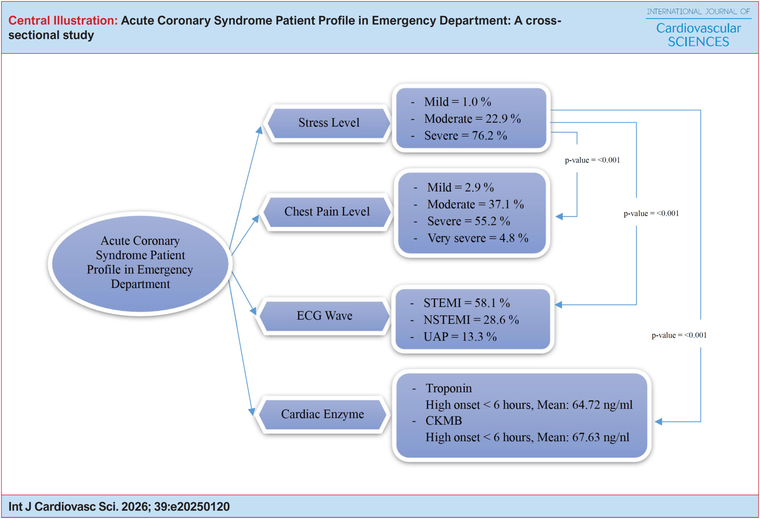
Stress scores were significantly associated with chest pain (p < 0.001), electrocardiographic changes (p < 0.001), troponin levels (p < 0.001), and CK-MB levels (p < 0.001). ECG findings showed that most STEMI patients (68.9%) experienced severe pain. Linear regression analysis indicated that troponin and CK-MB levels were significantly influenced by chest pain (p < 0.001).

Conclusions:

Stress is positively correlated with chest pain in ACS patients and is reflected in ECG changes and elevated cardiac enzymes (troponin and CK-MB).

  • Acute coronary syndrome
  • Chest Pain
  • Electrocardiography
Read Full Article
Creative Commons Attribution (CC BY) Creative Commons license
01/06/2026 DOI: 10.36660/ijcs.20250120 Views 1
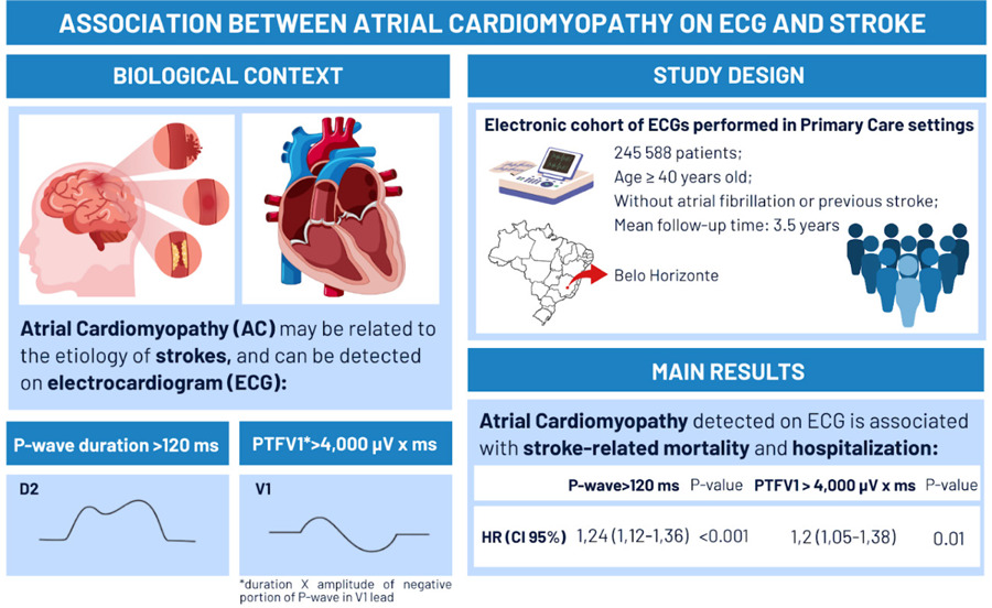
Atrial Cardiomyopathy Detected by Electrocardiogram: Association with Stroke in a Brazilian Electronic Cohort
ABC
Original Article

Atrial Cardiomyopathy Detected by Electrocardiogram: Association with Stroke in a Brazilian Electronic Cohort

Filipe Antunes de Lêu et al. Gabriela Miana de Mattos Paixão Ana Cecilia Oliveira Carla Paula Moreira Soares … Paulo R. Gomes Peter Macfarlane Antônio Luiz Pinho Ribeiro

Abstract

Background

Atrial cardiomyopathy (AC) is a condition detectable by electrocardiogram (ECG) that may play a significant role in the pathophysiology of the etiology of strokes, independently of atrial fibrillation (AF).

Objectives

To evaluate the association between ECG markers of AC (prolonged P-wave duration >120 ms and P-terminal force in V1 (PTFV1) >4,000 µV·ms) as well as the occurrence of mortality and hospitalization due to stroke.

Methods

This retrospective cohort study included patients from Belo Horizonte who underwent ECGs between 2006 and 2018. Patients aged ≥40 years, in sinus rhythm, and without a previous history of stroke at baseline ECG were included. Clinical and ECG data were linked to mortality (SIM) and hospitalization (SIH) public databases. Cox regression was used to calculate hazard ratios (HRs), and incremental adjustment models for age, sex, cardiovascular risk factors, and left ventricular hypertrophy were employed. Statistical significance was set at p<0.05.

Results

245,588 patients were included. 26.3% had prolonged P-wave duration >120 ms, and 10.1% had elevated PTFV1 >4,000 µV·ms. Mean follow-up duration was 3.5 years. AC was associated with mortality and hospitalization due to stroke (HR 1.24; 95% CI, 1.12–1.36 for P-wave duration >120 ms; p4,000 µV·ms; p<0.001).

Conclusion

ECG markers of AC are associated with death or hospitalization due to stroke, as well as cardiovascular mortality and incident AF in a large, representative Brazilian cohort, highlighting their prognostic value.

  • Cardiomyopathies
  • Cohort Studies
  • Electrocardiography
  • Mortality
  • Stroke
Read Full Article
Creative Commons Attribution (CC BY) Creative Commons license
01/05/2026 DOI: 10.36660/abc.20250625i Views 2
Figure 1
IJCS
Brief Communication

S-Wave Amplitude in Lead V3 Identifies Other Electrocardiography Criteria of Left Ventricular Hypertrophy

Bernardo Augusto Silveira Corrêa et al. Gabriel Pinheiro Soares Alencar e Silva Felipe Campelo de Miranda Marcio Henrique de Jesus Oliveira … Gabriel Scarpioni Barbosa Victor Hudson de Lacerda Borges José Nunes de Alencar

Introduction

Electrocardiographic (ECG) criteria for detecting left ventricular hypertrophy (LVH) are integral to clinical decision-making. Although these methods are quick to apply, low cost, and widely available, their sensitivity is modest. Multiple voltage-based criteria exist, such as the Sokolow-Lyon, Cornell, and Peguero-Lo Presti criteria, but accurate use often requires familiarity with several indices and can be time-consuming for trainees. Simple, reliable markers could improve the efficiency and consistency with which ECGs are flagged as possibly indicating LVH.

The S-wave amplitude in lead V3 (SV3) is a plausible single-lead marker. It is a component of the Cornell criterion (SV3 + RaVL), and it is closely related to the Peguero-Lo Presti criterion (SV4 plus the deepest precordial S). Because the largest precordial S wave often occurs in V3, a single SV3 measurement could serve as a rapid screening tool to identify the presence of established LVH criteria, especially for those with less experience interpreting ECGs.

[…]

  • Electrocardiography
  • Left Ventricular Hypertrophy
Read Full Article
Creative Commons Attribution (CC BY) Creative Commons license
27/04/2026 DOI: 10.36660/ijcs.20250050
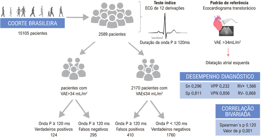
Diagnostic Utility of P-Wave Duration in Identifying Left Atrial Enlargement: Insights from the ELSA-Brasil Cohort
ABC
Original Article

Diagnostic Utility of P-Wave Duration in Identifying Left Atrial Enlargement: Insights from the ELSA-Brasil Cohort

Ana Cecília de Sena Oliveira et al. Vinicius Tostes Carvalho Marcelo Martins Pinto-Filho Luisa Caldeira Brant … Sandhi Maria Barreto Murilo Foppa Antonio Luiz Pinho Ribeiro

Abstract

Background

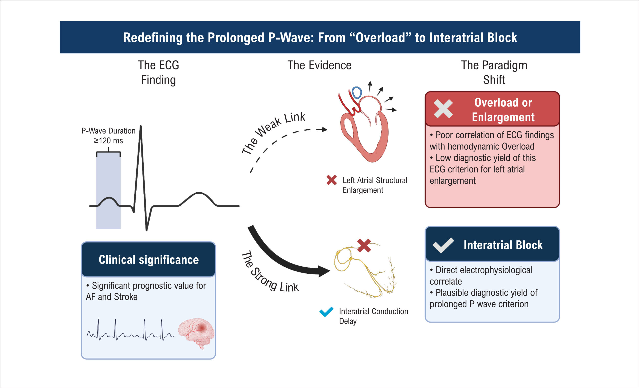
Atrial cardiomyopathy encompasses various atrial abnormalities, including left atrial enlargement (LAE), which is associated with adverse cardiovascular and neurocognitive outcomes. P-wave duration ≥120 ms on 12-lead electrocardiogram (ECG) is a widely available, cost-effective tool and one of the main diagnostic criteria adopted. However, its diagnostic performance in large-scale studies remains underexplored.

Objectives

To evaluate the correlation between P-wave duration and left atrial volume (LAV) measured by transthoracic echocardiogram (TTE) and determine the diagnostic accuracy of P-wave duration ≥120 ms for identifying LAE.

Methods

We conducted a cross-sectional analysis of baseline data from the ELSA-Brasil cohort participants in sinus rhythm. P-wave duration was automatically measured via standard 12-lead ECG, with ≥120 ms indicating enlargement. LAE was defined as LAV>34 mL/m2 by TTE. Spearman’s rank test assessed correlation. Diagnostic performance metrics – sensitivity (Sn), specificity (Sp), positive predictive value (PPV), and negative predictive value (NPV), positive and negative likelihood ratio (LR+ and LR-) – were calculated. Also, a Receiver Operating Characteristic (ROC) was constructed. Statistical significance was set at p-value<0.05 with 95% confidence interval.

Results

The study included 2589 participants, 419 (16,1%) with LAV index>34 mL/m2. P-wave duration correlated weakly with LAV (Spearman’s ρ = 0.120, p < 0.001). Diagnostic accuracy measurements were AUC = 0.557 ± 0.032; Sn = 0.296; Sp = 0.811; PPV = 0.232; NPV = 0.856; LR+ 1.566; LR- 0.868.

Conclusion

P-wave duration demonstrates limited diagnostic value for identifying LAE, suggesting against its recommendation as a standalone diagnostic criterion for this condition.

  • Cardiomyopathies
  • Echocardiography
  • Electrocardiography
  • Heart Atria
Read Full Article
Creative Commons Attribution (CC BY) Creative Commons license
15/12/2025 DOI: 10.36660/abc.20250151i Views 1
ABC
Letter to the Editor

Reflections on Survival Rates in Patients with Brugada Phenocopy

Gabriel Scarpioni Barbosa et al. José Nunes de Alencar Adrian Baranchuk

We read with great interest the article “Survival in Patients with Brugada Phenocopy. Case Series” by Fonseca et al. The authors describe a case series of patients with Brugada Phenocopy (BrP) and report in-hospital survival. We commend the authors for addressing an important topic related to BrP; however, we would like to comment on methodological considerations and propose recommendations that may strengthen interpretation and guide future BrP reports.

BrP refers to clinical conditions that lead to Brugada-ECG pattern (Br-ECGp) without the genetic substrate of Brugada Syndrome (BS) and an underlying condition that justifies the ECG change. The term was first used to describe the Br-ECGp caused by the propofol infusion syndrome; however, after several corrections to the concept, drugs that do block the sodium channels were no longer considered part of the BrP spectrum. BrP represents a diagnostic challenge even for experts. The mean accuracy to identify BrP based only on the 12-lead ECGs was 43±33% among 10 international experts. Therefore, the clinical context is essential to differentiate BrP from BS. Anselm et al. presented a systematic diagnostic criterion () for the diagnosis of BrP. Emphasis was placed on the recognition of the underlying clinical condition and the low pretest probability for BrP, in addition to the resolution of the Br-ECGp immediately after resolving the underlying condition. Based on the limited description provided by Fonseca et al., we cannot exclude the possibility that some cases represented concealed BS unmasked by hyperthermia or other triggers. This misclassification bias could alone skew the observed mortality.

[…]

  • Brugada Syndrome
  • Electrocardiography
  • Survival Rate
Read Full Article
Creative Commons Attribution (CC BY) Creative Commons license
14/11/2025 DOI: 10.36660/abc.20250330i Views 3
ABC
Editorial

LBBB and the Paradigm Shift from STEMI to Occlusion MI

Jesse T.T McLaren et al. Hans C. Helseth Stephen W. Smith

The current paradigm for acute coronary syndrome (ACS) is based on ST elevation myocardial infarction (STEMI) electrocardiogram (ECG) criteria. This is defined as ST elevation in the absence of left bundle branch block (LBBB). Nevertheless, this creates an obvious dilemma: what about patients with LBBB? In their systematic review and meta-analysis of 51 studies, Alencar et al. help resolve longstanding debates by comparing guidelines with evidence. This not only clarifies how to address this specific diagnostic dilemma, but also how a paradigm shift from STEMI to Occlusion MI (OMI) could transform patient care.

As Alencar found, LBBB appeared in only 3.3% of ACS, but had higher mortality than acute MI without BBB. The current paradigm creates the twin dangers of unnecessary cath lab activation or even thrombolytics for LBBB without OMI, or delayed reperfusion for LBBB with OMI.

[…]

  • Electrocardiography
  • Myocardial Infarction
  • Non-ST Elevated Myocardial Infarction
Read Full Article
Creative Commons Attribution (CC BY) Creative Commons license
03/11/2025 DOI: 10.36660/abc.20250497i Views 1
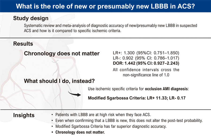
Accuracy of Left Bundle Branch Block Chronology and Electrocardiography Criteria for Acute Myocardial Infarction Diagnosis: A Systematic Review and Meta-analysis
ABC
Original Article

Accuracy of Left Bundle Branch Block Chronology and Electrocardiography Criteria for Acute Myocardial Infarction Diagnosis: A Systematic Review and Meta-analysis

José Nunes de Alencar et al. Gleydson Wesley Freire Lima Haissa Assad dos Santos Geraldo Rinaldo Carvalho Fernandes … Matheus Kiszka Scheffer Sandro Pinelli Felicioni Mariana Fuziy Nogueira De Marchi

Abstract

Background

The diagnostic utility of new or presumed new left bundle branch block (LBBB) for acute myocardial infarction (AMI) in the setting of acute coronary syndrome (ACS) remains controversial.

Objective

To evaluate whether the timing of LBBB predicts AMI and to compare its diagnostic accuracy with ischemic electrocardiography (ECG) criteria, particularly the Modified Sgarbossa Criteria (MSC).

Methods

We searched PubMed and Scopus for studies involving patients with ACS with LBBB through December 2023. Sensitivity, specificity, positive (LR+) and negative (LR–) likelihood ratios, and diagnostic odds ratios (DOR) were calculated to assess diagnostic accuracy. Incidence and mortality data were also analyzed. Risk of bias was evaluated using the Newcastle-Ottawa Scale (NOS) and the revised Quality Assessment of Diagnostic Accuracy Studies (QUADAS-2) tool.

Results

A total of 51 studies were included. LBBB occurred in 3.3% of ACS presentations and was associated with higher in-hospital mortality. Differentiating new from old LBBB was diagnostically neutral: LR+ 1.30 (95% CI: 0.75 to 1.85), LR– 0.90 (95% CI: 0.79 to 1.02), and DOR 1.44 (95% CI: 0.93 to 2.24); all confidence intervals crossed the null value of 1.0. In contrast, MSC demonstrated 83.6% sensitivity (95% CI: 55.4 to 95.5%) and 92.6% specificity (95% CI: 78.9 to 97.7%) for angiographically confirmed occlusive AMI, with LR+ 11.34 (95% CI: 3.67 to 34.99) and LR– 0.18 (95% CI: 0.054 to 0.575).

Conclusion

LBBB chronology alone does not significantly impact the likelihood of AMI. Ischemic ECG criteria — especially the MSC — provide substantially greater diagnostic accuracy and should guide clinical decision-making in ACS patients with LBBB.

  • Acute coronary syndrome
  • Electrocardiography
  • Myocardial Infarction
Read Full Article
Creative Commons Attribution (CC BY) Creative Commons license
03/11/2025 DOI: 10.36660/abc.20250109i
Left Ventricular Hypertrophy on Electrocardiography and Chronic Kidney Disease: A Case–Control Pilot Study
IJCS
Original Article

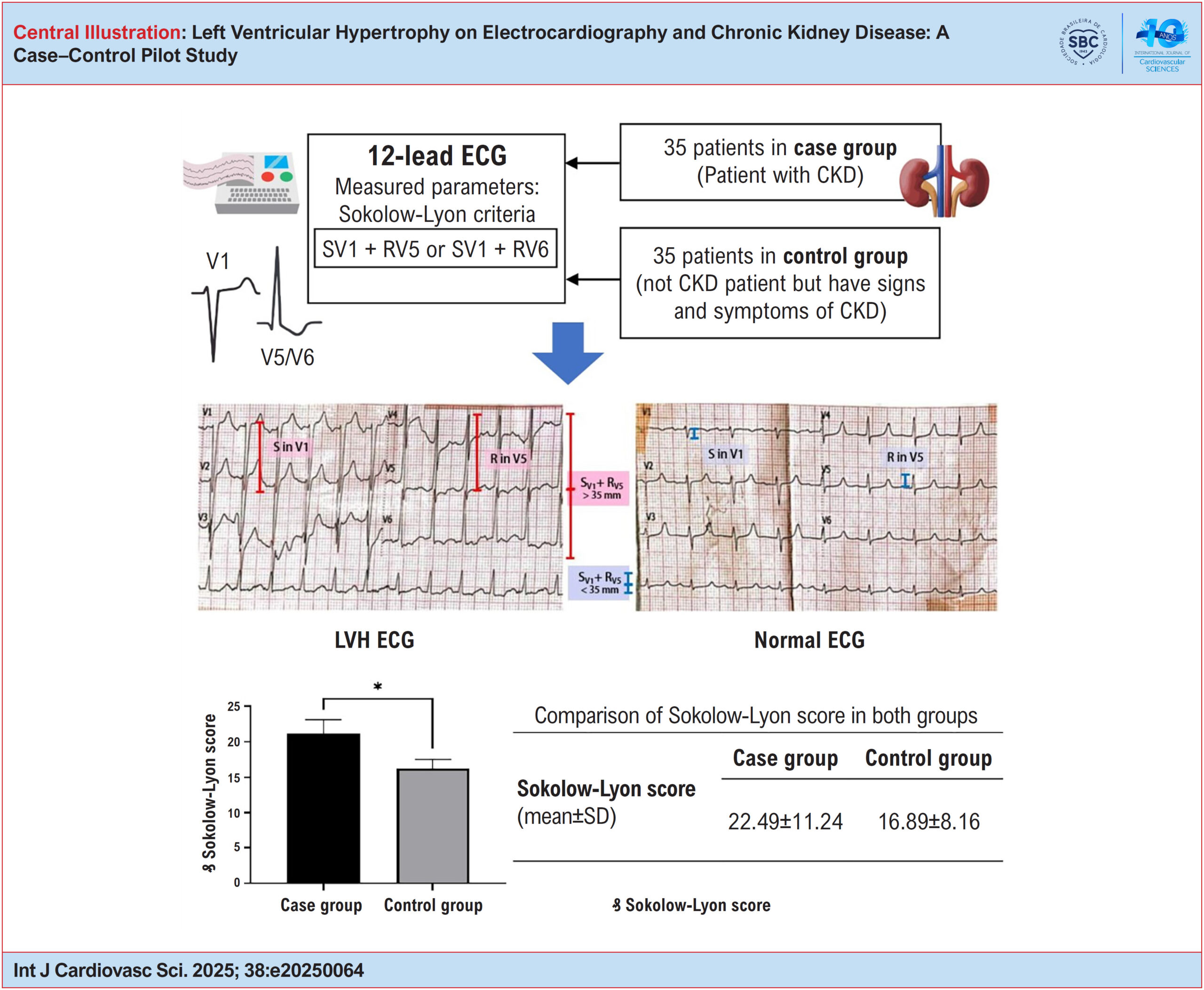
Left Ventricular Hypertrophy on Electrocardiography and Chronic Kidney Disease: A Case–Control Pilot Study

Nabil Hajar et al. Nabila Nuralya Joveazhari Shofa Chasani Zulfachmi Wahab … Galih Prakasa Adhyatma Indira Aura Balqis Zeidan Ahmad Fahdalhaq Bintang Adhiyoso Wicaksono

Abstract

Background:

Chronic kidney disease (CKD) is a condition characterized by abnormalities in kidney structure or function lasting more than 3 months. CKD is known to increase the risk of cardiovascular events, which may be predicted by electrocardiography (ECG) abnormalities, such as left ventricular hypertrophy (LVH). ECG is widely used in under-resourced health care settings due to its practicality and accessibility.

Objective:

This study aimed to analyze the relationship between LVH and CKD using ECG.

Methods:

This analytical observational study used a case–control design. The case group included patients diagnosed with CKD, while the control group consisted of patients presenting with signs and symptoms suggestive of CKD but without a confirmed diagnosis. Each group included 35 participants. Simple random sampling was used for the case group, and stratified random sampling for the control group. ECG examinations were performed, and measurements were taken using calipers in millimeters (mm). The Sokolow–Lyon criteria were applied to identify LVH. Data were analyzed using an independent t-test, with statistical significance set at p < 0.05.

Results:

The Sokolow–Lyon score was significantly higher in the case group than in the control group (p = 0.03).

Conclusion:

There is a significant association between LVH, as identified on ECG, and CKD. These findings may assist health professionals in resource-limited settings, where access to ECG is restricted.

  • Cardiovascular Diseases
  • chronic renal insufficiency
  • Electrocardiography
  • Left Ventricular Hypertrophy
Read Full Article
Creative Commons Attribution (CC BY) Creative Commons license
01/09/2025 DOI: 10.36660/ijcs.20250064 Views 2
Applicability of Machine Learning Algorithms in Diagnosis of Atrial Fibrillation and LQTS by Electrocardiogram Interpretation: A Systematic Review
ABC
Original Article

Applicability of Machine Learning Algorithms in Diagnosis of Atrial Fibrillation and LQTS by Electrocardiogram Interpretation: A Systematic Review

Paulo Cainan Guimarães do Nascimento et al. Matthews Silva Martins Alex Cleber Improta-Caria Roque Aras Junior

Abstract

Background

Machine Learning (ML) is a type of algorithm that autonomously learns to recognize complex patterns. In the diagnostic context of cardiac arrhythmias, these algorithms have shown significant advancements due to their ability to provide automated interpretation and pattern recognition in electrocardiograms (ECGs).

Objective

To analyze and identify the applicability, validity, and feasibility of ML algorithm models in the diagnostic process of cardiac arrhythmias through automated electrocardiogram interpretation.

Methods

This systematic literature review was reported according to the PRISMA guidelines. The searches were conducted in the Cochrane Library, EMBASE, LILACS, and PubMed between February 2022 and November 2022. The study period encompasses articles published between 2017 and 2022.

Results

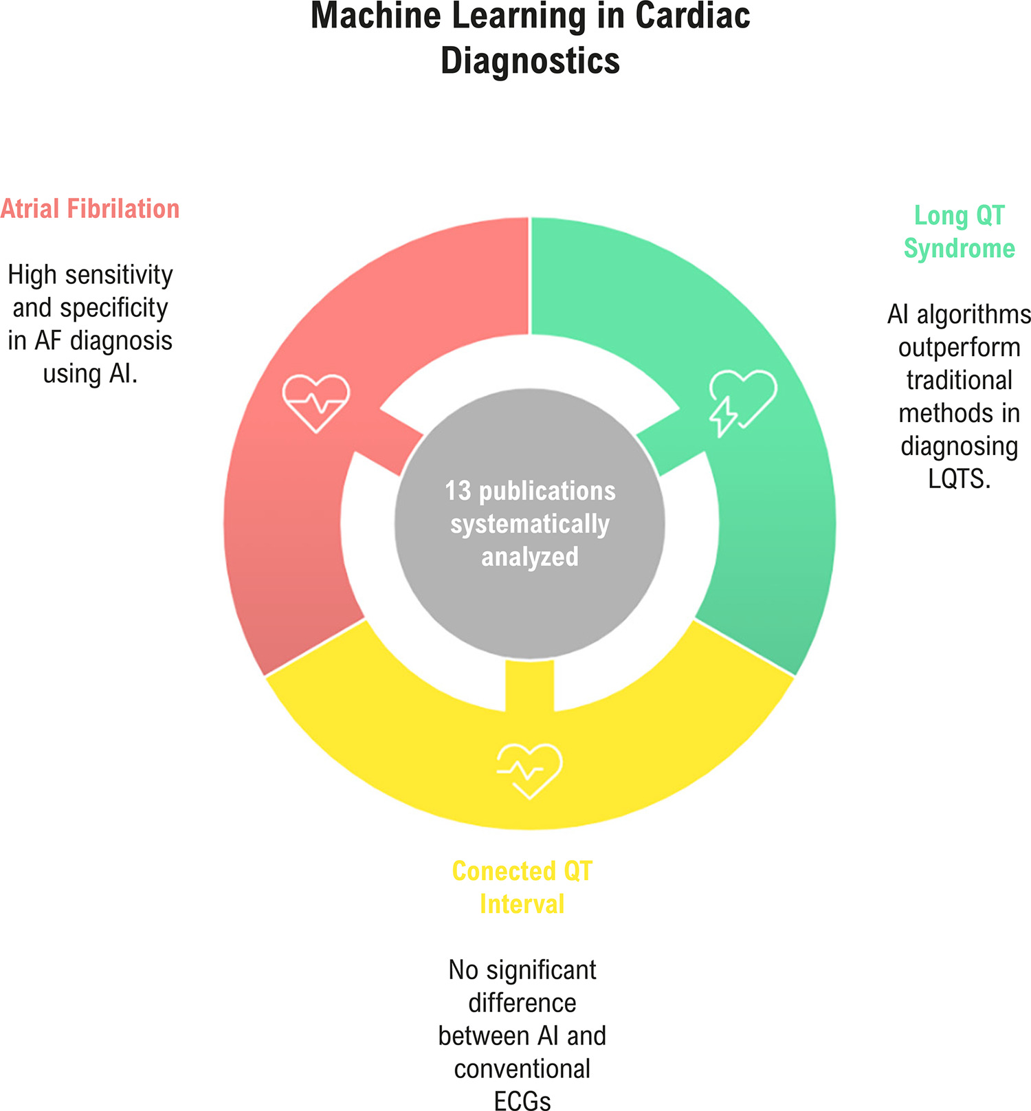
The database search yielded 119 results, covering three subthemes: Long QT Syndrome (LQTS), corrected QT interval (QTc), and atrial fibrillation (AF). AF was the most prevalent theme. The sample sizes were quite variable. The outcomes were mostly satisfactory. In the diagnosis of LQTS using Artificial Intelligence (AI), the algorithm outperformed conventional methods in diagnostic distinction. In the evaluation of QTc, there was no difference between the AI-integrated ECG and the conventional ECG. In the diagnosis of AF, the algorithms, models, and devices demonstrated high sensitivity and specificity, along with greater accuracy.

Conclusion

ML models in the diagnostic process of cardiac arrhythmias are feasible and rapidly developing. They demonstrate accuracy values between 96.4% and 98.2%, sensitivity between 92.8% and 99.4%, and specificity between 95% and 98.1%, particularly in the diagnosis of atrial fibrillation.

  • Artificial Intelligence
  • Cardiac Arrhythmias
  • Electrocardiography
  • Machine Learning
  • Neural Network
Read Full Article
Creative Commons Attribution (CC BY) Creative Commons license
01/09/2025 DOI: 10.36660/abc.20240843i Views 1
Figure 1
ABCIC
Original Article

Conventional and Partially ECG-Gated Triple Rule-Out Computed Tomography Angiography with Extension to Abdominal Aorta: Comparative Radiation Dose and Imaging Quality

Pamela Bertolazzi et al. Carla Franco Greco Silva Leonardo Iunes Fernando Freitas de Oliveira … Fabio Payão Pereira Publio Cesar Cavalcanti Viana Isac Castro Natally Horvat José Arimateia Batista Araújo-Filho

Abstract

Background:

Triple rule-out (TRO) computed tomography angiography (CTA) is an ECG-gated protocol that enables the simultaneous evaluation of the coronary arteries, thoracic aorta, and pulmonary arteries in a single scan. It is especially useful for patients in the emergency department with low to moderate risk of acute coronary syndrome, particularly when aortic dissection and pulmonary embolism are also considered in the differential diagnosis.

Objective:

This study aimed to compare two TRO protocols, fully electrocardiogram (ECG)-gated (protocol A) and partially ECG-gated (protocol B), both including coverage of the abdominal aorta, in terms of radiation dose and image quality. Such a comparison of protocols can be useful when iterative reconstruction algorithms are not available and a manual transition between partially ECG-synchronized protocols is required.

Methods:

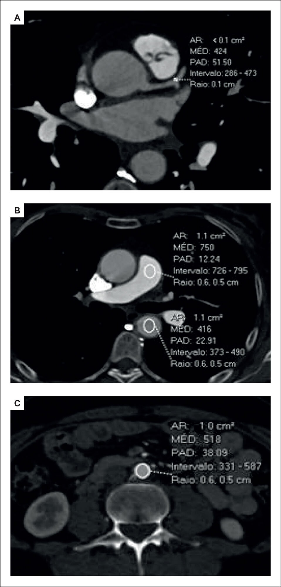
Radiation dose was evaluated using dose-length product (DLP), effective dose (ED), and virtual dose. Attenuation values were measured in the coronary and pulmonary arteries, as well as in the descending and abdominal aorta. Image quality and vessel conspicuity were assessed using a 5-point Likert scale.

Results:

A total of 56 patients were included. Protocol B demonstrated significantly lower radiation exposure compared to protocol A across all metrics: median DLP (1.1 mSv [interquartile range: 0.9 to 1.1] versus 2.2 mSv [interquartile range: 1.6 to 2.8]), ED (17.0 mSv [interquartile range: 14.3 to 18.1] versus 32.6 mSv [interquartile range: 24.4 to 42.7]), and virtual dose (16.2 [interquartile range: 9.3 to 20.4] versus 34.7 [interquartile range: 19.9 to 43.5]); all differences were statistically significant (p < 0.001). There were no significant differences in attenuation measurements or qualitative image assessment between the two protocols.

Conclusion:

Partially ECG-gated TRO CTA provides comparable image quality to the fully ECG-gated technique while significantly reducing radiation exposure, becoming a more radiation dose-efficient alternative when iterative reconstruction algorithms are not available and manual transition is necessary.

  • Drug Tapering
  • Electrocardiography
  • Radiation Dose-Response Relationship
Read Full Article
Creative Commons Attribution (CC BY) Creative Commons license
27/06/2025 DOI: 10.36660/abcimg.20250025i
ABC
Letter to the Editor

How to Assess Impaired Right Heart Electrical Conduction in Patients with Pulmonary Arterial Hypertension?

Naoya Kataoka et al. Teruhiko Imamura

To editor

The identification of an optimal modality to monitor the severity of pulmonary arterial hypertension (PAH) remains imperative. The authors demonstrated that a prolonged RS interval on the electrocardiogram was associated with adverse clinical outcomes in this cohort, under the hypothesis that right heart electrical conduction is compromised in patients with severe PAH. Nonetheless, several concerns merit consideration.

[…]

  • Bundle Branch Block
  • Electrocardiography
  • Heart Failure
Read Full Article
Creative Commons Attribution (CC BY) Creative Commons license
20/06/2025 DOI: 10.36660/abc.20250159i Views 2
ABC
Editorial

Does Early Repolarization Affect the Risk of Death and Ventricular Arrhythmias?

Guilherme Fenelon

Early repolarization (ER) pattern, defined as J-point elevation of ≥1 mm in ≥2 contiguous inferior and/or lateral electrocardiography (ECG) leads, is a common finding, with a prevalence of approximately 6% in the adult population., ER has long been considered a benign phenomenon, as most affected individuals — typically young males and athletes — have a very low incidence of sudden cardiac death.,

However, a pivotal study by Haïssaguerre et al. demonstrated a significantly higher occurrence of ER (31%) in patients with idiopathic ventricular fibrillation (VF) compared to controls (5%). Subsequent studies confirmed an association between ER and malignant ventricular arrhythmias (VA) in patients without structural heart disease., Additionally, ER has been linked to adverse outcomes in patients with primary electrical disorders such as long QT syndrome and Brugada syndrome.,

[…]

  • Cardiac Arrhythmias
  • Cardiac Sudden Death
  • Electrocardiography
Read Full Article
Creative Commons Attribution (CC BY) Creative Commons license
20/06/2025 DOI: 10.36660/abc.20250275i
Use of Artificial Intelligence Applied to Electrocardiogram for Diagnosis of Left Ventricular Systolic Dysfunction
ABC
Original Article

Use of Artificial Intelligence Applied to Electrocardiogram for Diagnosis of Left Ventricular Systolic Dysfunction

Wilton Batista de Santana Júnior et al. Marcelo M. Pinto Filho Sandhi Maria Barreto Murilo Foppa … Luana Giatti Rohan Khera Antonio Luiz Pinho Ribeiro

Abstract

Background

Heart failure (HF) is a disease associated with an important type of morbidity and mortality. The electrocardiogram (ECG), one of the tests used to evaluate HF, is low-cost and widely available.

Objective

To evaluate the performance of an artificial intelligence (AI) algorithm applied to ECG to detect HF and compare it with the predictive power of major electrocardiographic alterations (MEA).

Methods

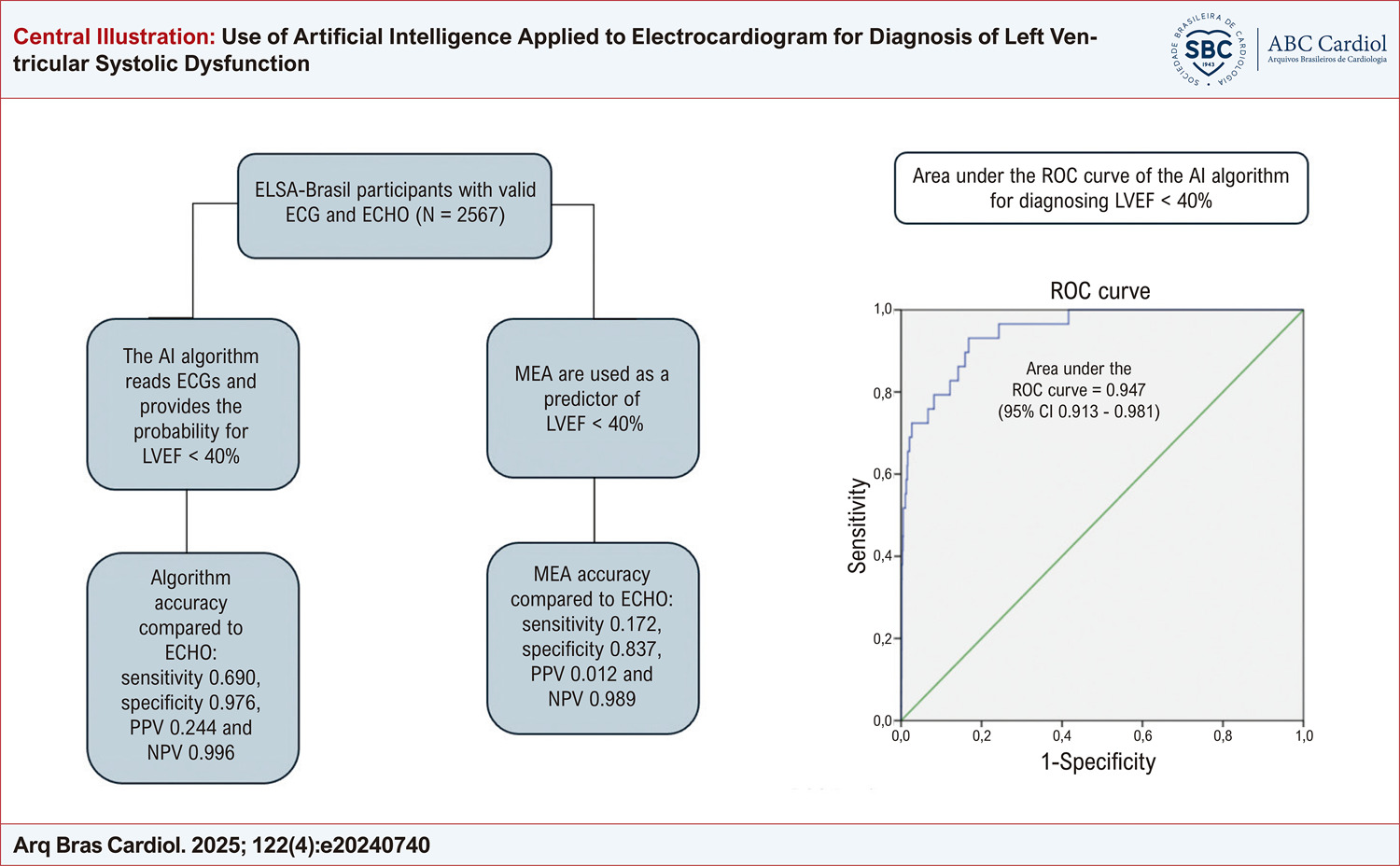
This work is a diagnostic accuracy cross-sectional study. All participants were from the Longitudinal Study of Adult Health (Estudo Longitudinal da Saúde do Adulto – ELSA-Brasil) and presented a valid ECG and echocardiogram (ECHO). The algorithm estimated probability values for left ventricular systolic dysfunction (LVSD). The assessed endpoint was left ventricular ejection fraction (LVEF) <40% in the ECHO. Sensitivity, specificity, positive predictive value (PPV), negative predictive value (NPV), positive likelihood ratio (LR+), negative likelihood ratio (LR-), and diagnostic odds ratio (DOR) were determined for both the algorithm and the MEA. The area under the ROC curve (AUC-ROC) for the algorithm was calculated.

Results

In the analytical sample of 2,567 individuals, the prevalence of LVEF <40% was 1.13% (29 individuals). The values obtained for sensitivity, specificity, PPV, NPV, LR+, LR-, and DOR for the algorithm were 0.690, 0.976, 0.244, 0.996, 27.6, 0.32, and 88.74, respectively. For the MEA, the values were 0.172, 0.837, 0.012, 0.989, 1.09, 0.990, and 1.07, respectively. The AUC-ROC of the algorithm to predict the LVEF <40% was 0.947 (95% CI: 0.913 – 0.981).

Conclusion

The AI algorithm performed well in detecting LVSD and can be used as a screening tool for LVSD.
  • Artificial Intelligence
  • Electrocardiography
  • Heart Failure
  • Left Ventricular Dysfunction
Read Full Article
Creative Commons Attribution (CC BY) Creative Commons license
16/05/2025 DOI: 10.36660/abc.20240740i
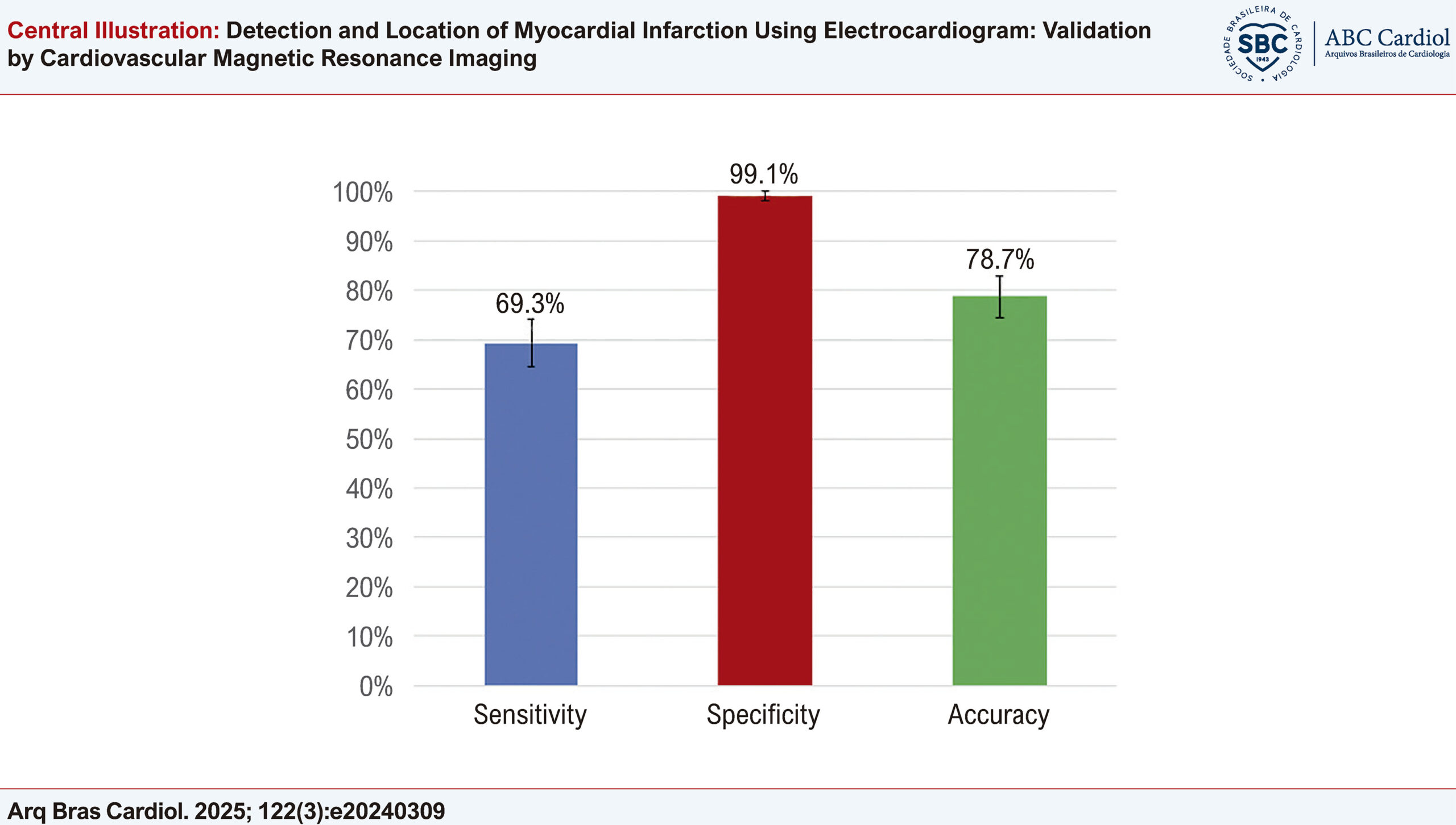
Detection and Location of Myocardial Infarction Using Electrocardiogram: Validation by Cardiovascular Magnetic Resonance Imaging
ABC
Original Article

Detection and Location of Myocardial Infarction Using Electrocardiogram: Validation by Cardiovascular Magnetic Resonance Imaging

Maria Catarina de Melo Dias Guerra et al. André Gustavo da Silva Rezende Tiago Augusto Magalhães William Azem Chalela … Augusto Hiroshi Uchida Rafael Almeida Fonseca Nevelton Heringer Filho Jürgen Beuther Guilherme Garcia Eduardo Cavalcanti Lapa dos Santos Ulisses Ramos Montarroyos Renata Ávila Cintra José Antônio Franchine Ramires Carlos Eduardo Rochitte

Abstract

Background

In the assessment of ischemic heart disease, cardiac magnetic resonance (CMR) is considered the gold standard for detecting and locating myocardial infarction (MI), but electrocardiogram (ECG) is less expensive and more widely available. Recognition of MI on ECG outside the acute phase is challenging; Q waves are absent in a significant proportion of patients and may reduce or disappear over time. Although ECG is widely used in the initial assessment of previous infarction, studies to validate ECG using CMR as a reference in the context of chronic coronary disease are limited.

Objectives

To evaluate the diagnostic performance of ECG in detecting and locating CMR-defined MI.

Methods

This study included 352 individuals who underwent CMR and ECG, 241 patients with previous MI confirmed by CMR and 111 controls with normal CMR. Their ECG tracings were analyzed by 2 observers, who were blinded to the CMR, for detection and location of MI following to the Fourth Brazilian Society of Cardiology Guidelines on the Analysis and Issuance of Electrocardiographic Reports. The significance level adopted was 5% (p < 0.05).

Results

ECG showed good performance for detecting previous MI, with sensitivity of 69.3% (64.5% to 74.1%), specificity of 99.1% (98.1% to 100%), and accuracy of 78.7% (74.4% to 83.0%). However, in locating MI in accordance with CMR, its accuracy was unsatisfactory.

Conclusions

When compared to CMR, ECG was shown to be a method with good accuracy for detecting previous MI, but not for defining its location.

  • Coronary Artery Disease
  • Electrocardiography
  • Magnetic Resonance Imaging
  • Myocardial Infarction
Read Full Article
Creative Commons Attribution (CC BY) Creative Commons license
12/05/2025 DOI: 10.36660/abc.20240309i Views 3

Could not load more articles. Please try again.

How to cite this article

EndNote RIS

Electronic Document Format (Vancouver)

Abreu JS, Diógenes TCP, Abreu MEB, Chagas ISM, Feitosa MPM. Apical Hypertrophic Cardiomyopathy with Microfistulas: Correlation Between Coronary Flow Steal, Positive Inotropic Effect, and Normalization of T-Wave Polarity. ABC Imagem Cardiovascular 2026;39(3):e20260090.

Electronic Document Format (ABNT)

Abreu, José Sebastiao de; Diógenes, Tereza Cristina Pinheiro; Abreu, Marília Esther Benevides; Chagas, Isadora Sucupira Machado; Feitosa, Mateus Paiva Marques. Apical Hypertrophic Cardiomyopathy with Microfistulas: Correlation Between Coronary Flow Steal, Positive Inotropic Effect, and Normalization of T-Wave Polarity. ABC Imagem Cardiovascular, v. 39, n. 3, e20260090, Sep. 2026.

Electronic Document Format (APA)

Abreu, J. S. , Diógenes, T. C. P., Abreu, M. E. B., Chagas, I. S. M., & Feitosa, M. P. M. (2026). Apical Hypertrophic Cardiomyopathy with Microfistulas: Correlation Between Coronary Flow Steal, Positive Inotropic Effect, and Normalization of T-Wave Polarity. ABC Imagem Cardiovascular, 39(3), e20260090.

Electronic Document Format (ISO)

Abreu, José Sebastiao de and Diógenes, Tereza Cristina Pinheiro and Abreu, Marília Esther Benevides and Chagas, Isadora Sucupira Machado and Feitosa, Mateus Paiva Marques. Apical Hypertrophic Cardiomyopathy with Microfistulas: Correlation Between Coronary Flow Steal, Positive Inotropic Effect, and Normalization of T-Wave Polarity. ABC Imagem Cardiovascular [online]. 2026, vol. 39, n. 3, [cited 2026-10-14], e20260090. Available from: <https://journals.cardiol.br/en/article/apical-hypertrophic-cardiomyopathy-with-microfistulas-correlation-between-coronary-flow-steal-positive-inotropic-effect-and-normalization-of-t-wave-polarity/>. ISSN 2318-8219

EndNote RIS

Electronic Document Format (Vancouver)

Pinheiro MCM, Felberg L, Bozzi ICRS, Ribeiro ALP, Paixão GMM. Artificial Intelligence Applied to Electrocardiogram-Based Cardiovascular Risk Assessment: A Systematic Review. Arquivos Brasileiros de Cardiologia 2026;123(7):e20250790.

Electronic Document Format (ABNT)

Pinheiro, Maria Clara Mantoan; Felberg, Lívia; Bozzi, Isadora Cristine Reis Sguizzato; Ribeiro, Antônio Luiz Pinho; Paixão, Gabriela Miana de Mattos. Artificial Intelligence Applied to Electrocardiogram-Based Cardiovascular Risk Assessment: A Systematic Review. Arquivos Brasileiros de Cardiologia, v. 123, n. 7, e20250790, Aug. 2026.

Electronic Document Format (APA)

Pinheiro, M. C. M., Felberg, L., Bozzi, I. C. R. S., Ribeiro, A. L. P., & Paixão, G. M. M. (2026). Artificial Intelligence Applied to Electrocardiogram-Based Cardiovascular Risk Assessment: A Systematic Review. Arquivos Brasileiros de Cardiologia, 123(7), e20250790.

Electronic Document Format (ISO)

Pinheiro, Maria Clara Mantoan and Felberg, Lívia and Bozzi, Isadora Cristine Reis Sguizzato and Ribeiro, Antônio Luiz Pinho and Paixão, Gabriela Miana de Mattos. Artificial Intelligence Applied to Electrocardiogram-Based Cardiovascular Risk Assessment: A Systematic Review. Arquivos Brasileiros de Cardiologia [online]. 2026, vol. 123, n. 7, [cited 2026-10-14], e20250790. Available from: <https://journals.cardiol.br/en/article/artificial-intelligence-applied-to-electrocardiogram-based-cardiovascular-risk-assessment-a-systematic-review/>. ISSN 0066-782X

EndNote RIS

Electronic Document Format (Vancouver)

Villacorta H. AI-Enhanced ECGs for the Diagnosis of HFpEF: A Promising Frontier, But Not Yet a Standalone Solution. International Journal of Cardiovascular Sciences 2026;39:e20250272.

Electronic Document Format (ABNT)

Villacorta, Humberto. AI-Enhanced ECGs for the Diagnosis of HFpEF: A Promising Frontier, But Not Yet a Standalone Solution. International Journal of Cardiovascular Sciences, v. 39, e20250272, Jul. 2026.

Electronic Document Format (APA)

Villacorta, H. (2026). AI-Enhanced ECGs for the Diagnosis of HFpEF: A Promising Frontier, But Not Yet a Standalone Solution. International Journal of Cardiovascular Sciences, 39, e20250272.

Electronic Document Format (ISO)

Villacorta, Humberto. AI-Enhanced ECGs for the Diagnosis of HFpEF: A Promising Frontier, But Not Yet a Standalone Solution. International Journal of Cardiovascular Sciences [online]. 2026, vol. 39, [cited 2026-10-14], e20250272. Available from: <https://journals.cardiol.br/en/article/ai-enhanced-ecgs-for-the-diagnosis-of-hfpef-a-promising-frontier-but-not-yet-a-standalone-solution/>. ISSN 2359-4802

EndNote RIS

Electronic Document Format (Vancouver)

Pedrosa RC. Artificial Intelligence Attack! The Rising Importance of Chagas Disease. Arquivos Brasileiros de Cardiologia 2026;123(5):e20260409.

Electronic Document Format (ABNT)

Pedrosa, Roberto Coury. Artificial Intelligence Attack! The Rising Importance of Chagas Disease. Arquivos Brasileiros de Cardiologia, v. 123, n. 5, e20260409, Jul. 2026.

Electronic Document Format (APA)

Pedrosa, R. C. (2026). Artificial Intelligence Attack! The Rising Importance of Chagas Disease. Arquivos Brasileiros de Cardiologia, 123(5), e20260409.

Electronic Document Format (ISO)

Pedrosa, Roberto Coury. Artificial Intelligence Attack! The Rising Importance of Chagas Disease. Arquivos Brasileiros de Cardiologia [online]. 2026, vol. 123, n. 5, [cited 2026-10-14], e20260409. Available from: <https://journals.cardiol.br/en/article/artificial-intelligence-attack-the-rising-importance-of-chagas-disease/>. ISSN 0066-782X

EndNote RIS

Electronic Document Format (Vancouver)

Alencar JN, Felicioni SP, Scheffer MK, Marchi MND, Bacharova L. The Old Habits Need to be Abandoned and the Term “Overload” Replaced by “High Amplitude Criteria”. Arquivos Brasileiros de Cardiologia 2026;123(5):e20250709.

Electronic Document Format (ABNT)

Alencar, José Nunes de; Felicioni, Sandro Pinelli; Scheffer, Matheus Kiszka; Marchi, Mariana Nogueira De; Bacharova, Ljuba. The Old Habits Need to be Abandoned and the Term “Overload” Replaced by “High Amplitude Criteria”. Arquivos Brasileiros de Cardiologia, v. 123, n. 5, e20250709, Jul. 2026.

Electronic Document Format (APA)

Alencar, J. N. , Felicioni, S. P., Scheffer, M. K., Marchi, M. N. D., & Bacharova, L. (2026). The Old Habits Need to be Abandoned and the Term “Overload” Replaced by “High Amplitude Criteria”. Arquivos Brasileiros de Cardiologia, 123(5), e20250709.

Electronic Document Format (ISO)

Alencar, José Nunes de and Felicioni, Sandro Pinelli and Scheffer, Matheus Kiszka and Marchi, Mariana Nogueira De and Bacharova, Ljuba. The Old Habits Need to be Abandoned and the Term “Overload” Replaced by “High Amplitude Criteria”. Arquivos Brasileiros de Cardiologia [online]. 2026, vol. 123, n. 5, [cited 2026-10-14], e20250709. Available from: <https://journals.cardiol.br/en/article/the-old-habits-need-to-be-abandoned-and-the-term-overload-replaced-by-high-amplitude-criteria/>. ISSN 0066-782X

EndNote RIS

Electronic Document Format (Vancouver)

Fonseca IMG, Nunes MCP, Sable C, Mendoza RF, Santos FLN, Gomes PR, Carvalho APV, et al. Artificial Intelligence-Enhanced Electrocardiography Analysis for Diagnosing Chagas Disease. Arquivos Brasileiros de Cardiologia 2026;123(5):e20260032.

Electronic Document Format (ABNT)

Fonseca, Isabella Moreira Gonzalez; Nunes, Maria Carmo Pereira; Sable, Craig; Mendoza, Renata Fonseca; Santos, Fred Luciano Neves; Gomes, Paulo Rodrigues; Carvalho, Adson Patrik Vieira; Mendoza, Bernardo Fonseca; Souza, Noilson Lazaro; Barbosa, José Augusto de Almeida; Cardoso, Clareci Silva; Nascimento, Bruno Ramos; Ribeiro, Antonio L. P.. Artificial Intelligence-Enhanced Electrocardiography Analysis for Diagnosing Chagas Disease. Arquivos Brasileiros de Cardiologia, v. 123, n. 5, e20260032, Jul. 2026.

Electronic Document Format (APA)

Fonseca, I. M. G., Nunes, M. C. P., Sable, C., Mendoza, R. F., Santos, F. L. N., Gomes, P. R., Carvalho, A. P. V., Mendoza, B. F., Souza, N. L., Barbosa, J. A. A., Cardoso, C. S., Nascimento, B. R., & Ribeiro, A. L. P. (2026). Artificial Intelligence-Enhanced Electrocardiography Analysis for Diagnosing Chagas Disease. Arquivos Brasileiros de Cardiologia, 123(5), e20260032.

Electronic Document Format (ISO)

Fonseca, Isabella Moreira Gonzalez and Nunes, Maria Carmo Pereira and Sable, Craig and Mendoza, Renata Fonseca and Santos, Fred Luciano Neves and Gomes, Paulo Rodrigues and Carvalho, Adson Patrik Vieira and Mendoza, Bernardo Fonseca and Souza, Noilson Lazaro and Barbosa, José Augusto de Almeida and Cardoso, Clareci Silva and Nascimento, Bruno Ramos and Ribeiro, Antonio L. P.. Artificial Intelligence-Enhanced Electrocardiography Analysis for Diagnosing Chagas Disease. Arquivos Brasileiros de Cardiologia [online]. 2026, vol. 123, n. 5, [cited 2026-10-14], e20260032. Available from: <https://journals.cardiol.br/en/article/artificial-intelligence-enhanced-electrocardiography-analysis-for-diagnosing-chagas-disease/>. ISSN 0066-782X

EndNote RIS

Electronic Document Format (Vancouver)

Bozzi ICRS, Lima MCAG, Ribeiro ALP, Paixão GMM. Artificial Intelligence-Derived ECG-Age as a Predictor of Mortality and Cardiovascular Events: A Systematic Review and Meta-Analysis. Arquivos Brasileiros de Cardiologia 2026;123(4):e20250650.

Electronic Document Format (ABNT)

Bozzi, Isadora Cristine Reis Sguizzato; Lima, Maria Clara de Araujo Gontijo; Ribeiro, Antonio Luiz Pinho; Paixão, Gabriela Miana de Mattos. Artificial Intelligence-Derived ECG-Age as a Predictor of Mortality and Cardiovascular Events: A Systematic Review and Meta-Analysis. Arquivos Brasileiros de Cardiologia, v. 123, n. 4, e20250650, Jun. 2026.

Electronic Document Format (APA)

Bozzi, I. C. R. S., Lima, M. C. A. G., Ribeiro, A. L. P., & Paixão, G. M. M. (2026). Artificial Intelligence-Derived ECG-Age as a Predictor of Mortality and Cardiovascular Events: A Systematic Review and Meta-Analysis. Arquivos Brasileiros de Cardiologia, 123(4), e20250650.

Electronic Document Format (ISO)

Bozzi, Isadora Cristine Reis Sguizzato and Lima, Maria Clara de Araujo Gontijo and Ribeiro, Antonio Luiz Pinho and Paixão, Gabriela Miana de Mattos. Artificial Intelligence-Derived ECG-Age as a Predictor of Mortality and Cardiovascular Events: A Systematic Review and Meta-Analysis. Arquivos Brasileiros de Cardiologia [online]. 2026, vol. 123, n. 4, [cited 2026-10-14], e20250650. Available from: <https://journals.cardiol.br/en/article/artificial-intelligence-derived-ecg-age-as-a-predictor-of-mortality-and-cardiovascular-events-a-systematic-review-and-meta-analysis/>. ISSN 0066-782X

EndNote RIS

Electronic Document Format (Vancouver)

Pinto-Filho MM, Faria LG, Foppa M, Santos ABS, Barreto SM, Ribeiro ALP. Accuracy and Prognosis of Electrocardiographic and Echocardiographic Left Ventricular Hypertrophy in Brazilian Adults – ELSA-Brasil Study. Arquivos Brasileiros de Cardiologia 2026;123(4):e20250789.

Electronic Document Format (ABNT)

Pinto-Filho, Marcelo M.; Faria, Lillian Guimarães de; Foppa, Murilo; Santos, Angela Barreto Santiago; Barreto, Sandhi Maria; Ribeiro, Antonio Luiz Pinho. Accuracy and Prognosis of Electrocardiographic and Echocardiographic Left Ventricular Hypertrophy in Brazilian Adults – ELSA-Brasil Study. Arquivos Brasileiros de Cardiologia, v. 123, n. 4, e20250789, Jun. 2026.

Electronic Document Format (APA)

Pinto-Filho, M. M., Faria, L. G. , Foppa, M., Santos, A. B. S., Barreto, S. M., & Ribeiro, A. L. P. (2026). Accuracy and Prognosis of Electrocardiographic and Echocardiographic Left Ventricular Hypertrophy in Brazilian Adults – ELSA-Brasil Study. Arquivos Brasileiros de Cardiologia, 123(4), e20250789.

Electronic Document Format (ISO)

Pinto-Filho, Marcelo M. and Faria, Lillian Guimarães de and Foppa, Murilo and Santos, Angela Barreto Santiago and Barreto, Sandhi Maria and Ribeiro, Antonio Luiz Pinho. Accuracy and Prognosis of Electrocardiographic and Echocardiographic Left Ventricular Hypertrophy in Brazilian Adults – ELSA-Brasil Study. Arquivos Brasileiros de Cardiologia [online]. 2026, vol. 123, n. 4, [cited 2026-10-14], e20250789. Available from: <https://journals.cardiol.br/en/article/accuracy-and-prognosis-of-electrocardiographic-and-echocardiographic-left-ventricular-hypertrophy-in-brazilian-adults-elsa-brasil-study/>. ISSN 0066-782X

EndNote RIS

Electronic Document Format (Vancouver)

Alencar JN, Barreiro DTC, Baranchuk A. The End of “Left Atrial Overload”. Arquivos Brasileiros de Cardiologia 2026;123(4):e20250814.

Electronic Document Format (ABNT)

Alencar, José Nunes de; Barreiro, Daniel Teixeira de Campos e; Baranchuk, Adrian. The End of “Left Atrial Overload”. Arquivos Brasileiros de Cardiologia, v. 123, n. 4, e20250814, May. 2026.

Electronic Document Format (APA)

Alencar, J. N. , Barreiro, D. T. C. , & Baranchuk, A. (2026). The End of “Left Atrial Overload”. Arquivos Brasileiros de Cardiologia, 123(4), e20250814.

Electronic Document Format (ISO)

Alencar, José Nunes de and Barreiro, Daniel Teixeira de Campos e and Baranchuk, Adrian. The End of “Left Atrial Overload”. Arquivos Brasileiros de Cardiologia [online]. 2026, vol. 123, n. 4, [cited 2026-10-14], e20250814. Available from: <https://journals.cardiol.br/en/article/the-end-of-left-atrial-overload/>. ISSN 0066-782X

EndNote RIS

Electronic Document Format (Vancouver)

David GB, Ramos N, Huntermann R, Oliveira JP, Molinari ME, Bacca COF. Diagnostic Performance of Artificial Intelligence-Driven Electrocardiogram Algorithms in Detecting Heart Failure with Preserved Ejection Fraction: A Systematic Review and Meta-Analysis. International Journal of Cardiovascular Sciences 2026;39:e20250091.

Electronic Document Format (ABNT)

David, Gustavo Barbosa; Ramos, Nicolas; Huntermann, Ramon; Oliveira, Juan Peres de; Molinari, Maria Eduarda; Bacca, Caroline de Oliveira Fischer. Diagnostic Performance of Artificial Intelligence-Driven Electrocardiogram Algorithms in Detecting Heart Failure with Preserved Ejection Fraction: A Systematic Review and Meta-Analysis. International Journal of Cardiovascular Sciences, v. 39, e20250091, May. 2026.

Electronic Document Format (APA)

David, G. B., Ramos, N., Huntermann, R., Oliveira, J. P. , Molinari, M. E., & Bacca, C. O. F. (2026). Diagnostic Performance of Artificial Intelligence-Driven Electrocardiogram Algorithms in Detecting Heart Failure with Preserved Ejection Fraction: A Systematic Review and Meta-Analysis. International Journal of Cardiovascular Sciences, 39, e20250091.

Electronic Document Format (ISO)

David, Gustavo Barbosa and Ramos, Nicolas and Huntermann, Ramon and Oliveira, Juan Peres de and Molinari, Maria Eduarda and Bacca, Caroline de Oliveira Fischer. Diagnostic Performance of Artificial Intelligence-Driven Electrocardiogram Algorithms in Detecting Heart Failure with Preserved Ejection Fraction: A Systematic Review and Meta-Analysis. International Journal of Cardiovascular Sciences [online]. 2026, vol. 39, [cited 2026-10-14], e20250091. Available from: <https://journals.cardiol.br/en/article/diagnostic-performance-of-artificial-intelligence-driven-electrocardiogram-algorithms-in-detecting-heart-failure-with-preserved-ejection-fraction-a-systematic-review-and-meta-analysis/>. ISSN 2359-4802

EndNote RIS

Electronic Document Format (Vancouver)

Fikriana R, Afik A, Prasetya RE. Acute Coronary Syndrome Patient Profile in Emergency Department: A cross-sectional study. International Journal of Cardiovascular Sciences 2026;39:e20250120.

Electronic Document Format (ABNT)

Fikriana, Riza; Afik, Al; Prasetya, Randy Eka. Acute Coronary Syndrome Patient Profile in Emergency Department: A cross-sectional study. International Journal of Cardiovascular Sciences, v. 39, e20250120, May. 2026.

Electronic Document Format (APA)

Fikriana, R., Afik, A., & Prasetya, R. E. (2026). Acute Coronary Syndrome Patient Profile in Emergency Department: A cross-sectional study. International Journal of Cardiovascular Sciences, 39, e20250120.

Electronic Document Format (ISO)

Fikriana, Riza and Afik, Al and Prasetya, Randy Eka. Acute Coronary Syndrome Patient Profile in Emergency Department: A cross-sectional study. International Journal of Cardiovascular Sciences [online]. 2026, vol. 39, [cited 2026-10-14], e20250120. Available from: <https://journals.cardiol.br/en/article/acute-coronary-syndrome-patient-profile-in-emergency-department-a-cross-sectional-study/>. ISSN 2359-4802

EndNote RIS

Electronic Document Format (Vancouver)

Lêu FA, Paixão GMM, Oliveira AC, Soares CPM, Gomes PR, Macfarlane P, Ribeiro ALP, et al. Atrial Cardiomyopathy Detected by Electrocardiogram: Association with Stroke in a Brazilian Electronic Cohort. Arquivos Brasileiros de Cardiologia 2026;123(3):e20250625.

Electronic Document Format (ABNT)

Lêu, Filipe Antunes de; Paixão, Gabriela Miana de Mattos; Oliveira, Ana Cecilia; Soares, Carla Paula Moreira; Gomes, Paulo R.; Macfarlane, Peter; Ribeiro, Antônio Luiz Pinho. Atrial Cardiomyopathy Detected by Electrocardiogram: Association with Stroke in a Brazilian Electronic Cohort. Arquivos Brasileiros de Cardiologia, v. 123, n. 3, e20250625, Apr. 2026.

Electronic Document Format (APA)

Lêu, F. A. , Paixão, G. M. M., Oliveira, A. C., Soares, C. P. M., Gomes, P. R., Macfarlane, P., & Ribeiro, A. L. P. (2026). Atrial Cardiomyopathy Detected by Electrocardiogram: Association with Stroke in a Brazilian Electronic Cohort. Arquivos Brasileiros de Cardiologia, 123(3), e20250625.

Electronic Document Format (ISO)

Lêu, Filipe Antunes de and Paixão, Gabriela Miana de Mattos and Oliveira, Ana Cecilia and Soares, Carla Paula Moreira and Gomes, Paulo R. and Macfarlane, Peter and Ribeiro, Antônio Luiz Pinho. Atrial Cardiomyopathy Detected by Electrocardiogram: Association with Stroke in a Brazilian Electronic Cohort. Arquivos Brasileiros de Cardiologia [online]. 2026, vol. 123, n. 3, [cited 2026-10-14], e20250625. Available from: <https://journals.cardiol.br/en/article/atrial-cardiomyopathy-detected-by-electrocardiogram-association-with-stroke-in-a-brazilian-electronic-cohort/>. ISSN 0066-782X

EndNote RIS

Electronic Document Format (Vancouver)

Corrêa BAS, Silva GPSA, Miranda FC, Oliveira MHJ, Barbosa GS, Borges VHL, Alencar JN, et al. S-Wave Amplitude in Lead V3 Identifies Other Electrocardiography Criteria of Left Ventricular Hypertrophy. International Journal of Cardiovascular Sciences 2026;39:e20250050.

Electronic Document Format (ABNT)

Corrêa, Bernardo Augusto Silveira; Silva, Gabriel Pinheiro Soares Alencar e; Miranda, Felipe Campelo de; Oliveira, Marcio Henrique de Jesus; Barbosa, Gabriel Scarpioni; Borges, Victor Hudson de Lacerda; Alencar, José Nunes de. S-Wave Amplitude in Lead V3 Identifies Other Electrocardiography Criteria of Left Ventricular Hypertrophy. International Journal of Cardiovascular Sciences, v. 39, e20250050, Apr. 2026.

Electronic Document Format (APA)

Corrêa, B. A. S., Silva, G. P. S. A. , Miranda, F. C. , Oliveira, M. H. J., Barbosa, G. S., Borges, V. H. L., & Alencar, J. N. (2026). S-Wave Amplitude in Lead V3 Identifies Other Electrocardiography Criteria of Left Ventricular Hypertrophy. International Journal of Cardiovascular Sciences, 39, e20250050.

Electronic Document Format (ISO)

Corrêa, Bernardo Augusto Silveira and Silva, Gabriel Pinheiro Soares Alencar e and Miranda, Felipe Campelo de and Oliveira, Marcio Henrique de Jesus and Barbosa, Gabriel Scarpioni and Borges, Victor Hudson de Lacerda and Alencar, José Nunes de. S-Wave Amplitude in Lead V3 Identifies Other Electrocardiography Criteria of Left Ventricular Hypertrophy. International Journal of Cardiovascular Sciences [online]. 2026, vol. 39, [cited 2026-10-14], e20250050. Available from: <https://journals.cardiol.br/en/article/s-wave-amplitude-in-lead-v3-identifies-other-electrocardiography-criteria-of-left-ventricular-hypertrophy/>. ISSN 2359-4802

EndNote RIS

Electronic Document Format (Vancouver)

Oliveira ACS, Carvalho VT, Pinto-Filho MM, Brant LC, Barreto SM, Foppa M, Ribeiro ALP, et al. Diagnostic Utility of P-Wave Duration in Identifying Left Atrial Enlargement: Insights from the ELSA-Brasil Cohort. Arquivos Brasileiros de Cardiologia 2025;122(10):e20250151.

Electronic Document Format (ABNT)

Oliveira, Ana Cecília de Sena; Carvalho, Vinicius Tostes; Pinto-Filho, Marcelo Martins; Brant, Luisa Caldeira; Barreto, Sandhi Maria; Foppa, Murilo; Ribeiro, Antonio Luiz Pinho. Diagnostic Utility of P-Wave Duration in Identifying Left Atrial Enlargement: Insights from the ELSA-Brasil Cohort. Arquivos Brasileiros de Cardiologia, v. 122, n. 10, e20250151, Dec. 2025.

Electronic Document Format (APA)

Oliveira, A. C. S., Carvalho, V. T., Pinto-Filho, M. M., Brant, L. C., Barreto, S. M., Foppa, M., & Ribeiro, A. L. P. (2025). Diagnostic Utility of P-Wave Duration in Identifying Left Atrial Enlargement: Insights from the ELSA-Brasil Cohort. Arquivos Brasileiros de Cardiologia, 122(10), e20250151.

Electronic Document Format (ISO)

Oliveira, Ana Cecília de Sena and Carvalho, Vinicius Tostes and Pinto-Filho, Marcelo Martins and Brant, Luisa Caldeira and Barreto, Sandhi Maria and Foppa, Murilo and Ribeiro, Antonio Luiz Pinho. Diagnostic Utility of P-Wave Duration in Identifying Left Atrial Enlargement: Insights from the ELSA-Brasil Cohort. Arquivos Brasileiros de Cardiologia [online]. 2025, vol. 122, n. 10, [cited 2026-10-14], e20250151. Available from: <https://journals.cardiol.br/en/article/diagnostic-utility-of-p-wave-duration-in-identifying-left-atrial-enlargement-insights-from-the-elsa-brasil-cohort/>. ISSN 0066-782X

EndNote RIS

Electronic Document Format (Vancouver)

Barbosa GS, Alencar JN, Baranchuk A. Reflections on Survival Rates in Patients with Brugada Phenocopy. Arquivos Brasileiros de Cardiologia 2025;122(9):e20250330.

Electronic Document Format (ABNT)

Barbosa, Gabriel Scarpioni; Alencar, José Nunes de; Baranchuk, Adrian. Reflections on Survival Rates in Patients with Brugada Phenocopy. Arquivos Brasileiros de Cardiologia, v. 122, n. 9, e20250330, Nov. 2025.

Electronic Document Format (APA)

Barbosa, G. S., Alencar, J. N. , & Baranchuk, A. (2025). Reflections on Survival Rates in Patients with Brugada Phenocopy. Arquivos Brasileiros de Cardiologia, 122(9), e20250330.

Electronic Document Format (ISO)

Barbosa, Gabriel Scarpioni and Alencar, José Nunes de and Baranchuk, Adrian. Reflections on Survival Rates in Patients with Brugada Phenocopy. Arquivos Brasileiros de Cardiologia [online]. 2025, vol. 122, n. 9, [cited 2026-10-14], e20250330. Available from: <https://journals.cardiol.br/en/article/reflections-on-survival-rates-in-patients-with-brugada-phenocopy/>. ISSN 0066-782X

EndNote RIS

Electronic Document Format (Vancouver)

McLaren JT, Helseth HC, Smith SW. LBBB and the Paradigm Shift from STEMI to Occlusion MI. Arquivos Brasileiros de Cardiologia 2025;122(10):e20250497.

Electronic Document Format (ABNT)

McLaren, Jesse T.T; Helseth, Hans C.; Smith, Stephen W.. LBBB and the Paradigm Shift from STEMI to Occlusion MI. Arquivos Brasileiros de Cardiologia, v. 122, n. 10, e20250497, Nov. 2025.

Electronic Document Format (APA)

McLaren, J. T., Helseth, H. C., & Smith, S. W. (2025). LBBB and the Paradigm Shift from STEMI to Occlusion MI. Arquivos Brasileiros de Cardiologia, 122(10), e20250497.

Electronic Document Format (ISO)

McLaren, Jesse T.T and Helseth, Hans C. and Smith, Stephen W.. LBBB and the Paradigm Shift from STEMI to Occlusion MI. Arquivos Brasileiros de Cardiologia [online]. 2025, vol. 122, n. 10, [cited 2026-10-14], e20250497. Available from: <https://journals.cardiol.br/en/article/lbbb-and-the-paradigm-shift-from-stemi-to-occlusion-mi/>. ISSN 0066-782X

EndNote RIS

Electronic Document Format (Vancouver)

Alencar JN, Lima GWF, Geraldo HAS, Fernandes RC, Scheffer MK, Felicioni SP, Marchi MFND, et al. Accuracy of Left Bundle Branch Block Chronology and Electrocardiography Criteria for Acute Myocardial Infarction Diagnosis: A Systematic Review and Meta-analysis. Arquivos Brasileiros de Cardiologia 2025;122(10):e20250109.

Electronic Document Format (ABNT)

Alencar, José Nunes de; Lima, Gleydson Wesley Freire; Geraldo, Haissa Assad dos Santos; Fernandes, Rinaldo Carvalho; Scheffer, Matheus Kiszka; Felicioni, Sandro Pinelli; Marchi, Mariana Fuziy Nogueira De. Accuracy of Left Bundle Branch Block Chronology and Electrocardiography Criteria for Acute Myocardial Infarction Diagnosis: A Systematic Review and Meta-analysis. Arquivos Brasileiros de Cardiologia, v. 122, n. 10, e20250109, Nov. 2025.

Electronic Document Format (APA)

Alencar, J. N. , Lima, G. W. F., Geraldo, H. A. S., Fernandes, R. C., Scheffer, M. K., Felicioni, S. P., & Marchi, M. F. N. D. (2025). Accuracy of Left Bundle Branch Block Chronology and Electrocardiography Criteria for Acute Myocardial Infarction Diagnosis: A Systematic Review and Meta-analysis. Arquivos Brasileiros de Cardiologia, 122(10), e20250109.

Electronic Document Format (ISO)

Alencar, José Nunes de and Lima, Gleydson Wesley Freire and Geraldo, Haissa Assad dos Santos and Fernandes, Rinaldo Carvalho and Scheffer, Matheus Kiszka and Felicioni, Sandro Pinelli and Marchi, Mariana Fuziy Nogueira De. Accuracy of Left Bundle Branch Block Chronology and Electrocardiography Criteria for Acute Myocardial Infarction Diagnosis: A Systematic Review and Meta-analysis. Arquivos Brasileiros de Cardiologia [online]. 2025, vol. 122, n. 10, [cited 2026-10-14], e20250109. Available from: <https://journals.cardiol.br/en/article/accuracy-of-left-bundle-branch-block-chronology-and-electrocardiography-criteria-for-acute-myocardial-infarction-diagnosis-a-systematic-review-and-meta-analysis/>. ISSN 0066-782X

EndNote RIS

Electronic Document Format (Vancouver)

Hajar N, Joveazhari NN, Chasani S, Wahab Z, Adhyatma GP, Balqis IA, Fahdalhaq ZA, et al. Left Ventricular Hypertrophy on Electrocardiography and Chronic Kidney Disease: A Case–Control Pilot Study. International Journal of Cardiovascular Sciences 2025;38:e20250064.

Electronic Document Format (ABNT)

Hajar, Nabil; Joveazhari, Nabila Nuralya; Chasani, Shofa; Wahab, Zulfachmi; Adhyatma, Galih Prakasa; Balqis, Indira Aura; Fahdalhaq, Zeidan Ahmad; Wicaksono, Bintang Adhiyoso. Left Ventricular Hypertrophy on Electrocardiography and Chronic Kidney Disease: A Case–Control Pilot Study. International Journal of Cardiovascular Sciences, v. 38, e20250064, Aug. 2025.

Electronic Document Format (APA)

Hajar, N., Joveazhari, N. N., Chasani, S., Wahab, Z., Adhyatma, G. P., Balqis, I. A., Fahdalhaq, Z. A., & Wicaksono, B. A. (2025). Left Ventricular Hypertrophy on Electrocardiography and Chronic Kidney Disease: A Case–Control Pilot Study. International Journal of Cardiovascular Sciences, 38, e20250064.

Electronic Document Format (ISO)

Hajar, Nabil and Joveazhari, Nabila Nuralya and Chasani, Shofa and Wahab, Zulfachmi and Adhyatma, Galih Prakasa and Balqis, Indira Aura and Fahdalhaq, Zeidan Ahmad and Wicaksono, Bintang Adhiyoso. Left Ventricular Hypertrophy on Electrocardiography and Chronic Kidney Disease: A Case–Control Pilot Study. International Journal of Cardiovascular Sciences [online]. 2025, vol. 38, [cited 2026-10-14], e20250064. Available from: <https://journals.cardiol.br/en/article/left-ventricular-hypertrophy-on-electrocardiography-and-chronic-kidney-disease-a-case-control-pilot-study/>. ISSN 2359-4802

EndNote RIS

Electronic Document Format (Vancouver)

Nascimento PCG, Martins MS, Improta-Caria AC, Aras Junior R. Applicability of Machine Learning Algorithms in Diagnosis of Atrial Fibrillation and LQTS by Electrocardiogram Interpretation: A Systematic Review. Arquivos Brasileiros de Cardiologia 2025;122(8):e20240843.

Electronic Document Format (ABNT)

Nascimento, Paulo Cainan Guimarães do; Martins, Matthews Silva; Improta-Caria, Alex Cleber; Aras Junior, Roque. Applicability of Machine Learning Algorithms in Diagnosis of Atrial Fibrillation and LQTS by Electrocardiogram Interpretation: A Systematic Review. Arquivos Brasileiros de Cardiologia, v. 122, n. 8, e20240843, Aug. 2025.

Electronic Document Format (APA)

Nascimento, P. C. G. , Martins, M. S., Improta-Caria, A. C., & Aras Junior, R. (2025). Applicability of Machine Learning Algorithms in Diagnosis of Atrial Fibrillation and LQTS by Electrocardiogram Interpretation: A Systematic Review. Arquivos Brasileiros de Cardiologia, 122(8), e20240843.

Electronic Document Format (ISO)

Nascimento, Paulo Cainan Guimarães do and Martins, Matthews Silva and Improta-Caria, Alex Cleber and Aras Junior, Roque. Applicability of Machine Learning Algorithms in Diagnosis of Atrial Fibrillation and LQTS by Electrocardiogram Interpretation: A Systematic Review. Arquivos Brasileiros de Cardiologia [online]. 2025, vol. 122, n. 8, [cited 2026-10-14], e20240843. Available from: <https://journals.cardiol.br/en/article/applicability-of-machine-learning-algorithms-in-diagnosis-of-atrial-fibrillation-and-lqts-by-electrocardiogram-interpretation-a-systematic-review/>. ISSN 0066-782X

EndNote RIS

Electronic Document Format (Vancouver)

Bertolazzi P, Silva CFG, Iunes L, Oliveira FF, Pereira FP, Viana PCC, Castro I, et al. Conventional and Partially ECG-Gated Triple Rule-Out Computed Tomography Angiography with Extension to Abdominal Aorta: Comparative Radiation Dose and Imaging Quality. ABC Imagem Cardiovascular 2025;38(2):e20250025.

Electronic Document Format (ABNT)

Bertolazzi, Pamela; Silva, Carla Franco Greco; Iunes, Leonardo; Oliveira, Fernando Freitas de; Pereira, Fabio Payão; Viana, Publio Cesar Cavalcanti; Castro, Isac; Horvat, Natally; Araújo-Filho, José Arimateia Batista. Conventional and Partially ECG-Gated Triple Rule-Out Computed Tomography Angiography with Extension to Abdominal Aorta: Comparative Radiation Dose and Imaging Quality. ABC Imagem Cardiovascular, v. 38, n. 2, e20250025, Jun. 2025.

Electronic Document Format (APA)

Bertolazzi, P., Silva, C. F. G., Iunes, L., Oliveira, F. F. , Pereira, F. P., Viana, P. C. C., Castro, I., Horvat, N., & Araújo-Filho, J. A. B. (2025). Conventional and Partially ECG-Gated Triple Rule-Out Computed Tomography Angiography with Extension to Abdominal Aorta: Comparative Radiation Dose and Imaging Quality. ABC Imagem Cardiovascular, 38(2), e20250025.

Electronic Document Format (ISO)

Bertolazzi, Pamela and Silva, Carla Franco Greco and Iunes, Leonardo and Oliveira, Fernando Freitas de and Pereira, Fabio Payão and Viana, Publio Cesar Cavalcanti and Castro, Isac and Horvat, Natally and Araújo-Filho, José Arimateia Batista. Conventional and Partially ECG-Gated Triple Rule-Out Computed Tomography Angiography with Extension to Abdominal Aorta: Comparative Radiation Dose and Imaging Quality. ABC Imagem Cardiovascular [online]. 2025, vol. 38, n. 2, [cited 2026-10-14], e20250025. Available from: <https://journals.cardiol.br/en/article/conventional-and-partially-ecg-gated-triple-rule-out-computed-tomography-angiography-with-extension-to-abdominal-aorta-comparative-radiation-dose-and-imaging-quality/>. ISSN 2318-8219

EndNote RIS

Electronic Document Format (Vancouver)

Kataoka N, Imamura T. How to Assess Impaired Right Heart Electrical Conduction in Patients with Pulmonary Arterial Hypertension?. Arquivos Brasileiros de Cardiologia 2025;122(5):e20250159.

Electronic Document Format (ABNT)

Kataoka, Naoya; Imamura, Teruhiko. How to Assess Impaired Right Heart Electrical Conduction in Patients with Pulmonary Arterial Hypertension?. Arquivos Brasileiros de Cardiologia, v. 122, n. 5, e20250159, Jun. 2025.

Electronic Document Format (APA)

Kataoka, N., & Imamura, T. (2025). How to Assess Impaired Right Heart Electrical Conduction in Patients with Pulmonary Arterial Hypertension?. Arquivos Brasileiros de Cardiologia, 122(5), e20250159.

Electronic Document Format (ISO)

Kataoka, Naoya and Imamura, Teruhiko. How to Assess Impaired Right Heart Electrical Conduction in Patients with Pulmonary Arterial Hypertension?. Arquivos Brasileiros de Cardiologia [online]. 2025, vol. 122, n. 5, [cited 2026-10-14], e20250159. Available from: <https://journals.cardiol.br/en/article/how-to-assess-impaired-right-heart-electrical-conduction-in-patients-with-pulmonary-arterial-hypertension/>. ISSN 0066-782X

EndNote RIS

Electronic Document Format (Vancouver)

Fenelon G. Does Early Repolarization Affect the Risk of Death and Ventricular Arrhythmias?. Arquivos Brasileiros de Cardiologia 2025;122(4):e20250275.

Electronic Document Format (ABNT)

Fenelon, Guilherme. Does Early Repolarization Affect the Risk of Death and Ventricular Arrhythmias?. Arquivos Brasileiros de Cardiologia, v. 122, n. 4, e20250275, Jun. 2025.

Electronic Document Format (APA)

Fenelon, G. (2025). Does Early Repolarization Affect the Risk of Death and Ventricular Arrhythmias?. Arquivos Brasileiros de Cardiologia, 122(4), e20250275.

Electronic Document Format (ISO)

Fenelon, Guilherme. Does Early Repolarization Affect the Risk of Death and Ventricular Arrhythmias?. Arquivos Brasileiros de Cardiologia [online]. 2025, vol. 122, n. 4, [cited 2026-10-14], e20250275. Available from: <https://journals.cardiol.br/en/article/does-early-repolarization-affect-the-risk-of-death-and-ventricular-arrhythmias/>. ISSN 0066-782X

EndNote RIS

Electronic Document Format (Vancouver)

Santana Júnior WB, Pinto Filho MM, Barreto SM, Foppa M, Giatti L, Khera R, Ribeiro ALP, et al. Use of Artificial Intelligence Applied to Electrocardiogram for Diagnosis of Left Ventricular Systolic Dysfunction. Arquivos Brasileiros de Cardiologia 2025;122(4):e20240740.

Electronic Document Format (ABNT)

Santana Júnior, Wilton Batista de; Pinto Filho, Marcelo M.; Barreto, Sandhi Maria; Foppa, Murilo; Giatti, Luana; Khera, Rohan; Ribeiro, Antonio Luiz Pinho. Use of Artificial Intelligence Applied to Electrocardiogram for Diagnosis of Left Ventricular Systolic Dysfunction. Arquivos Brasileiros de Cardiologia, v. 122, n. 4, e20240740, May. 2025.

Electronic Document Format (APA)

Santana Júnior, W. B. , Pinto Filho, M. M., Barreto, S. M., Foppa, M., Giatti, L., Khera, R., & Ribeiro, A. L. P. (2025). Use of Artificial Intelligence Applied to Electrocardiogram for Diagnosis of Left Ventricular Systolic Dysfunction. Arquivos Brasileiros de Cardiologia, 122(4), e20240740.

Electronic Document Format (ISO)

Santana Júnior, Wilton Batista de and Pinto Filho, Marcelo M. and Barreto, Sandhi Maria and Foppa, Murilo and Giatti, Luana and Khera, Rohan and Ribeiro, Antonio Luiz Pinho. Use of Artificial Intelligence Applied to Electrocardiogram for Diagnosis of Left Ventricular Systolic Dysfunction. Arquivos Brasileiros de Cardiologia [online]. 2025, vol. 122, n. 4, [cited 2026-10-14], e20240740. Available from: <https://journals.cardiol.br/en/article/use-of-artificial-intelligence-applied-to-electrocardiogram-for-diagnosis-of-left-ventricular-systolic-dysfunction/>. ISSN 0066-782X

EndNote RIS

Electronic Document Format (Vancouver)

Guerra MCMD, Rezende AGS, Magalhães TA, Chalela WA, Uchida AH, Fonseca RA, Heringer Filho N, et al. Detection and Location of Myocardial Infarction Using Electrocardiogram: Validation by Cardiovascular Magnetic Resonance Imaging. Arquivos Brasileiros de Cardiologia 2025;122(3):e20240309.

Electronic Document Format (ABNT)

Guerra, Maria Catarina de Melo Dias; Rezende, André Gustavo da Silva; Magalhães, Tiago Augusto; Chalela, William Azem; Uchida, Augusto Hiroshi; Fonseca, Rafael Almeida; Heringer Filho, Nevelton; Beuther, Jürgen; Garcia, Guilherme; Santos, Eduardo Cavalcanti Lapa dos; Montarroyos, Ulisses Ramos; Cintra, Renata Ávila; Ramires, José Antônio Franchine; Rochitte, Carlos Eduardo. Detection and Location of Myocardial Infarction Using Electrocardiogram: Validation by Cardiovascular Magnetic Resonance Imaging. Arquivos Brasileiros de Cardiologia, v. 122, n. 3, e20240309, May. 2025.

Electronic Document Format (APA)

Guerra, M. C. M. D., Rezende, A. G. S., Magalhães, T. A., Chalela, W. A., Uchida, A. H., Fonseca, R. A., Heringer Filho, N., Beuther, J., Garcia, G., Santos, E. C. L. , Montarroyos, U. R., Cintra, R. , Ramires, J. A. F., & Rochitte, C. E. (2025). Detection and Location of Myocardial Infarction Using Electrocardiogram: Validation by Cardiovascular Magnetic Resonance Imaging. Arquivos Brasileiros de Cardiologia, 122(3), e20240309.

Electronic Document Format (ISO)

Guerra, Maria Catarina de Melo Dias and Rezende, André Gustavo da Silva and Magalhães, Tiago Augusto and Chalela, William Azem and Uchida, Augusto Hiroshi and Fonseca, Rafael Almeida and Heringer Filho, Nevelton and Beuther, Jürgen and Garcia, Guilherme and Santos, Eduardo Cavalcanti Lapa dos and Montarroyos, Ulisses Ramos and Cintra, Renata Ávila and Ramires, José Antônio Franchine and Rochitte, Carlos Eduardo. Detection and Location of Myocardial Infarction Using Electrocardiogram: Validation by Cardiovascular Magnetic Resonance Imaging. Arquivos Brasileiros de Cardiologia [online]. 2025, vol. 122, n. 3, [cited 2026-10-14], e20240309. Available from: <https://journals.cardiol.br/en/article/detection-and-location-of-myocardial-infarction-using-electrocardiogram-validation-by-cardiovascular-magnetic-resonance-imaging/>. ISSN 0066-782X

  • About ABC Family
  • Internal Regulation
  • Editorial Board
  • Brazilian Society of Cardiology (SBC)

Issues

SBC Guidelines

  • Videos
  • Podcasts
  • News