ABC Family Arquivos Brasileiros de Cardiologia International Journal of Cardiovascular Sciences ABC Heart Failure & Cardiomyopathy ABC Imagem Cardiovascular
Português English
ABC Family
Português English
  • About
    • About ABC Family
    • Internal Regulation
    • Editorial Board
    • Brazilian Society of Cardiology (SBC)
  • Issues
  • SBC Guidelines
  • Content
    • Videos
    • Podcasts
    • News
  • About
    • About ABC Family
    • Internal Regulation
    • Editorial Board
    • Brazilian Society of Cardiology (SBC)
  • Issues
  • SBC Guidelines
  • Content
    • Videos
    • Podcasts
    • News

ABC Family

  • ABC Family
  • Arquivos Brasileiros de Cardiologia
  • International Journal of Cardiovascular Sciences
  • ABC Heart Failure & Cardiomyopathy
  • ABC Imagem Cardiovascular
Search in

No content found.

Journals
Ordering
Article types
Others

  1. Home
  2. Keyword: Computed Tomography Angiography
Figure 1
ABC
Image

Ectopic Gas: A Sign for Potential Aortic Endoprosthesis Infection before Symptoms

Hongliang Li et al. Ying Zhao Qingyu Ji

Introduction

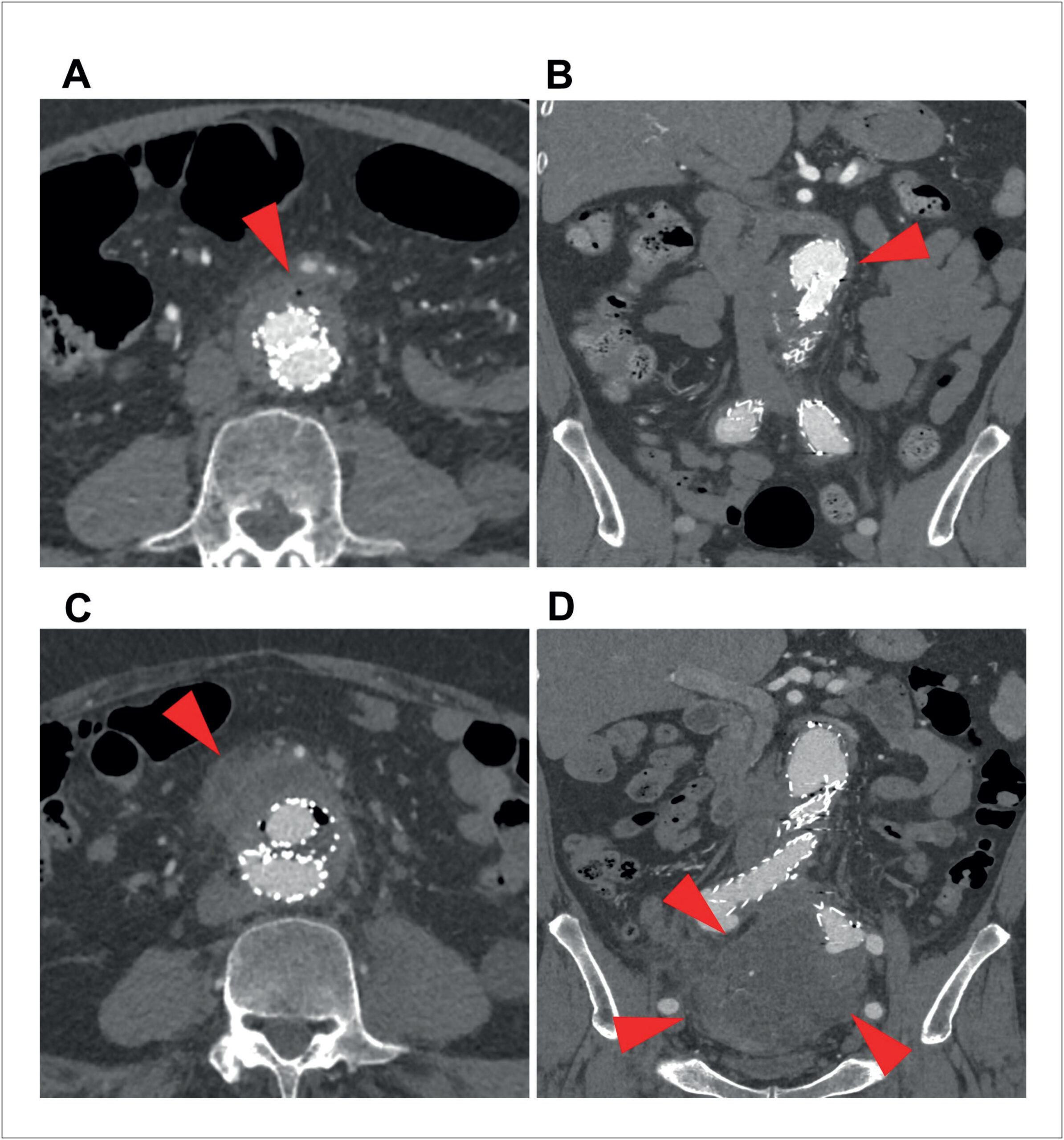
Abdominal aortic endoprosthesis (AAE) infection is the most dreaded complication of endovascular aortic repair (EVAR) for abdominal aortic aneurysm (AAA), with mortality rates reported as high as 51.8%. Recent meta-analyses emphasize that despite advances in surgical technique, the prognosis of AAE infection remains poor due to delayed diagnosis and the complexity of endoprosthesis explantation., The management of AAE infection is significantly hindered by diagnostic latency. Currently, computed tomography angiography (CTA) is typically reserved for evaluation only after the onset of infectious symptoms. Although periprosthetic exudation and ectopic gas are considered hallmark radiographic features, these signs are often subtle and minute during the early, pre-symptomatic stages of infection, rendering them easily overlooked. We present a case in which a radiologist detected minute periprosthetic ectopic gas via thin-section CT during a routine CTA follow-up, five days before the onset of infectious symptoms, thereby alerting clinicians to the possibility of AAE infection. Based on this case, we underscore the critical value of thin-section CT in the early identification of ectopic gas. Furthermore, we aim to demonstrate how advanced post-processing—specifically volume rendering (VR) and hyper-realistic rendering (HRR)—enhances the visualization of AAE infection, thereby enabling longitudinal monitoring of infection progression.

[…]

  • Computed Tomography Angiography
  • Endovascular Aneurysm Repair
  • Endovascular Procedures
  • Infections
Read Full Article
Creative Commons Attribution (CC BY) Creative Commons license
20/07/2026 DOI: 10.36660/abc.20250591i
Cardiac CT-Detected Vulnerable Plaques and Risk of Future Acute Myocardial Infarction: Predictive Value of CAC Scores and Inflammatory Burden Index
ABC
Original Article

Cardiac CT-Detected Vulnerable Plaques and Risk of Future Acute Myocardial Infarction: Predictive Value of CAC Scores and Inflammatory Burden Index

Sefa Erdi Ömür et al. Emin Koyun Gökhan Cabri

Abstract

Background

Computed Tomography (CT) angiography is widely used as a basic non-invasive modality to evaluate coronary artery disease. Inflammatory burden index (IBI) is a systemic inflammation indicator reflecting the inflammatory status. It is still difficult to determine the high risk of future acute myocardial infarction (MI) in patients with plaque detected by CT angiography.

Objective

This study aimed to evaluate the effectiveness of CAC scores and IBI in predicting MI risk among patients undergoing CT angiography for chest pain. We sought to assess the individual and combined predictive value of these indices, determine their optimal cut-off values, and compare MI risk across patient groups with varying CAC and IBI scores. We also aimed to evaluate the potential role of these markers in identifying ‘vulnerable patients’ at high risk for future cardiovascular events.

Methods

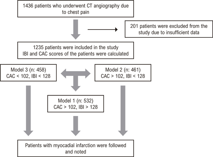
This retrospective study included 1235 patients who underwent CT angiography due to chest pain and were followed for 3 years (2.8-3.4). Patients were categorized into three models based on their CAC and IBI scores. The primary outcome was the occurrence of MI during the follow-up period. Statistical significance was established at p < 0.05 for all analyses.

Results

Patients who experienced MI had significantly higher IBI and CAC scores compared to those without MI. Logistic regression analysis identified CAC score and IBI as independent predictors of MI. ROC analysis determined optimal cut-off values for IBI (128) and CAC (102) in predicting MI. Kaplan-Meier analysis revealed a significant gradient in MI risk across the three models, with the highest risk observed in patients with both high IBI and CAC scores.

Conclusion

The combination of IBI and CAC scores provides improved risk stratification for the prediction of future MI (vulnerable plaque) in patients with plaque detected by CT angiography. This approach may facilitate more specific treatment strategies for high-risk patients.

  • Computed Tomography Angiography
  • Coronary Vessels
  • Myocardial Infarction
Read Full Article
Creative Commons Attribution (CC BY) Creative Commons license
09/01/2026 DOI: 10.36660/abc.20250052i Views 1
Figure 1
ABCIC
Editorial

Photon-Counting Computed Tomography in Cardiovascular Imaging: Where We Are and What Lies Ahead

Paulo Savoia et al. Rodrigo Bello

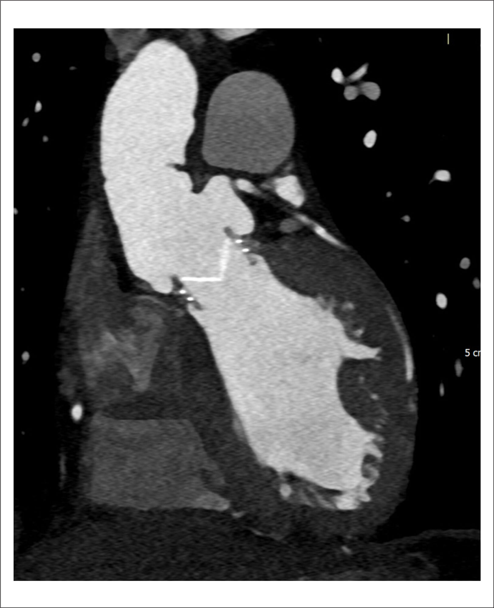
Computed Tomography Angiography (CTA) has been an important asset in cardiovascular diagnosis for decades. The continuous improvement of rapid data acquisition with often higher spatial resolution has established the method as indispensable for diagnosing and managing multiple cardiovascular entities such as aneurysms, aortic dissection, and pulmonary embolism., The use of CTA in heart and coronary imaging has also gained important territory and relevance., Computed Tomography (CT) techniques have been advancing significantly since its introduction in the medical field. Starting with single detectors, then helicoidal scanners, multidetector scanners with high pitch, improvement of reconstruction algorithms, the appearance of dual-energy technology, and lately the ability to acquire images of the whole heart in just one heartbeat with information from the entire cardiac cycle, are incredible milestones. Fractional Flow Reserve CT (FFR-CT) is also a very promising technique. Progression does not come without downsides: ionizing radiation is still concerning, and despite ways to reduce it such as dose modulation, some cardiovascular CTA exams still show relatively high levels of absorbed radiation, especially exams with multiple series, extremely thin slices, and retrospective reconstruction of larger parts of the cardiac cycle. Following this trend of improving image quality with less ionizing radiation as possible, a new complex technique emerged: Photon-counting CT (PCCT).

The main distinction between conventional CT and PCCT is in the detectors of X-rays. The conventional CT detector is based on receiving the X-ray photons that interact with the patient, converting them into light photons (it is an indirect system), and then converting this light into electrical signals that ultimately are going to be converted into digital signals. This system is called an Energy-Integrating Detector (EID). Basically, more light means more X-rays arriving and less interaction / more penetration in the patient’s tissues. It is not possible to differentiate exactly how many X-rays are arriving, nor the different energy levels of each X-ray photon. Moreover, the septa between each of the detectors also limit the reception of X-rays., On the other hand, PCCT detectors can detect separately each X-ray photon that is arriving and measure the energy of each one of the X-ray photons directly. This is possible because the PCCT detector has a high-voltage crystal layer that relocates electrons for each X-ray photon received, and the number of electrons relocated is proportional to the energy of each X-ray. The relocated electrons generate the electrical pulse that is going to be finally converted into the digital signal. And even smaller energy X-rays can be detected, improving the image contrast-to-noise ratio. It is a direct system, and there is no septa between the detectors. In the end, less X-rays are necessary (therefore less radiation) and it is possible to separate the X-rays according to the energy of each one, leading to applications of tissue recognition (different known energy X-rays are likely to have interacted with different known tissues) functioning as a dual energy CT.,

[…]

  • Cardiology
  • Cardiovascular Diagnostic Techniques
  • Computed Tomography Angiography
  • Radiology
Read Full Article
Creative Commons Attribution (CC BY) Creative Commons license
27/06/2025 DOI: 10.36660/abcimg.20250035i Views 1
Figure 1
ABCIC
Review Article

My Approach to Functional Analysis in Computed Tomography Angiography: Myocardial Perfusion and FFRct

Gustavo Corrêa de Almeida Teixeira et al. Bruno Vicente Gomes de Castro Douglas Carli Silva Tiago Magalhães

Abstract

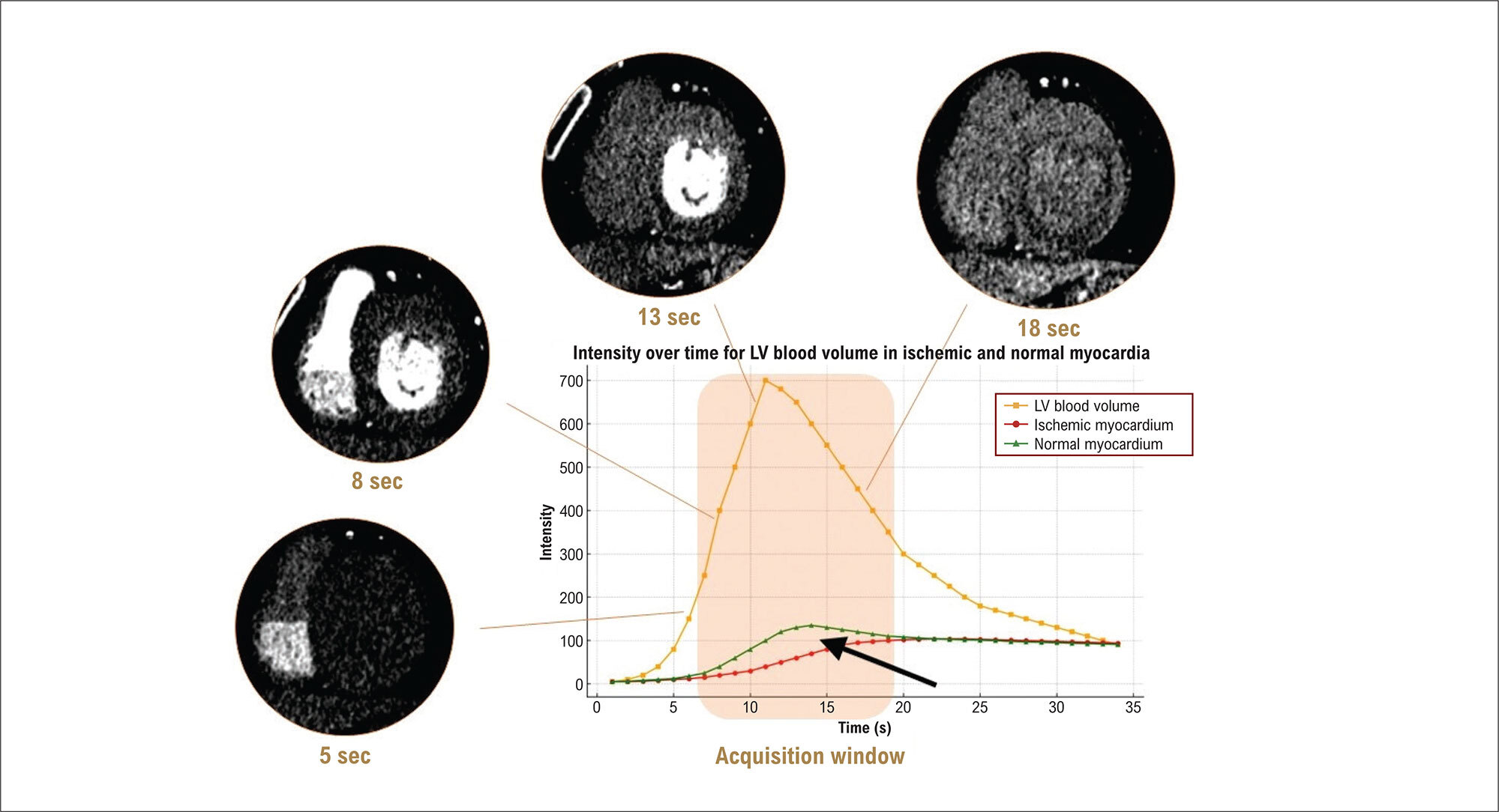
Coronary computed tomography angiography (CCTA) is an effective noninvasive method for detecting atherosclerotic disease, especially in symptomatic patients with low-to-moderate disease probability. Despite good accuracy in detecting obstructions, its relationship with functional lesion severity is limited. For a more accurate assessment, anatomical evaluation can be associated with myocardial computed tomography perfusion and fractional flow reserve derived from CCTA (FFRct). Myocardial perfusion computed tomography identifies areas of ischemia. FFRct, which assesses the hemodynamic significance of stenoses, has shown high accuracy and can reduce the need for invasive catheterizations. The image acquisition and interpretation protocols are essential to ensure reliable results and appropriate management of patients with coronary artery disease.

  • Computed Tomography Angiography
  • Myocardial Fractional Flow Reserve
  • Myocardial Perfusion Imaging
Read Full Article
Creative Commons Attribution (CC BY) Creative Commons license
13/12/2024 DOI: 10.36660/abcimg.20240113i Views 1
Figure 1
ABCIC
Case Report

Rare Presentation of an Anatomical Congenital Anomaly of the Coronary Arteries on Computed Tomography Angiography

Marcello Zapparoli et al. Alexandre Alessi Franco Camargo Zapparoli Mateus Rodrigues Alessi

Introduction

Anomalous origin of the coronary arteries represents a heterogeneous group of alterations in the origin and course of these vessels, ranging from the most common, asymptomatic and low risk, to the rarest and highest risk, which may eventually be symptomatic or have sudden death as their first manifestation., Their prevalence corresponds to 1.3% of patients undergoing coronary angiography, but this number may be higher depending on the diagnostic method and the population selected, representing the second leading cause of sudden death in young athletes, behind only hypertrophic cardiomyopathy.,

Within this group, coronary anomalies with a single ostium are present in a smaller proportion of the population, approximately 0.004% to 0.098%. They are most often asymptomatic, but they may manifest with chest pain, dyspnea, palpitation, myocardial infarction, and sudden death., The presentation of this anomaly has worse prognosis in the presence of an interarterial vessel course (between the aorta and pulmonary trunk), an acute angle of origin, and atherosclerotic plaque; the latter change has a higher incidence in anomalous vessels on account of the greater stress on the arterial wall., In addition to presenting in isolation, they may also be associated with other alterations such as cardiac fistulas or a bicuspid aortic valve.

[…]

  • Anatomic Variation
  • Chest Pain
  • Computed Tomography Angiography
  • Coronary Vessel Anomalies
  • Coronary Vessels
Read Full Article
Creative Commons Attribution (CC BY) Creative Commons license
21/11/2024 DOI: 10.36660/abcimg.20240075i Views 1
Figure 1
ABCIC
Case Report

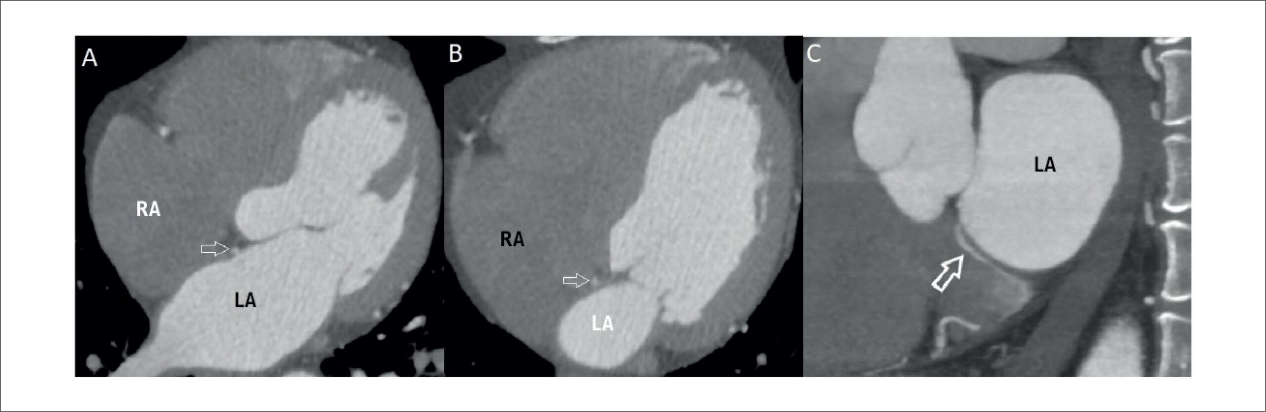
A Bridging Vein Connecting Left Atrium and Coronary Sinus: Coronary Computed Tomography Angiography Findings

Sercan Şahin et al. Nilgün Işıksalan Özbülbül

Introduction

Until recent years, coronary venous system imaging was overshadowed by coronary artery imaging. However, in recent times, the significance of coronary venous system imaging has risen markedly, particularly due to its pivotal role in diverse interventional cardiology procedures. These interventions encompass a spectrum of tasks, including left ventricular pacing, mapping and ablation of arrhythmias, and retrograde cardioplegia. These procedures are facilitated by utilizing the potential of the coronary sinus (CS). As a result, comprehending the anatomy and anomalies of the CS holds paramount importance, and coronary computed tomography (CT) angiography has proven invaluable for achieving this objective.

In this case report, we present an extraordinary cardiac venous anomaly: a bridging vein connecting the left atrium to the CS with non-specific chest pain. We believe that this case serves as an illustrative example for understanding rarely observed cardiac venous vascular anomalies, while also making a valuable contribution to the existing literature through the utilization of clear and direct coronary CT angiography images.

[…]

  • Computed Tomography Angiography
  • Coronary sinus
  • Heart Atria
Read Full Article
Creative Commons Attribution (CC BY) Creative Commons license
12/03/2024 DOI: 10.36660/abcimg.20230111i Views 1
Central Illustration
ABCIC
Review Article

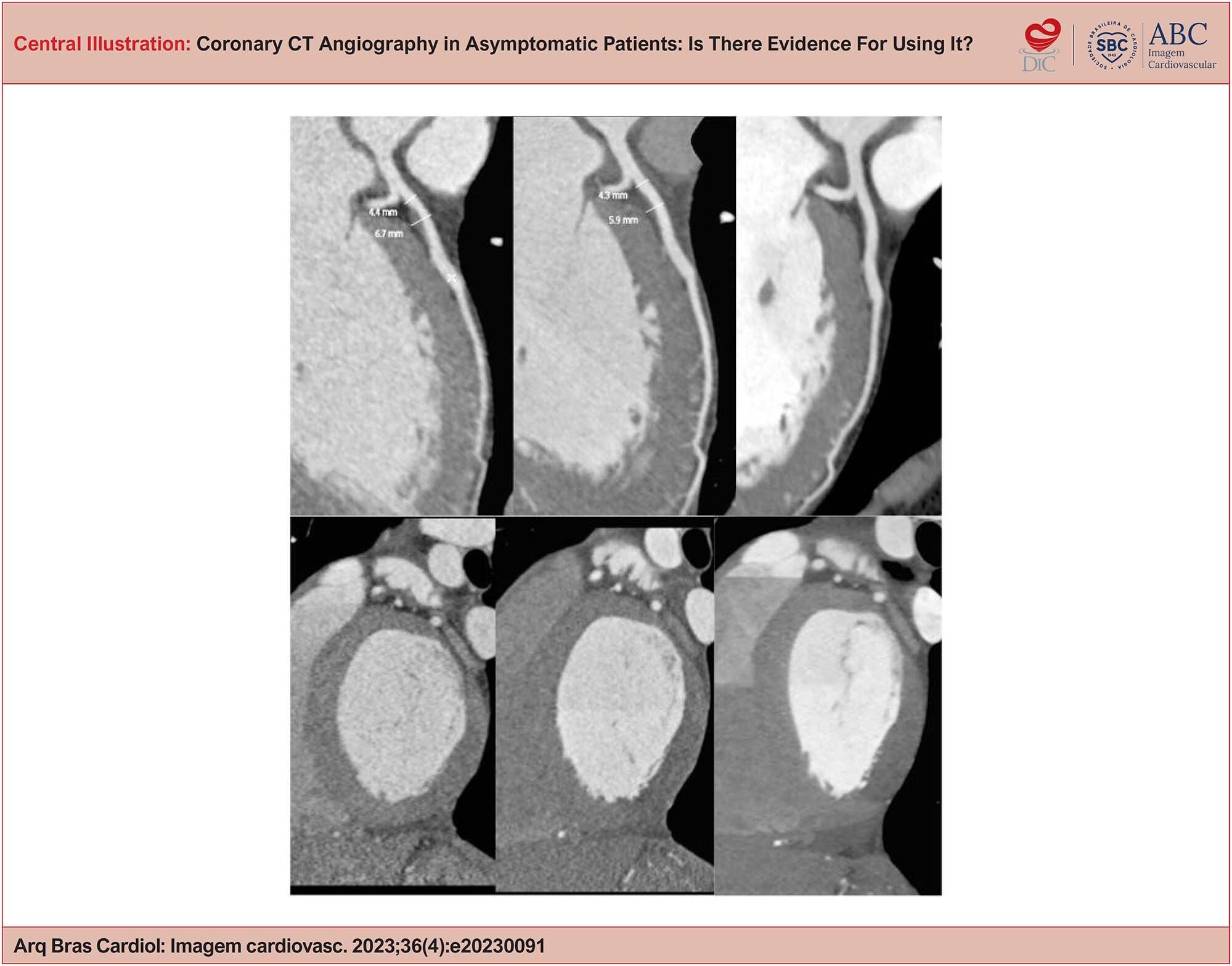
Coronary CT Angiography in Asymptomatic Patients: Is There Evidence For Using It?

Ilan Gottlieb

Abstract

Cardiovascular diseases are the leading cause of death worldwide, and their clinical management is complicated by 3 main pathophysiological factors: a) a long onset period, often decades before a clinical event; b) a silent clinical course; and c) a multifactorial and heterogeneous pathogenesis among different population subgroups. Coronary CT angiography is the best available tool for diagnosing and phenotyping coronary artery disease, whether obstructive or non-obstructive. This allows early diagnosis and monitoring of the disease, which is essential for deciding who should be treated, determining the therapeutic approach, and for better patient adherence to clinical treatment. Early and accurate diagnosis generally results in better clinical outcomes.

  • Asymptomatic Diseases
  • Computed Tomography Angiography
  • Coronary Artery Disease
Read Full Article
Creative Commons Attribution (CC BY) Creative Commons license
13/12/2023 DOI: 10.36660/abcimg.20230091i Views 2
Figure 1
ABCIC
Case Report

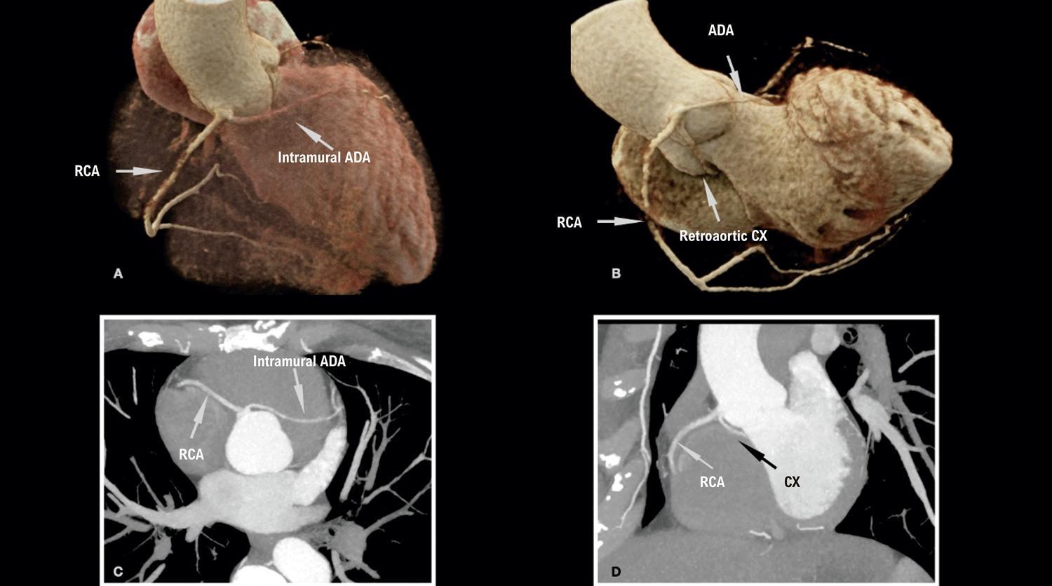
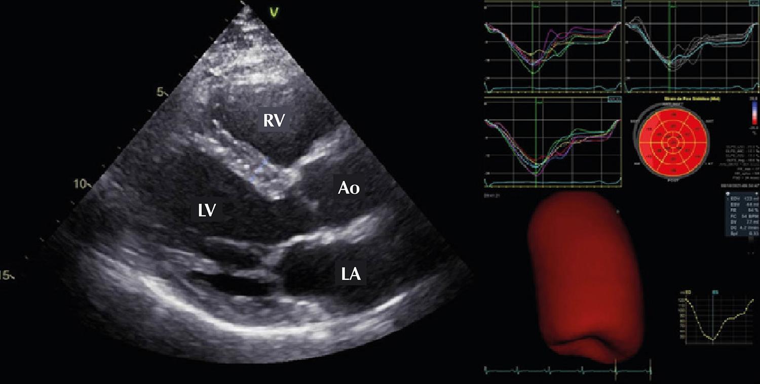
Diagnosis of Coronary Anomaly of the Circumflex Artery With Retroaortic Course by Transthoracic Echocardiography (Rac Sign) Confirmed by Computed Tomography Coronary Angiography: A Case Series

Raul Serra Valério et al. Alfredo A. Eyer Rodrigues Carlos Eduardo Suaide Silva Marly Uellendahl Luciano de Figueiredo Aguiar Filho

Introduction

Congenital coronary artery anomaly (CCAA) is a relatively rare condition with an estimated population prevalence of 0.21–5.79%. CCAA presents a wide spectrum of variations and can be classified into origin and course, intrinsic, and termination anomalies. , The CCAA with the highest prevalence is the anomalous origin of the circumflex artery (CxA) from the right coronary sinus (RCS) with an estimated prevalence of 0.3–0.8%.

CCAA usually travels along a retroaortic course and may present different originations, such as from a common ostium with the right coronary artery (RCA), from separate ostia in the RCS, or from the proximal segment of the RCA.

[…]

  • Cardiovascular Abnormalities
  • Computed Tomography Angiography
  • Echocardiography
Read Full Article
Creative Commons Attribution (CC BY) Creative Commons license
27/05/2022 DOI: 10.47593/2675-312X/20223502eabc271 Views 5
ABC
Editorial

It is Time for Coronary Computed Tomography Angiography to be Incorporated into the SUS

Nilson Tavares Poppi

In the evaluation of patients with stable coronary artery disease (CAD), also called chronic coronary syndrome (CCS), complementary tests are used both for diagnostic and prognostic purposes. , Anatomical (coronary angiography and coronary CT angiography) and functional (exercise testing, stress echocardiography, rest and stress myocardial perfusion imaging by scintigraphy, magnetic resonance, and positron emission tomography) tests are available. Coronary angiography, the gold standard, is invasive and therefore, indicated for clinically more severe patients, or those with poor prognostic findings in non-invasive testing, when myocardial revascularization is considered or planned.

The choice for the most appropriate diagnostic test is an important and challenging issue for the cardiologist in the clinical evaluation of CCS. The first step in this decision-making process is the assessment of the pre-test probability (PTP) of CAD. As recommended by the current SCC guidelines, , patients classified as having high PTP should receive medical therapy and undergo testing for prognostic information. The patients with low PTP should be assessed for an alternative diagnosis more likely than CAD. Patients with PTP calculated between 15-85% are in the intermediate range, where the complementary tests are more useful and important for CAD diagnosis. In addition to diagnostic accuracy and PTP, the selection of a non-invasive test depends on the clinical characteristics of patients, local expertise, and the availability of tests. In Brazil, it is estimated that up to 80% of the population depends exclusively on medical care provided by the public health system (SUS). In this context of managing economic resources, physicians and health managers should focus on the most cost-effective options for the diagnosis of CAD.

[…]

  • Computed Tomography Angiography
  • Coronary Artery Disease
  • Cost-Benefit Analysis
  • Diagnostic Imaging/methods
  • Stable Angina
  • Unified Health System
Read Full Article
Creative Commons Attribution (CC BY) Creative Commons license
18/03/2022 DOI: 10.36660/abc.20220033 Views 1
ABCIC
Case Report

Bilateral Internal Carotid Artery Hypoplasia in Asymptomatic Patient. Case Report

Márcia Azevedo Caldas et al. Luiza Braconi Grilo Lerrícia Pinheiro Soares Polyana da Silva Custódio … Laura Maria Vieira Duarte Carlos Eduardo Cordeiro Soares

[…]

  • Carotid Artery, Internal
  • Computed Tomography Angiography
  • Congenital Abnormality
  • Hypoplasia
Read Full Article
Creative Commons Attribution (CC BY) Creative Commons license
14/01/2022 DOI: 10.5935/2318-8219.20190050
ABCIC
Editorial

Computed Tomography Angiography and Cardiac Catheterization: Allies in the Treatment of Chronic Coronary Obstruction

Leonardo Sara da Silva et al. Juliana Kelendjian

[…]

  • Cardiac Catheterization
  • Computed Tomography Angiography
  • Coronary Occlusion
Read Full Article
Creative Commons Attribution (CC BY) Creative Commons license
14/01/2022 DOI: 10.5935/2318-8219.20190019
ABCIC
Case Report

Cardiac Metastasis Secondary to Endometrial Cancer: an Extremely Rare Presentation

Marcos Danillo Peixoto Oliveira et al. Marina Tulher Florenzano Laura Frontana C Santos Pedro Luiz Barbosa Navarro Roney Cesar Signorini Filho

[…]

  • Computed Tomography Angiography
  • Endometrial Neoplasms
  • Metastasis
Read Full Article
Creative Commons Attribution (CC BY) Creative Commons license
14/01/2022 DOI: 10.5935/2318-8219.20190029 Views 2
Figure 1
ABCIC
Case Report

Spontaneous Coronary Artery Dissection and Diagnosis by Computed Tomography Angiography: a Case Report

Mathias Silvestre de Brida et al. Georgia Pergher Postingher Fábio Vieira Caovilla Juarez Neuhaus Barbisan

Introduction

Spontaneous coronary artery dissection (SCAD), defined as separation of the layers of the coronary wall unrelated to trauma, iatrogenesis, or atherosclerosis, often occurs in young women without cardiovascular risk factors. First described almost a century ago, it was poorly understood for some time, known only to be associated with the peripartum and pregnancy periods. SCAD is a rare cause of acute coronary syndrome (ACS), and despite technological advances in invasive or non-invasive imaging tests increasing our understanding of its pathophysiology and treatment, it remains underdiagnosed. This report describes the case of an ACS patient without obstructive lesions on coronary angiography in which coronary computed tomography angiography (CCTA) was an indispensable tool in the diagnosis of SCAD with an unusual location, highlighting its growing importance based on the current scientific literature.

[…]

  • Acute coronary syndrome
  • Chest Pain
  • Computed Tomography Angiography
  • Dissection
Read Full Article
Creative Commons Attribution (CC BY) Creative Commons license
26/11/2021 DOI: 10.47593/2675-312X/20213403eabc191
Figure 1
ABCIC
Image

Late Diagnosis of Anomalous Left Coronary Artery from Pulmonary Artery in Oligosymptomatic Women

Carolina de Souza Galvão et al. Sterffeson Lamare Lucena de Abreu Magda Luciene de Sousa Carvalho José Eduardo da Cruz Sales Ana Bárbara Silva dos Santos Leite

A

[…]

  • Bland-White-Garland Syndrome
  • Computed Tomography Angiography
  • Diagnosis
  • Echocardiography
Read Full Article
Creative Commons Attribution (CC BY) Creative Commons license
11/06/2021 DOI: 10.47593/2675-312X/20213402eabc179
Figure 1
ABC
Image

Radiologic-Electrocardiography Correlation in Wellens Syndrome

Eduardo Kaiser Ururahy Nunes Fonseca et al. Nevelton Heringer Filho Marcelo L. Montemor Luiz Francisco Rodrigues de Ávila Carlos Eduardo Rochitte

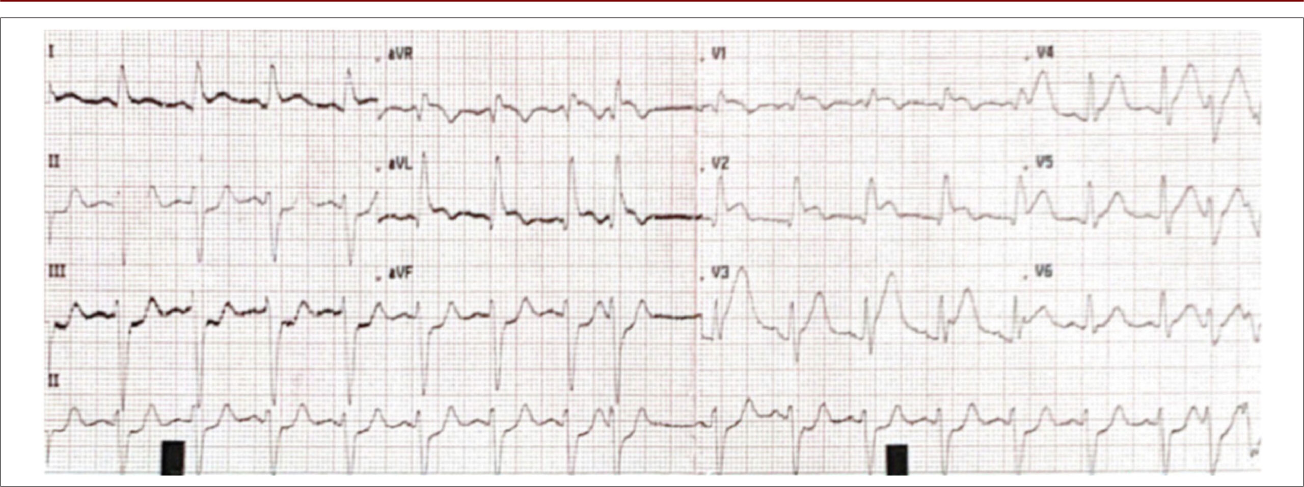
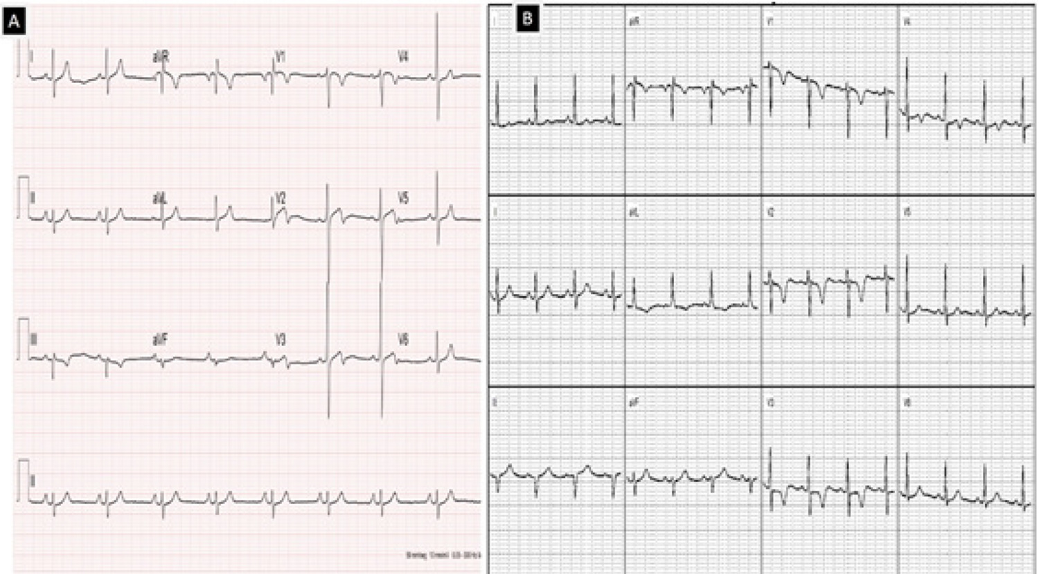
Wellens Syndrome, also known as “Anterior Descending Coronary T-wave syndrome”, was described in 1982 by Dr. Henrick Joan Joost (Hein) Wellens, a Dutch physician who also contributed to the characterization of the reentry mechanism in Wolff–Parkinson–White syndrome (WPWS).

Originally described during hospital admission (60% at admission and 40% at the follow-up) of patients with unstable angina, it was characterized by the occurrence of 2 electrocardiographic patterns, with pattern A in 25% of patients and B in 75% of patients.

[…]

  • Computed Tomography Angiography
  • Coronary Angiography
  • Coronary Vessels
  • Electrocardiography
  • Myocardial Infarction
Read Full Article
Creative Commons Attribution (CC BY) Creative Commons license
01/03/2021 DOI: 10.36660/abc.20200200 Views 1
ABC
Viewpoint

Coronary Computed Tomography Angiography Takes the Center Stage and Here is Why

Illan Gottlieb et al. Marcio Sommer Bittencourt Carlos Eduardo Rochitte João L. Cavalcante

Max Planck once said that “A new scientific truth does not triumph by convincing its opponents and making them see the light, but rather because its opponents eventually die, and a new generation grows up that is familiar with it.” In its beginnings, coronary computed tomography angiography (CCTA) was accused of having too low accuracy for the diagnosis of obstructive coronary artery disease (CAD) to be used in clinical practice. Over the last decade, major technical developments such as larger axial coverage (from 2 cm to 16 cm) and improved temporal resolution, have enabled CCTA to become by far the most accurate non-invasive imaging method for diagnosis of obstructive CAD, with sensitivity and specificity of approximately 95% and 90%, respectively.

Then CCTA was burdened with the accusation of exposing patients to radiation doses so high, that warranted some society guidelines to specifically point this out and limit its use. At that time, CCTA exposed patients to doses ranging from 20 to 25 mSV, while triphasic abdomen CT exposed patients to 30 to 40 mSv and scintigraphic myocardial perfusion studies with Thallium used up to 40 mSv. In 2018, radiation exposure from CCTA dropped to well below 5 mSv (most advanced clinical centers use much less), a fraction of the dose used in myocardial perfusion studies with MIBI tetrophosmin. Then the cost-effectiveness wave came with societies rightfully demanding proof that CCTA offered more value at an acceptable cost compared to other imaging modalities, and CCTA once again proved to be more cost-effective than other modalities. Although one hardly finds cost-effectiveness studies comparing nuclear scans with ECG treadmill tests, providing better diagnosis performance is not enough anymore. More recently, this strategy has even been put into challenge in large randomized clinical trials comparing CCTA with the standard of care in the investigation of suspected CAD both in the acute and in the outpatient settings.–

[…]

  • Computed Tomography Angiography
  • Coronary Artery Disease/diagnostic imaging
  • Coronary Artery Disease/physiopathology
  • Coronary Artery Disease/prevention and control
  • Tomography, X-Ray Computed
Read Full Article
Creative Commons Attribution (CC BY) Creative Commons license
01/01/2019 DOI: 10.5935/abc.20190003 Views 1
Figure 1
ABC
Image

Simultaneous Dual Coronary Fistulas

Ioannis Ntalas et al. John B. Chambers Júlia Karády Ronak Rajani

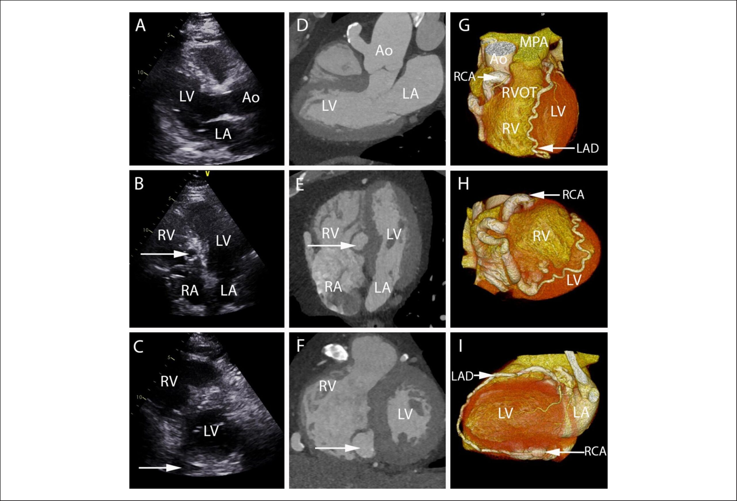
A 61-year-old man with type II diabetes mellitus was referred with breathlessness on exertion. On auscultation, there was a continuous ejection systolic murmur on the left upper sternal border. Transthoracic echocardiography showed a dilated vessel in aortic wall in the parasternal long axis view () and a spherical lesion in the apical 4-chamber view (). A coronary computed tomographic angiographic (CTCA) study revealed a dilated and ectatic right coronary artery (RCA). It arose from the ascending aorta at the 12 o’clock position and followed a tortuous course around the right sided atrioventricular groove before passing into the basal inferoseptum draining into the base of the right ventricle. An additional fistulous connection could be detected between the posterior descending artery of the RCA and the left anterior descending artery (LAD-RCA fistula) (, , ). After a normal dobutamine stress echocardiogram, a decision for continued medical therapy was taken.

[…]

  • Computed Tomography Angiography
  • Echocardiography
  • Systolic Murmurs
Read Full Article
Creative Commons Attribution (CC BY) Creative Commons license
01/04/2018 DOI: 10.5935/abc.20180057
Figure 1
ABC
Original Article

Prevalence and Prediction of Obstructive Coronary Artery Disease in Patients Undergoing Primary Heart Valve Surgery

José Guilherme Cazelli et al. Gabriel Cordeiro Camargo Dany David Kruczan Clara Weksler … Alexandre Rouge Felipe Ilan Gottlieb

Abstract

Background:

The prevalence of coronary artery disease (CAD) in valvular patients is similar to that of the general population, with the usual association with traditional risk factors. Nevertheless, the search for obstructive CAD is more aggressive in the preoperative period of patients with valvular heart disease, resulting in the indication of invasive coronary angiography (ICA) to almost all adult patients, because it is believed that coronary artery bypass surgery should be associated with valve replacement.

Objectives:

To evaluate the prevalence of obstructive CAD and factors associated with it in adult candidates for primary heart valve surgery between 2001 and 2014 at the National Institute of Cardiology (INC) and, thus, derive and validate a predictive obstructive CAD score.

Methods:

Cross-sectional study evaluating 2898 patients with indication for heart surgery of any etiology. Of those, 712 patients, who had valvular heart disease and underwent ICA in the 12 months prior to surgery, were included. The P value < 0.05 was adopted as statistical significance.

Results:

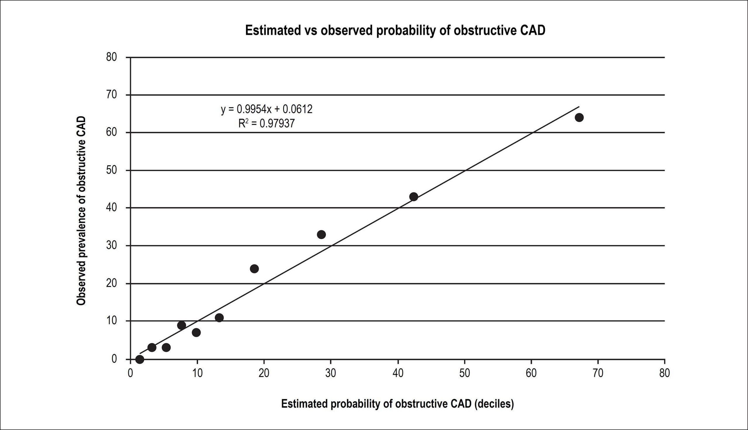
The prevalence of obstructive CAD was 20%. A predictive model of obstructive CAD was created from multivariate logistic regression, using the variables age, chest pain, family history of CAD, systemic arterial hypertension, diabetes mellitus, dyslipidemia, smoking, and male gender. The model showed excellent correlation and calibration (R² = 0.98), as well as excellent accuracy (ROC of 0.848; 95%CI: 0.817-0.879) and validation (ROC of 0.877; 95%CI: 0.830 – 0.923) in different valve populations.

Conclusions:

Obstructive CAD can be estimated from clinical data of adult candidates for valve repair surgery, using a simple, accurate and validated score, easy to apply in clinical practice, which may contribute to changes in the preoperative strategy of acquired heart valve surgery in patients with a lower probability of obstructive disease.

  • Computed Tomography Angiography
  • Coronary Angiography
  • Coronary Artery Disease
  • Heart Valve Disease
Read Full Article
Creative Commons Attribution (CC BY) Creative Commons license
28/09/2017 DOI: 10.5935/abc.20170135 Views 1

How to cite this article

EndNote RIS

Electronic Document Format (Vancouver)

Li H, Zhao Y, Ji Q. Ectopic Gas: A Sign for Potential Aortic Endoprosthesis Infection before Symptoms. Arquivos Brasileiros de Cardiologia 2026;123(5):e20250591.

Electronic Document Format (ABNT)

Li, Hongliang; Zhao, Ying; Ji, Qingyu. Ectopic Gas: A Sign for Potential Aortic Endoprosthesis Infection before Symptoms. Arquivos Brasileiros de Cardiologia, v. 123, n. 5, e20250591, Jul. 2026.

Electronic Document Format (APA)

Li, H., Zhao, Y., & Ji, Q. (2026). Ectopic Gas: A Sign for Potential Aortic Endoprosthesis Infection before Symptoms. Arquivos Brasileiros de Cardiologia, 123(5), e20250591.

Electronic Document Format (ISO)

Li, Hongliang and Zhao, Ying and Ji, Qingyu. Ectopic Gas: A Sign for Potential Aortic Endoprosthesis Infection before Symptoms. Arquivos Brasileiros de Cardiologia [online]. 2026, vol. 123, n. 5, [cited 2026-10-14], e20250591. Available from: <https://journals.cardiol.br/en/article/ectopic-gas-a-sign-for-potential-aortic-endoprosthesis-infection-before-symptoms/>. ISSN 0066-782X

EndNote RIS

Electronic Document Format (Vancouver)

Ömür SE, Koyun E, Cabri G. Cardiac CT-Detected Vulnerable Plaques and Risk of Future Acute Myocardial Infarction: Predictive Value of CAC Scores and Inflammatory Burden Index. Arquivos Brasileiros de Cardiologia 2026;122(11):e20250052.

Electronic Document Format (ABNT)

Ömür, Sefa Erdi; Koyun, Emin; Cabri, Gökhan. Cardiac CT-Detected Vulnerable Plaques and Risk of Future Acute Myocardial Infarction: Predictive Value of CAC Scores and Inflammatory Burden Index. Arquivos Brasileiros de Cardiologia, v. 122, n. 11, e20250052, Jan. 2026.

Electronic Document Format (APA)

Ömür, S. E., Koyun, E., & Cabri, G. (2026). Cardiac CT-Detected Vulnerable Plaques and Risk of Future Acute Myocardial Infarction: Predictive Value of CAC Scores and Inflammatory Burden Index. Arquivos Brasileiros de Cardiologia, 122(11), e20250052.

Electronic Document Format (ISO)

Ömür, Sefa Erdi and Koyun, Emin and Cabri, Gökhan. Cardiac CT-Detected Vulnerable Plaques and Risk of Future Acute Myocardial Infarction: Predictive Value of CAC Scores and Inflammatory Burden Index. Arquivos Brasileiros de Cardiologia [online]. 2026, vol. 122, n. 11, [cited 2026-10-14], e20250052. Available from: <https://journals.cardiol.br/en/article/cardiac-ct-detected-vulnerable-plaques-and-risk-of-future-acute-myocardial-infarction-predictive-value-of-cac-scores-and-inflammatory-burden-index/>. ISSN 0066-782X

EndNote RIS

Electronic Document Format (Vancouver)

Savoia P, Bello R. Photon-Counting Computed Tomography in Cardiovascular Imaging: Where We Are and What Lies Ahead. ABC Imagem Cardiovascular 2025;38(2):e20250035.

Electronic Document Format (ABNT)

Savoia, Paulo; Bello, Rodrigo. Photon-Counting Computed Tomography in Cardiovascular Imaging: Where We Are and What Lies Ahead. ABC Imagem Cardiovascular, v. 38, n. 2, e20250035, Jun. 2025.

Electronic Document Format (APA)

Savoia, P., & Bello, R. (2025). Photon-Counting Computed Tomography in Cardiovascular Imaging: Where We Are and What Lies Ahead. ABC Imagem Cardiovascular, 38(2), e20250035.

Electronic Document Format (ISO)

Savoia, Paulo and Bello, Rodrigo. Photon-Counting Computed Tomography in Cardiovascular Imaging: Where We Are and What Lies Ahead. ABC Imagem Cardiovascular [online]. 2025, vol. 38, n. 2, [cited 2026-10-14], e20250035. Available from: <https://journals.cardiol.br/en/article/photon-counting-computed-tomography-in-cardiovascular-imaging-where-we-are-and-what-lies-ahead/>. ISSN 2318-8219

EndNote RIS

Electronic Document Format (Vancouver)

Teixeira GCA, Castro BVG, Silva DC, Magalhães T. My Approach to Functional Analysis in Computed Tomography Angiography: Myocardial Perfusion and FFRct. ABC Imagem Cardiovascular 2024;37(4):e20240113.

Electronic Document Format (ABNT)

Teixeira, Gustavo Corrêa de Almeida; Castro, Bruno Vicente Gomes de; Silva, Douglas Carli; Magalhães, Tiago. My Approach to Functional Analysis in Computed Tomography Angiography: Myocardial Perfusion and FFRct. ABC Imagem Cardiovascular, v. 37, n. 4, e20240113, Dec. 2024.

Electronic Document Format (APA)

Teixeira, G. C. A., Castro, B. V. G. , Silva, D. C., & Magalhães, T. (2024). My Approach to Functional Analysis in Computed Tomography Angiography: Myocardial Perfusion and FFRct. ABC Imagem Cardiovascular, 37(4), e20240113.

Electronic Document Format (ISO)

Teixeira, Gustavo Corrêa de Almeida and Castro, Bruno Vicente Gomes de and Silva, Douglas Carli and Magalhães, Tiago. My Approach to Functional Analysis in Computed Tomography Angiography: Myocardial Perfusion and FFRct. ABC Imagem Cardiovascular [online]. 2024, vol. 37, n. 4, [cited 2026-10-14], e20240113. Available from: <https://journals.cardiol.br/en/article/my-approach-to-functional-analysis-in-computed-tomography-angiography-myocardial-perfusion-and-ffrct/>. ISSN 2318-8219

EndNote RIS

Electronic Document Format (Vancouver)

Zapparoli M, Alessi A, Zapparoli FC, Alessi MR. Rare Presentation of an Anatomical Congenital Anomaly of the Coronary Arteries on Computed Tomography Angiography. ABC Imagem Cardiovascular 2024;37(4):e20240075.

Electronic Document Format (ABNT)

Zapparoli, Marcello; Alessi, Alexandre; Zapparoli, Franco Camargo; Alessi, Mateus Rodrigues. Rare Presentation of an Anatomical Congenital Anomaly of the Coronary Arteries on Computed Tomography Angiography. ABC Imagem Cardiovascular, v. 37, n. 4, e20240075, Nov. 2024.

Electronic Document Format (APA)

Zapparoli, M., Alessi, A., Zapparoli, F. C., & Alessi, M. R. (2024). Rare Presentation of an Anatomical Congenital Anomaly of the Coronary Arteries on Computed Tomography Angiography. ABC Imagem Cardiovascular, 37(4), e20240075.

Electronic Document Format (ISO)

Zapparoli, Marcello and Alessi, Alexandre and Zapparoli, Franco Camargo and Alessi, Mateus Rodrigues. Rare Presentation of an Anatomical Congenital Anomaly of the Coronary Arteries on Computed Tomography Angiography. ABC Imagem Cardiovascular [online]. 2024, vol. 37, n. 4, [cited 2026-10-14], e20240075. Available from: <https://journals.cardiol.br/en/article/rare-presentation-of-an-anatomical-congenital-anomaly-of-the-coronary-arteries-on-computed-tomography-angiography/>. ISSN 2318-8219

EndNote RIS

Electronic Document Format (Vancouver)

Şahin S, Özbülbül NI. A Bridging Vein Connecting Left Atrium and Coronary Sinus: Coronary Computed Tomography Angiography Findings. ABC Imagem Cardiovascular 2024;37(1):e20230111.

Electronic Document Format (ABNT)

Şahin, Sercan; Özbülbül, Nilgün Işıksalan. A Bridging Vein Connecting Left Atrium and Coronary Sinus: Coronary Computed Tomography Angiography Findings. ABC Imagem Cardiovascular, v. 37, n. 1, e20230111, Mar. 2024.

Electronic Document Format (APA)

Şahin, S., & Özbülbül, N. I. (2024). A Bridging Vein Connecting Left Atrium and Coronary Sinus: Coronary Computed Tomography Angiography Findings. ABC Imagem Cardiovascular, 37(1), e20230111.

Electronic Document Format (ISO)

Şahin, Sercan and Özbülbül, Nilgün Işıksalan. A Bridging Vein Connecting Left Atrium and Coronary Sinus: Coronary Computed Tomography Angiography Findings. ABC Imagem Cardiovascular [online]. 2024, vol. 37, n. 1, [cited 2026-10-14], e20230111. Available from: <https://journals.cardiol.br/en/article/a-bridging-vein-connecting-left-atrium-and-coronary-sinus-coronary-computed-tomography-angiography-findings/>. ISSN 2318-8219

EndNote RIS

Electronic Document Format (Vancouver)

Gottlieb I. Coronary CT Angiography in Asymptomatic Patients: Is There Evidence For Using It?. ABC Imagem Cardiovascular 2023;36(4):e20230091.

Electronic Document Format (ABNT)

Gottlieb, Ilan. Coronary CT Angiography in Asymptomatic Patients: Is There Evidence For Using It?. ABC Imagem Cardiovascular, v. 36, n. 4, e20230091, Dec. 2023.

Electronic Document Format (APA)

Gottlieb, I. (2023). Coronary CT Angiography in Asymptomatic Patients: Is There Evidence For Using It?. ABC Imagem Cardiovascular, 36(4), e20230091.

Electronic Document Format (ISO)

Gottlieb, Ilan. Coronary CT Angiography in Asymptomatic Patients: Is There Evidence For Using It?. ABC Imagem Cardiovascular [online]. 2023, vol. 36, n. 4, [cited 2026-10-14], e20230091. Available from: <https://journals.cardiol.br/en/article/coronary-ct-angiography-in-asymptomatic-patients-is-there-evidence-for-using-it/>. ISSN 2318-8219

EndNote RIS

Electronic Document Format (Vancouver)

Valério RS, Rodrigues AAE, Silva CES, Uellendahl M, Aguiar Filho LF. Diagnosis of Coronary Anomaly of the Circumflex Artery With Retroaortic Course by Transthoracic Echocardiography (Rac Sign) Confirmed by Computed Tomography Coronary Angiography: A Case Series. Arquivos Brasileiros de Cardiologia Imagem cardiovascular 2022;35(2):eabc271.

Electronic Document Format (ABNT)

Valério, Raul Serra; Rodrigues, Alfredo A. Eyer; Silva, Carlos Eduardo Suaide; Uellendahl, Marly; Aguiar Filho, Luciano de Figueiredo. Diagnosis of Coronary Anomaly of the Circumflex Artery With Retroaortic Course by Transthoracic Echocardiography (Rac Sign) Confirmed by Computed Tomography Coronary Angiography: A Case Series. Arquivos Brasileiros de Cardiologia Imagem cardiovascular, v. 35, n. 2, eabc271, May. 2022.

Electronic Document Format (APA)

Valério, R. S., Rodrigues, A. A. E., Silva, C. E. S., Uellendahl, M., & Aguiar Filho, L. F. (2022). Diagnosis of Coronary Anomaly of the Circumflex Artery With Retroaortic Course by Transthoracic Echocardiography (Rac Sign) Confirmed by Computed Tomography Coronary Angiography: A Case Series. Arquivos Brasileiros de Cardiologia Imagem cardiovascular, 35(2), eabc271.

Electronic Document Format (ISO)

Valério, Raul Serra and Rodrigues, Alfredo A. Eyer and Silva, Carlos Eduardo Suaide and Uellendahl, Marly and Aguiar Filho, Luciano de Figueiredo. Diagnosis of Coronary Anomaly of the Circumflex Artery With Retroaortic Course by Transthoracic Echocardiography (Rac Sign) Confirmed by Computed Tomography Coronary Angiography: A Case Series. Arquivos Brasileiros de Cardiologia Imagem cardiovascular [online]. 2022, vol. 35, n. 2, [cited 2026-10-14], eabc271. Available from: <https://journals.cardiol.br/en/article/diagnosis-of-coronary-anomaly-of-the-circumflex-artery-with-retroaortic-course-by-transthoracic-echocardiography-rac-sign-confirmed-by-computed-tomography-coronary-angiography-a-case-series/>. ISSN 2318-8219

EndNote RIS

Electronic Document Format (Vancouver)

Poppi NT. It is Time for Coronary Computed Tomography Angiography to be Incorporated into the SUS. Arquivos Brasileiros de Cardiologia 2022;118(3):586-7.

Electronic Document Format (ABNT)

Poppi, Nilson Tavares. It is Time for Coronary Computed Tomography Angiography to be Incorporated into the SUS. Arquivos Brasileiros de Cardiologia, v. 118, n. 3, p. 586-587, Mar. 2022.

Electronic Document Format (APA)

Poppi, N. T. (2022). It is Time for Coronary Computed Tomography Angiography to be Incorporated into the SUS. Arquivos Brasileiros de Cardiologia, 118(3), 586-587.

Electronic Document Format (ISO)

Poppi, Nilson Tavares. It is Time for Coronary Computed Tomography Angiography to be Incorporated into the SUS. Arquivos Brasileiros de Cardiologia [online]. 2022, vol. 118, n. 3, [cited 2026-10-14], pp. 586-587. Available from: <https://journals.cardiol.br/en/article/it-is-time-for-coronary-computed-tomography-angiography-to-be-incorporated-into-the-sus/>. ISSN 0066-782X

EndNote RIS

Electronic Document Format (Vancouver)

Caldas MA, Grilo LB, Soares LP, Custódio PS, Duarte LMV, Soares CEC. Bilateral Internal Carotid Artery Hypoplasia in Asymptomatic Patient. Case Report. Arquivos Brasileiros de Cardiologia Imagem cardiovascular 2022;32(4):318-30.

Electronic Document Format (ABNT)

Caldas, Márcia Azevedo; Grilo, Luiza Braconi; Soares, Lerrícia Pinheiro; Custódio, Polyana da Silva; Duarte, Laura Maria Vieira; Soares, Carlos Eduardo Cordeiro. Bilateral Internal Carotid Artery Hypoplasia in Asymptomatic Patient. Case Report. Arquivos Brasileiros de Cardiologia Imagem cardiovascular, v. 32, n. 4, p. 318-330, Jan. 2022.

Electronic Document Format (APA)

Caldas, M. A., Grilo, L. B., Soares, L. P., Custódio, P. S., Duarte, L. M. V., & Soares, C. E. C. (2022). Bilateral Internal Carotid Artery Hypoplasia in Asymptomatic Patient. Case Report. Arquivos Brasileiros de Cardiologia Imagem cardiovascular, 32(4), 318-330.

Electronic Document Format (ISO)

Caldas, Márcia Azevedo and Grilo, Luiza Braconi and Soares, Lerrícia Pinheiro and Custódio, Polyana da Silva and Duarte, Laura Maria Vieira and Soares, Carlos Eduardo Cordeiro. Bilateral Internal Carotid Artery Hypoplasia in Asymptomatic Patient. Case Report. Arquivos Brasileiros de Cardiologia Imagem cardiovascular [online]. 2022, vol. 32, n. 4, [cited 2026-10-14], pp. 318-330. Available from: <https://journals.cardiol.br/en/article/bilateral-internal-carotid-artery-hypoplasia-in-asymptomatic-patient-case-report/>. ISSN 2318-8219

EndNote RIS

Electronic Document Format (Vancouver)

Silva LS, Kelendjian J. Computed Tomography Angiography and Cardiac Catheterization: Allies in the Treatment of Chronic Coronary Obstruction. Arquivos Brasileiros de Cardiologia Imagem cardiovascular 2022;32(2):94-5.

Electronic Document Format (ABNT)

Silva, Leonardo Sara da; Kelendjian, Juliana. Computed Tomography Angiography and Cardiac Catheterization: Allies in the Treatment of Chronic Coronary Obstruction. Arquivos Brasileiros de Cardiologia Imagem cardiovascular, v. 32, n. 2, p. 94-95, Jan. 2022.

Electronic Document Format (APA)

Silva, L. S. , & Kelendjian, J. (2022). Computed Tomography Angiography and Cardiac Catheterization: Allies in the Treatment of Chronic Coronary Obstruction. Arquivos Brasileiros de Cardiologia Imagem cardiovascular, 32(2), 94-95.

Electronic Document Format (ISO)

Silva, Leonardo Sara da and Kelendjian, Juliana. Computed Tomography Angiography and Cardiac Catheterization: Allies in the Treatment of Chronic Coronary Obstruction. Arquivos Brasileiros de Cardiologia Imagem cardiovascular [online]. 2022, vol. 32, n. 2, [cited 2026-10-14], pp. 94-95. Available from: <https://journals.cardiol.br/en/article/computed-tomography-angiography-and-cardiac-catheterization-allies-in-the-treatment-of-chronic-coronary-obstruction/>. ISSN 2318-8219

EndNote RIS

Electronic Document Format (Vancouver)

Oliveira MDP, Florenzano MT, Santos LFC, Navarro PLB, Signorini Filho RC. Cardiac Metastasis Secondary to Endometrial Cancer: an Extremely Rare Presentation. Arquivos Brasileiros de Cardiologia Imagem cardiovascular 2022;32(2):138-40.

Electronic Document Format (ABNT)

Oliveira, Marcos Danillo Peixoto; Florenzano, Marina Tulher; Santos, Laura Frontana C; Navarro, Pedro Luiz Barbosa; Signorini Filho, Roney Cesar. Cardiac Metastasis Secondary to Endometrial Cancer: an Extremely Rare Presentation. Arquivos Brasileiros de Cardiologia Imagem cardiovascular, v. 32, n. 2, p. 138-140, Jan. 2022.

Electronic Document Format (APA)

Oliveira, M. D. P., Florenzano, M. T., Santos, L. F. C., Navarro, P. L. B., & Signorini Filho, R. C. (2022). Cardiac Metastasis Secondary to Endometrial Cancer: an Extremely Rare Presentation. Arquivos Brasileiros de Cardiologia Imagem cardiovascular, 32(2), 138-140.

Electronic Document Format (ISO)

Oliveira, Marcos Danillo Peixoto and Florenzano, Marina Tulher and Santos, Laura Frontana C and Navarro, Pedro Luiz Barbosa and Signorini Filho, Roney Cesar. Cardiac Metastasis Secondary to Endometrial Cancer: an Extremely Rare Presentation. Arquivos Brasileiros de Cardiologia Imagem cardiovascular [online]. 2022, vol. 32, n. 2, [cited 2026-10-14], pp. 138-140. Available from: <https://journals.cardiol.br/en/article/cardiac-metastasis-secondary-to-endometrial-cancer-an-extremely-rare-presentation/>. ISSN 2318-8219

EndNote RIS

Electronic Document Format (Vancouver)

Brida MS, Postingher GP, Caovilla FV, Barbisan JN. Spontaneous Coronary Artery Dissection and Diagnosis by Computed Tomography Angiography: a Case Report. ABC Imagem Cardiovascular 2021;34(3):eabc191.

Electronic Document Format (ABNT)

Brida, Mathias Silvestre de; Postingher, Georgia Pergher; Caovilla, Fábio Vieira; Barbisan, Juarez Neuhaus. Spontaneous Coronary Artery Dissection and Diagnosis by Computed Tomography Angiography: a Case Report. ABC Imagem Cardiovascular, v. 34, n. 3, eabc191, Nov. 2021.

Electronic Document Format (APA)

Brida, M. S. , Postingher, G. P., Caovilla, F. V., & Barbisan, J. N. (2021). Spontaneous Coronary Artery Dissection and Diagnosis by Computed Tomography Angiography: a Case Report. ABC Imagem Cardiovascular, 34(3), eabc191.

Electronic Document Format (ISO)

Brida, Mathias Silvestre de and Postingher, Georgia Pergher and Caovilla, Fábio Vieira and Barbisan, Juarez Neuhaus. Spontaneous Coronary Artery Dissection and Diagnosis by Computed Tomography Angiography: a Case Report. ABC Imagem Cardiovascular [online]. 2021, vol. 34, n. 3, [cited 2026-10-14], eabc191. Available from: <https://journals.cardiol.br/en/article/spontaneous-coronary-artery-dissection-and-diagnosis-by-computed-tomography-angiography-a-case-report/>. ISSN 2318-8219

EndNote RIS

Electronic Document Format (Vancouver)

Galvão CS, Abreu SLL, Carvalho MLS, Sales JEC, Leite ABSS. Late Diagnosis of Anomalous Left Coronary Artery from Pulmonary Artery in Oligosymptomatic Women. ABC Imagem Cardiovascular 2021;34(2):eabc179.

Electronic Document Format (ABNT)

Galvão, Carolina de Souza; Abreu, Sterffeson Lamare Lucena de; Carvalho, Magda Luciene de Sousa; Sales, José Eduardo da Cruz; Leite, Ana Bárbara Silva dos Santos. Late Diagnosis of Anomalous Left Coronary Artery from Pulmonary Artery in Oligosymptomatic Women. ABC Imagem Cardiovascular, v. 34, n. 2, eabc179, Jun. 2021.

Electronic Document Format (APA)

Galvão, C. S., Abreu, S. L. L. , Carvalho, M. L. S., Sales, J. E. C., & Leite, A. B. S. S. (2021). Late Diagnosis of Anomalous Left Coronary Artery from Pulmonary Artery in Oligosymptomatic Women. ABC Imagem Cardiovascular, 34(2), eabc179.

Electronic Document Format (ISO)

Galvão, Carolina de Souza and Abreu, Sterffeson Lamare Lucena de and Carvalho, Magda Luciene de Sousa and Sales, José Eduardo da Cruz and Leite, Ana Bárbara Silva dos Santos. Late Diagnosis of Anomalous Left Coronary Artery from Pulmonary Artery in Oligosymptomatic Women. ABC Imagem Cardiovascular [online]. 2021, vol. 34, n. 2, [cited 2026-10-14], eabc179. Available from: <https://journals.cardiol.br/en/article/late-diagnosis-of-anomalous-left-coronary-artery-from-pulmonary-artery-in-oligosymptomatic-women/>. ISSN 2318-8219

EndNote RIS

Electronic Document Format (Vancouver)

Fonseca EKUN, Heringer Filho N, Montemor ML, Ávila LFR, Rochitte CE. Radiologic-Electrocardiography Correlation in Wellens Syndrome. Arquivos Brasileiros de Cardiologia 2021;116(2):363-6.

Electronic Document Format (ABNT)

Fonseca, Eduardo Kaiser Ururahy Nunes; Heringer Filho, Nevelton; Montemor, Marcelo L.; Ávila, Luiz Francisco Rodrigues de; Rochitte, Carlos Eduardo. Radiologic-Electrocardiography Correlation in Wellens Syndrome. Arquivos Brasileiros de Cardiologia, v. 116, n. 2, p. 363-366, Feb. 2021.

Electronic Document Format (APA)

Fonseca, E. K. U. N., Heringer Filho, N., Montemor, M. L., Ávila, L. F. R. , & Rochitte, C. E. (2021). Radiologic-Electrocardiography Correlation in Wellens Syndrome. Arquivos Brasileiros de Cardiologia, 116(2), 363-366.

Electronic Document Format (ISO)

Fonseca, Eduardo Kaiser Ururahy Nunes and Heringer Filho, Nevelton and Montemor, Marcelo L. and Ávila, Luiz Francisco Rodrigues de and Rochitte, Carlos Eduardo. Radiologic-Electrocardiography Correlation in Wellens Syndrome. Arquivos Brasileiros de Cardiologia [online]. 2021, vol. 116, n. 2, [cited 2026-10-14], pp. 363-366. Available from: <https://journals.cardiol.br/en/article/radiologic-electrocardiography-correlation-in-wellens-syndrome/>. ISSN 0066-782X

EndNote RIS

Electronic Document Format (Vancouver)

Gottlieb I, Bittencourt MS, Rochitte CE, Cavalcante JL. Coronary Computed Tomography Angiography Takes the Center Stage and Here is Why. Arquivos Brasileiros de Cardiologia 2019;112(1):104-6.

Electronic Document Format (ABNT)

Gottlieb, Illan; Bittencourt, Marcio Sommer; Rochitte, Carlos Eduardo; Cavalcante, João L.. Coronary Computed Tomography Angiography Takes the Center Stage and Here is Why. Arquivos Brasileiros de Cardiologia, v. 112, n. 1, p. 104-106, Dec. 2018.

Electronic Document Format (APA)

Gottlieb, I., Bittencourt, M. S., Rochitte, C. E., & Cavalcante, J. L. (2019). Coronary Computed Tomography Angiography Takes the Center Stage and Here is Why. Arquivos Brasileiros de Cardiologia, 112(1), 104-106.

Electronic Document Format (ISO)

Gottlieb, Illan and Bittencourt, Marcio Sommer and Rochitte, Carlos Eduardo and Cavalcante, João L.. Coronary Computed Tomography Angiography Takes the Center Stage and Here is Why. Arquivos Brasileiros de Cardiologia [online]. 2019, vol. 112, n. 1, [cited 2026-10-14], pp. 104-106. Available from: <https://journals.cardiol.br/en/article/coronary-computed-tomography-angiography-takes-the-center-stage-and-here-is-why/>. ISSN 0066-782X

EndNote RIS

Electronic Document Format (Vancouver)

Ntalas I, Chambers JB, Karády J, Rajani R. Simultaneous Dual Coronary Fistulas. Arquivos Brasileiros de Cardiologia 2018;110(4):397-8.

Electronic Document Format (ABNT)

Ntalas, Ioannis; Chambers, John B.; Karády, Júlia; Rajani, Ronak. Simultaneous Dual Coronary Fistulas. Arquivos Brasileiros de Cardiologia, v. 110, n. 4, p. 397-398, Mar. 2018.

Electronic Document Format (APA)

Ntalas, I., Chambers, J. B., Karády, J., & Rajani, R. (2018). Simultaneous Dual Coronary Fistulas. Arquivos Brasileiros de Cardiologia, 110(4), 397-398.

Electronic Document Format (ISO)

Ntalas, Ioannis and Chambers, John B. and Karády, Júlia and Rajani, Ronak. Simultaneous Dual Coronary Fistulas. Arquivos Brasileiros de Cardiologia [online]. 2018, vol. 110, n. 4, [cited 2026-10-14], pp. 397-398. Available from: <https://journals.cardiol.br/en/article/simultaneous-dual-coronary-fistulas/>. ISSN 0066-782X

EndNote RIS

Electronic Document Format (Vancouver)

Cazelli JG, Camargo GC, Kruczan DD, Weksler C, Felipe AR, Gottlieb I. Prevalence and Prediction of Obstructive Coronary Artery Disease in Patients Undergoing Primary Heart Valve Surgery. Arquivos Brasileiros de Cardiologia 2017;109(4):348-56.

Electronic Document Format (ABNT)

Cazelli, José Guilherme; Camargo, Gabriel Cordeiro; Kruczan, Dany David; Weksler, Clara; Felipe, Alexandre Rouge; Gottlieb, Ilan. Prevalence and Prediction of Obstructive Coronary Artery Disease in Patients Undergoing Primary Heart Valve Surgery. Arquivos Brasileiros de Cardiologia, v. 109, n. 4, p. 348-356, Sep. 2017.

Electronic Document Format (APA)

Cazelli, J. G., Camargo, G. C., Kruczan, D. D., Weksler, C., Felipe, A. R., & Gottlieb, I. (2017). Prevalence and Prediction of Obstructive Coronary Artery Disease in Patients Undergoing Primary Heart Valve Surgery. Arquivos Brasileiros de Cardiologia, 109(4), 348-356.

Electronic Document Format (ISO)

Cazelli, José Guilherme and Camargo, Gabriel Cordeiro and Kruczan, Dany David and Weksler, Clara and Felipe, Alexandre Rouge and Gottlieb, Ilan. Prevalence and Prediction of Obstructive Coronary Artery Disease in Patients Undergoing Primary Heart Valve Surgery. Arquivos Brasileiros de Cardiologia [online]. 2017, vol. 109, n. 4, [cited 2026-10-14], pp. 348-356. Available from: <https://journals.cardiol.br/en/article/prevalence-and-prediction-of-obstructive-coronary-artery-disease-in-patients-undergoing-primary-heart-valve-surgery/>. ISSN 0066-782X

  • About ABC Family
  • Internal Regulation
  • Editorial Board
  • Brazilian Society of Cardiology (SBC)

Issues

SBC Guidelines

  • Videos
  • Podcasts
  • News