ABC Family Arquivos Brasileiros de Cardiologia International Journal of Cardiovascular Sciences ABC Heart Failure & Cardiomyopathy ABC Imagem Cardiovascular
Português English
ABC Family
Português English
  • About
    • About ABC Family
    • Internal Regulation
    • Editorial Board
    • Brazilian Society of Cardiology (SBC)
  • Issues
  • SBC Guidelines
  • Content
    • Videos
    • Podcasts
    • News
  • About
    • About ABC Family
    • Internal Regulation
    • Editorial Board
    • Brazilian Society of Cardiology (SBC)
  • Issues
  • SBC Guidelines
  • Content
    • Videos
    • Podcasts
    • News

ABC Family

  • ABC Family
  • Arquivos Brasileiros de Cardiologia
  • International Journal of Cardiovascular Sciences
  • ABC Heart Failure & Cardiomyopathy
  • ABC Imagem Cardiovascular
Search in

No content found.

Journals
Ordering
Article types
Others

  1. Home
  2. Keyword: Echocardiography
ABC
Letter to the Editor

Point-of-Care Ultrasound in Cardiology: When a Positioning Statement Does Not Reflect the State of the Art

Guilherme Pinheiro Machado et al. Guilherme Heiden Telo Gustavo Neves de Araujo Fernando Luis Scolari … Anderson Donelli da Silveira Rodrigo Wainstein Marco Wainstein

Dear Editor,

We read with interest the recently published Positioning Statement on the Use of Point-of-Care Ultrasound in Cardiovascular Medicine – 2026, in Arquivos Brasileiros de Cardiologia. This is a relevant initiative that acknowledges the growing role of point-of-care ultrasound (POCUS) as a fundamental tool in contemporary cardiovascular practice. Nevertheless, despite its merits, we believe that the document presents an important limitation: the lack of discussion and incorporation of a substantial body of evidence regarding the assessment of cardiac patients across different clinical scenarios, particularly ST-segment elevation myocardial infarction (STEMI).

[…]

  • Diagnostic Ultrasound
  • Echocardiography
  • Heart Diseases
  • ST Elevation Myocardial Infarction
Read Full Article
Creative Commons Attribution (CC BY) Creative Commons license
21/09/2026 DOI: 10.36660/abc.20260312i Views 3
Serial Echocardiographic Assessment of Right Ventricular Function and Its Association With Functional Capacity, Dyspnea, and Pulmonary Diffusion in Post-COVID-19 Survivors
ABCIC
Original Article

Serial Echocardiographic Assessment of Right Ventricular Function and Its Association With Functional Capacity, Dyspnea, and Pulmonary Diffusion in Post-COVID-19 Survivors

Marcelo Rogelin et al. Adriana Ferraz Martins Graça Aranha Fernanda Rodrigues Fonseca Rosemeri Maurici da Silva Guilherme Loureiro Fialho

Abstract

Background

Survivors of post-COVID-19 may experience persistent functional impairment and late cardiopulmonary sequelae. The right ventricle (RV) is particularly susceptible to injury, and RV free wall longitudinal strain (RVFWLS), measured by 2D speckle-tracking echocardiography (2D-STE), can detect subclinical RV systolic dysfunction.

Objective

To evaluate longitudinal changes in RV systolic function, assessed by RVFWLS, tricuspid annular plane systolic excursion (TAPSE), fractional area change (FAC), and tricuspid annular systolic velocity (S′), and to investigate their associations with functional capacity (30-second sit-to-stand test [30STS]), functional status (Post-COVID-19 Functional Status Scale [PCFS]), dyspnea (modified Medical Research Council [mMRC] scale), and carbon monoxide transfer coefficient (KCO) in post-COVID-19 survivors.

Methods

This observational analysis included participants from a prospective post-COVID-19 cohort evaluated at two follow-up visits (AV1 and AV2), approximately 4 and 13 months after the acute infection. Adults with confirmed SARS-CoV-2 infection and analyzable transthoracic echocardiography using 2D-STE were included. Statistical significance was defined as a two-sided p < 0.05.

Results

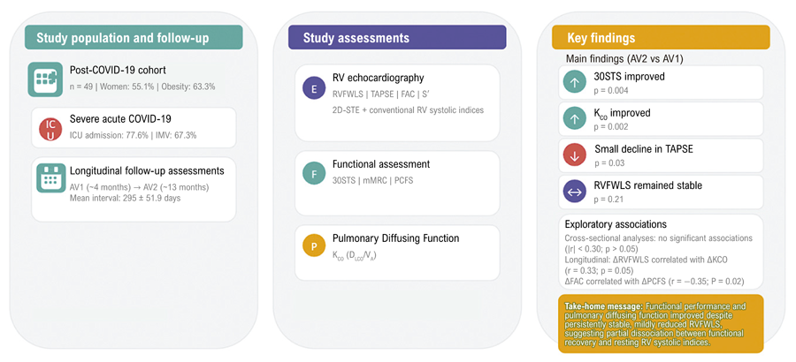
A total of 49 patients were included; 55.1% were women, the mean age was 50.7 ± 10.3 years, 63.3% had obesity, 77.6% required intensive care unit admission, and 67.3% underwent invasive mechanical ventilation. Performance on the 30STS improved (10.1 ± 3.3 vs 11.6 ± 3.1 repetitions; p = 0.004), and KCO increased (4.1 ± 0.7 vs 4.3 ± 0.8 mL/min/mmHg/L; p = 0.002), whereas mMRC scores (p = 0.43) and median PCFS scores (p = 0.11) remained unchanged. TAPSE decreased modestly but remained within the normal reference range (2.2 ± 0.3 vs 2.1 ± 0.2 cm; p = 0.03), while RVFWLS, FAC, and S′ showed no significant changes over time. Cross-sectional correlations between RV systolic indices and clinical outcomes were weak and nonsignificant at both AV1 and AV2 (|r| 0.05). Longitudinally, changes in RVFWLS were positively correlated with changes in KCO (r = 0.33; p = 0.05), whereas changes in FAC were inversely correlated with changes in PCFS scores (r = −0.35; p = 0.02).

Conclusios

In this cohort of post-COVID-19 survivors, functional capacity and pulmonary diffusing capacity improved over time, whereas RVFWLS remained persistently reduced without significant longitudinal change. These exploratory findings suggest that RV systolic indices should not be interpreted in isolation as markers of functional limitation during the late post-COVID-19 period and support the use of an integrated, multiparametric approach to patient assessment.
  • Echocardiography
  • Global Longitudinal Strain
  • Post-Acute COVID-19 Syndrome
  • Right Ventricular Dysfunction
Read Full Article
Creative Commons Attribution (CC BY) Creative Commons license
21/09/2026 DOI: 10.36660/abcimg.20260051i Views 2
Correlating Invasive and Echocardiographic Hemodynamic Measures in Patients with Heart Failure with Reduced Ejection Fraction
ABCIC
Original Article

Correlating Invasive and Echocardiographic Hemodynamic Measures in Patients with Heart Failure with Reduced Ejection Fraction

João Pedro da Rosa Barbato et al. Fernando Luis Scolari Guilherme Pinheiro Machado André Barcellos Amon … Pedro Castilhos de Freitas Crivelaro Willian R. Menegazzo Solano Vinicius Berger Gustavo Neves de Araujo Felipe Homem Valle Rodrigo Vugman Wainstein

Abstract

Background:

The management of heart failure with reduced ejection fraction (HFrEF) has advanced with the use of invasive hemodynamic markers such as cardiac power output (CPO) and pulmonary artery pulsatility index (PAPI). Simultaneously, interest has grown in noninvasive prognostic indicators, especially the left ventricular outflow tract velocity–time integral (LVOT-VTI).

Objective:

This study aimed to assess the correlation between invasive and echocardiographic parameters, particularly CPO, PAPI, and LVOT-VTI, in patients with HFrEF.

Methods:

This cross-sectional, single-center study included inpatients and outpatients with chronic stable or acutely decompensated HFrEF. Individuals with cardiogenic shock (CS) were excluded. Right heart catheterization (RHC) and echocardiography were performed on the same day. Statistical significance was defined as a two-sided p value < 0.05.

Results:

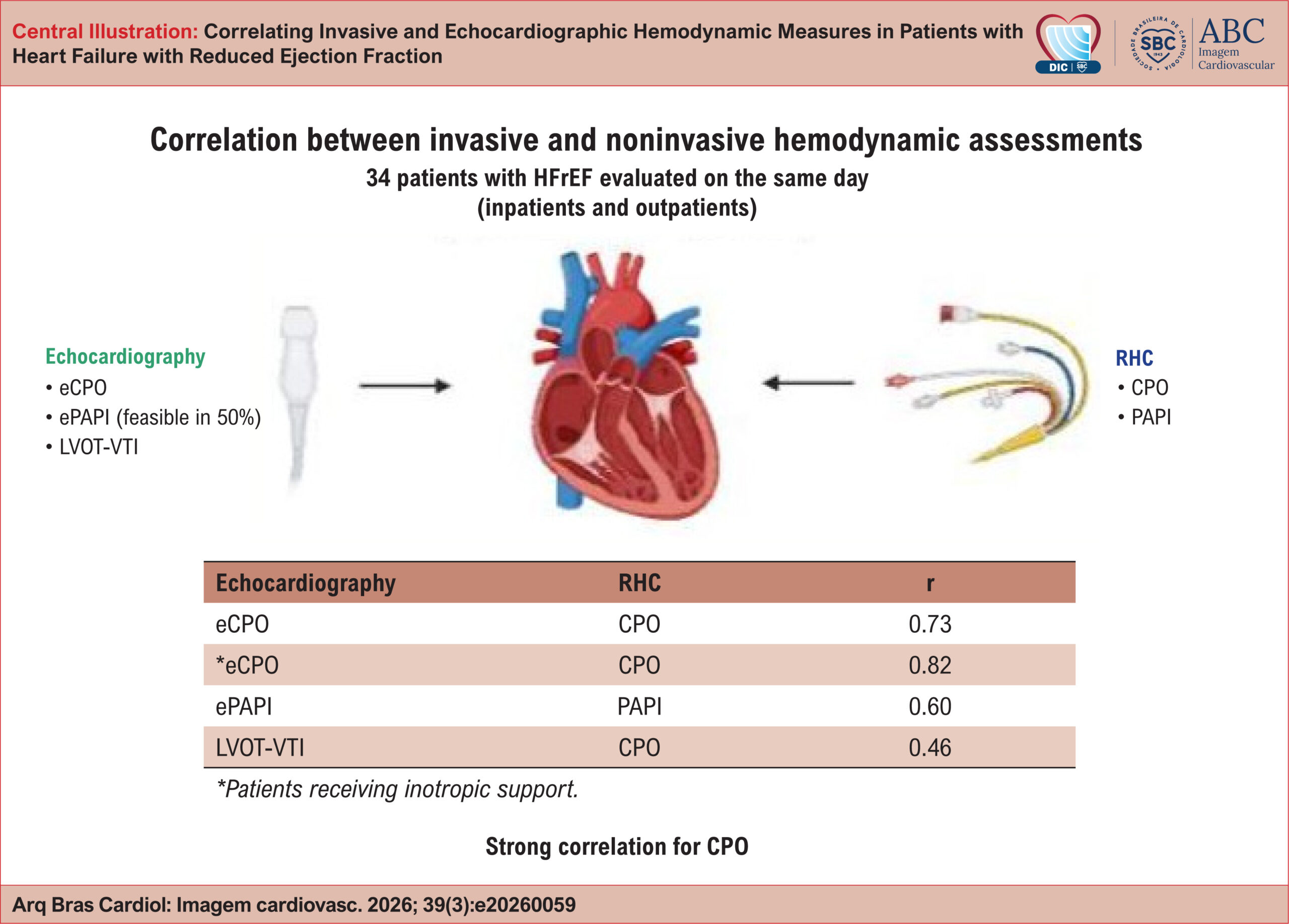
Significant correlations were observed between invasive and echocardiographic CPO (r = 0.737, p < 0.001) and PAPI (r = 0.604, p < 0.05). LVOT-VTI showed significant correlation with CPO measured via pulmonary artery catheter (PAC) (r = 0.469, p < 0.01). Echocardiographic PAPI was measurable in only 16 patients. A sensitivity analysis of those receiving inotropes showed an even stronger correlation between invasive and echocardiographic CPO (r = 0.812, p = 0.005).

Conclusion:

Echocardiographic and invasive hemodynamic parameters showed significant correlations in HFrEF, particularly CPO, supporting echocardiography as a potential alternative for hemodynamic assessment. However, the low feasibility of noninvasive PAPI measurement highlights important limitations in this population.

  • Echocardiography
  • Heart Rate
  • Hemodynamic Monitoring
  • Systolic Heart Failure
Read Full Article
Creative Commons Attribution (CC BY) Creative Commons license
03/09/2026 DOI: 10.36660/abcimg.20260059i Views 3
Multimodal Evaluation of Cardiac Myosin Inhibitor Response in Obstructive Hypertrophic Cardiomyopathy Using Echocardiography and Cardiopulmonary Exercise Testing: A Meta-Analysis A Meta-Analysis
ABCIC
Original Article

Multimodal Evaluation of Cardiac Myosin Inhibitor Response in Obstructive Hypertrophic Cardiomyopathy Using Echocardiography and Cardiopulmonary Exercise Testing: A Meta-Analysis A Meta-Analysis

João Victor de Oliveira Ramos et al. João Vitor Andrade Fernandes Luiz Henrique Cartaxo Fernandes Vera Louise Freire de Albuquerque Figueiredo … João Victor Oliveira Estrela Marcelo Tavares

Abstract

Background:

Cardiac Myosin Inhibitors (CMIs), including mavacamten and aficamten, represent a novel class of targeted therapies for obstructive Hypertrophic Cardiomyopathy (oHCM). Their effects on echocardiographic parameters and cardiopulmonary performance across Randomized Controlled Trials (RCTs) have not yet been systematically synthesized.

Objectives:

To conduct a systematic review and meta-analysis of RCTs in accordance with the PRISMA guidelines.

Methods:

Primary echocardiographic outcomes included Left Ventricular Ejection Fraction (LVEF), Left Ventricular Mass Index (LVMI), Interventricular Septal Thickness (IVST), E/e’ septal ratio, and resting, Valsalva, and post-exercise Left Ventricular Outflow Tract gradients (LVOT-G). Cardiopulmonary Exercise Testing (CPET) outcomes were also assessed. Data were pooled using random-effects models and reported as Mean Differences (MDs) or Standardized Mean Differences (SMDs), with 95% Confidence Intervals (CIs).

Results:

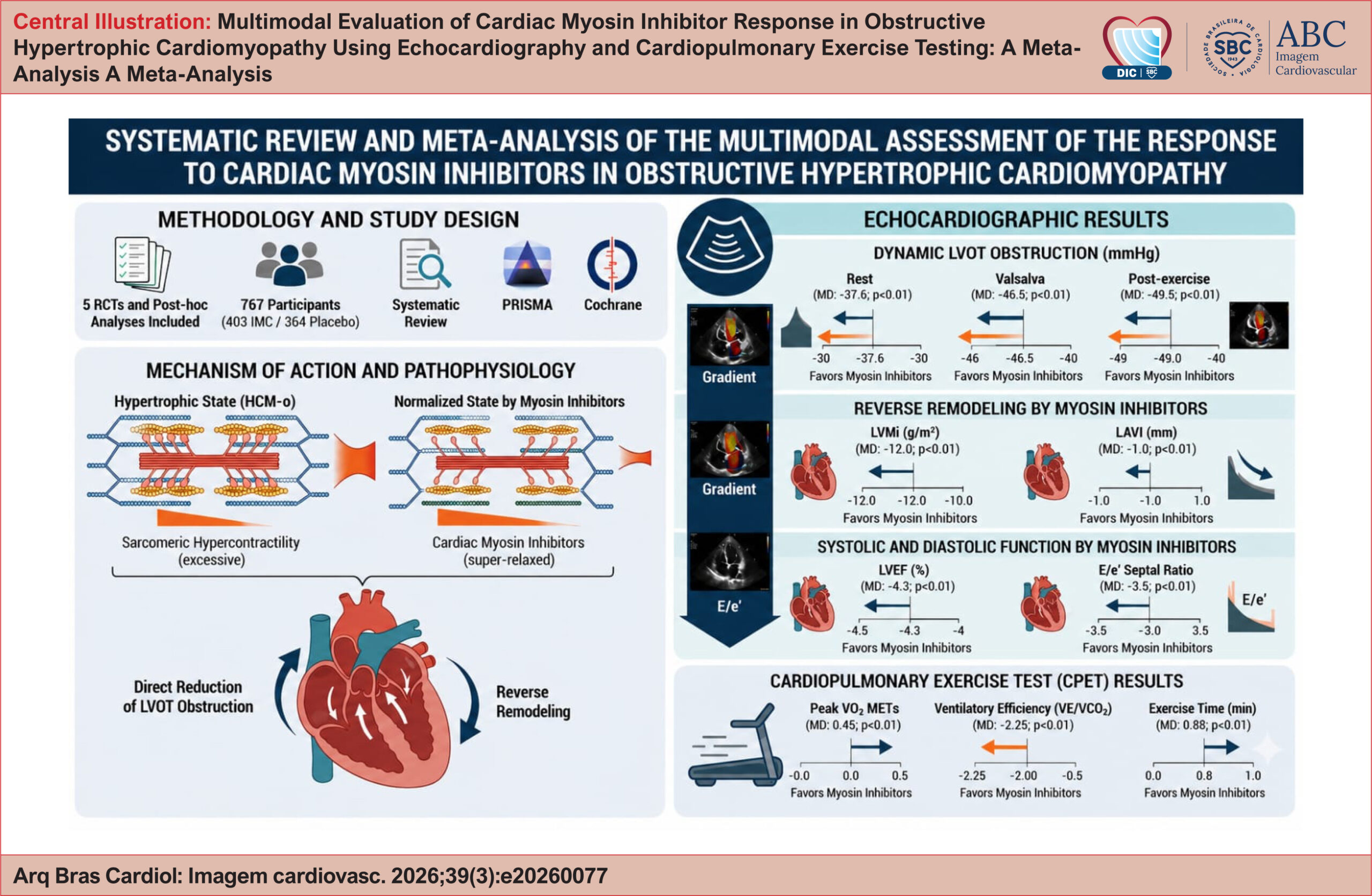
Five RCTs comprising 767 participants were included. CMIs significantly reduced resting LVOT-G (MD: −37.59 mmHg), Valsalva gradients (MD: −46.45 mmHg), and post-exercise gradients (MD: −49.45 mmHg; all p < 0.01). Reverse cardiac remodeling was reflected by reductions in LVMI (MD: −11.96 g/m²), IVST (SMD: −0.98), and E/e' septal ratio (MD: −3.53), along with an expected modest reduction in LVEF (MD: −4.33%). CPET outcomes also improved significantly, including ventilatory efficiency (MD: −2.25), peak metabolic equivalents (METs) (MD: +0.45), peak circulatory power (MD: +482.78 mL/kg/min × mmHg), and exercise duration (MD: +0.88 min; all p < 0.01). Subgroup analyses revealed no significant differences between the agents.

Conclusion:

CMIs promote reverse cardiac remodeling, improve hemodynamics, and enhance exercise tolerance in patients with oHCM, supporting their role as disease-modifying targeted therapies.

  • Echocardiography
  • Hypertrophic Cardiomyopathy
  • Meta-Analysis
Read Full Article
Creative Commons Attribution (CC BY) Creative Commons license
03/09/2026 DOI: 10.36660/abcimg.2026007i Views 2
Figure 1
ABCIC
Image

Beyond the Appendage: Left Atrial Wall Thrombosis in Rheumatic Mitral Stenosis

Bernardo Resende et al. Tomás Carlos Rogério Teixeira Lino Gonçalves

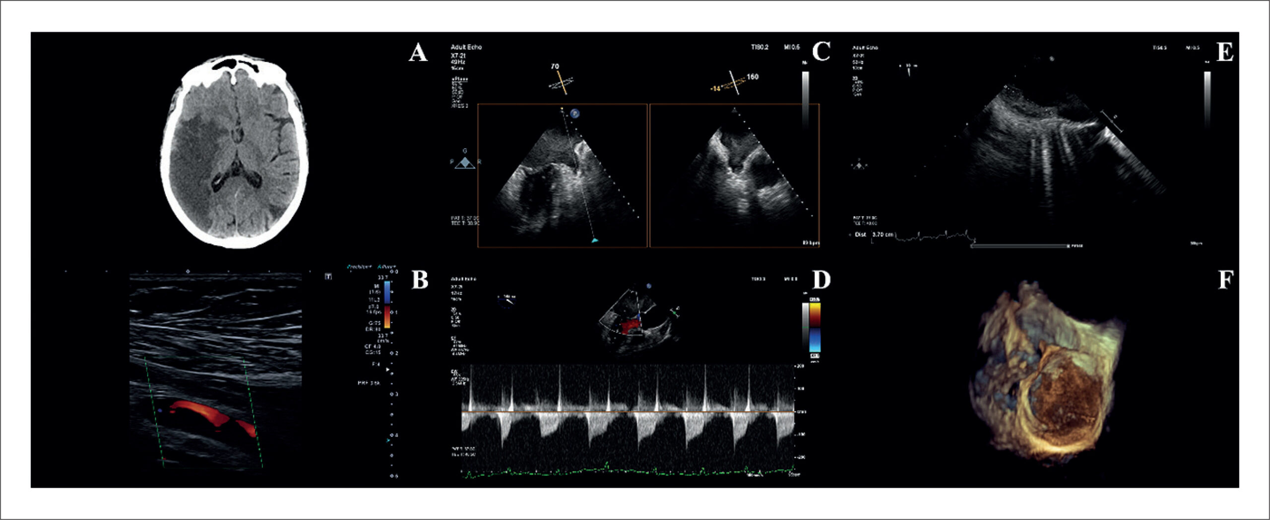
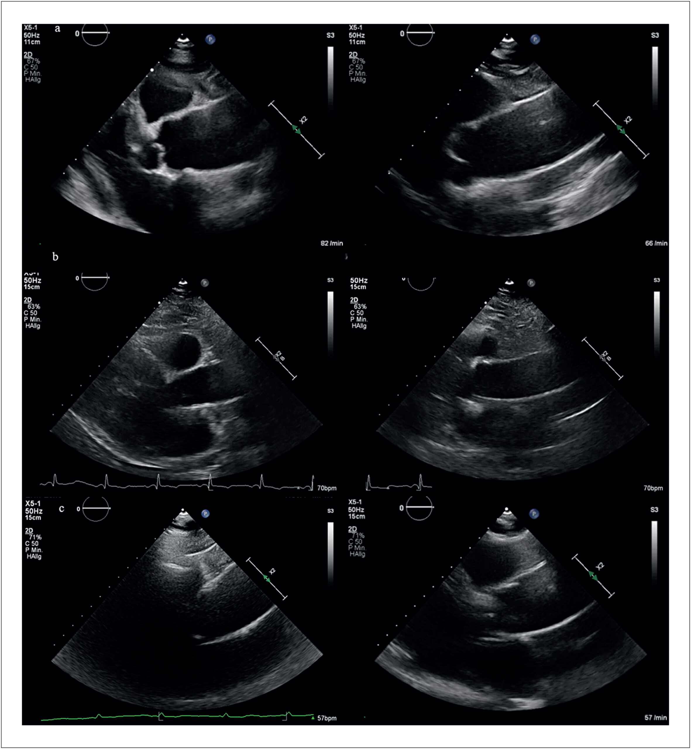
A 61-year-old woman with a history of rheumatic valvular heart disease and permanent atrial fibrillation (AF), chronically anticoagulated with rivaroxaban, presented with acute ischemic stroke and bilateral upper-limb ischemia. Neuroimaging revealed an acute infarction in the right middle cerebral artery territory and a subacute infarction in the left posterior cerebral artery territory (). Doppler ultrasound confirmed arterial occlusion in both upper limbs ().

Transesophageal echocardiography demonstrated marked left atrial (LA) enlargement, dense spontaneous echocardiographic contrast within the left atrial appendage (LAA), and reduced appendage flow velocities, without evidence of an organized thrombus (). The mitral valve exhibited features consistent with chronic rheumatic disease, with an estimated valve area of 1.0 cm² (). Notably, two- and three-dimensional imaging identified a large mural thrombus adherent to the LA wall, extending along the lateral and posterior walls and measuring approximately 4 × 0.8 cm (; Supplementary Videos 1-3).

[…]

  • Atrial Fibrillation
  • Echocardiography
  • Mitral Valve Stenosis
Read Full Article
Creative Commons Attribution (CC BY) Creative Commons license
14/08/2026 DOI: 10.36660/abcimg.20260069i Views 3
Figure 1
ABCIC
Case Report

Dilated Cardiomyopathy as a Rare Initial Manifestation of ANCA-positive Microscopic Polyangiitis: Case Report

Karoline Gonzaga Costa et al. Maria Estefânia Bosco Otto André Felipe Lobão Fernandes Nathália de Macêdo Assunção … Mariana Ubaldo Barbosa Paiva Rosyane Luz Rufino de Lima Rita Mikelle Soares Dias

Introduction

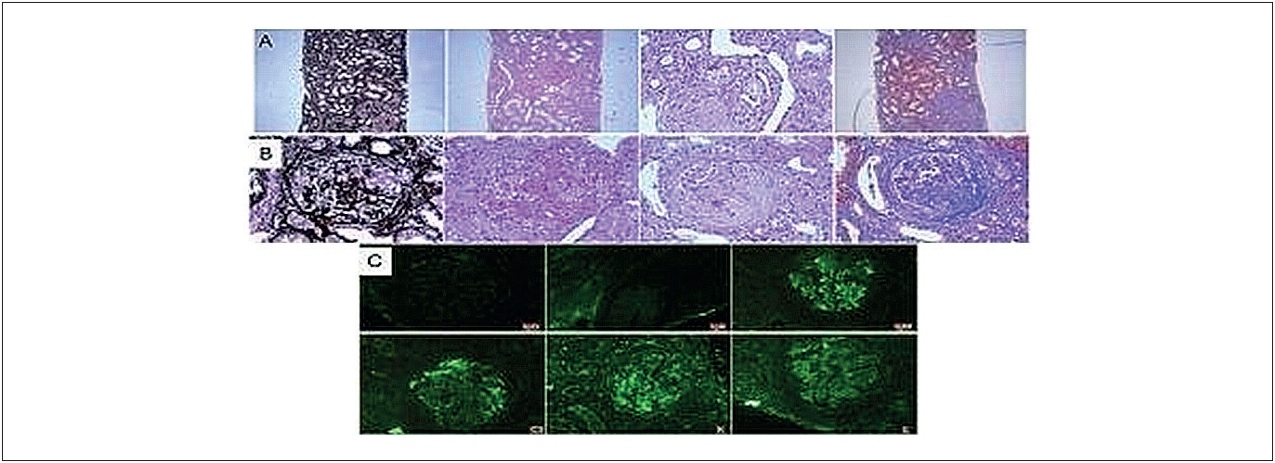
Antineutrophil cytoplasmic antibody (ANCA)-associated vasculitis constitute a group of systemic small-vessel inflammatory diseases characterized by pauci-immune necrotizing vasculitis and multisystem involvement, including granulomatosis with polyangiitis (GPA), eosinophilic granulomatosis with polyangiitis (EGPA), and microscopic polyangiitis (MPA).– MPA is classically associated with rapidly progressive glomerulonephritis and alveolar hemorrhage, conditions that carry high morbidity and mortality when not promptly recognized and treated.,

In addition to predominant renal and pulmonary involvement, there is growing evidence that patients with ANCA-associated vasculitis have an increased cardiovascular risk, related not only to traditional factors but also to disease-specific mechanisms such as persistent inflammation, endothelial dysfunction, and accelerated atherosclerosis.– Studies suggest a higher incidence of major cardiovascular events (myocardial infarction, stroke, and heart failure) compared with the general population.

[…]

  • Anti-Neutrophil Cytoplasmic Antibody-Associated Vasculitis
  • Dilated Cardiomyopathy
  • Echocardiography
  • Microscopic Polyangiitis
Read Full Article
Creative Commons Attribution (CC BY) Creative Commons license
31/07/2026 DOI: 10.36660/abcimg.20260038i Views 1
Recognition of Clinical Stages and Myocardial Damage in Aortic Stenosis: Beyond Diagnosis – To Op or Not To Op
ABCIC
Review Article

Recognition of Clinical Stages and Myocardial Damage in Aortic Stenosis: Beyond Diagnosis – To Op or Not To Op

Daniel Franca Vasconcelos

Abstract

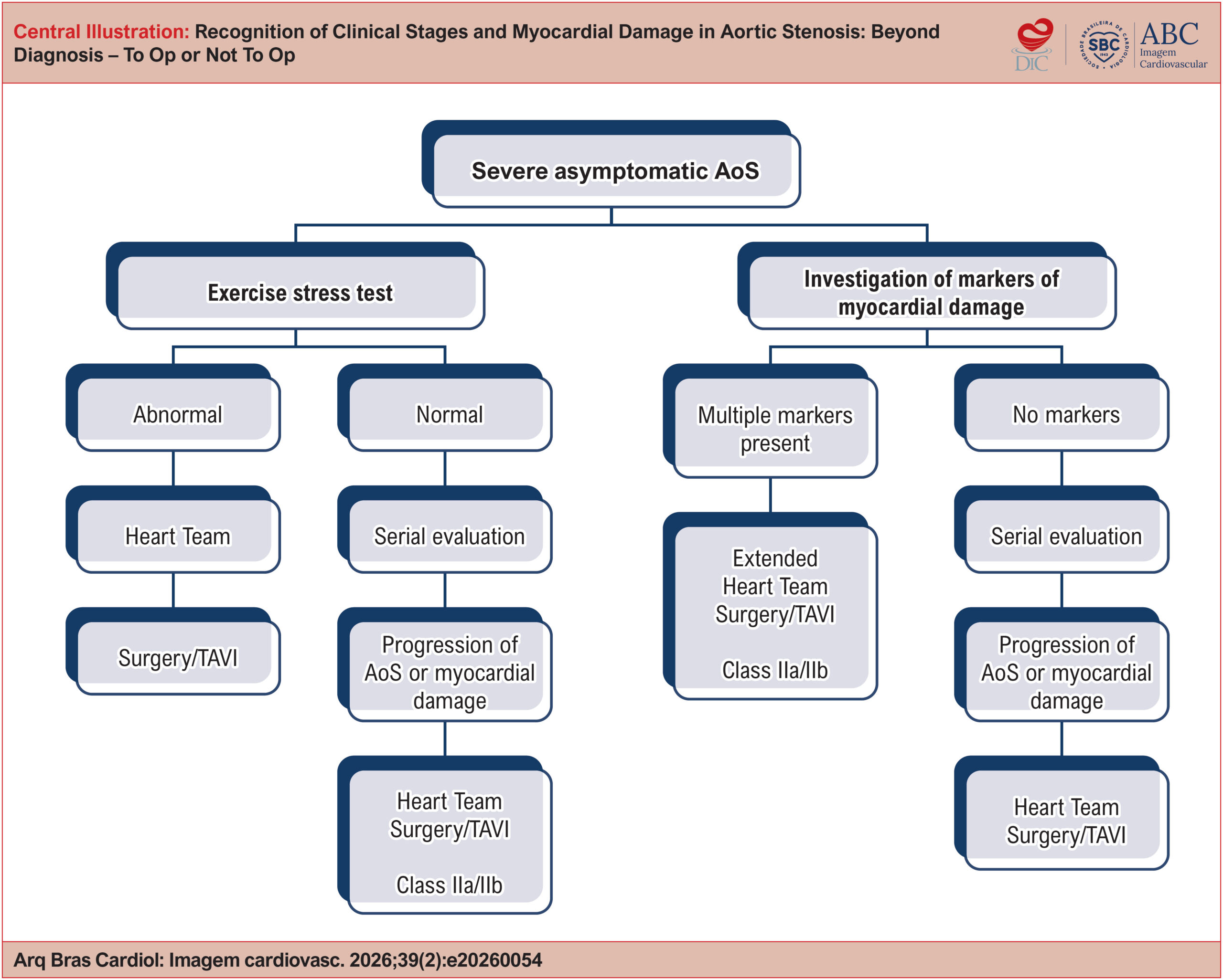
Severe aortic stenosis is a progressive clinical entity that has historically developed alongside descriptions of pathological anatomy, cardiovascular semiology, heart surgery, and percutaneous cardiovascular interventions. Indication for valve replacement in patients with severe aortic stenosis is still based on symptom onset. With advances in surgical techniques, especially transcatheter implantation, the focus has shifted to identifying early markers of myocardial damage that may indicate irreversible myocardial injury. This review article seeks to provide a comprehensive analysis of methods for identifying myocardial damage in patients with severe aortic stenosis, including clinical evaluation, electrocardiography (ECG), echocardiography, cardiac magnetic resonance imaging (CMR), and plasma biomarkers. It also discusses the staging of myocardial damage and the optimal timing for therapeutic intervention based on clinical, functional, and structural findings. Recent studies designed to address how to manage asymptomatic patients with aortic stenosis have yielded divergent results. Accordingly, the aim is to assist clinicians in deciding the optimal timing for valve replacement, taking individual patient characteristics into account.

  • Aortic Valve Stenosis
  • Echocardiography
  • Magnetic Resonance Imaging
Read Full Article
Creative Commons Attribution (CC BY) Creative Commons license
31/07/2026 DOI: 10.36660/abcimg.20260054i Views 3
Figure 1
ABCIC
Brief Communication

Visualization of the Ascending Aorta by Transthoracic Echocardiography: Could a Modified Parasternal Long-Axis View Provide Additional Imaging of a Longer Aortic Segment?

Heba Farouk et al. Karim El-Chilali Axel Kloppe

Introduction

Assessment of the ascending aorta is an essential component of standard transthoracic echocardiography (TTE). While visualization of the aortic root and proximal ascending aorta is routinely achieved using the standard parasternal long-axis view (PLAX), imaging of the mid and distal ascending aorta remains technically challenging.– Current recommendations suggest moving the transducer to upper left intercostal spaces or alternatively using right parasternal windows to improve visualization of these segments in patients with adequate acoustic windows.

In clinical practice, experienced echocardiographers often obtain satisfactory visualization of the tubular ascending aorta using individualized modifications of conventional views. However, less experienced operators and general cardiologists may fail to consistently image these segments because the upper left parasternal long-axis view (uPLAX) is poorly described in the literature, whereas acquisition of the right parasternal view is more technically demanding and time-consuming.

[…]

  • Aortic imaging
  • Ascending aorta
  • Echocardiography
Read Full Article
Creative Commons Attribution (CC BY) Creative Commons license
31/07/2026 DOI: 10.36660/abcimg.20260068i Views 3
Echocardiographic Changes Associated With Sacubitril/Valsartan Therapy in Patients With Heart Failure With Reduced Ejection Fraction (HFrEF): A Descriptive 12-Month Follow-Up Analysis
IJCS
Original Article

Echocardiographic Changes Associated With Sacubitril/Valsartan Therapy in Patients With Heart Failure With Reduced Ejection Fraction (HFrEF): A Descriptive 12-Month Follow-Up Analysis

Marcely Bonatto et al. Ana Karyn Ehrenfried de Freitas Caio Berwanger Takatuzi Luiz Gustavo Esmanhoto Franco … Danielle Rodrigues Vendramini Luka David Lechinewski Gustavo Gavazzoni Blume Lidia Zytynski Moura

Abstract

Background:

Although randomized controlled trials have demonstrated the efficacy of Sacubitril/Valsartan in heart failure (HF), real-world evidence regarding its impact on cardiac remodeling, particularly in Heart Failure With Reduced Ejection Fraction (HFrEF), remains limited.

Objectives:

This real-world study aimed to evaluate echocardiographic changes, including ejection fraction (EF), in a cohort of patients with HFrEF treated at a public hospital in Curitiba, Brazil, and to assess clinical outcomes at 6 and 12 months of follow-up.

Methods:

Longitudinal data were collected from 200 patients with HFrEF at baseline and at 6- and 12-month follow-up. Echocardiographic assessments included EF, left ventricular (LV) dimensions, and other cardiac parameters. Clinical outcomes included HF-related hospitalizations and New York Heart Association (NYHA) functional class.

Results:

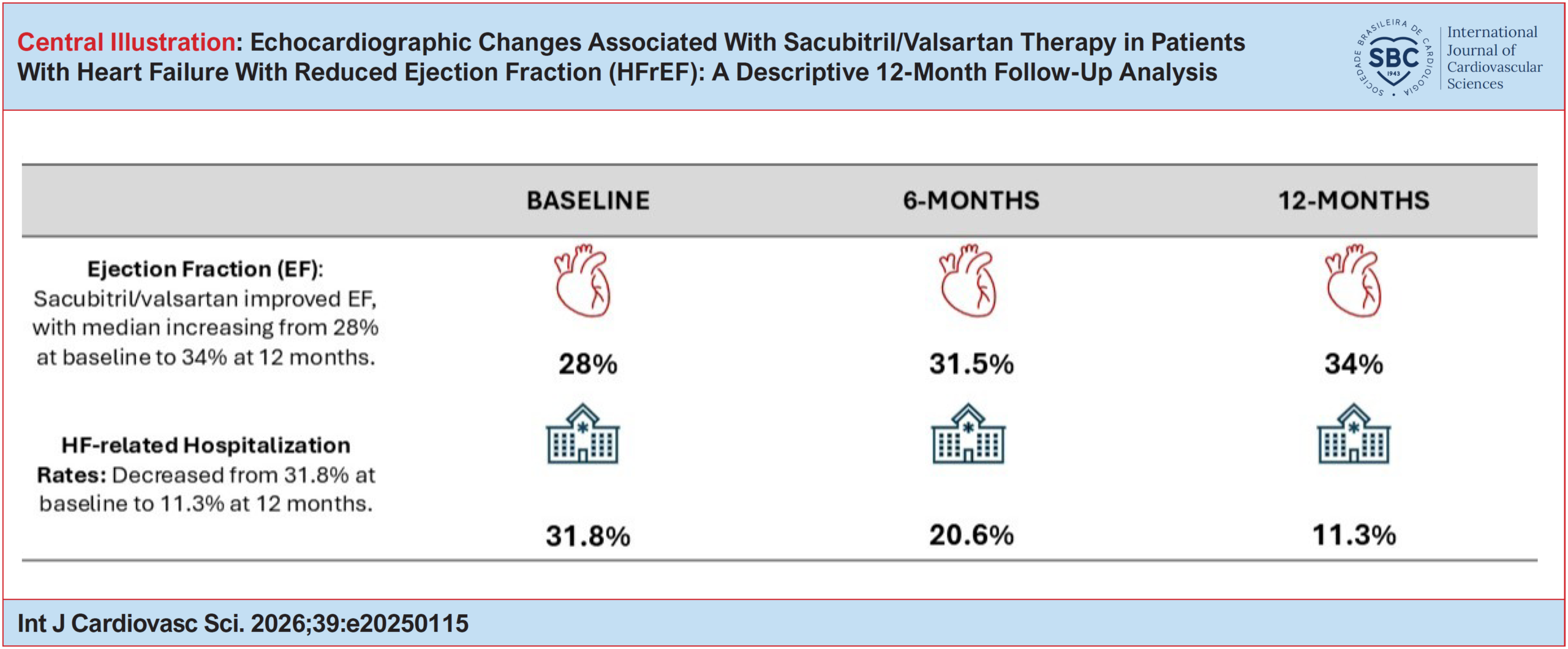
Sacubitril/Valsartan therapy was associated with improvements in EF, with the median increasing from 28% at baseline to 31.5% at 6 months and 34% at 12 months (Teichholz method). Structural remodeling was observed, including reductions in LV end-systolic and end-diastolic dimensions, as well as left atrial (LA) diameter. HF-related hospitalization rates decreased from 31.8% at baseline to 20.6% at 6 months and 11.3% at 12 months. NYHA functional class improved, with the proportion of patients in Classes III and IV decreasing from 53.2% at baseline to 17.4% at 6 months. Renal function remained stable throughout treatment. Improvements in EF were associated with a lower risk of hospitalization at 6 months (OR = 0.95) and 12 months (OR = 0.93), whereas a larger LA diameter was associated with a higher risk of hospitalization at 12 months (OR = 1.06).

Conclusions:

Sacubitril/Valsartan therapy was associated with improvements in EF, echocardiographic parameters, and NYHA functional class, as well as reductions in hospitalization rates. These findings underscore the role of the therapy in promoting reverse remodeling and suggest that its clinical benefits may extend beyond direct effects on cardiac structure.

  • Echocardiography
  • Heart Failure
  • Systolic Heart Failure
  • Ventricular Remodeling
Read Full Article
Creative Commons Attribution (CC BY) Creative Commons license
27/07/2026 DOI: 10.36660/ijcs.20250115 Views 3
Concordance Between Echocardiographic Left Ventricular Ejection Fraction by Simpson's Method, Global Longitudinal Strain, and Cardiac Magnetic Resonance
ABCIC
Original Article

Concordance Between Echocardiographic Left Ventricular Ejection Fraction by Simpson's Method, Global Longitudinal Strain, and Cardiac Magnetic Resonance

Álvaro Herrera-Escandón et al. Juan Pablo Morales-Grisales Sebastián Ayala-Zapata Stephany Barbosa-Balaguera … Álvaro José Muriel-Ruiz Juan Felipe Bravo-Rueda José Eduardo Citelli-Ramírez Luis Fernando Osío-Jimenez Luis Miguel Benitez-Gómez Carlos Javier Ramírez-Estupiñán

Abstract

Background:

Left ventricular ejection fraction (LVEF) measured by echocardiography is a widely used parameter in clinical practice for the assessment of ventricular function. More recently, global longitudinal strain (GLS) has emerged as a complementary method, as has the assessment of LVEF by cardiac magnetic resonance (CMR). However, regional evidence evaluating the concordance among these three techniques remains limited.

Objectives:

To assess the concordance between echocardiographic and CMR measurements in patients treated at a cardiovascular clinic in the city of Cali, Colombia.

Methods:

This cross-sectional, analytical, descriptive study included 35 patients with confirmed or suspected heart disease, in whom all three methods were performed consecutively. Concordance was evaluated using Lin’s concordance correlation coefficient (CCC), Bland-Altman plots for LVEF, and linear and quadratic weighted κ coefficients for agreement between LVEF classifications.

Results:

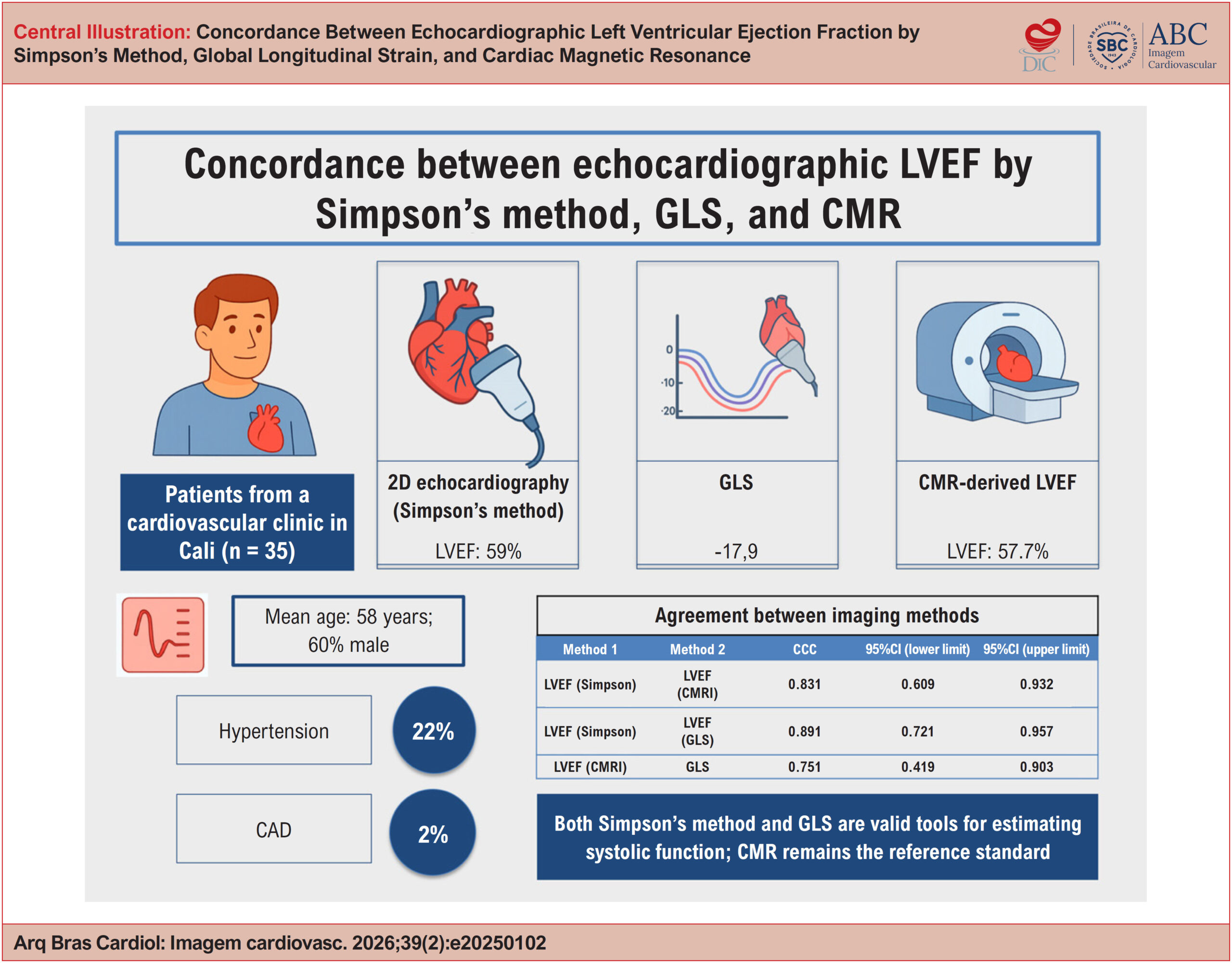
The mean age was 58 years, and 60% of participants were male. The most common comorbidities were hypertension (22%) and dyslipidemia (11%). The mean LVEF was 59% by Simpson’s method and 57.7% by CMR, while the mean GLS was −17.7%. Concordance was as follows: Simpson’s LVEF vs. CMR (CCC, 0.831; 95%CI, 0.609-0.932); GLS vs. CMR-derived LVEF (CCC, 0.751; 95%CI, 0.419-0.903); and Simpson’s LVEF vs. GLS (CCC, 0.891; 95%CI, 0.721-0.957).

Conclusions:

Both Simpson’s method and GLS are valid tools for estimating systolic function. CMR remains the reference standard.

  • Echocardiography
  • Magnetic Resonance Spectroscopy
  • Stroke Volume
Read Full Article
Creative Commons Attribution (CC BY) Creative Commons license
24/07/2026 DOI: 10.36660/abcimg.20250102i Views 1
My Approach to Echocardiographic Assessment of Left Ventricular Filling Pressures: From Ambiguity to Precision With New Guidelines
ABCIC
Review Article

My Approach to Echocardiographic Assessment of Left Ventricular Filling Pressures: From Ambiguity to Precision With New Guidelines

Marco Stephan Lofrano Alves et al. Larissa Maria Vosgerau Marcelo Vitola Dreckmann Roberto D´Ávila Martins … Cláudia Biondo Zanlorensi Eduardo Henrique Bonotto

Abstract

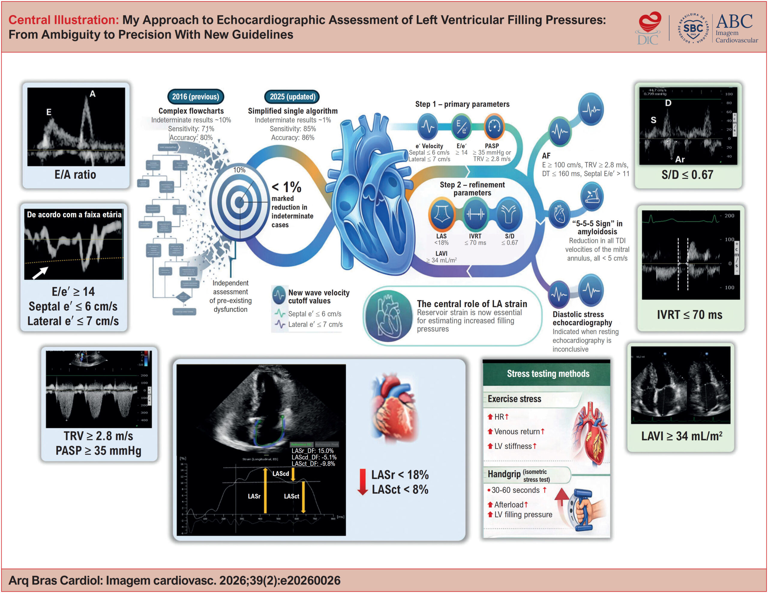
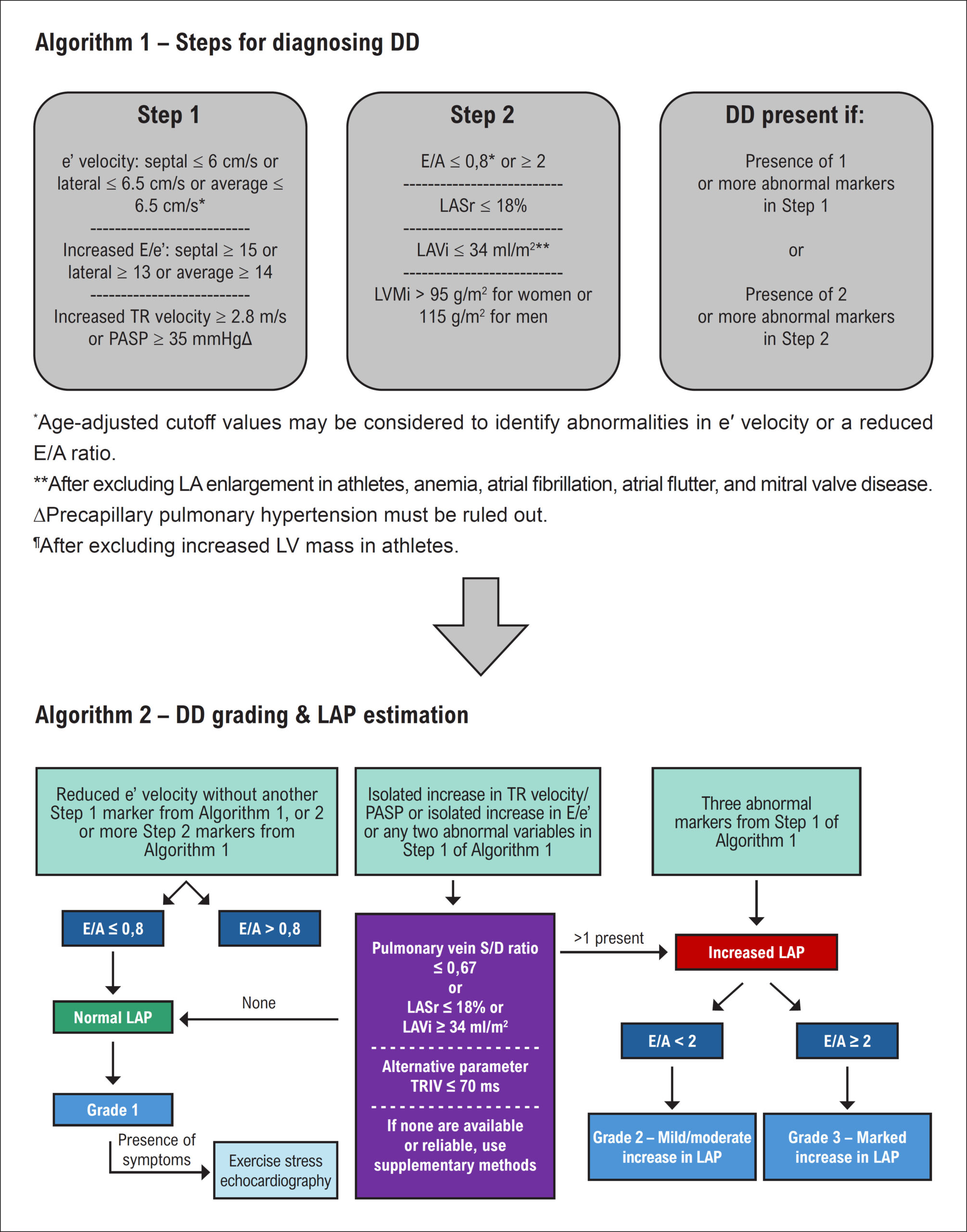
The echocardiographic estimation of left ventricular (LV) filling pressures is a cornerstone in the assessment of heart failure, particularly in patients with heart failure with preserved ejection fraction (HFpEF). The 2016 American Society of Echocardiography/European Association of cardiovascular Imaging algorithm standardized this evaluation using key variables, but a substantial proportion of cases remained indeterminate in clinical practice. The recent update of the American guideline reorganized the approach into a hierarchical framework, incorporating age-related adjustments and formalizing the role of left atrial strain (LAS) as a tie-breaking parameter, while also providing a more detailed characterization of special clinical scenarios. In parallel, the 2024 British Society of Echocardiography guideline emphasizes the pathophysiological interpretation of ventricular filling, complementing the operational framework proposed by the American document. Multicenter studies with invasive validation support these updates, establishing LAS as a robust marker of increased filling pressures. In this article, we present My Approach to echocardiographic assessment of LV filling pressures, based on an initial morphofunctional evaluation, followed by structured screening (Step 1) and further refinement (Step 2) incorporating LAS and additional parameters. We also provide a comparison between guidelines, discuss common pitfalls and algorithm limitations, and include case-based videos for practical application.

  • Echocardiography
  • Heart Failure
  • Left Ventricular Function
Read Full Article
Creative Commons Attribution (CC BY) Creative Commons license
24/07/2026 DOI: 10.36660/abcimg.20260026i Views 1
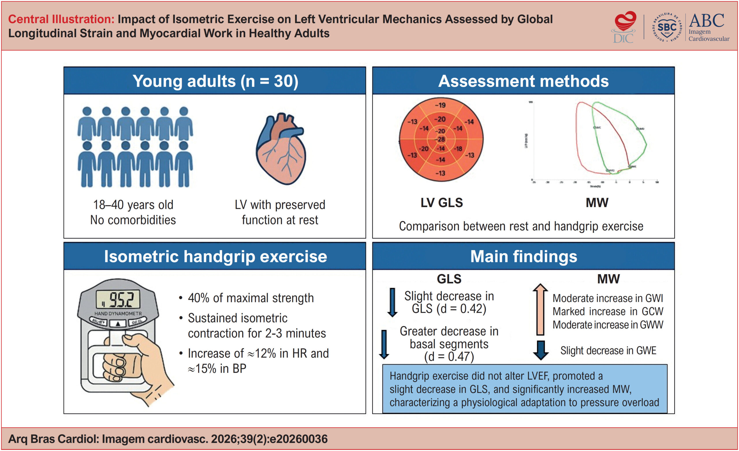
Impact of Isometric Exercise on Left Ventricular Mechanics Assessed by Global Longitudinal Strain and Myocardial Work in Healthy Adults
ABCIC
Original Article

Impact of Isometric Exercise on Left Ventricular Mechanics Assessed by Global Longitudinal Strain and Myocardial Work in Healthy Adults

Marcio Mendes Pereira et al. Maria Estefania Bosco Otto Juliana Lins da Paz Portela

Abstract

Background:

Traditional volumetric parameters have limitations in detecting subtle left ventricular (LV) systolic dysfunction. Global longitudinal strain (GLS) and myocardial work (MW) allow a more sensitive assessment of ventricular mechanics.

Objectives:

To evaluate changes in GLS and MW indices during isometric handgrip exercise compared with resting conditions.

Methods:

A total of 30 healthy individuals (29.3 ± 6.1 years; 50% male) were included in the sample. Echocardiography was performed at rest and during handgrip exercise (30%-40% of maximal strength). GLS, LV ejection fraction (LVEF), and MW indices were assessed: i) global work index (GWI), ii) global constructive work (GCW), iii) global wasted work (GWW), and iv) global work efficiency (GWE). Comparisons were performed using paired tests. Statistical significance was set at p < 0.05.

Results:

Handgrip exercise increased both systolic blood pressure (115 ± 16 vs 133 ± 18 mmHg; p < 0.0001) and diastolic blood pressure (69 ± 9 vs 79 ± 13 mmHg; p = 0.0002), without significant changes in LVEF (64.8% vs 64.4%; p = 0.62). A decrease in GLS was observed (20.38% ± 2.57% vs 19.60% ± 2.52%; p = 0.028), along with increases in GWI (+244 mmHg%; p = 0.0002), GCW (+313 mmHg%; p < 0.0001), and GWW (+52 mmHg%; p = 0.0008) as well as a decrease in GWE (94.8% ± 1.8% vs 93.6% ± 2.5%; p = 0.022).

Conclusions:

Handgrip exercise induces measurable ventricular mechanical changes in healthy individuals, reflecting a physiological response to acute pressure overload.

  • Echocardiography
  • Exercise
  • Left Ventricular Dysfunction
Read Full Article
Creative Commons Attribution (CC BY) Creative Commons license
20/07/2026 DOI: 10.36660/abcimg.20260036i Views 2
ABCIC
Editorial

Chemotherapy-Induced Cardiotoxicity in the Pediatric Population: What Are the Unique Aspects of Cardiovascular Imaging Follow-Up?

Jéssica Laureano Martins et al. Maria do Carmo Menezes Bezerra Duarte Fabiana Gomes Aragão Magalhães Feitosa Maria Verônica Câmara dos Santos

Special considerations for the pediatric population,

The pediatric perspective in cardio-oncology presents particular challenges, given that cardiotoxicity related to cancer treatment is not limited to an acute event, but represents a dynamic process that can interfere with myocardial growth and maturation over time. The vulnerability of the developing heart, associated with early exposure to potentially cardiotoxic therapies, confers a prolonged risk of progressive myocardial dysfunction. In this context, the use of more sensitive, reproducible, and integrated cardiovascular imaging strategies becomes fundamental. The multimodal approach, with emphasis on echocardiography (and cardiac magnetic resonance imaging in selected situations), increases diagnostic accuracy and contributes to better risk stratification. Additionally, structured longitudinal follow-up, with interpretation based on clinical individualization, is essential for adequate follow-up of these patients.

Finally, the incorporation of genetic, clinical, and therapeutic factors into risk models, coupled with continuous imaging monitoring, represents a promising perspective for precision medicine, with the goal of reducing cardiovascular morbidity and mortality and improving the quality of life of pediatric cancer survivors.

[…]

  • Cardiotoxicity
  • Child
  • Drug Therapy
  • Echocardiography
  • Neoplasms
Read Full Article
Creative Commons Attribution (CC BY) Creative Commons license
20/07/2026 DOI: 10.36660/abcimg.20260044i Views 1
My Approach to Transcatheter Closure of Atrial Septal Defect: Step-by-Step and Current Contraindications
ABCIC
Review Article

My Approach to Transcatheter Closure of Atrial Septal Defect: Step-by-Step and Current Contraindications

Angele Azevedo Alves Mattoso et al. Joberto Pinheiro Sena Gilson Soares Feitosa Filho Maria Lúcia Duarte

Abstract

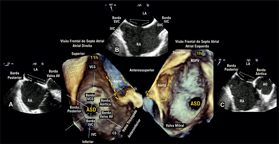
All patients who are candidates for transcatheter closure of an ostium secundum atrial septal defect (ASD) should undergo a detailed echocardiographic assessment using 2D transesophageal echocardiography (TEE) prior to the procedure. It is essential for a comprehensive analysis of interatrial septal anatomy and for determining defect eligibility for the procedure. The type, number, size, location and morphology of the ASD, as well as the quality of the surrounding septal tissue and its relationship with adjacent cardiac structures, should be carefully assessed. 3D TEE may complement this assessment by providing additional relevant anatomical information. During the procedure, TEE plays a fundamental role in real-time guidance, allowing proper deployment of the septal occluder device as well as immediate assessment of the result and early detection of potential complications. This article presents a practical approach to transcatheter closure of ostium secundum ASD, with emphasis on echocardiographic aspects in the pre-procedural, intraprocedural, and post-procedural phases.

  • Catheterization
  • Echocardiography
  • Heart Septal Defects, Atrial
Read Full Article
Creative Commons Attribution (CC BY) Creative Commons license
17/07/2026 DOI: 10.36660/abcimg.20260020i Views 1
Figure 1
ABCIC
Case Report

Single Coronary Artery and Stress Cardiomyopathy: An Association Demonstrated by Multimodality Imaging

Marcus Vinicius Silva Ferreira et al. Julia Pereira Afonso dos Santos Tormin Roberto Nery Dantas Jr Roberto Vitor Almeida Torres … Renan Andreos Cordeiro José de Arimateia Batista Araújo Filho Luiz Francisco Cardoso

Case Report

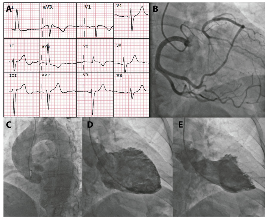
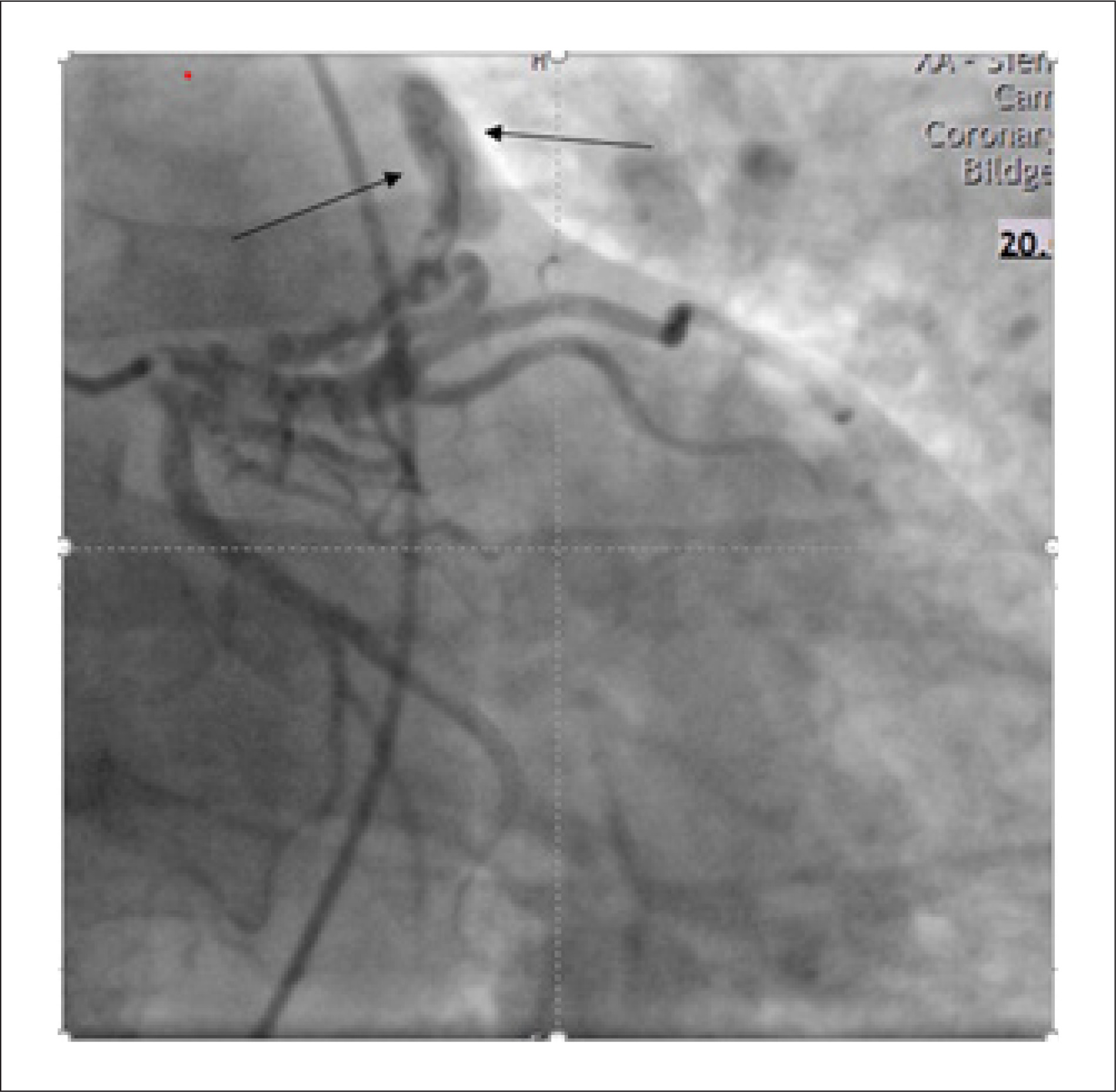
A 58-years-old female patient was admitted with retrosternal chest pain described as a continuous, oppressive tightness that had started two days earlier, after a tooth extraction procedure performed under ineffective local anesthesia. The patient’s past medical history included diabetes, dyslipidemia, and ovarian cancer, and she was receiving aspirin, rosuvastatin, metformin, dapagliflozin, and semaglutide. Physical examination was unremarkable. The chest pain protocol was initiated, and the initial electrocardiogram showed sinus rhythm with left anterior fascicular block, early repolarization in the inferior leads, and T-wave inversion in aVL, V1, and V2 (). The patient received 5 mg of sublingual isosorbide dinitrate, and blood samples were collected for laboratory testing. High-sensitivity troponin T (hs-cTnT) was 171 ng/L, confirming myocardial injury. An initial diagnosis of non–ST-elevation myocardial infarction (NSTEMI) was made, and the patient underwent coronary angiography (CA) for anatomic assessment ( / Video S1).

[…]

  • Coronary Angiography
  • Coronary Vessels
  • Echocardiography
  • Magnetic Resonance Imaging
  • Takotsubo Cardiomyopathy
Read Full Article
Creative Commons Attribution (CC BY) Creative Commons license
17/07/2026 DOI: 10.36660/abcimg.20260023i Views 1
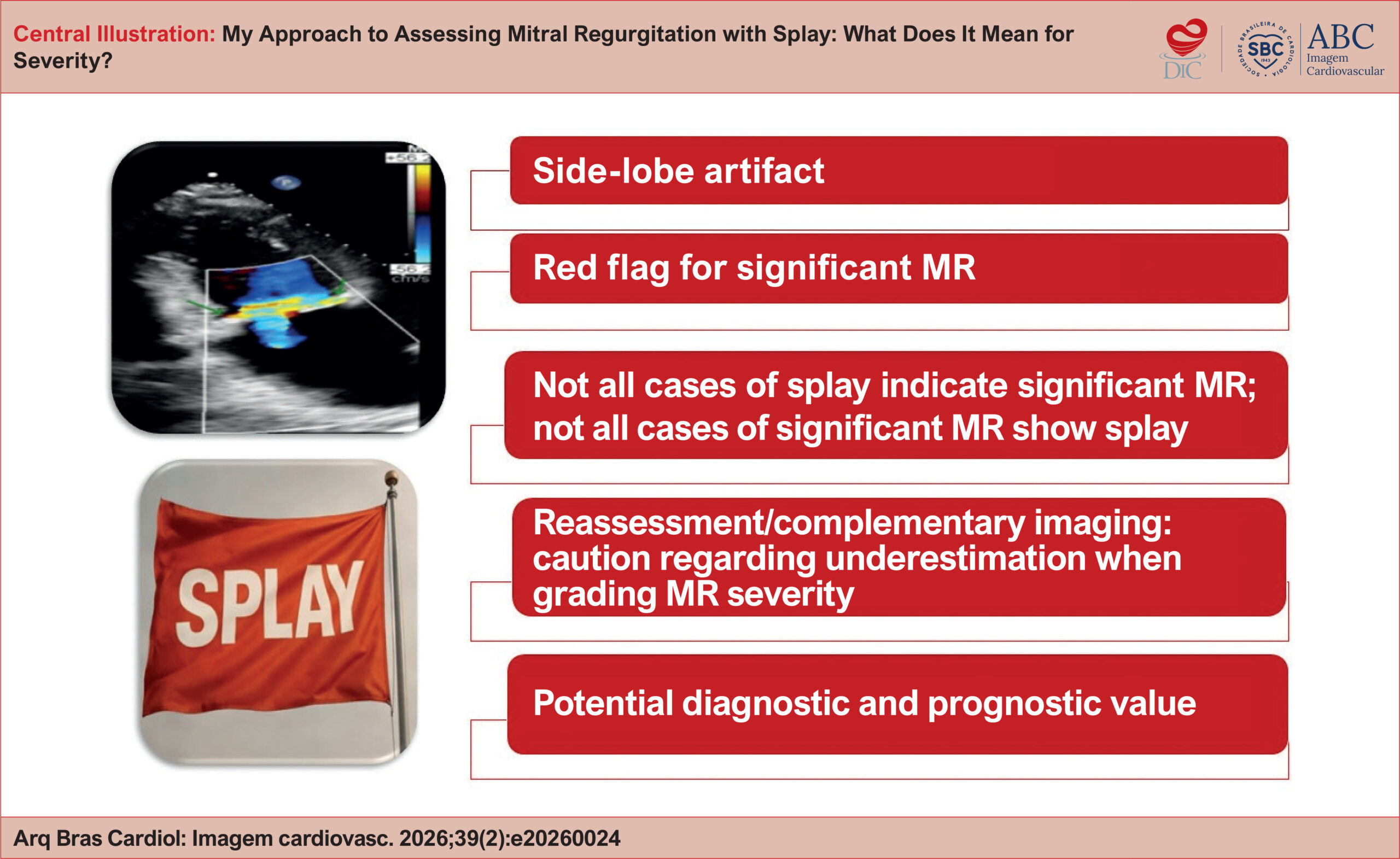
My Approach to Assessing Mitral Regurgitation with Splay: What Does It Mean for Severity?
ABCIC
Review Article

My Approach to Assessing Mitral Regurgitation with Splay: What Does It Mean for Severity?

Gustavo Nishida et al. Natasha Soares Simões dos Santos Jorge Eduardo Assef Andrea de Andrade Vilela

Abstract

Echocardiographic assessment of mitral regurgitation (MR) is multiparametric and often challenging. The artifact known as splay has been described as an additional tool for estimating the severity of regurgitation. The term refers to a side-lobe artifact that forms a horizontal arc on color Doppler imaging. This same phenomenon can also be observed in other valvular heart diseases, such as aortic regurgitation. In 2020, Wiener et al. reported that splay was present in 81% of cases of significant MR, reaching 93% in eccentric jets, whereas the prevalence was only 16% in mild MR. Verbeke et al. associated the artifact with larger regurgitant volumes. A splay width greater than 29 mm was identified as an independent predictor of adverse cardiovascular outcomes. Although its presence alone does not denote severe MR, splay acts as a red flag, suggesting that regurgitation may be greater than it appears and indicating the need for careful reassessment of the transthoracic echocardiogram or, possibly, complementary transesophageal echocardiography. Further evidence and systematic evaluations are needed to investigate the best strategy for incorporating these data into the multiparametric approach to MR.

  • Echocardiography
  • Mitral Valve Insufficiency
  • Severity of Illness Index
Read Full Article
Creative Commons Attribution (CC BY) Creative Commons license
10/07/2026 DOI: 10.36660/abcimg.20260024i
Figure 1
ABCIC
Case Report

Coexistence of Partial Anomalous Pulmonary Venous Connection and Coronary Artery Fistulas: A Rare Case Report

Mourad Haj Abdo et al. Hussain Latsh Mathias Wagner George König Sebastian Barth

Abstract

Background

Dyspnoea is a common clinical symptom that frequently prompts hospital admission and is associated with significant morbidity. While it most often results from prevalent cardiopulmonary conditions, rare congenital cardiovascular anomalies can also manifest with dyspnoea. Partial Anomalous Pulmonary Venous Connection (PAPVC) and Coronary Artery Fistulas (CAFs) are uncommon congenital malformations of the cardiovascular system, and their simultaneous presence is exceedingly rare. Early recognition of such anomalies is critical to avoid progressive hemodynamic compromise and to guide appropriate management strategies.

Case Presentation

We present the case of a 55-year-old man who experienced an acute onset of dyspnoea lasting approximately two hours. Initial clinical assessment and routine investigations—including physical examination, echocardiography, electrocardiography, and right and left heart catheterization—raised suspicion of an underlying cardiac abnormality, prompting further evaluation. Subsequent cardiac Magnetic Resonance Imaging (MRI) and Multi-Detector Computed Tomography (MDCT) revealed the presence of a Partial Anomalous Pulmonary Venous Connection (PAPVC) accompanied by Coronary Artery Fistulas (CAFs). Given the non-complex characteristics of the shunt in this case, a shared decision was reached with the patient to proceed with conservative management.

Discussion

PAPVC and Coronary Artery Fistulas CAFs are rare entities that should be considered in the differential diagnosis. However, recommending their evaluation as initial diagnostic hypotheses may result in unnecessary investigations.

  • Coronary Vessels
  • Differential Diagnosis
  • Echocardiography
Read Full Article
Creative Commons Attribution (CC BY) Creative Commons license
25/05/2026 DOI: 10.36660/abcimg.20250030i Views 1
Step-by-Step Approach to the Evaluation of Constrictive Pericarditis
ABCIC
Review Article

Step-by-Step Approach to the Evaluation of Constrictive Pericarditis

Aline Travessa et al. Paulo Henrique Pereira Louise Moutinho Machado Natalia Sousa Esteves

Abstract

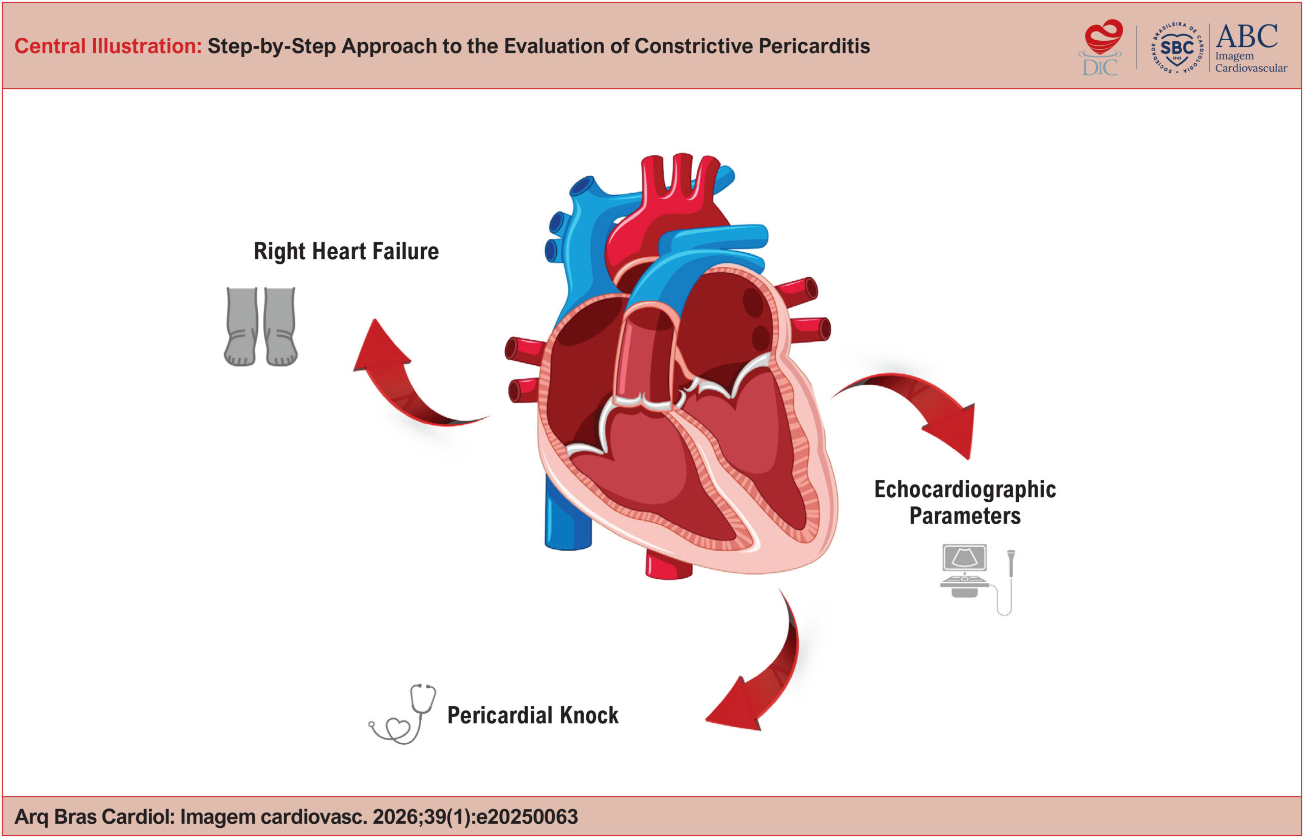
Constrictive Pericarditis (CP) is an uncommon but potentially curable condition, typically presenting with right-sided heart failure and impaired ventricular filling. Diagnosis can be challenging and requires a structured approach that integrates various cardiovascular imaging modalities. This article offers a practical guide, based on evidence and clinical experience, for the step-by-step recognition of Constrictive Pericarditis, highlighting echocardiographic findings.

  • Doppler Echocardiography
  • Echocardiography
  • Pericarditis
Read Full Article
Creative Commons Attribution (CC BY) Creative Commons license
25/05/2026 DOI: 10.36660/abcimg.20250063i
Cardiovascular Complications in Patients with COVID-19 and Their Relationship with Mortality: An Echocardiographic Perspective
ABCIC
Original Article

Cardiovascular Complications in Patients with COVID-19 and Their Relationship with Mortality: An Echocardiographic Perspective

João Henrique Andrade de Almeida et al. Gustavo Miranda de Azevedo Ferreira Marina de Azevedo Martins Andrade Heloísa Marceliano Nunes Igor Brasil Costa

Abstract

Background:

Echocardiography plays a fundamental role in the diagnosis of cardiovascular diseases related to SARS-CoV-2 infection. Despite prior investigation in the literature, studies focusing on cardiovascular manifestations among patients from the Brazilian Legal Amazon region remain scarce.

Objectives:

To identify cardiovascular complications of COVID-19 in patients admitted to a hospital in the North Region of Brazil and their potential association with mortality.

Methods:

We conducted a retrospective cohort study including 25 medical records of adults diagnosed with COVID-19. These patients were admitted to a hospital in Belém, Pará, in Northern Brazil, between March 2020 and December 2020. Demographic and clinical characteristics, as well as echocardiographic findings, were extracted from the medical records. Statistical analyses were performed adopting a significance level of α < 0.05. The project was approved by the Research Ethics Committee (approval number: 5.540.025).

Results:

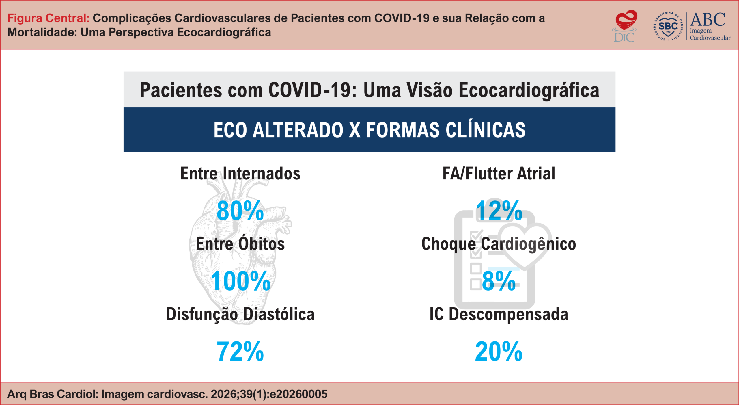
Among the 25 patients, 15 (60%) were male, with a mean age of 69.6 ± 14.4 years. Hypertension (n = 23; 92%) was the most prevalent cardiovascular risk factor, followed by diabetes (n = 11; 44%) and obesity (n = 8; 32%). We detected echocardiographic abnormalities in 20 (80%) individuals, with diastolic dysfunction being the most frequent (n = 18; 72%). All 7 patients who died showed abnormal echocardiography (p > 0.05). Atrial fibrillation or flutter (n = 5; 20%), decompensated heart failure (n = 3; 12%), and cardiogenic shock (n = 2; 8%) were the most common types of cardiovascular involvement.

Conclusions:

An increasing trend in COVID-19 mortality was observed among patients with cardiovascular complications. Nevertheless, the statistical power was limited by the small sample size.

  • Cardiovascular Diseases
  • COVID-19
  • Echocardiography
  • Mortality
Read Full Article
Creative Commons Attribution (CC BY) Creative Commons license
25/05/2026 DOI: 10.36660/abcimg.20260005i Views 3
Figure 1
ABC
Editorial

Echocardiographic Reference Values: Why Brazil Needs to Speak With its Own Voice

Imara Correia de Queiroz Barbosa et al. Israel Nilton de Almeida Feitosa

Echocardiographic measurements are cornerstones of diagnosis, risk stratification, and therapeutic decision-making across a wide range of cardiovascular conditions. Despite this, clinical practice in Brazil still relies largely on reference ranges derived from European and North American populations. In a country of continental dimensions, marked by substantial ethnic, anthropometric, and socioeconomic heterogeneity, such extrapolation raises legitimate concerns regarding diagnostic accuracy and equity in cardiovascular assessment.

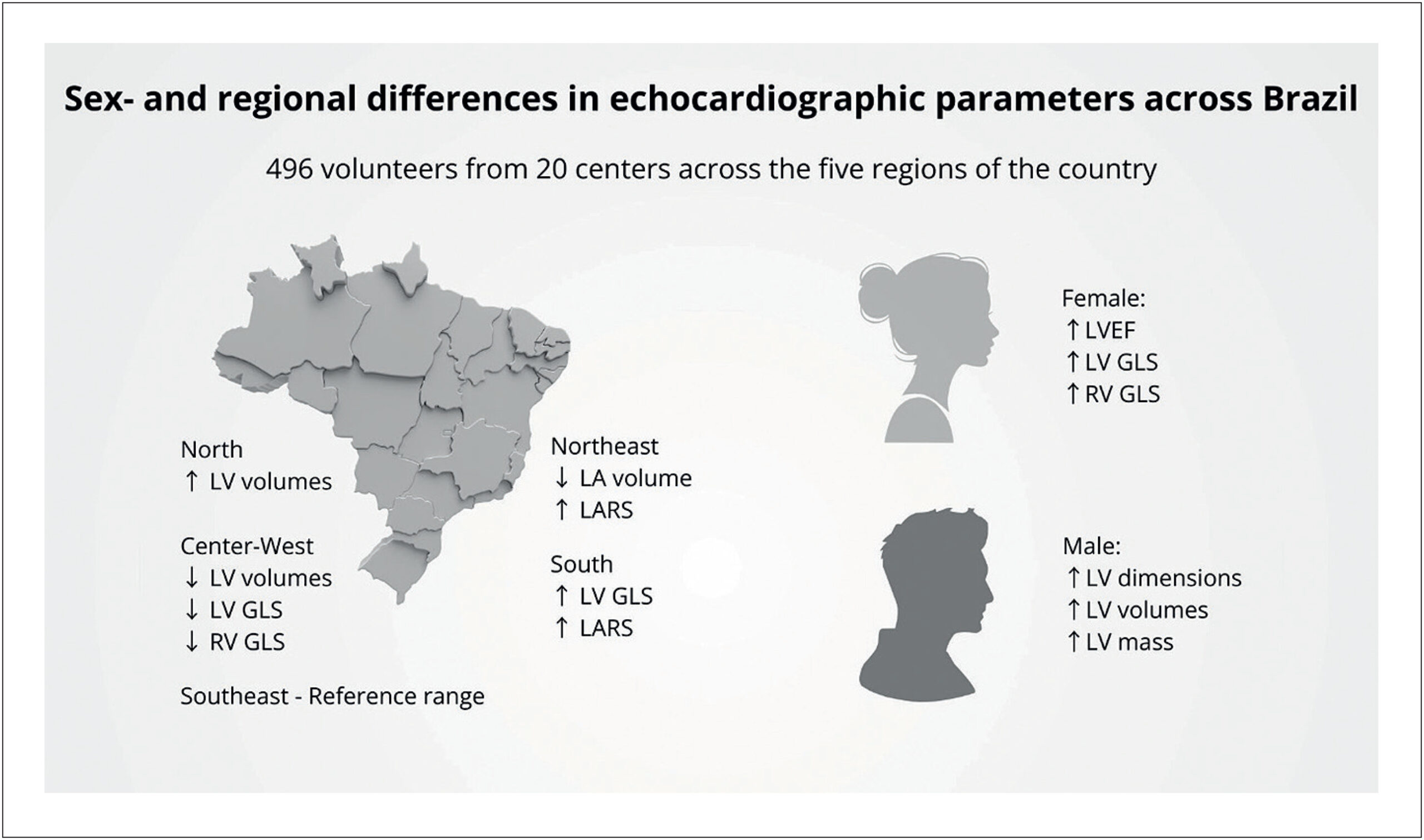
In this issue of the Arquivos Brasileiros de Cardiologia, Rodrigues et al. present a timely and methodologically sound study establishing contemporary reference values for echocardiographic parameters in healthy Brazilian adults. The inclusion of 496 volunteers recruited from 20 echocardiography laboratories across the five major geographic regions of the country provides unprecedented national representativeness and addresses a longstanding gap in Brazilian cardiology.

[…]

  • Echocardiography
  • Global Longitudinal Strain
  • Reference Values
Read Full Article
Creative Commons Attribution (CC BY) Creative Commons license
22/05/2026 DOI: 10.36660/abc.20260155i Views 2
Figure 1
ABC
Research Letter

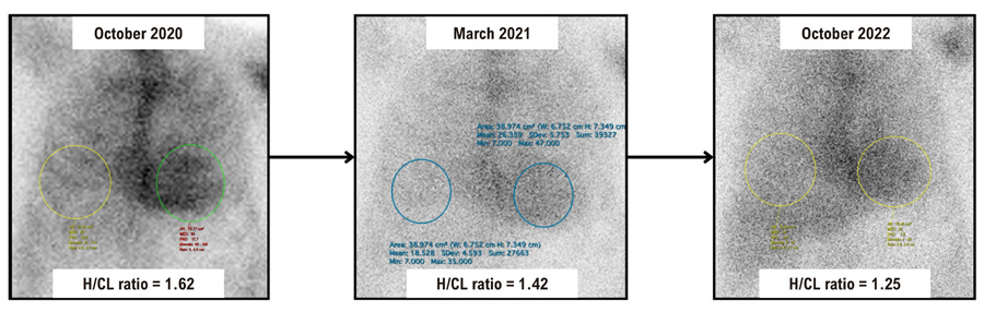
Biomarker–Imaging Dissociation in Wild-Type Cardiac Amyloidosis after Treatment with Patisiran

Claudio Tinoco Mesquita et al. Davi Shunji Yahiro Roberta Pereira dos Santos Coelho Giovane Leal de Azevedo Junior … Beatriz Robert Moreira Régina Limongi de Figueiredo Eliza Gripp Diane Xavier Ávila

Introduction

Amyloidosis is a progressive and complex disease caused by the extracellular deposition of insoluble fibrillar proteins in organs and tissues. Among the different types of amyloidosis, cardiac amyloidosis poses unique challenges due to its direct impact on cardiac function, structure, and electrical conduction. This condition can lead to restrictive cardiomyopathy, resulting in heart failure, arrhythmias, and other potentially life-threatening complications. Transthyretin amyloidosis (ATTR) is one of the more common forms of cardiac amyloidosis, occurring in both hereditary and non-hereditary forms. The hereditary form is caused by genetic mutations, whereas the non-hereditary form, known as wild-type transthyretin amyloidosis (ATTRwt), predominantly affects older adults and does not involve genetic mutations.

Recent therapeutic advances in the treatment of amyloidosis, particularly cardiac ATTRwt, have opened promising avenues for reducing amyloid burden in affected patients. Patisiran, an RNA interference (RNAi)-based therapy, targets the hepatic synthesis of transthyretin, thereby lowering circulating transthyretin protein levels available to form amyloid deposits in tissues. This mechanism represents a major advance because it directly reduces the source of the pathology rather than merely managing symptoms.

[…]

  • Amyloidosis
  • Biomarkers
  • Echocardiography
Read Full Article
Creative Commons Attribution (CC BY) Creative Commons license
11/05/2026 DOI: 10.36660/abc.20240757i Views 2
ABCIC
Editorial

Hypertrophic Cardiomyopathy: Standardization of Echocardiographic Assessment in an Era of New Therapies

Marcelo Goulart Paiva

Hypertrophic cardiomyopathy (HCM) has become the most common inherited myocardial disease, with an estimated global prevalence between 1:200 and 1:500. Despite its relatively high frequency in the general population, the disease remains significantly underdiagnosed. Only about 15% of affected individuals are clinically identified, due mainly to the wide variability of phenotypes and clinical manifestations., Slightly more than half of patients may develop progressive symptoms or experience adverse events throughout their lifetime. Early identification, risk stratification, and cardiovascular therapies and interventions have reduced mortality rates to < 1.0% per year.

The pathophysiology of HCM is based on myocardial hypertrophy in the absence of secondary causes, associated with hypercontractility and diastolic dysfunction, resulting from abnormal myosin activation. Approximately 75% of patients present with left ventricular outflow tract (LVOT) obstruction at rest or after provocative maneuvers. In the absence of an obstructive pattern, the disease course is usually favorable, oligosymptomatic, or asymptomatic, with a minority progressing to advanced stages.–

[…]

  • Echocardiography
  • Hypertrophic Cardiomyopathy
  • Left Ventricular Outflow Obstruction
Read Full Article
Creative Commons Attribution (CC BY) Creative Commons license
11/05/2026 DOI: 10.36660/abcimg.20260027i Views 2
Figure 1
ABCIC
Editorial

Recalibrating the Barometer: Echocardiography in Diastolic Dysfunction and the Era of New Algorithms

Maria Estefania Bosco Otto et al. Jorge Eduardo Assef Gustavo Nishida

Diastolic dysfunction (DD) remains a diagnostic challenge, not for lack of available parameters, but because uncertainty emerges when complex physiology is reduced to static labels in echocardiographic reports. In clinical practice, physicians are often less concerned with the specific grade of DD and more interested in its prognostic implications and in whether increased filling pressures may explain dyspnea, guide further investigation, and support therapeutic decisions. It is precisely in this variable, mean left atrial pressure (MLAP) and left ventricular (LV) filling pressure (LVFP), that echocardiography must be most pragmatic, minimizing the number of “indeterminate” reports and offering an operational conclusion grounded in integrated physiology.–

The structured reasoning that supports this goal began with the 2009 American Society of Echocardiography/European Association of Echocardiography guideline on the evaluation of LV diastolic function, a landmark document that organized modern thinking on diastole, established a shared language for key pathophysiological mechanisms, and systematized the interpretation of echocardiographic parameters. Beyond its conceptual value, it reinforced a critical principle for echocardiography laboratories: the assessment of diastole must translate into a clinically meaningful message, particularly when heart failure with preserved ejection fraction (HFpEF) is suspected.

[…]

  • Diastolic Heart Failure
  • Echocardiography
  • Left Ventricular Dysfunction
Read Full Article
Creative Commons Attribution (CC BY) Creative Commons license
20/04/2026 DOI: 10.36660/abcimg.20260004i Views 2
ABCIC
Editorial

Echocardiography with Ultrasound Enhancement Agents and the Diagnostic Challenge of Cardiac Masses: Solid Evidence for a Complex Clinical Problem

Rafael Bonafim Piveta et al. Miguel Osman Dias Aguiar

The proper characterization of intracardiac masses continues to be one of the most relevant challenges in contemporary cardiovascular imaging. Thrombi, benign tumors, and malignant neoplasms share some similar morphological characteristics in conventional echocardiography, but they entail radically different approaches, prognoses, and therapeutic urgencies. Despite significant advances in cardiovascular imaging diagnostic techniques, critical decisions, such as whether to anticoagulate or operate, investigate or observe, treat urgently or monitor, are still frequently challenging in clinical practice. Echocardiography, although indispensable as an initial method, often fails to differentiate thrombi, benign tumors, and malignant neoplasms in a significant number of patients. Given this scenario, it is legitimate to question: why does echocardiography with ultrasound enhancing agents (UEAs), available for decades, still play a secondary role in many diagnostic algorithms?

The meta-analysis, “Diagnostic performance of contrast-enhanced echocardiography in differentiating cardiac masses”, presents important arguments for this context. Using methodological rigor, aligned with PRISMA-DTA and Cochrane Handbook recommendations,, the authors demonstrate that echocardiography with UEAs has shown exceptional diagnostic performance in two of the most critical dilemmas in clinical practice: differentiating tumors from thrombi and distinguishing benign from malignant tumors.

[…]

  • cardiac masses
  • contrast
  • Echocardiography
Read Full Article
Creative Commons Attribution (CC BY) Creative Commons license
20/04/2026 DOI: 10.36660/abcimg.20260009i Views 1

Could not load more articles. Please try again.

How to cite this article

EndNote RIS

Electronic Document Format (Vancouver)

Machado GP, Telo GH, Araujo GN, Scolari FL, Silveira AD, Wainstein R, Wainstein M, et al. Point-of-Care Ultrasound in Cardiology: When a Positioning Statement Does Not Reflect the State of the Art. Arquivos Brasileiros de Cardiologia 2026;123(8):e20260312.

Electronic Document Format (ABNT)

Machado, Guilherme Pinheiro; Telo, Guilherme Heiden; Araujo, Gustavo Neves de; Scolari, Fernando Luis; Silveira, Anderson Donelli da; Wainstein, Rodrigo; Wainstein, Marco. Point-of-Care Ultrasound in Cardiology: When a Positioning Statement Does Not Reflect the State of the Art. Arquivos Brasileiros de Cardiologia, v. 123, n. 8, e20260312, Sep. 2026.

Electronic Document Format (APA)

Machado, G. P., Telo, G. H., Araujo, G. N. , Scolari, F. L., Silveira, A. D. , Wainstein, R., & Wainstein, M. (2026). Point-of-Care Ultrasound in Cardiology: When a Positioning Statement Does Not Reflect the State of the Art. Arquivos Brasileiros de Cardiologia, 123(8), e20260312.

Electronic Document Format (ISO)

Machado, Guilherme Pinheiro and Telo, Guilherme Heiden and Araujo, Gustavo Neves de and Scolari, Fernando Luis and Silveira, Anderson Donelli da and Wainstein, Rodrigo and Wainstein, Marco. Point-of-Care Ultrasound in Cardiology: When a Positioning Statement Does Not Reflect the State of the Art. Arquivos Brasileiros de Cardiologia [online]. 2026, vol. 123, n. 8, [cited 2026-10-14], e20260312. Available from: <https://journals.cardiol.br/en/article/point-of-care-ultrasound-in-cardiology-when-a-positioning-statement-does-not-reflect-the-state-of-the-art/>. ISSN 0066-782X

EndNote RIS

Electronic Document Format (Vancouver)

Rogelin M, Aranha AFMG, Fonseca FR, Silva RM, Fialho GL. Serial Echocardiographic Assessment of Right Ventricular Function and Its Association With Functional Capacity, Dyspnea, and Pulmonary Diffusion in Post-COVID-19 Survivors. ABC Imagem Cardiovascular 2026;39(3):e20260051.

Electronic Document Format (ABNT)

Rogelin, Marcelo; Aranha, Adriana Ferraz Martins Graça; Fonseca, Fernanda Rodrigues; Silva, Rosemeri Maurici da; Fialho, Guilherme Loureiro. Serial Echocardiographic Assessment of Right Ventricular Function and Its Association With Functional Capacity, Dyspnea, and Pulmonary Diffusion in Post-COVID-19 Survivors. ABC Imagem Cardiovascular, v. 39, n. 3, e20260051, Sep. 2026.

Electronic Document Format (APA)

Rogelin, M., Aranha, A. F. M. G., Fonseca, F. R., Silva, R. M. , & Fialho, G. L. (2026). Serial Echocardiographic Assessment of Right Ventricular Function and Its Association With Functional Capacity, Dyspnea, and Pulmonary Diffusion in Post-COVID-19 Survivors. ABC Imagem Cardiovascular, 39(3), e20260051.

Electronic Document Format (ISO)

Rogelin, Marcelo and Aranha, Adriana Ferraz Martins Graça and Fonseca, Fernanda Rodrigues and Silva, Rosemeri Maurici da and Fialho, Guilherme Loureiro. Serial Echocardiographic Assessment of Right Ventricular Function and Its Association With Functional Capacity, Dyspnea, and Pulmonary Diffusion in Post-COVID-19 Survivors. ABC Imagem Cardiovascular [online]. 2026, vol. 39, n. 3, [cited 2026-10-14], e20260051. Available from: <https://journals.cardiol.br/en/article/serial-echocardiographic-assessment-of-right-ventricular-function-and-its-association-with-functional-capacity-dyspnea-and-pulmonary-diffusion-in-post-covid-19-survivors/>. ISSN 2318-8219

EndNote RIS

Electronic Document Format (Vancouver)

Barbato JPR, Scolari FL, Machado GP, Amon AB, Crivelaro PCF, Menegazzo WR, Berger SV, et al. Correlating Invasive and Echocardiographic Hemodynamic Measures in Patients with Heart Failure with Reduced Ejection Fraction. ABC Imagem Cardiovascular 2026;39(3):e20260059.

Electronic Document Format (ABNT)

Barbato, João Pedro da Rosa; Scolari, Fernando Luis; Machado, Guilherme Pinheiro; Amon, André Barcellos; Crivelaro, Pedro Castilhos de Freitas; Menegazzo, Willian R.; Berger, Solano Vinicius; Araujo, Gustavo Neves de; Valle, Felipe Homem; Wainstein, Rodrigo Vugman. Correlating Invasive and Echocardiographic Hemodynamic Measures in Patients with Heart Failure with Reduced Ejection Fraction. ABC Imagem Cardiovascular, v. 39, n. 3, e20260059, Sep. 2026.

Electronic Document Format (APA)

Barbato, J. P. R., Scolari, F. L., Machado, G. P., Amon, A. B., Crivelaro, P. C. F., Menegazzo, W. R., Berger, S. V., Araujo, G. N. , Valle, F. H., & Wainstein, R. V. (2026). Correlating Invasive and Echocardiographic Hemodynamic Measures in Patients with Heart Failure with Reduced Ejection Fraction. ABC Imagem Cardiovascular, 39(3), e20260059.

Electronic Document Format (ISO)

Barbato, João Pedro da Rosa and Scolari, Fernando Luis and Machado, Guilherme Pinheiro and Amon, André Barcellos and Crivelaro, Pedro Castilhos de Freitas and Menegazzo, Willian R. and Berger, Solano Vinicius and Araujo, Gustavo Neves de and Valle, Felipe Homem and Wainstein, Rodrigo Vugman. Correlating Invasive and Echocardiographic Hemodynamic Measures in Patients with Heart Failure with Reduced Ejection Fraction. ABC Imagem Cardiovascular [online]. 2026, vol. 39, n. 3, [cited 2026-10-14], e20260059. Available from: <https://journals.cardiol.br/en/article/correlating-invasive-and-echocardiographic-hemodynamic-measures-in-patients-with-heart-failure-with-reduced-ejection-fraction/>. ISSN 2318-8219

EndNote RIS

Electronic Document Format (Vancouver)

Ramos JVO, Fernandes JVA, Fernandes LHC, Figueiredo VLFA, Estrela JVO, Tavares M. Multimodal Evaluation of Cardiac Myosin Inhibitor Response in Obstructive Hypertrophic Cardiomyopathy Using Echocardiography and Cardiopulmonary Exercise Testing: A Meta-Analysis A Meta-Analysis. ABC Imagem Cardiovascular 2026;39(3):e20260077.

Electronic Document Format (ABNT)

Ramos, João Victor de Oliveira; Fernandes, João Vitor Andrade; Fernandes, Luiz Henrique Cartaxo; Figueiredo, Vera Louise Freire de Albuquerque; Estrela, João Victor Oliveira; Tavares, Marcelo. Multimodal Evaluation of Cardiac Myosin Inhibitor Response in Obstructive Hypertrophic Cardiomyopathy Using Echocardiography and Cardiopulmonary Exercise Testing: A Meta-Analysis A Meta-Analysis. ABC Imagem Cardiovascular, v. 39, n. 3, e20260077, Sep. 2026.

Electronic Document Format (APA)

Ramos, J. V. O., Fernandes, J. V. A., Fernandes, L. H. C., Figueiredo, V. L. F. A., Estrela, J. V. O., & Tavares, M. (2026). Multimodal Evaluation of Cardiac Myosin Inhibitor Response in Obstructive Hypertrophic Cardiomyopathy Using Echocardiography and Cardiopulmonary Exercise Testing: A Meta-Analysis A Meta-Analysis. ABC Imagem Cardiovascular, 39(3), e20260077.

Electronic Document Format (ISO)

Ramos, João Victor de Oliveira and Fernandes, João Vitor Andrade and Fernandes, Luiz Henrique Cartaxo and Figueiredo, Vera Louise Freire de Albuquerque and Estrela, João Victor Oliveira and Tavares, Marcelo. Multimodal Evaluation of Cardiac Myosin Inhibitor Response in Obstructive Hypertrophic Cardiomyopathy Using Echocardiography and Cardiopulmonary Exercise Testing: A Meta-Analysis A Meta-Analysis. ABC Imagem Cardiovascular [online]. 2026, vol. 39, n. 3, [cited 2026-10-14], e20260077. Available from: <https://journals.cardiol.br/en/article/multimodal-evaluation-of-cardiac-myosin-inhibitor-response-in-obstructive-hypertrophic-cardiomyopathy-using-echocardiography-and-cardiopulmonary-exercise-testing-a-meta-analysis-a-meta-analysis/>. ISSN 2318-8219

EndNote RIS

Electronic Document Format (Vancouver)

Resende B, Carlos T, Teixeira R, Gonçalves L. Beyond the Appendage: Left Atrial Wall Thrombosis in Rheumatic Mitral Stenosis. ABC Imagem Cardiovascular 2026;39(3):e20260069.

Electronic Document Format (ABNT)

Resende, Bernardo; Carlos, Tomás; Teixeira, Rogério; Gonçalves, Lino. Beyond the Appendage: Left Atrial Wall Thrombosis in Rheumatic Mitral Stenosis. ABC Imagem Cardiovascular, v. 39, n. 3, e20260069, Aug. 2026.

Electronic Document Format (APA)

Resende, B., Carlos, T., Teixeira, R., & Gonçalves, L. (2026). Beyond the Appendage: Left Atrial Wall Thrombosis in Rheumatic Mitral Stenosis. ABC Imagem Cardiovascular, 39(3), e20260069.

Electronic Document Format (ISO)

Resende, Bernardo and Carlos, Tomás and Teixeira, Rogério and Gonçalves, Lino. Beyond the Appendage: Left Atrial Wall Thrombosis in Rheumatic Mitral Stenosis. ABC Imagem Cardiovascular [online]. 2026, vol. 39, n. 3, [cited 2026-10-14], e20260069. Available from: <https://journals.cardiol.br/en/article/beyond-the-appendage-left-atrial-wall-thrombosis-in-rheumatic-mitral-stenosis/>. ISSN 2318-8219

EndNote RIS

Electronic Document Format (Vancouver)

Costa KG, Otto MEB, Fernandes AFL, Assunção NM, Paiva MUB, Lima RLR, Dias RMS, et al. Dilated Cardiomyopathy as a Rare Initial Manifestation of ANCA-positive Microscopic Polyangiitis: Case Report. ABC Imagem Cardiovascular 2026;39(2):e20260038.

Electronic Document Format (ABNT)

Costa, Karoline Gonzaga; Otto, Maria Estefânia Bosco; Fernandes, André Felipe Lobão; Assunção, Nathália de Macêdo; Paiva, Mariana Ubaldo Barbosa; Lima, Rosyane Luz Rufino de; Dias, Rita Mikelle Soares. Dilated Cardiomyopathy as a Rare Initial Manifestation of ANCA-positive Microscopic Polyangiitis: Case Report. ABC Imagem Cardiovascular, v. 39, n. 2, e20260038, Jul. 2026.

Electronic Document Format (APA)

Costa, K. G., Otto, M. E. B., Fernandes, A. F. L., Assunção, N. M., Paiva, M. U. B., Lima, R. L. R. , & Dias, R. M. S. (2026). Dilated Cardiomyopathy as a Rare Initial Manifestation of ANCA-positive Microscopic Polyangiitis: Case Report. ABC Imagem Cardiovascular, 39(2), e20260038.

Electronic Document Format (ISO)

Costa, Karoline Gonzaga and Otto, Maria Estefânia Bosco and Fernandes, André Felipe Lobão and Assunção, Nathália de Macêdo and Paiva, Mariana Ubaldo Barbosa and Lima, Rosyane Luz Rufino de and Dias, Rita Mikelle Soares. Dilated Cardiomyopathy as a Rare Initial Manifestation of ANCA-positive Microscopic Polyangiitis: Case Report. ABC Imagem Cardiovascular [online]. 2026, vol. 39, n. 2, [cited 2026-10-14], e20260038. Available from: <https://journals.cardiol.br/en/article/dilated-cardiomyopathy-as-a-rare-initial-manifestation-of-anca-positive-microscopic-polyangiitis-case-report/>. ISSN 2318-8219

EndNote RIS

Electronic Document Format (Vancouver)

Vasconcelos DF. Recognition of Clinical Stages and Myocardial Damage in Aortic Stenosis: Beyond Diagnosis – To Op or Not To Op. ABC Imagem Cardiovascular 2026;39(2):e20260054.

Electronic Document Format (ABNT)

Vasconcelos, Daniel Franca. Recognition of Clinical Stages and Myocardial Damage in Aortic Stenosis: Beyond Diagnosis – To Op or Not To Op. ABC Imagem Cardiovascular, v. 39, n. 2, e20260054, Jul. 2026.

Electronic Document Format (APA)

Vasconcelos, D. F. (2026). Recognition of Clinical Stages and Myocardial Damage in Aortic Stenosis: Beyond Diagnosis – To Op or Not To Op. ABC Imagem Cardiovascular, 39(2), e20260054.

Electronic Document Format (ISO)

Vasconcelos, Daniel Franca. Recognition of Clinical Stages and Myocardial Damage in Aortic Stenosis: Beyond Diagnosis – To Op or Not To Op. ABC Imagem Cardiovascular [online]. 2026, vol. 39, n. 2, [cited 2026-10-14], e20260054. Available from: <https://journals.cardiol.br/en/article/recognition-of-clinical-stages-and-myocardial-damage-in-aortic-stenosis-beyond-diagnosis-to-op-or-not-to-op/>. ISSN 2318-8219

EndNote RIS

Electronic Document Format (Vancouver)

Farouk H, El-Chilali K, Kloppe A. Visualization of the Ascending Aorta by Transthoracic Echocardiography: Could a Modified Parasternal Long-Axis View Provide Additional Imaging of a Longer Aortic Segment?. ABC Imagem Cardiovascular 2026;39(2):e20260068.

Electronic Document Format (ABNT)

Farouk, Heba; El-Chilali, Karim; Kloppe, Axel. Visualization of the Ascending Aorta by Transthoracic Echocardiography: Could a Modified Parasternal Long-Axis View Provide Additional Imaging of a Longer Aortic Segment?. ABC Imagem Cardiovascular, v. 39, n. 2, e20260068, Jul. 2026.

Electronic Document Format (APA)

Farouk, H., El-Chilali, K., & Kloppe, A. (2026). Visualization of the Ascending Aorta by Transthoracic Echocardiography: Could a Modified Parasternal Long-Axis View Provide Additional Imaging of a Longer Aortic Segment?. ABC Imagem Cardiovascular, 39(2), e20260068.

Electronic Document Format (ISO)

Farouk, Heba and El-Chilali, Karim and Kloppe, Axel. Visualization of the Ascending Aorta by Transthoracic Echocardiography: Could a Modified Parasternal Long-Axis View Provide Additional Imaging of a Longer Aortic Segment?. ABC Imagem Cardiovascular [online]. 2026, vol. 39, n. 2, [cited 2026-10-14], e20260068. Available from: <https://journals.cardiol.br/en/article/visualization-of-the-ascending-aorta-by-transthoracic-echocardiography-could-a-modified-parasternal-long-axis-view-provide-additional-imaging-of-a-longer-aortic-segment/>. ISSN 2318-8219

EndNote RIS

Electronic Document Format (Vancouver)

Bonatto M, Freitas AKE, Takatuzi CB, Franco LGE, Vendramini DR, Lechinewski LD, Blume GG, et al. Echocardiographic Changes Associated With Sacubitril/Valsartan Therapy in Patients With Heart Failure With Reduced Ejection Fraction (HFrEF): A Descriptive 12-Month Follow-Up Analysis. International Journal of Cardiovascular Sciences 2026;39:e20250115.

Electronic Document Format (ABNT)

Bonatto, Marcely; Freitas, Ana Karyn Ehrenfried de; Takatuzi, Caio Berwanger; Franco, Luiz Gustavo Esmanhoto; Vendramini, Danielle Rodrigues; Lechinewski, Luka David; Blume, Gustavo Gavazzoni; Moura, Lidia Zytynski. Echocardiographic Changes Associated With Sacubitril/Valsartan Therapy in Patients With Heart Failure With Reduced Ejection Fraction (HFrEF): A Descriptive 12-Month Follow-Up Analysis. International Journal of Cardiovascular Sciences, v. 39, e20250115, Jul. 2026.

Electronic Document Format (APA)

Bonatto, M., Freitas, A. K. E. , Takatuzi, C. B., Franco, L. G. E., Vendramini, D. R., Lechinewski, L. D., Blume, G. G., & Moura, L. Z. (2026). Echocardiographic Changes Associated With Sacubitril/Valsartan Therapy in Patients With Heart Failure With Reduced Ejection Fraction (HFrEF): A Descriptive 12-Month Follow-Up Analysis. International Journal of Cardiovascular Sciences, 39, e20250115.

Electronic Document Format (ISO)

Bonatto, Marcely and Freitas, Ana Karyn Ehrenfried de and Takatuzi, Caio Berwanger and Franco, Luiz Gustavo Esmanhoto and Vendramini, Danielle Rodrigues and Lechinewski, Luka David and Blume, Gustavo Gavazzoni and Moura, Lidia Zytynski. Echocardiographic Changes Associated With Sacubitril/Valsartan Therapy in Patients With Heart Failure With Reduced Ejection Fraction (HFrEF): A Descriptive 12-Month Follow-Up Analysis. International Journal of Cardiovascular Sciences [online]. 2026, vol. 39, [cited 2026-10-14], e20250115. Available from: <https://journals.cardiol.br/en/article/echocardiographic-changes-associated-with-sacubitril-valsartan-therapy-in-patients-with-heart-failure-with-reduced-ejection-fraction-hfref-a-descriptive-12-month-follow-up-analysis/>. ISSN 2359-4802

EndNote RIS

Electronic Document Format (Vancouver)

Herrera-Escandón , Morales-Grisales JP, Ayala-Zapata S, Barbosa-Balaguera S, Muriel-Ruiz J, Bravo-Rueda JF, Citelli-Ramírez JE, et al. Concordance Between Echocardiographic Left Ventricular Ejection Fraction by Simpson’s Method, Global Longitudinal Strain, and Cardiac Magnetic Resonance. ABC Imagem Cardiovascular 2026;39(2):e20250102.

Electronic Document Format (ABNT)

Herrera-Escandón, Álvaro; Morales-Grisales, Juan Pablo; Ayala-Zapata, Sebastián; Barbosa-Balaguera, Stephany; Muriel-Ruiz, Álvaro José; Bravo-Rueda, Juan Felipe; Citelli-Ramírez, José Eduardo; Osío-Jimenez, Luis Fernando; Benitez-Gómez, Luis Miguel; Ramírez-Estupiñán, Carlos Javier. Concordance Between Echocardiographic Left Ventricular Ejection Fraction by Simpson’s Method, Global Longitudinal Strain, and Cardiac Magnetic Resonance. ABC Imagem Cardiovascular, v. 39, n. 2, e20250102, Jul. 2026.

Electronic Document Format (APA)

Herrera-Escandón, , Morales-Grisales, J. P., Ayala-Zapata, S., Barbosa-Balaguera, S., Muriel-Ruiz, J., Bravo-Rueda, J. F., Citelli-Ramírez, J. E., Osío-Jimenez, L. F., Benitez-Gómez, L. M., & Ramírez-Estupiñán, C. J. (2026). Concordance Between Echocardiographic Left Ventricular Ejection Fraction by Simpson’s Method, Global Longitudinal Strain, and Cardiac Magnetic Resonance. ABC Imagem Cardiovascular, 39(2), e20250102.

Electronic Document Format (ISO)

Herrera-Escandón, Álvaro and Morales-Grisales, Juan Pablo and Ayala-Zapata, Sebastián and Barbosa-Balaguera, Stephany and Muriel-Ruiz, Álvaro José and Bravo-Rueda, Juan Felipe and Citelli-Ramírez, José Eduardo and Osío-Jimenez, Luis Fernando and Benitez-Gómez, Luis Miguel and Ramírez-Estupiñán, Carlos Javier. Concordance Between Echocardiographic Left Ventricular Ejection Fraction by Simpson’s Method, Global Longitudinal Strain, and Cardiac Magnetic Resonance. ABC Imagem Cardiovascular [online]. 2026, vol. 39, n. 2, [cited 2026-10-14], e20250102. Available from: <https://journals.cardiol.br/en/article/concordance-between-echocardiographic-left-ventricular-ejection-fraction-by-simpsons-method-global-longitudinal-strain-and-cardiac-magnetic-resonance/>. ISSN 2318-8219

EndNote RIS

Electronic Document Format (Vancouver)

Alves MSL, Vosgerau LM, Dreckmann MV, Martins RD, Zanlorensi CB, Bonotto EH. My Approach to Echocardiographic Assessment of Left Ventricular Filling Pressures: From Ambiguity to Precision With New Guidelines. ABC Imagem Cardiovascular 2026;39(2):e20260026.

Electronic Document Format (ABNT)

Alves, Marco Stephan Lofrano; Vosgerau, Larissa Maria; Dreckmann, Marcelo Vitola; Martins, Roberto D´Ávila; Zanlorensi, Cláudia Biondo; Bonotto, Eduardo Henrique. My Approach to Echocardiographic Assessment of Left Ventricular Filling Pressures: From Ambiguity to Precision With New Guidelines. ABC Imagem Cardiovascular, v. 39, n. 2, e20260026, Jul. 2026.

Electronic Document Format (APA)

Alves, M. S. L., Vosgerau, L. M., Dreckmann, M. V., Martins, R. D., Zanlorensi, C. B., & Bonotto, E. H. (2026). My Approach to Echocardiographic Assessment of Left Ventricular Filling Pressures: From Ambiguity to Precision With New Guidelines. ABC Imagem Cardiovascular, 39(2), e20260026.

Electronic Document Format (ISO)

Alves, Marco Stephan Lofrano and Vosgerau, Larissa Maria and Dreckmann, Marcelo Vitola and Martins, Roberto D´Ávila and Zanlorensi, Cláudia Biondo and Bonotto, Eduardo Henrique. My Approach to Echocardiographic Assessment of Left Ventricular Filling Pressures: From Ambiguity to Precision With New Guidelines. ABC Imagem Cardiovascular [online]. 2026, vol. 39, n. 2, [cited 2026-10-14], e20260026. Available from: <https://journals.cardiol.br/en/article/my-approach-to-echocardiographic-assessment-of-left-ventricular-filling-pressures-from-ambiguity-to-precision-with-new-guidelines/>. ISSN 2318-8219

EndNote RIS

Electronic Document Format (Vancouver)

Pereira MM, Otto MEB, Portela JLP. Impact of Isometric Exercise on Left Ventricular Mechanics Assessed by Global Longitudinal Strain and Myocardial Work in Healthy Adults. ABC Imagem Cardiovascular 2026;39(2):e20260036.

Electronic Document Format (ABNT)

Pereira, Marcio Mendes; Otto, Maria Estefania Bosco; Portela, Juliana Lins da Paz. Impact of Isometric Exercise on Left Ventricular Mechanics Assessed by Global Longitudinal Strain and Myocardial Work in Healthy Adults. ABC Imagem Cardiovascular, v. 39, n. 2, e20260036, Jul. 2026.

Electronic Document Format (APA)

Pereira, M. M., Otto, M. E. B., & Portela, J. L. P. (2026). Impact of Isometric Exercise on Left Ventricular Mechanics Assessed by Global Longitudinal Strain and Myocardial Work in Healthy Adults. ABC Imagem Cardiovascular, 39(2), e20260036.

Electronic Document Format (ISO)

Pereira, Marcio Mendes and Otto, Maria Estefania Bosco and Portela, Juliana Lins da Paz. Impact of Isometric Exercise on Left Ventricular Mechanics Assessed by Global Longitudinal Strain and Myocardial Work in Healthy Adults. ABC Imagem Cardiovascular [online]. 2026, vol. 39, n. 2, [cited 2026-10-14], e20260036. Available from: <https://journals.cardiol.br/en/article/impact-of-isometric-exercise-on-left-ventricular-mechanics-assessed-by-global-longitudinal-strain-and-myocardial-work-in-healthy-adults/>. ISSN 2318-8219

EndNote RIS

Electronic Document Format (Vancouver)

Martins JL, Duarte MCMB, Feitosa FGAM, Santos MVC. Chemotherapy-Induced Cardiotoxicity in the Pediatric Population: What Are the Unique Aspects of Cardiovascular Imaging Follow-Up?. ABC Imagem Cardiovascular 2026;39(2):e20260044.

Electronic Document Format (ABNT)

Martins, Jéssica Laureano; Duarte, Maria do Carmo Menezes Bezerra; Feitosa, Fabiana Gomes Aragão Magalhães; Santos, Maria Verônica Câmara dos. Chemotherapy-Induced Cardiotoxicity in the Pediatric Population: What Are the Unique Aspects of Cardiovascular Imaging Follow-Up?. ABC Imagem Cardiovascular, v. 39, n. 2, e20260044, Jul. 2026.

Electronic Document Format (APA)

Martins, J. L., Duarte, M. C. M. B., Feitosa, F. G. A. M., & Santos, M. V. C. (2026). Chemotherapy-Induced Cardiotoxicity in the Pediatric Population: What Are the Unique Aspects of Cardiovascular Imaging Follow-Up?. ABC Imagem Cardiovascular, 39(2), e20260044.

Electronic Document Format (ISO)

Martins, Jéssica Laureano and Duarte, Maria do Carmo Menezes Bezerra and Feitosa, Fabiana Gomes Aragão Magalhães and Santos, Maria Verônica Câmara dos. Chemotherapy-Induced Cardiotoxicity in the Pediatric Population: What Are the Unique Aspects of Cardiovascular Imaging Follow-Up?. ABC Imagem Cardiovascular [online]. 2026, vol. 39, n. 2, [cited 2026-10-14], e20260044. Available from: <https://journals.cardiol.br/en/article/chemotherapy-induced-cardiotoxicity-in-the-pediatric-population-what-are-the-unique-aspects-of-cardiovascular-imaging-follow-up/>. ISSN 2318-8219

EndNote RIS

Electronic Document Format (Vancouver)

Mattoso AAA, Sena JP, Feitosa Filho GS, Duarte ML. My Approach to Transcatheter Closure of Atrial Septal Defect: Step-by-Step and Current Contraindications. ABC Imagem Cardiovascular 2026;39(2):e20260020.

Electronic Document Format (ABNT)

Mattoso, Angele Azevedo Alves; Sena, Joberto Pinheiro; Feitosa Filho, Gilson Soares; Duarte, Maria Lúcia. My Approach to Transcatheter Closure of Atrial Septal Defect: Step-by-Step and Current Contraindications. ABC Imagem Cardiovascular, v. 39, n. 2, e20260020, Jul. 2026.

Electronic Document Format (APA)

Mattoso, A. A. A., Sena, J. P., Feitosa Filho, G. S., & Duarte, M. L. (2026). My Approach to Transcatheter Closure of Atrial Septal Defect: Step-by-Step and Current Contraindications. ABC Imagem Cardiovascular, 39(2), e20260020.

Electronic Document Format (ISO)

Mattoso, Angele Azevedo Alves and Sena, Joberto Pinheiro and Feitosa Filho, Gilson Soares and Duarte, Maria Lúcia. My Approach to Transcatheter Closure of Atrial Septal Defect: Step-by-Step and Current Contraindications. ABC Imagem Cardiovascular [online]. 2026, vol. 39, n. 2, [cited 2026-10-14], e20260020. Available from: <https://journals.cardiol.br/en/article/my-approach-to-transcatheter-closure-of-atrial-septal-defect-step-by-step-and-current-contraindications/>. ISSN 2318-8219

EndNote RIS

Electronic Document Format (Vancouver)

Ferreira MVS, Tormin JPAS, Dantas Jr RN, Torres RVA, Cordeiro RA, Araújo Filho JAB, Cardoso LF, et al. Single Coronary Artery and Stress Cardiomyopathy: An Association Demonstrated by Multimodality Imaging. ABC Imagem Cardiovascular 2026;39(2):e20260023.

Electronic Document Format (ABNT)

Ferreira, Marcus Vinicius Silva; Tormin, Julia Pereira Afonso dos Santos; Dantas Jr, Roberto Nery; Torres, Roberto Vitor Almeida; Cordeiro, Renan Andreos; Araújo Filho, José de Arimateia Batista; Cardoso, Luiz Francisco. Single Coronary Artery and Stress Cardiomyopathy: An Association Demonstrated by Multimodality Imaging. ABC Imagem Cardiovascular, v. 39, n. 2, e20260023, Jul. 2026.

Electronic Document Format (APA)

Ferreira, M. V. S., Tormin, J. P. A. S., Dantas Jr, R. N., Torres, R. V. A., Cordeiro, R. A., Araújo Filho, J. A. B., & Cardoso, L. F. (2026). Single Coronary Artery and Stress Cardiomyopathy: An Association Demonstrated by Multimodality Imaging. ABC Imagem Cardiovascular, 39(2), e20260023.

Electronic Document Format (ISO)

Ferreira, Marcus Vinicius Silva and Tormin, Julia Pereira Afonso dos Santos and Dantas Jr, Roberto Nery and Torres, Roberto Vitor Almeida and Cordeiro, Renan Andreos and Araújo Filho, José de Arimateia Batista and Cardoso, Luiz Francisco. Single Coronary Artery and Stress Cardiomyopathy: An Association Demonstrated by Multimodality Imaging. ABC Imagem Cardiovascular [online]. 2026, vol. 39, n. 2, [cited 2026-10-14], e20260023. Available from: <https://journals.cardiol.br/en/article/single-coronary-artery-and-stress-cardiomyopathy-an-association-demonstrated-by-multimodality-imaging/>. ISSN 2318-8219

EndNote RIS

Electronic Document Format (Vancouver)

Nishida G, Santos NSS, Assef JE, Vilela AA. My Approach to Assessing Mitral Regurgitation with Splay: What Does It Mean for Severity?. ABC Imagem Cardiovascular 2026;39(2):e20260024.

Electronic Document Format (ABNT)

Nishida, Gustavo; Santos, Natasha Soares Simões dos; Assef, Jorge Eduardo; Vilela, Andrea de Andrade. My Approach to Assessing Mitral Regurgitation with Splay: What Does It Mean for Severity?. ABC Imagem Cardiovascular, v. 39, n. 2, e20260024, Jul. 2026.

Electronic Document Format (APA)

Nishida, G., Santos, N. S. S. , Assef, J. E., & Vilela, A. A. (2026). My Approach to Assessing Mitral Regurgitation with Splay: What Does It Mean for Severity?. ABC Imagem Cardiovascular, 39(2), e20260024.

Electronic Document Format (ISO)

Nishida, Gustavo and Santos, Natasha Soares Simões dos and Assef, Jorge Eduardo and Vilela, Andrea de Andrade. My Approach to Assessing Mitral Regurgitation with Splay: What Does It Mean for Severity?. ABC Imagem Cardiovascular [online]. 2026, vol. 39, n. 2, [cited 2026-10-14], e20260024. Available from: <https://journals.cardiol.br/en/article/my-approach-to-assessing-mitral-regurgitation-with-splay-what-does-it-mean-for-severity/>. ISSN 2318-8219

EndNote RIS

Electronic Document Format (Vancouver)

Abdo MH, Latsh H, Wagner M, König G, Barth S. Coexistence of Partial Anomalous Pulmonary Venous Connection and Coronary Artery Fistulas: A Rare Case Report. ABC Imagem Cardiovascular 2026;39(1):e20250030.

Electronic Document Format (ABNT)

Abdo, Mourad Haj; Latsh, Hussain; Wagner, Mathias; König, George; Barth, Sebastian. Coexistence of Partial Anomalous Pulmonary Venous Connection and Coronary Artery Fistulas: A Rare Case Report. ABC Imagem Cardiovascular, v. 39, n. 1, e20250030, May. 2026.

Electronic Document Format (APA)

Abdo, M. H., Latsh, H., Wagner, M., König, G., & Barth, S. (2026). Coexistence of Partial Anomalous Pulmonary Venous Connection and Coronary Artery Fistulas: A Rare Case Report. ABC Imagem Cardiovascular, 39(1), e20250030.

Electronic Document Format (ISO)

Abdo, Mourad Haj and Latsh, Hussain and Wagner, Mathias and König, George and Barth, Sebastian. Coexistence of Partial Anomalous Pulmonary Venous Connection and Coronary Artery Fistulas: A Rare Case Report. ABC Imagem Cardiovascular [online]. 2026, vol. 39, n. 1, [cited 2026-10-14], e20250030. Available from: <https://journals.cardiol.br/en/article/coexistence-of-partial-anomalous-pulmonary-venous-connection-and-coronary-artery-fistulas-a-rare-case-report/>. ISSN 2318-8219

EndNote RIS

Electronic Document Format (Vancouver)

Travessa A, Pereira PH, Machado LM, Esteves NS. Step-by-Step Approach to the Evaluation of Constrictive Pericarditis. ABC Imagem Cardiovascular 2026;39(1):e20250063.

Electronic Document Format (ABNT)

Travessa, Aline; Pereira, Paulo Henrique; Machado, Louise Moutinho; Esteves, Natalia Sousa. Step-by-Step Approach to the Evaluation of Constrictive Pericarditis. ABC Imagem Cardiovascular, v. 39, n. 1, e20250063, May. 2026.

Electronic Document Format (APA)

Travessa, A., Pereira, P. H., Machado, L. M., & Esteves, N. S. (2026). Step-by-Step Approach to the Evaluation of Constrictive Pericarditis. ABC Imagem Cardiovascular, 39(1), e20250063.

Electronic Document Format (ISO)

Travessa, Aline and Pereira, Paulo Henrique and Machado, Louise Moutinho and Esteves, Natalia Sousa. Step-by-Step Approach to the Evaluation of Constrictive Pericarditis. ABC Imagem Cardiovascular [online]. 2026, vol. 39, n. 1, [cited 2026-10-14], e20250063. Available from: <https://journals.cardiol.br/en/article/step-by-step-approach-to-the-evaluation-of-constrictive-pericarditis/>. ISSN 2318-8219

EndNote RIS

Electronic Document Format (Vancouver)

Almeida JHA, Ferreira GMA, Andrade MAM, Nunes HM, Costa IB. Cardiovascular Complications in Patients with COVID-19 and Their Relationship with Mortality: An Echocardiographic Perspective. ABC Imagem Cardiovascular 2026;39(1):e20260005.

Electronic Document Format (ABNT)

Almeida, João Henrique Andrade de; Ferreira, Gustavo Miranda de Azevedo; Andrade, Marina de Azevedo Martins; Nunes, Heloísa Marceliano; Costa, Igor Brasil. Cardiovascular Complications in Patients with COVID-19 and Their Relationship with Mortality: An Echocardiographic Perspective. ABC Imagem Cardiovascular, v. 39, n. 1, e20260005, May. 2026.

Electronic Document Format (APA)

Almeida, J. H. A. , Ferreira, G. M. A., Andrade, M. A. M., Nunes, H. M., & Costa, I. B. (2026). Cardiovascular Complications in Patients with COVID-19 and Their Relationship with Mortality: An Echocardiographic Perspective. ABC Imagem Cardiovascular, 39(1), e20260005.

Electronic Document Format (ISO)

Almeida, João Henrique Andrade de and Ferreira, Gustavo Miranda de Azevedo and Andrade, Marina de Azevedo Martins and Nunes, Heloísa Marceliano and Costa, Igor Brasil. Cardiovascular Complications in Patients with COVID-19 and Their Relationship with Mortality: An Echocardiographic Perspective. ABC Imagem Cardiovascular [online]. 2026, vol. 39, n. 1, [cited 2026-10-14], e20260005. Available from: <https://journals.cardiol.br/en/article/cardiovascular-complications-in-patients-with-covid-19-and-their-relationship-with-mortality-an-echocardiographic-perspective/>. ISSN 2318-8219

EndNote RIS

Electronic Document Format (Vancouver)

Barbosa ICQ, Feitosa INA. Echocardiographic Reference Values: Why Brazil Needs to Speak With its Own Voice. Arquivos Brasileiros de Cardiologia 2026;123(2):e20260155.

Electronic Document Format (ABNT)

Barbosa, Imara Correia de Queiroz; Feitosa, Israel Nilton de Almeida. Echocardiographic Reference Values: Why Brazil Needs to Speak With its Own Voice. Arquivos Brasileiros de Cardiologia, v. 123, n. 2, e20260155, May. 2026.

Electronic Document Format (APA)

Barbosa, I. C. Q., & Feitosa, I. N. A. (2026). Echocardiographic Reference Values: Why Brazil Needs to Speak With its Own Voice. Arquivos Brasileiros de Cardiologia, 123(2), e20260155.

Electronic Document Format (ISO)

Barbosa, Imara Correia de Queiroz and Feitosa, Israel Nilton de Almeida. Echocardiographic Reference Values: Why Brazil Needs to Speak With its Own Voice. Arquivos Brasileiros de Cardiologia [online]. 2026, vol. 123, n. 2, [cited 2026-10-14], e20260155. Available from: <https://journals.cardiol.br/en/article/echocardiographic-reference-values-why-brazil-needs-to-speak-with-its-own-voice/>. ISSN 0066-782X

EndNote RIS

Electronic Document Format (Vancouver)

Mesquita CT, Yahiro DS, Coelho RPS, Azevedo Junior GL, Moreira BR, Figueiredo RL, Gripp E, et al. Biomarker–Imaging Dissociation in Wild-Type Cardiac Amyloidosis after Treatment with Patisiran. Arquivos Brasileiros de Cardiologia 2026;123(3):e20240757.

Electronic Document Format (ABNT)

Mesquita, Claudio Tinoco; Yahiro, Davi Shunji; Coelho, Roberta Pereira dos Santos; Azevedo Junior, Giovane Leal de; Moreira, Beatriz Robert; Figueiredo, Régina Limongi de; Gripp, Eliza; Ávila, Diane Xavier. Biomarker–Imaging Dissociation in Wild-Type Cardiac Amyloidosis after Treatment with Patisiran. Arquivos Brasileiros de Cardiologia, v. 123, n. 3, e20240757, May. 2026.

Electronic Document Format (APA)

Mesquita, C. T., Yahiro, D. S., Coelho, R. P. S., Azevedo Junior, G. L. , Moreira, B. R., Figueiredo, R. L. , Gripp, E., & Ávila, D. X. (2026). Biomarker–Imaging Dissociation in Wild-Type Cardiac Amyloidosis after Treatment with Patisiran. Arquivos Brasileiros de Cardiologia, 123(3), e20240757.

Electronic Document Format (ISO)

Mesquita, Claudio Tinoco and Yahiro, Davi Shunji and Coelho, Roberta Pereira dos Santos and Azevedo Junior, Giovane Leal de and Moreira, Beatriz Robert and Figueiredo, Régina Limongi de and Gripp, Eliza and Ávila, Diane Xavier. Biomarker–Imaging Dissociation in Wild-Type Cardiac Amyloidosis after Treatment with Patisiran. Arquivos Brasileiros de Cardiologia [online]. 2026, vol. 123, n. 3, [cited 2026-10-14], e20240757. Available from: <https://journals.cardiol.br/en/article/biomarker-imaging-dissociation-in-wild-type-cardiac-amyloidosis-after-treatment-with-patisiran/>. ISSN 0066-782X

EndNote RIS

Electronic Document Format (Vancouver)

Paiva MG. Hypertrophic Cardiomyopathy: Standardization of Echocardiographic Assessment in an Era of New Therapies. ABC Imagem Cardiovascular 2026;39(1):e20260027.

Electronic Document Format (ABNT)

Paiva, Marcelo Goulart. Hypertrophic Cardiomyopathy: Standardization of Echocardiographic Assessment in an Era of New Therapies. ABC Imagem Cardiovascular, v. 39, n. 1, e20260027, May. 2026.

Electronic Document Format (APA)

Paiva, M. G. (2026). Hypertrophic Cardiomyopathy: Standardization of Echocardiographic Assessment in an Era of New Therapies. ABC Imagem Cardiovascular, 39(1), e20260027.

Electronic Document Format (ISO)

Paiva, Marcelo Goulart. Hypertrophic Cardiomyopathy: Standardization of Echocardiographic Assessment in an Era of New Therapies. ABC Imagem Cardiovascular [online]. 2026, vol. 39, n. 1, [cited 2026-10-14], e20260027. Available from: <https://journals.cardiol.br/en/article/hypertrophic-cardiomyopathy-standardization-of-echocardiographic-assessment-in-an-era-of-new-therapies/>. ISSN 2318-8219

EndNote RIS

Electronic Document Format (Vancouver)

Otto MEB, Assef JE, Nishida G. Recalibrating the Barometer: Echocardiography in Diastolic Dysfunction and the Era of New Algorithms. ABC Imagem Cardiovascular 2026;39(1):e20260004.

Electronic Document Format (ABNT)

Otto, Maria Estefania Bosco; Assef, Jorge Eduardo; Nishida, Gustavo. Recalibrating the Barometer: Echocardiography in Diastolic Dysfunction and the Era of New Algorithms. ABC Imagem Cardiovascular, v. 39, n. 1, e20260004, Apr. 2026.

Electronic Document Format (APA)

Otto, M. E. B., Assef, J. E., & Nishida, G. (2026). Recalibrating the Barometer: Echocardiography in Diastolic Dysfunction and the Era of New Algorithms. ABC Imagem Cardiovascular, 39(1), e20260004.

Electronic Document Format (ISO)

Otto, Maria Estefania Bosco and Assef, Jorge Eduardo and Nishida, Gustavo. Recalibrating the Barometer: Echocardiography in Diastolic Dysfunction and the Era of New Algorithms. ABC Imagem Cardiovascular [online]. 2026, vol. 39, n. 1, [cited 2026-10-14], e20260004. Available from: <https://journals.cardiol.br/en/article/recalibrating-the-barometer-echocardiography-in-diastolic-dysfunction-and-the-era-of-new-algorithms/>. ISSN 2318-8219

EndNote RIS

Electronic Document Format (Vancouver)

Piveta RB, Aguiar MOD. Echocardiography with Ultrasound Enhancement Agents and the Diagnostic Challenge of Cardiac Masses: Solid Evidence for a Complex Clinical Problem. ABC Imagem Cardiovascular 2026;39(1):e20260009.

Electronic Document Format (ABNT)

Piveta, Rafael Bonafim; Aguiar, Miguel Osman Dias. Echocardiography with Ultrasound Enhancement Agents and the Diagnostic Challenge of Cardiac Masses: Solid Evidence for a Complex Clinical Problem. ABC Imagem Cardiovascular, v. 39, n. 1, e20260009, Apr. 2026.

Electronic Document Format (APA)

Piveta, R. B., & Aguiar, M. O. D. (2026). Echocardiography with Ultrasound Enhancement Agents and the Diagnostic Challenge of Cardiac Masses: Solid Evidence for a Complex Clinical Problem. ABC Imagem Cardiovascular, 39(1), e20260009.

Electronic Document Format (ISO)

Piveta, Rafael Bonafim and Aguiar, Miguel Osman Dias. Echocardiography with Ultrasound Enhancement Agents and the Diagnostic Challenge of Cardiac Masses: Solid Evidence for a Complex Clinical Problem. ABC Imagem Cardiovascular [online]. 2026, vol. 39, n. 1, [cited 2026-10-14], e20260009. Available from: <https://journals.cardiol.br/en/article/echocardiography-with-ultrasound-enhancement-agents-and-the-diagnostic-challenge-of-cardiac-masses-solid-evidence-for-a-complex-clinical-problem/>. ISSN 2318-8219

  • About ABC Family
  • Internal Regulation
  • Editorial Board
  • Brazilian Society of Cardiology (SBC)

Issues

SBC Guidelines

  • Videos
  • Podcasts
  • News