ABC Family Arquivos Brasileiros de Cardiologia International Journal of Cardiovascular Sciences ABC Heart Failure & Cardiomyopathy ABC Imagem Cardiovascular
Português English
ABC Family
Português English
  • About
    • About ABC Family
    • Internal Regulation
    • Editorial Board
    • Brazilian Society of Cardiology (SBC)
  • Issues
  • SBC Guidelines
  • Content
    • Videos
    • Podcasts
    • News
  • About
    • About ABC Family
    • Internal Regulation
    • Editorial Board
    • Brazilian Society of Cardiology (SBC)
  • Issues
  • SBC Guidelines
  • Content
    • Videos
    • Podcasts
    • News

ABC Family

  • ABC Family
  • Arquivos Brasileiros de Cardiologia
  • International Journal of Cardiovascular Sciences
  • ABC Heart Failure & Cardiomyopathy
  • ABC Imagem Cardiovascular
Search in

No content found.

Journals
Ordering
Article types
Others

  1. Home
  2. Keyword: Diagnosis Imaging
ABC
Original Article

Clinical, Laboratory, and Imaging Profile in Patients with Systemic Amyloidosis in a Brazilian Cardiology Referral Center

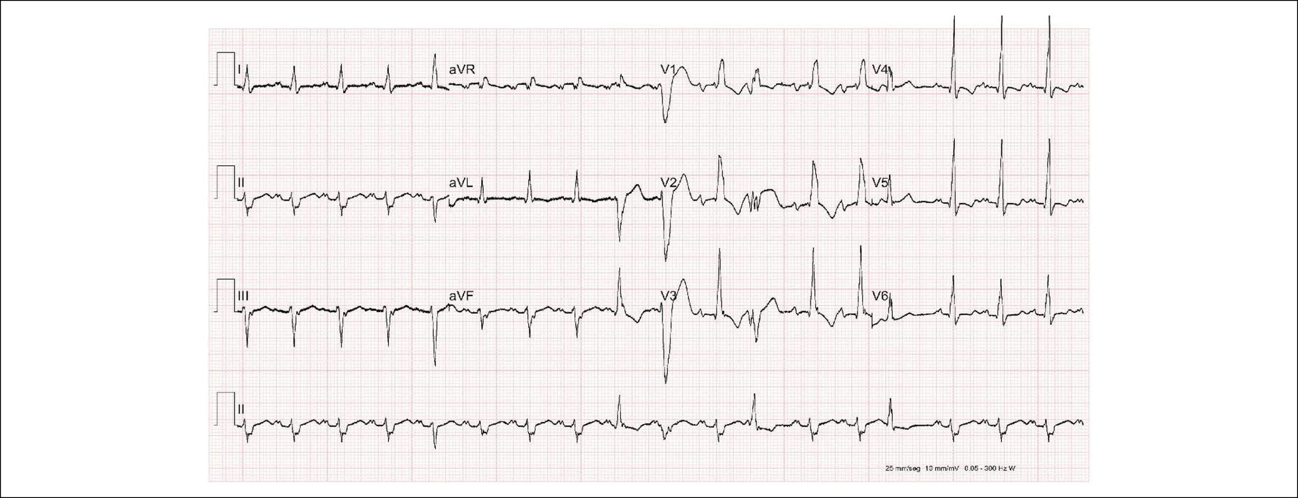
Fábio Fernandes et al. Aristóteles Comte de Alencar Neto Bruno Vaz Kerges Bueno Caio Rebouças Fonseca Cafezeiro … João Henrique Rissato Roberta Shcolnik Szor Mariana Lombardi Peres de Carvalho Wilson Mathias Júnior Angelina Maria Martins Lino Jussara Bianchi Castelli Evandro de Oliveira Souza Félix José Alvarez Ramires Viviane Tiemi Hotta José Soares Júnior Caio de Assis Moura Tavares José Eduardo Krieger Carlos Eduardo Rochitte André Dabarian Ludhmila Abrahão Hajjar Roberto Kalil Filho Charles Mady

Abstract

Background

Systemic amyloidosis is a disease with heterogeneous clinical manifestations. Diagnosis depends on clinical suspicion combined with specific complementary methods.

Objective

To describe the clinical, laboratory, electrocardiographic, and imaging profile in patients with systemic amyloidosis with cardiac involvement.

Methods

This study was conducted with a convenience sample, analyzing clinical, laboratory, electrocardiographic, echocardiographic, nuclear medicine, and magnetic resonance data. Statistical significance was set at p < 0.05.

Results

A total of 105 patients were evaluated (median age of 66 years), 62 of whom were male. Of all patients, 83 had transthyretin (ATTR) amyloidosis, and 22 had light chain (AL) amyloidosis. With respect to ATTR cases, 68.7% were the hereditary form (ATTRh), and 31.3% were wild type (ATTRw). The most prevalent mutations were Val142Ile (45.6%) and Val50Met (40.3%). Time from onset of symptoms to diagnosis was 0.54 and 2.15 years, in the AL and ATTR forms, respectively (p < 0.001). Cardiac involvement was observed in 77.9% of patients with ATTR and in 90.9% of those with AL. Alterations were observed in atrioventricular and intraventricular conduction in 20% and 27.6% of patients, respectively, with 33.7% in ATTR and 4.5% in AL (p = 0.006). In the ATTRw form, there were more atrial arrhythmias than in ATTRh (61.5% versus 22.8%; p = 0.001). On echocardiogram, median septum thickness in ATTRw, ATTRh, and AL was 15 mm, 12 mm, and 11 mm, respectively (p = 0.193). Elevated BNP was observed in 89.5% of patients (median 249, ICR 597.7), and elevated troponin was observed in 43.2%.

Conclusion

In this setting, it was possible to characterize cardiac involvement in systemic amyloidosis in its different subtypes by means of clinical history and the diagnostic methods described.
  • Amyloidosis
  • Diagnosis Imaging
  • Heart Failure
  • Hypertrophic Cardiomyopathy
  • Immunoglobulin Light Chains
  • Prealbumin
Read Full Article
Creative Commons Attribution (CC BY) Creative Commons license
07/03/2022 DOI: 10.36660/abc.20201003 Views 1
Figure 1
ABC
Original Article

Clinical Heart Failure Stratification Through Native T1 Mapping: Experience of a Referral Service

Thiago dos Santos Silva Marques et al. André Maurício de Souza Fernandes Roberto Nery Dantas Júnior Robert W. Biederman … Ana Paula Marques de Oliveira Melo Roque Aras

Abstract

Background:

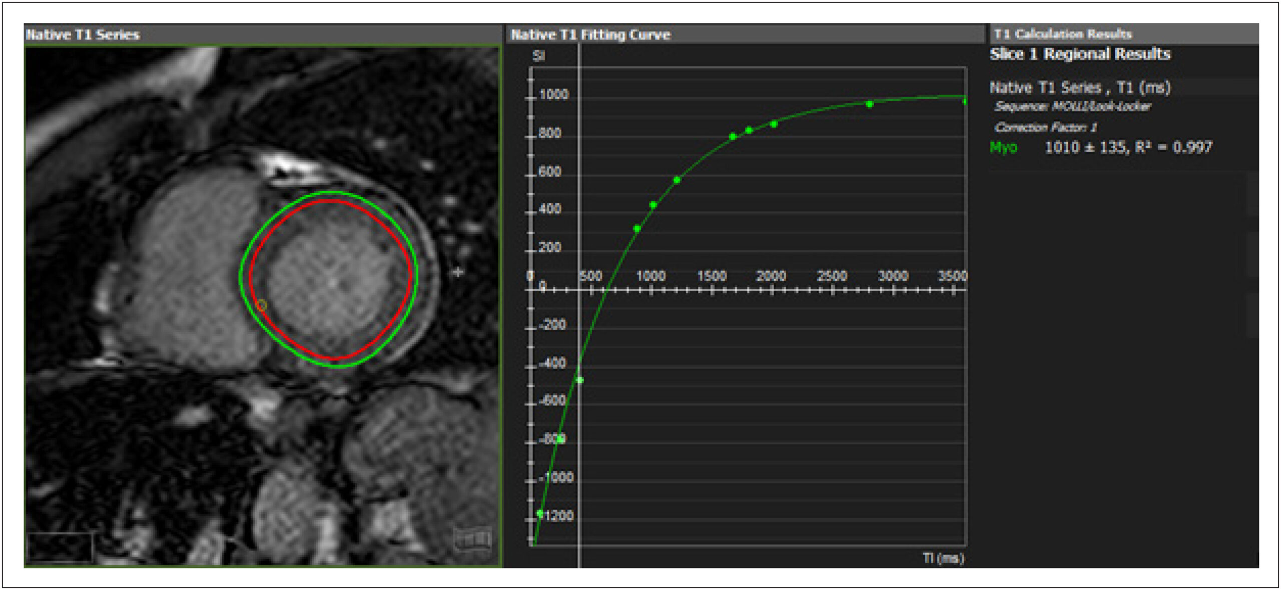
Diffuse cardiac fibrosis is an important factor in the prognostic assessment of patients with ventricular dysfunction. Cardiovascular magnetic resonance imaging (CMR) native T1 mapping is highly sensitive and considered an independent predictor of all-cause mortality and heart failure (HF) development in patients with cardiomyopathy.

Objectives:

To evaluate the feasibility of native T1 mapping assessment in patients with HF in a cardiology referral hospital and its association with structural parameters and functional profile.

Methods:

Cross-sectional study with adult patients with HF NYHA functional classes I and II, ischemic and non-ischemic, followed in a referral hospital, who underwent CMR. Native T1 values were analyzed for structural parameters, comorbidities, etiology, and categorization of HF by left ventricular ejection fraction (LVEF). Analyses were performed with a significance level of 5%.

Results:

Enrollment of 134 patients. Elevated native T1 values were found in patients with greater dilation (1004.9 vs 1042.7ms, p = 0.001), ventricular volumes (1021.3 vs 1050.3ms, p <0.01) and ventricular dysfunction (1010.1 vs 1053.4ms, p <0.001), also present when the non-ischemic group was analyzed separately. Patients classified as HF with reduced ejection fraction had higher T1 values than those with HF and preserved ejection fraction (HFPEF) (992.7 vs 1054.1ms, p <0.001). Of those with HFPEF, 55.2% had higher T1.

Conclusions:

CMR T1 mapping is feasible for clinical HF evaluation. There was a direct association between higher native T1 values and lower ejection fraction, and with larger LV diameters and volumes, regardless of the etiology of HF.

  • Cardiomyopathy, Dilated
  • Chagas Cardiomyopathy
  • Diagnosis Imaging
  • Fibrosis
  • Heart Failure
  • Magnetic Resonance Spectroscopy/ methods
  • Ventricular Dysfunction, Left
Read Full Article
Creative Commons Attribution (CC BY) Creative Commons license
17/05/2021 DOI: 10.36660/abc.20190782 Views 1
Figure 1
ABC
Case Report

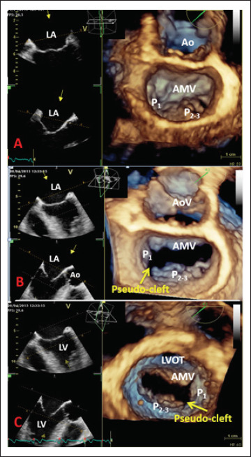
Three-dimensional Echocardiography Reveals the True Enemy in a Young Male with ST-Elevation Myocardial Infarction and Severe Mitral Regurgitation: Posterior Mitral Valve “Pseudo-Cleft” and Prolapse

Sorina Mihaila et al. Andreea Elena Velcea Luigi Paolo Badano Vinereanu Dragos Denisa Muraru

Introduction

Three-dimensional echocardiography (3DE) plays an increasingly important role in the diagnosis of valvular heart disease, in the assessment of valvular morphology in an anatomical manner, and in establishing valve repairability, beyond the limitations of conventional two-dimensional echocardiography (2DE).

We report the case of a young patient presenting with acute anterior ST-segment elevation myocardial infarction and severe mitral regurgitation (MR) after successful primary percutaneous coronary intervention (PCI) of the left anterior descending artery, whose three-dimensional transesophageal echocardiography (3D TEE) revealed an unexpected cause of the MR, namely, complex mitral valve (MV) pathology consisting of prolapse of the P2-3 scallops, flail chordae, and pseudo-cleft of the posterior leaflet separating the P1 from the P2 segment.

[…]

  • Diagnosis Imaging
  • Echocardiography, Three Dimensional / methods
  • Mitral Valve / abnormalities
  • Myocardial Infarction
  • Young Adult
Read Full Article
Creative Commons Attribution (CC BY) Creative Commons license
05/02/2021 DOI: 10.36660/abc.20190485
Figure 1
ABC
Case Report

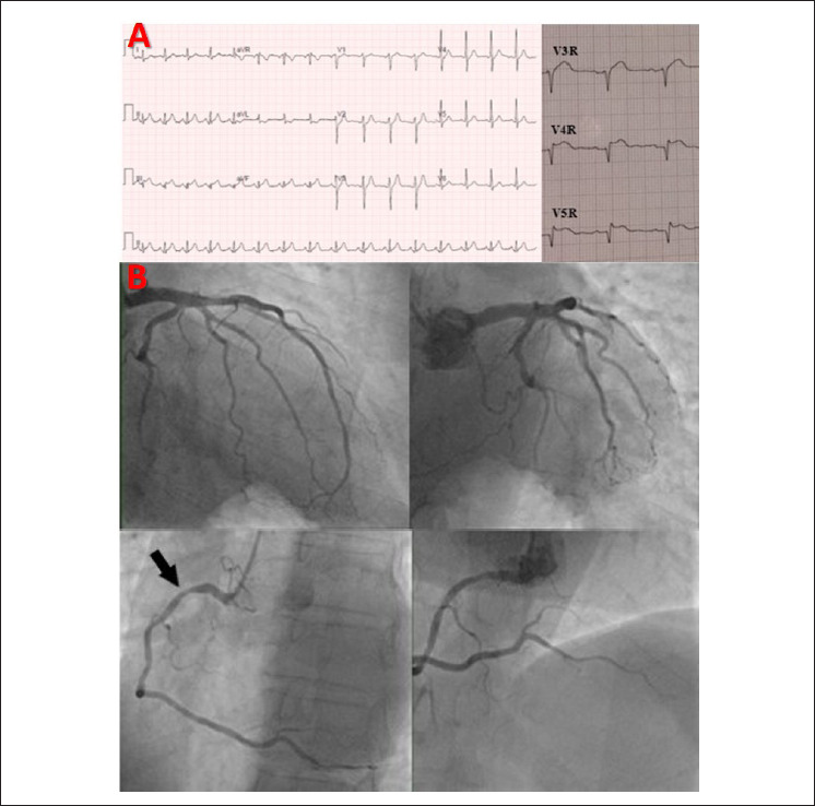
Isolated Right Ventricle Myocardial Infarction – Is the Right Ventricle Still the Forgotten Ventricle?

Ana Marques et al. Inês Cruz Alexandra Briosa Isabel João … Sofia Almeida Hélder Pereira

Introduction

Isolated right ventricular myocardial infarction is extremely rare, and it is often silent, with only 25% of patients developing clinically evident hemodynamic manifestations on presentation. Current management of acute myocardial infarction is based on prompt diagnosis and immediate revascularization. About 90% of patients presenting with ST-segment elevation myocardial infarction have an explanatory coronary artery stenosis or occlusion. Myocardial infarction with non-obstructive coronary arteries (MINOCA) should lead the treating physician to investigate underlying causes, since failure to identify the underlying cause may result in inadequate and inappropriate therapy in these patients.

We describe a case of isolated right ventricular myocardial infarction with normal physical examination, transthoracic echocardiograms, and non-obstructive coronary artery disease on coronary angiography, whose definitive diagnosis was established by cardiac magnetic resonance imaging.

[…]

  • Coronary Artery Disease
  • Diagnosis Imaging
  • Myocardial Infarction
  • Myocardial Revascularization
  • Percutaneous Coronary Intervention
Read Full Article
Creative Commons Attribution (CC BY) Creative Commons license
05/02/2021 DOI: 10.36660/abc.20200164
Figure 1
ABC
Research Letter

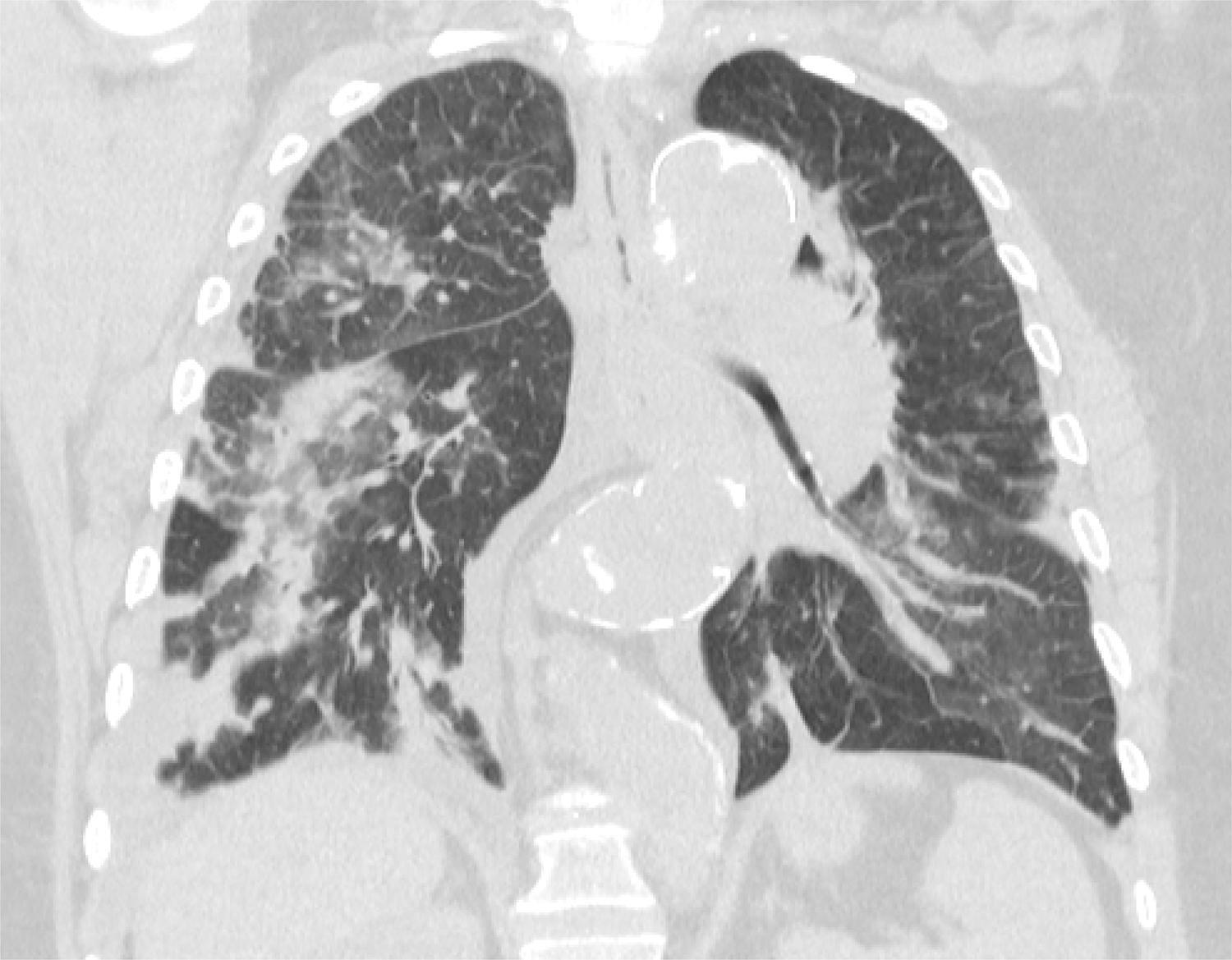
Mechanical Aortic Prosthetic Thrombosis in a 65-Year-Old Woman with SARS-CoV-2 Infection

Marcos H. F. Jacob et al. Tatiana C.A.T. Leal Paulo Rogério Soares Alexandre de Matos Soeiro

Clinical data

A 65-year-old female patient was hospitalized, initially due to an episode of melena. Anticoagulation was suspended, and upper gastrointestinal endoscopy was performed, without detecting bleeding. In 2005, she had late post-operative history of mechanical aortic prosthesis and Bentall and de Bono surgery due to Stanford A aortic dissection; she was a former tobacco user, with obesity and chronic atrial fibrillation under regular use of warfarin. The following day, she had an acute myocardial infarction with ST-segment elevation in the anterior wall; she was submitted to angioplasty with an anterior descending drug-eluting stent, and she received aspirin, clopidogrel 75 mg daily, and anticoagulation with enoxaparin 100 mg every 12 hours. On that occasion, she underwent transthoracic echocardiogram that demonstrated mild ventricular dysfunction (LVEF: 50%) and aortic prosthesis leaflets with preserved mobility; the peak systolic pressure gradient between the left ventricle and the aorta was 58 mmHg, with a mean of 33 mmHg. Two days later, she began to present anosmia, headache, subfebrile temperature, and hypoxemia, and diagnosis of SARS-CoV-2 was confirmed by oropharyngeal swab PCR. Seven days later, while still on full enoxaparin, the patient started presenting atypical chest pain associated with dyspnea. During physical examination, the patient’s overall condition was regular; she had dyspnea, without cyanosis. Pulse was present and full in all 4 limbs, and she had Glasgow score of 15, blood pressure 120/86 mmHg, heart rate 130 bpm, peripheral oximetry 97% while using a nasal oxygen catheter at 3 L/min, and capillary filling time of less than 3 seconds. Ictus cordis was in the left hemiclavicular line, at the level of the fifth intercostal space. She had normophonetic heart sounds, with moderate intensity ejective systolic murmur with metallic click in aortic focus. The patient’s liver was not palpable, and there were mild and sparking crackles in the pulmonary fields bilaterally.

[…]

  • Atrial Fibrillation
  • COVID-19
  • Diagnosis Imaging
  • Heart Valve Prosthesis
  • Myocardial Infarction
  • SARS-CoV-2
  • Thrombosis
Read Full Article
Creative Commons Attribution (CC BY) Creative Commons license
18/01/2021 DOI: 10.36660/abc.20200754
ABC
Editorial

Transseptal, Transcatheter Mitral Valve-In-Valve Replacement: Ready for Prime Time Treatment of Bioprosthetic Valve Failure in Brazil?

Dimytri Alexandre Alvim de Siqueira et al. Auristela Isabel de Oliveira Ramos Fausto Feres

Structural Bioprosthetic Valve Deterioration, Failure and Redo Valve Surgery

Bioprosthetic valves are the device of choice for older patients who require left heart valve replacement, and recent data indicate that bioprostheses are also increasingly used in patients less than 65 years of age who undergo aortic or mitral valve surgeries. Although numerous observational studies have shown excellent survival and freedom from valve-related complications, ultimately all bioprosthetic heart valves are prone to fail. The term structural valve deterioration (SVD) includes permanent, irreversible intrinsic changes of the valve (i.e. leaflet tears, calcification, pannus formation or fibrosis); other pathological causes of bioprosthetic valve dysfunction (i.e. thrombosis, endocarditis) are potentially reversible. When significant hemodynamic compromise (severe regurgitation or stenosis) ensues and/or there is a clinical event related to SVD (reintervention, death), the term bioprosthetic valve failure (BVF) is now suggested.

Redo surgical valve replacement is currently indicated for the majority of patients with bioprosthetic SVD. The risk of redo mitral surgery remains much higher than the risk of first mitral valve replacement (MVR). Advanced age, female gender, preoperative NYHA class, reduced left ventricular ejection fraction (LVEF), pulmonary hypertension and the number of prior operations have all been identified as predictors of mortality. Moreover, adhesions from previous surgery substantially increases bleeding complications. In a study with 260 patients enrolled in the European Redo Cardiac Operation Database (RECORD), in-hospital mortality was 9.2% after redo MVR. A high incidence of postoperative complications, such as low cardiac output syndrome (17.3%), need for circulatory support (9.2%), acute renal failure (16.5%) and need for transfusion (25%) was reported. Similarly, a contemporary study from the Society of Thoracic Surgery (STS) with 11,973 patients revealed that mitral reoperation (n=1096) was associated with higher operative mortality (11.1%) in comparison with the first MVR or repair (6.5%). Despite the paucity of data about redo MVR in Brazil, it is well-known that rheumatic heart disease with valvular involvement is the most frequent indication for cardiac valve surgery in young patients. Since BVF is influenced by age – in patients less than 40 years of age, for example, 15-year freedom from reoperation can be as low as 36%-, patients in our country require multiple reinterventions with increasing risks, substantial impact on their quality of life and costs to the healthcare system.

[…]

  • Aged
  • Diagnosis Imaging
  • Heart Valve Prosthesis
  • Mitral Valve Stenosis
  • Mitral Valve/physiopathology
  • Transcatheter Aortic Replacement/trends
Read Full Article
Creative Commons Attribution (CC BY) Creative Commons license
28/09/2020 DOI: 10.36660/abc.20200575 Views 1
Figure 1
ABC
Case Report

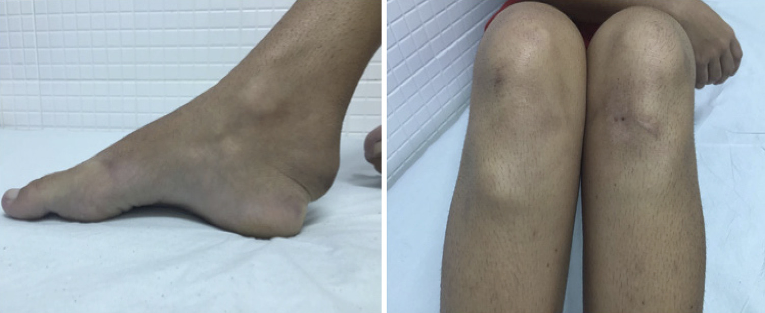
Spontaneous Coronary Artery Dissection in a Patient with Cerebrotendinous Xanthomatosis

Maria Júlia Silveira Souto et al. Marcos Antônio Almeida-Santos Eduardo José Pereira Ferreira Luiz Flávio Galvão Gonçalves … Joselina Luzia Menezes Oliveira Antônio Carlos Sobral Sousa

Introduction

Cerebrotendinous xanthomatosis (CTX) is an autosomal recessive disease characterized by the formation of xanthomatous lesions in many tissues, particularly the brain and tendons. The disorder is a consequence of the reduced production of bile acids, predominantly chenodeoxycholic acid (CDCA), and an increased formation of cholestanol. Common clinical manifestations include infant-onset diarrhea and juvenile-onset bilateral cataract, usually followed by tendon xanthomas and progressive neurological dysfunction. The final diagnosis is based on biochemical abnormalities, including elevated plasma cholestanol level and increased levels of bile alcohol in urine associated with a diminished biliary concentration of CDCA. The treatment is based on oral supplementation of CDCA, which, if initiated early, can prevent major clinical problems, as it produces a reduction in cholestanol synthesis and plasma levels.

Cardiovascular impairment in patients with CTX is mostly associated with premature atherosclerosis. Blood lipid analysis in patients with CTX revealed dramatically high levels of 27-hydroxycholesterol and low levels of high-density lipoprotein cholesterol (HDL), which place these patients at a high risk of cardiovascular disease.

[…]

  • Chenodeoxycholic Acid/adverse effects
  • Child, Adolescent
  • Cholestanol
  • Cholesterol
  • Diagnosis Imaging
  • Xanthomatosis Cerebrotendinous
Read Full Article
Creative Commons Attribution (CC BY) Creative Commons license
14/09/2020 DOI: 10.36660/abc.20190456 Views 1
ABC
Viewpoint

Echoes of Telecardiology Guideline

Silvio Henrique Barberato et al. Marcelo Antônio Cartaxo Queiroga Lopes

Echocardiography has an established role in the diagnosis, prognostic evaluation and therapeutic orientation in several cardiovascular diseases. The great technological development in the last decades has allowed the digitization and standardization of medical images (DICOM), miniaturization of echocardiography equipment (portable cardiac ultrasound) and the transfer of images over the internet. In this scenario, strategies for the use of telemedicine-associated echocardiography, called tele-echocardiography, have been employed in the context of clinical research with the support of teleconsultation for remote expert review, in real time or after image storage and submission. In recognition of the demands generated by the ongoing digital transformation in health, the Brazilian Society of Cardiology recently published the Telemedicine Guideline in Cardiology. The document recognizes tele-echocardiography as a strategy for early detection of congenital heart disease in newborns and screening for early detection of subclinical cases of rheumatic heart disease in children and adolescents (both recommended as indication class IIa, level of evidence B). In addition, it evokes potential application in primary health care in remote locations, where it could enable early detection of cases of heart disease and assist in prioritizing referrals to specialized care (indication class IIb, level of evidence C). It should be emphasized that such recommendations are made within the conditions of regular use of the method in Brazil, which would include the need for doctors at both ends, i.e., the execution and interpretation of the exam. The text of the Guideline explicitly states the need for regulation and legal provision for the participation of other professionals in performing diagnostic procedures (in this case, obtaining echocardiographic images by non-doctors), currently not allowed by the country’s legislation.

In recent years, tele-echocardiography has extended the application of the method to individuals in geographically distant locations, such as remote rural communities or even space. A rural area is classified as remote when 50% of the local population needs at least 45 to 60 minutes of travel by motor vehicle to reach a population center of at least 50,000 inhabitants. Several situations have been experimentally tested, with a study on focused echocardiographic by non-cardiologist physicians,, non-physicians,, or remote-operated robotic devices, combined with remote interpretation by echocardiography cardiologists.

[…]

  • Cardiovascular Diseases
  • Diagnosis Imaging
  • Echocardiography/methods
  • Image Interpretation, Computer-Assisted
  • Robotics/trends
  • Telemedicine/methods
  • Telemedicine/trends
  • Telemonitoring
  • Training
Read Full Article
Creative Commons Attribution (CC BY) Creative Commons license
10/02/2020 DOI: 10.36660/abc.20190720 Views 1
Figure 1
ABC
Image

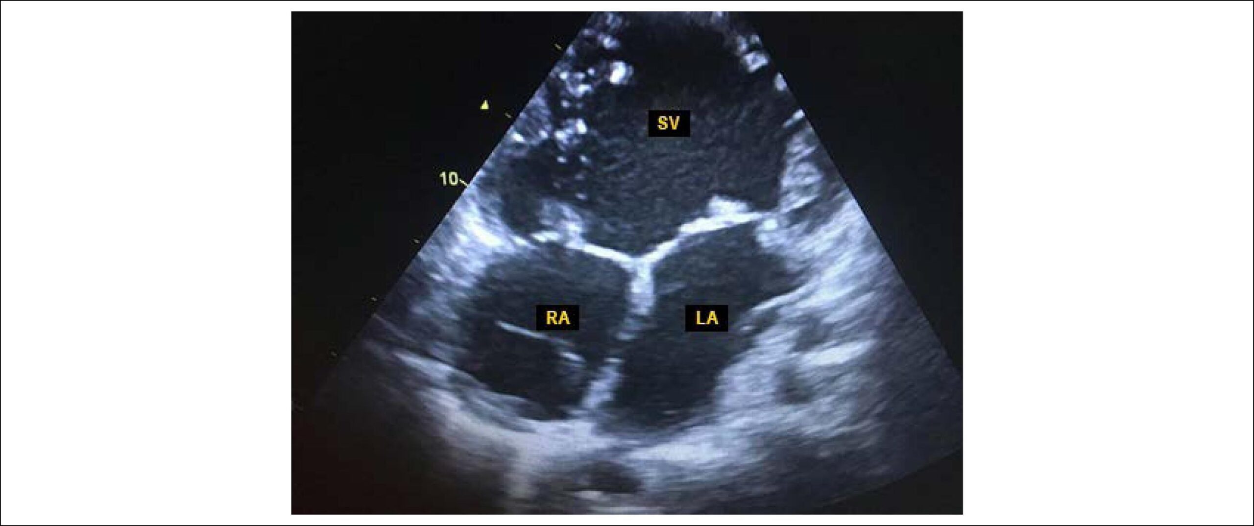
Double-Inlet Single Ventricle with Malposed Great Arteries

Paulo Andrade et al. Danilo Santos Magna Moreira Adail Almeida

A 62-year-old man came to the Echocardiography Service with a history of arterial hypertension and systolic murmur in the mitral area. At the subsequent evaluation, the patient reported dyspnea and fatigue on moderate exertion, but without an impact on social life. Peripheral oxygen saturation at rest ranged from 95% to 98%; extremities were warm and perfused, with no signs of peripheral hypoperfusion; cyanosis and digital clubbing were absent.

The echocardiogram disclosed a case of levocardia, with the presence of a double-inlet single ventricle with transposition of the great arteries (, and ), with situs solitus, enlargement of the atrial chambers associated with significant mitral regurgitation due to annulus dilatation.

[…]

  • Aged
  • Diagnosis Imaging
  • Echocardiography, Doppler/methods
  • Mitral Valve Insufficiency
  • Transposition of great vessels/ surgery
Read Full Article
Creative Commons Attribution (CC BY) Creative Commons license
10/10/2019 DOI: 10.5935/abc.20190160
Figure 1
ABC
Anatomopathological Correlation

Case 4/2019 – 26-Year-Old Man with Congenital Chagas Disease and Heart Transplantation

Henrique Trombini Pinesi et al. Tânia Mara Varejão Strabelli Vera Demarchi Aiello

Clinical aspects

Chagas’ disease was first described by Brazilian physician and scientist Carlos Chagas in 1909. This multifaceted disease is caused by the protozoan Trypanosoma cruzi, which can be transmitted in different ways. Vector transmission through hematophagous insects is the most classic one, although it has declined in importance in recent years with measures to control the vector population. Transfusion transmission, as well as vector transmission, have shown a drastic reduction in the last decades, and no cases have been reported in Brazil for years. In contrast, there was an increase in reports of oral transmission. This form of contagion was little known but gained importance, with several descriptions of micro-epidemic events in the country, especially related to the consumption of açai berry. The development of the acute phase of the disease is more common in the oral transmission. In the context of controlling the main forms of contagion, vertical transmission has become relevant.

The World Health Organization (WHO) estimates there are approximately 8 million individuals infected with Chagas disease worldwide, with an annual mortality of 10,000 people due to disease complications. Most of these cases are found in Latin America, and Brazil is the country with the majority of infected individuals (approximately 4.6 million individuals). The decrease in transmission was accompanied by a reduction in mortality, estimated in 2007 to be 2.78 deaths/year for every 10,000 inhabitants. Another change in the disease epidemiology in recent years has been the increase in cases in non-endemic regions, such as the USA and Europe, which has contributed to the increased attention given by the international scientific community to Chagas’ disease.

[…]

  • Chagas Cardiomyopathy
  • Diagnosis Imaging
  • Heart Defects, Congenital
  • Heart Failure
  • Heart Transplantation
Read Full Article
Creative Commons Attribution (CC BY) Creative Commons license
02/09/2019 DOI: 10.5935/abc.20190162 Views 1
Figure 1
ABC
Case Report

Extensive Anterior Myocardial Infarction … and Something Else?

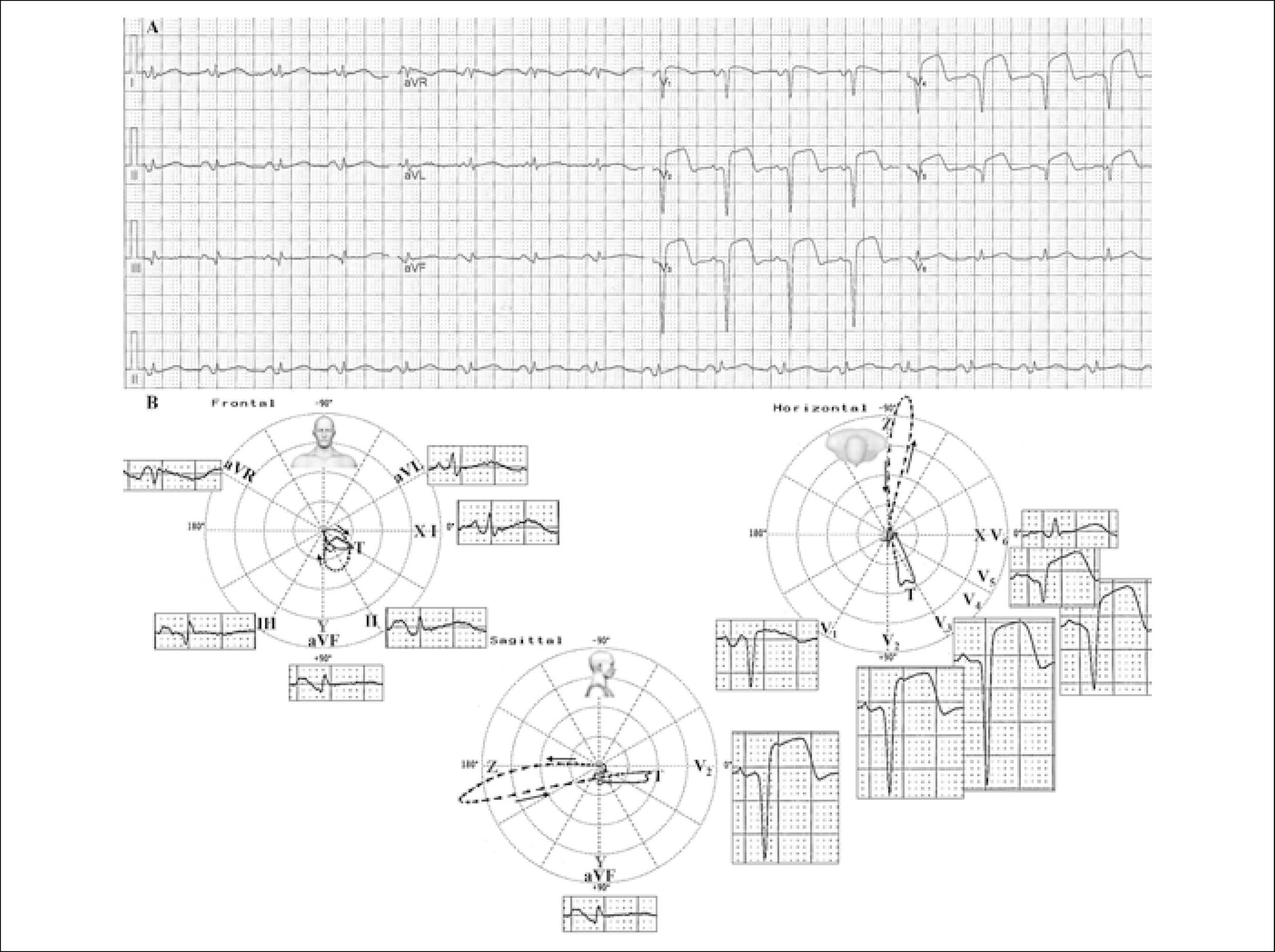
Andrés Ricardo Pérez Riera et al. Raimundo Barbosa Barros Antônio Fernandes Silva e Sousa Neto Rodrigo Daminello Raimundo … Luiz Carlos de Abreu Kjell Nikus

Case report

A 64-year-old Caucasian female, with a one-week history of stress angina. She was admitted to the hospital 2 hours after the onset of oppressive retrosternal pain at rest.

Risk factors: hypertension, smoker, dyslipidemia and diabetes.

[…]

  • Arrhythmias Cardiac
  • Diagnosis Imaging
  • Drug-Eluting Stents
  • Myocardial Infarction/physiopathology
  • P Wave
  • Percutaneous Coronary Intervention
  • Risk Factors
Read Full Article
Creative Commons Attribution (CC BY) Creative Commons license
15/07/2019 DOI: 10.5935/abc.20190096 Views 2
Figure 1
ABC
Image

A Large Cardiac Metastasis of a Parathyroid Tumour Presenting with Ventricular Tachycardia

Rita Ilhão Moreira et al. Sílvia Aguiar Rosa Ana Galrinho Nuno Jalles Tavares Rui Cruz Ferreira

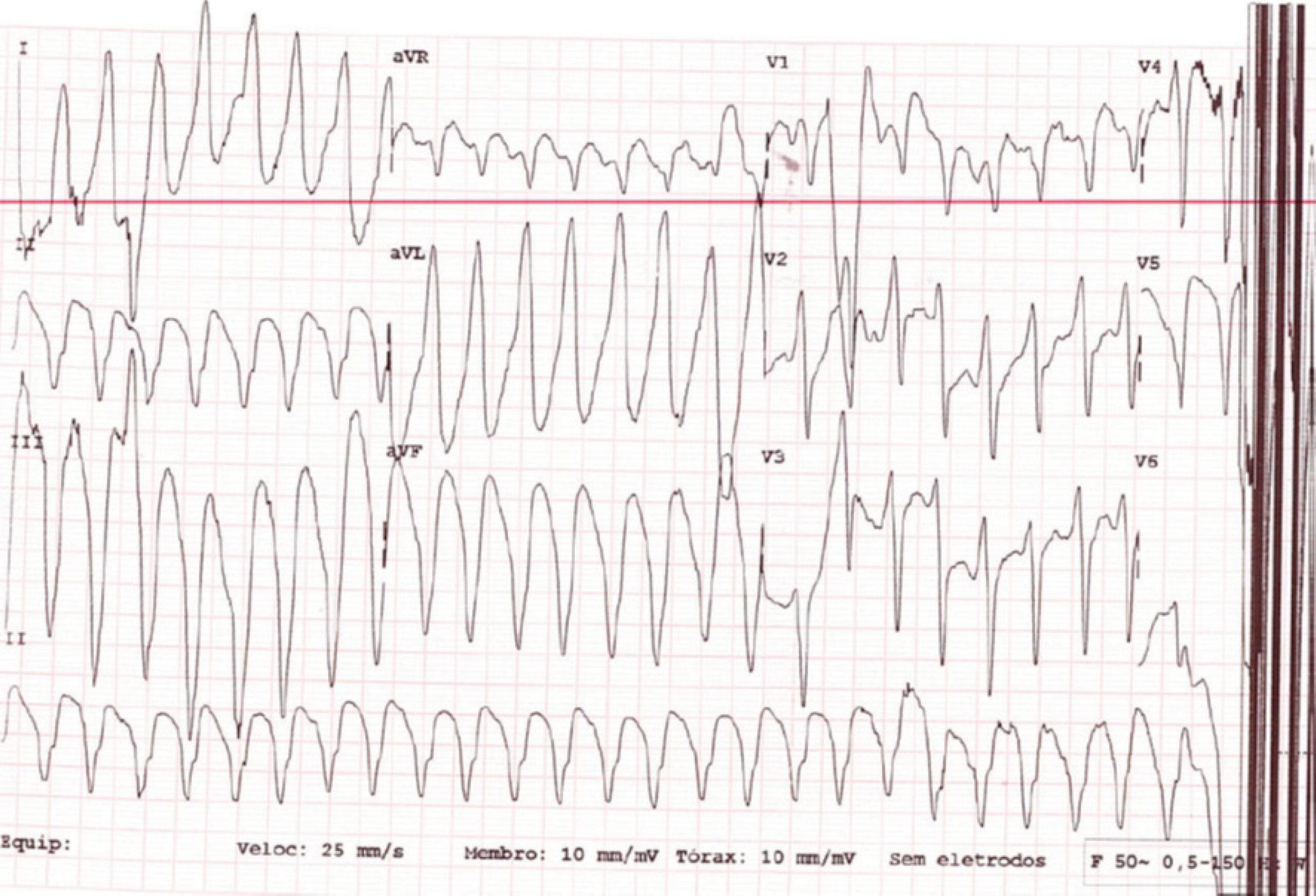
A-81-years old woman was admitted after an episode of ventricular tachycardia with hemodynamic instability converted after electrical cardioversion (). Past medical history was significant for poorly differentiated squamous cell carcinoma of left parathyroid, diabetes and hypertension.

[…]

  • Arrhythmias Cardiac
  • Carcinoma, Squamous Cell
  • Diagnosis Imaging
  • Echocardiography/methods
  • Neoplasms Metastasis
  • Neoplasms Metastasis/therapy
  • Parathyroid Neoplasms
Read Full Article
Creative Commons Attribution (CC BY) Creative Commons license
01/01/2019 DOI: 10.5935/abc.20180255 Views 3
IJCS
Editorial

To Occlude or not to Occlude. That is the Question

Wilson Mathias Junior

In this issue of the International Journal of Cardiovascular Sciences, Madeira et al. evaluated the influence of percutaneous left atrial appendage (LAA) occlusion device on left atrial (LA) function. They assessed several parameters of atrial function, including LA strain and strain rate by speckle-tracking echocardiography (STE) of 16 patients, 75% with permanent atrial fibrillation, undergoing percutaneous LAA closure. No differences were found in maximum and minimum LA volume or LA emptying fraction before and 3 months after the procedure. Similarly, no differences were noted in LA strain (13.7 ± 11.1 vs. 13.0 ± 8.8%; p = 0.63) or strain rate (1.06 ± 0.26 vs. 1.13 ± 0.34 s-1; p = 0.38) in the reservoir phase. Based on those results, the authors concluded that LAA occlusion had no impact on LA function.

This is among the first studies to examine the effect of this procedure on LA function, for which the authors should be congratulated.

[…]

  • Atrial Appendage
  • Atrial Fibrillation
  • Balloon Device
  • Diagnosis Imaging
  • Echocardiography/methods
Read Full Article
Creative Commons Attribution (CC BY) Creative Commons license
01/01/2018 DOI: 10.5935/2359-4802.20180089 Views 1

How to cite this article

EndNote RIS

Electronic Document Format (Vancouver)

Fernandes F, Alencar Neto AC, Bueno BVK, Cafezeiro CRF, Rissato JH, Szor RS, Carvalho MLP, et al. Clinical, Laboratory, and Imaging Profile in Patients with Systemic Amyloidosis in a Brazilian Cardiology Referral Center. Arquivos Brasileiros de Cardiologia 2022;118(2):422-3.

Electronic Document Format (ABNT)

Fernandes, Fábio; Alencar Neto, Aristóteles Comte de; Bueno, Bruno Vaz Kerges; Cafezeiro, Caio Rebouças Fonseca; Rissato, João Henrique; Szor, Roberta Shcolnik; Carvalho, Mariana Lombardi Peres de; Mathias Júnior, Wilson; Lino, Angelina Maria Martins; Castelli, Jussara Bianchi; Souza, Evandro de Oliveira; Ramires, Félix José Alvarez; Hotta, Viviane Tiemi; Soares Júnior, José; Tavares, Caio de Assis Moura; Krieger, José Eduardo; Rochitte, Carlos Eduardo; Dabarian, André; Hajjar, Ludhmila Abrahão; Kalil Filho, Roberto; Mady, Charles. Clinical, Laboratory, and Imaging Profile in Patients with Systemic Amyloidosis in a Brazilian Cardiology Referral Center. Arquivos Brasileiros de Cardiologia, v. 118, n. 2, p. 422-432, Mar. 2022.

Electronic Document Format (APA)

Fernandes, F., Alencar Neto, A. C. , Bueno, B. V. K., Cafezeiro, C. R. F., Rissato, J. H., Szor, R. S., Carvalho, M. L. P. , Mathias Júnior, W., Lino, A. M. M., Castelli, J. B., Souza, E. O., Ramires, F. J. A., Hotta, V. T., Soares Júnior, J., Tavares, C. A. M., Krieger, J. E., Rochitte, C. E., Dabarian, A., Hajjar, L. A., Kalil Filho, R., & Mady, C. (2022). Clinical, Laboratory, and Imaging Profile in Patients with Systemic Amyloidosis in a Brazilian Cardiology Referral Center. Arquivos Brasileiros de Cardiologia, 118(2), 422-432.

Electronic Document Format (ISO)

Fernandes, Fábio and Alencar Neto, Aristóteles Comte de and Bueno, Bruno Vaz Kerges and Cafezeiro, Caio Rebouças Fonseca and Rissato, João Henrique and Szor, Roberta Shcolnik and Carvalho, Mariana Lombardi Peres de and Mathias Júnior, Wilson and Lino, Angelina Maria Martins and Castelli, Jussara Bianchi and Souza, Evandro de Oliveira and Ramires, Félix José Alvarez and Hotta, Viviane Tiemi and Soares Júnior, José and Tavares, Caio de Assis Moura and Krieger, José Eduardo and Rochitte, Carlos Eduardo and Dabarian, André and Hajjar, Ludhmila Abrahão and Kalil Filho, Roberto and Mady, Charles. Clinical, Laboratory, and Imaging Profile in Patients with Systemic Amyloidosis in a Brazilian Cardiology Referral Center. Arquivos Brasileiros de Cardiologia [online]. 2022, vol. 118, n. 2, [cited 2026-10-14], pp. 422-432. Available from: <https://journals.cardiol.br/en/article/clinical-laboratory-and-imaging-profile-in-patients-with-systemic-amyloidosis-in-a-brazilian-cardiology-referral-center/>. ISSN 0066-782X

EndNote RIS

Electronic Document Format (Vancouver)

Marques TSS, Fernandes AMS, Dantas Júnior RN, Biederman RW, Melo APMO, Aras R. Clinical Heart Failure Stratification Through Native T1 Mapping: Experience of a Referral Service. Arquivos Brasileiros de Cardiologia 2021;116(5):919-25.

Electronic Document Format (ABNT)

Marques, Thiago dos Santos Silva; Fernandes, André Maurício de Souza; Dantas Júnior, Roberto Nery; Biederman, Robert W.; Melo, Ana Paula Marques de Oliveira; Aras, Roque. Clinical Heart Failure Stratification Through Native T1 Mapping: Experience of a Referral Service. Arquivos Brasileiros de Cardiologia, v. 116, n. 5, p. 919-925, May. 2021.

Electronic Document Format (APA)

Marques, T. S. S., Fernandes, A. M. S., Dantas Júnior, R. N., Biederman, R. W., Melo, A. P. M. O., & Aras, R. (2021). Clinical Heart Failure Stratification Through Native T1 Mapping: Experience of a Referral Service. Arquivos Brasileiros de Cardiologia, 116(5), 919-925.

Electronic Document Format (ISO)

Marques, Thiago dos Santos Silva and Fernandes, André Maurício de Souza and Dantas Júnior, Roberto Nery and Biederman, Robert W. and Melo, Ana Paula Marques de Oliveira and Aras, Roque. Clinical Heart Failure Stratification Through Native T1 Mapping: Experience of a Referral Service. Arquivos Brasileiros de Cardiologia [online]. 2021, vol. 116, n. 5, [cited 2026-10-14], pp. 919-925. Available from: <https://journals.cardiol.br/en/article/clinical-heart-failure-stratification-through-native-t1-mapping-experience-of-a-referral-service/>. ISSN 0066-782X

EndNote RIS

Electronic Document Format (Vancouver)

Mihaila S, Velcea AE, Badano LP, Dragos V, Muraru D. Three-dimensional Echocardiography Reveals the True Enemy in a Young Male with ST-Elevation Myocardial Infarction and Severe Mitral Regurgitation: Posterior Mitral Valve “Pseudo-Cleft” and Prolapse. Arquivos Brasileiros de Cardiologia 2021;116(2 suppl 1):36-8.

Electronic Document Format (ABNT)

Mihaila, Sorina; Velcea, Andreea Elena; Badano, Luigi Paolo; Dragos, Vinereanu; Muraru, Denisa. Three-dimensional Echocardiography Reveals the True Enemy in a Young Male with ST-Elevation Myocardial Infarction and Severe Mitral Regurgitation: Posterior Mitral Valve “Pseudo-Cleft” and Prolapse. Arquivos Brasileiros de Cardiologia, v. 116, n. 2 suppl 1, p. 36-38, Feb. 2021.

Electronic Document Format (APA)

Mihaila, S., Velcea, A. E., Badano, L. P., Dragos, V., & Muraru, D. (2021). Three-dimensional Echocardiography Reveals the True Enemy in a Young Male with ST-Elevation Myocardial Infarction and Severe Mitral Regurgitation: Posterior Mitral Valve “Pseudo-Cleft” and Prolapse. Arquivos Brasileiros de Cardiologia, 116(2 suppl 1), 36-38.

Electronic Document Format (ISO)

Mihaila, Sorina and Velcea, Andreea Elena and Badano, Luigi Paolo and Dragos, Vinereanu and Muraru, Denisa. Three-dimensional Echocardiography Reveals the True Enemy in a Young Male with ST-Elevation Myocardial Infarction and Severe Mitral Regurgitation: Posterior Mitral Valve “Pseudo-Cleft” and Prolapse. Arquivos Brasileiros de Cardiologia [online]. 2021, vol. 116, n. 2 suppl 1, [cited 2026-10-14], pp. 36-38. Available from: <https://journals.cardiol.br/en/article/three-dimensional-echocardiography-reveals-the-true-enemy-in-a-young-male-with-st-elevation-myocardial-infarction-and-severe-mitral-regurgitation-posterior-mitral-valve-pseudo-cleft/>. ISSN 0066-782X

EndNote RIS

Electronic Document Format (Vancouver)

Marques A, Cruz I, Briosa A, João I, Almeida S, Pereira H. Isolated Right Ventricle Myocardial Infarction – Is the Right Ventricle Still the Forgotten Ventricle?. Arquivos Brasileiros de Cardiologia 2021;116(2 suppl 1):32-5.

Electronic Document Format (ABNT)

Marques, Ana; Cruz, Inês; Briosa, Alexandra; João, Isabel; Almeida, Sofia; Pereira, Hélder. Isolated Right Ventricle Myocardial Infarction – Is the Right Ventricle Still the Forgotten Ventricle?. Arquivos Brasileiros de Cardiologia, v. 116, n. 2 suppl 1, p. 32-35, Feb. 2021.

Electronic Document Format (APA)

Marques, A., Cruz, I., Briosa, A., João, I., Almeida, S., & Pereira, H. (2021). Isolated Right Ventricle Myocardial Infarction – Is the Right Ventricle Still the Forgotten Ventricle?. Arquivos Brasileiros de Cardiologia, 116(2 suppl 1), 32-35.

Electronic Document Format (ISO)

Marques, Ana and Cruz, Inês and Briosa, Alexandra and João, Isabel and Almeida, Sofia and Pereira, Hélder. Isolated Right Ventricle Myocardial Infarction – Is the Right Ventricle Still the Forgotten Ventricle?. Arquivos Brasileiros de Cardiologia [online]. 2021, vol. 116, n. 2 suppl 1, [cited 2026-10-14], pp. 32-35. Available from: <https://journals.cardiol.br/en/article/isolated-right-ventricle-myocardial-infarction-is-the-right-ventricle-still-the-forgotten-ventricle/>. ISSN 0066-782X

EndNote RIS

Electronic Document Format (Vancouver)

Jacob MHF, Leal TC, Soares PR, Soeiro AM. Mechanical Aortic Prosthetic Thrombosis in a 65-Year-Old Woman with SARS-CoV-2 Infection. Arquivos Brasileiros de Cardiologia 2021;115(6):1180-3.

Electronic Document Format (ABNT)

Jacob, Marcos H. F.; Leal, Tatiana C.A.T.; Soares, Paulo Rogério; Soeiro, Alexandre de Matos. Mechanical Aortic Prosthetic Thrombosis in a 65-Year-Old Woman with SARS-CoV-2 Infection. Arquivos Brasileiros de Cardiologia, v. 115, n. 6, p. 1180-1183, Jan. 2021.

Electronic Document Format (APA)

Jacob, M. H. F., Leal, T. C., Soares, P. R., & Soeiro, A. M. (2021). Mechanical Aortic Prosthetic Thrombosis in a 65-Year-Old Woman with SARS-CoV-2 Infection. Arquivos Brasileiros de Cardiologia, 115(6), 1180-1183.

Electronic Document Format (ISO)

Jacob, Marcos H. F. and Leal, Tatiana C.A.T. and Soares, Paulo Rogério and Soeiro, Alexandre de Matos. Mechanical Aortic Prosthetic Thrombosis in a 65-Year-Old Woman with SARS-CoV-2 Infection. Arquivos Brasileiros de Cardiologia [online]. 2021, vol. 115, n. 6, [cited 2026-10-14], pp. 1180-1183. Available from: <https://journals.cardiol.br/en/article/mechanical-aortic-prosthetic-thrombosis-in-a-65-year-old-woman-with-sars-cov-2-infection/>. ISSN 0066-782X

EndNote RIS

Electronic Document Format (Vancouver)

Siqueira DAA, Ramos AIO, Feres F. Transseptal, Transcatheter Mitral Valve-In-Valve Replacement: Ready for Prime Time Treatment of Bioprosthetic Valve Failure in Brazil?. Arquivos Brasileiros de Cardiologia 2020;115(3):525-7.

Electronic Document Format (ABNT)

Siqueira, Dimytri Alexandre Alvim de; Ramos, Auristela Isabel de Oliveira; Feres, Fausto. Transseptal, Transcatheter Mitral Valve-In-Valve Replacement: Ready for Prime Time Treatment of Bioprosthetic Valve Failure in Brazil?. Arquivos Brasileiros de Cardiologia, v. 115, n. 3, p. 525-527, Sep. 2020.

Electronic Document Format (APA)

Siqueira, D. A. A. , Ramos, A. I. O., & Feres, F. (2020). Transseptal, Transcatheter Mitral Valve-In-Valve Replacement: Ready for Prime Time Treatment of Bioprosthetic Valve Failure in Brazil?. Arquivos Brasileiros de Cardiologia, 115(3), 525-527.

Electronic Document Format (ISO)

Siqueira, Dimytri Alexandre Alvim de and Ramos, Auristela Isabel de Oliveira and Feres, Fausto. Transseptal, Transcatheter Mitral Valve-In-Valve Replacement: Ready for Prime Time Treatment of Bioprosthetic Valve Failure in Brazil?. Arquivos Brasileiros de Cardiologia [online]. 2020, vol. 115, n. 3, [cited 2026-10-14], pp. 525-527. Available from: <https://journals.cardiol.br/en/article/transseptal-transcatheter-mitral-valve-in-valve-replacement-ready-for-prime-time-treatment-of-bioprosthetic-valve-failure-in-brazil/>. ISSN 0066-782X

EndNote RIS

Electronic Document Format (Vancouver)

Souto MJS, Almeida-Santos MA, Ferreira EJP, Gonçalves LFG, Oliveira JLM, Sousa ACS. Spontaneous Coronary Artery Dissection in a Patient with Cerebrotendinous Xanthomatosis. Arquivos Brasileiros de Cardiologia 2020;115(1 suppl 1):18-21.

Electronic Document Format (ABNT)

Souto, Maria Júlia Silveira; Almeida-Santos, Marcos Antônio; Ferreira, Eduardo José Pereira; Gonçalves, Luiz Flávio Galvão; Oliveira, Joselina Luzia Menezes; Sousa, Antônio Carlos Sobral. Spontaneous Coronary Artery Dissection in a Patient with Cerebrotendinous Xanthomatosis. Arquivos Brasileiros de Cardiologia, v. 115, n. 1 suppl 1, p. 18-21, Sep. 2020.

Electronic Document Format (APA)

Souto, M. J. S., Almeida-Santos, M. A., Ferreira, E. J. P., Gonçalves, L. F. G., Oliveira, J. L. M., & Sousa, A. C. S. (2020). Spontaneous Coronary Artery Dissection in a Patient with Cerebrotendinous Xanthomatosis. Arquivos Brasileiros de Cardiologia, 115(1 suppl 1), 18-21.

Electronic Document Format (ISO)

Souto, Maria Júlia Silveira and Almeida-Santos, Marcos Antônio and Ferreira, Eduardo José Pereira and Gonçalves, Luiz Flávio Galvão and Oliveira, Joselina Luzia Menezes and Sousa, Antônio Carlos Sobral. Spontaneous Coronary Artery Dissection in a Patient with Cerebrotendinous Xanthomatosis. Arquivos Brasileiros de Cardiologia [online]. 2020, vol. 115, n. 1 suppl 1, [cited 2026-10-14], pp. 18-21. Available from: <https://journals.cardiol.br/en/article/spontaneous-coronary-artery-dissection-in-a-patient-with-cerebrotendinous-xanthomatosis/>. ISSN 0066-782X

EndNote RIS

Electronic Document Format (Vancouver)

Barberato SH, Lopes MACQ. Echoes of Telecardiology Guideline. Arquivos Brasileiros de Cardiologia 2020;114(1):130-2.

Electronic Document Format (ABNT)

Barberato, Silvio Henrique; Lopes, Marcelo Antônio Cartaxo Queiroga. Echoes of Telecardiology Guideline. Arquivos Brasileiros de Cardiologia, v. 114, n. 1, p. 130-132, Feb. 2020.

Electronic Document Format (APA)

Barberato, S. H., & Lopes, M. A. C. Q. (2020). Echoes of Telecardiology Guideline. Arquivos Brasileiros de Cardiologia, 114(1), 130-132.

Electronic Document Format (ISO)

Barberato, Silvio Henrique and Lopes, Marcelo Antônio Cartaxo Queiroga. Echoes of Telecardiology Guideline. Arquivos Brasileiros de Cardiologia [online]. 2020, vol. 114, n. 1, [cited 2026-10-14], pp. 130-132. Available from: <https://journals.cardiol.br/en/article/echoes-of-telecardiology-guideline/>. ISSN 0066-782X

EndNote RIS

Electronic Document Format (Vancouver)

Andrade P, Santos D, Moreira M, Almeida A. Double-Inlet Single Ventricle with Malposed Great Arteries. Arquivos Brasileiros de Cardiologia 2019;113(3):444-6.

Electronic Document Format (ABNT)

Andrade, Paulo; Santos, Danilo; Moreira, Magna; Almeida, Adail. Double-Inlet Single Ventricle with Malposed Great Arteries. Arquivos Brasileiros de Cardiologia, v. 113, n. 3, p. 444-446, Oct. 2019.

Electronic Document Format (APA)

Andrade, P., Santos, D., Moreira, M., & Almeida, A. (2019). Double-Inlet Single Ventricle with Malposed Great Arteries. Arquivos Brasileiros de Cardiologia, 113(3), 444-446.

Electronic Document Format (ISO)

Andrade, Paulo and Santos, Danilo and Moreira, Magna and Almeida, Adail. Double-Inlet Single Ventricle with Malposed Great Arteries. Arquivos Brasileiros de Cardiologia [online]. 2019, vol. 113, n. 3, [cited 2026-10-14], pp. 444-446. Available from: <https://journals.cardiol.br/en/article/double-inlet-single-ventricle-with-malposed-great-arteries/>. ISSN 0066-782X

EndNote RIS

Electronic Document Format (Vancouver)

Pinesi HT, Strabelli TMV, Aiello VD. Case 4/2019 – 26-Year-Old Man with Congenital Chagas Disease and Heart Transplantation. Arquivos Brasileiros de Cardiologia 2019;113(2):286-93.

Electronic Document Format (ABNT)

Pinesi, Henrique Trombini; Strabelli, Tânia Mara Varejão; Aiello, Vera Demarchi. Case 4/2019 – 26-Year-Old Man with Congenital Chagas Disease and Heart Transplantation. Arquivos Brasileiros de Cardiologia, v. 113, n. 2, p. 286-293, Sep. 2019.

Electronic Document Format (APA)

Pinesi, H. T., Strabelli, T. M. V., & Aiello, V. D. (2019). Case 4/2019 – 26-Year-Old Man with Congenital Chagas Disease and Heart Transplantation. Arquivos Brasileiros de Cardiologia, 113(2), 286-293.

Electronic Document Format (ISO)

Pinesi, Henrique Trombini and Strabelli, Tânia Mara Varejão and Aiello, Vera Demarchi. Case 4/2019 – 26-Year-Old Man with Congenital Chagas Disease and Heart Transplantation. Arquivos Brasileiros de Cardiologia [online]. 2019, vol. 113, n. 2, [cited 2026-10-14], pp. 286-293. Available from: <https://journals.cardiol.br/en/article/case-4-2019-26-year-old-man-with-congenital-chagas-disease-and-heart-transplantation/>. ISSN 0066-782X

EndNote RIS

Electronic Document Format (Vancouver)

Riera ARP, Barros RB, Sousa Neto AFS, Raimundo RD, Abreu LC, Nikus K. Extensive Anterior Myocardial Infarction … and Something Else?. Arquivos Brasileiros de Cardiologia 2019;112(6):803-6.

Electronic Document Format (ABNT)

Riera, Andrés Ricardo Pérez; Barros, Raimundo Barbosa; Sousa Neto, Antônio Fernandes Silva e; Raimundo, Rodrigo Daminello; Abreu, Luiz Carlos de; Nikus, Kjell. Extensive Anterior Myocardial Infarction … and Something Else?. Arquivos Brasileiros de Cardiologia, v. 112, n. 6, p. 803-806, Jul. 2019.

Electronic Document Format (APA)

Riera, A. R. P., Barros, R. B., Sousa Neto, A. F. S. , Raimundo, R. D., Abreu, L. C. , & Nikus, K. (2019). Extensive Anterior Myocardial Infarction … and Something Else?. Arquivos Brasileiros de Cardiologia, 112(6), 803-806.

Electronic Document Format (ISO)

Riera, Andrés Ricardo Pérez and Barros, Raimundo Barbosa and Sousa Neto, Antônio Fernandes Silva e and Raimundo, Rodrigo Daminello and Abreu, Luiz Carlos de and Nikus, Kjell. Extensive Anterior Myocardial Infarction … and Something Else?. Arquivos Brasileiros de Cardiologia [online]. 2019, vol. 112, n. 6, [cited 2026-10-14], pp. 803-806. Available from: <https://journals.cardiol.br/en/article/extensive-anterior-myocardial-infarction-and-something-else/>. ISSN 0066-782X

EndNote RIS

Electronic Document Format (Vancouver)

Moreira RI, Rosa SA, Galrinho A, Tavares NJ, Ferreira RC. A Large Cardiac Metastasis of a Parathyroid Tumour Presenting with Ventricular Tachycardia. Arquivos Brasileiros de Cardiologia 2019;112(1):113-5.

Electronic Document Format (ABNT)

Moreira, Rita Ilhão; Rosa, Sílvia Aguiar; Galrinho, Ana; Tavares, Nuno Jalles; Ferreira, Rui Cruz. A Large Cardiac Metastasis of a Parathyroid Tumour Presenting with Ventricular Tachycardia. Arquivos Brasileiros de Cardiologia, v. 112, n. 1, p. 113-115, Dec. 2018.

Electronic Document Format (APA)

Moreira, R. I., Rosa, S. A., Galrinho, A., Tavares, N. J., & Ferreira, R. C. (2019). A Large Cardiac Metastasis of a Parathyroid Tumour Presenting with Ventricular Tachycardia. Arquivos Brasileiros de Cardiologia, 112(1), 113-115.

Electronic Document Format (ISO)

Moreira, Rita Ilhão and Rosa, Sílvia Aguiar and Galrinho, Ana and Tavares, Nuno Jalles and Ferreira, Rui Cruz. A Large Cardiac Metastasis of a Parathyroid Tumour Presenting with Ventricular Tachycardia. Arquivos Brasileiros de Cardiologia [online]. 2019, vol. 112, n. 1, [cited 2026-10-14], pp. 113-115. Available from: <https://journals.cardiol.br/en/article/a-large-cardiac-metastasis-of-a-parathyroid-tumour-presenting-with-ventricular-tachycardia/>. ISSN 0066-782X

EndNote RIS

Electronic Document Format (Vancouver)

Mathias Junior W. To Occlude or not to Occlude. That is the Question. International Journal of Cardiovascular Sciences 2018;31(6):560-1.

Electronic Document Format (ABNT)

Mathias Junior, Wilson. To Occlude or not to Occlude. That is the Question. International Journal of Cardiovascular Sciences, v. 31, n. 6, p. 560-561, Dec. 2017.

Electronic Document Format (APA)

Mathias Junior, W. (2018). To Occlude or not to Occlude. That is the Question. International Journal of Cardiovascular Sciences, 31(6), 560-561.

Electronic Document Format (ISO)

Mathias Junior, Wilson. To Occlude or not to Occlude. That is the Question. International Journal of Cardiovascular Sciences [online]. 2018, vol. 31, n. 6, [cited 2026-10-14], pp. 560-561. Available from: <https://journals.cardiol.br/en/article/to-occlude-or-not-to-occlude-that-is-the-question/>. ISSN 2359-4802

  • About ABC Family
  • Internal Regulation
  • Editorial Board
  • Brazilian Society of Cardiology (SBC)

Issues

SBC Guidelines

  • Videos
  • Podcasts
  • News