ABC Family Arquivos Brasileiros de Cardiologia International Journal of Cardiovascular Sciences ABC Heart Failure & Cardiomyopathy ABC Imagem Cardiovascular
Português English
ABC Family
Português English
  • About
    • About ABC Family
    • Internal Regulation
    • Editorial Board
    • Brazilian Society of Cardiology (SBC)
  • Issues
  • SBC Guidelines
  • Content
    • Videos
    • Podcasts
    • News
  • About
    • About ABC Family
    • Internal Regulation
    • Editorial Board
    • Brazilian Society of Cardiology (SBC)
  • Issues
  • SBC Guidelines
  • Content
    • Videos
    • Podcasts
    • News

ABC Family

  • ABC Family
  • Arquivos Brasileiros de Cardiologia
  • International Journal of Cardiovascular Sciences
  • ABC Heart Failure & Cardiomyopathy
  • ABC Imagem Cardiovascular
Search in

No content found.

Journals
Ordering
Article types
Others

  1. Home
  2. Keyword: Magnetic Resonance Spectroscopy/ methods
Figure 1
ABC
Image

Rare Presentation of Yolk Sac Tumor with Cardiac Involvement: Characteristics Detected by MRI

Cristhian Espinoza Romero et al. Williams Roberto Lata Guacho Kevin Rafael de Paula Robert Paladines Jimenez Eduardo Kaiser Ururahy Nunes Fonseca

Introduction

Primary cardiac tumors are extremely rare, with their incidence varying between 0.0017 and 0.28%, among which is the malignant germ cell yolk sac tumor (YST).

Although transthoracic echocardiography (TTE) is often the first line in the evaluation of cardiac tumors, currently, due to its good spatial resolution and tissue characterization, cardiac magnetic resonance (CMR) imaging is the technique of choice for the evaluation of these tumors., The intracardiac YST is a rare neoplasm, with few reported cases.–

[…]

  • Diagnostic Imaging
  • Drug Therapy
  • Echocardiography, Transthoracic/methods
  • Heart Failure
  • Heart Neoplasms
  • Magnetic Resonance Spectroscopy/ methods
  • Neoplasms, Germ Cell and Embryonal
Read Full Article
Creative Commons Attribution (CC BY) Creative Commons license
11/07/2022 DOI: 10.36660/abc.20210335 Views 1
ABC
Editorial

Stress CMR in the Elderly: Does It Provide the Answers?

Vera Vaz Ferreira et al. Boban Thomas

After the ISCHEMIA clinical trial, despite the ongoing debate, clinicians need to contemplate all strategies for patients with chronic stable ischemic heart disease (SIHD). The management of SIHD has two aims – improve prognosis and/or relieve symptoms. Most studies have shown that revascularisation provides greater symptom benefits than optimal medical therapy (OMT) alone, but data on hard outcomes have been elusive. Therefore, improving our clinical decision-making tools in selecting patients for revascularization is important. This becomes particularly important when we select elderly patients for revascularisation because the risk-benefit ratio is probably more tenuous than a younger cohort. The elderly present difficulties in the evaluation of SIHD due to multiple reasons. One refinement is the adequate selection of the method to evaluate SIHD. A panoply of non-invasive techniques exists for imaging, including cardiac computed tomography, echocardiography, nuclear medicine techniques and cardiac magnetic resonance (CMR). Combining the right patient and the right method of evaluation followed by the right therapeutic strategy improves outcomes. In clinical medicine, what is important is not how much we do but how well our patients do after what we do.

CMR stress perfusion is a valuable resource with a high negative predictive value in those who do not have perfusion defects irrespective of the presence or absence of coronary artery disease. Few studies exist on the prognostic value of adenosine stress perfusion CMR in elderly patients with or without established SIHD. The study by Boonyasirinant and Kaolawanich, although not the first, is a welcome addition to the repository of existing literature that provides a cogent reason for using stress CMR for the evaluation of ischemia now addressing the elderly age group. Their findings demonstrate that while clinical data were combined with information on left ventricular function, no incremental value was seen compared to clinical data alone in predicting serious cardiac events. The presence of ischemia as detected by CMR helped predict events significantly better. The predictive power was not increased with the addition of information on late gadolinium enhancement. Esteban-Fernandez et al. found that elderly patients with a moderate or severe degree of ischemia on stress CMR have a higher risk of having an event during follow-up. Taken together, these two studies provide evidence for the use of adenosine stress CMR in predicting outcomes in elderly patients. A notable feature of both studies is the absence of significant adverse events of adenosine. However, the ISCHEMIA clinical trial reflected the limited availability of both equipment and expertise, where CMR was the least chosen method to evaluate for ischemia (5%), reflecting real-world practice.

[…]

  • Aged
  • Coronary Artery Disease/surgery
  • Diagnostic,Imaging
  • Exercise Test
  • Magnetic Resonance Spectroscopy/ methods
Read Full Article
Creative Commons Attribution (CC BY) Creative Commons license
11/07/2022 DOI: 10.36660/abc.20220385
Figure 1
IJCS
Case Report

Cardiac Magnetic Resonance and Positron Emission Tomography in the Diagnosis and Follow-Up of Acute Rheumatic Fever – Case Report

David Salazar et al. Cristhian Espinoza Romero Mariana Pezzute Lopes Carlos Eduardo de Barros Branco … José Soares Junior Antonio de Santis Roney Orismar Sampaio Flávio Tarasoutchi

Introduction

Rheumatic fever (RF) is a prevalent condition in developing countries, accounting for a cardiac mortality of rate 7.9% in Brazil (DATASUS). Cases of rheumatic fever are often underdiagnosed and recognized only after permanent valve damage due to previous carditis. Secondary prevention of RF aims to limit the development and/or progression of valvular lesions and their complications. However, even on therapy with benzathine penicillin every 21 days, patients may experience recurrence of acute RF, making diagnosis a greater challenge. The development of new diagnostic methods can contribute to the early detection of rheumatic carditis (RC) considering that 90% of cases are subclinical. In addition, not only the usual gallium-67 scintigraphy, but also, tests such as cardiac nuclear magnetic resonance imaging (MRI) and positron emission tomography (PET-CT) may have the potential to be diagnostic tools in acute rheumatic carditis. However, more studies are needed to validate these methods. ,

[…]

  • Diagnostic Imaging
  • Epidemiology
  • Magnetic Resonance Spectroscopy/ methods
  • Positron Emission Tomogaphy Computed Tomography/methods
  • Rheumatic Fever/physiopathology
  • rheumatic heart disease
Read Full Article
Creative Commons Attribution (CC BY) Creative Commons license
22/04/2022 DOI: 10.36660/ijcs.20210093 Views 1
Figure 1
ABC
Original Article

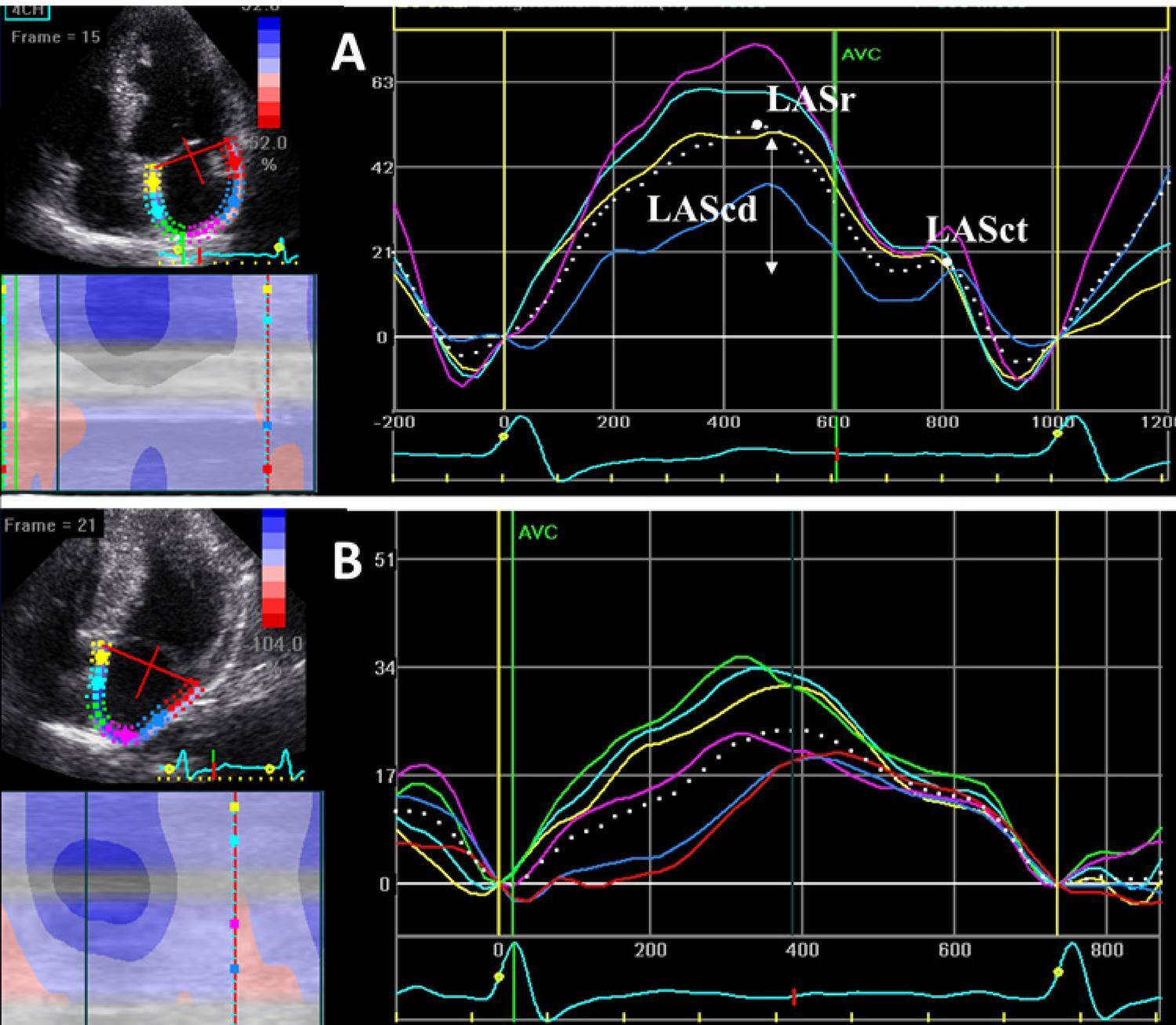
Atrial Mechanics in Hypertrophic Cardiomyopathy: Discriminating between Ventricular Hypertrophy and Fibrosis

Patrícia Marques-Alves et al. João André Ferreira André Azul Freitas José Paulo Almeida … Rui Baptista Graça Castro Rui Martins Paulo Donato Maria João Ferreira Lino Gonçalves

Abstract

Background

Hypertrophic cardiomyopathy (HCM) and left ventricular hypertrophy (LVH) secondary to systemic hypertension (HTN) may be associated with left atrial (LA) functional abnormalities.

Objectives

We aimed to characterize LA mechanics in HCM and HTN and determine any correlation with the extent of left ventricular (LV) fibrosis measured by cardiac magnetic resonance (CMR) in HCM patients.

Methods

Two-dimensional speckle tracking-derived longitudinal LA function was acquired from apical views in 60 HCM patients, 60 HTN patients, and 34 age-matched controls. HCM patients also underwent CMR, with measurement of late gadolinium enhancement (LGE) extension. Association with LA strain parameters was analyzed. Statistical significance was set at p<0.05.

Results

Mean LV ejection fraction was not different between the groups. The E/e’ ratio was impaired in the HCM group and preserved in the control group. LA mechanics was significantly reduced in HCM, compared to the HTN group. LA strain rate in reservoir (LASRr) and in contractile (LASRct) phases were the best discriminators of HCM, with an area under the curve (AUC) of 0.8, followed by LA strain in reservoir phase (LASr) (AUC 0.76). LASRr and LASR-ct had high specificity (89% and 91%, respectively) and LASr had sensitivity of 80%. A decrease in 2.79% of LA strain rate in conduit phase (LASRcd) predicted an increase of 1cm in LGE extension (r2=0.42, β 2.79, p=0.027).

Conclusions

LASRr and LASRct were the best discriminators for LVH secondary to HCM. LASRcd predicted the degree of LV fibrosis assessed by CMR. These findings suggest that LA mechanics is a potential predictor of disease severity in HCM.

  • Cardiomyopathy, Hypertrophic
  • Echocardiography/methods
  • Hypertension
  • Left Ventricular Hypertrophy
  • Magnetic Resonance Spectroscopy/ methods
Read Full Article
Creative Commons Attribution (CC BY) Creative Commons license
21/02/2022 DOI: 10.36660/abc.20200890 Views 1
ABCIC
Original Article

The Value of Two-dimensional Strain in the Diagnosis of Acute Myocarditis: Comparison with Cardiac Magnetic Resonance Imaging

Marcio Vinícius Lins Barros et al. Maria Helena Albernaz Siqueira Fábio Junior Modesto Silva Isabelle Penna Santos … Leandro Minelli Reis Pedro Henrique Costa Silva Carlos Eduardo Ornelas

Abstract

Introduction

Acute myocarditis is a major cause of sudden death in young patients. Cardiac magnetic resonance imaging (CMR) is a sensitive noninvasive method to detect myocarditis, but it is costly and unavailable in most medical centers. Two-dimensional strain is a new echocardiographic technique that evaluates myocardial strain to analyze global and regional myocardial function.

Objetive

To assess the value of two-dimensional strain in patients diagnosed with myocarditis.

Material and methods

We prospectively studied patients with acute myocarditis and normal cardiac contractility using CMR, who underwent conventional echocardiography and two-dimensional strain. The ventricular myocardium was divided into 16 segments and these segments were divided into two groups. Group 0: normal myocardial segment by CMR. Group 1: myocardial segment compatible with myocarditis using CMR.

Results

The study evaluated 28 patients, including 82,1% (n = 46) males with a mean age of 35.6 ± 8.9 years. Of the 448 myocardial segments evaluated, 316 segments were normal (group 0) and 132 segments (group 1) were diagnosed with myocarditis using the late enhancement technique on CMR. The analysis of two-dimensional strain showed a significant difference between the groups (19.6 ± 2.9 versus 15.4 ± 2.8 p = 0.001), with 75% sensitivity and 79% specificity and AUC of 0.86 (95% CI 0.82 to 0.89).

Conclusion

Two-dimensional strain can be useful in the evaluation of patients with myocarditis, normal contractility by CMR and conventional echocardiography. (Arq Bras Cardiol: Imagem cardiovasc. 2017;30(1):3-7)

  • Echocardiography/methods
  • Magnetic Resonance Spectroscopy/ methods
  • Myocarditis
Read Full Article
Creative Commons Attribution (CC BY) Creative Commons license
20/01/2022 DOI: 10.5935/2318-8219.20170003 Views 2
ABCIC
Viewpoint

Non-compaction cardiomyopathy: How Can Imaging Contribute?

José de Arimateia Batista Araújo Filho et al. Antonildes Nascimento Assunção-Jr José Rodrigues Parga

[…]

  • Arrhythmias Cardiac
  • Cardiomyopathies/physiopathology
  • Diagnostic Imaging/methods
  • Heart Failure
  • Magnetic Resonance Spectroscopy/ methods
  • Thromboembolism
Read Full Article
Creative Commons Attribution (CC BY) Creative Commons license
20/01/2022 DOI: 10.5935/2318-8219.20190004
ABCIC
Editorial

Choice of Imaging Methods in Hypertrophic Cardiomyopathy

Murillo de Oliveira Antunes et al. Edmundo Arteaga-Fernández Charles Mady

[…]

  • Cardiomyopathy,Hypertrophic
  • Diagnostic Imaging
  • Discrete Subaortic Stenosis
  • Echocardiography/methods
  • Electrocardiography/methods
  • Magnetic Resonance Spectroscopy/ methods
  • Mitral Valve Insufficiency
Read Full Article
Creative Commons Attribution (CC BY) Creative Commons license
20/01/2022 DOI: 10.5935/2318-8219.20190003 Views 1
ABCIC
Case Report

Right Ventricular Myxoma

Cristina Schneider et al. Maria Natividade Santos Costa Lopes Simone Ferreira Leite Camila Costa Oliveira … Nestor Sabatovicz Jr Maria Cristina Resende

[…]

  • Echocardiography/methods
  • Magnetic Resonance Spectroscopy/ methods
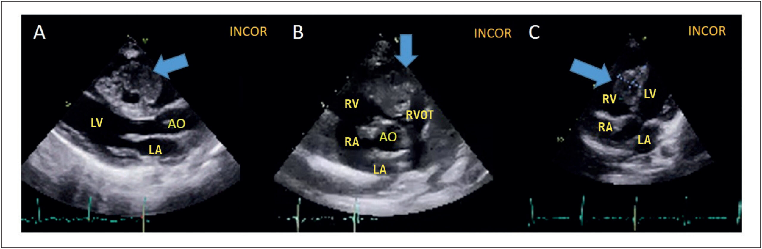
  • Mixoma/ diagnostic imaging
  • Mixoma/ surgery
  • Ventricular Dysfunction Right
Read Full Article
Creative Commons Attribution (CC BY) Creative Commons license
20/01/2022 DOI: 10.5935/2318-8219.20180007
Figure 1
ABC
Original Article

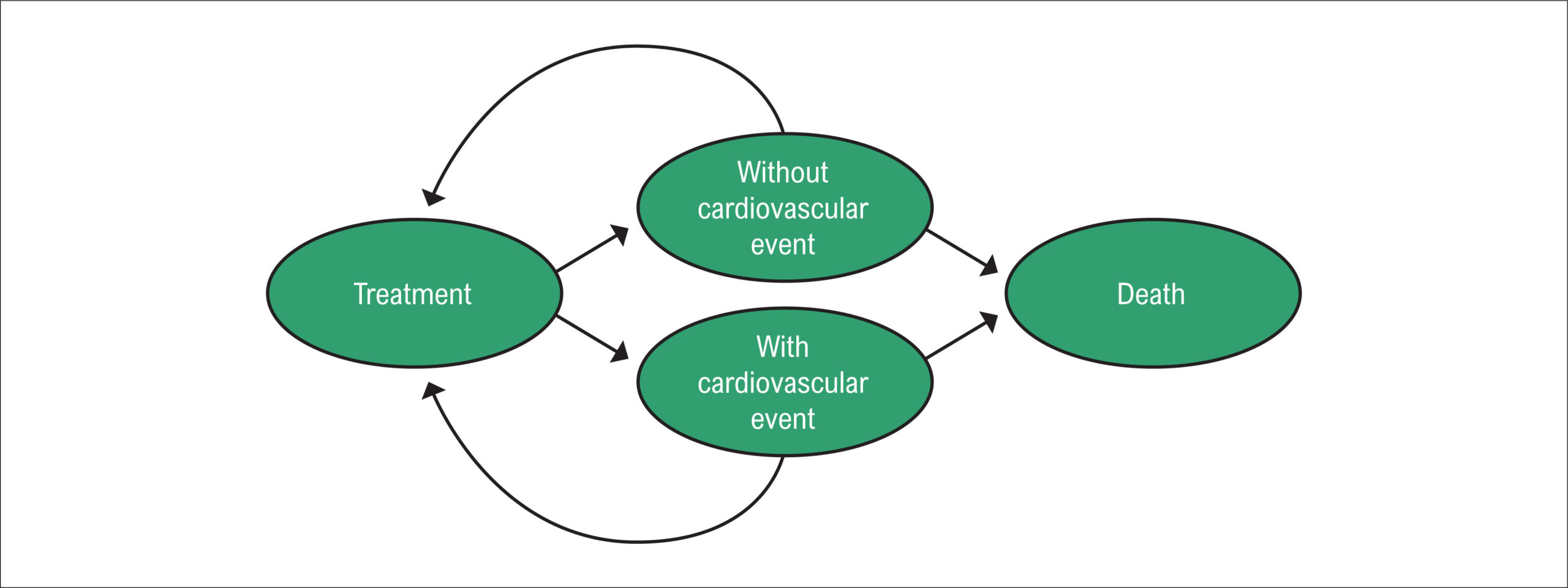
Cost-Effectiveness Analysis of Evolocumab Therapy in Patients at High Risk of Cardiovascular Events in the Context of the Brazilian Unified Health System

Luiza Latado et al. Rodrigo Morel Vieira de Melo Sóstenes Mistro Adriana Lopes Latado … Harrison Floriano do Nascimento Yasmin Menezes Lira Natalia Ferreira Cardoso de Oliveira Yuri de Santana Galindo Tainá Viana Luiz Carlos Santana Passos

Abstract

Background:

Hypertrophic cardiomyopathy (HCM) and left ventricular hypertrophy (LVH) secondary to systemic hypertension (HTN) may be associated with left atrial (LA) functional abnormalities.

Objectives:

We aimed to characterize LA mechanics in HCM and HTN and determine any correlation with the extent of left ventricular (LV) fibrosis measured by cardiac magnetic resonance (CMR) in HCM patients.

Methods:

Two-dimensional speckle tracking-derived longitudinal LA function was acquired from apical views in 60 HCM patients, 60 HTN patients, and 34 age-matched controls. HCM patients also underwent CMR, with measurement of late gadolinium enhancement (LGE) extension. Association with LA strain parameters was analyzed. Statistical significance was set at p<0.05.

Results:

Mean LV ejection fraction was not different between the groups. The E/e’ ratio was impaired in the HCM group and preserved in the control group. LA mechanics was significantly reduced in HCM, compared to the HTN group. LA strain rate in reservoir (LASRr) and in contractile (LASRct) phases were the best discriminators of HCM, with an area under the curve (AUC) of 0.8, followed by LA strain in reservoir phase (LASr) (AUC 0.76). LASRr and LASR-ct had high specificity (89% and 91%, respectively) and LASr had sensitivity of 80%. A decrease in 2.79% of LA strain rate in conduit phase (LASRcd) predicted an increase of 1cm in LGE extension (r2=0.42, β 2.79, p=0.027).

Conclusions:

LASRr and LASRct were the best discriminators for LVH secondary to HCM. LASRcd predicted the degree of LV fibrosis assessed by CMR. These findings suggest that LA mechanics is a potential predictor of disease severity in HCM.

  • Cardiomyopathy, Hypertrophic
  • Echocardiography/methods
  • Hypertension
  • Left Ventricular Hypertrophy
  • Magnetic Resonance Spectroscopy/ methods
Read Full Article
Creative Commons Attribution (CC BY) Creative Commons license
22/11/2021 DOI: 10.36660/abc.20200690 Views 1
ABC
Editorial

The Challenge of Echocardiography in the Accurate Assessment of the Right Ventricle and Pulmonary Insufficiency

Claudia R. Pinheiro de Castro Grau

The current recognition of the importance of the right ventricle (RV) role in the evolution and prognosis of various cardiovascular conditions is indisputable and the proper assessment of its dimension and function has a direct impact on clinical management.

Pulmonary insufficiency (PI) is common after tetralogy of Fallot repair (POTF) and approach of congenital heart disease with pulmonary obstruction, being is an important determinant of outcome due to its consequences, causing RV dilation and dysfunction, exercise intolerance, increased risk of arrhythmias and sudden death.

[…]

  • Heart Defects,Congenital
  • Magnetic Resonance Spectroscopy/ methods
  • Pulmonary Valve, Insufficiency
  • Tetralogy of Fallot/surgery
  • Ventricular Function, Right
Read Full Article
Creative Commons Attribution (CC BY) Creative Commons license
25/10/2021 DOI: 10.36660/abc.20210744 Views 2
ABC
Editorial

Myocardial Scar and Ventricular Repolarization on the electrocardiogram: Insights from Cardiac Magnetic Resonance Imaging

Luciano C. Amado

Myocardial fibrosis is a final common feature of most inflammatory pathway and is frequently seen in patients with heart failure with reduced ejection fraction (HFrEF). , Myocardial scar, detected by contrast enhanced magnetic resonance imaging (ce-MRI), has been shown to be a predictor of prognosis and sudden cardiac death in patients with HFrEF. – Cardiac MRI has emerged as a non-invasive gold standard for the assessment of scar and risk prediction of ventricular arrhythmias. , , , Nevertheless, cardiac MRI can be time consuming, expensive, and not easily available.

The 12-lead surface electrocardiogram (ECG) is a simple and inexpensive diagnostic tool that is widely used in clinical practice for the diagnosis of cardiac arrhythmias. The ECG waveform is the expression of the transmembrane action potentials and is composed of two distinct processes: depolarization and repolarization. Ventricular repolarization is a complex electrical phenomenon which in clinical practice is measured from the beginning of the QRS complex to the end of the T-wave. Cardiac structural and electrical changes may cause action potential abnormalities, affecting the refractory period and thus favoring the onset of ventricular arrhythmias.

[…]

  • Death, Sudden, Cardiac
  • Electrocardiography/methods
  • Endomyocardial Fibrosis
  • Heart Failure
  • Magnetic Resonance Spectroscopy/ methods
  • Prognosis
  • Stroke Volume
Read Full Article
Creative Commons Attribution (CC BY) Creative Commons license
25/10/2021 DOI: 10.36660/abc.20210712 Views 2
ABC
Editorial

T1 Mapping in Heart Failure: Prognostic Implications

Sarah Moharem-Elgamal

Myocardial fibrosis leads to impaired diastolic and systolic function and is associated with increased major adverse cardiovascular events. It is a structural correlate that can be found in the different stages of heart failure. The two main types of myocardial fibrosis are interstitial fibrosis and replacement fibrosis. Interstitial fibrosis is a reversible process that occurs early in the disease process as increased collagen synthesis in diffuse microscopic distribution within the myocardium and sometimes by localized perivascular distribution. Replacement fibrosis typically occurs in the later stages of the disease after irreversible myocyte injury or death, in which cell apoptosis triggers fibroblasts and promotes macroscopic deposition of collagen fibrous tissue in the myocardium.

Cardiac magnetic resonance (CMR) has the capability to accurately quantify ventricular volumes and ejection fraction, as well as the non-invasive characterization of the myocardium. These unique features have led to the increased use of CMR in the assessment of patients with heart failure (HF). CMR can detect the presence and extent of replacement fibrosis through late gadolinium enhancement imaging and diffuse interstitial fibrosis through native T1 mapping. Interstitial fibrosis identified by native T1 mapping has been used as a marker of disease activity,– risk stratification and monitoring of the therapeutic management in heart failure patients.

[…]

  • Cellular Apoptose Susceptibility Protein
  • Endomyocardial Fibrosis
  • Fibroblasts
  • Heart Failure
  • Magnetic Resonance Spectroscopy/ methods
  • Mortality
Read Full Article
Creative Commons Attribution (CC BY) Creative Commons license
17/05/2021 DOI: 10.36660/abc.20210205
Figure 1
ABC
Original Article

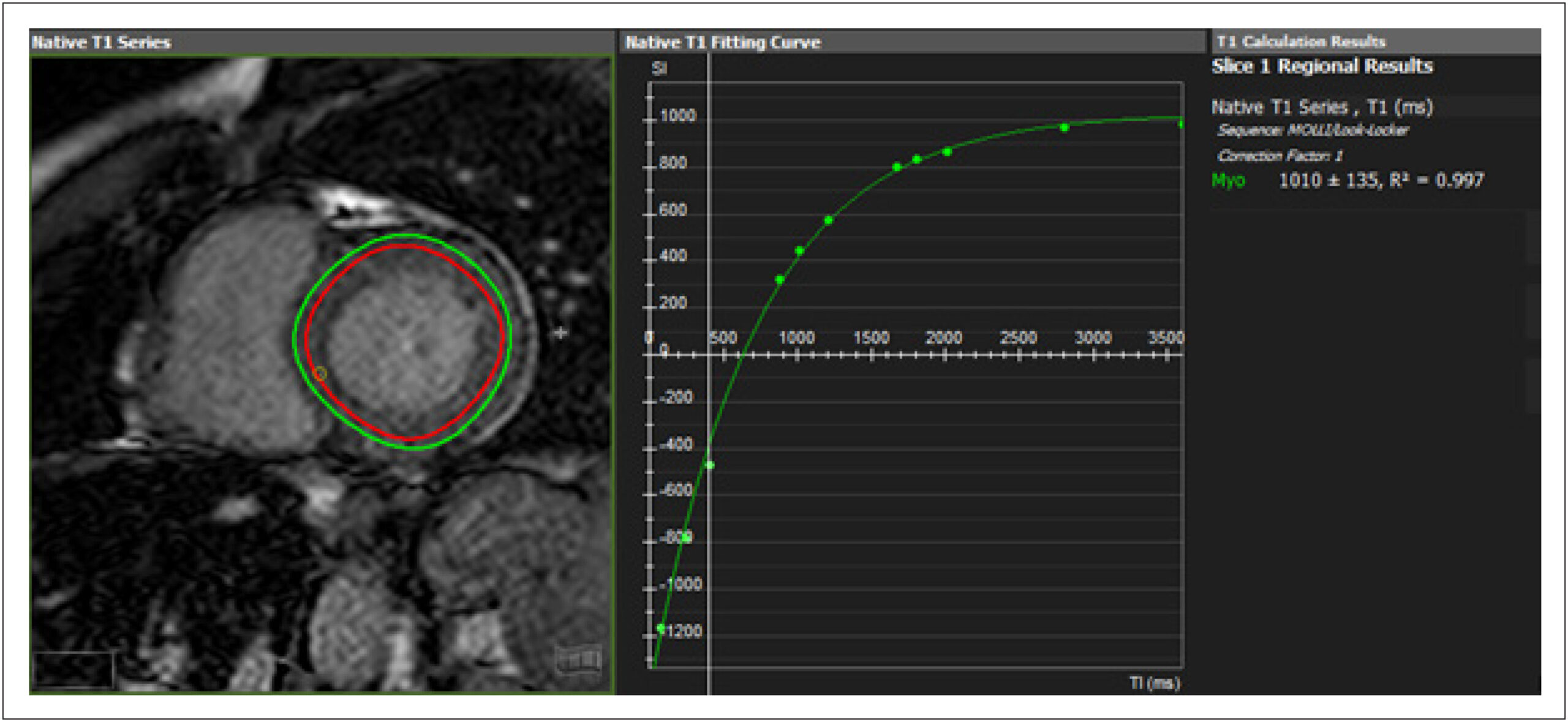
Clinical Heart Failure Stratification Through Native T1 Mapping: Experience of a Referral Service

Thiago dos Santos Silva Marques et al. André Maurício de Souza Fernandes Roberto Nery Dantas Júnior Robert W. Biederman … Ana Paula Marques de Oliveira Melo Roque Aras

Abstract

Background:

Diffuse cardiac fibrosis is an important factor in the prognostic assessment of patients with ventricular dysfunction. Cardiovascular magnetic resonance imaging (CMR) native T1 mapping is highly sensitive and considered an independent predictor of all-cause mortality and heart failure (HF) development in patients with cardiomyopathy.

Objectives:

To evaluate the feasibility of native T1 mapping assessment in patients with HF in a cardiology referral hospital and its association with structural parameters and functional profile.

Methods:

Cross-sectional study with adult patients with HF NYHA functional classes I and II, ischemic and non-ischemic, followed in a referral hospital, who underwent CMR. Native T1 values were analyzed for structural parameters, comorbidities, etiology, and categorization of HF by left ventricular ejection fraction (LVEF). Analyses were performed with a significance level of 5%.

Results:

Enrollment of 134 patients. Elevated native T1 values were found in patients with greater dilation (1004.9 vs 1042.7ms, p = 0.001), ventricular volumes (1021.3 vs 1050.3ms, p <0.01) and ventricular dysfunction (1010.1 vs 1053.4ms, p <0.001), also present when the non-ischemic group was analyzed separately. Patients classified as HF with reduced ejection fraction had higher T1 values than those with HF and preserved ejection fraction (HFPEF) (992.7 vs 1054.1ms, p <0.001). Of those with HFPEF, 55.2% had higher T1.

Conclusions:

CMR T1 mapping is feasible for clinical HF evaluation. There was a direct association between higher native T1 values and lower ejection fraction, and with larger LV diameters and volumes, regardless of the etiology of HF.
  • Cardiomyopathy, Dilated
  • Chagas Cardiomyopathy
  • Diagnosis Imaging
  • Fibrosis
  • Heart Failure
  • Magnetic Resonance Spectroscopy/ methods
  • Ventricular Dysfunction, Left
Read Full Article
Creative Commons Attribution (CC BY) Creative Commons license
17/05/2021 DOI: 10.36660/abc.20190782 Views 1
Figure 1
ABC
Image

Non-Progressive Hepatic Form of Andersen Disease as a Mimic of Hypertrophic Cardiomyopathy

Fabio Mastrocola et al. William Santos de Oliveira Adalberto Atsushi Porto Roberto Moreno Mendonça Nestor Rodrigues de Oliveira Neto

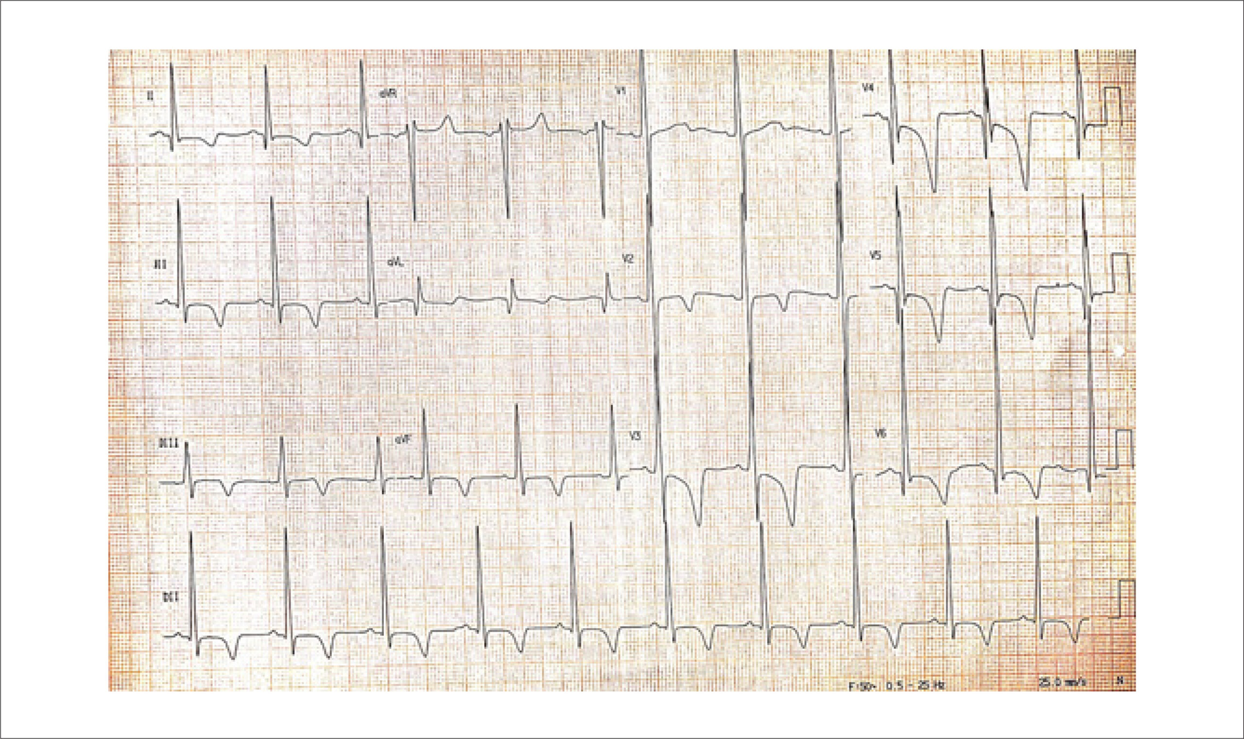
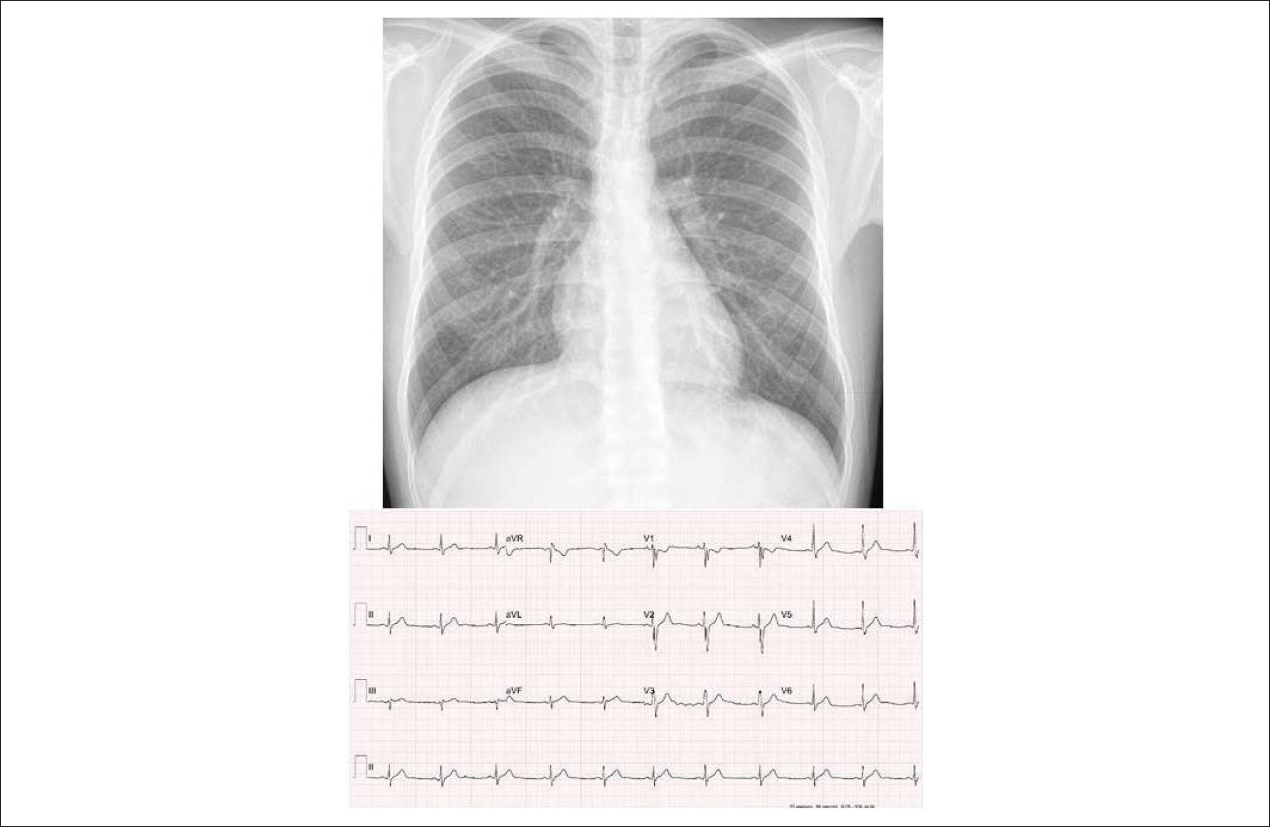
An 18-year-old male patient presented to our ambulatory services due to a 3-month course of progressive dyspnea and non-productive cough. Symptoms were present even at rest, and he exhibited severe limitation at physical activity. He had a diagnosis of glycogen storage disease (GSD) type IV attained by liver biopsy since three years of age, when he developed hepatomegaly and mild hepatic dysfunction. At the time, histopathological examination showed grade 2 fibrosis along with numerous intracytoplasmic PAS-positive deposits that were resistant to diastase. Thenceforth, his liver function remained stable, and he persisted otherwise asymptomatic. His physical examination was unremarkable, and routine laboratory evaluation was within the normal ranges. However, his basal ECG () showed signs of left ventricular hypertrophy. He was submitted to a cardiovascular magnetic resonance (CMR), which showed asymmetrical hypertrophy with predominance in the interventricular septum (). Late gadolinium enhancement (LGE) was present in a patchy, multifocal pattern (). Medical therapy for heart failure was initiated; the patient is now asymptomatic and maintains regular follow-up in our ambulatory.

Patients with classic GSD type IV have unrelenting liver disease with fast progression to cirrhosis during childhood; however, a small subset of affected individuals may present with milder hepatic dysfunction that does not advance to end-stage liver disease., Accordingly, our patient had the diagnosis of GSD type IV by three years of age, but did not develop cirrhosis afterwards.

[…]

  • Cardiomyopathy, Hypertrophic
  • Fibrosis
  • Glycogen Storage Disease Type IV
  • Heart Failure
  • Magnetic Resonance Spectroscopy/ methods
  • PRKAG2 Syndrome
  • Prognosis
Read Full Article
Creative Commons Attribution (CC BY) Creative Commons license
07/12/2020 DOI: 10.36660/abc.20200218
Figure 1
ABC
Image

Evolved Inferior Wall Myocardial Infarction with Left Ventricular Pseudoaneurysm: A Diagnostic Dilemma

Sónia Gomes Coelho et al. Clara F. Jorge Pedro B. Carlos Anne Delgado Leopoldina Vicente

Introduction

The left ventricular (LV) pseudoaneurysm (PA) is a rare mechanical complication of acute myocardial infarction (AMI). It results from myocardial rupture, in which the hemorrhagic process is contained by the adherent pericardium. It occurs most commonly in the inferior and posterior ventricular wall, since the rupture of the anterior ventricular wall usually leads to cardiac tamponade and immediate death, while the inferior-posterior face of the heart rests on the diaphragm, facilitating the ventricular cavity containment by the pericardium. – Imaging methods are crucial to establish the diagnosis. Transthoracic (TTE) and transesophageal echocardiography (TEE) allow the definitive diagnosis in 26% and 75% of cases, respectively. , Cardiac magnetic resonance (CMR) imaging is useful in the differential diagnosis of LV PA and aneurysm, with a reported sensitivity of 100%. The presence of late pericardial enhancement in the CMR is highly suggestive of LV PA, which may represent the effect of the passage of blood into the pericardial space at the time of myocardial rupture, with subsequent pericardial inflammation and fibrosis. , ,

[…]

  • Echocardiography/methods
  • Heart Rupture
  • Magnetic Resonance Spectroscopy/ methods
  • Myocardial Infarction/complications
  • Pseudoaneurysm
Read Full Article
Creative Commons Attribution (CC BY) Creative Commons license
29/05/2020 DOI: 10.36660/abc.20200029
ABC
Editorial

Is Magnetic Resonance Imaging Already an Appropriate Method for Evaluating Patients after Atrial Fibrillation Catheter Ablation?

Cristiano F. Pisani et al. Mauricio Scanavacca

For a long time, lack of knowledge regarding the physiopathology of atrial fibrillation (AF) limited the development of interventional techniques for treating it. Demonstrations that paroxysmal AF was triggered by extrasystoles and tachycardias mainly originating from inside the pulmonary veins ushered in a new era for treatment of AF. Since then, electrical isolation of the pulmonary veins has been the standard treatment for AF. Among specialists, achieving lasting electrical isolation of the pulmonary veins has been the main technical challenge, which has gradually been overcome in recent years with the implementation of new technology for more effective ablation, given that reconnections or previously isolated veins are the main cause of recurrences observed in these patients.

The challenge has been greater in patients with persistent AF, owing to its more complex physiopathology, which involves additional mechanisms that are little known, in addition to the pulmonary venous foci. It is known that metabolic alterations induced by excessive atrial work during repeated episodes of AF initially induce atrial electrical remodeling, characterized by functional and transient changes in ion channels of cell membranes that modulate atrial electrical activity, thus facilitating the appearance of trigger foci in other regions of the atria and conditions that favor increased persistence of AF.

[…]

  • Atria Premature Complexes
  • Atrial Fibrillation
  • Catheter Ablation
  • Gadolinium
  • Magnetic Resonance Spectroscopy/ methods
Read Full Article
Creative Commons Attribution (CC BY) Creative Commons license
29/05/2020 DOI: 10.36660/abc.20200204 Views 2
ABC
Editorial

Admission NT-ProBNP in Myocardial Infarction: an Alert Sign?

Luís Beck-da-Silva

This issue of Arquivos Brasileiros de Cardiologia brings a paper entitled “The Usefulness of Admission Plasma NT-pro BNP Level to Predict Left Ventricular Aneurysm Formation after Acute ST-Segment Elevation Myocardial Infarction”. The authors bring a cohort of 1,519 post-acute ST-segment elevation myocardial infarction (STEMI) who were followed-uP for at least six months. Despite its observational and retrograde design, the authors were straightforward in looking for predictive variables that could foresee the occurrence of left ventricular aneurysms (LVA). Among other major clinical aspects such as previous coronary artery bypass graft, post-MI heart failure, younger age, smoking and no-reflow phenomenon; authors highlighted the importance of high NT-proBNP at admission as a predictor of LVA formation after acute STEMI.

I would probably highlight one weakness and a potentially positive aspect of their work.

[…]

  • Echocardiography/diagnostic Imaging
  • Heart Failure
  • Magnetic Resonance Spectroscopy/ methods
  • Myocardial Infarction
  • Natriuretic Peptide Brain/metabolism
  • Natriuretic Peptides/metabolismo
  • Ventricular Dysfunction, Left
Read Full Article
Creative Commons Attribution (CC BY) Creative Commons license
02/12/2019 DOI: 10.36660/abc.20190685 Views 1
Figure 1
ABC
Clinicoradiological Correlation

Case 5/2019 – Naturally Evolving Ebstein’s Anomaly of Discrete Repercussion in 24-Year-old Asymptomatic Adult

Edmar Atik et al. Maria Angélica Binotto Alessandra Costa Barreto

Clinical data

Patient reports 3 episodes of paroxysmal tachycardia of up to 20 minutes, accompanied by precordial pain in the past 7 years, all with spontaneous reversal. Four years prior, the patient presented another tachycardia with signs of low output, including pallor, visual turbidity, cold extremities and mental confusion, requiring electrical reversion. On that occasion, ventricular tachycardia ablation in the right ventricular (RV) inflow tract through the right posterior accessory pathway was successfully performed. Ebstein’s anomaly diagnosis was given for the first time by echocardiography scan, at that time. Ever since, the patient remained asymptomatic, living a normal life and with no medication.

Physical examination: Good general condition, eupneic, acyanotic, normal pulses in the 4 limbs. Weight: 80 kg, Height: 180 cm, BP: 110 x 70 mmHg, HR: 70 bpm, Sat O2 = 94%.

[…]

  • Cardiac Output,Low
  • Ebstein Anomaly
  • Echocardiography/methods
  • Magnetic Resonance Spectroscopy/ methods
  • Tachycardia,Paroxysmal
  • Tricuspide Valve Insufficiency
Read Full Article
Creative Commons Attribution (CC BY) Creative Commons license
10/10/2019 DOI: 10.5935/abc.20190158 Views 1
ABC
Editorial

Myocardial Fibrosis in Hypertrophic Cardiomyopathy: What Remains to be Proven?

Leonardo Sara

Hypertrophic cardiomyopathy (HCM) is a complex and well-characterized disease. Its prevalence in the general population is 1 in 500 individuals, with some data suggesting an even higher prevalence when using associated genetic criteria. This means that, based on a conservative estimate, cities such as Goiânia-GO or Recife-PE have approximately more than 3,000 individuals with the disease, a figure that can be as high as 25,000 individuals in a large city such as São Paulo.

Although HCM is perceived as a rare and usually very severe disease, related to dramatic episodes of sudden death (SD) in young individuals and athletes, it is a disease with low mortality. Due to advances in the diagnosis, treatment and prevention of SD, current estimates indicate a mortality rate of 0.5% a year, similar to that found in the general population, with most patients with the disease being asymptomatic or minimally symptomatic. In this scenario, the correct identification of subgroups of individuals with a higher risk of unfavorable outcome and, therefore, a greater chance of benefit when submitted to specific therapeutic strategies, such as the use of implantable cardioverter defibrillators (ICD), is of utmost importance.

[…]

  • Cardiomyopathy, Hypertrophic
  • Death, Sudden, Cardiac/prevention & control
  • Heart Failure
  • Magnetic Resonance Spectroscopy/ methods
  • Prevalence
Read Full Article
Creative Commons Attribution (CC BY) Creative Commons license
01/03/2019 DOI: 10.5935/abc.20190043
Figure 1
ABC
Case Report

Myocardial Infarction Due to an Anomalous Origin of the Left Coronary Artery with Unique Aggravating Features

Jorge Alberto Silva Estrada et al. Alejandra Domínguez Camacho Lorenzo Reyes de la Cruz Jesus Reyna Figueroa

Introduction

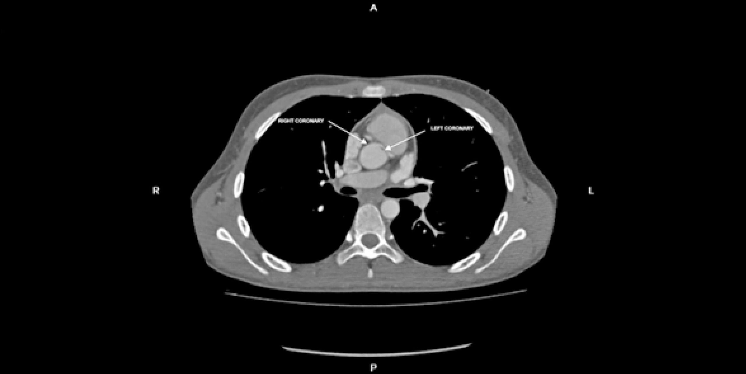
Anomalous left coronary artery arising at the right sinus of Valsalva is a relatively rare congenital cardiac anomaly that can cause myocardial ischemia. It can follow one out of five aberrant courses: interarterial, subpulmonic, pre-pulmonic, retroaortic, or retrocardiac.

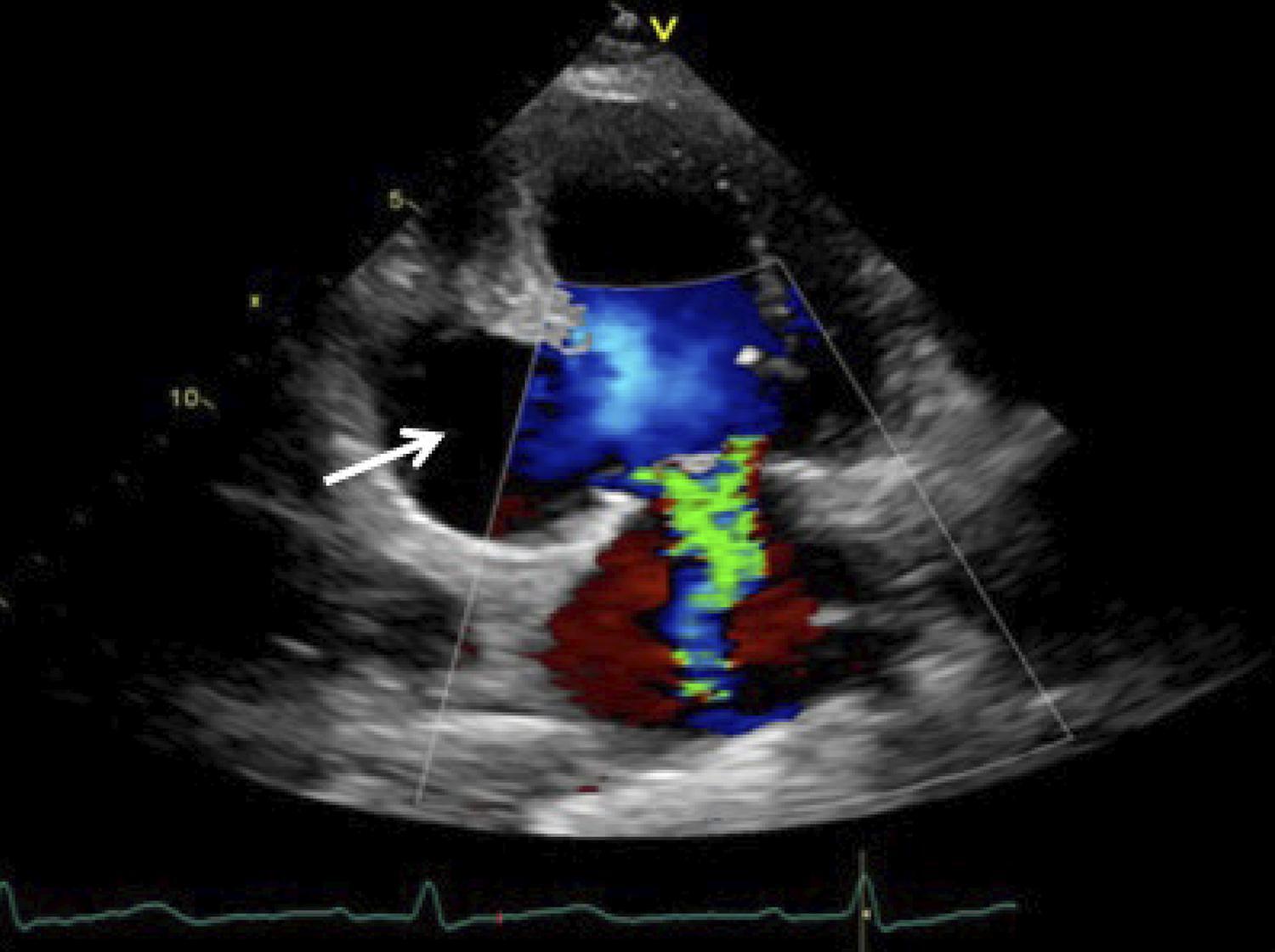
We present the case of a young patient with an anomalous left coronary artery arising at the right sinus of Valsalva with severe stenosis and hypoplasia throughout the interarterial segment, which hampered corrective surgery. The timely detection and adequate treatment of this specific anomaly gains relevance because of its association with increased risk of sudden cardiac death. Although the true prevalence of interarterial anomalous left coronary artery is unknown, owing to the lack of population-wide screening studies and at times asymptomatic course, its frequency has been reported at 0.03%. Imaging techniques allow the characterization of the coronary anomalous origin, course, morphology and surrounding structures. Transthoracic echocardiography, magnetic resonance angiography, and computed tomography (CT) angiography are first line noninvasive tests available while invasive tests such as coronary angiography and intravascular ultrasound are second line diagnostic alternatives. Treatment approaches are still controversial and election of the optimal surgical procedure, whenever applicable, must be an individualized and patient-centered decision.

[…]

  • Echocardiography/methods
  • Heart Defects, Congenital
  • Magnetic Resonance Spectroscopy/ methods
  • Myocardial Infarction
  • Myocardial Revascularization
Read Full Article
Creative Commons Attribution (CC BY) Creative Commons license
01/01/2019 DOI: 10.5935/abc.20180217

How to cite this article

EndNote RIS

Electronic Document Format (Vancouver)

Romero CE, Guacho WRL, Paula KR, Jimenez RP, Fonseca EKUN. Rare Presentation of Yolk Sac Tumor with Cardiac Involvement: Characteristics Detected by MRI. Arquivos Brasileiros de Cardiologia 2022;119(1):136-8.

Electronic Document Format (ABNT)

Romero, Cristhian Espinoza; Guacho, Williams Roberto Lata; Paula, Kevin Rafael de; Jimenez, Robert Paladines; Fonseca, Eduardo Kaiser Ururahy Nunes. Rare Presentation of Yolk Sac Tumor with Cardiac Involvement: Characteristics Detected by MRI. Arquivos Brasileiros de Cardiologia, v. 119, n. 1, p. 136-138, Jul. 2022.

Electronic Document Format (APA)

Romero, C. E., Guacho, W. R. L., Paula, K. R. , Jimenez, R. P., & Fonseca, E. K. U. N. (2022). Rare Presentation of Yolk Sac Tumor with Cardiac Involvement: Characteristics Detected by MRI. Arquivos Brasileiros de Cardiologia, 119(1), 136-138.

Electronic Document Format (ISO)

Romero, Cristhian Espinoza and Guacho, Williams Roberto Lata and Paula, Kevin Rafael de and Jimenez, Robert Paladines and Fonseca, Eduardo Kaiser Ururahy Nunes. Rare Presentation of Yolk Sac Tumor with Cardiac Involvement: Characteristics Detected by MRI. Arquivos Brasileiros de Cardiologia [online]. 2022, vol. 119, n. 1, [cited 2026-10-14], pp. 136-138. Available from: <https://journals.cardiol.br/en/article/rare-presentation-of-yolk-sac-tumor-with-cardiac-involvement-characteristics-detected-by-mri/>. ISSN 0066-782X

EndNote RIS

Electronic Document Format (Vancouver)

Ferreira VV, Thomas B. Stress CMR in the Elderly: Does It Provide the Answers?. Arquivos Brasileiros de Cardiologia 2022;119(1):107-8.

Electronic Document Format (ABNT)

Ferreira, Vera Vaz; Thomas, Boban. Stress CMR in the Elderly: Does It Provide the Answers?. Arquivos Brasileiros de Cardiologia, v. 119, n. 1, p. 107-108, Jul. 2022.

Electronic Document Format (APA)

Ferreira, V. V., & Thomas, B. (2022). Stress CMR in the Elderly: Does It Provide the Answers?. Arquivos Brasileiros de Cardiologia, 119(1), 107-108.

Electronic Document Format (ISO)

Ferreira, Vera Vaz and Thomas, Boban. Stress CMR in the Elderly: Does It Provide the Answers?. Arquivos Brasileiros de Cardiologia [online]. 2022, vol. 119, n. 1, [cited 2026-10-14], pp. 107-108. Available from: <https://journals.cardiol.br/en/article/stress-cmr-in-the-elderly-does-it-provide-the-answers/>. ISSN 0066-782X

EndNote RIS

Electronic Document Format (Vancouver)

Salazar D, Romero CE, Lopes MP, Branco CEB, Soares Junior J, Santis A, Sampaio RO, et al. Cardiac Magnetic Resonance and Positron Emission Tomography in the Diagnosis and Follow-Up of Acute Rheumatic Fever – Case Report. International Journal of Cardiovascular Sciences 2022;36:e20210093.

Electronic Document Format (ABNT)

Salazar, David; Romero, Cristhian Espinoza; Lopes, Mariana Pezzute; Branco, Carlos Eduardo de Barros; Soares Junior, José; Santis, Antonio de; Sampaio, Roney Orismar; Tarasoutchi, Flávio. Cardiac Magnetic Resonance and Positron Emission Tomography in the Diagnosis and Follow-Up of Acute Rheumatic Fever – Case Report. International Journal of Cardiovascular Sciences, v. 36, e20210093, Apr. 2022.

Electronic Document Format (APA)

Salazar, D., Romero, C. E., Lopes, M. P., Branco, C. E. B., Soares Junior, J., Santis, A. , Sampaio, R. O., & Tarasoutchi, F. (2022). Cardiac Magnetic Resonance and Positron Emission Tomography in the Diagnosis and Follow-Up of Acute Rheumatic Fever – Case Report. International Journal of Cardiovascular Sciences, 36, e20210093.

Electronic Document Format (ISO)

Salazar, David and Romero, Cristhian Espinoza and Lopes, Mariana Pezzute and Branco, Carlos Eduardo de Barros and Soares Junior, José and Santis, Antonio de and Sampaio, Roney Orismar and Tarasoutchi, Flávio. Cardiac Magnetic Resonance and Positron Emission Tomography in the Diagnosis and Follow-Up of Acute Rheumatic Fever – Case Report. International Journal of Cardiovascular Sciences [online]. 2022, vol. 36, [cited 2026-10-14], e20210093. Available from: <https://journals.cardiol.br/en/article/cardiac-magnetic-resonance-and-positron-emission-tomography-in-the-diagnosis-and-follow-up-of-acute-rheumatic-fever-case-report/>. ISSN 2359-4802

EndNote RIS

Electronic Document Format (Vancouver)

Marques-Alves P, Ferreira JA, Freitas AA, Almeida JP, Baptista R, Castro G, Martins R, et al. Atrial Mechanics in Hypertrophic Cardiomyopathy: Discriminating between Ventricular Hypertrophy and Fibrosis. Arquivos Brasileiros de Cardiologia 2022;118(1):77-8.

Electronic Document Format (ABNT)

Marques-Alves, Patrícia; Ferreira, João André; Freitas, André Azul; Almeida, José Paulo; Baptista, Rui; Castro, Graça; Martins, Rui; Donato, Paulo; Ferreira, Maria João; Gonçalves, Lino. Atrial Mechanics in Hypertrophic Cardiomyopathy: Discriminating between Ventricular Hypertrophy and Fibrosis. Arquivos Brasileiros de Cardiologia, v. 118, n. 1, p. 77-87, Feb. 2022.

Electronic Document Format (APA)

Marques-Alves, P., Ferreira, J. A., Freitas, A. A., Almeida, J. P., Baptista, R., Castro, G., Martins, R., Donato, P., Ferreira, M. J., & Gonçalves, L. (2022). Atrial Mechanics in Hypertrophic Cardiomyopathy: Discriminating between Ventricular Hypertrophy and Fibrosis. Arquivos Brasileiros de Cardiologia, 118(1), 77-87.

Electronic Document Format (ISO)

Marques-Alves, Patrícia and Ferreira, João André and Freitas, André Azul and Almeida, José Paulo and Baptista, Rui and Castro, Graça and Martins, Rui and Donato, Paulo and Ferreira, Maria João and Gonçalves, Lino. Atrial Mechanics in Hypertrophic Cardiomyopathy: Discriminating between Ventricular Hypertrophy and Fibrosis. Arquivos Brasileiros de Cardiologia [online]. 2022, vol. 118, n. 1, [cited 2026-10-14], pp. 77-87. Available from: <https://journals.cardiol.br/en/article/atrial-mechanics-in-hypertrophic-cardiomyopathy-discriminating-between-ventricular-hypertrophy-and-fibrosis/>. ISSN 0066-782X

EndNote RIS

Electronic Document Format (Vancouver)

Barros MVL, Siqueira MHA, Silva FJM, Santos IP, Reis LM, Silva PHC, Ornelas CE, et al. The Value of Two-dimensional Strain in the Diagnosis of Acute Myocarditis: Comparison with Cardiac Magnetic Resonance Imaging. Arquivos Brasileiros de Cardiologia Imagem cardiovascular 2022;30(1):3-7.

Electronic Document Format (ABNT)

Barros, Marcio Vinícius Lins; Siqueira, Maria Helena Albernaz; Silva, Fábio Junior Modesto; Santos, Isabelle Penna; Reis, Leandro Minelli; Silva, Pedro Henrique Costa; Ornelas, Carlos Eduardo. The Value of Two-dimensional Strain in the Diagnosis of Acute Myocarditis: Comparison with Cardiac Magnetic Resonance Imaging. Arquivos Brasileiros de Cardiologia Imagem cardiovascular, v. 30, n. 1, p. 3-7, Jan. 2022.

Electronic Document Format (APA)

Barros, M. V. L., Siqueira, M. H. A., Silva, F. J. M., Santos, I. P., Reis, L. M., Silva, P. H. C., & Ornelas, C. E. (2022). The Value of Two-dimensional Strain in the Diagnosis of Acute Myocarditis: Comparison with Cardiac Magnetic Resonance Imaging. Arquivos Brasileiros de Cardiologia Imagem cardiovascular, 30(1), 3-7.

Electronic Document Format (ISO)

Barros, Marcio Vinícius Lins and Siqueira, Maria Helena Albernaz and Silva, Fábio Junior Modesto and Santos, Isabelle Penna and Reis, Leandro Minelli and Silva, Pedro Henrique Costa and Ornelas, Carlos Eduardo. The Value of Two-dimensional Strain in the Diagnosis of Acute Myocarditis: Comparison with Cardiac Magnetic Resonance Imaging. Arquivos Brasileiros de Cardiologia Imagem cardiovascular [online]. 2022, vol. 30, n. 1, [cited 2026-10-14], pp. 3-7. Available from: <https://journals.cardiol.br/en/article/the-value-of-two-dimensional-strain-in-the-diagnosis-of-acute-myocarditis-comparison-with-cardiac-magnetic-resonance-imaging/>. ISSN 2318-8219

EndNote RIS

Electronic Document Format (Vancouver)

Araújo Filho JAB, Assunção-Jr AN, Parga JR. Non-compaction cardiomyopathy: How Can Imaging Contribute?. Arquivos Brasileiros de Cardiologia Imagem cardiovascular 2022;32(1):83-4.

Electronic Document Format (ABNT)

Araújo Filho, José de Arimateia Batista; Assunção-Jr, Antonildes Nascimento; Parga, José Rodrigues. Non-compaction cardiomyopathy: How Can Imaging Contribute?. Arquivos Brasileiros de Cardiologia Imagem cardiovascular, v. 32, n. 1, p. 83-84, Jan. 2022.

Electronic Document Format (APA)

Araújo Filho, J. A. B., Assunção-Jr, A. N., & Parga, J. R. (2022). Non-compaction cardiomyopathy: How Can Imaging Contribute?. Arquivos Brasileiros de Cardiologia Imagem cardiovascular, 32(1), 83-84.

Electronic Document Format (ISO)

Araújo Filho, José de Arimateia Batista and Assunção-Jr, Antonildes Nascimento and Parga, José Rodrigues. Non-compaction cardiomyopathy: How Can Imaging Contribute?. Arquivos Brasileiros de Cardiologia Imagem cardiovascular [online]. 2022, vol. 32, n. 1, [cited 2026-10-14], pp. 83-84. Available from: <https://journals.cardiol.br/en/article/non-compaction-cardiomyopathy-how-can-imaging-contribute/>. ISSN 2318-8219

EndNote RIS

Electronic Document Format (Vancouver)

Antunes MO, Arteaga-Fernández E, Mady C. Choice of Imaging Methods in Hypertrophic Cardiomyopathy. Arquivos Brasileiros de Cardiologia Imagem cardiovascular 2022;32(1):4-5.

Electronic Document Format (ABNT)

Antunes, Murillo de Oliveira; Arteaga-Fernández, Edmundo; Mady, Charles. Choice of Imaging Methods in Hypertrophic Cardiomyopathy. Arquivos Brasileiros de Cardiologia Imagem cardiovascular, v. 32, n. 1, p. 4-5, Jan. 2022.

Electronic Document Format (APA)

Antunes, M. O., Arteaga-Fernández, E., & Mady, C. (2022). Choice of Imaging Methods in Hypertrophic Cardiomyopathy. Arquivos Brasileiros de Cardiologia Imagem cardiovascular, 32(1), 4-5.

Electronic Document Format (ISO)

Antunes, Murillo de Oliveira and Arteaga-Fernández, Edmundo and Mady, Charles. Choice of Imaging Methods in Hypertrophic Cardiomyopathy. Arquivos Brasileiros de Cardiologia Imagem cardiovascular [online]. 2022, vol. 32, n. 1, [cited 2026-10-14], pp. 4-5. Available from: <https://journals.cardiol.br/en/article/choice-of-imaging-methods-in-hypertrophic-cardiomyopathy/>. ISSN 2318-8219

EndNote RIS

Electronic Document Format (Vancouver)

Schneider C, Lopes MNSC, Leite SF, Oliveira CC, Sabatovicz Jr N, Resende MC. Right Ventricular Myxoma. Arquivos Brasileiros de Cardiologia Imagem cardiovascular 2022;31(1):19-21.

Electronic Document Format (ABNT)

Schneider, Cristina; Lopes, Maria Natividade Santos Costa; Leite, Simone Ferreira; Oliveira, Camila Costa; Sabatovicz Jr, Nestor; Resende, Maria Cristina. Right Ventricular Myxoma. Arquivos Brasileiros de Cardiologia Imagem cardiovascular, v. 31, n. 1, p. 19-21, Jan. 2022.

Electronic Document Format (APA)

Schneider, C., Lopes, M. N. S. C., Leite, S. F., Oliveira, C. C., Sabatovicz Jr, N., & Resende, M. C. (2022). Right Ventricular Myxoma. Arquivos Brasileiros de Cardiologia Imagem cardiovascular, 31(1), 19-21.

Electronic Document Format (ISO)

Schneider, Cristina and Lopes, Maria Natividade Santos Costa and Leite, Simone Ferreira and Oliveira, Camila Costa and Sabatovicz Jr, Nestor and Resende, Maria Cristina. Right Ventricular Myxoma. Arquivos Brasileiros de Cardiologia Imagem cardiovascular [online]. 2022, vol. 31, n. 1, [cited 2026-10-14], pp. 19-21. Available from: <https://journals.cardiol.br/en/article/right-ventricular-myxoma/>. ISSN 2318-8219

EndNote RIS

Electronic Document Format (Vancouver)

Latado L, Melo RMV, Mistro S, Latado AL, Nascimento HF, Lira YM, Oliveira NFC, et al. Cost-Effectiveness Analysis of Evolocumab Therapy in Patients at High Risk of Cardiovascular Events in the Context of the Brazilian Unified Health System. Arquivos Brasileiros de Cardiologia 2021;117(5):988-96.

Electronic Document Format (ABNT)

Latado, Luiza; Melo, Rodrigo Morel Vieira de; Mistro, Sóstenes; Latado, Adriana Lopes; Nascimento, Harrison Floriano do; Lira, Yasmin Menezes; Oliveira, Natalia Ferreira Cardoso de; Galindo, Yuri de Santana; Viana, Tainá; Passos, Luiz Carlos Santana. Cost-Effectiveness Analysis of Evolocumab Therapy in Patients at High Risk of Cardiovascular Events in the Context of the Brazilian Unified Health System. Arquivos Brasileiros de Cardiologia, v. 117, n. 5, p. 988-996, Nov. 2021.

Electronic Document Format (APA)

Latado, L., Melo, R. M. V. , Mistro, S., Latado, A. L., Nascimento, H. F. , Lira, Y. M., Oliveira, N. F. C. , Galindo, Y. S., Viana, T., & Passos, L. C. S. (2021). Cost-Effectiveness Analysis of Evolocumab Therapy in Patients at High Risk of Cardiovascular Events in the Context of the Brazilian Unified Health System. Arquivos Brasileiros de Cardiologia, 117(5), 988-996.

Electronic Document Format (ISO)

Latado, Luiza and Melo, Rodrigo Morel Vieira de and Mistro, Sóstenes and Latado, Adriana Lopes and Nascimento, Harrison Floriano do and Lira, Yasmin Menezes and Oliveira, Natalia Ferreira Cardoso de and Galindo, Yuri de Santana and Viana, Tainá and Passos, Luiz Carlos Santana. Cost-Effectiveness Analysis of Evolocumab Therapy in Patients at High Risk of Cardiovascular Events in the Context of the Brazilian Unified Health System. Arquivos Brasileiros de Cardiologia [online]. 2021, vol. 117, n. 5, [cited 2026-10-14], pp. 988-996. Available from: <https://journals.cardiol.br/en/article/cost-effectiveness-analysis-of-evolocumab-therapy-in-patients-at-high-risk-of-cardiovascular-events-in-the-context-of-the-brazilian-unified-health-system/>. ISSN 0066-782X

EndNote RIS

Electronic Document Format (Vancouver)

Grau CRPC. The Challenge of Echocardiography in the Accurate Assessment of the Right Ventricle and Pulmonary Insufficiency. Arquivos Brasileiros de Cardiologia 2021;117(4):699-700.

Electronic Document Format (ABNT)

Grau, Claudia R. Pinheiro de Castro. The Challenge of Echocardiography in the Accurate Assessment of the Right Ventricle and Pulmonary Insufficiency. Arquivos Brasileiros de Cardiologia, v. 117, n. 4, p. 699-700, Oct. 2021.

Electronic Document Format (APA)

Grau, C. R. P. C. (2021). The Challenge of Echocardiography in the Accurate Assessment of the Right Ventricle and Pulmonary Insufficiency. Arquivos Brasileiros de Cardiologia, 117(4), 699-700.

Electronic Document Format (ISO)

Grau, Claudia R. Pinheiro de Castro. The Challenge of Echocardiography in the Accurate Assessment of the Right Ventricle and Pulmonary Insufficiency. Arquivos Brasileiros de Cardiologia [online]. 2021, vol. 117, n. 4, [cited 2026-10-14], pp. 699-700. Available from: <https://journals.cardiol.br/en/article/the-challenge-of-echocardiography-in-the-accurate-assessment-of-the-right-ventricle-and-pulmonary-insufficiency/>. ISSN 0066-782X

EndNote RIS

Electronic Document Format (Vancouver)

Amado LC. Myocardial Scar and Ventricular Repolarization on the electrocardiogram: Insights from Cardiac Magnetic Resonance Imaging. Arquivos Brasileiros de Cardiologia 2021;117(4):688-9.

Electronic Document Format (ABNT)

Amado, Luciano C.. Myocardial Scar and Ventricular Repolarization on the electrocardiogram: Insights from Cardiac Magnetic Resonance Imaging. Arquivos Brasileiros de Cardiologia, v. 117, n. 4, p. 688-689, Oct. 2021.

Electronic Document Format (APA)

Amado, L. C. (2021). Myocardial Scar and Ventricular Repolarization on the electrocardiogram: Insights from Cardiac Magnetic Resonance Imaging. Arquivos Brasileiros de Cardiologia, 117(4), 688-689.

Electronic Document Format (ISO)

Amado, Luciano C.. Myocardial Scar and Ventricular Repolarization on the electrocardiogram: Insights from Cardiac Magnetic Resonance Imaging. Arquivos Brasileiros de Cardiologia [online]. 2021, vol. 117, n. 4, [cited 2026-10-14], pp. 688-689. Available from: <https://journals.cardiol.br/en/article/myocardial-scar-and-ventricular-repolarization-on-the-electrocardiogram-insights-from-cardiac-magnetic-resonance-imaging/>. ISSN 0066-782X

EndNote RIS

Electronic Document Format (Vancouver)

Moharem-Elgamal S. T1 Mapping in Heart Failure: Prognostic Implications. Arquivos Brasileiros de Cardiologia 2021;116(5):926-7.

Electronic Document Format (ABNT)

Moharem-Elgamal, Sarah. T1 Mapping in Heart Failure: Prognostic Implications. Arquivos Brasileiros de Cardiologia, v. 116, n. 5, p. 926-927, May. 2021.

Electronic Document Format (APA)

Moharem-Elgamal, S. (2021). T1 Mapping in Heart Failure: Prognostic Implications. Arquivos Brasileiros de Cardiologia, 116(5), 926-927.

Electronic Document Format (ISO)

Moharem-Elgamal, Sarah. T1 Mapping in Heart Failure: Prognostic Implications. Arquivos Brasileiros de Cardiologia [online]. 2021, vol. 116, n. 5, [cited 2026-10-14], pp. 926-927. Available from: <https://journals.cardiol.br/en/article/t1-mapping-in-heart-failure-prognostic-implications/>. ISSN 0066-782X

EndNote RIS

Electronic Document Format (Vancouver)

Marques TSS, Fernandes AMS, Dantas Júnior RN, Biederman RW, Melo APMO, Aras R. Clinical Heart Failure Stratification Through Native T1 Mapping: Experience of a Referral Service. Arquivos Brasileiros de Cardiologia 2021;116(5):919-25.

Electronic Document Format (ABNT)

Marques, Thiago dos Santos Silva; Fernandes, André Maurício de Souza; Dantas Júnior, Roberto Nery; Biederman, Robert W.; Melo, Ana Paula Marques de Oliveira; Aras, Roque. Clinical Heart Failure Stratification Through Native T1 Mapping: Experience of a Referral Service. Arquivos Brasileiros de Cardiologia, v. 116, n. 5, p. 919-925, May. 2021.

Electronic Document Format (APA)

Marques, T. S. S., Fernandes, A. M. S., Dantas Júnior, R. N., Biederman, R. W., Melo, A. P. M. O., & Aras, R. (2021). Clinical Heart Failure Stratification Through Native T1 Mapping: Experience of a Referral Service. Arquivos Brasileiros de Cardiologia, 116(5), 919-925.

Electronic Document Format (ISO)

Marques, Thiago dos Santos Silva and Fernandes, André Maurício de Souza and Dantas Júnior, Roberto Nery and Biederman, Robert W. and Melo, Ana Paula Marques de Oliveira and Aras, Roque. Clinical Heart Failure Stratification Through Native T1 Mapping: Experience of a Referral Service. Arquivos Brasileiros de Cardiologia [online]. 2021, vol. 116, n. 5, [cited 2026-10-14], pp. 919-925. Available from: <https://journals.cardiol.br/en/article/clinical-heart-failure-stratification-through-native-t1-mapping-experience-of-a-referral-service/>. ISSN 0066-782X

EndNote RIS

Electronic Document Format (Vancouver)

Mastrocola F, Oliveira WS, Porto AA, Mendonça RM, Oliveira Neto NR. Non-Progressive Hepatic Form of Andersen Disease as a Mimic of Hypertrophic Cardiomyopathy. Arquivos Brasileiros de Cardiologia 2020;115(5):964-6.

Electronic Document Format (ABNT)

Mastrocola, Fabio; Oliveira, William Santos de; Porto, Adalberto Atsushi; Mendonça, Roberto Moreno; Oliveira Neto, Nestor Rodrigues de. Non-Progressive Hepatic Form of Andersen Disease as a Mimic of Hypertrophic Cardiomyopathy. Arquivos Brasileiros de Cardiologia, v. 115, n. 5, p. 964-966, Dec. 2020.

Electronic Document Format (APA)

Mastrocola, F., Oliveira, W. S. , Porto, A. A., Mendonça, R. M., & Oliveira Neto, N. R. (2020). Non-Progressive Hepatic Form of Andersen Disease as a Mimic of Hypertrophic Cardiomyopathy. Arquivos Brasileiros de Cardiologia, 115(5), 964-966.

Electronic Document Format (ISO)

Mastrocola, Fabio and Oliveira, William Santos de and Porto, Adalberto Atsushi and Mendonça, Roberto Moreno and Oliveira Neto, Nestor Rodrigues de. Non-Progressive Hepatic Form of Andersen Disease as a Mimic of Hypertrophic Cardiomyopathy. Arquivos Brasileiros de Cardiologia [online]. 2020, vol. 115, n. 5, [cited 2026-10-14], pp. 964-966. Available from: <https://journals.cardiol.br/en/article/non-progressive-hepatic-form-of-andersen-disease-as-a-mimic-of-hypertrophic-cardiomyopathy/>. ISSN 0066-782X

EndNote RIS

Electronic Document Format (Vancouver)

Coelho SG, Jorge CF, Carlos PB, Delgado A, Vicente L. Evolved Inferior Wall Myocardial Infarction with Left Ventricular Pseudoaneurysm: A Diagnostic Dilemma. Arquivos Brasileiros de Cardiologia 2020;114(4):730-1.

Electronic Document Format (ABNT)

Coelho, Sónia Gomes; Jorge, Clara F.; Carlos, Pedro B.; Delgado, Anne; Vicente, Leopoldina. Evolved Inferior Wall Myocardial Infarction with Left Ventricular Pseudoaneurysm: A Diagnostic Dilemma. Arquivos Brasileiros de Cardiologia, v. 114, n. 4, p. 730-731, May. 2020.

Electronic Document Format (APA)

Coelho, S. G., Jorge, C. F., Carlos, P. B., Delgado, A., & Vicente, L. (2020). Evolved Inferior Wall Myocardial Infarction with Left Ventricular Pseudoaneurysm: A Diagnostic Dilemma. Arquivos Brasileiros de Cardiologia, 114(4), 730-731.

Electronic Document Format (ISO)

Coelho, Sónia Gomes and Jorge, Clara F. and Carlos, Pedro B. and Delgado, Anne and Vicente, Leopoldina. Evolved Inferior Wall Myocardial Infarction with Left Ventricular Pseudoaneurysm: A Diagnostic Dilemma. Arquivos Brasileiros de Cardiologia [online]. 2020, vol. 114, n. 4, [cited 2026-10-14], pp. 730-731. Available from: <https://journals.cardiol.br/en/article/evolved-inferior-wall-myocardial-infarction-with-left-ventricular-pseudoaneurysm-a-diagnostic-dilemma/>. ISSN 0066-782X

EndNote RIS

Electronic Document Format (Vancouver)

Pisani CF, Scanavacca M. Is Magnetic Resonance Imaging Already an Appropriate Method for Evaluating Patients after Atrial Fibrillation Catheter Ablation?. Arquivos Brasileiros de Cardiologia 2020;114(4):636-7.

Electronic Document Format (ABNT)

Pisani, Cristiano F.; Scanavacca, Mauricio. Is Magnetic Resonance Imaging Already an Appropriate Method for Evaluating Patients after Atrial Fibrillation Catheter Ablation?. Arquivos Brasileiros de Cardiologia, v. 114, n. 4, p. 636-637, May. 2020.

Electronic Document Format (APA)

Pisani, C. F., & Scanavacca, M. (2020). Is Magnetic Resonance Imaging Already an Appropriate Method for Evaluating Patients after Atrial Fibrillation Catheter Ablation?. Arquivos Brasileiros de Cardiologia, 114(4), 636-637.

Electronic Document Format (ISO)

Pisani, Cristiano F. and Scanavacca, Mauricio. Is Magnetic Resonance Imaging Already an Appropriate Method for Evaluating Patients after Atrial Fibrillation Catheter Ablation?. Arquivos Brasileiros de Cardiologia [online]. 2020, vol. 114, n. 4, [cited 2026-10-14], pp. 636-637. Available from: <https://journals.cardiol.br/en/article/is-magnetic-resonance-imaging-already-an-appropriate-method-for-evaluating-patients-after-atrial-fibrillation-catheter-ablation/>. ISSN 0066-782X

EndNote RIS

Electronic Document Format (Vancouver)

Beck-da-Silva L. Admission NT-ProBNP in Myocardial Infarction: an Alert Sign?. Arquivos Brasileiros de Cardiologia 2019;113(6):1138.

Electronic Document Format (ABNT)

Beck-da-Silva, Luís. Admission NT-ProBNP in Myocardial Infarction: an Alert Sign?. Arquivos Brasileiros de Cardiologia, v. 113, n. 6, p. 1138-1138, Dec. 2019.

Electronic Document Format (APA)

Beck-da-Silva, L. (2019). Admission NT-ProBNP in Myocardial Infarction: an Alert Sign?. Arquivos Brasileiros de Cardiologia, 113(6), 1138-1138.

Electronic Document Format (ISO)

Beck-da-Silva, Luís. Admission NT-ProBNP in Myocardial Infarction: an Alert Sign?. Arquivos Brasileiros de Cardiologia [online]. 2019, vol. 113, n. 6, [cited 2026-10-14], pp. 1138-1138. Available from: <https://journals.cardiol.br/en/article/admission-nt-probnp-in-myocardial-infarction-an-alert-sign/>. ISSN 0066-782X

EndNote RIS

Electronic Document Format (Vancouver)

Atik E, Binotto MA, Barreto AC. Case 5/2019 – Naturally Evolving Ebstein’s Anomaly of Discrete Repercussion in 24-Year-old Asymptomatic Adult. Arquivos Brasileiros de Cardiologia 2019;113(3):435-7.

Electronic Document Format (ABNT)

Atik, Edmar; Binotto, Maria Angélica; Barreto, Alessandra Costa. Case 5/2019 – Naturally Evolving Ebstein’s Anomaly of Discrete Repercussion in 24-Year-old Asymptomatic Adult. Arquivos Brasileiros de Cardiologia, v. 113, n. 3, p. 435-437, Oct. 2019.

Electronic Document Format (APA)

Atik, E., Binotto, M. A., & Barreto, A. C. (2019). Case 5/2019 – Naturally Evolving Ebstein’s Anomaly of Discrete Repercussion in 24-Year-old Asymptomatic Adult. Arquivos Brasileiros de Cardiologia, 113(3), 435-437.

Electronic Document Format (ISO)

Atik, Edmar and Binotto, Maria Angélica and Barreto, Alessandra Costa. Case 5/2019 – Naturally Evolving Ebstein’s Anomaly of Discrete Repercussion in 24-Year-old Asymptomatic Adult. Arquivos Brasileiros de Cardiologia [online]. 2019, vol. 113, n. 3, [cited 2026-10-14], pp. 435-437. Available from: <https://journals.cardiol.br/en/article/case-5-2019-naturally-evolving-ebsteins-anomaly-of-discrete-repercussion-in-24-year-old-asymptomatic-adult/>. ISSN 0066-782X

EndNote RIS

Electronic Document Format (Vancouver)

Sara L. Myocardial Fibrosis in Hypertrophic Cardiomyopathy: What Remains to be Proven?. Arquivos Brasileiros de Cardiologia 2019;112(3):290-1.

Electronic Document Format (ABNT)

Sara, Leonardo. Myocardial Fibrosis in Hypertrophic Cardiomyopathy: What Remains to be Proven?. Arquivos Brasileiros de Cardiologia, v. 112, n. 3, p. 290-291, Feb. 2019.

Electronic Document Format (APA)

Sara, L. (2019). Myocardial Fibrosis in Hypertrophic Cardiomyopathy: What Remains to be Proven?. Arquivos Brasileiros de Cardiologia, 112(3), 290-291.

Electronic Document Format (ISO)

Sara, Leonardo. Myocardial Fibrosis in Hypertrophic Cardiomyopathy: What Remains to be Proven?. Arquivos Brasileiros de Cardiologia [online]. 2019, vol. 112, n. 3, [cited 2026-10-14], pp. 290-291. Available from: <https://journals.cardiol.br/en/article/myocardial-fibrosis-in-hypertrophic-cardiomyopathy-what-remains-to-be-proven/>. ISSN 0066-782X

EndNote RIS

Electronic Document Format (Vancouver)

Estrada JAS, Camacho AD, Cruz LR, Figueroa JR. Myocardial Infarction Due to an Anomalous Origin of the Left Coronary Artery with Unique Aggravating Features. Arquivos Brasileiros de Cardiologia 2019;112(1):110-2.

Electronic Document Format (ABNT)

Estrada, Jorge Alberto Silva; Camacho, Alejandra Domínguez; Cruz, Lorenzo Reyes de la; Figueroa, Jesus Reyna. Myocardial Infarction Due to an Anomalous Origin of the Left Coronary Artery with Unique Aggravating Features. Arquivos Brasileiros de Cardiologia, v. 112, n. 1, p. 110-112, Dec. 2018.

Electronic Document Format (APA)

Estrada, J. A. S., Camacho, A. D., Cruz, L. R. , & Figueroa, J. R. (2019). Myocardial Infarction Due to an Anomalous Origin of the Left Coronary Artery with Unique Aggravating Features. Arquivos Brasileiros de Cardiologia, 112(1), 110-112.

Electronic Document Format (ISO)

Estrada, Jorge Alberto Silva and Camacho, Alejandra Domínguez and Cruz, Lorenzo Reyes de la and Figueroa, Jesus Reyna. Myocardial Infarction Due to an Anomalous Origin of the Left Coronary Artery with Unique Aggravating Features. Arquivos Brasileiros de Cardiologia [online]. 2019, vol. 112, n. 1, [cited 2026-10-14], pp. 110-112. Available from: <https://journals.cardiol.br/en/article/myocardial-infarction-due-to-an-anomalous-origin-of-the-left-coronary-artery-with-unique-aggravating-features/>. ISSN 0066-782X

  • About ABC Family
  • Internal Regulation
  • Editorial Board
  • Brazilian Society of Cardiology (SBC)

Issues

SBC Guidelines

  • Videos
  • Podcasts
  • News