ABC Family Arquivos Brasileiros de Cardiologia International Journal of Cardiovascular Sciences ABC Heart Failure & Cardiomyopathy ABC Imagem Cardiovascular
Português English
ABC Family
Português English
  • About
    • About ABC Family
    • Internal Regulation
    • Editorial Board
    • Brazilian Society of Cardiology (SBC)
  • Issues
  • SBC Guidelines
  • Content
    • Videos
    • Podcasts
    • News
  • About
    • About ABC Family
    • Internal Regulation
    • Editorial Board
    • Brazilian Society of Cardiology (SBC)
  • Issues
  • SBC Guidelines
  • Content
    • Videos
    • Podcasts
    • News

ABC Family

  • ABC Family
  • Arquivos Brasileiros de Cardiologia
  • International Journal of Cardiovascular Sciences
  • ABC Heart Failure & Cardiomyopathy
  • ABC Imagem Cardiovascular
Search in

No content found.

Journals
Ordering
Article types
Others

  1. Home
  2. Keyword: Heart Septal Defects, Ventricular
ABCIC
Case Report

Atypical Presentation of Ventricular Septal Defect Following Myocardial Infarction

Eduardo Cavalcanti Lapa Santos et al. Sandro Gonçalves Lima Brivaldo Markman Filho Andréa Bezerra de Melo da Silveira Lordsleem … George Augusto da Fonseca Carvalho Antunes Lima Priscila Costa dos Santos Moreira

[…]

  • Echocardiography
  • Heart Septal Defects, Ventricular
  • Myocardial Infarction
  • Thoracic Surgery
Read Full Article
Creative Commons Attribution (CC BY) Creative Commons license
20/01/2022 DOI: 10.5935/2318-8219.20170029 Views 2
Figure 1
ABCIC
Case Report

Left Ventricular-to-right Atrial Shunt (Gerbode Defect) Associated with Interventricular Communication in a 42-year-old Patient

Andressa Sardá Maiochi Takagui et al. Thales Cantelle Baggio Mariana Baretta Savariz Giulia Theilacker Thalia Elisa Baggio

Introduction

The Gerbode defect, a rare ventricular septal anomaly characterized by communication between the left ventricle (LV) and the right atrium (RA), represents approximately 0.08% of intracardiac shunts and less than 1% of congenital heart defects. Although it classically has a congenital etiology, secondary causes of infective endocarditis, myocardial infarction (MI), and iatrogenesis have been reported. The Gerbode defect was first described in 1857, and its first successful surgical treatment was reported by Kirby at the Hospital of the University of Pennsylvania.

Here, we report an unusual case of congenital Gerbode defect in a 42-year-old patient whose diagnosis was challenging due to the presence of an additional shunt at the ventricular level.

[…]

  • Congenital Heart Defects
  • Heart Septal Defects, Ventricular
  • Ventricular septal defects
Read Full Article
Creative Commons Attribution (CC BY) Creative Commons license
16/06/2021 DOI: 10.47593/2675-312X/20213401eabc142 Views 1
Figure 1
ABC
Research Letter

Double Outlet Right Ventricle with Unrelated Ventricular Septal Defect and Pulmonary Stenosis, in Natural Evolution, in a 36-Year-Old Woman

Edmar Atik

Introduction

The congenital defect, characterized by the exit of both large arteries from the right ventricle (RV), completely or even when overlapping more than 50% of one of the arteries over the interventricular septum, presents variable relationships between these arteries and the ventricular septal defect (VSD), as well as association with other anatomical and functional variables.

In the most common associated defect, the isolated VSD, in a subaortic position or in that unrelated to the large arteries, the functional condition is expressed with a predominance of volume overload of the heart as a whole, increased by pressure overload, causing early heart failure.

[…]

  • Adult
  • Congenital/surgery
  • diagnostic
  • Double outlet Right Ventricle
  • Heart Defects
  • Heart Septal Defects, Ventricular
  • Imaging
  • Pulmonary Valve Stenosis
Read Full Article
Creative Commons Attribution (CC BY) Creative Commons license
16/04/2021 DOI: 10.36660/abc.20200971 Views 1
IJCS
Editorial

Risk Factors for Mortality in Pediatric Cardiac Intensive Care Unit

Cristiane Martins et al. Bruna M. N. Gama

Congenital heart defect commonly requires care in pediatric intensive care unit (PICU). They are a heterogeneous group of disorders with an annual incidence of 25,757 new cases in Brazil, 12 cases per 1,000 inhabitants . In this paper, Guimarães et al., published a cross-sectional study to describe the epidemiology of patients admitted to PICU from a tertiary hospital in Brazil. In this editorial, we review recent progress in understanding the risk factors for mortality in PICU. The editorial was produced by searching Pubmed and Scielo, using the terms “PICU”, “RACHS”, “CHD”, and “mortality”.

Congenital heart defects (CHD) are serious and common conditions that have a significant impact on morbidity, mortality and healthcare costs in both children and adults. It is estimated that at least 32,000 infants in the United States will be affected each year by CHD. Of these, approximately 25%, or 2.4 per 1,000 live births require invasive treatment in the first year of life. While advances in treatment in the last decades have decreased infant mortality, they have also led to an increase in the number of children and adults with CHD. Despite these advances and developments in interventional and surgical techniques, heart disease in children remains an important cause of morbidity and mortality.

[…]

  • Aortic Coarctation
  • Arterial Switch Operation
  • Heart Defects Congenital/mortality
  • Heart Septal Defects, Ventricular
  • Hospitalization
  • Intensive Care Units
  • Mordity
  • Risk Factors
Read Full Article
Creative Commons Attribution (CC BY) Creative Commons license
31/07/2020 DOI: 10.36660/ijcs.20200130 Views 1
Figure 1
ABC
Clinicoradiological Correlation

Case 6/2019 – Total Atrioventricular Septal Defect, 12 Years After Operative Correction, No Residual Defects

Edmar Atik et al. Maria Angélica Binotto Alessandra Costa Barreto

Clinical Data

A 14-year-old patient was asymptomatic in all routine postoperative evaluations and in full physical activity, compared to others. During this period, which began when the patient was 21 months old, due to the type A atrioventricular (AV) septal defect totally corrected, no cardiac murmur was heard, the cardiac area remained normal and did not require the use of specific medication. An Echocardiography showed no residual defects, normal heart cavities and no valve regurgitation. The defect was diagnosed from birth and follow-up before the surgery was marked by obvious signs of heart failure due to the volume overload imposed by valve insufficiency and intercavity communications.

Physical exam: good general conditions, eupneic, acyanotic, normal 4-limb pulses. Weight: 30 Kg, Height: 140 cm, BP: 105 x 60 mmHg, HR = 76 bpm, O2 Sat = 95%.

[…]

  • Heart Defects, Congenital/surgery
  • Heart Septal Defects, Ventricular
  • Postoperative evolution
Read Full Article
Creative Commons Attribution (CC BY) Creative Commons license
02/12/2019 DOI: 10.5935/abc.20190192
Figure 1
ABC
Clinicoradiological Correlation

Case 4/2019 – Pulmonary Atresia, Ventricular Septal Defect, and Systemic-Pulmonary Collateral Circulation developing after prior Blalock-Taussig, in a Symptomatic 47-Year-Old Adult

Edmar Atik et al. Maria Angélica Binotto Alessandra Costa Barreto Walther Ishikawa

Clinical data

The patient had no symptoms from birth to young adulthood, when he developed more severe hypoxia, which required a Blalock-Taussig anastomosis on the right. Five years ago, worsening of hypoxia was again observed, with hemapheresis every 6 months, in addition to fatigue on small to medium exertion and with associated ventricular dysfunction. He used f warfarin, carvedilol and enalapril, in addition to nasal oxygen at night.

[…]

  • Adult
  • Clinical Evolution
  • Collateral Circulation
  • Heart Septal Defects, Ventricular
  • Pulmonary Atresia
Read Full Article
Creative Commons Attribution (CC BY) Creative Commons license
08/08/2019 DOI: 10.5935/abc.20190121 Views 2
Figure 1
ABC
Clinicoradiological Correlation

Case 1 / 2019 – Natural Evolution of Double Outlet Right Ventricle with Noncommitted Ventricular Septal Defect and Pulmonary Stenosis in a 28-Year-Old Asymptomatic Woman

Edmar Atik et al. Alessandra Costa Barreto Maria Angélica Binotto

Clinical data

The patient evolved without symptoms from birth, when the diagnosis of heart disease was made, as evidenced by heart murmur. Two years ago, supraventricular extrasystoles caused the use of atenolol, propafenone, and magnesium, without improvement. Infective endocarditis was effectively treated 10 years ago. The patient uses levothyroxine 50 mcg for hypothyroidism.

Physical examination: good overall status, eupneic, acyanotic, normal pulses in the four limbs. Weight: 60 Kg, Height 160 cm, right upper extremity blood pressure: 120 x 70 mmHg, HR: 60 bpm.

[…]

  • Adult
  • Clinical Evolution
  • Double outlet Right Ventricle
  • Heart Septal Defects, Ventricular
  • Pulmonary Valve Stenosis
Read Full Article
Creative Commons Attribution (CC BY) Creative Commons license
01/01/2019 DOI: 10.5935/abc.20180260 Views 1
Figure 1
ABC
Clinicoradiological Session

Case 6/2016 – The Patient is a 29-Year-Old Male with Spontaneous Closure of Ventricular Septal Defect in Adulthood

Edmar Atik

Complementary tests:

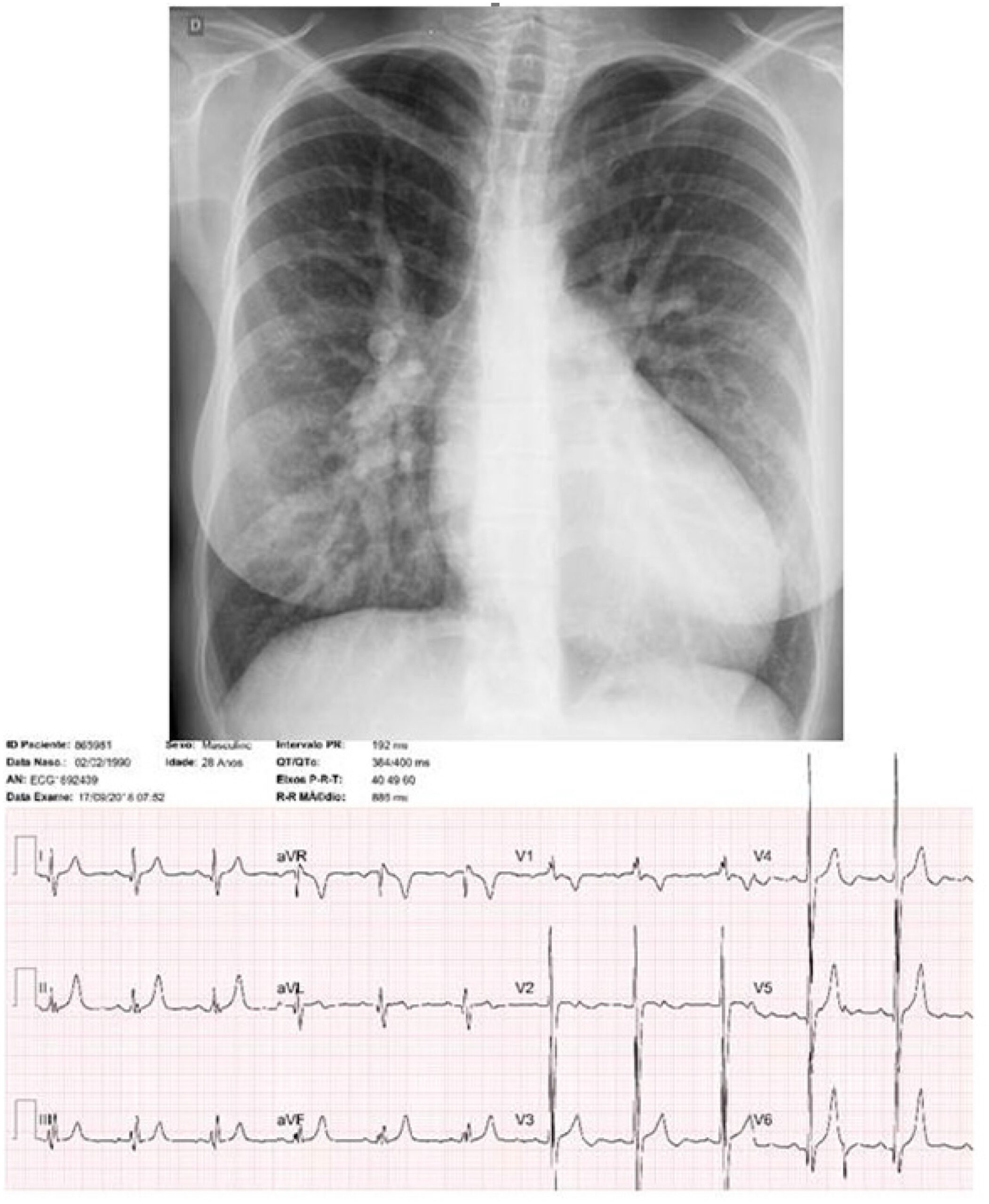
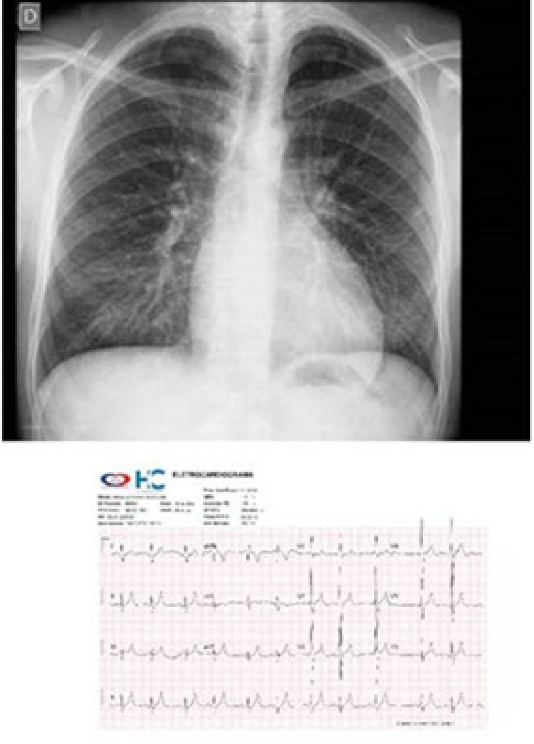
Electrocardiogram: Showed sinus rhythm, no chamber overload and no changes in ventricular repolarization. AP= +40º, AQRS= +60º, AT= +30º (). The ECGs performed before the closure of the defect were normal, in accordance with the corresponding ages.

Chest radiography: Showed heart area within the normal range (cardiothoracic index = 0.50) at the age of 13 years, prior to closure of the defect ().

[…]

  • Down Syndrome
  • Heart Defects, Congenital
  • Heart Septal Defects, Ventricular
Read Full Article
Creative Commons Attribution (CC BY) Creative Commons license
01/10/2016 DOI: 10.5935/abc.20160148
Congenital Muscular Interventricular Septal Malformation with Complex Anatomical Features
ABC
Image

Congenital Muscular Interventricular Septal Malformation with Complex Anatomical Features

Zafer ışılak et al. Mehmet Uzun Ejder Kardeşoğlu Ömer Uz Uğur Küçük

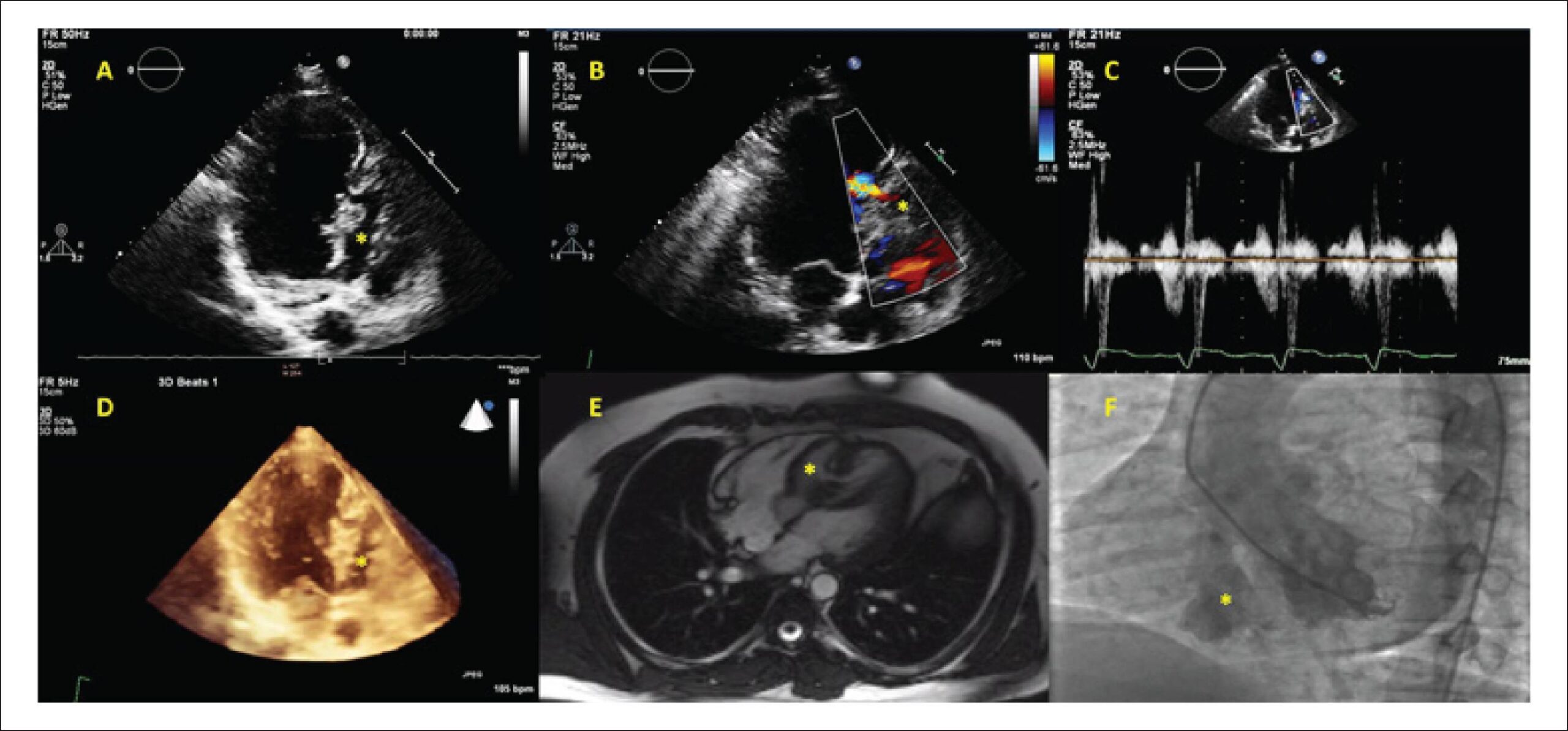
A 21 year-old male patient was admitted with symptoms of exertional dyspnea and palpitation. The physical examination revealed a grade 3/6 systolic murmur, best heard over the left 3-4th intercostal space. Transthoracic echocardiography disclosed a separate chamber (asterisk) in the interventricular septum. The apical portion of the chamber consisted of muscle tissue and the basal portion consisted of membranous and aneurysmatic tissue (Panel A). There was a muscular “tunnel-like” structure connecting the left ventricle and chamber at mid ventricular level. The color and continuous wave Doppler imaging revealed a bidirectional flow across the passage (Panel B, C).

The patient underwent three dimensional transthoracic echocardiographic examinations, which revealed two separate septa. Between these septa there was a third chamber (asterisk). It was connected to both left (via the tunnel at the muscular septum) and right ventricles (via the defect in the membranous septal aneurysm). The membranous septal aneurysm was separated from the left ventricle by a thin membrane, without any passage across it (Panel D). MRI findings were consistent with echocardiographic images (Panel E). Ventriculography and coronary angiography were performed. The coronary arteries were normal; the left ventriculography showed the muscular and membranous/aneurysmatic portions of the malformation (Panel F). The patient was advised to undergo a surgical procedure, but he refused and was discharged with recommendations about control visits.

[…]

  • Heart Defects, Congenital
  • Heart Septal Defects, Ventricular
  • ventricular septum
Read Full Article
Creative Commons Attribution (CC BY) Creative Commons license
01/04/2016 DOI: 10.5935/abc.20160045 Views 1
ABC
Original Article

Investigation of Copy Number Variation in Children with Conotruncal Heart Defects

Carla Marques Rondon Campos et al. Evelin Aline Zanardo Roberta Lelis Dutra Leslie Domenici Kulikowski Chong Ae Kim

Background:

Congenital heart defects (CHD) are the most prevalent group of structural abnormalities at birth and one of the main causes of infant morbidity and mortality. Studies have shown a contribution of the copy number variation in the genesis of cardiac malformations.

Objectives:

Investigate gene copy number variation (CNV) in children with conotruncal heart defect.

Methods:

Multiplex ligation-dependent probe amplification (MLPA) was performed in 39 patients with conotruncal heart defect. Clinical and laboratory assessments were conducted in all patients. The parents of the probands who presented abnormal findings were also investigated.

Results:

Gene copy number variation was detected in 7/39 patients: 22q11.2 deletion, 22q11.2 duplication, 15q11.2 duplication, 20p12.2 duplication, 19p deletion, 15q and 8p23.2 duplication with 10p12.31 duplication. The clinical characteristics were consistent with those reported in the literature associated with the encountered microdeletion/microduplication. None of these changes was inherited from the parents.

Conclusions:

Our results demonstrate that the technique of MLPA is useful in the investigation of microdeletions and microduplications in conotruncal congenital heart defects. Early diagnosis of the copy number variation in patients with congenital heart defect assists in the prevention of morbidity and decreased mortality in these patients.

  • DNA, Truncus Arteriosus
  • Genetic Variation
  • Heart Defects, Congenital
  • Heart Septal Defects, Ventricular
Read Full Article
Creative Commons Attribution (CC BY) Creative Commons license
11/11/2014 DOI: 10.5935/abc.20140169 Views 1
Figure 1
ABC
Clinicoradiological Session

Case 3/2014 – 26-year-old Man with Pulmonary Atresia and Ventricular Septal Defect in Late Postoperative Evolution

Edmar Atik

Complementary tests

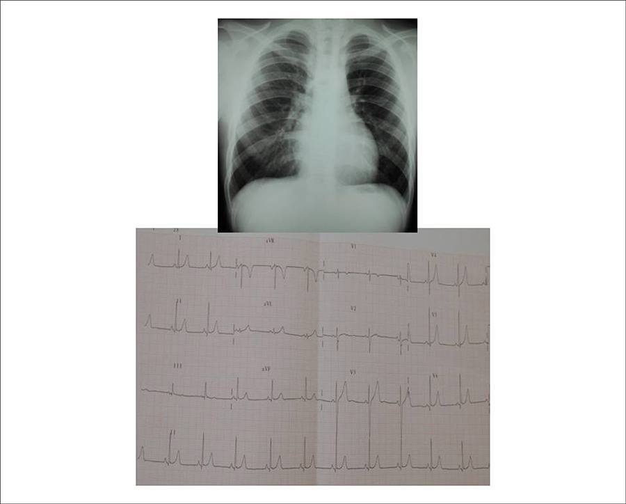
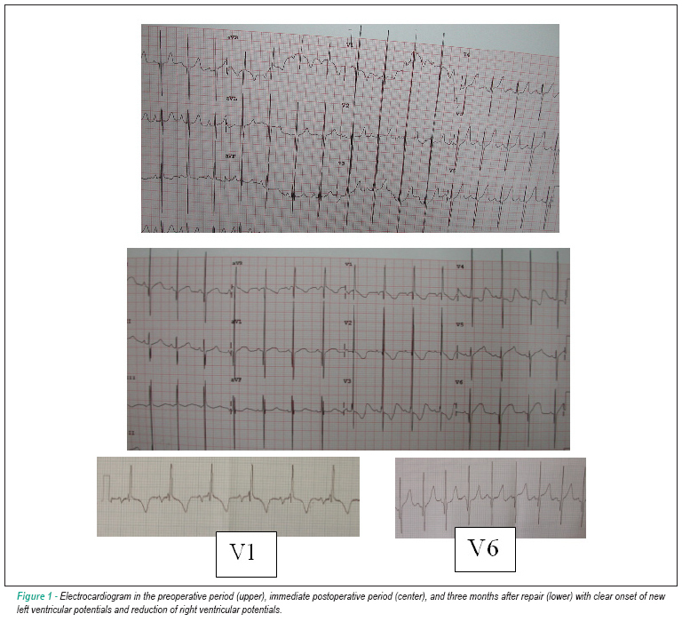
Electrocardiogram: Showed sinus rhythm, moderate right ventricular (RV) overload without right bundle branch block. Rs morphology in V1 and qRs in V6. Until six years ago, maintained rsr’ morphology in V1. QRS duration was 0.94 from the total correction. AP: +70º, AQRS: +90º, AT: +70º.

Chest radiography: Shows normal cardiac silhouette, rounded shape and normal pulmonary vasculature ().

[…]

  • Adult
  • Heart Septal Defects, Ventricular
  • Pulmonary Atresia/surgery
Read Full Article
Creative Commons Attribution (CC BY) Creative Commons license
01/04/2014 DOI: 10.5935/abc.20140045 Views 1
Occlusion of post-myocardial infarction ventricular defect with prosthesis CERA
ABC
Case Report

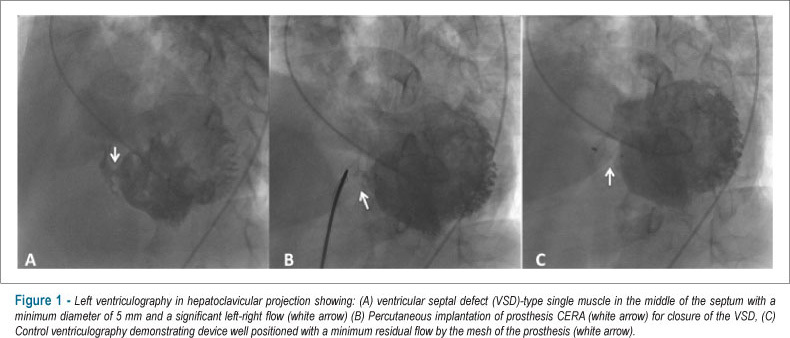
Occlusion of post-myocardial infarction ventricular defect with prosthesis CERA

Henrique Barbosa Ribeiro et al. Luciano Moreira Baracioli Luiz Junya Kajita Martina Battistini Pinheiro … Expedito E. Ribeiro José Carlos Nicolau

CASE REPORT

Occlusion of ventricular defect post-myocardial infarction with prothesis CERA

[…]

  • Heart Septal Defects, Ventricular
  • Myocardial Infarction
  • Septal occluder device
Read Full Article
Creative Commons Attribution (CC BY) Creative Commons license
04/09/2012 DOI: 10.1590/S0066-782X2012001000017 Views 1
Atypical truncus arteriosus operated at 28 years of age: importance of differential diagnosis
ABC
Case Report

Atypical truncus arteriosus operated at 28 years of age: importance of differential diagnosis

Lilian Maria Lopes et al. Jose Pedro da Silva Luciana da Fonseca Sonia Meiken … André Bavaresco Cristóvão Salvador Gustavo Spadaccia dos Santos Fernandes

This is the case of 28 year-old adult with suspected congenital heart disease since birth, not treated in childhood at the his family’s choice. At 27 years old, he was diagnosed with pulmonary atresia with ventricular septal defect and systemic-pulmonary collaterals, where surgery was contraindicated. A new review in our department showed that it was an atypical form of truncus arteriosus. The fact that a common arterial trunk with left-right shunt was viewed by echocardiography was a crucial fact for the indication of new catheterization, opening the prospect of surgical correction. Currently, the patient is well, with 7 years of postoperative outcome.

  • Congenital
  • Heart Defects
  • Heart Septal Defects, Ventricular
  • Pulmonary Atresia
  • truncus arteriosus
Read Full Article
Creative Commons Attribution (CC BY) Creative Commons license
04/10/2011 DOI: 10.1590/S0066-782X2011001100016 Views 1
Congenital heart disease in adults: outpatient clinic profile at the Hospital das Clínicas of Ribeirão Preto
ABC
Artigo Original

Congenital heart disease in adults: outpatient clinic profile at the Hospital das Clínicas of Ribeirão Preto

Fernando Amaral et al. Paulo Henrique Manso João Antonio Granzotti Walter Villela de Andrade Vicente Andre Schmidt

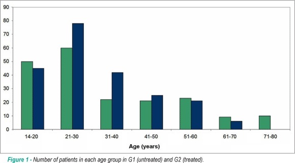
BACKGROUND: Service experiences for adults with congenital heart disease have not been reported in our country. OBJECTIVE: To describe the basic clinical profile of adults with congenital heart disease in an outpatient tertiary care center. METHODS: We compiled data on age, gender, place of residence, primary diagnosis, and secondary diagnoses of 413 patients treated for seven years. RESULTS: G1 (untreated): 195 patients, 51% women, 57% between 14 and 30 years, 80% living in the region. The most frequent heart diseases were ventricular septal defect (VSD) (31%), atrial septal defect (ASD) (29%), and pulmonary stenosis (7%). The predominant secondary diagnoses were hypertension (9%) and arrhythmias (5%). G2 (treated): 218 patients, 56% women, 57% between 14 and 30 years, 81% living in the region. The most frequently treated heart diseases were: ASD (36%), tetralogy of Fallot (14%), coarctation of the aorta (12%), and VSD (11%). Sixty-nine (32%) patients were operated on for congenital heart diseases in adulthood. Sixteen (7%) underwent an interventional catheterization. The predominant secondary diagnoses were hypertension (18%) and arrhythmias (8%). CONCLUSION: In the study, most patients were treated invasively, all of them were residents in the region, and most of them were under 40 years of age. Defects such as ASD, VSD, and pulmonary stenosis predominated in the untreated group, whereas in the treated group, most patients had undergone surgical correction of ASD, tetralogy of Fallot, aortic coarctation, and VSD. Hypertension and arrhythmias were relevant in both groups, and a large variety of other comorbidities were also observed.

  • Brazil
  • Health Profile
  • Heart Defects, Congenital
  • Heart Septal Defects, Atrial
  • Heart Septal Defects, Ventricular
  • outpatients
  • Ribeirão Preto (SP)
  • Tetralogy of Fallot
Read Full Article
Creative Commons Attribution (CC BY) Creative Commons license
21/05/2010 DOI: 10.1590/S0066-782X2010005000053 Views 1
Biventricular repair in unbalanced atrioventricular septal defect
ABC
Case Report

Biventricular repair in unbalanced atrioventricular septal defect

Edmar Atik et al. Patrícia O. Marques Rogério A. Miranda Vitor C. Guerra … Lucília Santana Faria Marcelo Jatene

We present the case of a 2.5 year-old child with unbalanced atrioventricular septal defect due to a small left ventricle (LV) (mitral annulus of 10mm and a 0.4 ratio in relation to the tricuspid annulus, LVDD: 17 mm, LV Vd2: 15 ml/m² and LV/RV long-axis ratio of 0.71); he had a favorable outcome after biventricular surgical repair. Normal LV development was observed three months after the operation (mitral annulus of 22 mm, with a 0.84 ratio in relation to the tricuspid annulus, and LVDD of 30 mm). Current parameters for utilization of the hypoplastic ventricle are discussed.

  • Heart Defects, Congenital
  • Heart Septal Defects, Ventricular
  • Heart Ventricles
Read Full Article
Creative Commons Attribution (CC BY) Creative Commons license
29/06/2009 DOI: 10.1590/S0066-782X2009000400015 Views 1

How to cite this article

EndNote RIS

Electronic Document Format (Vancouver)

Santos ECL, Lima SG, Markman Filho B, Lordsleem ABMS, Lima GAFCA, Moreira PCS. Atypical Presentation of Ventricular Septal Defect Following Myocardial Infarction. Arquivos Brasileiros de Cardiologia Imagem cardiovascular 2022;30(4):149-51.

Electronic Document Format (ABNT)

Santos, Eduardo Cavalcanti Lapa; Lima, Sandro Gonçalves; Markman Filho, Brivaldo; Lordsleem, Andréa Bezerra de Melo da Silveira; Lima, George Augusto da Fonseca Carvalho Antunes; Moreira, Priscila Costa dos Santos. Atypical Presentation of Ventricular Septal Defect Following Myocardial Infarction. Arquivos Brasileiros de Cardiologia Imagem cardiovascular, v. 30, n. 4, p. 149-151, Jan. 2022.

Electronic Document Format (APA)

Santos, E. C. L., Lima, S. G., Markman Filho, B., Lordsleem, A. B. M. S., Lima, G. A. F. C. A., & Moreira, P. C. S. (2022). Atypical Presentation of Ventricular Septal Defect Following Myocardial Infarction. Arquivos Brasileiros de Cardiologia Imagem cardiovascular, 30(4), 149-151.

Electronic Document Format (ISO)

Santos, Eduardo Cavalcanti Lapa and Lima, Sandro Gonçalves and Markman Filho, Brivaldo and Lordsleem, Andréa Bezerra de Melo da Silveira and Lima, George Augusto da Fonseca Carvalho Antunes and Moreira, Priscila Costa dos Santos. Atypical Presentation of Ventricular Septal Defect Following Myocardial Infarction. Arquivos Brasileiros de Cardiologia Imagem cardiovascular [online]. 2022, vol. 30, n. 4, [cited 2026-10-14], pp. 149-151. Available from: <https://journals.cardiol.br/en/article/atypical-presentation-of-ventricular-septal-defect-following-myocardial-infarction/>. ISSN 2318-8219

EndNote RIS

Electronic Document Format (Vancouver)

Takagui ASM, Baggio TC, Savariz MB, Theilacker G, Baggio TE. Left Ventricular-to-right Atrial Shunt (Gerbode Defect) Associated with Interventricular Communication in a 42-year-old Patient. ABC Imagem Cardiovascular 2021;34(1):eabc142.

Electronic Document Format (ABNT)

Takagui, Andressa Sardá Maiochi; Baggio, Thales Cantelle; Savariz, Mariana Baretta; Theilacker, Giulia; Baggio, Thalia Elisa. Left Ventricular-to-right Atrial Shunt (Gerbode Defect) Associated with Interventricular Communication in a 42-year-old Patient. ABC Imagem Cardiovascular, v. 34, n. 1, eabc142, Jun. 2021.

Electronic Document Format (APA)

Takagui, A. S. M., Baggio, T. C., Savariz, M. B., Theilacker, G., & Baggio, T. E. (2021). Left Ventricular-to-right Atrial Shunt (Gerbode Defect) Associated with Interventricular Communication in a 42-year-old Patient. ABC Imagem Cardiovascular, 34(1), eabc142.

Electronic Document Format (ISO)

Takagui, Andressa Sardá Maiochi and Baggio, Thales Cantelle and Savariz, Mariana Baretta and Theilacker, Giulia and Baggio, Thalia Elisa. Left Ventricular-to-right Atrial Shunt (Gerbode Defect) Associated with Interventricular Communication in a 42-year-old Patient. ABC Imagem Cardiovascular [online]. 2021, vol. 34, n. 1, [cited 2026-10-14], eabc142. Available from: <https://journals.cardiol.br/en/article/left-ventricular-to-right-atrial-shunt-gerbode-defect-associated-with-interventricular-communication-in-a-42-year-old-patient/>. ISSN 2318-8219

EndNote RIS

Electronic Document Format (Vancouver)

Atik E. Double Outlet Right Ventricle with Unrelated Ventricular Septal Defect and Pulmonary Stenosis, in Natural Evolution, in a 36-Year-Old Woman. Arquivos Brasileiros de Cardiologia 2021;116(4):836-9.

Electronic Document Format (ABNT)

Atik, Edmar. Double Outlet Right Ventricle with Unrelated Ventricular Septal Defect and Pulmonary Stenosis, in Natural Evolution, in a 36-Year-Old Woman. Arquivos Brasileiros de Cardiologia, v. 116, n. 4, p. 836-839, Apr. 2021.

Electronic Document Format (APA)

Atik, E. (2021). Double Outlet Right Ventricle with Unrelated Ventricular Septal Defect and Pulmonary Stenosis, in Natural Evolution, in a 36-Year-Old Woman. Arquivos Brasileiros de Cardiologia, 116(4), 836-839.

Electronic Document Format (ISO)

Atik, Edmar. Double Outlet Right Ventricle with Unrelated Ventricular Septal Defect and Pulmonary Stenosis, in Natural Evolution, in a 36-Year-Old Woman. Arquivos Brasileiros de Cardiologia [online]. 2021, vol. 116, n. 4, [cited 2026-10-14], pp. 836-839. Available from: <https://journals.cardiol.br/en/article/double-outlet-right-ventricle-with-unrelated-ventricular-septal-defect-and-pulmonary-stenosis-in-natural-evolution-in-a-36-year-old-woman/>. ISSN 0066-782X

EndNote RIS

Electronic Document Format (Vancouver)

Martins C, Gama BMN. Risk Factors for Mortality in Pediatric Cardiac Intensive Care Unit. International Journal of Cardiovascular Sciences 2020;33(4):337-9.

Electronic Document Format (ABNT)

Martins, Cristiane; Gama, Bruna M. N.. Risk Factors for Mortality in Pediatric Cardiac Intensive Care Unit. International Journal of Cardiovascular Sciences, v. 33, n. 4, p. 337-339, Jul. 2020.

Electronic Document Format (APA)

Martins, C., & Gama, B. M. N. (2020). Risk Factors for Mortality in Pediatric Cardiac Intensive Care Unit. International Journal of Cardiovascular Sciences, 33(4), 337-339.

Electronic Document Format (ISO)

Martins, Cristiane and Gama, Bruna M. N.. Risk Factors for Mortality in Pediatric Cardiac Intensive Care Unit. International Journal of Cardiovascular Sciences [online]. 2020, vol. 33, n. 4, [cited 2026-10-14], pp. 337-339. Available from: <https://journals.cardiol.br/en/article/risk-factors-for-mortality-in-pediatric-cardiac-intensive-care-unit/>. ISSN 2359-4802

EndNote RIS

Electronic Document Format (Vancouver)

Atik E, Binotto MA, Barreto AC. Case 6/2019 – Total Atrioventricular Septal Defect, 12 Years After Operative Correction, No Residual Defects. Arquivos Brasileiros de Cardiologia 2019;113(5):999-1001.

Electronic Document Format (ABNT)

Atik, Edmar; Binotto, Maria Angélica; Barreto, Alessandra Costa. Case 6/2019 – Total Atrioventricular Septal Defect, 12 Years After Operative Correction, No Residual Defects. Arquivos Brasileiros de Cardiologia, v. 113, n. 5, p. 999-1001, Dec. 2019.

Electronic Document Format (APA)

Atik, E., Binotto, M. A., & Barreto, A. C. (2019). Case 6/2019 – Total Atrioventricular Septal Defect, 12 Years After Operative Correction, No Residual Defects. Arquivos Brasileiros de Cardiologia, 113(5), 999-1001.

Electronic Document Format (ISO)

Atik, Edmar and Binotto, Maria Angélica and Barreto, Alessandra Costa. Case 6/2019 – Total Atrioventricular Septal Defect, 12 Years After Operative Correction, No Residual Defects. Arquivos Brasileiros de Cardiologia [online]. 2019, vol. 113, n. 5, [cited 2026-10-14], pp. 999-1001. Available from: <https://journals.cardiol.br/en/article/case-6-2019-total-atrioventricular-septal-defect-12-years-after-operative-correction-no-residual-defects/>. ISSN 0066-782X

EndNote RIS

Electronic Document Format (Vancouver)

Atik E, Binotto MA, Barreto AC, Ishikawa W. Case 4/2019 – Pulmonary Atresia, Ventricular Septal Defect, and Systemic-Pulmonary Collateral Circulation developing after prior Blalock-Taussig, in a Symptomatic 47-Year-Old Adult. Arquivos Brasileiros de Cardiologia 2019;113(1):103-5.

Electronic Document Format (ABNT)

Atik, Edmar; Binotto, Maria Angélica; Barreto, Alessandra Costa; Ishikawa, Walther. Case 4/2019 – Pulmonary Atresia, Ventricular Septal Defect, and Systemic-Pulmonary Collateral Circulation developing after prior Blalock-Taussig, in a Symptomatic 47-Year-Old Adult. Arquivos Brasileiros de Cardiologia, v. 113, n. 1, p. 103-105, Aug. 2019.

Electronic Document Format (APA)

Atik, E., Binotto, M. A., Barreto, A. C., & Ishikawa, W. (2019). Case 4/2019 – Pulmonary Atresia, Ventricular Septal Defect, and Systemic-Pulmonary Collateral Circulation developing after prior Blalock-Taussig, in a Symptomatic 47-Year-Old Adult. Arquivos Brasileiros de Cardiologia, 113(1), 103-105.

Electronic Document Format (ISO)

Atik, Edmar and Binotto, Maria Angélica and Barreto, Alessandra Costa and Ishikawa, Walther. Case 4/2019 – Pulmonary Atresia, Ventricular Septal Defect, and Systemic-Pulmonary Collateral Circulation developing after prior Blalock-Taussig, in a Symptomatic 47-Year-Old Adult. Arquivos Brasileiros de Cardiologia [online]. 2019, vol. 113, n. 1, [cited 2026-10-14], pp. 103-105. Available from: <https://journals.cardiol.br/en/article/case-4-2019-pulmonary-atresia-ventricular-septal-defect-and-systemic-pulmonary-collateral-circulation-developing-after-prior-blalock-taussig-in-a-symptomatic-47-year-old-adult/>. ISSN 0066-782X

EndNote RIS

Electronic Document Format (Vancouver)

Atik E, Barreto AC, Binotto MA. Case 1 / 2019 – Natural Evolution of Double Outlet Right Ventricle with Noncommitted Ventricular Septal Defect and Pulmonary Stenosis in a 28-Year-Old Asymptomatic Woman. Arquivos Brasileiros de Cardiologia 2019;112(1):107-9.

Electronic Document Format (ABNT)

Atik, Edmar; Barreto, Alessandra Costa; Binotto, Maria Angélica. Case 1 / 2019 – Natural Evolution of Double Outlet Right Ventricle with Noncommitted Ventricular Septal Defect and Pulmonary Stenosis in a 28-Year-Old Asymptomatic Woman. Arquivos Brasileiros de Cardiologia, v. 112, n. 1, p. 107-109, Dec. 2018.

Electronic Document Format (APA)

Atik, E., Barreto, A. C., & Binotto, M. A. (2019). Case 1 / 2019 – Natural Evolution of Double Outlet Right Ventricle with Noncommitted Ventricular Septal Defect and Pulmonary Stenosis in a 28-Year-Old Asymptomatic Woman. Arquivos Brasileiros de Cardiologia, 112(1), 107-109.

Electronic Document Format (ISO)

Atik, Edmar and Barreto, Alessandra Costa and Binotto, Maria Angélica. Case 1 / 2019 – Natural Evolution of Double Outlet Right Ventricle with Noncommitted Ventricular Septal Defect and Pulmonary Stenosis in a 28-Year-Old Asymptomatic Woman. Arquivos Brasileiros de Cardiologia [online]. 2019, vol. 112, n. 1, [cited 2026-10-14], pp. 107-109. Available from: <https://journals.cardiol.br/en/article/case-1-2019-natural-evolution-of-double-outlet-right-ventricle-with-noncommitted-ventricular-septal-defect-and-pulmonary-stenosis-in-a-28-year-old-asymptomatic-woman/>. ISSN 0066-782X

EndNote RIS

Electronic Document Format (Vancouver)

Atik E. Case 6/2016 – The Patient is a 29-Year-Old Male with Spontaneous Closure of Ventricular Septal Defect in Adulthood. Arquivos Brasileiros de Cardiologia 2016;107(4):375-7.

Electronic Document Format (ABNT)

Atik, Edmar. Case 6/2016 – The Patient is a 29-Year-Old Male with Spontaneous Closure of Ventricular Septal Defect in Adulthood. Arquivos Brasileiros de Cardiologia, v. 107, n. 4, p. 375-377, Sep. 2016.

Electronic Document Format (APA)

Atik, E. (2016). Case 6/2016 – The Patient is a 29-Year-Old Male with Spontaneous Closure of Ventricular Septal Defect in Adulthood. Arquivos Brasileiros de Cardiologia, 107(4), 375-377.

Electronic Document Format (ISO)

Atik, Edmar. Case 6/2016 – The Patient is a 29-Year-Old Male with Spontaneous Closure of Ventricular Septal Defect in Adulthood. Arquivos Brasileiros de Cardiologia [online]. 2016, vol. 107, n. 4, [cited 2026-10-14], pp. 375-377. Available from: <https://journals.cardiol.br/en/article/case-6-2016-the-patient-is-a-29-year-old-male-with-spontaneous-closure-of-ventricular-septal-defect-in-adulthood/>. ISSN 0066-782X

EndNote RIS

Electronic Document Format (Vancouver)

ışılak Z, Uzun M, Kardeşoğlu E, Uz , Küçük U. Congenital Muscular Interventricular Septal Malformation with Complex Anatomical Features. Arquivos Brasileiros de Cardiologia 2016;106(4):349.

Electronic Document Format (ABNT)

ışılak, Zafer; Uzun, Mehmet; Kardeşoğlu, Ejder; Uz, Ömer; Küçük, Uğur. Congenital Muscular Interventricular Septal Malformation with Complex Anatomical Features. Arquivos Brasileiros de Cardiologia, v. 106, n. 4, p. 349-349, Mar. 2016.

Electronic Document Format (APA)

ışılak, Z., Uzun, M., Kardeşoğlu, E., Uz, , & Küçük, U. (2016). Congenital Muscular Interventricular Septal Malformation with Complex Anatomical Features. Arquivos Brasileiros de Cardiologia, 106(4), 349-349.

Electronic Document Format (ISO)

ışılak, Zafer and Uzun, Mehmet and Kardeşoğlu, Ejder and Uz, Ömer and Küçük, Uğur. Congenital Muscular Interventricular Septal Malformation with Complex Anatomical Features. Arquivos Brasileiros de Cardiologia [online]. 2016, vol. 106, n. 4, [cited 2026-10-14], pp. 349-349. Available from: <https://journals.cardiol.br/en/article/congenital-muscular-interventricular-septal-malformation-with-complex-anatomical-features/>. ISSN 0066-782X

EndNote RIS

Electronic Document Format (Vancouver)

Campos CMR, Zanardo EA, Dutra RL, Kulikowski LD, Kim CA. Investigation of Copy Number Variation in Children with Conotruncal Heart Defects. Arquivos Brasileiros de Cardiologia 2014;104(1):24-31.

Electronic Document Format (ABNT)

Campos, Carla Marques Rondon; Zanardo, Evelin Aline; Dutra, Roberta Lelis; Kulikowski, Leslie Domenici; Kim, Chong Ae. Investigation of Copy Number Variation in Children with Conotruncal Heart Defects. Arquivos Brasileiros de Cardiologia, v. 104, n. 1, p. 24-31, Nov. 2014.

Electronic Document Format (APA)

Campos, C. M. R., Zanardo, E. A., Dutra, R. L., Kulikowski, L. D., & Kim, C. A. (2014). Investigation of Copy Number Variation in Children with Conotruncal Heart Defects. Arquivos Brasileiros de Cardiologia, 104(1), 24-31.

Electronic Document Format (ISO)

Campos, Carla Marques Rondon and Zanardo, Evelin Aline and Dutra, Roberta Lelis and Kulikowski, Leslie Domenici and Kim, Chong Ae. Investigation of Copy Number Variation in Children with Conotruncal Heart Defects. Arquivos Brasileiros de Cardiologia [online]. 2014, vol. 104, n. 1, [cited 2026-10-14], pp. 24-31. Available from: <https://journals.cardiol.br/en/article/investigation-of-copy-number-variation-in-children-with-conotruncal-heart-defects/>. ISSN 0066-782X

EndNote RIS

Electronic Document Format (Vancouver)

Atik E. Case 3/2014 – 26-year-old Man with Pulmonary Atresia and Ventricular Septal Defect in Late Postoperative Evolution. Arquivos Brasileiros de Cardiologia 2014;102(4):e34.

Electronic Document Format (ABNT)

Atik, Edmar. Case 3/2014 – 26-year-old Man with Pulmonary Atresia and Ventricular Septal Defect in Late Postoperative Evolution. Arquivos Brasileiros de Cardiologia, v. 102, n. 4, e34, Mar. 2014.

Electronic Document Format (APA)

Atik, E. (2014). Case 3/2014 – 26-year-old Man with Pulmonary Atresia and Ventricular Septal Defect in Late Postoperative Evolution. Arquivos Brasileiros de Cardiologia, 102(4), e34.

Electronic Document Format (ISO)

Atik, Edmar. Case 3/2014 – 26-year-old Man with Pulmonary Atresia and Ventricular Septal Defect in Late Postoperative Evolution. Arquivos Brasileiros de Cardiologia [online]. 2014, vol. 102, n. 4, [cited 2026-10-14], e34. Available from: <https://journals.cardiol.br/en/article/case-3-2014-26-year-old-man-with-pulmonary-atresia-and-ventricular-septal-defect-in-late-postoperative-evolution/>. ISSN 0066-782X

EndNote RIS

Electronic Document Format (Vancouver)

Ribeiro HB, Baracioli LM, Kajita LJ, Pinheiro MB, Ribeiro EE, Nicolau JC. Occlusion of post-myocardial infarction ventricular defect with prosthesis CERA. Arquivos Brasileiros de Cardiologia 2012;99(1):e112-3.

Electronic Document Format (ABNT)

Ribeiro, Henrique Barbosa; Baracioli, Luciano Moreira; Kajita, Luiz Junya; Pinheiro, Martina Battistini; Ribeiro, Expedito E.; Nicolau, José Carlos. Occlusion of post-myocardial infarction ventricular defect with prosthesis CERA. Arquivos Brasileiros de Cardiologia, v. 99, n. 1, p. e112-e113, Sep. 2012.

Electronic Document Format (APA)

Ribeiro, H. B., Baracioli, L. M., Kajita, L. J., Pinheiro, M. B., Ribeiro, E. E., & Nicolau, J. C. (2012). Occlusion of post-myocardial infarction ventricular defect with prosthesis CERA. Arquivos Brasileiros de Cardiologia, 99(1), e112-e113.

Electronic Document Format (ISO)

Ribeiro, Henrique Barbosa and Baracioli, Luciano Moreira and Kajita, Luiz Junya and Pinheiro, Martina Battistini and Ribeiro, Expedito E. and Nicolau, José Carlos. Occlusion of post-myocardial infarction ventricular defect with prosthesis CERA. Arquivos Brasileiros de Cardiologia [online]. 2012, vol. 99, n. 1, [cited 2026-10-14], pp. e112-e113. Available from: <https://journals.cardiol.br/en/article/occlusion-of-post-myocardial-infarction-ventricular-defect-with-prosthesis-cera/>. ISSN 0066-782X

EndNote RIS

Electronic Document Format (Vancouver)

Lopes LM, Silva JP, Fonseca L, Meiken S, Salvador ABC, Fernandes GSS. Atypical truncus arteriosus operated at 28 years of age: importance of differential diagnosis. Arquivos Brasileiros de Cardiologia 2011;97(2):e29-32.

Electronic Document Format (ABNT)

Lopes, Lilian Maria; Silva, Jose Pedro da; Fonseca, Luciana da; Meiken, Sonia; Salvador, André Bavaresco Cristóvão; Fernandes, Gustavo Spadaccia dos Santos. Atypical truncus arteriosus operated at 28 years of age: importance of differential diagnosis. Arquivos Brasileiros de Cardiologia, v. 97, n. 2, p. e29-e32, Oct. 2011.

Electronic Document Format (APA)

Lopes, L. M., Silva, J. P. , Fonseca, L. , Meiken, S., Salvador, A. B. C., & Fernandes, G. S. S. (2011). Atypical truncus arteriosus operated at 28 years of age: importance of differential diagnosis. Arquivos Brasileiros de Cardiologia, 97(2), e29-e32.

Electronic Document Format (ISO)

Lopes, Lilian Maria and Silva, Jose Pedro da and Fonseca, Luciana da and Meiken, Sonia and Salvador, André Bavaresco Cristóvão and Fernandes, Gustavo Spadaccia dos Santos. Atypical truncus arteriosus operated at 28 years of age: importance of differential diagnosis. Arquivos Brasileiros de Cardiologia [online]. 2011, vol. 97, n. 2, [cited 2026-10-14], pp. e29-e32. Available from: <https://journals.cardiol.br/en/article/atypical-truncus-arteriosus-operated-at-28-years-of-age-importance-of-differential-diagnosis/>. ISSN 0066-782X

EndNote RIS

Electronic Document Format (Vancouver)

Amaral F, Manso PH, Granzotti JA, Vicente WVA, Schmidt A. Congenital heart disease in adults: outpatient clinic profile at the Hospital das Clínicas of Ribeirão Preto. Arquivos Brasileiros de Cardiologia 2010;94(6):707-13.

Electronic Document Format (ABNT)

Amaral, Fernando; Manso, Paulo Henrique; Granzotti, João Antonio; Vicente, Walter Villela de Andrade; Schmidt, Andre. Congenital heart disease in adults: outpatient clinic profile at the Hospital das Clínicas of Ribeirão Preto. Arquivos Brasileiros de Cardiologia, v. 94, n. 6, p. 707-713, May. 2010.

Electronic Document Format (APA)

Amaral, F., Manso, P. H., Granzotti, J. A., Vicente, W. V. A., & Schmidt, A. (2010). Congenital heart disease in adults: outpatient clinic profile at the Hospital das Clínicas of Ribeirão Preto. Arquivos Brasileiros de Cardiologia, 94(6), 707-713.

Electronic Document Format (ISO)

Amaral, Fernando and Manso, Paulo Henrique and Granzotti, João Antonio and Vicente, Walter Villela de Andrade and Schmidt, Andre. Congenital heart disease in adults: outpatient clinic profile at the Hospital das Clínicas of Ribeirão Preto. Arquivos Brasileiros de Cardiologia [online]. 2010, vol. 94, n. 6, [cited 2026-10-14], pp. 707-713. Available from: <https://journals.cardiol.br/en/article/congenital-heart-disease-in-adults-outpatient-clinic-profile-at-the-hospital-das-clinicas-of-ribeirao-preto/>. ISSN 0066-782X

EndNote RIS

Electronic Document Format (Vancouver)

Atik E, Marques PO, Miranda RA, Guerra VC, Faria LS, Jatene M. Biventricular repair in unbalanced atrioventricular septal defect. Arquivos Brasileiros de Cardiologia 2009;92(4):e46-9.

Electronic Document Format (ABNT)

Atik, Edmar; Marques, Patrícia O.; Miranda, Rogério A.; Guerra, Vitor C.; Faria, Lucília Santana; Jatene, Marcelo. Biventricular repair in unbalanced atrioventricular septal defect. Arquivos Brasileiros de Cardiologia, v. 92, n. 4, p. e46-e49, Jun. 2009.

Electronic Document Format (APA)

Atik, E., Marques, P. O., Miranda, R. A., Guerra, V. C., Faria, L. S., & Jatene, M. (2009). Biventricular repair in unbalanced atrioventricular septal defect. Arquivos Brasileiros de Cardiologia, 92(4), e46-e49.

Electronic Document Format (ISO)

Atik, Edmar and Marques, Patrícia O. and Miranda, Rogério A. and Guerra, Vitor C. and Faria, Lucília Santana and Jatene, Marcelo. Biventricular repair in unbalanced atrioventricular septal defect. Arquivos Brasileiros de Cardiologia [online]. 2009, vol. 92, n. 4, [cited 2026-10-14], pp. e46-e49. Available from: <https://journals.cardiol.br/en/article/biventricular-repair-in-unbalanced-atrioventricular-septal-defect/>. ISSN 0066-782X

  • About ABC Family
  • Internal Regulation
  • Editorial Board
  • Brazilian Society of Cardiology (SBC)

Issues

SBC Guidelines

  • Videos
  • Podcasts
  • News