ABC Family Arquivos Brasileiros de Cardiologia International Journal of Cardiovascular Sciences ABC Heart Failure & Cardiomyopathy ABC Imagem Cardiovascular
Português English
ABC Family
Português English
  • About
    • About ABC Family
    • Internal Regulation
    • Editorial Board
    • Brazilian Society of Cardiology (SBC)
  • Issues
  • SBC Guidelines
  • Content
    • Videos
    • Podcasts
    • News
  • About
    • About ABC Family
    • Internal Regulation
    • Editorial Board
    • Brazilian Society of Cardiology (SBC)
  • Issues
  • SBC Guidelines
  • Content
    • Videos
    • Podcasts
    • News

ABC Family

  • ABC Family
  • Arquivos Brasileiros de Cardiologia
  • International Journal of Cardiovascular Sciences
  • ABC Heart Failure & Cardiomyopathy
  • ABC Imagem Cardiovascular
Search in

No content found.

Journals
Ordering
Article types
Others

  1. Home
  2. Keyword: Magnetic Resonance Imaging
ABC
Editorial

Reference Values in Cardiovascular Magnetic Resonance Imaging: Between Local Validation and Global Harmonization

Miguel Correia

Cardiovascular magnetic resonance imaging (CMR) has become established in recent decades as the gold standard method for cardiac morphofunctional assessment, allowing for the precise quantification of ventricular volumes, ejection fraction, and myocardial mass, as well as the tissue characterization of multiple heart diseases.– The high reproducibility of its measurements is one of its main advantages over other imaging modalities, making CMR an essential tool in both clinical practice and cardiovascular research.,

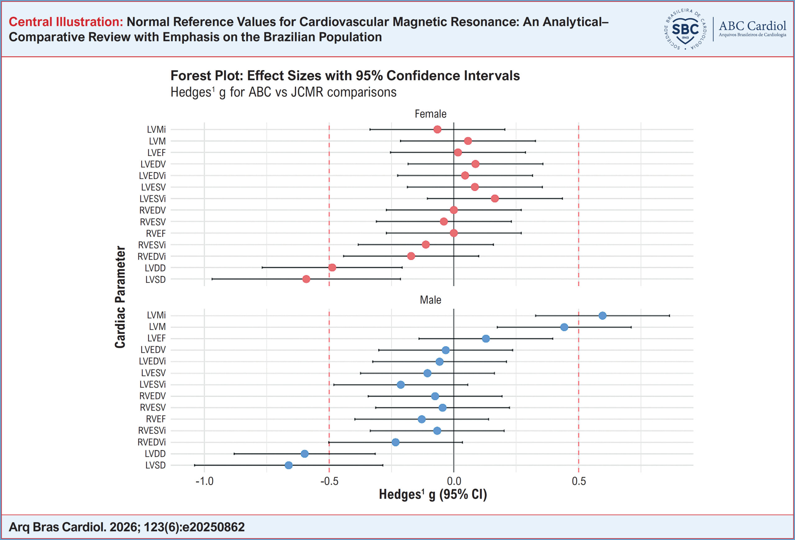
However, the proper interpretation of parameters obtained by CMR depends on the existence of robust reference values applicable to the studied population. In this context, the article “Normal Reference Values for Cardiovascular Magnetic Resonance: An Analytical–Comparative Review with Emphasis on the Brazilian Population”, published in this issue of the Arquivos Brasileiros de Cardiologia, addresses a question that is particularly relevant to Brazilian cardiology: the applicability of international normative values to the national reality.

[…]

  • Magnetic Resonance Imaging
  • Reference Standards
  • Reference Values
Read Full Article
Creative Commons Attribution (CC BY) Creative Commons license
10/08/2026 DOI: 10.36660/abc.2026458i Views 2
Normal Reference Values for Cardiovascular Magnetic Resonance: An Analytical–Comparative Review with Emphasis on the Brazilian Population
ABC
Original Article

Normal Reference Values for Cardiovascular Magnetic Resonance: An Analytical–Comparative Review with Emphasis on the Brazilian Population

Jorge A. Torreão et al. Isabela Bispo Santos da Silva Costa Antonildes N. Assunção-Jr Ilan Gottlieb … Leonardo Sara da Silva Tiago Senra Tiago Augusto Magalhães Marly Uellendahl Hugo Bizetto Zampa Gabriela Liberato Walther Yoshiharu Ishikawa Henrique Trad Marcelo Hadlich Otavio Coelho-Filho Roberto Sasdelli Neto Juliano Lara Fernandes Luiz Francisco Rodrigues de Ávila Ibraim Masciarelli F. Pinto Paulo R. Schvartzman Carlos E. Rochitte

ABSTRACT

Background:

Cardiovascular magnetic resonance (CMR) is the reference standard for morphofunctional cardiac assessment. In Brazil, the application of international normal reference values faces challenges related to population admixture, technical heterogeneity among centers, and the absence of national standardization, all of which may affect diagnostic accuracy.

Objectives:

To compare normal reference values for morphofunctional parameters obtained by CMR between a multicenter international review and a Brazilian publication, discussing concordance, methodological differences, and implications for Brazilian clinical practice.

Methods:

An analytical–comparative review was performed using summarized data from an international reference-value review and a Brazilian study. Means and standard deviations of key ventricular variables were compared, with assessment of 95% CI overlap, Welch’s t-test, and effect size (Hedges’ g). Standard-deviation–based cut-off points were proposed for grading abnormality.

Results:

Overall concordance between means was observed for most variables. Statistically significant differences occurred in a few parameters, mainly ventricular diameters and left ventricular mass, with small to moderate effect sizes. Discrepancies were largely attributed to methodological differences in measurement and indexation.

Conclusions:

International normal reference values may be used in Brazilian clinical practice, provided they are adjusted for body surface area and interpreted in light of local particularities. Standardization of protocols and national multicenter studies are needed to strengthen regional validation. Keywords: Normal Values, Cardiac Magnetic Resonance, Reference Interval.

  • Cardiovascular disease
  • Magnetic Resonance Imaging
  • Reference Values
Read Full Article
Creative Commons Attribution (CC BY) Creative Commons license
03/08/2026 DOI: 10.36660/abc.20250862i Views 1
Recognition of Clinical Stages and Myocardial Damage in Aortic Stenosis: Beyond Diagnosis – To Op or Not To Op
ABCIC
Review Article

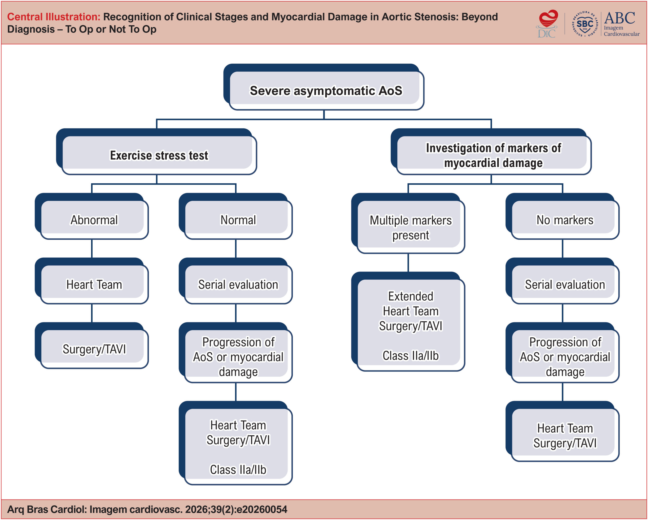
Recognition of Clinical Stages and Myocardial Damage in Aortic Stenosis: Beyond Diagnosis – To Op or Not To Op

Daniel Franca Vasconcelos

Abstract

Severe aortic stenosis is a progressive clinical entity that has historically developed alongside descriptions of pathological anatomy, cardiovascular semiology, heart surgery, and percutaneous cardiovascular interventions. Indication for valve replacement in patients with severe aortic stenosis is still based on symptom onset. With advances in surgical techniques, especially transcatheter implantation, the focus has shifted to identifying early markers of myocardial damage that may indicate irreversible myocardial injury. This review article seeks to provide a comprehensive analysis of methods for identifying myocardial damage in patients with severe aortic stenosis, including clinical evaluation, electrocardiography (ECG), echocardiography, cardiac magnetic resonance imaging (CMR), and plasma biomarkers. It also discusses the staging of myocardial damage and the optimal timing for therapeutic intervention based on clinical, functional, and structural findings. Recent studies designed to address how to manage asymptomatic patients with aortic stenosis have yielded divergent results. Accordingly, the aim is to assist clinicians in deciding the optimal timing for valve replacement, taking individual patient characteristics into account.

  • Aortic Valve Stenosis
  • Echocardiography
  • Magnetic Resonance Imaging
Read Full Article
Creative Commons Attribution (CC BY) Creative Commons license
31/07/2026 DOI: 10.36660/abcimg.20260054i Views 3
ABCIC
Editorial

Beyond Ejection Fraction: Integrating Myocardial Strain and Cardiac Magnetic Resonance into the Contemporary Assessment of Left Ventricular Function

Marly Uellendahl

The assessment of left ventricular function is undergoing a profound transformation. For decades, left ventricular ejection fraction (LVEF) has occupied a central role in clinical practice, guiding diagnosis, risk stratification, and therapeutic decision-making across a broad spectrum of cardiovascular diseases. Its extensive clinical validation, ease of acquisition, and reproducibility have established two-dimensional echocardiography as the first-line modality for evaluating left ventricular systolic function.,

However, advances in cardiovascular imaging have demonstrated that myocardial function cannot be fully characterized by a single volumetric parameter. Although LVEF remains a robust marker of global left ventricular performance, its dependence on loading conditions and its limited sensitivity for detecting early contractile abnormalities underscore the need for a more comprehensive functional assessment.

[…]

  • Left Ventricular Dysfunction
  • Magnetic Resonance Imaging
  • Stroke Volume
Read Full Article
Creative Commons Attribution (CC BY) Creative Commons license
27/07/2026 DOI: 10.36660/abcimg.20260087i Views 1
Figure 1
ABCIC
Case Report

Single Coronary Artery and Stress Cardiomyopathy: An Association Demonstrated by Multimodality Imaging

Marcus Vinicius Silva Ferreira et al. Julia Pereira Afonso dos Santos Tormin Roberto Nery Dantas Jr Roberto Vitor Almeida Torres … Renan Andreos Cordeiro José de Arimateia Batista Araújo Filho Luiz Francisco Cardoso

Case Report

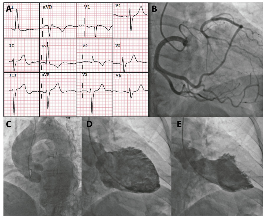
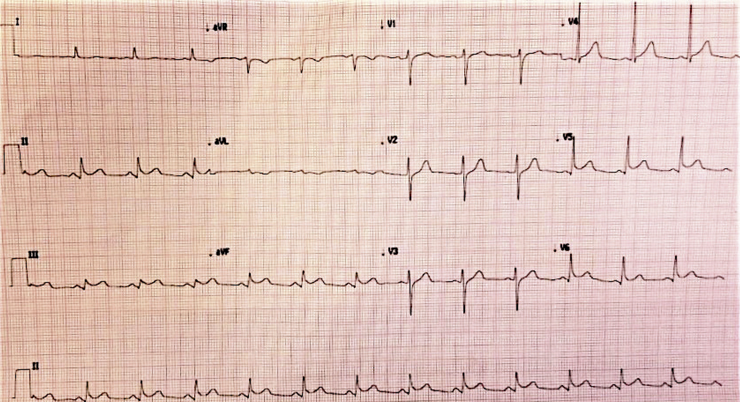
A 58-years-old female patient was admitted with retrosternal chest pain described as a continuous, oppressive tightness that had started two days earlier, after a tooth extraction procedure performed under ineffective local anesthesia. The patient’s past medical history included diabetes, dyslipidemia, and ovarian cancer, and she was receiving aspirin, rosuvastatin, metformin, dapagliflozin, and semaglutide. Physical examination was unremarkable. The chest pain protocol was initiated, and the initial electrocardiogram showed sinus rhythm with left anterior fascicular block, early repolarization in the inferior leads, and T-wave inversion in aVL, V1, and V2 (). The patient received 5 mg of sublingual isosorbide dinitrate, and blood samples were collected for laboratory testing. High-sensitivity troponin T (hs-cTnT) was 171 ng/L, confirming myocardial injury. An initial diagnosis of non–ST-elevation myocardial infarction (NSTEMI) was made, and the patient underwent coronary angiography (CA) for anatomic assessment ( / Video S1).

[…]

  • Coronary Angiography
  • Coronary Vessels
  • Echocardiography
  • Magnetic Resonance Imaging
  • Takotsubo Cardiomyopathy
Read Full Article
Creative Commons Attribution (CC BY) Creative Commons license
17/07/2026 DOI: 10.36660/abcimg.20260023i Views 1
ABC
Editorial

Cardiac Magnetic Resonance Imaging and Histology in Aortic Valve Disease: Convergences and Limitations

Helder Jorge Andrade Gomes et al. Tiago Senra

Myocardial fibrosis is a central element of ventricular remodeling in severe aortic valve disease and is associated with ventricular dysfunction and worse outcomes, even after valve intervention.– Although the collagen volume fraction obtained by histology represents the gold standard for quantifying interstitial fibrosis, myocardial biopsy is not part of routine clinical practice due to its invasive nature and sample limitations. In this context, cardiac magnetic resonance imaging (CMR) has become established as a non-invasive method for tissue characterization, allowing the identification of regional fibrosis through late enhancement and the estimation of interstitial expansion through T1 mapping and calculation of extracellular volume (ECV).–

The study published in this issue of the Arquivos Brasileiros de Cardiologia makes a significant contribution by directly comparing CMR parameters with the collagen volume fraction obtained by myocardial biopsy in 108 patients with severe aortic valve disease who underwent surgery. Unlike many previous series composed exclusively of aortic stenosis, this cohort also included patients with aortic insufficiency, broadening the applicability of the findings and incorporating a more comprehensive pathophysiological spectrum of ventricular overload.

[…]

  • Aortic Valve Disease
  • Fibrosis
  • Magnetic Resonance Imaging
Read Full Article
Creative Commons Attribution (CC BY) Creative Commons license
22/05/2026 DOI: 10.36660/abc.20260140i Views 1
Interstitial Myocardial Fibrosis Analysis by Histology and Cardiac Magnetic Resonance in Severe Aortic Valve Disease
ABC
Original Article

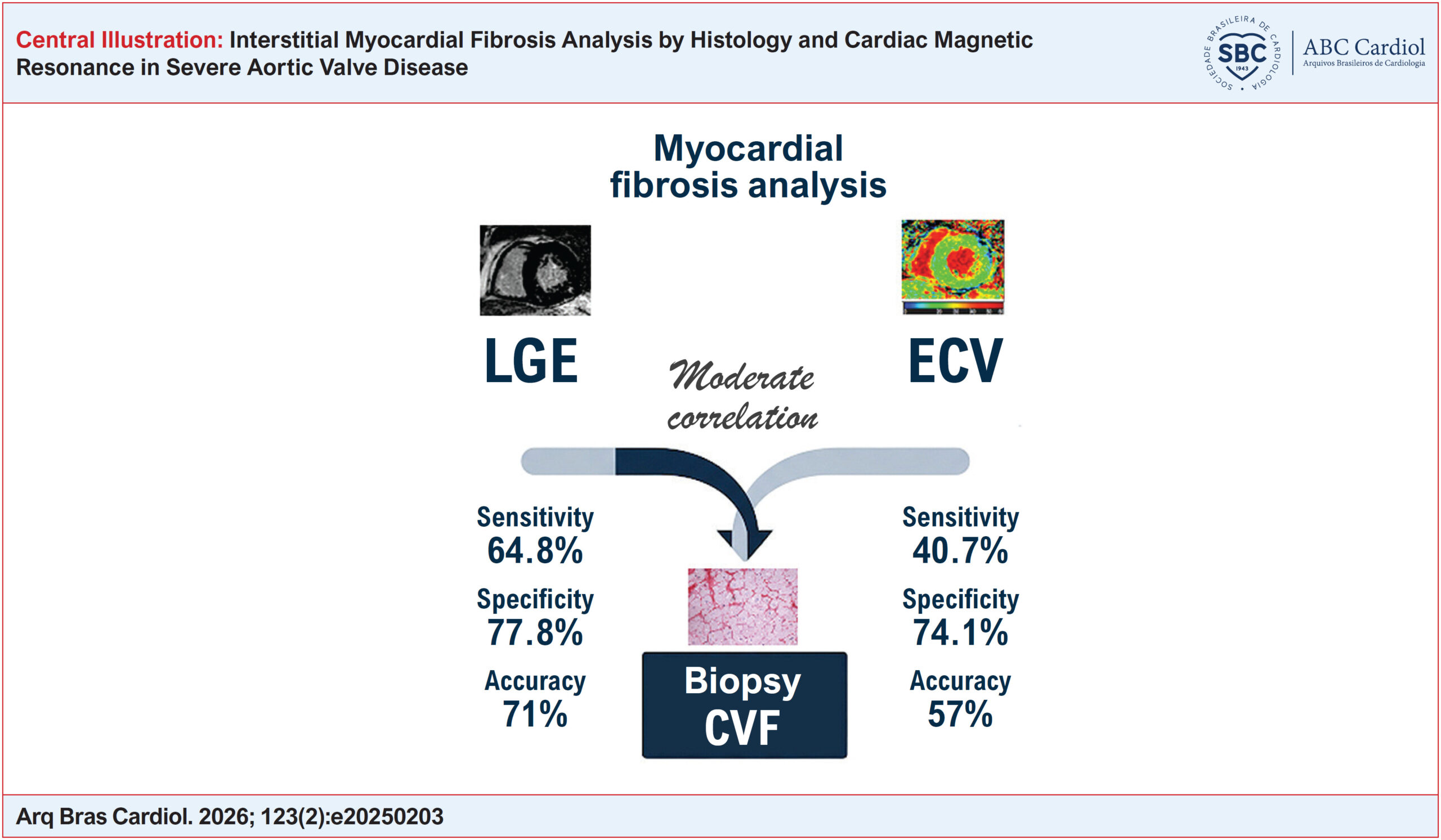
Interstitial Myocardial Fibrosis Analysis by Histology and Cardiac Magnetic Resonance in Severe Aortic Valve Disease

Juliana Hiromi Silva Matsumoto Bello et al. Lea M.M.F. Demarchi Rafael Almeida Fonseca Gabriele N. Lima … Lucas José Tachotti Pires Vitor E. E. Rosa Roney Orismar Sampaio Paulo S. Gutierrez Vera D. Aiello Flávio Tarasoutchi Carlos E. Rochitte

Abstract

Background:

Aortic valve disease may lead to the accumulation of myocardial fibrosis (MF). Cardiac magnetic resonance (CMR) with late gadolinium enhancement (LGE) evaluates regional fibrosis but does not detect interstitial fibrosis, which can be estimated by extracellular volume (ECV) using native (pre-contrast) and post-contrast T1 mapping.

Objectives:

To compare MF measured by collagen volume fraction (CVF) in myocardial biopsy with LGE and ECV in patients with severe aortic valve disease.

Methods:

A total of 108 patients undergoing aortic valve replacement with myocardial biopsy and preoperative CMR were included. LGE and ECV were compared with CVF using Pearson or Spearman correlation coefficients, diagnostic performance analysis, and agreement testing. Statistical significance was set at p < 0.05.

Results:

Increased CVF was observed in 96% of patients (21.9 ± 5.5%), LGE in 44% (1.3 ± 3%), and increased ECV in 33.3% (29.0 ± 4.0%). Moderate correlations were found between LGE and CVF and between ECV and CVF (r = 0.44 and r = 0.36, respectively; p = 0.001). LGE and ECV demonstrated sensitivities of 64.8% and 40.7%, specificities of 77.8% and 74.1%, and accuracies of 71% and 57%, respectively, using CVF above the median as the reference for MF. Agreement between CVF and ECV revealed a mean difference of −0.167 and a Cohen’s kappa value of 0.171.

Conclusions:

CMR detects MF in patients with severe aortic valve disease confirmed by biopsy. LGE and ECV show moderate correlation with CVF. Combined analysis of LGE and ECV may provide complementary information; however, additional studies are needed to determine their prognostic impact.

  • Aortic Valve Disease
  • Fibrosis
  • Histology
  • Magnetic Resonance Imaging
  • Miocardio
Read Full Article
Creative Commons Attribution (CC BY) Creative Commons license
15/05/2026 DOI: 10.36660/abc.20250203i Views 1
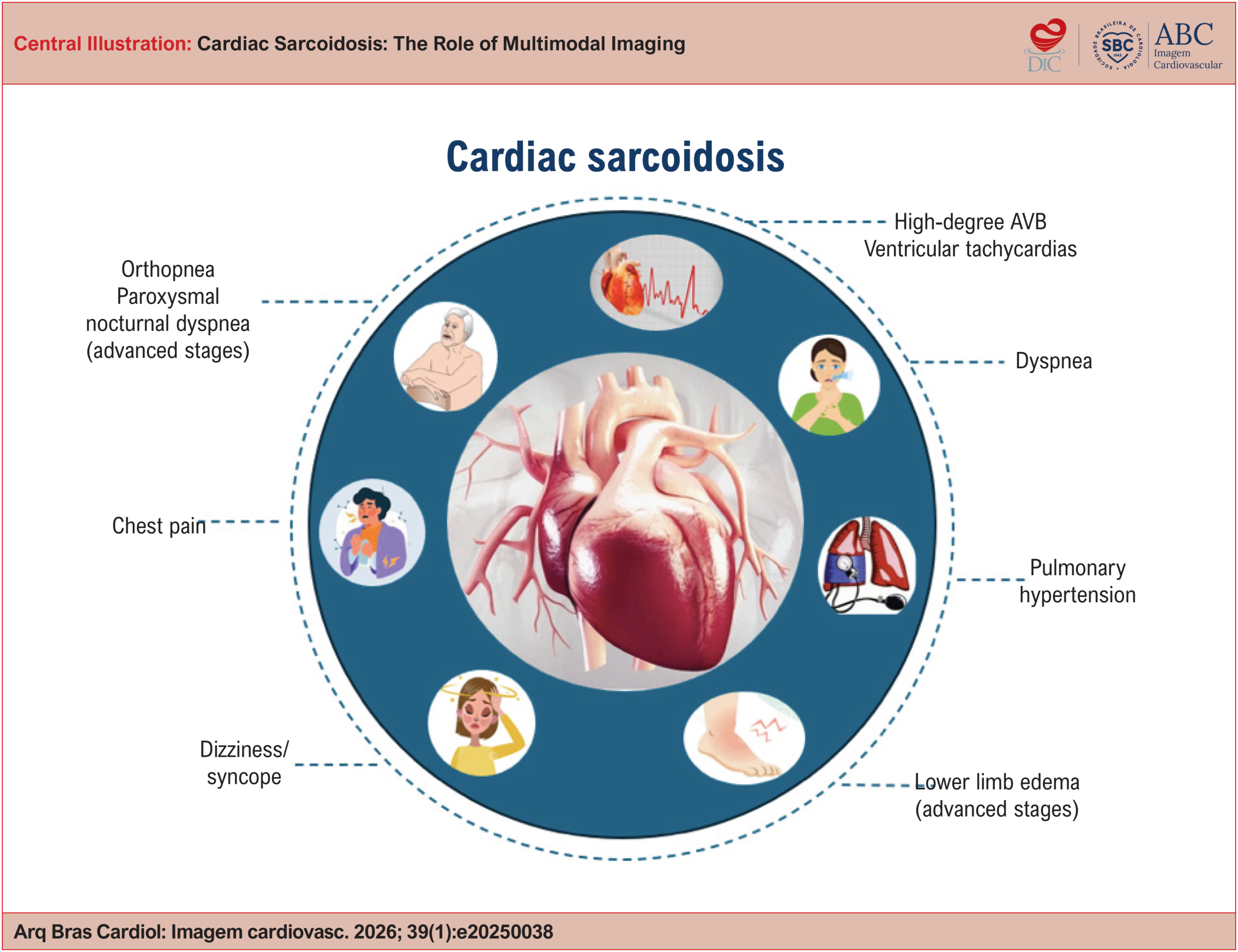
Cardiac Sarcoidosis: The Role of Multimodal Imaging
ABCIC
Review Article

Cardiac Sarcoidosis: The Role of Multimodal Imaging

Lara Cristiane Terra Ferreira Carreira et al. Lívia Carreira Adriana Soares Xavier de Brito

ABSTRACT

Cardiac sarcoidosis (CS) is a potentially severe manifestation of systemic sarcoidosis, associated with advanced atrioventricular block, ventricular arrhythmias, heart failure, and sudden death. Diagnosis remains challenging due to phenotypic variability and the limitations of conventional diagnostic methods. Advances in imaging techniques, especially the combination of positron emission tomography/computed tomography (PET/CT) using 18F-FDG and cardiac magnetic resonance imaging (CMR), have revolutionized the diagnostic approach and therapeutic monitoring of CS. This article reviews current concepts of CS and its diagnosis, with a focus on the role of PET/CT, the importance of appropriate patient preparation, and integration with CMR.

  • Fluorodeoxyglucose F18
  • Magnetic Resonance Imaging
  • Multimodal Imaging
  • Positron-Emission Tomography
  • Sarcoidosis
Read Full Article
Creative Commons Attribution (CC BY) Creative Commons license
20/04/2026 DOI: 10.36660/abcimg.20250038i Views 1
ABCIC
Editorial

Transesophageal Echocardiogram Always Before TAVI?

Jairo Alves Pinheiro et al. João Lucas Pinheiro Lucas Velloso Dutra Luciano Holanda Renata de Sá Cassar

Cardiovascular imaging is crucial in the selection and planning of interventions for structural heart disease.

Transcatheter aortic valve implantation (TAVI) has transformed the management of severe aortic stenosis (AS) in high-risk surgical populations and is currently being expanded to intermediate- and low-risk populations.

[…]

  • Aortic Valve Stenosis
  • Magnetic Resonance Imaging
  • Transcatheter Aortic Valve Replacement
  • Transesophageal echocardiography
Read Full Article
Creative Commons Attribution (CC BY) Creative Commons license
08/12/2025 DOI: 10.36660/abcimg.20250060i
Figure 1
ABCIC
Original Article

Overview of Professional Practice in Computed Tomography and Cardiac Magnetic Resonance Imaging in Brazil

Isabela Bispo Santos da Silva Costa et al. Jorge Andion Torreão Juliano de Lara Fernandes Henrique Trad … Gabriela Liberato Roberto Sasdelli Neto Silvio Henrique Barberato Ibraim Masciarelli Pinto Tiago Senra

Abstract

Background:

Non-invasive imaging for cardiovascular disease diagnosis has grown in volume and relevance in recent years, and it is important for early detection of cardiovascular diseases. There is a lack of data from Brazil on the availability of methods such as computed tomography (CT) and cardiac magnetic resonance imaging (CMR).

Objective:

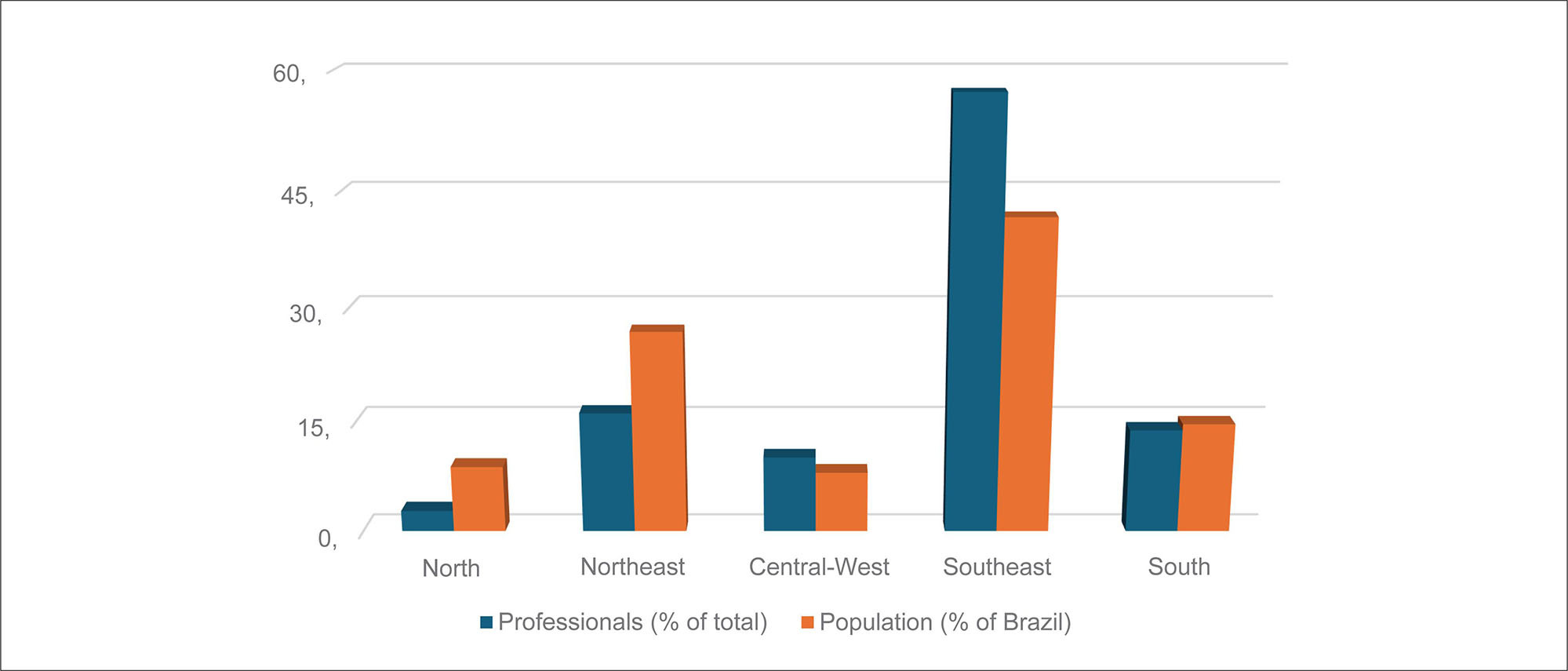
This study assessed the regional distribution of professional practice and the exams performed, correlating these factors with population data and the number of active professionals.

Methods:

A nationwide survey was conducted by means of an online questionnaire applied to professionals who perform CT and CMR, compared with demographic data and the number of scans performed within Brazilian public and private healthcare systems.

Results:

A total of 219 professionals filled out the questionnaire, 139 (63.8%) male, 161 (73.9%) cardiologists, and 46 (21.1%) radiologists. Regarding regional distribution, the Southeast had 125 professionals (57.3%), the Northeast 35 (16.1%), the South 30 (13.8%), the Central-West 22 (10.1%), and the North 6 (2.8%). The profile of these professionals in Brazilian regions was similar, with no statistical differences observed regarding the proportion of men (p = 0.2451), cardiologists (p = 0.1325), radiologists (p = 0.4564), or training time > 2 years (p = 0.8519). The Southeast had the highest absolute number of exams, and the North had the lowest.

Conclusion:

The data revealed structural disparities in access to cardiovascular imaging throughout Brazil, in terms of both number of professionals and number of exams performed. The regional discrepancies observed in this study reflect distinct demographic and economic circumstances in Brazilian regions.

  • Cardiovascular Diseases
  • Magnetic Resonance Imaging
  • Supplemental Health
  • Unified Health System
  • X-Ray Computed Tomography
Read Full Article
Creative Commons Attribution (CC BY) Creative Commons license
17/11/2025 DOI: 10.36660/abcimg.20250053i Views 1
Figure 1
ABCIC
Research Letter

3D Printing of a Heart With Amyloidosis

Mariana de Paula Cruz et al. Davi Shunji Yahiro Daniel Gama das Neves Renato Pereira Barbosa … Alexandre Todorovic Fabro Pedro Manoel Marques Garibaldi Marcus Simões Claudio Tinoco Mesquita

Abstract

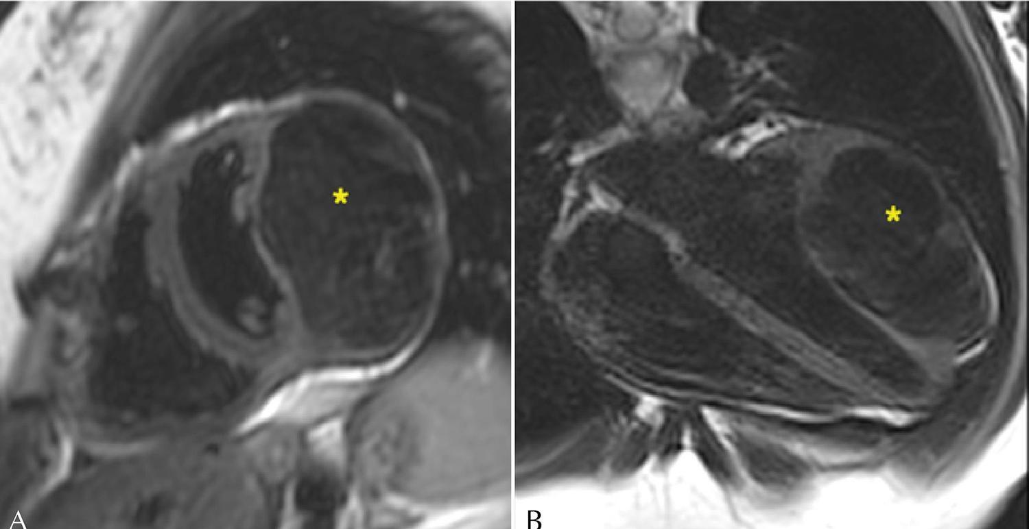
Cardiac Amyloidosis is a restrictive cardiomyopathy caused by the accumulation of amyloid protein in the heart. The advances in non-invasive diagnosis have been increasing its identification among patients with Heart Failure (HF), being previously considered to be a rare condition. Currently, it represents around 13% of the cases of Heart Failure with preserved Ejection Fraction (HFpEF). We report the case of a 72-year-old male patient who presented to the clinic reporting dyspnea and fatigue in low-effort activities. Laboratory exams were conducted, revealing increased cardiac markers, along with evidence of ventricular hypertrophy in electrocardiographic analysis. Cardiac imaging, serum free light chain (AL) assay, and endomyocardial biopsy indicated the diagnosis of AL amyloidosis. A three-dimensional model was constructed using magnetic resonance imaging (MRI) data. This model is intended to increase the awareness around amyloidosis as a possible differential diagnosis of HF, while also promoting patients’ and families’ education, demonstrating the typical structural abnormalities of the disease.

  • Amyloidosis
  • Heart Failure
  • Magnetic Resonance Imaging
  • Three-Dimensional Printing
Read Full Article
Creative Commons Attribution (CC BY) Creative Commons license
17/11/2025 DOI: 10.36660/abcimg.20250003i Views 1
ABC
Editorial

From Electrocardiogram to Magnetic Resonance Imaging – The Quest for Unrecognized Myocardial Infarction

Pedro Sousa Mateus

In the article “Detection and localization of myocardial infarction using electrocardiogram: validation by cardiovascular Magnetic Resonance Imaging”, the authors systematically and thoroughly analyze the relationship between the presence of pathological Q waves on the electrocardiogram (ECG) and delayed enhancement with an ischemic pattern on magnetic resonance imaging (MRI). Very relevant data on the diagnostic performance of the ECG in detecting and localizing myocardial infarction (MI) are presented, expanding the evidence accumulated in previous studies.

The authors studied two groups of patients: one consisting of patients with a documented history of MI and the other selected from diabetic patients without significant cardiac abnormalities on MRI.

[…]

  • Electrocardiography
  • Magnetic Resonance Imaging
  • Myocardial Infarction
Read Full Article
Creative Commons Attribution (CC BY) Creative Commons license
12/05/2025 DOI: 10.36660/abc.20250147i Views 1
Detection and Location of Myocardial Infarction Using Electrocardiogram: Validation by Cardiovascular Magnetic Resonance Imaging
ABC
Original Article

Detection and Location of Myocardial Infarction Using Electrocardiogram: Validation by Cardiovascular Magnetic Resonance Imaging

Maria Catarina de Melo Dias Guerra et al. André Gustavo da Silva Rezende Tiago Augusto Magalhães William Azem Chalela … Augusto Hiroshi Uchida Rafael Almeida Fonseca Nevelton Heringer Filho Jürgen Beuther Guilherme Garcia Eduardo Cavalcanti Lapa dos Santos Ulisses Ramos Montarroyos Renata Ávila Cintra José Antônio Franchine Ramires Carlos Eduardo Rochitte

Abstract

Background

In the assessment of ischemic heart disease, cardiac magnetic resonance (CMR) is considered the gold standard for detecting and locating myocardial infarction (MI), but electrocardiogram (ECG) is less expensive and more widely available. Recognition of MI on ECG outside the acute phase is challenging; Q waves are absent in a significant proportion of patients and may reduce or disappear over time. Although ECG is widely used in the initial assessment of previous infarction, studies to validate ECG using CMR as a reference in the context of chronic coronary disease are limited.

Objectives

To evaluate the diagnostic performance of ECG in detecting and locating CMR-defined MI.

Methods

This study included 352 individuals who underwent CMR and ECG, 241 patients with previous MI confirmed by CMR and 111 controls with normal CMR. Their ECG tracings were analyzed by 2 observers, who were blinded to the CMR, for detection and location of MI following to the Fourth Brazilian Society of Cardiology Guidelines on the Analysis and Issuance of Electrocardiographic Reports. The significance level adopted was 5% (p < 0.05).

Results

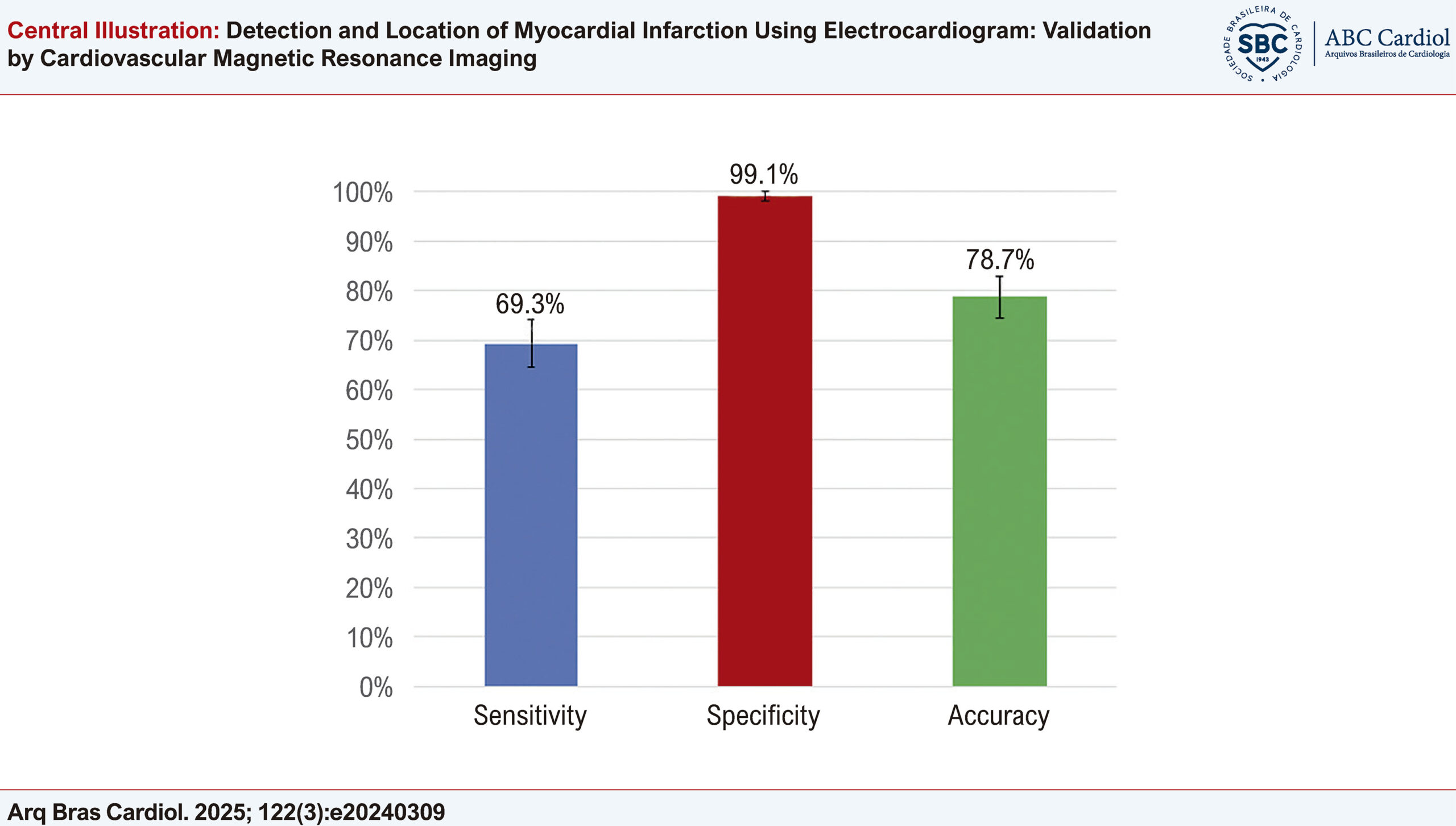
ECG showed good performance for detecting previous MI, with sensitivity of 69.3% (64.5% to 74.1%), specificity of 99.1% (98.1% to 100%), and accuracy of 78.7% (74.4% to 83.0%). However, in locating MI in accordance with CMR, its accuracy was unsatisfactory.

Conclusions

When compared to CMR, ECG was shown to be a method with good accuracy for detecting previous MI, but not for defining its location.

  • Coronary Artery Disease
  • Electrocardiography
  • Magnetic Resonance Imaging
  • Myocardial Infarction
Read Full Article
Creative Commons Attribution (CC BY) Creative Commons license
12/05/2025 DOI: 10.36660/abc.20240309i Views 3
Figure 1
ABCIC
Case Report

A Common Primary Cardiac Tumor With an Uncommon Presentation

Juan Felipe Vasquez-Rodriguez et al. Carolina Paola Idrovo-Turbay Jaime Alberto Rodríguez Lorena Montes Villalobos

Abstract

Background

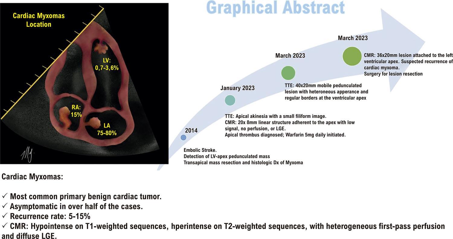
Cardiac tumors are rare, and most are secondary (metastatic) lesions. Primary tumors, though uncommon, are typically benign, with myxomas being the most frequent and occasionally displaying unique features. Diagnosis often requires multimodality imaging.

Case summary

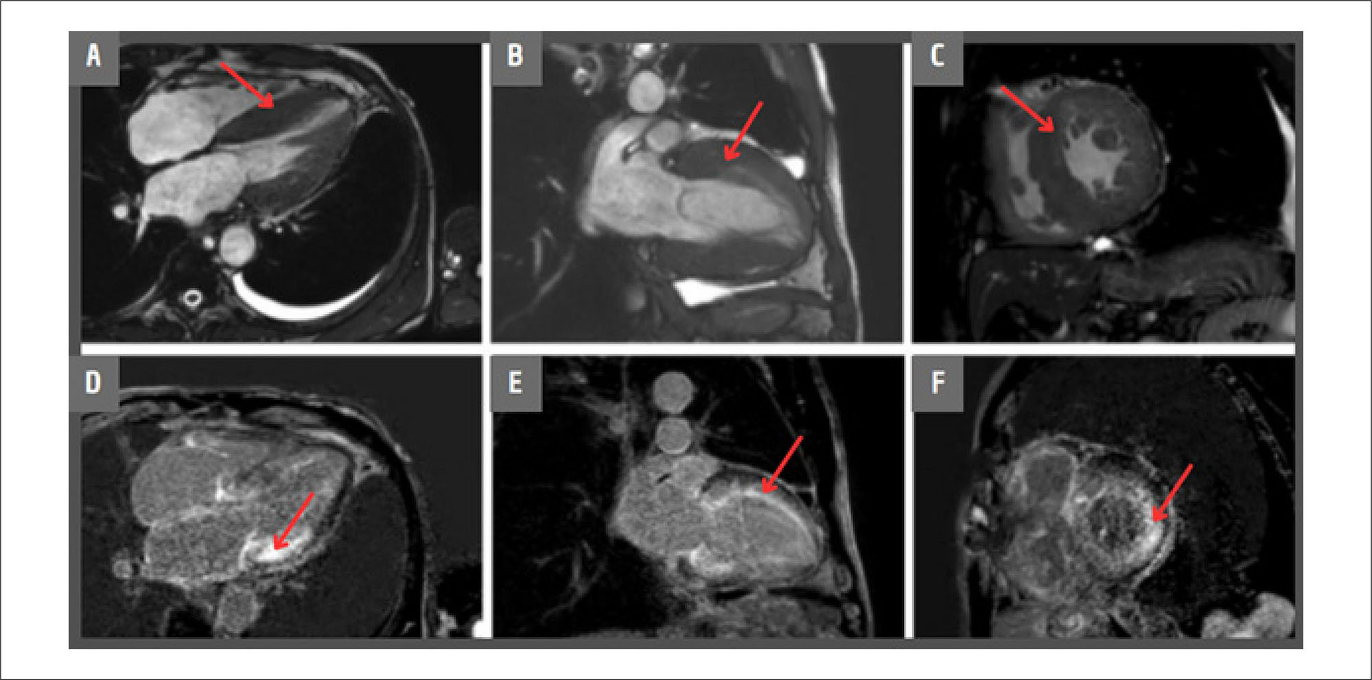
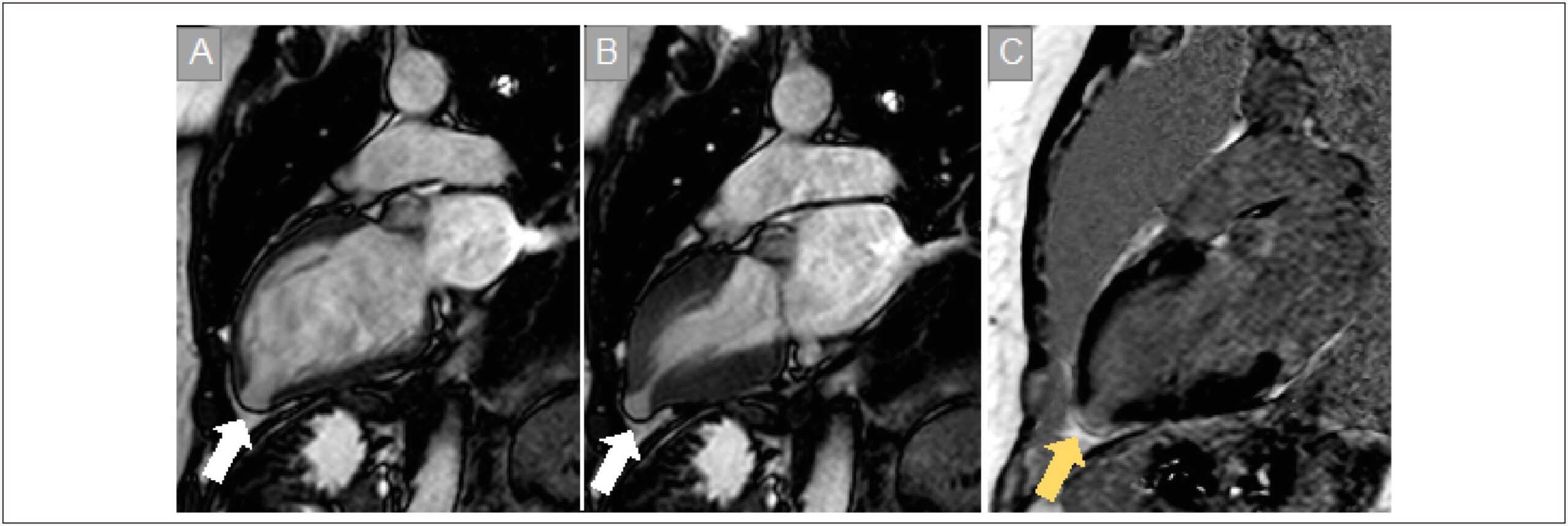
A 41-year-old man with a history of ischemic stroke and resection of an apical left ventricular myxoma presented for evaluation after a follow-up transthoracic echocardiogram (TTE) revealed a mobile, ovoid apical lesion with high embolic risk, despite being asymptomatic. Cardiac magnetic resonance (CMR) imaging suggested recurrence of the ventricular myxoma. Surgical resection was performed, and histopathology confirmed the diagnosis of cardiac myxoma (CM).

Discussion

CMs are the most common primary benign cardiac tumors, typically located in the left atrium and attached to the interatrial septum. They are less frequently found in the ventricular cavities. About half of cases are asymptomatic, and recurrence is possible after resection. This case highlights a rare instance of late recurrence in an atypical location.

  • Echocardiography
  • Heart Neoplasms
  • Magnetic Resonance Imaging
  • Myxoma
Read Full Article
Creative Commons Attribution (CC BY) Creative Commons license
27/03/2025 DOI: 10.36660/abcimg.20240123i Views 1
Central Illustration
ABCIC
Review Article

Pericardial Agenesis: When Should This Diagnosis Be Considered?

Bruna Barbosa Chagas de Almeida et al. Bruna Zanforlin Jácome Bernardo Nascimento Lourenço Alessandro Zakhia de Seixas … Juliana Serafim da Silveira Eliza de Almeida Gripp

Abstract

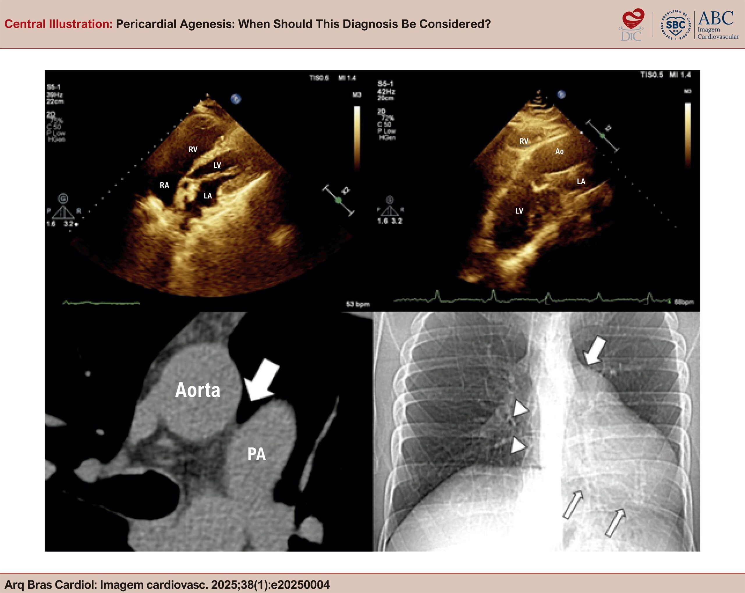
Pericardial agenesis (PA) is a rare condition characterized by the partial or complete absence of the pericardium. The consequences associated with this condition can be quite serious. Therefore, understanding the peculiarities of this disease is essential for accurate diagnosis and treatment.

  • Magnetic Resonance Imaging
  • Pericarditis
  • Pericardium
  • X-Ray Computed Tomography
Read Full Article
Creative Commons Attribution (CC BY) Creative Commons license
27/03/2025 DOI: 10.36660/abcimg.20250004i Views 1
ABCIC
Editorial

Quantification of Myocardial Fibrosis by Cardiac Magnetic Resonance Imaging: Advances, Impacts, and Perspectives

Isabela Bispo Santos da Silva Costa

Cardiac magnetic resonance imaging (CMR) has been established as a fundamental tool for the diagnosis and management of cardiomyopathies, especially due to its accuracy in tissue characterization, identification, and quantification of myocardial fibrosis., Late gadolinium enhancement (LGE) allows accurate assessment, identifying areas of myocardial fibrosis, and it serves to characterize, locate, and quantify them. In recent years, the role of LGE in the risk stratification of patients with hypertrophic cardiomyopathy (HCM) and other genetic cardiomyopathies has been the subject of several studies that aimed to correlate the presence and extent of myocardial fibrosis with the risk of sudden cardiac death (SCD) and major cardiovascular events.–

Different techniques are currently used to acquire, analyze, and quantify LGE by CMR. The presence of LGE is based on the differential tissue retention of gadolinium, which is preferentially absorbed in areas of fibrosis, where scar tissue is altered due to the presence of collagen. LGE acquisition techniques nowadays are fast, safe, and can be performed without requiring long respiratory pauses, as was the case in the past, with progressively better resolution and image definition. To quantify areas of fibrosis, different software programs that allow adequate quantification in a semi-automated manner are currently available.

[…]

  • Cardiomyopathies
  • Endomyocardial Fibrosis
  • Magnetic Resonance Imaging
Read Full Article
Creative Commons Attribution (CC BY) Creative Commons license
13/12/2024 DOI: 10.36660/abcimg.20240114i Views 1
Figure 1
ABCIC
Case Report

Giant Left Atrium in Rheumatic Mitral Valve Disease

Alice Cunha Darzé et al. Antonio Tito Paladino Filho Amanda Marsiaj Rassi Carlos Eduardo Prazeres … Ibraim Masciarelli Francisco Pinto João Marcelo Caneschi Jose Roberto Tuma da Ponte Junior Paul Salvador Morales Rafael Castro Hendges Tiago Senra

Abstract

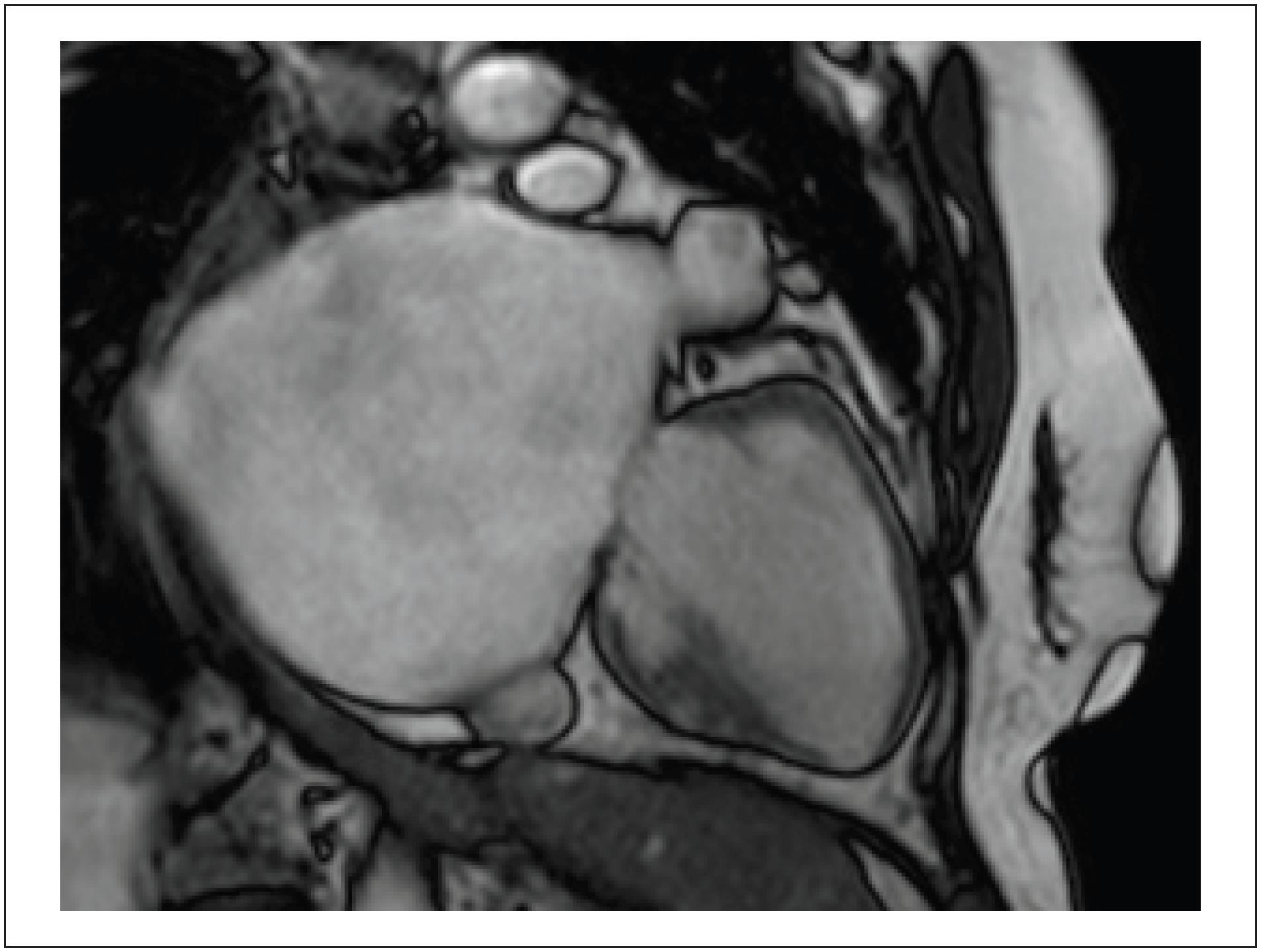
The Giant Left Atrium (GLA) is a rare condition, more commonly related to rheumatic mitral valve disease; it can be mistaken with a few possible differential diagnoses when less accurate methods are implemented, potentially leading to particularly dangerous and unnecessary procedures in this setting. Cardiovascular imaging methods, such as Cardiac Magnetic Resonance (CMR), can be valuable for this differentiation and provide high spatial resolution, supporting accurate diagnosis and surgical planning when indicated. Selecting the most precise diagnostic method, however, requires deep knowledge of the GLA condition and clinical data, leading to a correct suspicion. Furthermore, opting for using volumetry rather than the biplanar method in cases of significant deformity ensures greater accuracy in the values obtained.

  • Heart Atria
  • Heart Valve Diseases
  • Magnetic Resonance Imaging
Read Full Article
Creative Commons Attribution (CC BY) Creative Commons license
05/12/2024 DOI: 10.36660/abcimg.20240083i Views 1
Figure 1
ABCHF
Original Article

Myocarditis: Whole Heart Involvement Revealed by Cardiac Magnetic Resonance Mapping. A Case-control Study

Tiago Bartzen Pereira et al. Maurício Balk Gabriela Bartzen Pereira Victória Schmidt Ramos … Luciano Giordani Paulo R. Schvartzman Luís Beck-da-Silva

Abstract

Background:

Late gadolinium enhancement (LGE) on cardiac magnetic resonance (CMR) only demonstrates regional abnormalities in myocarditis and does not adequately assess diffuse myocardial involvement.

Objective:

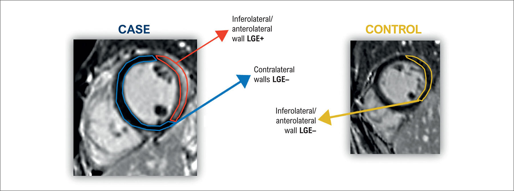
To evaluate possible differences in T1 and T2 mapping between ventricular wall segments with and without LGE in patients with myocarditis, compared to control subjects.

Methods:

In a case-control design, 22 patients with CMR evidence of myocarditis and 18 controls with normal CMR were assessed. The study included: (1) T1 mapping (shortened modified Look-Locker Inversion recovery); (2) LGE; (3) T2 mapping (steady-state free precession); and (4) the T2 signal intensity of the myocardium divided by that of skeletal muscle (T2 ratio). T1 and T2 mapping of affected (LGE+) and unaffected (LGE−) ventricular segments of cases were compared, as were those of controls versus cases. The level of significance was set at a two-sided alpha level of 0.05.

Results:

Comparing only patients with myocarditis, ventricular segments with evidence of late enhancement (LGE+) showed a mean T1 value significantly different from that of unaffected (LGE−) ventricular walls (1057 ± 30 versus 1028 ± 48; p = 0.0001). Comparing myocarditis versus controls, the mean T1 value of negative LGE segments in cases (myocarditis +) was significantly different from the mean of the corresponding walls in controls (1028 ± 48 versus 996 ± 10; p < 0.0001). The mean T2 maps of negative LGE walls in cases were not statistically different from those of controls (49 ± 4 versus 49 ± 1; p = 0.9229).

Conclusion:

This case-control study suggests that T1 mapping demonstrates significant involvement of the myocardium of patients with myocarditis, even in the absence of LGE. Specifically, T1 mapping could reveal diffuse myocardial involvement not evidenced by LGE imaging. T2 mapping was noncontributory.

  • Contrast Media
  • Magnetic Resonance Imaging
  • Myocarditis
Read Full Article
Creative Commons Attribution (CC BY) Creative Commons license
01/12/2022 DOI: 10.36660/abchf.20220075 Views 2
Figure 1
ABCIC
Review Article

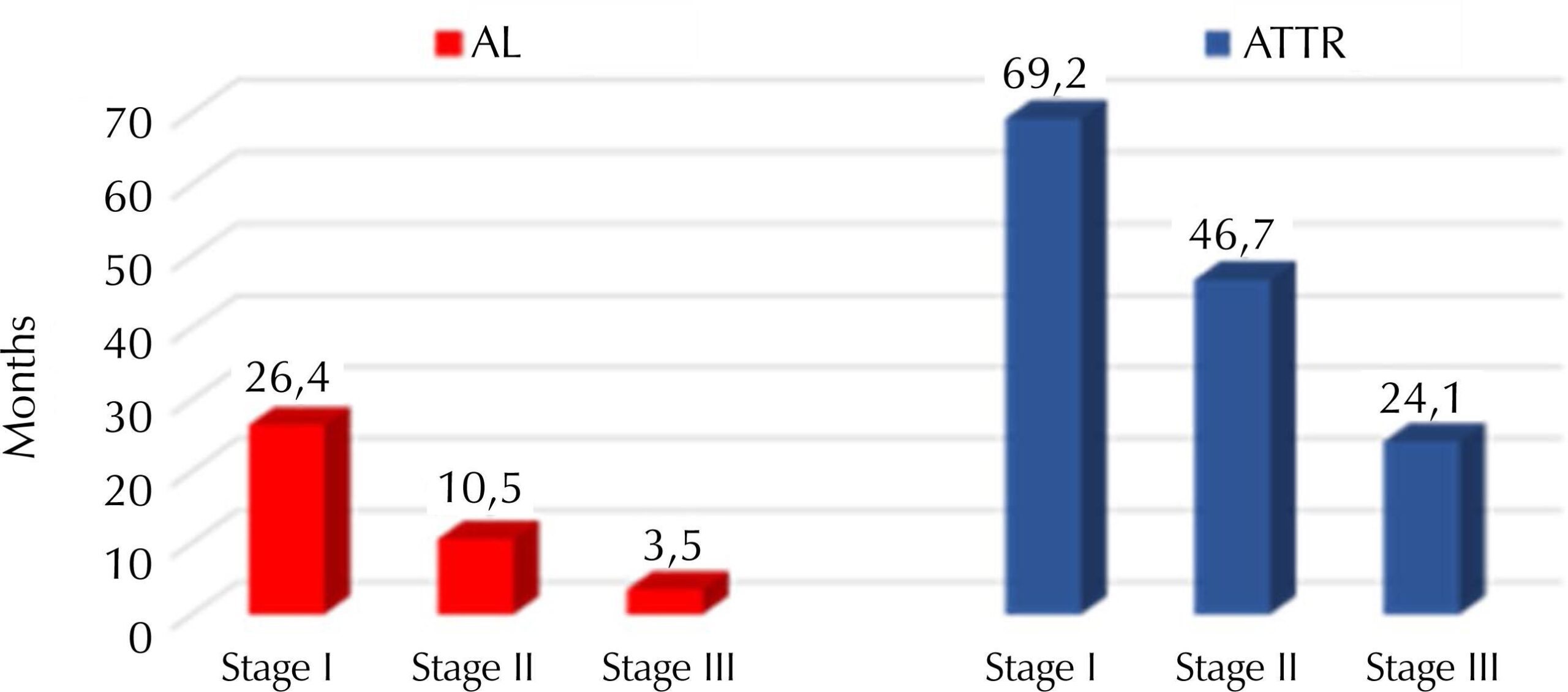
The Role of Cardiac Magnetic Resonance in patients with Amyloidosis and Aortic Stenosis

Pedro Macedo Esmeraldo Barbosa et al. Tiago Augusto Magalhães

Abstract

Cardiac magnetic resonance imaging (CMR) plays an important role in the diagnosis and prognosis of various heart diseases. The incidence of cardiac amyloidosis and aortic stenosis has been increasing in recent years, especially with the aging population, and their possible coexistence has created frequent diagnostic challenges in clinical practice. CMR assumes an important value in this context due to its unique capacity for multiparametric assessment evaluations that include anatomical, functional, and histopathological aspects. The present review aimed to increase our knowledge of the diagnosis and prognosis of amyloidosis in the presence of aortic stenosis and highlighted the importance of CMR in this scenario.

  • Amyloidosis
  • Aortic Valve Stenosis
  • Heart
  • Magnetic Resonance Imaging
Read Full Article
Creative Commons Attribution (CC BY) Creative Commons license
01/12/2022 DOI: 10.47593/2675-312X/20223503eabc320i Views 1
Figure 1
ABC
Research Letter

COVID-19 Myocarditis Mimicking ST-Segment Elevation Myocardial Infarction

Anthony Medina Conceição et al. César A. C. Pereira Maria Júlia Rahal Walther Yoshiharu Ishikawa Carlos E. Rochitte

Introduction

The new SARS-CoV-2, that causes the coronavirus disease 2019 (COVID-19) has proven to be a virus that affect not only the respiratory system, but to cause several systemic manifestations, including cardiovascular ones. , Patients with previous cardiovascular disease who develop myocardial injury usually have worse outcomes, , such as acute coronary syndrome (ACS) , and myocarditis. – Myocarditis is mostly asymptomatic but can manifest with angina, cardiac failure, and arrythmias. –

The clinical diagnosis of myocarditis without the aid of complementary exams is usually difficult to be made. A meta-analysis with 2,866 with myocardial infarction without obstructive coronary artery disease (MINOCA) who underwent cardiac magnetic resonance (CMR) showed a prevalence of myocarditis of 34.5%. In COVID-19, a study carried out in Germany reported that 60% of recently recovered patients had signs of myocardial inflammation at CMR.

[…]

  • Acute coronary syndrome
  • COVID-19
  • Magnetic Resonance Imaging
  • Myocarditis
Read Full Article
Creative Commons Attribution (CC BY) Creative Commons license
18/07/2022 DOI: 10.36660/abc.20210749 Views 1
Figure 1
ABCIC
Review Article

Myocardial Involvement in Chagas Disease: From the Perspective of Cardiovascular Magnetic Ressonance Assessment

Henrique Turin Moreira et al. Gustavo Jardim Volpe André Schmidt

Abstract

Chagas disease represents an important public health problem, especially in endemic countries in Latin America. Chronic cardiomyopathy is its most frequent clinical presentation. Myocardial involvement has a multifactorial pathogenesis and can lead to heart failure, thromboembolic events, arrhythmias, and sudden death. In this context, cardiovascular magnetic resonance imaging (CMR) is an excellent noninvasive method for investigating myocardial damage and understanding the mechanisms and consequences of these injuries. CMR has high spatial resolution and tissue characterization capacity, enabling a highly reliable morphofunctional analysis and the identification of risk markers for adverse events in patients with Chagas disease. This exam is very useful for the diagnosis and follow-up of these patients in the routine clinical setting.

  • Chagas disease
  • Magnetic Resonance Imaging
  • Ventricular Dysfunction
Read Full Article
Creative Commons Attribution (CC BY) Creative Commons license
30/03/2022 DOI: 10.47593/2675-312X/20223501eabc285
Figure 1
ABCIC
Case Report

Cardiac Fibroma: Long-Term Follow-up by Cardiac Magnetic Resonance

Fernanda Sayuri Oshiro et al. Raul Serra Valério Alfredo Augusto Eyer Rodrigues Gilberto Szarf … Marly Uellendahl Maria Eduarda Menezes de Siqueira

Abstract

Background

Primary cardiac neoplasms are rare, and a correct diagnosis is essential to planning the most appropriate treatment.

Objective

To demonstrate the role of cardiac magnetic resonance imaging (CMR) in the assessment, diagnosis, and follow-up of cardiac fibroma.

Case report

Female 21-year-old patient with a myocardial mass detected on echocardiogram. CMR confirmed a diagnosis of cardiac fibroma. The patient chose to be followed up, and her condition remained stable after six years.

Discussion

Cardiac fibromas are the second most common neoplasm in children and young people. On CMR, it is characterized by intense and homogeneous delayed enhancement. CMR plays an important role in the assessment of cardiac masses.

  • Fibromas
  • Heart Neoplasms
  • Magnetic Resonance Imaging
Read Full Article
Creative Commons Attribution (CC BY) Creative Commons license
04/03/2022 DOI: 10.47593/2675-312X/20223501eabc263 Views 1
ABCIC
Image

Circumferential Strain in the Diagnosis of Acute Myocarditis

Antonio Carvalho Leme Neto et al. Gustavo Carvalho

[…]

  • Diagnostic Imaging
  • Magnetic Resonance Imaging
  • Myocarditis
  • Myocardium/necrosis
Read Full Article
Creative Commons Attribution (CC BY) Creative Commons license
04/02/2022 DOI: 10.5935/2318-8219.20170023 Views 2
ABCIC
Case Report

Multimodality Evaluation in Cardiovascular Imaging in the Diagnosis of Atypical Cases of Chest Pain: Myasthenia Gravis

Roberta de Alvarenga Batista et al. Alexandre Henrique Cobucci Santana Roberto Luiz Marino Bárbara Campos Abreu Marino

[…]

  • Chest Pain
  • Diagnostic Imaging
  • Echocardiography
  • Electrocardiography
  • Magnetic Resonance Imaging
  • Myasthenia Gravis/diagnosis
Read Full Article
Creative Commons Attribution (CC BY) Creative Commons license
20/01/2022 DOI: 10.5935/2318-8219.20190012 Views 3

Could not load more articles. Please try again.

How to cite this article

EndNote RIS

Electronic Document Format (Vancouver)

Correia M. Reference Values in Cardiovascular Magnetic Resonance Imaging: Between Local Validation and Global Harmonization. Arquivos Brasileiros de Cardiologia 2026;123(6):e20260458.

Electronic Document Format (ABNT)

Correia, Miguel. Reference Values in Cardiovascular Magnetic Resonance Imaging: Between Local Validation and Global Harmonization. Arquivos Brasileiros de Cardiologia, v. 123, n. 6, e20260458, Aug. 2026.

Electronic Document Format (APA)

Correia, M. (2026). Reference Values in Cardiovascular Magnetic Resonance Imaging: Between Local Validation and Global Harmonization. Arquivos Brasileiros de Cardiologia, 123(6), e20260458.

Electronic Document Format (ISO)

Correia, Miguel. Reference Values in Cardiovascular Magnetic Resonance Imaging: Between Local Validation and Global Harmonization. Arquivos Brasileiros de Cardiologia [online]. 2026, vol. 123, n. 6, [cited 2026-10-14], e20260458. Available from: <https://journals.cardiol.br/en/article/reference-values-in-cardiovascular-magnetic-resonance-imaging-between-local-validation-and-global-harmonization/>. ISSN 0066-782X

EndNote RIS

Electronic Document Format (Vancouver)

Torreão JA, Costa IBSS, Assunção-Jr AN, Gottlieb I, Silva LS, Senra T, Magalhães TA, et al. Normal Reference Values for Cardiovascular Magnetic Resonance: An Analytical–Comparative Review with Emphasis on the Brazilian Population. Arquivos Brasileiros de Cardiologia 2026;123(6):e20250862.

Electronic Document Format (ABNT)

Torreão, Jorge A.; Costa, Isabela Bispo Santos da Silva; Assunção-Jr, Antonildes N.; Gottlieb, Ilan; Silva, Leonardo Sara da; Senra, Tiago; Magalhães, Tiago Augusto; Uellendahl, Marly; Zampa, Hugo Bizetto; Liberato, Gabriela; Ishikawa, Walther Yoshiharu; Trad, Henrique; Hadlich, Marcelo; Coelho-Filho, Otavio; Sasdelli Neto, Roberto; Fernandes, Juliano Lara; Ávila, Luiz Francisco Rodrigues de; Pinto, Ibraim Masciarelli F.; Schvartzman, Paulo R.; Rochitte, Carlos E.. Normal Reference Values for Cardiovascular Magnetic Resonance: An Analytical–Comparative Review with Emphasis on the Brazilian Population. Arquivos Brasileiros de Cardiologia, v. 123, n. 6, e20250862, Aug. 2026.

Electronic Document Format (APA)

Torreão, J. A., Costa, I. B. S. S., Assunção-Jr, A. N., Gottlieb, I., Silva, L. S. , Senra, T., Magalhães, T. A., Uellendahl, M., Zampa, H. B., Liberato, G., Ishikawa, W. Y., Trad, H., Hadlich, M., Coelho-Filho, O., Sasdelli Neto, R., Fernandes, J. L., Ávila, L. F. R. , Pinto, I. M. F., Schvartzman, P. R., & Rochitte, C. E. (2026). Normal Reference Values for Cardiovascular Magnetic Resonance: An Analytical–Comparative Review with Emphasis on the Brazilian Population. Arquivos Brasileiros de Cardiologia, 123(6), e20250862.

Electronic Document Format (ISO)

Torreão, Jorge A. and Costa, Isabela Bispo Santos da Silva and Assunção-Jr, Antonildes N. and Gottlieb, Ilan and Silva, Leonardo Sara da and Senra, Tiago and Magalhães, Tiago Augusto and Uellendahl, Marly and Zampa, Hugo Bizetto and Liberato, Gabriela and Ishikawa, Walther Yoshiharu and Trad, Henrique and Hadlich, Marcelo and Coelho-Filho, Otavio and Sasdelli Neto, Roberto and Fernandes, Juliano Lara and Ávila, Luiz Francisco Rodrigues de and Pinto, Ibraim Masciarelli F. and Schvartzman, Paulo R. and Rochitte, Carlos E.. Normal Reference Values for Cardiovascular Magnetic Resonance: An Analytical–Comparative Review with Emphasis on the Brazilian Population. Arquivos Brasileiros de Cardiologia [online]. 2026, vol. 123, n. 6, [cited 2026-10-14], e20250862. Available from: <https://journals.cardiol.br/en/article/normal-reference-values-for-cardiovascular-magnetic-resonance-an-analytical-comparative-review-with-emphasis-on-the-brazilian-population/>. ISSN 0066-782X

EndNote RIS

Electronic Document Format (Vancouver)

Vasconcelos DF. Recognition of Clinical Stages and Myocardial Damage in Aortic Stenosis: Beyond Diagnosis – To Op or Not To Op. ABC Imagem Cardiovascular 2026;39(2):e20260054.

Electronic Document Format (ABNT)

Vasconcelos, Daniel Franca. Recognition of Clinical Stages and Myocardial Damage in Aortic Stenosis: Beyond Diagnosis – To Op or Not To Op. ABC Imagem Cardiovascular, v. 39, n. 2, e20260054, Jul. 2026.

Electronic Document Format (APA)

Vasconcelos, D. F. (2026). Recognition of Clinical Stages and Myocardial Damage in Aortic Stenosis: Beyond Diagnosis – To Op or Not To Op. ABC Imagem Cardiovascular, 39(2), e20260054.

Electronic Document Format (ISO)

Vasconcelos, Daniel Franca. Recognition of Clinical Stages and Myocardial Damage in Aortic Stenosis: Beyond Diagnosis – To Op or Not To Op. ABC Imagem Cardiovascular [online]. 2026, vol. 39, n. 2, [cited 2026-10-14], e20260054. Available from: <https://journals.cardiol.br/en/article/recognition-of-clinical-stages-and-myocardial-damage-in-aortic-stenosis-beyond-diagnosis-to-op-or-not-to-op/>. ISSN 2318-8219

EndNote RIS

Electronic Document Format (Vancouver)

Uellendahl M. Beyond Ejection Fraction: Integrating Myocardial Strain and Cardiac Magnetic Resonance into the Contemporary Assessment of Left Ventricular Function. ABC Imagem Cardiovascular 2026;39(2):e20260087.

Electronic Document Format (ABNT)

Uellendahl, Marly. Beyond Ejection Fraction: Integrating Myocardial Strain and Cardiac Magnetic Resonance into the Contemporary Assessment of Left Ventricular Function. ABC Imagem Cardiovascular, v. 39, n. 2, e20260087, Jul. 2026.

Electronic Document Format (APA)

Uellendahl, M. (2026). Beyond Ejection Fraction: Integrating Myocardial Strain and Cardiac Magnetic Resonance into the Contemporary Assessment of Left Ventricular Function. ABC Imagem Cardiovascular, 39(2), e20260087.

Electronic Document Format (ISO)

Uellendahl, Marly. Beyond Ejection Fraction: Integrating Myocardial Strain and Cardiac Magnetic Resonance into the Contemporary Assessment of Left Ventricular Function. ABC Imagem Cardiovascular [online]. 2026, vol. 39, n. 2, [cited 2026-10-14], e20260087. Available from: <https://journals.cardiol.br/en/article/beyond-ejection-fraction-integrating-myocardial-strain-and-cardiac-magnetic-resonance-into-the-contemporary-assessment-of-left-ventricular-function/>. ISSN 2318-8219

EndNote RIS

Electronic Document Format (Vancouver)

Ferreira MVS, Tormin JPAS, Dantas Jr RN, Torres RVA, Cordeiro RA, Araújo Filho JAB, Cardoso LF, et al. Single Coronary Artery and Stress Cardiomyopathy: An Association Demonstrated by Multimodality Imaging. ABC Imagem Cardiovascular 2026;39(2):e20260023.

Electronic Document Format (ABNT)

Ferreira, Marcus Vinicius Silva; Tormin, Julia Pereira Afonso dos Santos; Dantas Jr, Roberto Nery; Torres, Roberto Vitor Almeida; Cordeiro, Renan Andreos; Araújo Filho, José de Arimateia Batista; Cardoso, Luiz Francisco. Single Coronary Artery and Stress Cardiomyopathy: An Association Demonstrated by Multimodality Imaging. ABC Imagem Cardiovascular, v. 39, n. 2, e20260023, Jul. 2026.

Electronic Document Format (APA)

Ferreira, M. V. S., Tormin, J. P. A. S., Dantas Jr, R. N., Torres, R. V. A., Cordeiro, R. A., Araújo Filho, J. A. B., & Cardoso, L. F. (2026). Single Coronary Artery and Stress Cardiomyopathy: An Association Demonstrated by Multimodality Imaging. ABC Imagem Cardiovascular, 39(2), e20260023.

Electronic Document Format (ISO)

Ferreira, Marcus Vinicius Silva and Tormin, Julia Pereira Afonso dos Santos and Dantas Jr, Roberto Nery and Torres, Roberto Vitor Almeida and Cordeiro, Renan Andreos and Araújo Filho, José de Arimateia Batista and Cardoso, Luiz Francisco. Single Coronary Artery and Stress Cardiomyopathy: An Association Demonstrated by Multimodality Imaging. ABC Imagem Cardiovascular [online]. 2026, vol. 39, n. 2, [cited 2026-10-14], e20260023. Available from: <https://journals.cardiol.br/en/article/single-coronary-artery-and-stress-cardiomyopathy-an-association-demonstrated-by-multimodality-imaging/>. ISSN 2318-8219

EndNote RIS

Electronic Document Format (Vancouver)

Gomes HJA, Senra T. Cardiac Magnetic Resonance Imaging and Histology in Aortic Valve Disease: Convergences and Limitations. Arquivos Brasileiros de Cardiologia 2026;123(2):e20260140.

Electronic Document Format (ABNT)

Gomes, Helder Jorge Andrade; Senra, Tiago. Cardiac Magnetic Resonance Imaging and Histology in Aortic Valve Disease: Convergences and Limitations. Arquivos Brasileiros de Cardiologia, v. 123, n. 2, e20260140, May. 2026.

Electronic Document Format (APA)

Gomes, H. J. A., & Senra, T. (2026). Cardiac Magnetic Resonance Imaging and Histology in Aortic Valve Disease: Convergences and Limitations. Arquivos Brasileiros de Cardiologia, 123(2), e20260140.

Electronic Document Format (ISO)

Gomes, Helder Jorge Andrade and Senra, Tiago. Cardiac Magnetic Resonance Imaging and Histology in Aortic Valve Disease: Convergences and Limitations. Arquivos Brasileiros de Cardiologia [online]. 2026, vol. 123, n. 2, [cited 2026-10-14], e20260140. Available from: <https://journals.cardiol.br/en/article/cardiac-magnetic-resonance-imaging-and-histology-in-aortic-valve-disease-convergences-and-limitations/>. ISSN 0066-782X

EndNote RIS

Electronic Document Format (Vancouver)

Bello JHSM, Demarchi LM, Fonseca RA, Lima GN, Pires LJT, Rosa VEE, Sampaio RO, et al. Interstitial Myocardial Fibrosis Analysis by Histology and Cardiac Magnetic Resonance in Severe Aortic Valve Disease. Arquivos Brasileiros de Cardiologia 2026;123(2):e20250203.

Electronic Document Format (ABNT)

Bello, Juliana Hiromi Silva Matsumoto; Demarchi, Lea M.M.F.; Fonseca, Rafael Almeida; Lima, Gabriele N.; Pires, Lucas José Tachotti; Rosa, Vitor E. E.; Sampaio, Roney Orismar; Gutierrez, Paulo S.; Aiello, Vera D.; Tarasoutchi, Flávio; Rochitte, Carlos E.. Interstitial Myocardial Fibrosis Analysis by Histology and Cardiac Magnetic Resonance in Severe Aortic Valve Disease. Arquivos Brasileiros de Cardiologia, v. 123, n. 2, e20250203, May. 2026.

Electronic Document Format (APA)

Bello, J. H. S. M., Demarchi, L. M., Fonseca, R. A., Lima, G. N., Pires, L. J. T., Rosa, V. E. E., Sampaio, R. O., Gutierrez, P. S., Aiello, V. D., Tarasoutchi, F., & Rochitte, C. E. (2026). Interstitial Myocardial Fibrosis Analysis by Histology and Cardiac Magnetic Resonance in Severe Aortic Valve Disease. Arquivos Brasileiros de Cardiologia, 123(2), e20250203.

Electronic Document Format (ISO)

Bello, Juliana Hiromi Silva Matsumoto and Demarchi, Lea M.M.F. and Fonseca, Rafael Almeida and Lima, Gabriele N. and Pires, Lucas José Tachotti and Rosa, Vitor E. E. and Sampaio, Roney Orismar and Gutierrez, Paulo S. and Aiello, Vera D. and Tarasoutchi, Flávio and Rochitte, Carlos E.. Interstitial Myocardial Fibrosis Analysis by Histology and Cardiac Magnetic Resonance in Severe Aortic Valve Disease. Arquivos Brasileiros de Cardiologia [online]. 2026, vol. 123, n. 2, [cited 2026-10-14], e20250203. Available from: <https://journals.cardiol.br/en/article/interstitial-myocardial-fibrosis-analysis-by-histology-and-cardiac-magnetic-resonance-in-severe-aortic-valve-disease/>. ISSN 0066-782X

EndNote RIS

Electronic Document Format (Vancouver)

Carreira LCTF, Carreira L, Brito ASX. Cardiac Sarcoidosis: The Role of Multimodal Imaging. ABC Imagem Cardiovascular 2026;39(1):e20250038.

Electronic Document Format (ABNT)

Carreira, Lara Cristiane Terra Ferreira; Carreira, Lívia; Brito, Adriana Soares Xavier de. Cardiac Sarcoidosis: The Role of Multimodal Imaging. ABC Imagem Cardiovascular, v. 39, n. 1, e20250038, Apr. 2026.

Electronic Document Format (APA)

Carreira, L. C. T. F., Carreira, L., & Brito, A. S. X. (2026). Cardiac Sarcoidosis: The Role of Multimodal Imaging. ABC Imagem Cardiovascular, 39(1), e20250038.

Electronic Document Format (ISO)

Carreira, Lara Cristiane Terra Ferreira and Carreira, Lívia and Brito, Adriana Soares Xavier de. Cardiac Sarcoidosis: The Role of Multimodal Imaging. ABC Imagem Cardiovascular [online]. 2026, vol. 39, n. 1, [cited 2026-10-14], e20250038. Available from: <https://journals.cardiol.br/en/article/cardiac-sarcoidosis-the-role-of-multimodal-imaging/>. ISSN 2318-8219

EndNote RIS

Electronic Document Format (Vancouver)

Pinheiro JA, Pinheiro JL, Dutra LV, Holanda L, Cassar RS. Transesophageal Echocardiogram Always Before TAVI?. ABC Imagem Cardiovascular 2025;38(3):e20250060.

Electronic Document Format (ABNT)

Pinheiro, Jairo Alves; Pinheiro, João Lucas; Dutra, Lucas Velloso; Holanda, Luciano; Cassar, Renata de Sá. Transesophageal Echocardiogram Always Before TAVI?. ABC Imagem Cardiovascular, v. 38, n. 3, e20250060, Dec. 2025.

Electronic Document Format (APA)

Pinheiro, J. A., Pinheiro, J. L., Dutra, L. V., Holanda, L., & Cassar, R. S. (2025). Transesophageal Echocardiogram Always Before TAVI?. ABC Imagem Cardiovascular, 38(3), e20250060.

Electronic Document Format (ISO)

Pinheiro, Jairo Alves and Pinheiro, João Lucas and Dutra, Lucas Velloso and Holanda, Luciano and Cassar, Renata de Sá. Transesophageal Echocardiogram Always Before TAVI?. ABC Imagem Cardiovascular [online]. 2025, vol. 38, n. 3, [cited 2026-10-14], e20250060. Available from: <https://journals.cardiol.br/en/article/transesophageal-echocardiogram-always-before-tavi/>. ISSN 2318-8219

EndNote RIS

Electronic Document Format (Vancouver)

Costa IBSS, Torreão JA, Fernandes JL, Trad H, Liberato G, Sasdelli Neto R, Barberato SH, et al. Overview of Professional Practice in Computed Tomography and Cardiac Magnetic Resonance Imaging in Brazil. ABC Imagem Cardiovascular 2025;38(3):e20250053.

Electronic Document Format (ABNT)

Costa, Isabela Bispo Santos da Silva; Torreão, Jorge Andion; Fernandes, Juliano de Lara; Trad, Henrique; Liberato, Gabriela; Sasdelli Neto, Roberto; Barberato, Silvio Henrique; Pinto, Ibraim Masciarelli; Senra, Tiago. Overview of Professional Practice in Computed Tomography and Cardiac Magnetic Resonance Imaging in Brazil. ABC Imagem Cardiovascular, v. 38, n. 3, e20250053, Nov. 2025.

Electronic Document Format (APA)

Costa, I. B. S. S., Torreão, J. A., Fernandes, J. L., Trad, H., Liberato, G., Sasdelli Neto, R., Barberato, S. H., Pinto, I. M., & Senra, T. (2025). Overview of Professional Practice in Computed Tomography and Cardiac Magnetic Resonance Imaging in Brazil. ABC Imagem Cardiovascular, 38(3), e20250053.

Electronic Document Format (ISO)

Costa, Isabela Bispo Santos da Silva and Torreão, Jorge Andion and Fernandes, Juliano de Lara and Trad, Henrique and Liberato, Gabriela and Sasdelli Neto, Roberto and Barberato, Silvio Henrique and Pinto, Ibraim Masciarelli and Senra, Tiago. Overview of Professional Practice in Computed Tomography and Cardiac Magnetic Resonance Imaging in Brazil. ABC Imagem Cardiovascular [online]. 2025, vol. 38, n. 3, [cited 2026-10-14], e20250053. Available from: <https://journals.cardiol.br/en/article/overview-of-professional-practice-in-computed-tomography-and-cardiac-magnetic-resonance-imaging-in-brazil/>. ISSN 2318-8219

EndNote RIS

Electronic Document Format (Vancouver)

Cruz MP, Yahiro DS, Neves DG, Barbosa RP, Fabro AT, Garibaldi PMM, Simões M, et al. 3D Printing of a Heart With Amyloidosis. ABC Imagem Cardiovascular 2025;38(3):e20250003.

Electronic Document Format (ABNT)

Cruz, Mariana de Paula; Yahiro, Davi Shunji; Neves, Daniel Gama das; Barbosa, Renato Pereira; Fabro, Alexandre Todorovic; Garibaldi, Pedro Manoel Marques; Simões, Marcus; Mesquita, Claudio Tinoco. 3D Printing of a Heart With Amyloidosis. ABC Imagem Cardiovascular, v. 38, n. 3, e20250003, Nov. 2025.

Electronic Document Format (APA)

Cruz, M. P., Yahiro, D. S., Neves, D. G. , Barbosa, R. P., Fabro, A. T., Garibaldi, P. M. M., Simões, M., & Mesquita, C. T. (2025). 3D Printing of a Heart With Amyloidosis. ABC Imagem Cardiovascular, 38(3), e20250003.

Electronic Document Format (ISO)

Cruz, Mariana de Paula and Yahiro, Davi Shunji and Neves, Daniel Gama das and Barbosa, Renato Pereira and Fabro, Alexandre Todorovic and Garibaldi, Pedro Manoel Marques and Simões, Marcus and Mesquita, Claudio Tinoco. 3D Printing of a Heart With Amyloidosis. ABC Imagem Cardiovascular [online]. 2025, vol. 38, n. 3, [cited 2026-10-14], e20250003. Available from: <https://journals.cardiol.br/en/article/3d-printing-of-a-heart-with-amyloidosis/>. ISSN 2318-8219

EndNote RIS

Electronic Document Format (Vancouver)

Mateus PS. From Electrocardiogram to Magnetic Resonance Imaging – The Quest for Unrecognized Myocardial Infarction. Arquivos Brasileiros de Cardiologia 2025;122(3):e20250147.

Electronic Document Format (ABNT)

Mateus, Pedro Sousa. From Electrocardiogram to Magnetic Resonance Imaging – The Quest for Unrecognized Myocardial Infarction. Arquivos Brasileiros de Cardiologia, v. 122, n. 3, e20250147, May. 2025.

Electronic Document Format (APA)

Mateus, P. S. (2025). From Electrocardiogram to Magnetic Resonance Imaging – The Quest for Unrecognized Myocardial Infarction. Arquivos Brasileiros de Cardiologia, 122(3), e20250147.

Electronic Document Format (ISO)

Mateus, Pedro Sousa. From Electrocardiogram to Magnetic Resonance Imaging – The Quest for Unrecognized Myocardial Infarction. Arquivos Brasileiros de Cardiologia [online]. 2025, vol. 122, n. 3, [cited 2026-10-14], e20250147. Available from: <https://journals.cardiol.br/en/article/from-electrocardiogram-to-magnetic-resonance-imaging-the-quest-for-unrecognized-myocardial-infarction/>. ISSN 0066-782X

EndNote RIS

Electronic Document Format (Vancouver)

Guerra MCMD, Rezende AGS, Magalhães TA, Chalela WA, Uchida AH, Fonseca RA, Heringer Filho N, et al. Detection and Location of Myocardial Infarction Using Electrocardiogram: Validation by Cardiovascular Magnetic Resonance Imaging. Arquivos Brasileiros de Cardiologia 2025;122(3):e20240309.

Electronic Document Format (ABNT)

Guerra, Maria Catarina de Melo Dias; Rezende, André Gustavo da Silva; Magalhães, Tiago Augusto; Chalela, William Azem; Uchida, Augusto Hiroshi; Fonseca, Rafael Almeida; Heringer Filho, Nevelton; Beuther, Jürgen; Garcia, Guilherme; Santos, Eduardo Cavalcanti Lapa dos; Montarroyos, Ulisses Ramos; Cintra, Renata Ávila; Ramires, José Antônio Franchine; Rochitte, Carlos Eduardo. Detection and Location of Myocardial Infarction Using Electrocardiogram: Validation by Cardiovascular Magnetic Resonance Imaging. Arquivos Brasileiros de Cardiologia, v. 122, n. 3, e20240309, May. 2025.

Electronic Document Format (APA)

Guerra, M. C. M. D., Rezende, A. G. S., Magalhães, T. A., Chalela, W. A., Uchida, A. H., Fonseca, R. A., Heringer Filho, N., Beuther, J., Garcia, G., Santos, E. C. L. , Montarroyos, U. R., Cintra, R. , Ramires, J. A. F., & Rochitte, C. E. (2025). Detection and Location of Myocardial Infarction Using Electrocardiogram: Validation by Cardiovascular Magnetic Resonance Imaging. Arquivos Brasileiros de Cardiologia, 122(3), e20240309.

Electronic Document Format (ISO)

Guerra, Maria Catarina de Melo Dias and Rezende, André Gustavo da Silva and Magalhães, Tiago Augusto and Chalela, William Azem and Uchida, Augusto Hiroshi and Fonseca, Rafael Almeida and Heringer Filho, Nevelton and Beuther, Jürgen and Garcia, Guilherme and Santos, Eduardo Cavalcanti Lapa dos and Montarroyos, Ulisses Ramos and Cintra, Renata Ávila and Ramires, José Antônio Franchine and Rochitte, Carlos Eduardo. Detection and Location of Myocardial Infarction Using Electrocardiogram: Validation by Cardiovascular Magnetic Resonance Imaging. Arquivos Brasileiros de Cardiologia [online]. 2025, vol. 122, n. 3, [cited 2026-10-14], e20240309. Available from: <https://journals.cardiol.br/en/article/detection-and-location-of-myocardial-infarction-using-electrocardiogram-validation-by-cardiovascular-magnetic-resonance-imaging/>. ISSN 0066-782X

EndNote RIS

Electronic Document Format (Vancouver)

Vasquez-Rodriguez JF, Idrovo-Turbay CP, Rodríguez JA, Villalobos LM. A Common Primary Cardiac Tumor With an Uncommon Presentation. ABC Imagem Cardiovascular 2025;38(1):e20240123.

Electronic Document Format (ABNT)

Vasquez-Rodriguez, Juan Felipe; Idrovo-Turbay, Carolina Paola; Rodríguez, Jaime Alberto; Villalobos, Lorena Montes. A Common Primary Cardiac Tumor With an Uncommon Presentation. ABC Imagem Cardiovascular, v. 38, n. 1, e20240123, Mar. 2025.

Electronic Document Format (APA)

Vasquez-Rodriguez, J. F., Idrovo-Turbay, C. P., Rodríguez, J. A., & Villalobos, L. M. (2025). A Common Primary Cardiac Tumor With an Uncommon Presentation. ABC Imagem Cardiovascular, 38(1), e20240123.

Electronic Document Format (ISO)

Vasquez-Rodriguez, Juan Felipe and Idrovo-Turbay, Carolina Paola and Rodríguez, Jaime Alberto and Villalobos, Lorena Montes. A Common Primary Cardiac Tumor With an Uncommon Presentation. ABC Imagem Cardiovascular [online]. 2025, vol. 38, n. 1, [cited 2026-10-14], e20240123. Available from: <https://journals.cardiol.br/en/article/a-common-primary-cardiac-tumor-with-an-uncommon-presentation/>. ISSN 2318-8219

EndNote RIS

Electronic Document Format (Vancouver)

Almeida BBC, Jácome BZ, Lourenço BN, Seixas AZ, Silveira JS, Gripp EA. Pericardial Agenesis: When Should This Diagnosis Be Considered?. ABC Imagem Cardiovascular 2025;38(1):e20250004.

Electronic Document Format (ABNT)

Almeida, Bruna Barbosa Chagas de; Jácome, Bruna Zanforlin; Lourenço, Bernardo Nascimento; Seixas, Alessandro Zakhia de; Silveira, Juliana Serafim da; Gripp, Eliza de Almeida. Pericardial Agenesis: When Should This Diagnosis Be Considered?. ABC Imagem Cardiovascular, v. 38, n. 1, e20250004, Mar. 2025.

Electronic Document Format (APA)

Almeida, B. B. C. , Jácome, B. Z., Lourenço, B. N., Seixas, A. Z. , Silveira, J. S. , & Gripp, E. A. (2025). Pericardial Agenesis: When Should This Diagnosis Be Considered?. ABC Imagem Cardiovascular, 38(1), e20250004.

Electronic Document Format (ISO)

Almeida, Bruna Barbosa Chagas de and Jácome, Bruna Zanforlin and Lourenço, Bernardo Nascimento and Seixas, Alessandro Zakhia de and Silveira, Juliana Serafim da and Gripp, Eliza de Almeida. Pericardial Agenesis: When Should This Diagnosis Be Considered?. ABC Imagem Cardiovascular [online]. 2025, vol. 38, n. 1, [cited 2026-10-14], e20250004. Available from: <https://journals.cardiol.br/en/article/pericardial-agenesis-when-should-this-diagnosis-be-considered/>. ISSN 2318-8219

EndNote RIS

Electronic Document Format (Vancouver)

Costa IBSS. Quantification of Myocardial Fibrosis by Cardiac Magnetic Resonance Imaging: Advances, Impacts, and Perspectives. ABC Imagem Cardiovascular 2024;37(4):e20240114.

Electronic Document Format (ABNT)

Costa, Isabela Bispo Santos da Silva. Quantification of Myocardial Fibrosis by Cardiac Magnetic Resonance Imaging: Advances, Impacts, and Perspectives. ABC Imagem Cardiovascular, v. 37, n. 4, e20240114, Dec. 2024.

Electronic Document Format (APA)

Costa, I. B. S. S. (2024). Quantification of Myocardial Fibrosis by Cardiac Magnetic Resonance Imaging: Advances, Impacts, and Perspectives. ABC Imagem Cardiovascular, 37(4), e20240114.

Electronic Document Format (ISO)

Costa, Isabela Bispo Santos da Silva. Quantification of Myocardial Fibrosis by Cardiac Magnetic Resonance Imaging: Advances, Impacts, and Perspectives. ABC Imagem Cardiovascular [online]. 2024, vol. 37, n. 4, [cited 2026-10-14], e20240114. Available from: <https://journals.cardiol.br/en/article/quantification-of-myocardial-fibrosis-by-cardiac-magnetic-resonance-imaging-advances-impacts-and-perspectives/>. ISSN 2318-8219

EndNote RIS

Electronic Document Format (Vancouver)

Darzé AC, Paladino Filho AT, Rassi AM, Prazeres CE, Pinto IMF, Caneschi JM, Ponte Junior JRT, et al. Giant Left Atrium in Rheumatic Mitral Valve Disease. ABC Imagem Cardiovascular 2024;37(4):e20240083.

Electronic Document Format (ABNT)

Darzé, Alice Cunha; Paladino Filho, Antonio Tito; Rassi, Amanda Marsiaj; Prazeres, Carlos Eduardo; Pinto, Ibraim Masciarelli Francisco; Caneschi, João Marcelo; Ponte Junior, Jose Roberto Tuma da; Morales, Paul Salvador; Hendges, Rafael Castro; Senra, Tiago. Giant Left Atrium in Rheumatic Mitral Valve Disease. ABC Imagem Cardiovascular, v. 37, n. 4, e20240083, Dec. 2024.

Electronic Document Format (APA)

Darzé, A. C., Paladino Filho, A. T., Rassi, A. M., Prazeres, C. E., Pinto, I. M. F., Caneschi, J. M., Ponte Junior, J. R. T. , Morales, P. S., Hendges, R. C., & Senra, T. (2024). Giant Left Atrium in Rheumatic Mitral Valve Disease. ABC Imagem Cardiovascular, 37(4), e20240083.

Electronic Document Format (ISO)

Darzé, Alice Cunha and Paladino Filho, Antonio Tito and Rassi, Amanda Marsiaj and Prazeres, Carlos Eduardo and Pinto, Ibraim Masciarelli Francisco and Caneschi, João Marcelo and Ponte Junior, Jose Roberto Tuma da and Morales, Paul Salvador and Hendges, Rafael Castro and Senra, Tiago. Giant Left Atrium in Rheumatic Mitral Valve Disease. ABC Imagem Cardiovascular [online]. 2024, vol. 37, n. 4, [cited 2026-10-14], e20240083. Available from: <https://journals.cardiol.br/en/article/giant-left-atrium-in-rheumatic-mitral-valve-disease/>. ISSN 2318-8219

EndNote RIS

Electronic Document Format (Vancouver)

Pereira TB, Balk M, Pereira GB, Ramos VS, Giordani L, Schvartzman PR, Beck-da-Silva L, et al. Myocarditis: Whole Heart Involvement Revealed by Cardiac Magnetic Resonance Mapping. A Case-control Study. ABC: Heart Failure & Cardiomyopathy 2022;2(3):250-8.

Electronic Document Format (ABNT)

Pereira, Tiago Bartzen; Balk, Maurício; Pereira, Gabriela Bartzen; Ramos, Victória Schmidt; Giordani, Luciano; Schvartzman, Paulo R.; Beck-da-Silva, Luís. Myocarditis: Whole Heart Involvement Revealed by Cardiac Magnetic Resonance Mapping. A Case-control Study. ABC: Heart Failure & Cardiomyopathy, v. 2, n. 3, p. 250-258, Nov. 2022.

Electronic Document Format (APA)

Pereira, T. B., Balk, M., Pereira, G. B., Ramos, V. S., Giordani, L., Schvartzman, P. R., & Beck-da-Silva, L. (2022). Myocarditis: Whole Heart Involvement Revealed by Cardiac Magnetic Resonance Mapping. A Case-control Study. ABC: Heart Failure & Cardiomyopathy, 2(3), 250-258.

Electronic Document Format (ISO)

Pereira, Tiago Bartzen and Balk, Maurício and Pereira, Gabriela Bartzen and Ramos, Victória Schmidt and Giordani, Luciano and Schvartzman, Paulo R. and Beck-da-Silva, Luís. Myocarditis: Whole Heart Involvement Revealed by Cardiac Magnetic Resonance Mapping. A Case-control Study. ABC: Heart Failure & Cardiomyopathy [online]. 2022, vol. 2, n. 3, [cited 2026-10-14], pp. 250-258. Available from: <https://journals.cardiol.br/en/article/myocarditis-whole-heart-involvement-revealed-by-cardiac-magnetic-resonance-mapping-a-case-control-study/>

EndNote RIS

Electronic Document Format (Vancouver)

Barbosa PME, Magalhães TA. The Role of Cardiac Magnetic Resonance in patients with Amyloidosis and Aortic Stenosis. ABC Imagem Cardiovascular 2022;35(4):eabc320.

Electronic Document Format (ABNT)

Barbosa, Pedro Macedo Esmeraldo; Magalhães, Tiago Augusto. The Role of Cardiac Magnetic Resonance in patients with Amyloidosis and Aortic Stenosis. ABC Imagem Cardiovascular, v. 35, n. 4, eabc320, Nov. 2022.

Electronic Document Format (APA)

Barbosa, P. M. E., & Magalhães, T. A. (2022). The Role of Cardiac Magnetic Resonance in patients with Amyloidosis and Aortic Stenosis. ABC Imagem Cardiovascular, 35(4), eabc320.

Electronic Document Format (ISO)

Barbosa, Pedro Macedo Esmeraldo and Magalhães, Tiago Augusto. The Role of Cardiac Magnetic Resonance in patients with Amyloidosis and Aortic Stenosis. ABC Imagem Cardiovascular [online]. 2022, vol. 35, n. 4, [cited 2026-10-14], eabc320. Available from: <https://journals.cardiol.br/en/article/the-role-of-cardiac-magnetic-resonance-in-patients-with-amyloidosis-and-aortic-stenosis/>. ISSN 2318-8219

EndNote RIS

Electronic Document Format (Vancouver)

Conceição AM, Pereira CAC, Rahal MJ, Ishikawa WY, Rochitte CE. COVID-19 Myocarditis Mimicking ST-Segment Elevation Myocardial Infarction. Arquivos Brasileiros de Cardiologia 2022;119(3):480-4.

Electronic Document Format (ABNT)

Conceição, Anthony Medina; Pereira, César A. C.; Rahal, Maria Júlia; Ishikawa, Walther Yoshiharu; Rochitte, Carlos E.. COVID-19 Myocarditis Mimicking ST-Segment Elevation Myocardial Infarction. Arquivos Brasileiros de Cardiologia, v. 119, n. 3, p. 480-484, Jul. 2022.

Electronic Document Format (APA)

Conceição, A. M., Pereira, C. A. C., Rahal, M. J., Ishikawa, W. Y., & Rochitte, C. E. (2022). COVID-19 Myocarditis Mimicking ST-Segment Elevation Myocardial Infarction. Arquivos Brasileiros de Cardiologia, 119(3), 480-484.

Electronic Document Format (ISO)

Conceição, Anthony Medina and Pereira, César A. C. and Rahal, Maria Júlia and Ishikawa, Walther Yoshiharu and Rochitte, Carlos E.. COVID-19 Myocarditis Mimicking ST-Segment Elevation Myocardial Infarction. Arquivos Brasileiros de Cardiologia [online]. 2022, vol. 119, n. 3, [cited 2026-10-14], pp. 480-484. Available from: <https://journals.cardiol.br/en/article/covid-19-myocarditis-mimicking-st-segment-elevation-myocardial-infarction/>. ISSN 0066-782X

EndNote RIS

Electronic Document Format (Vancouver)

Moreira HT, Volpe GJ, Schmidt A. Myocardial Involvement in Chagas Disease: From the Perspective of Cardiovascular Magnetic Ressonance Assessment. ABC Imagem Cardiovascular 2022;35(1):eabc285.

Electronic Document Format (ABNT)

Moreira, Henrique Turin; Volpe, Gustavo Jardim; Schmidt, André. Myocardial Involvement in Chagas Disease: From the Perspective of Cardiovascular Magnetic Ressonance Assessment. ABC Imagem Cardiovascular, v. 35, n. 1, eabc285, Mar. 2022.

Electronic Document Format (APA)

Moreira, H. T., Volpe, G. J., & Schmidt, A. (2022). Myocardial Involvement in Chagas Disease: From the Perspective of Cardiovascular Magnetic Ressonance Assessment. ABC Imagem Cardiovascular, 35(1), eabc285.

Electronic Document Format (ISO)

Moreira, Henrique Turin and Volpe, Gustavo Jardim and Schmidt, André. Myocardial Involvement in Chagas Disease: From the Perspective of Cardiovascular Magnetic Ressonance Assessment. ABC Imagem Cardiovascular [online]. 2022, vol. 35, n. 1, [cited 2026-10-14], eabc285. Available from: <https://journals.cardiol.br/en/article/myocardial-involvement-in-chagas-disease-from-the-perspective-of-cardiovascular-magnetic-ressonance-assessment/>. ISSN 2318-8219

EndNote RIS

Electronic Document Format (Vancouver)

Oshiro FS, Valério RS, Rodrigues AAE, Szarf G, Uellendahl M, Siqueira MEM. Cardiac Fibroma: Long-Term Follow-up by Cardiac Magnetic Resonance. Arquivos Brasileiros de Cardiologia Imagem cardiovascular 2022;35(1):eabc263.

Electronic Document Format (ABNT)

Oshiro, Fernanda Sayuri; Valério, Raul Serra; Rodrigues, Alfredo Augusto Eyer; Szarf, Gilberto; Uellendahl, Marly; Siqueira, Maria Eduarda Menezes de. Cardiac Fibroma: Long-Term Follow-up by Cardiac Magnetic Resonance. Arquivos Brasileiros de Cardiologia Imagem cardiovascular, v. 35, n. 1, eabc263, Mar. 2022.

Electronic Document Format (APA)

Oshiro, F. S., Valério, R. S., Rodrigues, A. A. E., Szarf, G., Uellendahl, M., & Siqueira, M. E. M. (2022). Cardiac Fibroma: Long-Term Follow-up by Cardiac Magnetic Resonance. Arquivos Brasileiros de Cardiologia Imagem cardiovascular, 35(1), eabc263.

Electronic Document Format (ISO)

Oshiro, Fernanda Sayuri and Valério, Raul Serra and Rodrigues, Alfredo Augusto Eyer and Szarf, Gilberto and Uellendahl, Marly and Siqueira, Maria Eduarda Menezes de. Cardiac Fibroma: Long-Term Follow-up by Cardiac Magnetic Resonance. Arquivos Brasileiros de Cardiologia Imagem cardiovascular [online]. 2022, vol. 35, n. 1, [cited 2026-10-14], eabc263. Available from: <https://journals.cardiol.br/en/article/cardiac-fibroma-long-term-follow-up-by-cardiac-magnetic-resonance/>. ISSN 2318-8219

EndNote RIS

Electronic Document Format (Vancouver)

Leme Neto AC, Carvalho G. Circumferential Strain in the Diagnosis of Acute Myocarditis. Arquivos Brasileiros de Cardiologia Imagem cardiovascular 2022;30(3):114-5.

Electronic Document Format (ABNT)

Leme Neto, Antonio Carvalho; Carvalho, Gustavo. Circumferential Strain in the Diagnosis of Acute Myocarditis. Arquivos Brasileiros de Cardiologia Imagem cardiovascular, v. 30, n. 3, p. 114-115, Feb. 2022.

Electronic Document Format (APA)

Leme Neto, A. C., & Carvalho, G. (2022). Circumferential Strain in the Diagnosis of Acute Myocarditis. Arquivos Brasileiros de Cardiologia Imagem cardiovascular, 30(3), 114-115.

Electronic Document Format (ISO)

Leme Neto, Antonio Carvalho and Carvalho, Gustavo. Circumferential Strain in the Diagnosis of Acute Myocarditis. Arquivos Brasileiros de Cardiologia Imagem cardiovascular [online]. 2022, vol. 30, n. 3, [cited 2026-10-14], pp. 114-115. Available from: <https://journals.cardiol.br/en/article/circumferential-strain-in-the-diagnosis-of-acute-myocarditis/>. ISSN 2318-8219

EndNote RIS

Electronic Document Format (Vancouver)

Batista RA, Santana AHC, Marino RL, Marino BCA. Multimodality Evaluation in Cardiovascular Imaging in the Diagnosis of Atypical Cases of Chest Pain: Myasthenia Gravis. Arquivos Brasileiros de Cardiologia Imagem cardiovascular 2022;32(1):63-6.

Electronic Document Format (ABNT)

Batista, Roberta de Alvarenga; Santana, Alexandre Henrique Cobucci; Marino, Roberto Luiz; Marino, Bárbara Campos Abreu. Multimodality Evaluation in Cardiovascular Imaging in the Diagnosis of Atypical Cases of Chest Pain: Myasthenia Gravis. Arquivos Brasileiros de Cardiologia Imagem cardiovascular, v. 32, n. 1, p. 63-66, Jan. 2022.

Electronic Document Format (APA)

Batista, R. A., Santana, A. H. C., Marino, R. L., & Marino, B. C. A. (2022). Multimodality Evaluation in Cardiovascular Imaging in the Diagnosis of Atypical Cases of Chest Pain: Myasthenia Gravis. Arquivos Brasileiros de Cardiologia Imagem cardiovascular, 32(1), 63-66.

Electronic Document Format (ISO)

Batista, Roberta de Alvarenga and Santana, Alexandre Henrique Cobucci and Marino, Roberto Luiz and Marino, Bárbara Campos Abreu. Multimodality Evaluation in Cardiovascular Imaging in the Diagnosis of Atypical Cases of Chest Pain: Myasthenia Gravis. Arquivos Brasileiros de Cardiologia Imagem cardiovascular [online]. 2022, vol. 32, n. 1, [cited 2026-10-14], pp. 63-66. Available from: <https://journals.cardiol.br/en/article/multimodality-evaluation-in-cardiovascular-imaging-in-the-diagnosis-of-atypical-cases-of-chest-pain-myasthenia-gravis/>. ISSN 2318-8219

  • About ABC Family
  • Internal Regulation
  • Editorial Board
  • Brazilian Society of Cardiology (SBC)

Issues

SBC Guidelines

  • Videos
  • Podcasts
  • News