ABC Family Arquivos Brasileiros de Cardiologia International Journal of Cardiovascular Sciences ABC Heart Failure & Cardiomyopathy ABC Imagem Cardiovascular
Português English
ABC Family
Português English
  • About
    • About ABC Family
    • Internal Regulation
    • Editorial Board
    • Brazilian Society of Cardiology (SBC)
  • Issues
  • SBC Guidelines
  • Content
    • Videos
    • Podcasts
    • News
  • About
    • About ABC Family
    • Internal Regulation
    • Editorial Board
    • Brazilian Society of Cardiology (SBC)
  • Issues
  • SBC Guidelines
  • Content
    • Videos
    • Podcasts
    • News

ABC Family

  • ABC Family
  • Arquivos Brasileiros de Cardiologia
  • International Journal of Cardiovascular Sciences
  • ABC Heart Failure & Cardiomyopathy
  • ABC Imagem Cardiovascular
Search in

No content found.

Journals
Ordering
Article types
Others

  1. Home
  2. Keyword: Diagnostic Imaging
Figure 1
ABC
Research Letter

Idiopathic Primary Chylopericardium

Camila Helena Ruffo et al. Alfredo Aurélio Marinho Rosa Filho Guilherme Lima Favaro Juliana Sayuri Maia Hirose … Raissa Matias Lewinter Natan Melillo Leonardo Izzo Silva Ramez Anbar Murilo Martins Coelho Giuliano Generoso

Introduction

Chylopericardium is a rare entity, defined by the accumulation of chyle in the pericardial space, characterized by a high concentration of triglycerides and lymphocyte predominance. It was first described in 1954, and since then, this condition has remained infrequent, with most evidence coming from case reports and small series, which hinders early recognition and standardization of management.

The main causes include congenital lymphangiectasia, iatrogenic injury after cardiac or thoracic surgery, neoplasms, trauma, infection, radiation, pulmonary hypertension, subclavian vein thrombosis, and primary or idiopathic causes. Primary chylopericardium has a variable incidence in the literature and is a diagnosis of exclusion.

[…]
  • Diagnostic Imaging
  • Pericardial Effusion
  • Pericardium
Read Full Article
Creative Commons Attribution (CC BY) Creative Commons license
18/09/2026 DOI: 10.36660/abc.20260066i Views 1
ABC
Editorial

The Thickness That Deceives: The Role of Imaging, Biopsy, and Genetics in the Evaluation of Ventricular Hypertrophy

Murillo de Oliveira Antunes

Increased ventricular wall thickness is a common finding in cardiovascular practice and is frequently attributed either to secondary myocardial hypertrophy or to sarcomeric hypertrophic cardiomyopathy (HCM). However, this phenotype may conceal a broad spectrum of genetic, infiltrative, and storage diseases, each associated with distinct prognostic implications and therapeutic strategies. Accurate differentiation between true myocardial hypertrophy and HCM phenocopies—or genocopies—remains a major diagnostic challenge and requires a multimodality approach integrating advanced cardiac imaging, endomyocardial biopsy (EMB), and genetic testing.–

Cardiac imaging has evolved beyond morphological assessment. Cardiac magnetic resonance (CMR) with tissue characterization has become a cornerstone of noninvasive evaluation in patients with increased myocardial thickness. Parametric mapping techniques, including T1 and T2 mapping and extracellular volume (ECV) quantification, allow the detection of myocardial compositional changes such as edema, fibrosis, and infiltration, providing important diagnostic clues for conditions such as cardiac amyloidosis, Fabry disease, and glycogen storage cardiomyopathies (e.g., PRKAG2-related disease).– In addition, specific late gadolinium enhancement patterns may further support the suspicion of non-sarcomeric etiologies.,

[…]

  • Biopsy
  • Cardiomyopathies
  • Diagnostic Imaging
  • Genetics
Read Full Article
Creative Commons Attribution (CC BY) Creative Commons license
15/05/2026 DOI: 10.36660/abc.20260119i Views 1
Central Illustration
ABCIC
Review Article

Infective Endocarditis in Brazil: A Narrative Review and Critical Analysis of Population and Hospital Case Data

Diego Augusto Medeiros Santos et al. Flávio Tarasoutchi João Ricardo Cordeiro Fernandes Milena Ribeiro Paixão … Tânia Mara Varejão Strabelli Karen Francine Köohler Alfredo José Mansur Rinaldo Focaccia Siciliano

Abstract

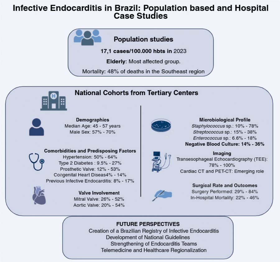
Infective endocarditis remains a serious condition with high morbidity and mortality rates despite advances in diagnostic and therapy. While the Global North has seen a shift toward older patients with prosthetic valve infections, Brazil’s understanding of the disease is hindered by reliance on fragmented, hospital-based data, primarily from the Southeast region. This narrative review synthesizes recent evidence to outline the national panorama. Population studies indicate an increase in incidence and mortality, primarily affecting men and the elderly, but with marked regional heterogeneity. Analyses of hospital case series reveal an epidemiological profile characterized by patients younger than international averages, with a high prevalence of comorbidities, underlying structural heart disease, and prosthetic valves. The microbiological landscape is diverse, with no consistent predominance between staphylococci and streptococci. Nevertheless, a particularly concerning finding is the high incidence of negative blood cultures. Severe complications are frequent, and variability exists in surgical intervention rates and in-hospital mortallity across studies. In light of this scenario, the authors propose a set of strategic actions to guide future advances: the creation of a Brazilian Registry of Infective Endocarditis, the development of Contextualized National Guidelines, the strengthening of multidisciplinary teams (Endocarditis Teams), and the implementation of measures with the potential to reduce regional disparities, such as telemedicine and the regionalization of care. The consolidation of these initiatives has the potential to transform the current landscape by enhancing epidemiological knowledge, standardizing care practices, and ultimately improving clinical outcomes nationwide.

  • Diagnostic Imaging
  • Endocarditis
  • Heart Valve Diseases
  • Unified Health System
Read Full Article
Creative Commons Attribution (CC BY) Creative Commons license
12/02/2026 DOI: 10.36660/abcimg.20250098i Views 2
Figure 1
ABCIC
Case Report

Late Discovery of Left Ventricular Pseudoaneurysm: A Rare Clinical Case

Angélica Pedreira da Silva et al. Gilson Soares Feitosa Gabriel de Castro Vaz Leal Rilson Fraga Moitinho Jorge Andion Torreão

Abstract

Left ventricular (LV) pseudoaneurysm is a rare complication of acute myocardial infarction (AMI). We report the case of a 67-year-old male patient seen in 2024. Following an AMI in 2019, the patient was diagnosed with a LV pseudoaneurysm after complaining of dyspnea and undergoing imaging tests including echocardiogram and magnetic resonance imaging. The patient underwent aneurysmectomy and revascularization surgery, progressing satisfactorily during the postoperative period.

  • Diagnostic Imaging
  • False Aneurysm
  • Post-Infarction Heart Rupture
Read Full Article
Creative Commons Attribution (CC BY) Creative Commons license
15/08/2025 DOI: 10.36660/abcimg.20250006i Views 2
ABC
Editorial

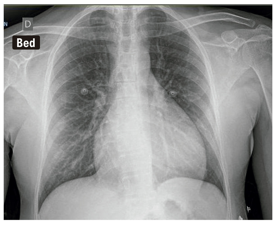
A Fresh Glimpse on Chest X-Ray: Revealing Significance of Aortic Arch Calcifications for Stroke Recurrence

José Ribeiro

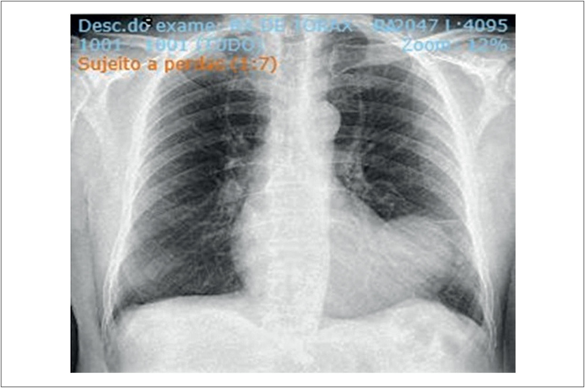
The global impact of cardiovascular disease continues to be significant, despite improvements in diagnosis and treatment. The epidemiological significance and associated clinical outcomes emphasize the importance of risk stratification and preventive measures.

In the realm of medical diagnostics, every subtle observation holds the potential to unlock crucial insights into the prognosis and management of various health conditions. One such observation that has garnered attention is the presence of calcification in the aortic arch as detected on chest X-ray. While traditionally viewed as a marker of cardiovascular risk, recent studies have ignited a debate on whether aortic arch calcification (AAC) could serve as an independent predictor of recurrent stroke.

[…]

  • Diagnostic Imaging
  • Heart Disease Risk Factors
  • Mass Chest X-Ray
  • Stroke
  • Thoracic aorta
Read Full Article
Creative Commons Attribution (CC BY) Creative Commons license
22/11/2024 DOI: 10.36660/abc.20240404i Views 1
Figure 1
ABCIC
Case Report

Exploring Complexities in Differential Diagnosis: Torn Chord Tendinea in the Aortic Valve

Alexandre Costa et al. Endy de Santana Alves Priscila Pinheiro Leonardo Flausino … Marcus Vinicius Freire Rodrigo Vieira Morel

Introduction

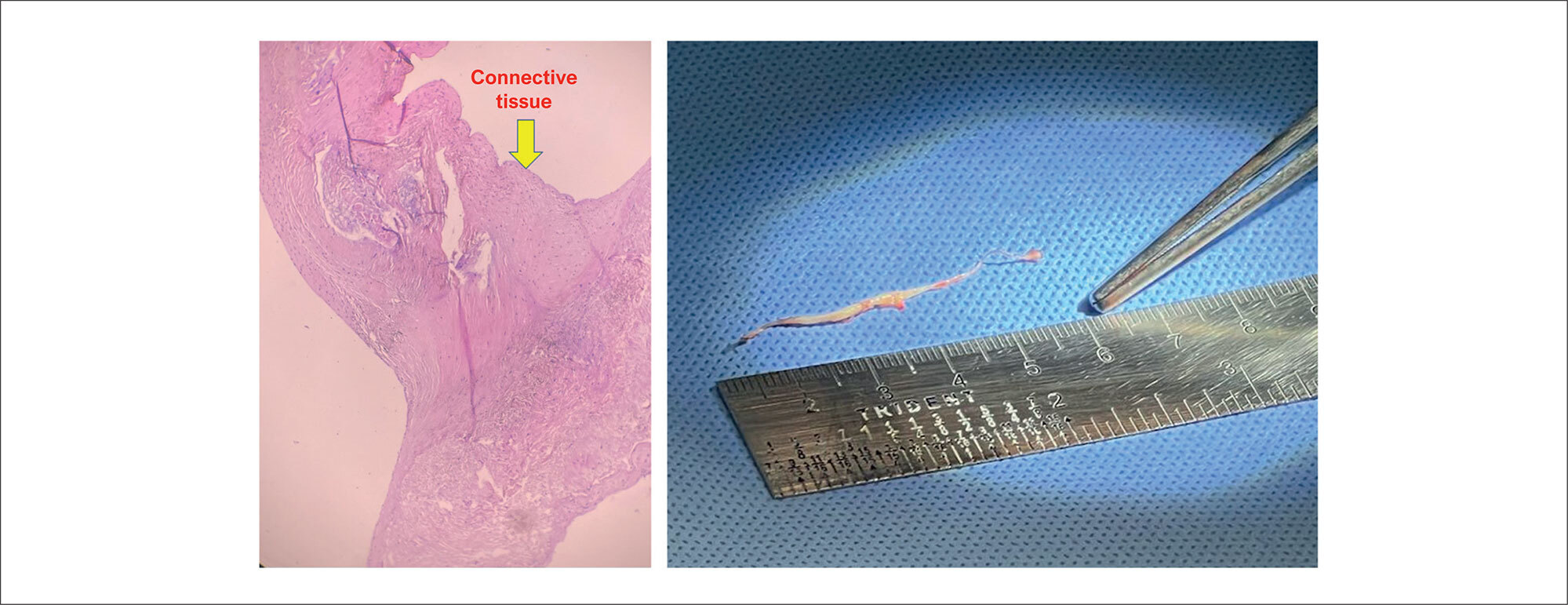
The prevalence of anomalous chordae in the aortic valve is rare in clinical practice. These anomalies, although uncommon, can have significant clinical implications, including valve insufficiency, arrhythmias and/or embolic risk. Accurate diagnosis and understanding of its pathophysiology are essential for adequate patient management.

The uncommon nature of anomalous chordae in the aortic valve means that their presence can often go unnoticed or misdiagnosed. Medical literature presents few reports on these structures of probable embryonic origin, leaving gaps in clinical knowledge. Furthermore, due to its ability to influence cardiac hemodynamics and potential to complicate clinical conditions, it is essential to expand the understanding of its incidence, clinical manifestations, and best management practices.

[…]

  • Aortic Valve
  • Cardiac Surgical Procedures
  • Diagnostic Imaging
  • Heart Valve Diseases
Read Full Article
Creative Commons Attribution (CC BY) Creative Commons license
18/12/2023 DOI: 10.36660/abcimg.20230084i Views 1
Figure 1
ABCIC
Case Report

Prosthetic Aortic Valve Endocarditis With Extensive Para-Aortic Abscess: The Relevance of Multimodal Imaging

Bernardo Boccalon et al. André Barcellos Amon Thiago Ferreira Serafini Álvaro Schmidt Albrecht … Murilo Foppa Angela Barreto Santiago Santos

Introduction

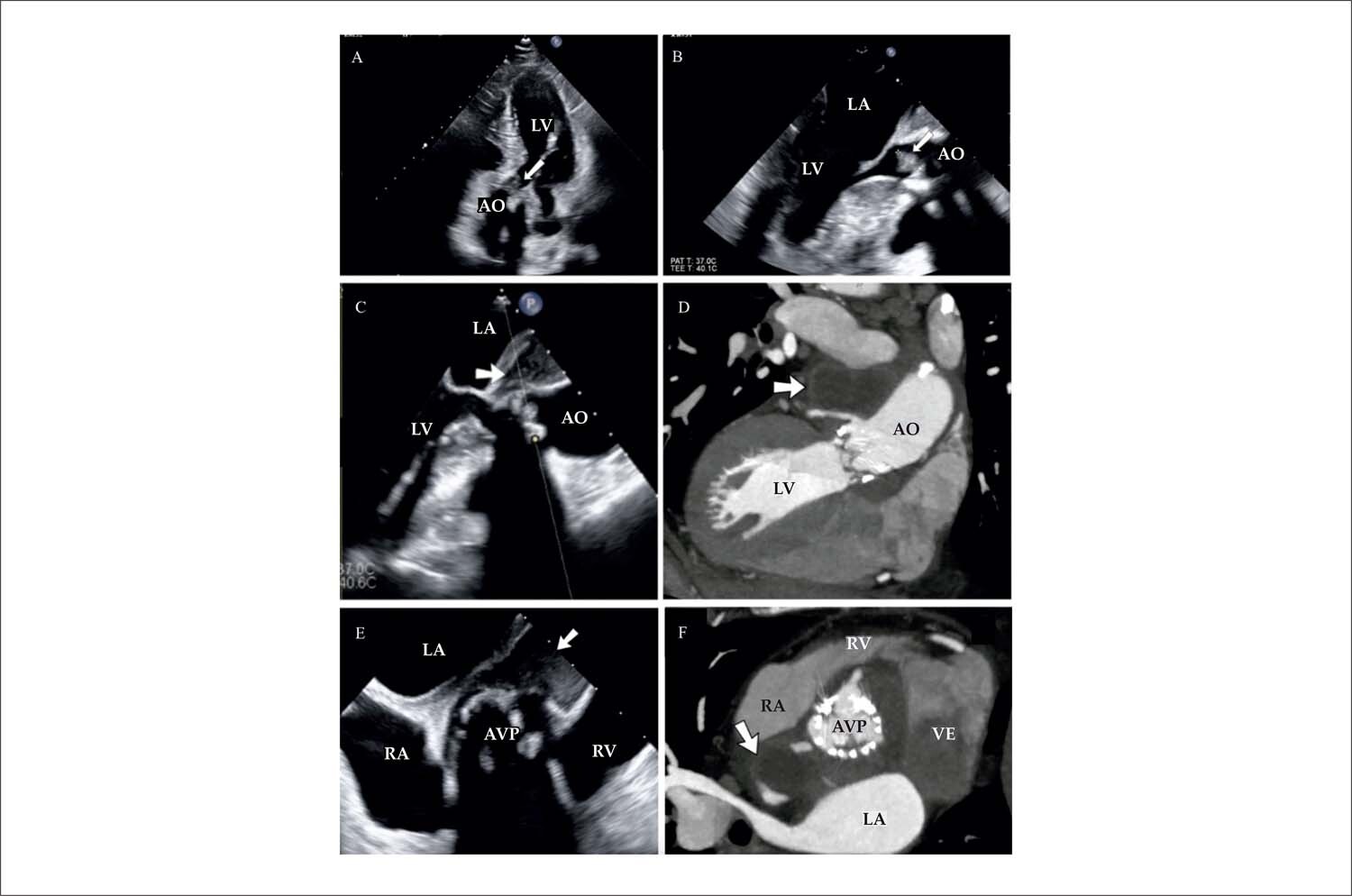
Infectious endocarditis (IE) is defined as the infection of the endocardium or prosthetic material in the heart, whose pathogenesis is bacterial adherence to the vulnerable endocardium or implanted prosthetic material during a bacteremia. Its clinical presentation is highly variable, depending on the micro-organism that causes it, underlying heart conditions and preexisting comorbidities. The annual estimated incidence of IE is up to 10/100,000 in western countries, and mortality of up to 30% in 30 days.

[…]

  • Diagnostic Imaging
  • Endocarditis
  • Heart Valve Prosthesis Implantation
Read Full Article
Creative Commons Attribution (CC BY) Creative Commons license
26/09/2023 DOI: 10.36660/abcimg.20230068i Views 1
ABCHF
Review Article

Diagnosis and Treatment of Hypertrophic Cardiomyopathy

Fernando Saraiva et al. Fabio Fernandes Minna Moreira Dias Romano Juliano Novaes Cardoso … Marília Taily Soliani Mariani Mendes Madisson Bernardo André Schmidt Marcus Vinicius Simões

Abstract

The concept of hypertrophic cardiomyopathy has undergone profound changes with the advent of cardiogenomics and the increase in knowledge about the sarcomeric structure and the mutations that lead to cardiomyocyte hypertrophy. In this review article, we address the state of the art in terms of diagnosis, contemplating the genetic heart diseases responsible for differential diagnoses of hypertrophic cardiomyopathy, known as phenocopies, which are a major challenge in clinical practice. We also discuss the current evidence that guides treatment.

  • Cardiomyopathy, Hypertrophic
  • Death, Sudden, Cardiac
  • Diagnostic Imaging
  • Drug Therapy
Read Full Article
Creative Commons Attribution (CC BY) Creative Commons license
01/08/2023 DOI: 10.36660/abchf.20230040 Views 1
IJCS
Original Article

Echocardiogram in Critically ill Patients with COVID-19: ECOVID Study

Bruno Ferraz de Oliveira Gomes et al. Armando Luis Cantisano Iliana Regina Ribeiro Menezes Anny de Sousa Azevedo … Thiago Moreira Bastos da Silva Valdilene Lima Silva Giovanni Possamai Dutra Suzanna Andressa Morais de Paula Barbara Ferreira da Silva Mendes Plinio Resende do Carmo Junior Basilio de Bragança Pereira Joao Luiz Fernandes Petriz Glaucia Maria Moraes de Oliveira

Abstract

Background

Literature is scarce on echocardiographic characteristics of COVID-19 patients admitted to the intensive care unit (ICU).

Objectives

To describe echocardiographic characteristics of ICU COVID-19 patients and associate them with clinical signals/symptoms, laboratory findings and outcomes.

Methods

Patients with RT-PCR-confirmed COVID-19, admitted to the ICU, who underwent echocardiography were included. Clinical characteristics associated with an abnormal echocardiogram (systolic ventricular dysfunction of any degree — left and/or right ventricle — and/or high filling pressures and/or moderate to severe pericardial effusion) were analyzed. Groups were compared using the Student’s t-test, chi-square, and logistic regression. A p < 0.05 was considered statistically significant.

Results

A total of 140 patients met inclusion criteria, and 74 (52.9%) had an abnormal echocardiogram. A low number of left and right ventricular systolic dysfunction was observed, and 35% of the population had a normal diastolic function. In the univariate analysis, characteristics associated with abnormal echocardiogram were age, chronic kidney disease, elevated troponin, previous heart failure, and simplified acute physiology score 3 (SAPS 3). In the regression model, troponin and SAPS3 score were independent markers of abnormal echocardiogram. An abnormal echocardiogram was associated with a higher prevalence of in-hospital death (RR 2.10; 95% CI 1.04-4.24) and orotracheal intubation (RR 2.3; 95% CI 1.14-4.78).

Conclusions

COVID-19 has little effect on ventricular function, but it is common to find increased filling pressures. Elevated serum troponin level and SAPS3 score were the independent markers of an abnormal echocardiogram. In addition, the prevalence of in-hospital death and need for mechanical ventilation were higher in patients with abnormal echocardiogram.

  • COVID-19, Critical Care
  • Diagnostic Imaging
  • Echocardiography/methods
  • Troponin/therapeutic use
Read Full Article
Creative Commons Attribution (CC BY) Creative Commons license
03/04/2023 DOI: 10.36660/ijcs.20210114 Views 1
Figure 1
ABC
Image

Tuberous Sclerosis: Unusual Findings in the Setting of a Rare Disease

Ines Oliveira et al. Rafaela Lopes Isabel Cruz Bruno Bragança … João Azevedo Aurora Andrade

Case Description

A 40-year-old woman diagnosed with tuberous sclerosis (TS) at the age of one, with cognitive impairment, epilepsy and renal angiomyolipomas, was referred to Cardiology for complaints of fatigue. She had no known family history of TS. Although her mother could not confirm if a genetic test had been performed, the patient had a definite diagnosis of TS, according to International Tuberous Sclerosis Consensus Group revised criteria, due to the presence of several major criteria, as hypomelanotic macules, facial angiofibromas, cortical dysplasia and subependymal nodules.

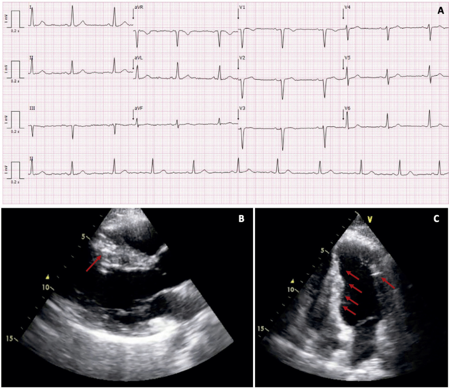
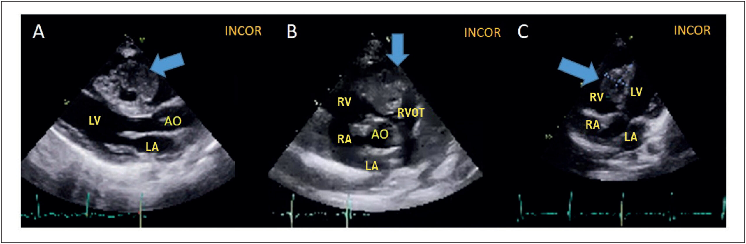
On cardiovascular evaluation, she had no dyspnea on exertion, chest pain, palpitations or history of syncope. On physical examination, she had multiple cutaneous angiofibromas and hypomelanotic macules, cardiac auscultation with no murmurs or signs of congestion. Electrocardiogram (ECG) showed nonspecific repolarization abnormalities ( ) and no arrhythmic events were detected by the Holter method. Transthoracic echocardiography revealed the presence of multiple intracardiac hyperechogenic lesions, with no blood flow obstruction or valvular impairment, and preserved ventricular systolic function ( ). Cardiac magnetic resonance (CMR) showed the nature of the lesions ( ) – homogeneous high signal intensity in T1 and T2-weighted images (A, B, C), with uniform suppression with the application of fat-saturation pulses, with an intensity similar to adipose tissue (D). The lesions also showed chemical shift , revealing its lipomatous nature (F). No evidence of perfusion, late gadolinium enhancement (E) or other masses. The patient remained asymptomatic after five years of follow-up, performing an annual ECG and echocardiogram, with no increase in the number or size of lipomas.

[…]

  • Atherosclerosis
  • Diagnostic Imaging
  • Familial Multiple Lipomatosis
  • Neoplasms
  • Tuberous Sclerosis/complications
  • Tumor Supressor/genetics
Read Full Article
Creative Commons Attribution (CC BY) Creative Commons license
13/02/2023 DOI: 10.36660/abc.20220147
Figure 1
ABCIC
Case Report

Primary Cardiac Angiosarcoma Evaluated on PET/CT

Pedro Henrique Rodrigues da Silva et al. Carlos Eduardo Lucena Montenegro Maria Eduarda Duarte de Mello Flamini Mariana Vila Nova de Oliveira Pontual Simone Cristina Soares Brandão

Introduction

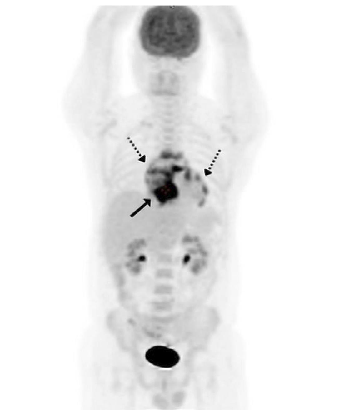
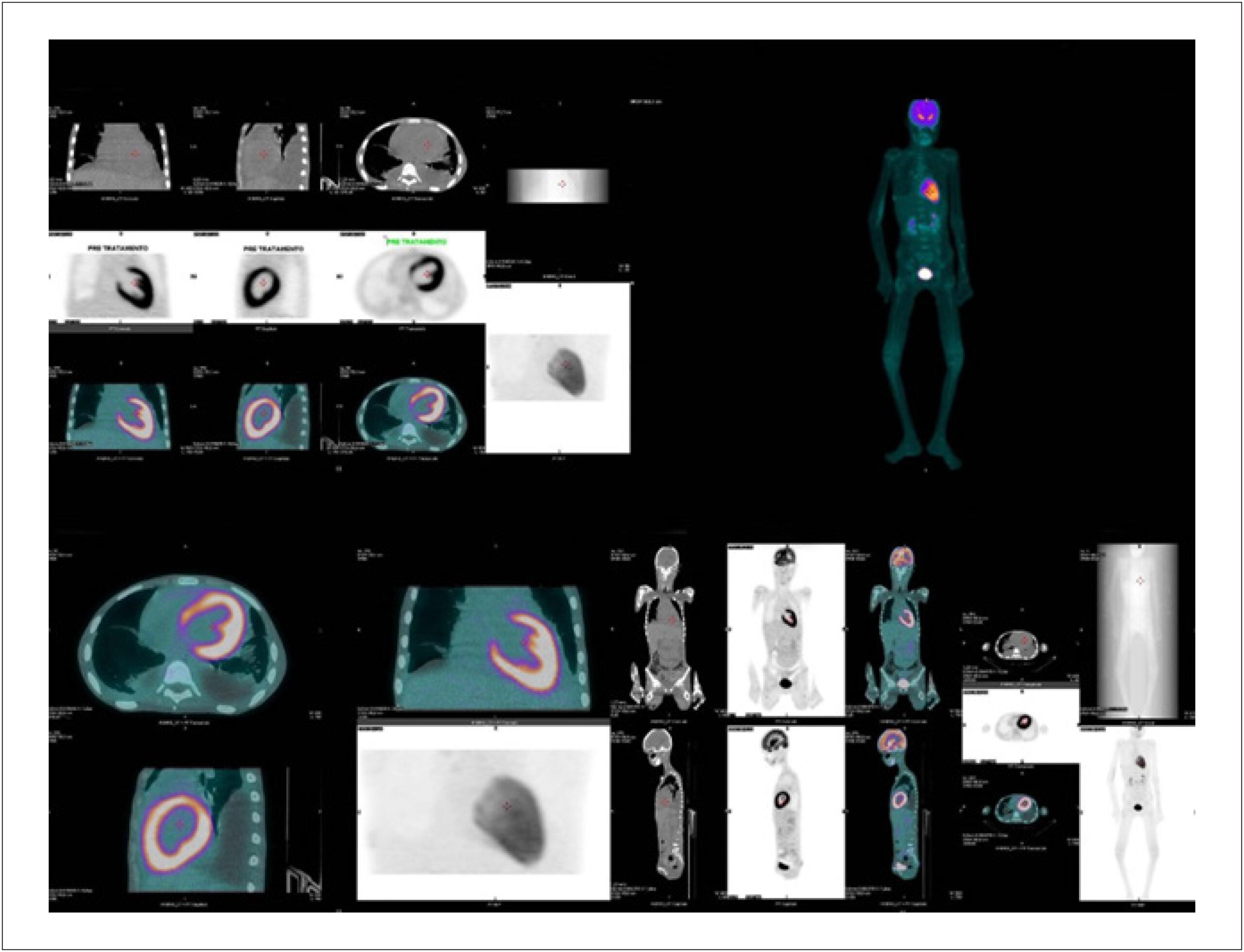
Cardiac angiosarcoma is a rare malignant endothelial tumor that is aggressive and has an adverse prognosis., It features a high mortality rate because of the tendency for local recurrence and high incidence of systemic metastases. Therefore, its early diagnosis is very important, as is the determination of its local and distant extension, to ensure its proper therapeutic management. The objective of this case report was to show the value of positron emission tomography associated with computed tomography with 18-fluorodeoxyglucose (18F-FDG PET/CT) in the diagnosis and staging of cardiac angiosarcoma.

[…]

  • Angiosarcoma
  • Diagnostic Imaging
  • Positron-Emission Tomography
Read Full Article
Creative Commons Attribution (CC BY) Creative Commons license
21/12/2022 DOI: 10.47593/2675-312X/20223504eabc337i Views 1
Figure 1
ABCHF
Review Article

Obstructive Cardiac Tumors

Sanderson Antonio Cauduro et al. João Pedro Passos Dutra Fabio Fernandes Marcely Bonatto … Maria Verônica Câmara Santos Letícia dos Santos de Oliveira Rocha Talita Ribeiro Mialski Ana Paula Konig da Nobrega Simone Cristina Soares Brandão Silvio Henrique Barberato

Abstract

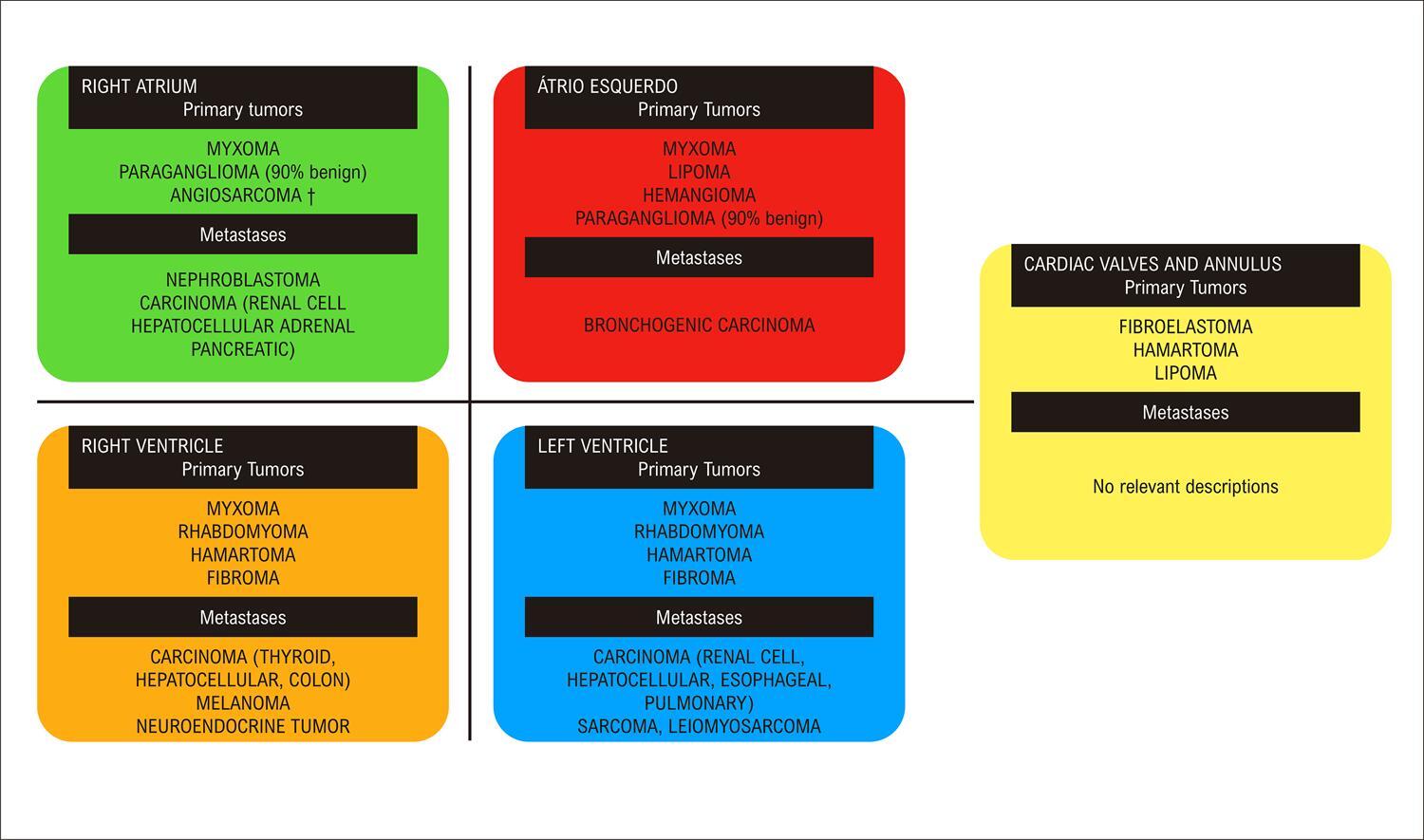
Cardiac tumors are considered rare clinical entities and can affect any cardiac tissue. Metastatic (secondary) cardiac tumors are more frequently diagnosed than primary tumors (malignant or benign). Both types can cause valve and/or inflow and outflow tract obstructions in any cardiac chamber, leading to symptoms of heart failure, as well as embolization and arrhythmia. Treatment of benign tumors is usually surgical, and that of metastatic and primary malignant tumors will depend on their origin and type, with poor prognosis. Recurrence of benign tumors is frequent. The aim of this article is to provide the clinician with tools to optimize diagnosis, differential diagnosis, and treatment of tumors with obstructive features causing heart failure.

  • Diagnostic Imaging
  • Echocardiography
  • Heart Failure
  • Heart Neoplasms
  • Magnetic Resonance Spectroscopy
Read Full Article
Creative Commons Attribution (CC BY) Creative Commons license
18/12/2022 DOI: 10.36660/abchf.20230001 Views 1
ABC
Research Letter

Mitral Valve Perforated Aneurysm: An Issue of Inflammation or Pressure Gradients?

Inês Oliveira et al. Isabel Cruz Ana Neto Bruno Bragança … Glória Abreu João Azevedo Aurora Andrade

Case Report

Mitral valve aneurysms (MVA) are uncommon and usually develop acutely in the setting of infective endocarditis (IE). MVA as a late complication of IE in a patient with obstructive hypertrophic cardiomyopathy (HCM) is rather unusual, raising considerations regarding the role of the infectious process and the hemodynamic conditions inherent to the cardiomyopathy. , We present a case report of a patient with HCM and a ruptured aneurysm of the mitral valve (MV) leaflet, secondary to previously treated IE.

A 68-year-old male patient with type 2 diabetes mellitus and dyslipidemia was admitted to a hospital with a 3-week history of malaise, fever, and recent left-sided abdominal pain. His physical examination revealed a grade II/VI systolic heart murmur at the cardiac apex, fever, abdominal tenderness in the left upper quadrant, and purpuric lesions in the inferior limbs. Blood analyses showed neutrophilia and C-Reactive Protein of 211mg/L. Positive blood cultures indicated Staphylococcus aureus methicillin-sensitive. Abdominal Computed Tomography revealed spleen embolization, with no abscess. Transthoracic (TTE) and transesophageal echocardiography (TEE) disclosed a highly mobile polypoid mass in the atrial side of the anterior MV leaflet, suggestive of a vegetation, with mild mitral regurgitation and no evidence of abscess, aneurysm, or valve perforation; asymmetrical left ventricular (LV) hypertrophy with the presence of an increased intraventricular systolic gradient (IVSG) and systolic anterior motion (SAM) of the MV ( ). The diagnosis of IE in HCM was established and treated accordingly with flucloxacillin, presenting a favorable clinical evolution. After three months of follow-up, cardiac magnetic resonance was performed, confirming the diagnosis of HCM: an increased LV mass (96g/m ) with asymmetrical left ventricular hypertrophy (maximal thickness of 20mm in the inferoseptal wall), with no perfusion defects but with evidence of intramural late gadolinium enhancement in the inferoseptal wall,and SAM of the MV ( ). Echocardiography was repeated and, in addition to evidence of obstructive HCM with a rest IVSG of 44 mmHg, an aneurysm of the anterior leaflet of the MV was identified. Two mitral regurgitant jets were observed, one due to incomplete coaptation of the leaflets and another through the perforated aneurysm, quantifying global mitral regurgitation (MR) in a moderate (grade II/IV) ( ). The beta-blocker dose was increased, and a strategy of close follow-up was adopted. Maintenance of the characteristics of the aneurysm was verified in outpatient follow-up after 2 years.

[…]

  • Aneurysm, Valvular
  • Cardiomyopathy, Hypertrophic / complications
  • Diabetes Mellitus/complications
  • Diagnostic Imaging
  • Dyslipidemia/complications
  • Endocarditis, bacterial
  • Inflammation
  • Mitral Valve / abnormalities
Read Full Article
Creative Commons Attribution (CC BY) Creative Commons license
30/09/2022 DOI: 10.36660/abc.20211031 Views 1
Figure 1
ABC
Original Article

Association between Atrioventricular Block and Mortality in Primary Care Patients: The CODE Study

Gabriela Miana de Mattos Paixão et al. Emilly M. Lima André B. Quadros Daniel P. R. Cabral … Renato R. Coelho Derick M. Oliveira Jamil de Souza Nascimento Paulo R. Gomes Antonio L. Ribeiro

Abstract

Background

Atrioventricular block (AVB) describes an impairment of conduction from the atria to the ventricles. Although the clinical course of AVB has been evaluated, the findings are from high-income countries and, therefore, cannot be extrapolated to the Latinx population.

Objective

Evaluate the association between AVB and mortality.

Methods

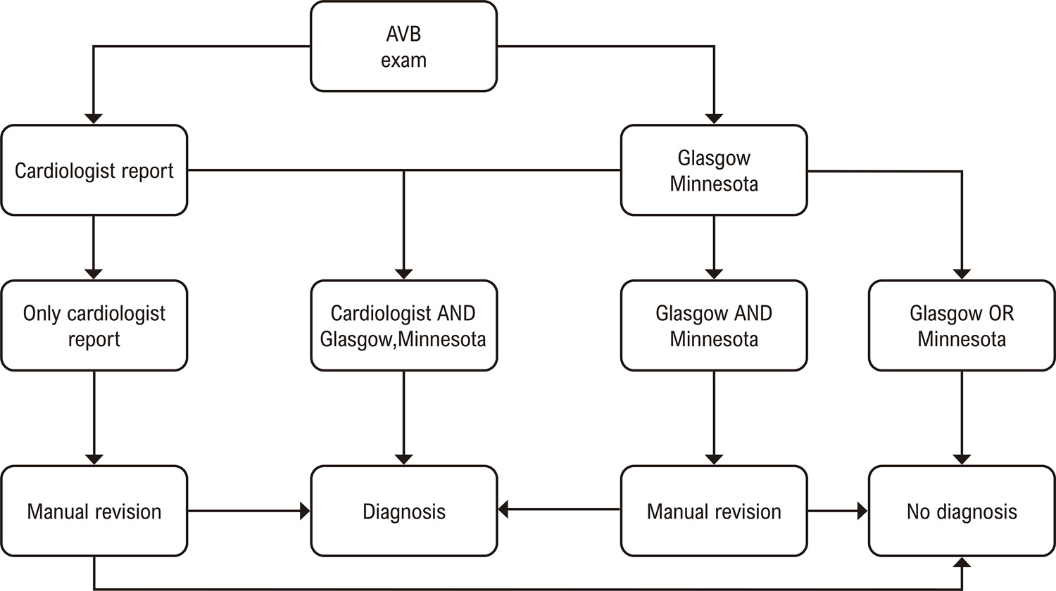
Patients from the CODE (Clinical Outcomes in Digital Electrocardiology) study, older than 16 years who underwent digital electrocardiogram (ECG) from 2010 to 2017 were included. ECGs were reported by cardiologists and by automated software. To assess the relationship between AVB and mortality, the log-normal model and the Kaplan-Meier curves were used with two-tailed p-values < 0.05 considered statistically significant.

Results

The study included 1,557,901 patients; 40.2% were men, and mean age was 51.7 (standard deviation ± 17.6) years. In a mean follow-up of 3.7 years, the mortality rate was 3.35%. The AVB prevalence was 1.38% (21,538). Patients with first-, second-, and third-degree AVB were associated with 24% (relative survival rate [RS] = 0.76; 95% confidence interval [CI]: 0.71-0.81; p < 0.001), 55% (RS = 0.45; 95% CI: 0.27-0.77; p = 0.01), and 64% (RS = 0.36; 95% CI: 0.26-0.49; p < 0.001) lower survival rate when compared to the control group, respectively. Patients with 2:1 AVB had 79% (RS = 0.21; 95% CI: 0.08-0.52; p = 0.005) lower survival rate than the control group. Only Mobitz type I was not associated with higher mortality (p = 0.27).

Conclusion

AVB was an independent risk factor for overall mortality, with the exception of Mobitz type I.

  • Atrioventricular Block/complications, Mortality
  • Atrioventricular Block/physiopathology
  • Cardiovascular Diseases/complications
  • Diagnostic Imaging
  • Electrocardiography/methods
Read Full Article
Creative Commons Attribution (CC BY) Creative Commons license
18/07/2022 DOI: 10.36660/abc.20210763
Figure 1
ABC
Image

Rare Presentation of Yolk Sac Tumor with Cardiac Involvement: Characteristics Detected by MRI

Cristhian Espinoza Romero et al. Williams Roberto Lata Guacho Kevin Rafael de Paula Robert Paladines Jimenez Eduardo Kaiser Ururahy Nunes Fonseca

Introduction

Primary cardiac tumors are extremely rare, with their incidence varying between 0.0017 and 0.28%, among which is the malignant germ cell yolk sac tumor (YST).

Although transthoracic echocardiography (TTE) is often the first line in the evaluation of cardiac tumors, currently, due to its good spatial resolution and tissue characterization, cardiac magnetic resonance (CMR) imaging is the technique of choice for the evaluation of these tumors., The intracardiac YST is a rare neoplasm, with few reported cases.–

[…]

  • Diagnostic Imaging
  • Drug Therapy
  • Echocardiography, Transthoracic/methods
  • Heart Failure
  • Heart Neoplasms
  • Magnetic Resonance Spectroscopy/ methods
  • Neoplasms, Germ Cell and Embryonal
Read Full Article
Creative Commons Attribution (CC BY) Creative Commons license
11/07/2022 DOI: 10.36660/abc.20210335 Views 1
Figure 1
ABC
Original Article

Assessment of the Relationship Between the Adropin Levels and the Coronary Collateral Circulation in Patients wıth Chronic Coronary Syndrome

Hasan Akkaya et al. Ertuğrul Emre Güntürk Fulya Akkaya Uğur Karabıyık … İnayet Güntürk Samet Yılmaz

Abstract

Background

Coronary collateral circulation (CCC) provides an alternative blood flow to myocardial tissue exposed to ischemia and helps to preserve myocardial functions. Endothelial-derived nitric-oxide (NO) production and vascular endothelial growth factor (VEGF) have been suggested as the most important factors in the development of CCC. Adropin is a peptide hormone responsible for energy hemostasis, and is known for its positive effects on the endothelium through NO and VEGF.

Objective

The aim of this study is to investigate the association between adropin and the presence of CCC in patients with chronic coronary syndrome (CCS).

Methods

A total of 102 patients with CCS, who had complete occlusion of at least one major epicardial coronary artery, were included in the study and were divided into two groups: the group of patients (n:50) with poor CCC (Rentrop 0-1) and the group of patients (n:52) with good CCC (Rentrop 2-3). The level of significance adopted in the statistical analysis was 5%.

Results

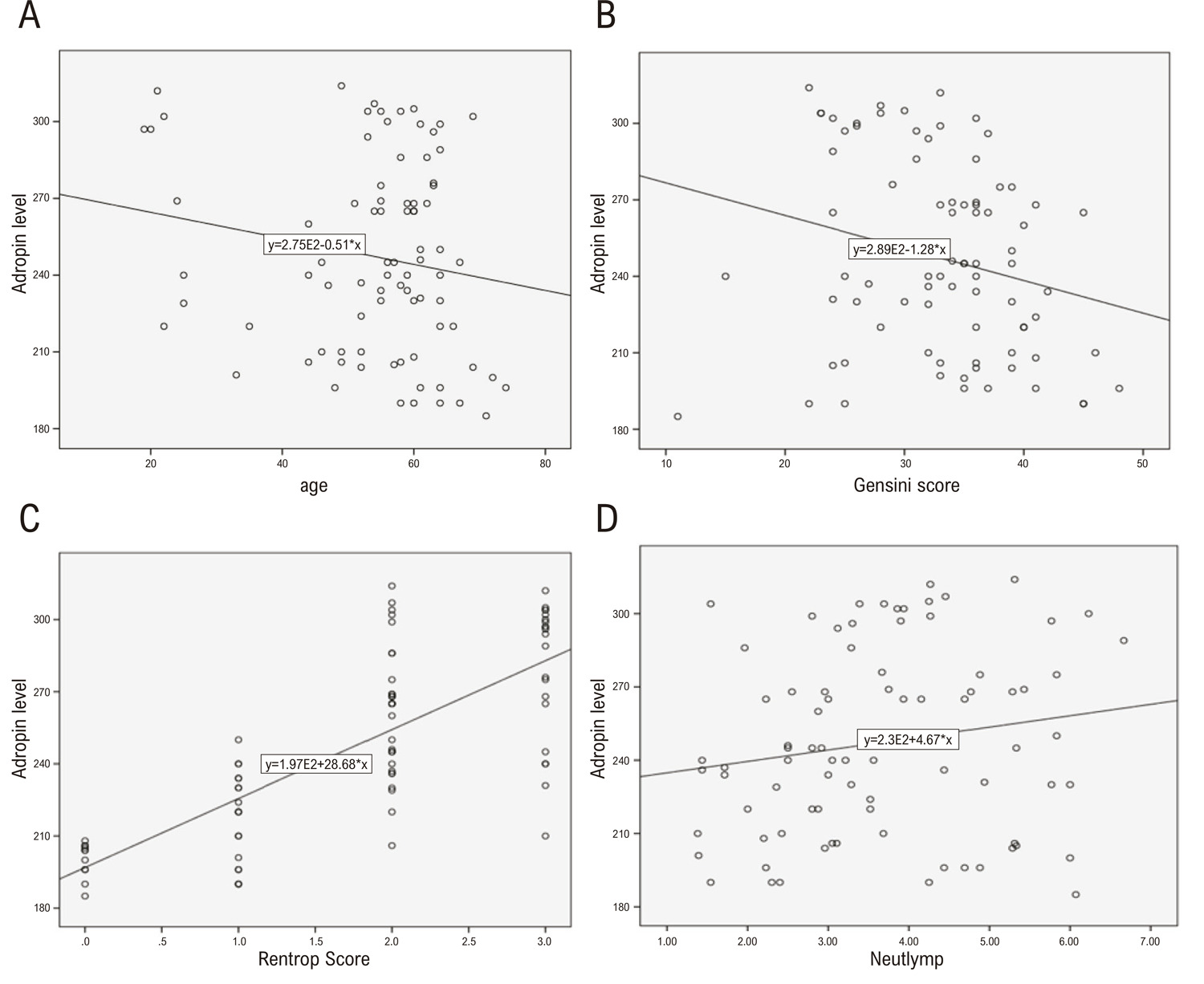
Mean adropine levels were found as 210.83±17.76 pg/mL and 268.25±28.94 pg/mL in the poor and good CCC groups, respectively (p<0.001). Adropin levels proved to be positively correlated with neutrophil-to-lymphocyte ratios (r:0.17, p:0.04) and the rentrop scores (r:0.76, p<0.001), and negatively correlated with age (r:-0.23, p:0.01) and Gensini scores (r:-0.19, p:0.02). Adropin level is a strong independent predictor of good CCC development (OR:1.12, 95% CI:(1.06–1.18), p<0.001).

Conclusion

This study suggests that adropin levels may be a possible factor associated with the presence of CCC in CCS patients.

  • Acute coronary syndrome
  • Adropin
  • Atherosclerosis
  • Coronary Angiography
  • Coronary Artery Diseases
  • Coronary Collateral Circulation
  • Diagnostic Imaging
  • Peptides
Read Full Article
Creative Commons Attribution (CC BY) Creative Commons license
27/06/2022 DOI: 10.36660/abc.20210573 Views 1
Figure 1
ABC
Image

Value of18F-FDG PET/CT in the Diagnosis and Assessment of Response to Treatment of Lupus Myocarditis

Alvaro M. Perazzo et al. Larissa G. F. Andrade Leonardo G. A. Venancio Pedro Alves da Cruz Gouveia … Mariana Feitosa Ramalho Galvão Esdras M. Lins Fernando Moraes Neto Simone Cristina Soares Brandão

Systemic Lupus Erythematosus (SLE) is an autoimmune disease with a wide spectrum of clinical manifestations. The cardiovascular system has relevant clinical importance among the affected organs because it is associated with higher mortality in these patients. The heart can be affected in any structure, and lupus myocarditis is a major diagnostic challenge in clinical practice.

Non-invasive tests such as electrocardiograms and echocardiograms are not sensitive or specific enough for this diagnosis. Cardiac magnetic resonance imaging (CMR) is the preferred imaging modality for diagnosing myocarditis, but it has contraindications, such as patients with metallic implants or using gadolinium in chronic kidney disease.

[…]

  • Diagnostic Imaging
  • Immunosupressive Agents/therapeutic use
  • Lupus Erythematosus, Systemic/complications
  • Lupus Myocarditis
  • Tomography Computed Emission Positrons Tomography/methods
Read Full Article
Creative Commons Attribution (CC BY) Creative Commons license
10/06/2022 DOI: 10.36660/abc.20210523 Views 1
Figure 1
ABC
Research Letter

An Unusual Manifestation of Rejection

Carlos Xavier Correia de Resende et al. Pedro Grilo Diogo Sandra Amorim Gonçalo Pestana … José Pinheiro Torres Filipe Macedo

Introduction

Orthotopic heart transplantation is the current treatment of choice for selected patients with end-stage heart failure. With the improvement of surgical techniques and the efficiency of new immunosuppressive treatments, short-term survival has markedly improved throughout the years. However, these patients still suffer from important comorbidities caused by chronic transplant complications such as rejection, coronary allograft vasculopathy (CAV) and malignancy. Rhythm disorders are common in heart transplant patients and in some circumstances, they can be the first clinical manifestation of rejection. Coronary vasospasm has been recently associated with acute rejection and CAV, but the mechanisms underlying this phenomenon are still speculative.

We present a clinical case of acute heart transplant rejection, manifested by coronary vasospasm and advanced rhythm disorder.

[…]

  • Arrhythmias Cardiac
  • Coronary Vasospasm
  • Diagnostic Imaging
  • Drug Therapy
  • Graft Rejection
  • Heart block
  • Heart Failure, Heart Transplantation
  • Research
Read Full Article
Creative Commons Attribution (CC BY) Creative Commons license
13/05/2022 DOI: 10.36660/abc.20210671
ABC
Editorial

Aortic Stiffness by Cardiac Magnetic Resonance: Prognostic tool or Bystander?

Sérgio Figueiredo Câmara et al. Henrique Barbosa Ribeiro

Arterial stiffness increases with age and may relate to higher rates of cardiovascular events, including mortality. – This predictive capacity has been demonstrated in various longitudinal cohorts, including ‘healthy’ community population studies and those with diabetes, hypertension, chronic kidney disease, and established coronary artery disease. , There are several ways to measure arterial stiffness, such as doppler-ultrasound, carotid-femoral tonometer, and cardiac magnetic resonance (CMR). CMR provides information regarding cardiac function, perfusion, and myocardial scarring in a single exam and may also be the preferred method for assessing arterial stiffness using aortic pulse wave velocity (PWV). – While the association between aortic stiffness and myocardial ischemia has been demonstrated, as well as the prognostic value of aortic stiffness using CMR, there is limited data regarding the prognostic value of PWV by CMR in elderly patients in whom cardiovascular diseases (CVD) account for the vast majority of mortality causes.

In this issue of the journal, Kaolawanich and Boonyasirinant evaluated the occurrence of major adverse cardiac and cerebrovascular events (MACCE), including cardiac mortality, nonfatal myocardial infarction, hospitalization for heart failure, late revascularization (>180 days after CMR) and ischemic stroke in elderly patients (> 70 years) with suspected or confirmed CAD undergoing adenosine stress CMR including PWV. The main objective was to determine the prognostic value of aortic stiffness using CMR-based PWV in elderly patients with CAD. Two hundred sixty-three consecutive patients (55% female; 77±5 years) between 2010 and 2014 were included with a median follow-up of 59.6 months and a mean PWV of 13.98 ± 9.00 m/s. A higher PWV (>13.98 m/s) was associated with greater MACCE rates (HR 1.75; 95% CI 1.05 – 2.94; p=0.03), as compared to non-elevated PWV (<13.98 m/s). By multivariable analysis, diastolic blood pressure, left ventricular ejection fraction (LVEF), myocardial ischemia and elevated PWV were independent predictors of MACCE at long-term follow-up (p<0.05 for all). PWV had an incremental prognostic value concerning clinical history, LVEF and ischemia (increased global chi-square = 7.25; p=0.01). In this evaluation, elderly patients with elevated PWV also had a higher prevalence of hypertension, diabetes mellitus and higher systolic blood pressure than those with non-elevated PWV, consistent with prior studies in younger populations.

[…]

  • Aortic Stiffness
  • Diagnostic Imaging
  • Magnetic Resonance Imaging/methods
  • Prognosis
  • Pulse Wave Analysis/methods
  • Vascular Stiffness
Read Full Article
Creative Commons Attribution (CC BY) Creative Commons license
13/05/2022 DOI: 10.36660/abc.20220231 Views 1
ABC
Original Article

Simple Echocardiographic Parameters are Strong Predictors of the Cardiovascular Risk in Asymptomatic Individuals: Elsa-Brasil Cohort

Luciana Pereira Fernandes et al. Maria da Conceição Chagas de Almeida Sheila Alvim de Matos Ana Clara Paixão Campos … Edmundo José Nassri Câmara Murilo Foppa Antônio Luiz Pinho Ribeiro Sandhi Maria Barreto Roque Aras Júnior

Abstract

Background

Several studies have evaluated echocardiographic abnormalities as predictors of cardiovascular risk; however, none have associated the global cardiovascular risk with echocardiographic abnormalities in the Brazilian population.

Objective

This study evaluates the association between the global cardiovascular risk (ASCVD score) and three echocardiographic abnormalities: left ventricular hypertrophy (LVH), left ventricular diastolic dysfunction (LVDD), and increased left atrium (LA) volume.

Methods

The study population was composed of participants from ELSA-Brasil who underwent echocardiography between 2008 and 2010 (n = 2973). They were asymptomatic and had no history of cardiovascular disease. The ASCVD score was calculated in two periods: 2008-2010 and 2012-2014. Prevalence ratios (PR) were estimated with 95% confidence intervals (CI).

Results

There is an association between echocardiographic abnormalities and high global cardiovascular risk (ASCVD score ≥ 7.5) in both study periods, separately. The combined global risk (low risk in the first period and high risk in the second period) was significantly associated only with LVDD (PR = 3.68, CI 95% 2.63–5.15) and LVH (PR = 2.20, 95% CI 1.62–3.00).

Conclusion

Echocardiographic abnormalities (LVDD, LVH, and increased LA volume) are independent predictors of cardiovascular risk in Brazilian adults.

  • Atherosclerosis
  • Cardiovascular Diseases
  • Diagnostic Imaging
  • Echocardiography/methods
  • Left Atrial Volume
  • Left Ventricular Diastolic Dysfunction
  • Risk Factors
  • Sedentarism
Read Full Article
Creative Commons Attribution (CC BY) Creative Commons license
13/05/2022 DOI: 10.36660/abc.20210101
Figure 1
IJCS
Case Report

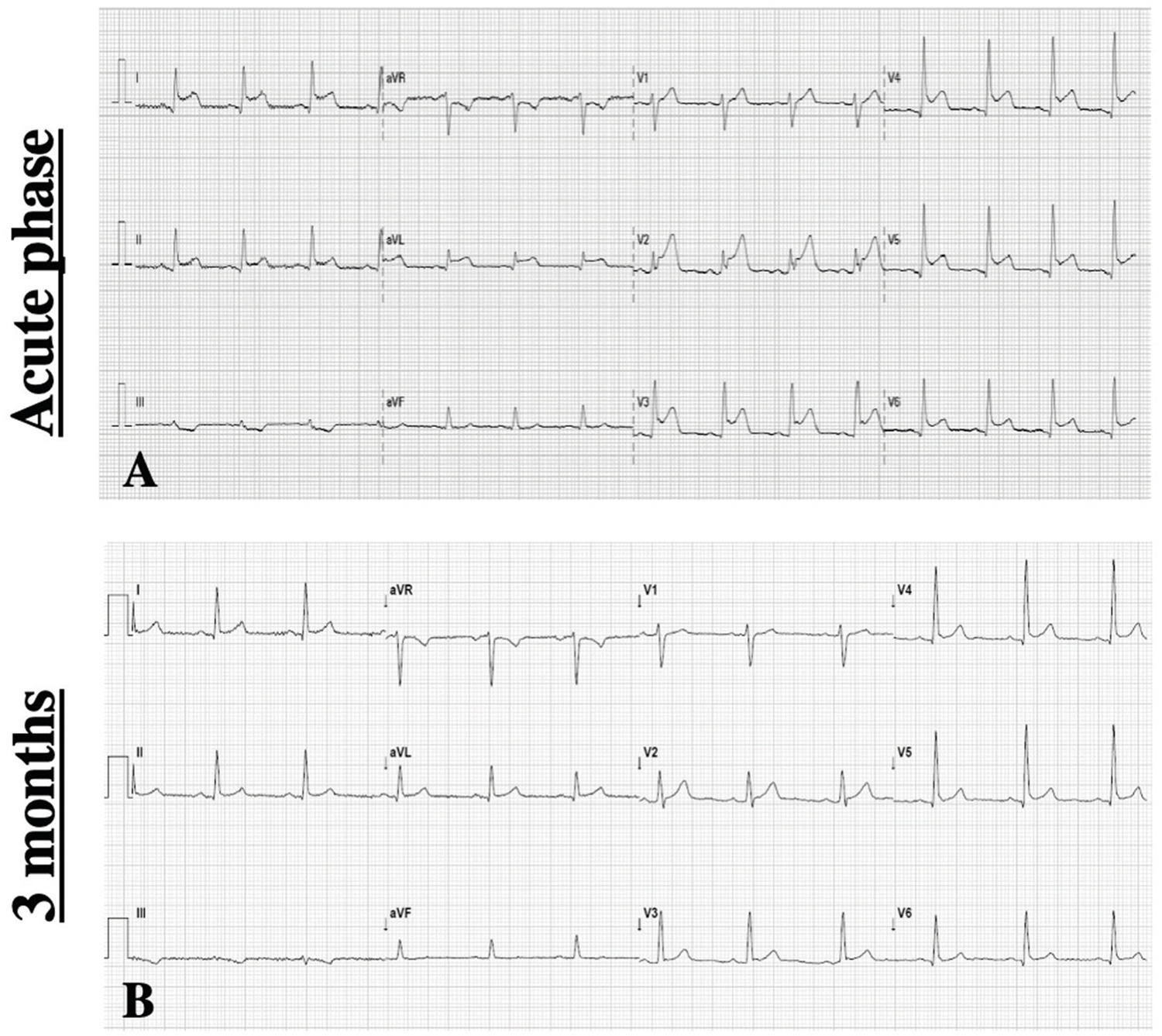
First-degree Atrioventricular Block as an Early Marker of Advanced Disease of the Conduction System in a Patient with Hereditary Val142Ile Cardiac Amyloidosis

Nágela S. V. Nunes et al. Evandro Tinoco Mesquita Claudio Tinoco Mesquita Paulo Roberto Benchimol-Barbosa

Introduction

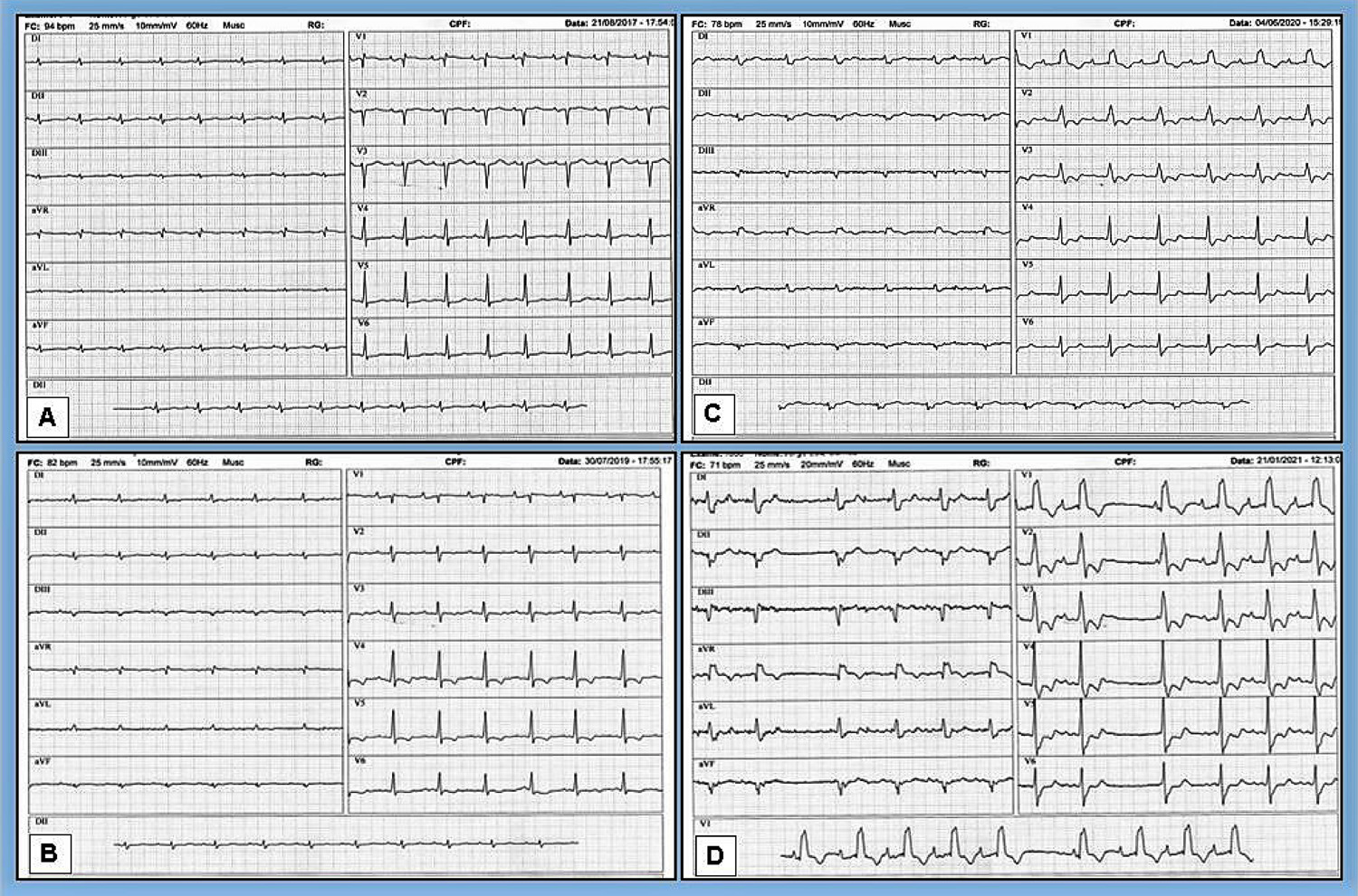
Cardiac amyloidosis (CA) is a type of restrictive cardiomyopathy secondary to amyloid infiltration in the heart. The most common clinical forms are immunoglobulin light chain amyloidosis (AL) and amyloidosis resulting from amyloid deposits secondary to the destabilization of the protein tetramer transthyretin (ATTR), which is divided into two types: the hereditary form (ATTRm) and the wild type (ATTRwt).

Cardiac impulse conduction disorders and atrial arrhythmias are more frequent in ATTR than in the AL form, and this seems to reflect the longer survival of patients affected by ATTR, given that the AL form manifests with high mortality up to 6 months from diagnosis, because conduction system disease appears to be a later manifestation.

[…]

  • Artificial Pacemaker
  • Atrioventricular Block/genetics
  • Bradycardia/complications
  • cardiomyopathy
  • Diagnostic Imaging
  • Familial Amyloidodis/complications
  • Sick Sinus Syndrome/genetics
Read Full Article
Creative Commons Attribution (CC BY) Creative Commons license
09/05/2022 DOI: 10.36660/ijcs.20210081 Views 2
Figure 1
ABC
Research Letter

Acute Myocarditis Following mRNA COVID-19 Vaccine

Daniel A. Gomes et al. Rita R. Santos Pedro Freitas Mariana S. Paiva … Jorge Ferreira Marisa Trabulo

Introduction

Vaccination is one of the most important breakthroughs of modern medicine and constitute a major advance in the prevention of infectious diseases.Overall, vaccines are effective and have an excellent general safety profile.In fact, reports of severe adverse effects following vaccination are extremely rare and idiosyncratic.

As the vaccine’s mechanism of action is based on the host immune response, a close relationship with autoimmunity cannot be disregarded.Cases of immunologic reactogenicity, such as Guillain-Barré syndrome and acute myocarditis following vaccination have been previously reported.,

[…]

  • Coronavirus-19
  • COVID-19
  • Diagnostic Imaging
  • Immunogenicity Vaccine
  • Molecular Mimicry
  • Myocarditis
  • Vaccine/adverse effects
Read Full Article
Creative Commons Attribution (CC BY) Creative Commons license
29/04/2022 DOI: 10.36660/abc.20210469 Views 1
Figure 1
IJCS
Case Report

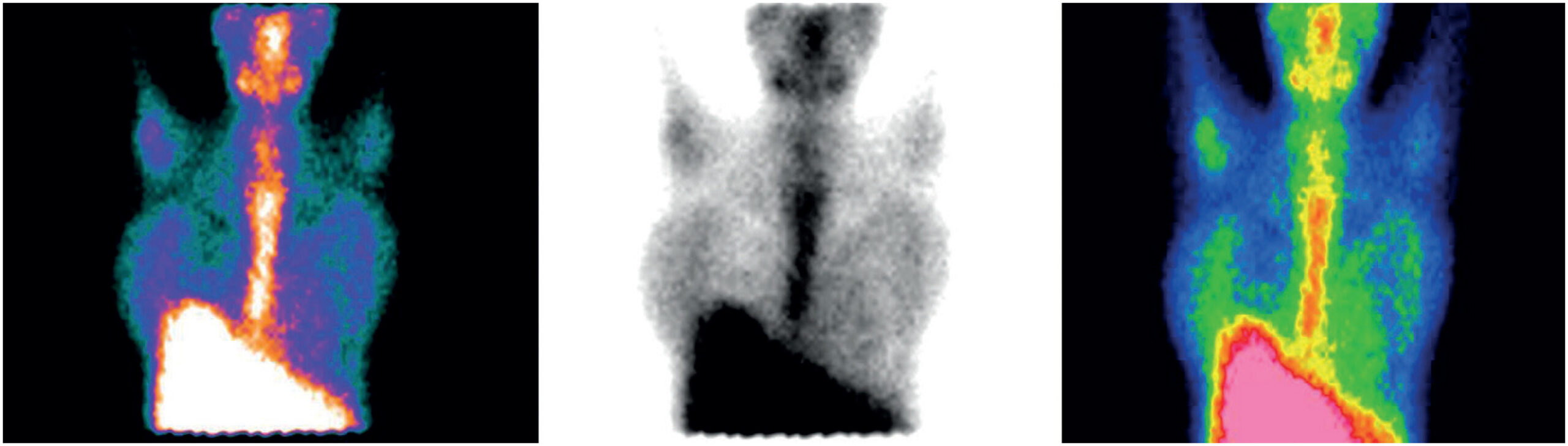
Cardiac Magnetic Resonance and Positron Emission Tomography in the Diagnosis and Follow-Up of Acute Rheumatic Fever – Case Report

David Salazar et al. Cristhian Espinoza Romero Mariana Pezzute Lopes Carlos Eduardo de Barros Branco … José Soares Junior Antonio de Santis Roney Orismar Sampaio Flávio Tarasoutchi

Introduction

Rheumatic fever (RF) is a prevalent condition in developing countries, accounting for a cardiac mortality of rate 7.9% in Brazil (DATASUS). Cases of rheumatic fever are often underdiagnosed and recognized only after permanent valve damage due to previous carditis. Secondary prevention of RF aims to limit the development and/or progression of valvular lesions and their complications. However, even on therapy with benzathine penicillin every 21 days, patients may experience recurrence of acute RF, making diagnosis a greater challenge. The development of new diagnostic methods can contribute to the early detection of rheumatic carditis (RC) considering that 90% of cases are subclinical. In addition, not only the usual gallium-67 scintigraphy, but also, tests such as cardiac nuclear magnetic resonance imaging (MRI) and positron emission tomography (PET-CT) may have the potential to be diagnostic tools in acute rheumatic carditis. However, more studies are needed to validate these methods. ,

[…]

  • Diagnostic Imaging
  • Epidemiology
  • Magnetic Resonance Spectroscopy/ methods
  • Positron Emission Tomogaphy Computed Tomography/methods
  • Rheumatic Fever/physiopathology
  • rheumatic heart disease
Read Full Article
Creative Commons Attribution (CC BY) Creative Commons license
22/04/2022 DOI: 10.36660/ijcs.20210093 Views 1
ABC
Editorial

Interleukin 35 (IL35): A New Biomarker for Coronary Artery Disease?

Gilson Soares Feitosa

In general terms, biomarkers are biologic parameters associated with an illness’s presence and severity. Biomarkers can be detected by several means, including the physical exam and evaluation by laboratory tests or by image.

A more recent and objectively directed definition restricts its realms to a proteic substance – enzyme, hormone or without known function- that relates to health status, indicating a good status of health or disease, thus allowing for diagnosing a given disease, or its severity, or else being a therapeutic target, or serving the purpose of therapeutic guidance, that can be obtained from the blood, urine, saliva or other body fluids or tissues.

[…]

  • Biomarkers
  • Coronary Artery Disease
  • Cytokines
  • Diagnostic Imaging
  • Interleukin 35
  • Troponin
Read Full Article
Creative Commons Attribution (CC BY) Creative Commons license
07/03/2022 DOI: 10.36660/abc.20210991 Views 2

Could not load more articles. Please try again.

How to cite this article

EndNote RIS

Electronic Document Format (Vancouver)

Ruffo CH, Rosa Filho AAM, Favaro GL, Hirose JSM, Lewinter RM, Melillo N, Silva LI, et al. Idiopathic Primary Chylopericardium. Arquivos Brasileiros de Cardiologia 2026;123(7):e20260062.

Electronic Document Format (ABNT)

Ruffo, Camila Helena; Rosa Filho, Alfredo Aurélio Marinho; Favaro, Guilherme Lima; Hirose, Juliana Sayuri Maia; Lewinter, Raissa Matias; Melillo, Natan; Silva, Leonardo Izzo; Anbar, Ramez; Coelho, Murilo Martins; Generoso, Giuliano. Idiopathic Primary Chylopericardium. Arquivos Brasileiros de Cardiologia, v. 123, n. 7, e20260062, Sep. 2026.

Electronic Document Format (APA)

Ruffo, C. H., Rosa Filho, A. A. M., Favaro, G. L., Hirose, J. S. M., Lewinter, R. M., Melillo, N., Silva, L. I., Anbar, R., Coelho, M. M., & Generoso, G. (2026). Idiopathic Primary Chylopericardium. Arquivos Brasileiros de Cardiologia, 123(7), e20260062.

Electronic Document Format (ISO)

Ruffo, Camila Helena and Rosa Filho, Alfredo Aurélio Marinho and Favaro, Guilherme Lima and Hirose, Juliana Sayuri Maia and Lewinter, Raissa Matias and Melillo, Natan and Silva, Leonardo Izzo and Anbar, Ramez and Coelho, Murilo Martins and Generoso, Giuliano. Idiopathic Primary Chylopericardium. Arquivos Brasileiros de Cardiologia [online]. 2026, vol. 123, n. 7, [cited 2026-10-14], e20260062. Available from: <https://journals.cardiol.br/en/article/idiopathic-primary-chylopericardium/>. ISSN 0066-782X

EndNote RIS

Electronic Document Format (Vancouver)

Antunes MO. The Thickness That Deceives: The Role of Imaging, Biopsy, and Genetics in the Evaluation of Ventricular Hypertrophy. Arquivos Brasileiros de Cardiologia 2026;123(2):e20260119.

Electronic Document Format (ABNT)

Antunes, Murillo de Oliveira. The Thickness That Deceives: The Role of Imaging, Biopsy, and Genetics in the Evaluation of Ventricular Hypertrophy. Arquivos Brasileiros de Cardiologia, v. 123, n. 2, e20260119, May. 2026.

Electronic Document Format (APA)

Antunes, M. O. (2026). The Thickness That Deceives: The Role of Imaging, Biopsy, and Genetics in the Evaluation of Ventricular Hypertrophy. Arquivos Brasileiros de Cardiologia, 123(2), e20260119.

Electronic Document Format (ISO)

Antunes, Murillo de Oliveira. The Thickness That Deceives: The Role of Imaging, Biopsy, and Genetics in the Evaluation of Ventricular Hypertrophy. Arquivos Brasileiros de Cardiologia [online]. 2026, vol. 123, n. 2, [cited 2026-10-14], e20260119. Available from: <https://journals.cardiol.br/en/article/the-thickness-that-deceives-the-role-of-imaging-biopsy-and-genetics-in-the-evaluation-of-ventricular-hypertrophy/>. ISSN 0066-782X

EndNote RIS

Electronic Document Format (Vancouver)

Santos DAM, Tarasoutchi F, Fernandes JRC, Paixão MR, Strabelli TMV, Köohler KF, Mansur AJ, et al. Infective Endocarditis in Brazil: A Narrative Review and Critical Analysis of Population and Hospital Case Data. ABC Imagem Cardiovascular 2026;38(4):e20250098.

Electronic Document Format (ABNT)

Santos, Diego Augusto Medeiros; Tarasoutchi, Flávio; Fernandes, João Ricardo Cordeiro; Paixão, Milena Ribeiro; Strabelli, Tânia Mara Varejão; Köohler, Karen Francine; Mansur, Alfredo José; Siciliano, Rinaldo Focaccia. Infective Endocarditis in Brazil: A Narrative Review and Critical Analysis of Population and Hospital Case Data. ABC Imagem Cardiovascular, v. 38, n. 4, e20250098, Feb. 2026.

Electronic Document Format (APA)

Santos, D. A. M., Tarasoutchi, F., Fernandes, J. R. C., Paixão, M. R., Strabelli, T. M. V., Köohler, K. F., Mansur, A. J., & Siciliano, R. F. (2026). Infective Endocarditis in Brazil: A Narrative Review and Critical Analysis of Population and Hospital Case Data. ABC Imagem Cardiovascular, 38(4), e20250098.

Electronic Document Format (ISO)

Santos, Diego Augusto Medeiros and Tarasoutchi, Flávio and Fernandes, João Ricardo Cordeiro and Paixão, Milena Ribeiro and Strabelli, Tânia Mara Varejão and Köohler, Karen Francine and Mansur, Alfredo José and Siciliano, Rinaldo Focaccia. Infective Endocarditis in Brazil: A Narrative Review and Critical Analysis of Population and Hospital Case Data. ABC Imagem Cardiovascular [online]. 2026, vol. 38, n. 4, [cited 2026-10-14], e20250098. Available from: <https://journals.cardiol.br/en/article/infective-endocarditis-in-brazil-a-narrative-review-and-critical-analysis-of-population-and-hospital-case-data/>. ISSN 2318-8219

EndNote RIS

Electronic Document Format (Vancouver)

Silva AP, Feitosa GS, Leal GCV, Moitinho RF, Torreão JA. Late Discovery of Left Ventricular Pseudoaneurysm: A Rare Clinical Case. ABC Imagem Cardiovascular 2025;38(3):e20250006.

Electronic Document Format (ABNT)

Silva, Angélica Pedreira da; Feitosa, Gilson Soares; Leal, Gabriel de Castro Vaz; Moitinho, Rilson Fraga; Torreão, Jorge Andion. Late Discovery of Left Ventricular Pseudoaneurysm: A Rare Clinical Case. ABC Imagem Cardiovascular, v. 38, n. 3, e20250006, Aug. 2025.

Electronic Document Format (APA)

Silva, A. P. , Feitosa, G. S., Leal, G. C. V., Moitinho, R. F., & Torreão, J. A. (2025). Late Discovery of Left Ventricular Pseudoaneurysm: A Rare Clinical Case. ABC Imagem Cardiovascular, 38(3), e20250006.

Electronic Document Format (ISO)

Silva, Angélica Pedreira da and Feitosa, Gilson Soares and Leal, Gabriel de Castro Vaz and Moitinho, Rilson Fraga and Torreão, Jorge Andion. Late Discovery of Left Ventricular Pseudoaneurysm: A Rare Clinical Case. ABC Imagem Cardiovascular [online]. 2025, vol. 38, n. 3, [cited 2026-10-14], e20250006. Available from: <https://journals.cardiol.br/en/article/late-discovery-of-left-ventricular-pseudoaneurysm-a-rare-clinical-case/>. ISSN 2318-8219

EndNote RIS

Electronic Document Format (Vancouver)

Ribeiro J. A Fresh Glimpse on Chest X-Ray: Revealing Significance of Aortic Arch Calcifications for Stroke Recurrence. Arquivos Brasileiros de Cardiologia 2024;121(7):e20240404.

Electronic Document Format (ABNT)

Ribeiro, José. A Fresh Glimpse on Chest X-Ray: Revealing Significance of Aortic Arch Calcifications for Stroke Recurrence. Arquivos Brasileiros de Cardiologia, v. 121, n. 7, e20240404, Nov. 2024.

Electronic Document Format (APA)

Ribeiro, J. (2024). A Fresh Glimpse on Chest X-Ray: Revealing Significance of Aortic Arch Calcifications for Stroke Recurrence. Arquivos Brasileiros de Cardiologia, 121(7), e20240404.

Electronic Document Format (ISO)

Ribeiro, José. A Fresh Glimpse on Chest X-Ray: Revealing Significance of Aortic Arch Calcifications for Stroke Recurrence. Arquivos Brasileiros de Cardiologia [online]. 2024, vol. 121, n. 7, [cited 2026-10-14], e20240404. Available from: <https://journals.cardiol.br/en/article/a-fresh-glimpse-on-chest-x-ray-revealing-significance-of-aortic-arch-calcifications-for-stroke-recurrence/>. ISSN 0066-782X

EndNote RIS

Electronic Document Format (Vancouver)

Costa A, Alves ES, Pinheiro P, Flausino L, Freire MV, Morel RV. Exploring Complexities in Differential Diagnosis: Torn Chord Tendinea in the Aortic Valve. ABC Imagem Cardiovascular 2023;36(4):e20230084.

Electronic Document Format (ABNT)

Costa, Alexandre; Alves, Endy de Santana; Pinheiro, Priscila; Flausino, Leonardo; Freire, Marcus Vinicius; Morel, Rodrigo Vieira. Exploring Complexities in Differential Diagnosis: Torn Chord Tendinea in the Aortic Valve. ABC Imagem Cardiovascular, v. 36, n. 4, e20230084, Dec. 2023.

Electronic Document Format (APA)

Costa, A., Alves, E. S., Pinheiro, P., Flausino, L., Freire, M. V., & Morel, R. V. (2023). Exploring Complexities in Differential Diagnosis: Torn Chord Tendinea in the Aortic Valve. ABC Imagem Cardiovascular, 36(4), e20230084.

Electronic Document Format (ISO)

Costa, Alexandre and Alves, Endy de Santana and Pinheiro, Priscila and Flausino, Leonardo and Freire, Marcus Vinicius and Morel, Rodrigo Vieira. Exploring Complexities in Differential Diagnosis: Torn Chord Tendinea in the Aortic Valve. ABC Imagem Cardiovascular [online]. 2023, vol. 36, n. 4, [cited 2026-10-14], e20230084. Available from: <https://journals.cardiol.br/en/article/exploring-complexities-in-differential-diagnosis-torn-chord-tendinea-in-the-aortic-valve/>. ISSN 2318-8219

EndNote RIS

Electronic Document Format (Vancouver)

Boccalon B, Amon AB, Serafini TF, Albrecht S, Foppa M, Santos ABS. Prosthetic Aortic Valve Endocarditis With Extensive Para-Aortic Abscess: The Relevance of Multimodal Imaging. ABC Imagem Cardiovascular 2023;36(3):e20230068.

Electronic Document Format (ABNT)

Boccalon, Bernardo; Amon, André Barcellos; Serafini, Thiago Ferreira; Albrecht, Álvaro Schmidt; Foppa, Murilo; Santos, Angela Barreto Santiago. Prosthetic Aortic Valve Endocarditis With Extensive Para-Aortic Abscess: The Relevance of Multimodal Imaging. ABC Imagem Cardiovascular, v. 36, n. 3, e20230068, Sep. 2023.

Electronic Document Format (APA)

Boccalon, B., Amon, A. B., Serafini, T. F., Albrecht, S., Foppa, M., & Santos, A. B. S. (2023). Prosthetic Aortic Valve Endocarditis With Extensive Para-Aortic Abscess: The Relevance of Multimodal Imaging. ABC Imagem Cardiovascular, 36(3), e20230068.

Electronic Document Format (ISO)

Boccalon, Bernardo and Amon, André Barcellos and Serafini, Thiago Ferreira and Albrecht, Álvaro Schmidt and Foppa, Murilo and Santos, Angela Barreto Santiago. Prosthetic Aortic Valve Endocarditis With Extensive Para-Aortic Abscess: The Relevance of Multimodal Imaging. ABC Imagem Cardiovascular [online]. 2023, vol. 36, n. 3, [cited 2026-10-14], e20230068. Available from: <https://journals.cardiol.br/en/article/prosthetic-aortic-valve-endocarditis-with-extensive-para-aortic-abscess-the-relevance-of-multimodal-imaging/>. ISSN 2318-8219

EndNote RIS

Electronic Document Format (Vancouver)

Saraiva F, Fernandes F, Romano MMD, Cardoso JN, Soliani MT, Bernardo MMM, Schmidt A, et al. Diagnosis and Treatment of Hypertrophic Cardiomyopathy. ABC Heart Fail Cardiomyop 2023;3(1):e20230040.

Electronic Document Format (ABNT)

Saraiva, Fernando; Fernandes, Fabio; Romano, Minna Moreira Dias; Cardoso, Juliano Novaes; Soliani, Marília Taily; Bernardo, Mariani Mendes Madisson; Schmidt, André; Simões, Marcus Vinicius. Diagnosis and Treatment of Hypertrophic Cardiomyopathy. ABC Heart Fail Cardiomyop, v. 3, n. 1, e20230040, Jul. 2023.

Electronic Document Format (APA)

Saraiva, F., Fernandes, F., Romano, M. M. D., Cardoso, J. N., Soliani, M. T., Bernardo, M. M. M., Schmidt, A., & Simões, M. V. (2023). Diagnosis and Treatment of Hypertrophic Cardiomyopathy. ABC Heart Fail Cardiomyop, 3(1), e20230040.

Electronic Document Format (ISO)

Saraiva, Fernando and Fernandes, Fabio and Romano, Minna Moreira Dias and Cardoso, Juliano Novaes and Soliani, Marília Taily and Bernardo, Mariani Mendes Madisson and Schmidt, André and Simões, Marcus Vinicius. Diagnosis and Treatment of Hypertrophic Cardiomyopathy. ABC Heart Fail Cardiomyop [online]. 2023, vol. 3, n. 1, [cited 2026-10-14], e20230040. Available from: <https://journals.cardiol.br/en/article/diagnosis-and-treatment-of-hypertrophic-cardiomyopathy/>

EndNote RIS

Electronic Document Format (Vancouver)

Gomes BFO, Cantisano AL, Menezes IRR, Azevedo AS, Silva TMB, Silva VL, Dutra GP, et al. Echocardiogram in Critically ill Patients with COVID-19: ECOVID Study. International Journal of Cardiovascular Sciences 2023;36:e20210114.

Electronic Document Format (ABNT)

Gomes, Bruno Ferraz de Oliveira; Cantisano, Armando Luis; Menezes, Iliana Regina Ribeiro; Azevedo, Anny de Sousa; Silva, Thiago Moreira Bastos da; Silva, Valdilene Lima; Dutra, Giovanni Possamai; Paula, Suzanna Andressa Morais de; Mendes, Barbara Ferreira da Silva; Carmo Junior, Plinio Resende do; Pereira, Basilio de Bragança; Petriz, Joao Luiz Fernandes; Oliveira, Glaucia Maria Moraes de. Echocardiogram in Critically ill Patients with COVID-19: ECOVID Study. International Journal of Cardiovascular Sciences, v. 36, e20210114, Apr. 2023.

Electronic Document Format (APA)

Gomes, B. F. O., Cantisano, A. L., Menezes, I. R. R., Azevedo, A. S., Silva, T. M. B. , Silva, V. L., Dutra, G. P., Paula, S. A. M. , Mendes, B. F. S., Carmo Junior, P. R. , Pereira, B. B., Petriz, J. L. F., & Oliveira, G. M. M. (2023). Echocardiogram in Critically ill Patients with COVID-19: ECOVID Study. International Journal of Cardiovascular Sciences, 36, e20210114.

Electronic Document Format (ISO)

Gomes, Bruno Ferraz de Oliveira and Cantisano, Armando Luis and Menezes, Iliana Regina Ribeiro and Azevedo, Anny de Sousa and Silva, Thiago Moreira Bastos da and Silva, Valdilene Lima and Dutra, Giovanni Possamai and Paula, Suzanna Andressa Morais de and Mendes, Barbara Ferreira da Silva and Carmo Junior, Plinio Resende do and Pereira, Basilio de Bragança and Petriz, Joao Luiz Fernandes and Oliveira, Glaucia Maria Moraes de. Echocardiogram in Critically ill Patients with COVID-19: ECOVID Study. International Journal of Cardiovascular Sciences [online]. 2023, vol. 36, [cited 2026-10-14], e20210114. Available from: <https://journals.cardiol.br/en/article/echocardiogram-in-critically-ill-patients-with-covid-19-ecovid-study/>. ISSN 2359-4802

EndNote RIS

Electronic Document Format (Vancouver)

Oliveira I, Lopes R, Cruz I, Bragança B, Azevedo J, Andrade A. Tuberous Sclerosis: Unusual Findings in the Setting of a Rare Disease. Arquivos Brasileiros de Cardiologia 2023;120(1):e20220147.

Electronic Document Format (ABNT)

Oliveira, Ines; Lopes, Rafaela; Cruz, Isabel; Bragança, Bruno; Azevedo, João; Andrade, Aurora. Tuberous Sclerosis: Unusual Findings in the Setting of a Rare Disease. Arquivos Brasileiros de Cardiologia, v. 120, n. 1, e20220147, Feb. 2023.

Electronic Document Format (APA)

Oliveira, I., Lopes, R., Cruz, I., Bragança, B., Azevedo, J., & Andrade, A. (2023). Tuberous Sclerosis: Unusual Findings in the Setting of a Rare Disease. Arquivos Brasileiros de Cardiologia, 120(1), e20220147.

Electronic Document Format (ISO)

Oliveira, Ines and Lopes, Rafaela and Cruz, Isabel and Bragança, Bruno and Azevedo, João and Andrade, Aurora. Tuberous Sclerosis: Unusual Findings in the Setting of a Rare Disease. Arquivos Brasileiros de Cardiologia [online]. 2023, vol. 120, n. 1, [cited 2026-10-14], e20220147. Available from: <https://journals.cardiol.br/en/article/tuberous-sclerosis-unusual-findings-in-the-setting-of-a-rare-disease/>. ISSN 0066-782X

EndNote RIS

Electronic Document Format (Vancouver)

Silva PHR, Montenegro CEL, Flamini MEDM, Pontual MVNO, Brandão SCS. Primary Cardiac Angiosarcoma Evaluated on PET/CT. ABC Imagem Cardiovascular 2022;35(4):eabc337.

Electronic Document Format (ABNT)

Silva, Pedro Henrique Rodrigues da; Montenegro, Carlos Eduardo Lucena; Flamini, Maria Eduarda Duarte de Mello; Pontual, Mariana Vila Nova de Oliveira; Brandão, Simone Cristina Soares. Primary Cardiac Angiosarcoma Evaluated on PET/CT. ABC Imagem Cardiovascular, v. 35, n. 4, eabc337, Dec. 2022.

Electronic Document Format (APA)

Silva, P. H. R. , Montenegro, C. E. L., Flamini, M. E. D. M., Pontual, M. V. N. O., & Brandão, S. C. S. (2022). Primary Cardiac Angiosarcoma Evaluated on PET/CT. ABC Imagem Cardiovascular, 35(4), eabc337.

Electronic Document Format (ISO)

Silva, Pedro Henrique Rodrigues da and Montenegro, Carlos Eduardo Lucena and Flamini, Maria Eduarda Duarte de Mello and Pontual, Mariana Vila Nova de Oliveira and Brandão, Simone Cristina Soares. Primary Cardiac Angiosarcoma Evaluated on PET/CT. ABC Imagem Cardiovascular [online]. 2022, vol. 35, n. 4, [cited 2026-10-14], eabc337. Available from: <https://journals.cardiol.br/en/article/primary-cardiac-angiosarcoma-evaluated-on-pet-ct/>. ISSN 2318-8219

EndNote RIS

Electronic Document Format (Vancouver)

Cauduro SA, Dutra JPP, Fernandes F, Bonatto M, Santos MVC, Rocha LSO, Mialski TR, et al. Obstructive Cardiac Tumors. ABC: Heart Failure & Cardiomyopathy 2022;2(4):367-73.

Electronic Document Format (ABNT)

Cauduro, Sanderson Antonio; Dutra, João Pedro Passos; Fernandes, Fabio; Bonatto, Marcely; Santos, Maria Verônica Câmara; Rocha, Letícia dos Santos de Oliveira; Mialski, Talita Ribeiro; Nobrega, Ana Paula Konig da; Brandão, Simone Cristina Soares; Barberato, Silvio Henrique. Obstructive Cardiac Tumors. ABC: Heart Failure & Cardiomyopathy, v. 2, n. 4, p. 367-373, Dec. 2022.

Electronic Document Format (APA)

Cauduro, S. A., Dutra, J. P. P., Fernandes, F., Bonatto, M., Santos, M. V. C., Rocha, L. S. O., Mialski, T. R., Nobrega, A. P. K. , Brandão, S. C. S., & Barberato, S. H. (2022). Obstructive Cardiac Tumors. ABC: Heart Failure & Cardiomyopathy, 2(4), 367-373.

Electronic Document Format (ISO)

Cauduro, Sanderson Antonio and Dutra, João Pedro Passos and Fernandes, Fabio and Bonatto, Marcely and Santos, Maria Verônica Câmara and Rocha, Letícia dos Santos de Oliveira and Mialski, Talita Ribeiro and Nobrega, Ana Paula Konig da and Brandão, Simone Cristina Soares and Barberato, Silvio Henrique. Obstructive Cardiac Tumors. ABC: Heart Failure & Cardiomyopathy [online]. 2022, vol. 2, n. 4, [cited 2026-10-14], pp. 367-373. Available from: <https://journals.cardiol.br/en/article/obstructive-cardiac-tumors/>

EndNote RIS

Electronic Document Format (Vancouver)

Oliveira I, Cruz I, Ana Neto , Bragança B, Abreu G, Azevedo J, Andrade A, et al. Mitral Valve Perforated Aneurysm: An Issue of Inflammation or Pressure Gradients?. Arquivos Brasileiros de Cardiologia 2022;119(5):809-12.

Electronic Document Format (ABNT)

Oliveira, Inês; Cruz, Isabel; Ana Neto, ; Bragança, Bruno; Abreu, Glória; Azevedo, João; Andrade, Aurora. Mitral Valve Perforated Aneurysm: An Issue of Inflammation or Pressure Gradients?. Arquivos Brasileiros de Cardiologia, v. 119, n. 5, p. 809-812, Sep. 2022.

Electronic Document Format (APA)

Oliveira, I., Cruz, I., Ana Neto, , Bragança, B., Abreu, G., Azevedo, J., & Andrade, A. (2022). Mitral Valve Perforated Aneurysm: An Issue of Inflammation or Pressure Gradients?. Arquivos Brasileiros de Cardiologia, 119(5), 809-812.

Electronic Document Format (ISO)

Oliveira, Inês and Cruz, Isabel and Ana Neto and Bragança, Bruno and Abreu, Glória and Azevedo, João and Andrade, Aurora. Mitral Valve Perforated Aneurysm: An Issue of Inflammation or Pressure Gradients?. Arquivos Brasileiros de Cardiologia [online]. 2022, vol. 119, n. 5, [cited 2026-10-14], pp. 809-812. Available from: <https://journals.cardiol.br/en/article/mitral-valve-perforated-aneurysm-an-issue-of-inflammation-or-pressure-gradients/>. ISSN 0066-782X

EndNote RIS

Electronic Document Format (Vancouver)

Paixão GMM, Lima EM, Quadros AB, Cabral DPR, Coelho RR, Oliveira DM, Nascimento JS, et al. Association between Atrioventricular Block and Mortality in Primary Care Patients: The CODE Study. Arquivos Brasileiros de Cardiologia 2022;119(4):564-71.

Electronic Document Format (ABNT)

Paixão, Gabriela Miana de Mattos; Lima, Emilly M.; Quadros, André B.; Cabral, Daniel P. R.; Coelho, Renato R.; Oliveira, Derick M.; Nascimento, Jamil de Souza; Gomes, Paulo R.; Ribeiro, Antonio L.. Association between Atrioventricular Block and Mortality in Primary Care Patients: The CODE Study. Arquivos Brasileiros de Cardiologia, v. 119, n. 4, p. 564-571, Jul. 2022.

Electronic Document Format (APA)

Paixão, G. M. M., Lima, E. M., Quadros, A. B., Cabral, D. P. R., Coelho, R. R., Oliveira, D. M., Nascimento, J. S., Gomes, P. R., & Ribeiro, A. L. (2022). Association between Atrioventricular Block and Mortality in Primary Care Patients: The CODE Study. Arquivos Brasileiros de Cardiologia, 119(4), 564-571.

Electronic Document Format (ISO)

Paixão, Gabriela Miana de Mattos and Lima, Emilly M. and Quadros, André B. and Cabral, Daniel P. R. and Coelho, Renato R. and Oliveira, Derick M. and Nascimento, Jamil de Souza and Gomes, Paulo R. and Ribeiro, Antonio L.. Association between Atrioventricular Block and Mortality in Primary Care Patients: The CODE Study. Arquivos Brasileiros de Cardiologia [online]. 2022, vol. 119, n. 4, [cited 2026-10-14], pp. 564-571. Available from: <https://journals.cardiol.br/en/article/association-between-atrioventricular-block-and-mortality-in-primary-care-patients-the-code-study/>. ISSN 0066-782X

EndNote RIS

Electronic Document Format (Vancouver)

Romero CE, Guacho WRL, Paula KR, Jimenez RP, Fonseca EKUN. Rare Presentation of Yolk Sac Tumor with Cardiac Involvement: Characteristics Detected by MRI. Arquivos Brasileiros de Cardiologia 2022;119(1):136-8.

Electronic Document Format (ABNT)

Romero, Cristhian Espinoza; Guacho, Williams Roberto Lata; Paula, Kevin Rafael de; Jimenez, Robert Paladines; Fonseca, Eduardo Kaiser Ururahy Nunes. Rare Presentation of Yolk Sac Tumor with Cardiac Involvement: Characteristics Detected by MRI. Arquivos Brasileiros de Cardiologia, v. 119, n. 1, p. 136-138, Jul. 2022.

Electronic Document Format (APA)

Romero, C. E., Guacho, W. R. L., Paula, K. R. , Jimenez, R. P., & Fonseca, E. K. U. N. (2022). Rare Presentation of Yolk Sac Tumor with Cardiac Involvement: Characteristics Detected by MRI. Arquivos Brasileiros de Cardiologia, 119(1), 136-138.

Electronic Document Format (ISO)

Romero, Cristhian Espinoza and Guacho, Williams Roberto Lata and Paula, Kevin Rafael de and Jimenez, Robert Paladines and Fonseca, Eduardo Kaiser Ururahy Nunes. Rare Presentation of Yolk Sac Tumor with Cardiac Involvement: Characteristics Detected by MRI. Arquivos Brasileiros de Cardiologia [online]. 2022, vol. 119, n. 1, [cited 2026-10-14], pp. 136-138. Available from: <https://journals.cardiol.br/en/article/rare-presentation-of-yolk-sac-tumor-with-cardiac-involvement-characteristics-detected-by-mri/>. ISSN 0066-782X

EndNote RIS

Electronic Document Format (Vancouver)

Akkaya H, Güntürk EE, Akkaya F, Karabıyık U, Güntürk , Yılmaz S. Assessment of the Relationship Between the Adropin Levels and the Coronary Collateral Circulation in Patients wıth Chronic Coronary Syndrome. Arquivos Brasileiros de Cardiologia 2022;119(3):402-10.

Electronic Document Format (ABNT)

Akkaya, Hasan; Güntürk, Ertuğrul Emre; Akkaya, Fulya; Karabıyık, Uğur; Güntürk, İnayet; Yılmaz, Samet. Assessment of the Relationship Between the Adropin Levels and the Coronary Collateral Circulation in Patients wıth Chronic Coronary Syndrome. Arquivos Brasileiros de Cardiologia, v. 119, n. 3, p. 402-410, Jun. 2022.

Electronic Document Format (APA)

Akkaya, H., Güntürk, E. E., Akkaya, F., Karabıyık, U., Güntürk, , & Yılmaz, S. (2022). Assessment of the Relationship Between the Adropin Levels and the Coronary Collateral Circulation in Patients wıth Chronic Coronary Syndrome. Arquivos Brasileiros de Cardiologia, 119(3), 402-410.

Electronic Document Format (ISO)

Akkaya, Hasan and Güntürk, Ertuğrul Emre and Akkaya, Fulya and Karabıyık, Uğur and Güntürk, İnayet and Yılmaz, Samet. Assessment of the Relationship Between the Adropin Levels and the Coronary Collateral Circulation in Patients wıth Chronic Coronary Syndrome. Arquivos Brasileiros de Cardiologia [online]. 2022, vol. 119, n. 3, [cited 2026-10-14], pp. 402-410. Available from: <https://journals.cardiol.br/en/article/assessment-of-the-relationship-between-the-adropin-levels-and-the-coronary-collateral-circulation-in-patients-with-chronic-coronary-syndrome/>. ISSN 0066-782X

EndNote RIS

Electronic Document Format (Vancouver)

Perazzo AM, Andrade LGF, Venancio LGA, Gouveia PAC, Galvão MFR, Lins EM, Moraes Neto F, et al. Value of18F-FDG PET/CT in the Diagnosis and Assessment of Response to Treatment of Lupus Myocarditis. Arquivos Brasileiros de Cardiologia 2022;118(6):1150-2.

Electronic Document Format (ABNT)

Perazzo, Alvaro M.; Andrade, Larissa G. F.; Venancio, Leonardo G. A.; Gouveia, Pedro Alves da Cruz; Galvão, Mariana Feitosa Ramalho; Lins, Esdras M.; Moraes Neto, Fernando; Brandão, Simone Cristina Soares. Value of18F-FDG PET/CT in the Diagnosis and Assessment of Response to Treatment of Lupus Myocarditis. Arquivos Brasileiros de Cardiologia, v. 118, n. 6, p. 1150-1152, Jun. 2022.

Electronic Document Format (APA)

Perazzo, A. M., Andrade, L. G. F., Venancio, L. G. A., Gouveia, P. A. C., Galvão, M. F. R., Lins, E. M., Moraes Neto, F., & Brandão, S. C. S. (2022). Value of18F-FDG PET/CT in the Diagnosis and Assessment of Response to Treatment of Lupus Myocarditis. Arquivos Brasileiros de Cardiologia, 118(6), 1150-1152.

Electronic Document Format (ISO)

Perazzo, Alvaro M. and Andrade, Larissa G. F. and Venancio, Leonardo G. A. and Gouveia, Pedro Alves da Cruz and Galvão, Mariana Feitosa Ramalho and Lins, Esdras M. and Moraes Neto, Fernando and Brandão, Simone Cristina Soares. Value of18F-FDG PET/CT in the Diagnosis and Assessment of Response to Treatment of Lupus Myocarditis. Arquivos Brasileiros de Cardiologia [online]. 2022, vol. 118, n. 6, [cited 2026-10-14], pp. 1150-1152. Available from: <https://journals.cardiol.br/en/article/value-of18f-fdg-pet-ct-in-the-diagnosis-and-assessment-of-response-to-treatment-of-lupus-myocarditis/>. ISSN 0066-782X

EndNote RIS

Electronic Document Format (Vancouver)

Resende CXC, Diogo PG, Amorim S, Pestana G, Torres JP, Macedo F. An Unusual Manifestation of Rejection. Arquivos Brasileiros de Cardiologia 2022;118(5):996-9.

Electronic Document Format (ABNT)

Resende, Carlos Xavier Correia de; Diogo, Pedro Grilo; Amorim, Sandra; Pestana, Gonçalo; Torres, José Pinheiro; Macedo, Filipe. An Unusual Manifestation of Rejection. Arquivos Brasileiros de Cardiologia, v. 118, n. 5, p. 996-999, May. 2022.

Electronic Document Format (APA)

Resende, C. X. C. , Diogo, P. G., Amorim, S., Pestana, G., Torres, J. P., & Macedo, F. (2022). An Unusual Manifestation of Rejection. Arquivos Brasileiros de Cardiologia, 118(5), 996-999.

Electronic Document Format (ISO)

Resende, Carlos Xavier Correia de and Diogo, Pedro Grilo and Amorim, Sandra and Pestana, Gonçalo and Torres, José Pinheiro and Macedo, Filipe. An Unusual Manifestation of Rejection. Arquivos Brasileiros de Cardiologia [online]. 2022, vol. 118, n. 5, [cited 2026-10-14], pp. 996-999. Available from: <https://journals.cardiol.br/en/article/an-unusual-manifestation-of-rejection/>. ISSN 0066-782X

EndNote RIS

Electronic Document Format (Vancouver)

Câmara SF, Ribeiro HB. Aortic Stiffness by Cardiac Magnetic Resonance: Prognostic tool or Bystander?. Arquivos Brasileiros de Cardiologia 2022;118(5):972-3.

Electronic Document Format (ABNT)

Câmara, Sérgio Figueiredo; Ribeiro, Henrique Barbosa. Aortic Stiffness by Cardiac Magnetic Resonance: Prognostic tool or Bystander?. Arquivos Brasileiros de Cardiologia, v. 118, n. 5, p. 972-973, May. 2022.

Electronic Document Format (APA)

Câmara, S. F., & Ribeiro, H. B. (2022). Aortic Stiffness by Cardiac Magnetic Resonance: Prognostic tool or Bystander?. Arquivos Brasileiros de Cardiologia, 118(5), 972-973.

Electronic Document Format (ISO)

Câmara, Sérgio Figueiredo and Ribeiro, Henrique Barbosa. Aortic Stiffness by Cardiac Magnetic Resonance: Prognostic tool or Bystander?. Arquivos Brasileiros de Cardiologia [online]. 2022, vol. 118, n. 5, [cited 2026-10-14], pp. 972-973. Available from: <https://journals.cardiol.br/en/article/aortic-stiffness-by-cardiac-magnetic-resonance-prognostic-tool-or-bystander/>. ISSN 0066-782X

EndNote RIS

Electronic Document Format (Vancouver)

Fernandes LP, Almeida MCC, Matos SA, Campos ACP, Câmara EJN, Foppa M, Ribeiro ALP, et al. Simple Echocardiographic Parameters are Strong Predictors of the Cardiovascular Risk in Asymptomatic Individuals: Elsa-Brasil Cohort. Arquivos Brasileiros de Cardiologia 2022;118(5):916-24.

Electronic Document Format (ABNT)

Fernandes, Luciana Pereira; Almeida, Maria da Conceição Chagas de; Matos, Sheila Alvim de; Campos, Ana Clara Paixão; Câmara, Edmundo José Nassri; Foppa, Murilo; Ribeiro, Antônio Luiz Pinho; Barreto, Sandhi Maria; Aras Júnior, Roque. Simple Echocardiographic Parameters are Strong Predictors of the Cardiovascular Risk in Asymptomatic Individuals: Elsa-Brasil Cohort. Arquivos Brasileiros de Cardiologia, v. 118, n. 5, p. 916-924, May. 2022.

Electronic Document Format (APA)

Fernandes, L. P., Almeida, M. C. C. , Matos, S. A. , Campos, A. C. P., Câmara, E. J. N., Foppa, M., Ribeiro, A. L. P., Barreto, S. M., & Aras Júnior, R. (2022). Simple Echocardiographic Parameters are Strong Predictors of the Cardiovascular Risk in Asymptomatic Individuals: Elsa-Brasil Cohort. Arquivos Brasileiros de Cardiologia, 118(5), 916-924.

Electronic Document Format (ISO)

Fernandes, Luciana Pereira and Almeida, Maria da Conceição Chagas de and Matos, Sheila Alvim de and Campos, Ana Clara Paixão and Câmara, Edmundo José Nassri and Foppa, Murilo and Ribeiro, Antônio Luiz Pinho and Barreto, Sandhi Maria and Aras Júnior, Roque. Simple Echocardiographic Parameters are Strong Predictors of the Cardiovascular Risk in Asymptomatic Individuals: Elsa-Brasil Cohort. Arquivos Brasileiros de Cardiologia [online]. 2022, vol. 118, n. 5, [cited 2026-10-14], pp. 916-924. Available from: <https://journals.cardiol.br/en/article/simple-echocardiographic-parameters-are-strong-predictors-of-the-cardiovascular-risk-in-asymptomatic-individuals-elsa-brasil-cohort/>. ISSN 0066-782X

EndNote RIS

Electronic Document Format (Vancouver)

Nunes NSV, Mesquita ET, Mesquita CT, Benchimol-Barbosa PR. First-degree Atrioventricular Block as an Early Marker of Advanced Disease of the Conduction System in a Patient with Hereditary Val142Ile Cardiac Amyloidosis. International Journal of Cardiovascular Sciences 2022;36:e20210081.

Electronic Document Format (ABNT)

Nunes, Nágela S. V.; Mesquita, Evandro Tinoco; Mesquita, Claudio Tinoco; Benchimol-Barbosa, Paulo Roberto. First-degree Atrioventricular Block as an Early Marker of Advanced Disease of the Conduction System in a Patient with Hereditary Val142Ile Cardiac Amyloidosis. International Journal of Cardiovascular Sciences, v. 36, e20210081, May. 2022.

Electronic Document Format (APA)

Nunes, N. S. V., Mesquita, E. T., Mesquita, C. T., & Benchimol-Barbosa, P. R. (2022). First-degree Atrioventricular Block as an Early Marker of Advanced Disease of the Conduction System in a Patient with Hereditary Val142Ile Cardiac Amyloidosis. International Journal of Cardiovascular Sciences, 36, e20210081.

Electronic Document Format (ISO)

Nunes, Nágela S. V. and Mesquita, Evandro Tinoco and Mesquita, Claudio Tinoco and Benchimol-Barbosa, Paulo Roberto. First-degree Atrioventricular Block as an Early Marker of Advanced Disease of the Conduction System in a Patient with Hereditary Val142Ile Cardiac Amyloidosis. International Journal of Cardiovascular Sciences [online]. 2022, vol. 36, [cited 2026-10-14], e20210081. Available from: <https://journals.cardiol.br/en/article/first-degree-atrioventricular-block-as-an-early-marker-of-advanced-disease-of-the-conduction-system-in-a-patient-with-hereditary-val142ile-cardiac-amyloidosis/>. ISSN 2359-4802

EndNote RIS

Electronic Document Format (Vancouver)

Gomes DA, Santos RR, Freitas P, Paiva MS, Ferreira J, Trabulo M. Acute Myocarditis Following mRNA COVID-19 Vaccine. Arquivos Brasileiros de Cardiologia 2022;118(4):783-6.

Electronic Document Format (ABNT)

Gomes, Daniel A.; Santos, Rita R.; Freitas, Pedro; Paiva, Mariana S.; Ferreira, Jorge; Trabulo, Marisa. Acute Myocarditis Following mRNA COVID-19 Vaccine. Arquivos Brasileiros de Cardiologia, v. 118, n. 4, p. 783-786, Apr. 2022.

Electronic Document Format (APA)

Gomes, D. A., Santos, R. R., Freitas, P., Paiva, M. S., Ferreira, J., & Trabulo, M. (2022). Acute Myocarditis Following mRNA COVID-19 Vaccine. Arquivos Brasileiros de Cardiologia, 118(4), 783-786.

Electronic Document Format (ISO)

Gomes, Daniel A. and Santos, Rita R. and Freitas, Pedro and Paiva, Mariana S. and Ferreira, Jorge and Trabulo, Marisa. Acute Myocarditis Following mRNA COVID-19 Vaccine. Arquivos Brasileiros de Cardiologia [online]. 2022, vol. 118, n. 4, [cited 2026-10-14], pp. 783-786. Available from: <https://journals.cardiol.br/en/article/acute-myocarditis-following-mrna-covid-19-vaccine/>. ISSN 0066-782X

EndNote RIS

Electronic Document Format (Vancouver)

Salazar D, Romero CE, Lopes MP, Branco CEB, Soares Junior J, Santis A, Sampaio RO, et al. Cardiac Magnetic Resonance and Positron Emission Tomography in the Diagnosis and Follow-Up of Acute Rheumatic Fever – Case Report. International Journal of Cardiovascular Sciences 2022;36:e20210093.

Electronic Document Format (ABNT)

Salazar, David; Romero, Cristhian Espinoza; Lopes, Mariana Pezzute; Branco, Carlos Eduardo de Barros; Soares Junior, José; Santis, Antonio de; Sampaio, Roney Orismar; Tarasoutchi, Flávio. Cardiac Magnetic Resonance and Positron Emission Tomography in the Diagnosis and Follow-Up of Acute Rheumatic Fever – Case Report. International Journal of Cardiovascular Sciences, v. 36, e20210093, Apr. 2022.

Electronic Document Format (APA)

Salazar, D., Romero, C. E., Lopes, M. P., Branco, C. E. B., Soares Junior, J., Santis, A. , Sampaio, R. O., & Tarasoutchi, F. (2022). Cardiac Magnetic Resonance and Positron Emission Tomography in the Diagnosis and Follow-Up of Acute Rheumatic Fever – Case Report. International Journal of Cardiovascular Sciences, 36, e20210093.

Electronic Document Format (ISO)

Salazar, David and Romero, Cristhian Espinoza and Lopes, Mariana Pezzute and Branco, Carlos Eduardo de Barros and Soares Junior, José and Santis, Antonio de and Sampaio, Roney Orismar and Tarasoutchi, Flávio. Cardiac Magnetic Resonance and Positron Emission Tomography in the Diagnosis and Follow-Up of Acute Rheumatic Fever – Case Report. International Journal of Cardiovascular Sciences [online]. 2022, vol. 36, [cited 2026-10-14], e20210093. Available from: <https://journals.cardiol.br/en/article/cardiac-magnetic-resonance-and-positron-emission-tomography-in-the-diagnosis-and-follow-up-of-acute-rheumatic-fever-case-report/>. ISSN 2359-4802

EndNote RIS

Electronic Document Format (Vancouver)

Feitosa GS. Interleukin 35 (IL35): A New Biomarker for Coronary Artery Disease?. Arquivos Brasileiros de Cardiologia 2022;118(2):409-10.

Electronic Document Format (ABNT)

Feitosa, Gilson Soares. Interleukin 35 (IL35): A New Biomarker for Coronary Artery Disease?. Arquivos Brasileiros de Cardiologia, v. 118, n. 2, p. 409-410, Mar. 2022.

Electronic Document Format (APA)

Feitosa, G. S. (2022). Interleukin 35 (IL35): A New Biomarker for Coronary Artery Disease?. Arquivos Brasileiros de Cardiologia, 118(2), 409-410.

Electronic Document Format (ISO)

Feitosa, Gilson Soares. Interleukin 35 (IL35): A New Biomarker for Coronary Artery Disease?. Arquivos Brasileiros de Cardiologia [online]. 2022, vol. 118, n. 2, [cited 2026-10-14], pp. 409-410. Available from: <https://journals.cardiol.br/en/article/interleukin-35-il35-a-new-biomarker-for-coronary-artery-disease/>. ISSN 0066-782X

  • About ABC Family
  • Internal Regulation
  • Editorial Board
  • Brazilian Society of Cardiology (SBC)

Issues

SBC Guidelines

  • Videos
  • Podcasts
  • News