ABC Family Arquivos Brasileiros de Cardiologia International Journal of Cardiovascular Sciences ABC Heart Failure & Cardiomyopathy ABC Imagem Cardiovascular
Português English
ABC Family
Português English
  • About
    • About ABC Family
    • Internal Regulation
    • Editorial Board
    • Brazilian Society of Cardiology (SBC)
  • Issues
  • SBC Guidelines
  • Content
    • Videos
    • Podcasts
    • News
  • About
    • About ABC Family
    • Internal Regulation
    • Editorial Board
    • Brazilian Society of Cardiology (SBC)
  • Issues
  • SBC Guidelines
  • Content
    • Videos
    • Podcasts
    • News

ABC Family

  • ABC Family
  • Arquivos Brasileiros de Cardiologia
  • International Journal of Cardiovascular Sciences
  • ABC Heart Failure & Cardiomyopathy
  • ABC Imagem Cardiovascular
Search in

No content found.

Journals
Ordering
Article types
Others

  1. Home
  2. Keyword: Magnetic Resonance Spectroscopy
Concordance Between Echocardiographic Left Ventricular Ejection Fraction by Simpson's Method, Global Longitudinal Strain, and Cardiac Magnetic Resonance
ABCIC
Original Article

Concordance Between Echocardiographic Left Ventricular Ejection Fraction by Simpson's Method, Global Longitudinal Strain, and Cardiac Magnetic Resonance

Álvaro Herrera-Escandón et al. Juan Pablo Morales-Grisales Sebastián Ayala-Zapata Stephany Barbosa-Balaguera … Álvaro José Muriel-Ruiz Juan Felipe Bravo-Rueda José Eduardo Citelli-Ramírez Luis Fernando Osío-Jimenez Luis Miguel Benitez-Gómez Carlos Javier Ramírez-Estupiñán

Abstract

Background:

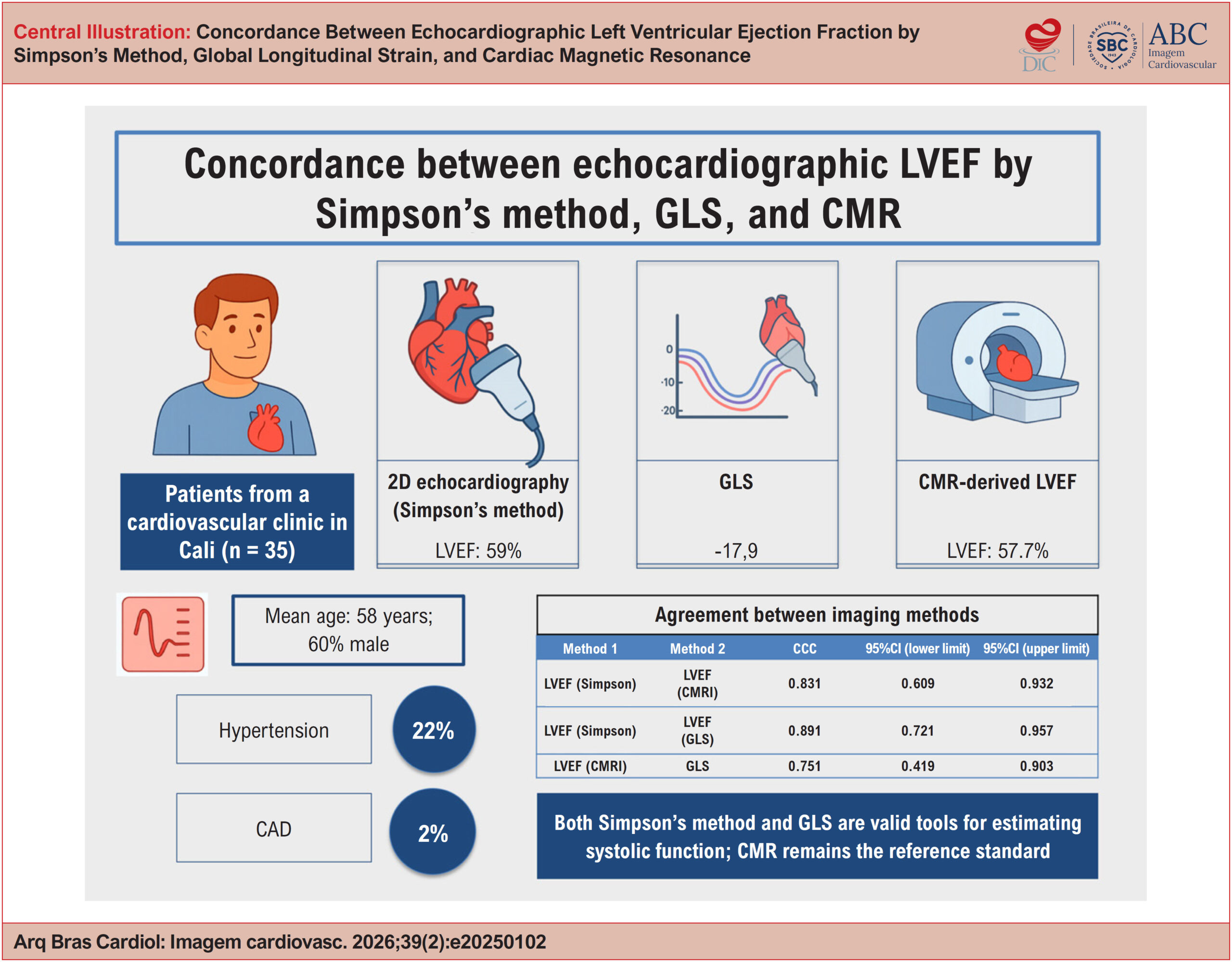
Left ventricular ejection fraction (LVEF) measured by echocardiography is a widely used parameter in clinical practice for the assessment of ventricular function. More recently, global longitudinal strain (GLS) has emerged as a complementary method, as has the assessment of LVEF by cardiac magnetic resonance (CMR). However, regional evidence evaluating the concordance among these three techniques remains limited.

Objectives:

To assess the concordance between echocardiographic and CMR measurements in patients treated at a cardiovascular clinic in the city of Cali, Colombia.

Methods:

This cross-sectional, analytical, descriptive study included 35 patients with confirmed or suspected heart disease, in whom all three methods were performed consecutively. Concordance was evaluated using Lin’s concordance correlation coefficient (CCC), Bland-Altman plots for LVEF, and linear and quadratic weighted κ coefficients for agreement between LVEF classifications.

Results:

The mean age was 58 years, and 60% of participants were male. The most common comorbidities were hypertension (22%) and dyslipidemia (11%). The mean LVEF was 59% by Simpson’s method and 57.7% by CMR, while the mean GLS was −17.7%. Concordance was as follows: Simpson’s LVEF vs. CMR (CCC, 0.831; 95%CI, 0.609-0.932); GLS vs. CMR-derived LVEF (CCC, 0.751; 95%CI, 0.419-0.903); and Simpson’s LVEF vs. GLS (CCC, 0.891; 95%CI, 0.721-0.957).

Conclusions:

Both Simpson’s method and GLS are valid tools for estimating systolic function. CMR remains the reference standard.
  • Echocardiography
  • Magnetic Resonance Spectroscopy
  • Stroke Volume
Read Full Article
Creative Commons Attribution (CC BY) Creative Commons license
24/07/2026 DOI: 10.36660/abcimg.20250102i Views 1
Figure 1
ABCIC
Review Article

My Approach to Cardiovascular Computed Tomography and Magnetic Resonance Imaging in the Evaluation of Cardiac Pseudotumors: A Brief Review

Roberto Vitor Almeida Torres et al. Bruno Maeda Fuzissima Eduardo Kaiser Ururahy Nunes Fonseca Lucas de Pádua Gomes de Farias … Walther Yoshiharu Ishikawa José de Arimatéia Batista Araújo Filho

Abstract

Cardiac pseudotumoral lesions are non-neoplastic conditions that are often overlooked in the differential diagnosis of cardiac masses. They present a variable clinical picture, ranging from asymptomatic to causing complications such as ventricular filling restriction and outflow tract obstruction. Echocardiography is the first-line imaging method but has limitations, such as dependence on the acoustic window and operator variability. However, a multimodality approach, including CT and MRI, is essential for seeking an accurate diagnosis. CT, with its excellent spatial resolution, allows for anatomical detailing, assessment of intralesional calcifications and fat, and contributes to therapeutic planning. MRI is preferred for tissue characterization and differentiation between benign and malignant lesions. Normal anatomical structures, such as the Eustachian valve and Chiari network, can be confused with thrombi or tumors, requiring correct identification. Thrombi are common in patients with atrial fibrillation or mitral valve disease, with MRI being important for differentiating them from neoplasms. Other pseudotumoral conditions include vegetation, gossypibomas, caseous calcification of the mitral valve annulus, and lipomatous hypertrophy of the interatrial septum. The integration of advanced cardiovascular imaging modalities is fundamental for the diagnosis and management of these lesions, optimizing patient care.

  • Emission-Computed Tomography
  • Heart Neoplasms
  • Magnetic Resonance Spectroscopy
Read Full Article
Creative Commons Attribution (CC BY) Creative Commons license
29/08/2024 DOI: 10.36660/abcimg.20240044i Views 1
Figure 1
ABC
Research Letter

Cardiac Magnetic Resonance to Evaluate Complete Substrate Elimination after Endocardial Ventricular Tachycardia Ablation in Chagas Disease

Mauricio I. Scanavacca et al. Rodrigo M. Kulchetscki Carlos E. Rochitte Cristiano F. Pisani

Introduction

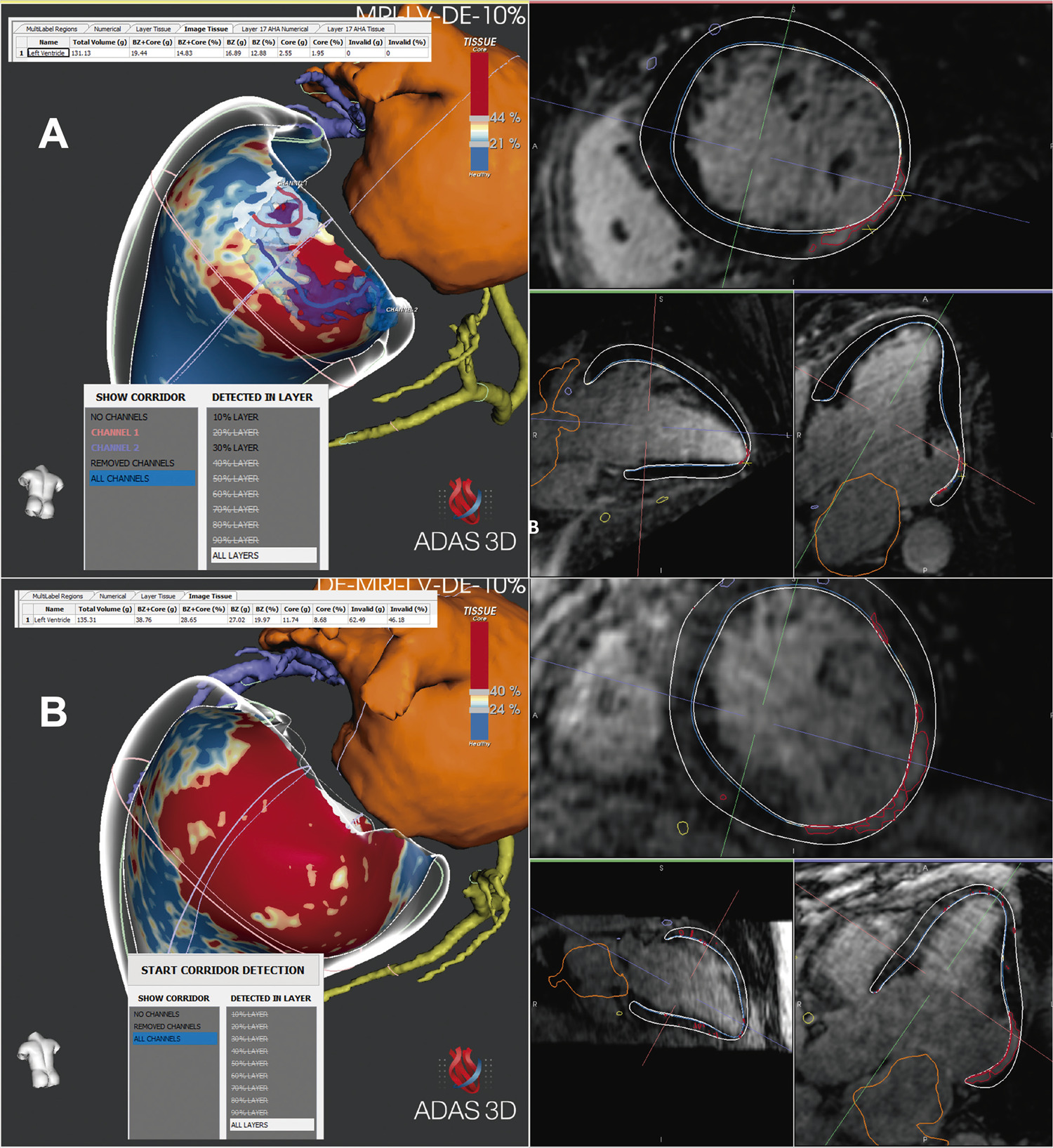
The main endpoint of ventricular tachycardia (VT) ablation includes elimination of the abnormal substrate on remapping and rendering non-inducible VT with programmed ventricular stimulation. ,

Cardiac magnetic resonance (CMR) has been increasingly used as a tool for locating the arrhythmogenic substrate of patients with scar-related VT. The images can be integrated with the electroanatomical mapping to guide ablation to the identified channels responsible for the VT-reentry circuits, optimizing the ablation results. However, until now, CMR has not been used to confirm the complete substrate elimination and effectiveness of VT ablation.

[…]

  • Ablation Techniques/methods
  • Chagas disease
  • Magnetic Resonance Spectroscopy
  • Tacchycardia, Ventricular
Read Full Article
Creative Commons Attribution (CC BY) Creative Commons license
26/02/2024 DOI: 10.36660/abc.20230421 Views 3
Figure 1
ABCHF
Image

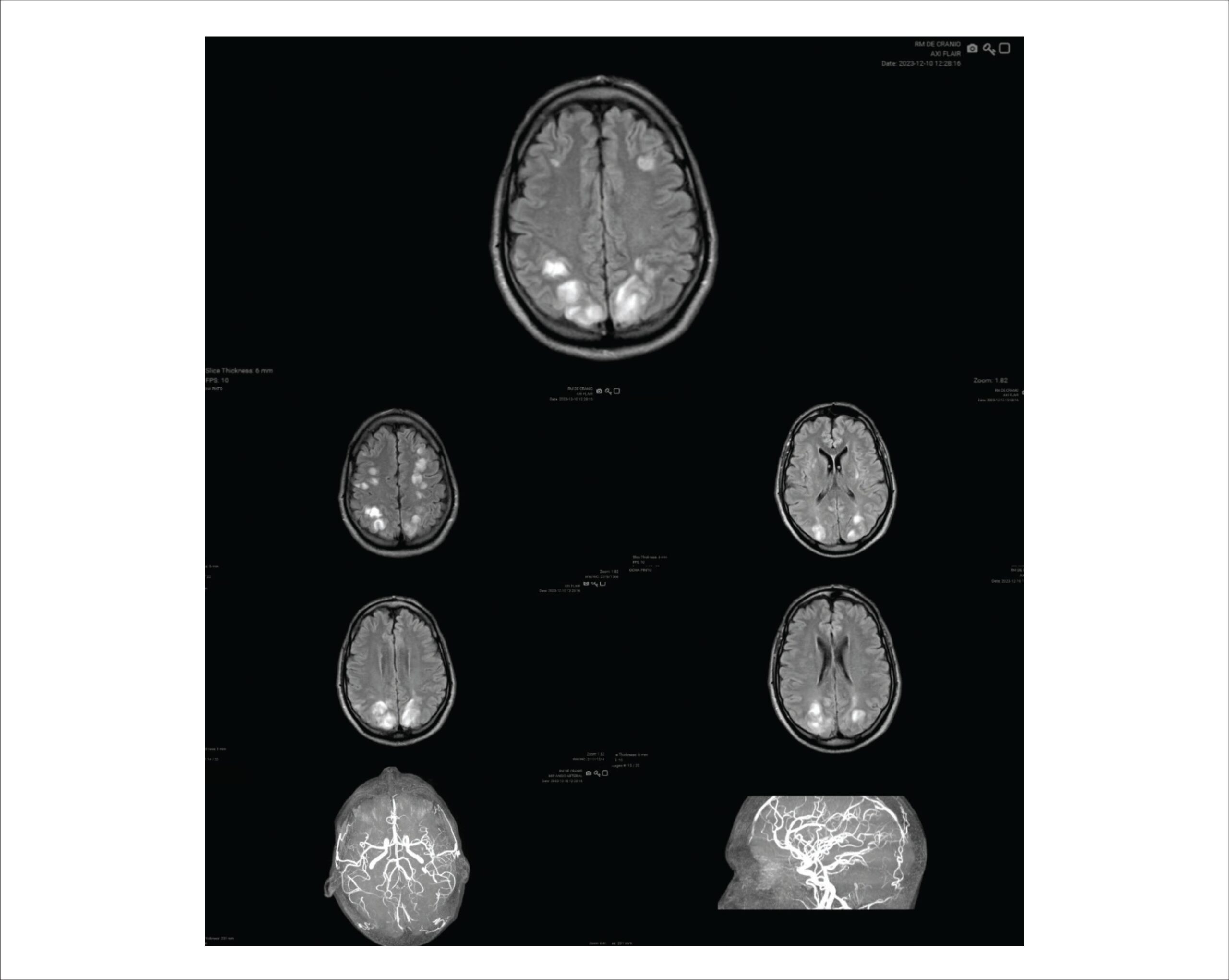
Tacrolimus-Associated Posterior Reversible Encephalopathy Syndrome after Heart Transplantation

Gabriel Gomes de Sousa et al. José Erirtônio Façanha Barreto Júnior Rivania Beatriz Novais Lima Pimentel Thiago Antônio Pessoa Carneiro Jefferson Luís Vieira

T

h

[…]

  • Magnetic Resonance Spectroscopy
  • Posterior Leukoencephalopathy Syndrome
  • Tacrolimus
Read Full Article
Creative Commons Attribution (CC BY) Creative Commons license
31/12/2023 DOI: 10.36660/abchf.20240005
ABC
Editorial

The Challenge of Making Cardiac Resonance a Global Reality

Hélder Jorge Andrade Gomes et al. Alcides Rocha de Figueredo Junior

Despite substantially reducing age-adjusted cardiovascular mortality rates in recent decades, cardiovascular disease remains the most common cause of death. Studies estimate that sudden cardiac death is responsible for approximately 50% of all deaths from cardiovascular causes, with ventricular tachycardia with degeneration to ventricular fibrillation and asystole being the most common fatal sequence of pathophysiological events.

The hard work done by authors of the article published in the Arquivos Brasileiros de Cardiologia illustrates the role of cardiac magnetic resonance (CMR) in the etiological elucidation of sudden death events encountered in clinical practice. In addition to the already expected Chagas and ischemic diseases, a wide variety of other diagnoses have been recognized: myocarditis, hypertrophic cardiomyopathy, dilated cardiomyopathy, arrhythmogenic heart disease, muscular dystrophy, hypertensive cardiomyopathy, metastases, non-compacted myocardium, and adrenergic cardiomyopathy (Takotsubo syndrome). The CMR was conclusive in more than 90% of cases and is a key tool for excluding structural heart disease and attributing the fatal event to extracardiac causes.

[…]

  • Cardiomyopathes
  • Cardiovascular Diseases, mortality
  • Death,Sudden
  • Diagnostic Imaging/trends
  • Endomyocardial Fibrosis
  • Magnetic Resonance Spectroscopy
  • Myocardial Ischemia
  • Stroke Volume
Read Full Article
Creative Commons Attribution (CC BY) Creative Commons license
21/04/2023 DOI: 10.36660/abc.20230187 Views 2
Figure 1
ABCHF
Review Article

Obstructive Cardiac Tumors

Sanderson Antonio Cauduro et al. João Pedro Passos Dutra Fabio Fernandes Marcely Bonatto … Maria Verônica Câmara Santos Letícia dos Santos de Oliveira Rocha Talita Ribeiro Mialski Ana Paula Konig da Nobrega Simone Cristina Soares Brandão Silvio Henrique Barberato

Abstract

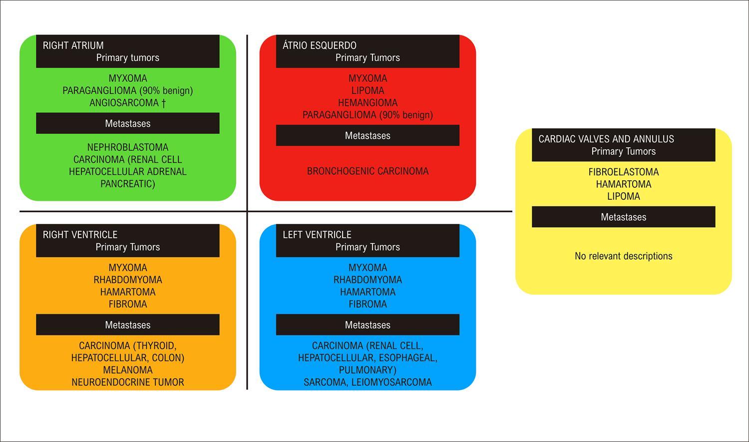
Cardiac tumors are considered rare clinical entities and can affect any cardiac tissue. Metastatic (secondary) cardiac tumors are more frequently diagnosed than primary tumors (malignant or benign). Both types can cause valve and/or inflow and outflow tract obstructions in any cardiac chamber, leading to symptoms of heart failure, as well as embolization and arrhythmia. Treatment of benign tumors is usually surgical, and that of metastatic and primary malignant tumors will depend on their origin and type, with poor prognosis. Recurrence of benign tumors is frequent. The aim of this article is to provide the clinician with tools to optimize diagnosis, differential diagnosis, and treatment of tumors with obstructive features causing heart failure.

  • Diagnostic Imaging
  • Echocardiography
  • Heart Failure
  • Heart Neoplasms
  • Magnetic Resonance Spectroscopy
Read Full Article
Creative Commons Attribution (CC BY) Creative Commons license
18/12/2022 DOI: 10.36660/abchf.20230001 Views 1
ABCIC
Image

Intrathoracic Mesothelial Cyst in Pediatric Patient

João Carlos Geber Júnior et al. Cláudia Regina Zaramello Ana Maria Silveira Rosendo Pedreira

[…]

  • Diagnostic Imaging
  • Echocardiography
  • Magnetic Resonance Spectroscopy
  • Mediastinal Cyst
  • Pericardium/abnormalities
Read Full Article
Creative Commons Attribution (CC BY) Creative Commons license
20/01/2022 DOI: 10.5935/2318-8219.20170018
ABCIC
Case Report

Progression of Left Valve Disease during Use of Dopaminergic Drugs

Lívia Santana Barbosa et al. Ana Clara Tude Rodrigues Shirlei Novillo Pereira Hugo Leonardo Medeiros Vieira … Kamila Fernanda Staszko José Lázaro Andrade

Abstract

Cabergoline and bromocriptine are dopaminergic drugs deriving from ergot and used for treating hyperprolactinemic idiopathic disorders or pituitary adenomas, whose mechanism of action results from the reduction of prolactin secretion. Some reports in the literature show that cabergoline can cause valvular heart disease after long-term administration. We report the case of a patient diagnosed with macroprolactinoma who did interleaved use of cabergoline and bromocriptine and developed valvular abnormalities that did not exist before.

  • Bromocriptine/effects adverse
  • Echocardiography
  • Heart Valve Diseases/physiopathology
  • Magnetic Resonance Spectroscopy
  • Prolactinoma/diagnosis
  • Prolactinoma/surgery
Read Full Article
Creative Commons Attribution (CC BY) Creative Commons license
20/01/2022 DOI: 10.5935/2318-8219.20170017 Views 1
ABCIC
Original Article

Cardiac Magnetic Resonance Evaluation of Patients with Cirrhosis Eligible to Liver Transplantation

Jeniffer Araújo Ribeiro et al. Priscilla Pereira dos Reis Paulo Victor Alves Pinto Eduardo Belisário Falquetto Antônio Márcio de Faria Andrade

Summary

Background

Liver transplantation (LT) is a huge surgery performed to treat patients with advanced liver cirrhosis and is associated with several risks. For this reason, is necessary to stratify the risk in the pre-transplantation period through the evaluation of myocardial function and ischemia

Objective

To demonstrate the applicability of cardiac magnetic resonance (CMR) in cardiac morphologic and functional evaluation, as well use in the evaluation of myocardial ischemia in pre-transplantation.

Methods

Retrospective, descriptive study. Data from patients with cirrhosis referred to the liver transplant outpatient clinic from January 2014 to July 2016 were analyzed they underwent CMR for cardiac evaluation and as provocative test of myocardial ischemia.

Results

135 patients were referred of these, 39 performed CMR. The mean age was 60 (50 to 71). About 87% (n = 34) were males. Alcoholic etiology prevailed 56% (n = 22). Most were of CHILD C patients with MELD ≥ 18, (n = 26). CMR showed myocardial ischemia in 03 patients (7,6%). Coronary angiography was performed and presence of severe coronary artery disease (obstruction > 70%) was confirmed, with consequent myocardial revascularization. At a follow-up of 2 years and 7 months, the survival of transplanted patients was 87%, without cardiologic complications.

Conclusions

The realization of CMR in the evaluation of cirrhotic patients in the pre-transplantation proved to be a safe strategy by showing presence of morphologic and functional changes of the cirrhotic cardiomyopathy and the presence of myocardial ischemia. However, more studies should be performed to standardize methods and criteria for cardiovascular evaluation in cirrhotic patients before the liver transplantation. (Arq Bras Cardiol: Imagem cardiovasc. 2017;30(4):119-125)

  • Coronary Artery Disease
  • Diagnostic Imaging
  • Liver Transplantation
  • Magnetic Resonance Spectroscopy
Read Full Article
Creative Commons Attribution (CC BY) Creative Commons license
20/01/2022 DOI: 10.5935/2318-8219.20170031 Views 1
Figure 1
ABC
Original Article

Relationship between Late Gadolinium Enhancement and Ventricular Repolarization Parameters in Heart Failure Patients with Reduced Ejection Fraction

Ali Riza Demir et al. Omer Celik Songül Ustündağ Begum Uygur … Mustafa Umut Somuncu Emre Yilmaz Yalcin Avci Gokhan Demirci Serkan Kahraman Mehmet Erturk

Abstract

Background

Heart failure with reduced ejection fraction (HFrEF) is a highly prevalent disease that requires repeating hospitalizations, causes significant morbidity and mortality. Therefore, early recognition of poor outcome predictors is essential for patient management.

Objective

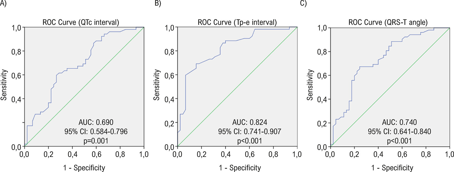
The aim of the present study is to investigate the relationship between late gadolinium enhancement (LGE) detected by cardiac magnetic resonance (CMR) and repolarization parameters such as corrected QT (QTc) interval, Tp-e interval, frontal QRS-T angle detected by 12 lead electrocardiograph (ECG) in HFrEF.

Method

In this single-center, retrospective observational study included 97 consecutive HFrEF patients who had CMR scan. Study population was divided into two groups according to the presence of LGE. Echocardiographic and CMR measurements and demographic features were recorded. QTc intervals, Tp-e intervals, frontal QRS-T angles were calculated from the ECG. A p-value less than 0.05 was considered statistically significant.

Results

LGE was detected in 52 (53.6%) out of 97 HFrEF patients. QTc intervals (p=0.001), Tp-e intervals (p<0.001), frontal QRS-T angles (p<0.001) were found to be significantly higher in LGE group when compared to non-LGE group. In univariate regression analysis which was performed to investigate the predictors of LGE in HFrEF, all three repolarization parameters were reached significant values but in multivariate analysis the only repolarization parameter remained significant was Tp-e interval (OR=1.085 95% CI 1.032-1.140, p=0.001).

Conclusion

With the prolongation of the Tp-e interval, the presence of myocardial fibrosis which is an arrhythmogenic substrate, can be predicted in patients with HFrEF.

  • Continuity of Patient Care/ethic
  • Diagnostic Imaging
  • Electrocardiography/methods
  • Gadolinium
  • Heart Failure
  • Magnetic Resonance Spectroscopy
  • Stroke Volume
Read Full Article
Creative Commons Attribution (CC BY) Creative Commons license
02/07/2021 DOI: 10.36660/abc.20200149
Figure 1
ABC
Original Article

Early Changes in Circulating Interleukins and Residual Inflammatory Risk After Acute Myocardial Infarction

Maria E. R. Coste et al. Carolina N. França Maria Cristina Izar Daniela Teixeira … Mayari E. Ishimura Ieda Longo-Maugeri Amanda S. Bacchin Henrique Tria Bianco Flavio T. Moreira Ibraim Masciarelli Pinto Gilberto Szarf Adriano Mendes Caixeta Otavio Berwanger Iran Gonçalves Jr Francisco A. H. Fonseca

Abstract

Background

Patients with acute myocardial infarction may have a large infarcted area and ventricular dysfunction despite early thrombolysis and revascularization.

Objective

To investigate the behavior of circulating cytokines in patients with ST-segment elevation myocardial infarction (STEMI) and their relationship with ventricular function.

Methods

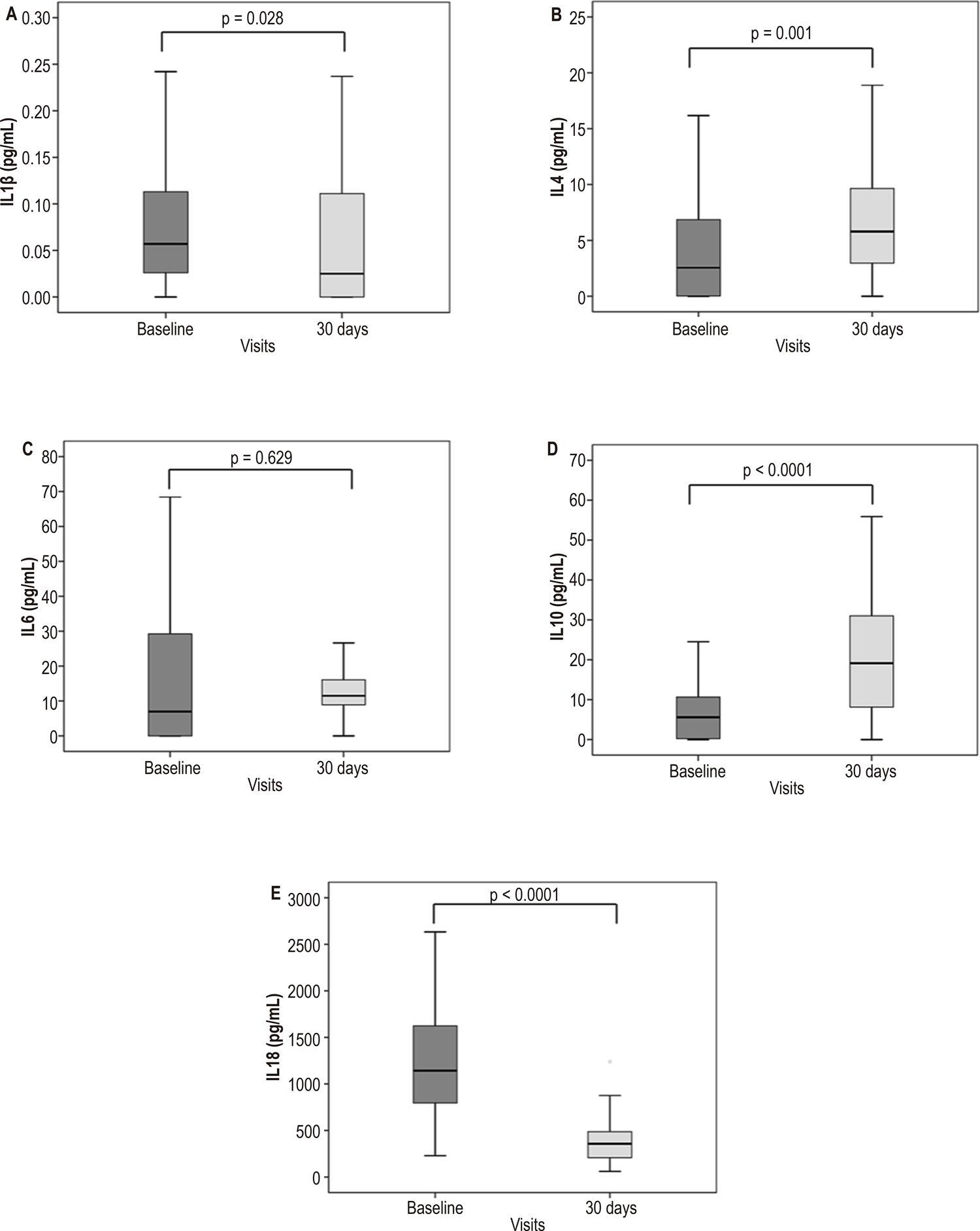
In the BATTLE-AMI (B and T Types of Lymphocytes Evaluation in Acute Myocardial Infarction) trial, patients with STEMI were treated with a pharmacoinvasive strategy. The plasma levels of cytokines (IL-1 β , IL-4, IL-6, IL-10, and IL-18) were tested using enzyme-linked immunosorbent assay (ELISA) at baseline and after 30 days. Infarcted mass and left ventricular ejection fraction (LVEF) were examined by 3-T cardiac magnetic resonance imaging. All p-values < 0.05 were considered statistically significant.

Results

Compared to baseline, lower levels were detected for IL-1 β (p = 0.028) and IL-18 (p < 0.0001) 30 days after STEMI, whereas higher levels were observed for IL-4 (p = 0.001) and IL-10 (p < 0.0001) at that time point. Conversely, no changes were detected for IL-6 levels (p = 0.63). The levels of high-sensitivity C-reactive protein and IL-6 correlated at baseline (rho = 0.45, p < 0.0001) and 30 days after STEMI (rho = 0.29, p = 0.009). At baseline, correlation between IL-6 levels and LVEF was also observed (rho = -0.50, p = 0.004).

Conclusions

During the first month post-MI, we observed a marked improvement in the balance of pro- and anti-inflammatory cytokines, except for IL-6. These findings suggest residual inflammatory risk. (Arq Bras Cardiol. 2020; [online].ahead print, PP.0-0)

  • C-Reactive Protein
  • Interleukin-6, Interleukin-10, Interleukin-18
  • Magnetic Resonance Spectroscopy
  • ST Elevation Myocardial Infarction
Read Full Article
Creative Commons Attribution (CC BY) Creative Commons license
28/08/2020 DOI: 10.36660/abc.20190567 Views 2
ABC
Editorial

Coronary Slow Flow Phenomenon – Adding Myocardial Fibrosis to the Equation

Filipe Penna de Carvalho et al. Clério Francisco de Azevedo

Initially described more than 40 years ago by Tambe et al., coronary slow flow phenomenon (CSFP) is characterized by delayed contrast medium progression in the absence of obstructive coronary epicardial disease during invasive coronary angiography. CSFP typically affects young male smokers, who often presents with acute coronary syndrome (ACS) or recurrent refractory resting angina requiring hospital admission.– Moreover, life-threatening arrhythmias and sudden cardiac death have also been associated with CSFP.

Despite increased awareness and research, CSFP remains an elusive and poorly understood condition, with many proposed pathogenic mechanisms including endothelial, vasomotor and microvascular dysfunction., Indeed, an abnormal regulation of microvascular tone that occurs only during resting conditions, while coronary flow reserve is within normal range, has been described in CSFP.

[…]

  • Cicatrix, Hypertrophic
  • Endomyocardial Fibrosis
  • Fractional Flow Reserve
  • Heart Failure
  • Magnetic Resonance Spectroscopy
  • Natriuretic-Peptide, C-Type
  • Prognosis
Read Full Article
Creative Commons Attribution (CC BY) Creative Commons license
06/04/2020 DOI: 10.36660/abc.20200187
Figure 1
ABC
Original Article

Longitudinal Shortening of the Left Ventricle by Cine-CMR for Assessment of Diastolic Function in Patients with Aortic Valve Disease

Sergio Marrone Ribeiro et al. Clerio Francisco de Azevedo Filho Roney Sampaio Flávio Tarasoutchi … Max Grinberg Roberto Kalil-Filho Carlos Eduardo Rochitte

Abstract

Background:

Diastolic dysfunction, commonly evaluated by echocardiography, is an important early finding in many cardiomyopathies. Cardiac magnetic resonance (CMR) often requires specialized sequences that extends the test time. Recently, feature-tracking imaging has been made available, but still requires expensive software and lacks clinical validation.

Objective:

To assess diastolic function in patients with aortic valve disease (AVD) and compare it with normal controls by evaluating left ventricular (LV) longitudinal displacement by CMR.

Methods:

We compared 26 AVD patients with 19 normal controls. Diastolic function was evaluated as LV longitudinal displacement in 4-chamber view cine-CMR images using steady state free precession (SSFP) sequence during the entire cardiac cycle with temporal resolution < 50 ms. The resulting plot of atrioventricular junction (AVJ) position versus time generated variables of AVJ motion. Significance level of p < 0.05 was used.

Results:

Maximum longitudinal displacement (0.12 vs. 0.17 cm), maximum velocity during early diastole (MVED, 0.6 vs. 1.4s-1), slope of the best-fit line of displacement in diastasis (VDS, 0.22 vs. 0.03s-1), and VDS/MVED ratio (0.35 vs. 0.02) were significantly reduced in AVD patients compared with controls, respectively. Aortic regurgitation showed significantly worse longitudinal LV shortening compared with aortic stenosis. Higher LV mass indicated worse diastolic dysfunction.

Conclusions:

A simple linear measurement detected significant differences on LV diastolic function between AVD patients and controls. LV mass was the only independent predictor of diastolic dysfunction in these patients. This method can help in the evaluation of diastolic dysfunction, improving cardiomyopathy detection by CMR, without prolonging exam time or depending on expensive software.

  • Aortic Valve Insufficiency
  • Cardiomyopathy, Hypertrophic / complications
  • Cardiovascular Diseases, mortality
  • Diagnostic Imaging
  • Echocardiography
  • Heart Failure
  • Magnetic Resonance Spectroscopy
Read Full Article
Creative Commons Attribution (CC BY) Creative Commons license
23/09/2019 DOI: 10.5935/abc.20190193 Views 1
Figure 1
ABC
Original Article

Intra-Atrial Dyssynchrony Using Cardiac Magnetic Resonance to Quantify Tissue Remodeling in Patients with Atrial Fibrillation

Luisa Allen Ciuffo et al. João Lima Henrique Doria de Vasconcellos Muhammad Balouch … Susumu Tao Saman Nazarian David D. Spragg Joseph E. Marine Ronald D. Berger Hugh Calkins Hiroshi Ashikaga

Abstract

Background:

Recent studies suggest that left atrial (LA) late gadolinium enhancement (LGE) can quantify the underlying tissue remodeling that harbors atrial fibrillation (AF). However, quantification of LA-LGE requires labor-intensive magnetic resonance imaging acquisition and postprocessing at experienced centers. LA intra-atrial dyssynchrony assessment is an emerging imaging technique that predicts AF recurrence after catheter ablation. We hypothesized that 1) LA intra-atrial dyssynchrony is associated with LA-LGE in patients with AF and 2) LA intra-atrial dyssynchrony is greater in patients with persistent AF than in those with paroxysmal AF.

Method:

We conducted a cross-sectional study comparing LA intra-atrial dyssynchrony and LA-LGE in 146 patients with a history of AF (60.0 ± 10.0 years, 30.1% nonparoxysmal AF) who underwent pre-AF ablation cardiac magnetic resonance (CMR) in sinus rhythm. Using tissue-tracking CMR, we measured the LA longitudinal strain in two- and four-chamber views. We defined intra-atrial dyssynchrony as the standard deviation (SD) of the time to peak longitudinal strain (SD-TPS, in %) and the SD of the time to the peak pre-atrial contraction strain corrected by the cycle length (SD-TPSpreA, in %). We used the image intensity ratio (IIR) to quantify LA-LGE.

Results:

Intra-atrial dyssynchrony analysis took 5 ± 9 minutes per case. Multivariable analysis showed that LA intra-atrial dyssynchrony was independently associated with LA-LGE. In addition, LA intra-atrial dyssynchrony was significantly greater in patients with persistent AF than those with paroxysmal AF. In contrast, there was no significant difference in LA-LGE between patients with persistent and paroxysmal AF. LA intra-atrial dyssynchrony showed excellent reproducibility and its analysis was less time-consuming (5 ± 9 minutes) than the LA-LGE (60 ± 20 minutes).

Conclusion:

LA Intra-atrial dyssynchrony is a quick and reproducible index that is independently associated with LA-LGE to reflect the underlying tissue remodeling.

  • Atrial Fibrillation
  • Diagnostic Imaging
  • Echocardiography/methods
  • Heart Atria
  • Magnetic Resonance Spectroscopy
Read Full Article
Creative Commons Attribution (CC BY) Creative Commons license
15/04/2019 DOI: 10.5935/abc.20190064 Views 1
Figure 1
ABC
Original Article

Correlation of Electrocardiographic Changes with Cardiac Magnetic Resonance Findings in Patients with Hypertrophic Cardiomyopathy

Gabriela Miana de Mattos Paixão et al. Horácio Eduardo Veronesi Halsted Alarcão Gomes Pereira da Silva José Nunes de Alencar Neto … Carolina de Paulo Maldi Luciano de Figueiredo Aguiar Filho Ibrahim Masciarelli Francisco Pinto Francisco Faustino de Albuquerque Carneiro de França Edileide de Barros Correia

Abstract

Background:

Electrocardiogram is the initial test in the investigation of heart disease. Electrocardiographic changes in hypertrophic cardiomyopathy have no set pattern, and correlates poorly with echocardiographic findings. Cardiac magnetic resonance imaging has been gaining momentum for better assessment of hypertrophy, as well as the detection of myocardial fibrosis.

Objectives:

To correlate the electrocardiographic changes with the location of hypertrophy in hypertrophic cardiomyopathy by cardiac magnetic resonance.

Methods:

This descriptive cross-sectional study evaluated 68 patients with confirmed diagnosis of hypertrophic cardiomyopathy by cardiac magnetic resonance. The patients’ electrocardiogram was compared with the location of the greatest myocardial hypertrophy by cardiac magnetic resonance. Statistical significance level of 5% and 95% confidence interval were adopted.

Results:

Of 68 patients, 69% had septal hypertrophy, 21% concentric and 10% apical hypertrophies. Concentric hypertrophy showed the greatest myocardial fibrosis mass (p < 0.001) and the greatest R wave size in D1 (p = 0.0280). The amplitudes of R waves in V5 and V6 (p = 0.0391, p = 0.0148) were higher in apical hypertrophy, with statistical significance. Apical hypertrophy was also associated with higher T wave negativity in D1, V5 and V6 (p < 0.001). Strain pattern was found in 100% of the patients with apical hypertrophy (p < 0.001).

Conclusion:

The location of myocardial hypertrophy by cardiac magnetic resonance can be correlated with electrocardiographic changes, especially for apical hypertrophy.

  • Electrocardiography
  • Hypertrophic cardiomyopathy / genetic
  • Magnetic Resonance Spectroscopy
Read Full Article
Creative Commons Attribution (CC BY) Creative Commons license
01/01/2018 DOI: 10.5935/abc.20170189
Figure 1
ABC
Review Article

Myocardial Viability on Cardiac Magnetic Resonance

Ana Luiza Mansur Souto et al. Rafael Mansur Souto Isabella Cristina Resende Teixeira Marcelo Souto Nacif

Abstract

The study of myocardial viability is of great importance in the orientation and management of patients requiring myocardial revascularization or angioplasty. The technique of delayed enhancement (DE) is accurate and has transformed the study of viability into an easy test, not only for the detection of fibrosis but also as a binary test detecting what is viable or not. On DE, fibrosis equal to or greater than 50% of the segmental area is considered as non-viable, whereas that below 50% is considered viable. During the same evaluation, cardiac magnetic resonance (CMR) may also use other techniques for functional and perfusion studies to obtain a global evaluation of ischemic heart disease. This study aims to highlight the current concepts and broadly emphasize the use of CMR as a method that over the last 20 years has become a reference in the detection of infarction and assessment of myocardial viability.

  • Angioplasty
  • Magnetic Resonance Spectroscopy
  • Myocardial Infarction / metabolism
  • Myocardial Revascularization
  • Tissue Survival
Read Full Article
Creative Commons Attribution (CC BY) Creative Commons license
01/05/2017 DOI: 10.5935/abc.20170056
Figure 1
ABC
Original Article

Cardiac Magnetic Resonance-Verified Myocardial Fibrosis in Chagas Disease: Clinical Correlates and Risk Stratification

Marly Uellendahl et al. Maria Eduarda Menezes de Siqueira Eveline Barros Calado Roberto Kalil-Filho … Dário Sobral Clébia Ribeiro Wilson Oliveira Silvia Martins Jagat Narula Carlos Eduardo Rochitte

Abstract

Background:

Chagas disease (CD) is an important cause of heart failure and mortality, mainly in Latin America. This study evaluated the morphological and functional characteristics of the heart as well the extent of myocardial fibrosis (MF) in patients with CD by cardiac magnetic resonance (CMR). The prognostic value of MF evaluated by myocardial-delayed enhancement (MDE) was compared with that via Rassi score.

Methods:

This study assessed 39 patients divided into 2 groups: 28 asymptomatic patients as indeterminate form group (IND); and symptomatic patients as Chagas Heart Disease (CHD) group. All patients underwent CMR using the techniques of cine-MRI and MDE, and the amount of MF was compared with the Rassi score.

Results:

Regarding the morphological and functional analysis, significant differences were observed between both groups (p < 0.001). Furthermore, there was a strong correlation between the extent of MF and the Rassi score (r = 0.76).

Conclusions:

CMR is an important technique for evaluating patients with CD, stressing morphological and functional differences in all clinical presentations. The strong correlation with the Rassi score and the extent of MF detected by CMR emphasizes its role in the prognostic stratification of patients with CD.

  • Chagas Cardiomyopathy
  • Chagas disease
  • Magnetic Resonance Spectroscopy
  • Risk factors, Prognosis
Read Full Article
Creative Commons Attribution (CC BY) Creative Commons license
01/11/2016 DOI: 10.5935/abc.20160168 Views 3
Figure 1
ABC
Case Report

Complicated Sinus of Valsalva Aneurysm Initially Diagnosed as Atrial Myxoma

Rafael Plens Teixeira et al. Pedro Felipe Gomes Nicz Felipe Lourenço Fernandes Renner Augusto Raposo Pereira … Roney Orismar Sampaio Flavio Tarasoutchi

Introduction

Atrial myxoma and sinus of Valsalva aneurysm (SVA) are rare conditions and possibly underdiagnosed in clinical practice. We report an unusual presentation of a left SVA after an episode of infective endocarditis (IE). The SVA extrinsically compressed the left main coronary artery (LMCA), which was initially diagnosed as a left atrial myxoma.

[…]

  • Aortic Aneurysm/surgery
  • Aortic Valve Insufficiency/surgery
  • Echocardiography, Transesophageal
  • Magnetic Resonance Spectroscopy
  • Mitral Valve Insufficiency / surgery
  • Sinus of Valsalva
Read Full Article
Creative Commons Attribution (CC BY) Creative Commons license
01/09/2016 DOI: 10.5935/abc.20160126 Views 1
ABC
Original Article

Aerobic Training after Myocardial Infarction: Remodeling Evaluated by Cardiac Magnetic Resonance

Nataly Lino Izeli et al. Aurélia Juliana dos Santos Júlio César Crescêncio Ana Clara Campagnolo Real Gonçalves … Valéria Papa Fabiana Marques Antônio Pazin-Filho Lourenço Gallo-Júnior André Schmidt

Abstract

Background:

Numerous studies show the benefits of exercise training after myocardial infarction (MI). Nevertheless, the effects on function and remodeling are still controversial.

Objectives:

To evaluate, in patients after (MI), the effects of aerobic exercise of moderate intensity on ventricular remodeling by cardiac magnetic resonance imaging (CMR).

Methods:

26 male patients, 52.9 ± 7.9 years, after a first MI, were assigned to groups: trained group (TG), 18; and control group (CG), 8. The TG performed supervised aerobic exercise on treadmill twice a week, and unsupervised sessions on 2 additional days per week, for at least 3 months. Laboratory tests, anthropometric measurements, resting heart rate (HR), exercise test, and CMR were conducted at baseline and follow-up.

Results:

The TG showed a 10.8% reduction in fasting blood glucose (p = 0.01), and a 7.3-bpm reduction in resting HR in both sitting and supine positions (p < 0.0001). There was an increase in oxygen uptake only in the TG (35.4 ± 8.1 to 49.1 ± 9.6 mL/kg/min, p < 0.0001). There was a statistically significant decrease in the TG left ventricular mass (LVmass) (128.7 ± 38.9 to 117.2 ± 27.2 g, p = 0.0032). There were no statistically significant changes in the values of left ventricular end-diastolic volume (LVEDV) and ejection fraction in the groups. The LVmass/EDV ratio demonstrated a statistically significant positive remodeling in the TG (p = 0.015).

Conclusions:

Aerobic exercise of moderate intensity improved physical capacity and other cardiovascular variables. A positive remodeling was identified in the TG, where a left ventricular diastolic dimension increase was associated with LVmass reduction.

  • Exercise
  • Magnetic Resonance Spectroscopy
  • Myocardial Infarction
  • Rehabilitation
Read Full Article
Creative Commons Attribution (CC BY) Creative Commons license
08/03/2016 DOI: 10.5935/abc.20160031 Views 1
Figure 1
ABC
Original Article

Prognostic Value of Pulmonary Vascular Resistance by Magnetic Resonance in Systolic Heart Failure

Óscar Fabregat-Andrés et al. Jordi Estornell-Erill Francisco Ridocci-Soriano José Leandro Pérez-Boscá … Pilar García-González Rafael Payá-Serrano Salvador Morell Julio Cortijo

Abstract

Background:

Pulmonary hypertension is associated with poor prognosis in heart failure. However, non-invasive diagnosis is still challenging in clinical practice.

Objective:

We sought to assess the prognostic utility of non-invasive estimation of pulmonary vascular resistances (PVR) by cardiovascular magnetic resonance to predict adverse cardiovascular outcomes in heart failure with reduced ejection fraction (HFrEF).

Methods:

Prospective registry of patients with left ventricular ejection fraction (LVEF) < 40% and recently admitted for decompensated heart failure during three years. PVRwere calculated based on right ventricular ejection fraction and average velocity of the pulmonary artery estimated during cardiac magnetic resonance. Readmission for heart failure and all-cause mortality were considered as adverse events at follow-up.

Results:

105 patients (average LVEF 26.0 ±7.7%, ischemic etiology 43%) were included. Patients with adverse events at long-term follow-up had higher values of PVR (6.93 ± 1.9 vs. 4.6 ± 1.7estimated Wood Units (eWu), p < 0.001). In multivariate Cox regression analysis, PVR ≥ 5 eWu(cutoff value according to ROC curve) was independently associated with increased risk of adverse events at 9 months follow-up (HR2.98; 95% CI 1.12-7.88; p < 0.03).

Conclusions:

In patients with HFrEF, the presence of PVR ≥ 5.0 Wu is associated with significantly worse clinical outcome at follow-up. Non-invasive estimation of PVR by cardiac magnetic resonance might be useful for risk stratification in HFrEF, irrespective of etiology, presence of late gadolinium enhancement or LVEF.

  • Heart Failure
  • Hypertension Pulmonary
  • Magnetic Resonance Spectroscopy
  • Prognosis
  • Vascular Resistance
Read Full Article
Creative Commons Attribution (CC BY) Creative Commons license
02/02/2016 DOI: 10.5935/abc.20160020
Cardiovascular magnetic resonance imaging-derived mitral valve geometry in determining mitral regurgitation severity
ABC
Artigo Original

Cardiovascular magnetic resonance imaging-derived mitral valve geometry in determining mitral regurgitation severity

Andre Mauricio Fernandes et al. Vikas Rathi Robert W. Biederman Mark Doyle … June A. Yamrozik Ronald B. Willians Vinayak Hedge Saundra Graunt Roque Aras Jr.

BACKGROUND: Mitral regurgitation is the most common valvular heart disease worldwide. Magnetic resonance may be a useful tool to analyze mitral valve parameters. OBJECTIVE: To distinguish mitral valve geometric patterns in patients with different severities of mitral regurgitation (MR) based on cardiovascular magnetic resonance imaging. METHODS: Sixty-three patients underwent cardiovascular magnetic resonance imaging. Mitral valve parameters analyzed were: tenting area (mm2) and angle (degrees), ventricle height (mm), tenting height (mm), anterior leaflet, posterior leaflet length and annulus diameter (mm). Patients were divided into two groups, one including patients who required mitral valve surgery and another which did not. RESULTS: Thirty-six patients had trace to mild (1-2+) MR and 27 had moderate to severe MR (3-4+). Ten (15.9%) out of 63 patients underwent surgery. Patients with more severe MR had a larger left ventricle end systolic diameter (38.6 ± 10.2 vs 45.4 ± 16.8, p<0.05) and left end diastolic diameter (52.9 ± 6.8 vs 60.1 ± 12.3, p= 0.005). On multivariate analysis, the tenting area was the strongest determinant of MR severity (r= 0.62, p=0.035). Annulus length (36.1 ± 4.7 vs 41 ± 6.7, p< 0.001), tenting area (190.7 ± 149.7 vs 130 ± 71.3, p= 0.048) and posterior leaflet length (15.1 ± 4.1 vs 12.2 ± 3.5, p= 0.023) were larger on patients requiring mitral valve surgery. CONCLUSIONS: Tenting area, annulus and posterior leaflet length are possible determinants of MR severity. These geometric parameters could be used to determine severity and could, in the future, direct specific patient care based on individual mitral apparatus anatomy.

  • Magnetic Resonance Spectroscopy
  • Mitral Valve Insufficiency
  • Mitral Valve Prolapse
Read Full Article
Creative Commons Attribution (CC BY) Creative Commons license
14/05/2013 DOI: 10.5935/abc.20130103
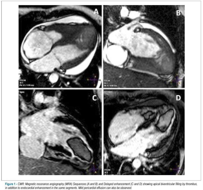
Biventricular thrombus and endomyocardial fibrosis in antiphospholipid syndrome
ABC
Case Report

Biventricular thrombus and endomyocardial fibrosis in antiphospholipid syndrome

Luiz Flávio Galvão Gonçalves et al. Fernanda Maria Silveira Souto Fernanda Nascimento Faro Joselina Luzia Menezes Oliveira … José Augusto Soares Barreto-Filho Antônio Carlos Sobral Sousa

CASE REPORT

Biventricular thrombus and endomyocardial fibrosis in antiphospholipid syndrome

[…]

  • Antibodies
  • Antiphospholipid
  • Antiphospholipid syndrome
  • Endomyocardial Fibrosis
  • Magnetic Resonance Spectroscopy
  • Thrombosis
Read Full Article
Creative Commons Attribution (CC BY) Creative Commons license
22/11/2012 DOI: 10.1590/S0066-782X2012001400017
Transthoracic impedance compared to magnetic resonance imaging in the assessment of cardiac output
ABC
Artigo Original

Transthoracic impedance compared to magnetic resonance imaging in the assessment of cardiac output

Humberto Villacorta Junior et al. Aline Sterque Villacorta Fernanda Amador Marcelo Hadlich … Denilson Campos de Albuquerque Clerio Francisco Azevedo

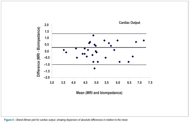
BACKGROUND: Cardiac magnetic resonance imaging is considered the gold-standard method for the calculation of cardiac volumes. Transthoracic impedance cardiography assesses the cardiac output. No studies validating this measurement, in comparison to that obtained by magnetic resonance imaging, are available. OBJECTIVE: To evaluate the performance of transthoracic impedance cardiography in the calculation of the cardiac output, cardiac index and stroke volume using magnetic resonance imaging as the gold-standard. METHODS: 31 patients with a mean age of 56.7 ± 18 years were assessed; of these, 18 (58%) were males. Patients whose indication for magnetic resonance imaging required pharmacologic stress test were excluded. Correlation between methods was assessed using the Pearson’s coefficient, and dispersion of absolute differences in relation to the mean was demonstrated using the Bland-Altman’s method. Agreement between methods was analyzed using the intraclass correlation coefficient. RESULTS: The mean cardiac output by transthoracic impedance cardiography and by magnetic resonance imaging was 5.16 ± 0.9 and 5.13 ± 0.9 L/min, respectively. Good agreement between methods was observed for cardiac output (r = 0.79; p = 0.0001), cardiac index (r = 0.74; p = 0.0001) and stroke volume (r = 0.88; p = 0.0001). The analysis by the Bland-Altman plot showed low dispersion of differences in relation to the mean, with a low amplitude of agreement intervals. Good agreement between the two methods was observed when analyzed by the intraclass correlation coefficient, with coefficients for cardiac output, cardiac index and stroke volume of 0.78, 0.73 and 0.88, respectively (p < 0.0001 for all comparisons). CONCLUSION: Transthoracic impedance cardiography proved accurate in the calculation of the cardiac output in comparison to cardiac magnetic resonance imaging.

  • Cardiac Ouput
  • Electric Impedance
  • Heart Failure
  • Magnetic Resonance Spectroscopy
Read Full Article
Creative Commons Attribution (CC BY) Creative Commons license
16/11/2012 DOI: 10.1590/S0066-782X2012005000104 Views 1
Assessment of aortic valve area combining echocardiography and magnetic resonance imaging
ABC
Artigo Original

Assessment of aortic valve area combining echocardiography and magnetic resonance imaging

Werner Nickl et al. Reiner Füth Jan Smettan Till Köhler … Mark Lankisch Frank Kramer Thomas Krahn Michael Coll Barroso Rolf Michael Klein Wilfried Dinh

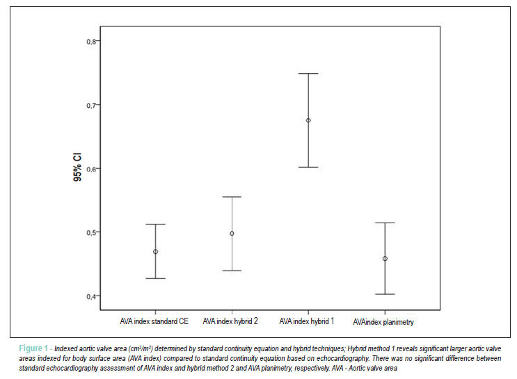
BACKGROUND: Transthoracic echocardiography (TTE) is routinely used to calculate aortic valve area (AVA) by continuity equation (CE). However, accurate measurement of the left ventricular outflow tract (LVOT) can be difficult and flow acceleration in the LVOT may lead to miscalculation of the AVA. OBJECTIVE: The aim of our study was to compare AVA measurements by standard TTE, cardiac magnetic resonance imaging (MRI) and a hybrid approach combining both techniques. METHODS: AVA was calculated in 38 patients (age 73±9 years) with standard TTE, cine-MRI planimetry and a hybrid approach: Hybrid Method 1: TTE-derived LVOT measurement in the CE numerator was replaced by the MRI assessment of the LVOT and AVA was calculated: (LVOT MRI/*LVOT-VTI TTE)/transaortic-VTI TTE. Method 2: We replaced the SV in the numerator by the MRI-derived SV and calculated AVA = SV MRI/ transaortic-VTI TTE. RESULTS: Mean AVA derived by TTE was 0.86 cm²±0.23 cm² and 0.83 cm²±0.3 cm² by MRI- planimetry, respectively. The mean absolute difference in AVA was 0.03cm² for TTE vs. MRI planimetry. AVA calculated with method 1 and method 2 was 1.23 cm²±0.4cm² and 0.92cm²±0.32cm², respectively. The mean absolute difference between TTE and method 1 and method 2 was 0.37cm² and 0.06cm², respectively (p<0.001). CONCLUSION: MRI-planimetry of AVA and hybrid method 2 are accurate and showed a good agreement with standard TTE measurements. Therefore, hybrid method 1 is a reasonable alternative if poor acoustic windows or LVOT flow accelerations limit the accuracy of TTE, particularly in patients at high risk for an invasive hemodynamic study.

  • Aortic Valve Stenosis
  • Echocardiography
  • Magnetic Resonance Spectroscopy
Read Full Article
Creative Commons Attribution (CC BY) Creative Commons license
29/02/2012 DOI: 10.1590/S0066-782X2012005000017 Views 1

How to cite this article

EndNote RIS

Electronic Document Format (Vancouver)

Herrera-Escandón , Morales-Grisales JP, Ayala-Zapata S, Barbosa-Balaguera S, Muriel-Ruiz J, Bravo-Rueda JF, Citelli-Ramírez JE, et al. Concordance Between Echocardiographic Left Ventricular Ejection Fraction by Simpson’s Method, Global Longitudinal Strain, and Cardiac Magnetic Resonance. ABC Imagem Cardiovascular 2026;39(2):e20250102.

Electronic Document Format (ABNT)

Herrera-Escandón, Álvaro; Morales-Grisales, Juan Pablo; Ayala-Zapata, Sebastián; Barbosa-Balaguera, Stephany; Muriel-Ruiz, Álvaro José; Bravo-Rueda, Juan Felipe; Citelli-Ramírez, José Eduardo; Osío-Jimenez, Luis Fernando; Benitez-Gómez, Luis Miguel; Ramírez-Estupiñán, Carlos Javier. Concordance Between Echocardiographic Left Ventricular Ejection Fraction by Simpson’s Method, Global Longitudinal Strain, and Cardiac Magnetic Resonance. ABC Imagem Cardiovascular, v. 39, n. 2, e20250102, Jul. 2026.

Electronic Document Format (APA)

Herrera-Escandón, , Morales-Grisales, J. P., Ayala-Zapata, S., Barbosa-Balaguera, S., Muriel-Ruiz, J., Bravo-Rueda, J. F., Citelli-Ramírez, J. E., Osío-Jimenez, L. F., Benitez-Gómez, L. M., & Ramírez-Estupiñán, C. J. (2026). Concordance Between Echocardiographic Left Ventricular Ejection Fraction by Simpson’s Method, Global Longitudinal Strain, and Cardiac Magnetic Resonance. ABC Imagem Cardiovascular, 39(2), e20250102.

Electronic Document Format (ISO)

Herrera-Escandón, Álvaro and Morales-Grisales, Juan Pablo and Ayala-Zapata, Sebastián and Barbosa-Balaguera, Stephany and Muriel-Ruiz, Álvaro José and Bravo-Rueda, Juan Felipe and Citelli-Ramírez, José Eduardo and Osío-Jimenez, Luis Fernando and Benitez-Gómez, Luis Miguel and Ramírez-Estupiñán, Carlos Javier. Concordance Between Echocardiographic Left Ventricular Ejection Fraction by Simpson’s Method, Global Longitudinal Strain, and Cardiac Magnetic Resonance. ABC Imagem Cardiovascular [online]. 2026, vol. 39, n. 2, [cited 2026-10-14], e20250102. Available from: <https://journals.cardiol.br/en/article/concordance-between-echocardiographic-left-ventricular-ejection-fraction-by-simpsons-method-global-longitudinal-strain-and-cardiac-magnetic-resonance/>. ISSN 2318-8219

EndNote RIS

Electronic Document Format (Vancouver)

Torres RVA, Fuzissima BM, Fonseca EKUN, Farias LPG, Ishikawa WY, Araújo Filho JAB. My Approach to Cardiovascular Computed Tomography and Magnetic Resonance Imaging in the Evaluation of Cardiac Pseudotumors: A Brief Review. ABC Imagem Cardiovascular 2024;37(3):e20240044.

Electronic Document Format (ABNT)

Torres, Roberto Vitor Almeida; Fuzissima, Bruno Maeda; Fonseca, Eduardo Kaiser Ururahy Nunes; Farias, Lucas de Pádua Gomes de; Ishikawa, Walther Yoshiharu; Araújo Filho, José de Arimatéia Batista. My Approach to Cardiovascular Computed Tomography and Magnetic Resonance Imaging in the Evaluation of Cardiac Pseudotumors: A Brief Review. ABC Imagem Cardiovascular, v. 37, n. 3, e20240044, Aug. 2024.

Electronic Document Format (APA)

Torres, R. V. A., Fuzissima, B. M., Fonseca, E. K. U. N., Farias, L. P. G. , Ishikawa, W. Y., & Araújo Filho, J. A. B. (2024). My Approach to Cardiovascular Computed Tomography and Magnetic Resonance Imaging in the Evaluation of Cardiac Pseudotumors: A Brief Review. ABC Imagem Cardiovascular, 37(3), e20240044.

Electronic Document Format (ISO)

Torres, Roberto Vitor Almeida and Fuzissima, Bruno Maeda and Fonseca, Eduardo Kaiser Ururahy Nunes and Farias, Lucas de Pádua Gomes de and Ishikawa, Walther Yoshiharu and Araújo Filho, José de Arimatéia Batista. My Approach to Cardiovascular Computed Tomography and Magnetic Resonance Imaging in the Evaluation of Cardiac Pseudotumors: A Brief Review. ABC Imagem Cardiovascular [online]. 2024, vol. 37, n. 3, [cited 2026-10-14], e20240044. Available from: <https://journals.cardiol.br/en/article/my-approach-to-cardiovascular-computed-tomography-and-magnetic-resonance-imaging-in-the-evaluation-of-cardiac-pseudotumors-a-brief-review/>. ISSN 2318-8219

EndNote RIS

Electronic Document Format (Vancouver)

Scanavacca MI, Kulchetscki RM, Rochitte CE, Pisani CF. Cardiac Magnetic Resonance to Evaluate Complete Substrate Elimination after Endocardial Ventricular Tachycardia Ablation in Chagas Disease. Arquivos Brasileiros de Cardiologia 2024;121(1):e20230421.

Electronic Document Format (ABNT)

Scanavacca, Mauricio I.; Kulchetscki, Rodrigo M.; Rochitte, Carlos E.; Pisani, Cristiano F.. Cardiac Magnetic Resonance to Evaluate Complete Substrate Elimination after Endocardial Ventricular Tachycardia Ablation in Chagas Disease. Arquivos Brasileiros de Cardiologia, v. 121, n. 1, e20230421, Feb. 2024.

Electronic Document Format (APA)

Scanavacca, M. I., Kulchetscki, R. M., Rochitte, C. E., & Pisani, C. F. (2024). Cardiac Magnetic Resonance to Evaluate Complete Substrate Elimination after Endocardial Ventricular Tachycardia Ablation in Chagas Disease. Arquivos Brasileiros de Cardiologia, 121(1), e20230421.

Electronic Document Format (ISO)

Scanavacca, Mauricio I. and Kulchetscki, Rodrigo M. and Rochitte, Carlos E. and Pisani, Cristiano F.. Cardiac Magnetic Resonance to Evaluate Complete Substrate Elimination after Endocardial Ventricular Tachycardia Ablation in Chagas Disease. Arquivos Brasileiros de Cardiologia [online]. 2024, vol. 121, n. 1, [cited 2026-10-14], e20230421. Available from: <https://journals.cardiol.br/en/article/cardiac-magnetic-resonance-to-evaluate-complete-substrate-elimination-after-endocardial-ventricular-tachycardia-ablation-in-chagas-disease/>. ISSN 0066-782X

EndNote RIS

Electronic Document Format (Vancouver)

Sousa GG, Barreto Júnior JEF, Pimentel RBNL, Carneiro TAP, Vieira JL. Tacrolimus-Associated Posterior Reversible Encephalopathy Syndrome after Heart Transplantation. ABC Heart Fail Cardiomyop 2023;3(4):e20240005.

Electronic Document Format (ABNT)

Sousa, Gabriel Gomes de; Barreto Júnior, José Erirtônio Façanha; Pimentel, Rivania Beatriz Novais Lima; Carneiro, Thiago Antônio Pessoa; Vieira, Jefferson Luís. Tacrolimus-Associated Posterior Reversible Encephalopathy Syndrome after Heart Transplantation. ABC Heart Fail Cardiomyop, v. 3, n. 4, e20240005, Dec. 2023.

Electronic Document Format (APA)

Sousa, G. G. , Barreto Júnior, J. E. F., Pimentel, R. B. N. L., Carneiro, T. A. P., & Vieira, J. L. (2023). Tacrolimus-Associated Posterior Reversible Encephalopathy Syndrome after Heart Transplantation. ABC Heart Fail Cardiomyop, 3(4), e20240005.

Electronic Document Format (ISO)

Sousa, Gabriel Gomes de and Barreto Júnior, José Erirtônio Façanha and Pimentel, Rivania Beatriz Novais Lima and Carneiro, Thiago Antônio Pessoa and Vieira, Jefferson Luís. Tacrolimus-Associated Posterior Reversible Encephalopathy Syndrome after Heart Transplantation. ABC Heart Fail Cardiomyop [online]. 2023, vol. 3, n. 4, [cited 2026-10-14], e20240005. Available from: <https://journals.cardiol.br/en/article/tacrolimus-associated-posterior-reversible-encephalopathy-syndrome-after-heart-transplantation/>

EndNote RIS

Electronic Document Format (Vancouver)

Gomes HJA, Figueredo Junior AR. The Challenge of Making Cardiac Resonance a Global Reality. Arquivos Brasileiros de Cardiologia 2023;120(4):e20230187.

Electronic Document Format (ABNT)

Gomes, Hélder Jorge Andrade; Figueredo Junior, Alcides Rocha de. The Challenge of Making Cardiac Resonance a Global Reality. Arquivos Brasileiros de Cardiologia, v. 120, n. 4, e20230187, Apr. 2023.

Electronic Document Format (APA)

Gomes, H. J. A., & Figueredo Junior, A. R. (2023). The Challenge of Making Cardiac Resonance a Global Reality. Arquivos Brasileiros de Cardiologia, 120(4), e20230187.

Electronic Document Format (ISO)

Gomes, Hélder Jorge Andrade and Figueredo Junior, Alcides Rocha de. The Challenge of Making Cardiac Resonance a Global Reality. Arquivos Brasileiros de Cardiologia [online]. 2023, vol. 120, n. 4, [cited 2026-10-14], e20230187. Available from: <https://journals.cardiol.br/en/article/the-challenge-of-making-cardiac-resonance-a-global-reality/>. ISSN 0066-782X

EndNote RIS

Electronic Document Format (Vancouver)

Cauduro SA, Dutra JPP, Fernandes F, Bonatto M, Santos MVC, Rocha LSO, Mialski TR, et al. Obstructive Cardiac Tumors. ABC: Heart Failure & Cardiomyopathy 2022;2(4):367-73.

Electronic Document Format (ABNT)

Cauduro, Sanderson Antonio; Dutra, João Pedro Passos; Fernandes, Fabio; Bonatto, Marcely; Santos, Maria Verônica Câmara; Rocha, Letícia dos Santos de Oliveira; Mialski, Talita Ribeiro; Nobrega, Ana Paula Konig da; Brandão, Simone Cristina Soares; Barberato, Silvio Henrique. Obstructive Cardiac Tumors. ABC: Heart Failure & Cardiomyopathy, v. 2, n. 4, p. 367-373, Dec. 2022.

Electronic Document Format (APA)

Cauduro, S. A., Dutra, J. P. P., Fernandes, F., Bonatto, M., Santos, M. V. C., Rocha, L. S. O., Mialski, T. R., Nobrega, A. P. K. , Brandão, S. C. S., & Barberato, S. H. (2022). Obstructive Cardiac Tumors. ABC: Heart Failure & Cardiomyopathy, 2(4), 367-373.

Electronic Document Format (ISO)

Cauduro, Sanderson Antonio and Dutra, João Pedro Passos and Fernandes, Fabio and Bonatto, Marcely and Santos, Maria Verônica Câmara and Rocha, Letícia dos Santos de Oliveira and Mialski, Talita Ribeiro and Nobrega, Ana Paula Konig da and Brandão, Simone Cristina Soares and Barberato, Silvio Henrique. Obstructive Cardiac Tumors. ABC: Heart Failure & Cardiomyopathy [online]. 2022, vol. 2, n. 4, [cited 2026-10-14], pp. 367-373. Available from: <https://journals.cardiol.br/en/article/obstructive-cardiac-tumors/>

EndNote RIS

Electronic Document Format (Vancouver)

Geber Júnior JC, Zaramello CR, Pedreira AMSR. Intrathoracic Mesothelial Cyst in Pediatric Patient. Arquivos Brasileiros de Cardiologia Imagem cardiovascular 2022;30(2):84-5.

Electronic Document Format (ABNT)

Geber Júnior, João Carlos; Zaramello, Cláudia Regina; Pedreira, Ana Maria Silveira Rosendo. Intrathoracic Mesothelial Cyst in Pediatric Patient. Arquivos Brasileiros de Cardiologia Imagem cardiovascular, v. 30, n. 2, p. 84-85, Jan. 2022.

Electronic Document Format (APA)

Geber Júnior, J. C., Zaramello, C. R., & Pedreira, A. M. S. R. (2022). Intrathoracic Mesothelial Cyst in Pediatric Patient. Arquivos Brasileiros de Cardiologia Imagem cardiovascular, 30(2), 84-85.

Electronic Document Format (ISO)

Geber Júnior, João Carlos and Zaramello, Cláudia Regina and Pedreira, Ana Maria Silveira Rosendo. Intrathoracic Mesothelial Cyst in Pediatric Patient. Arquivos Brasileiros de Cardiologia Imagem cardiovascular [online]. 2022, vol. 30, n. 2, [cited 2026-10-14], pp. 84-85. Available from: <https://journals.cardiol.br/en/article/intrathoracic-mesothelial-cyst-in-pediatric-patient/>. ISSN 2318-8219

EndNote RIS

Electronic Document Format (Vancouver)

Barbosa LS, Rodrigues ACT, Pereira SN, Vieira HLM, Staszko KF, Andrade JL. Progression of Left Valve Disease during Use of Dopaminergic Drugs. Arquivos Brasileiros de Cardiologia Imagem cardiovascular 2022;30(2):77-83.

Electronic Document Format (ABNT)

Barbosa, Lívia Santana; Rodrigues, Ana Clara Tude; Pereira, Shirlei Novillo; Vieira, Hugo Leonardo Medeiros; Staszko, Kamila Fernanda; Andrade, José Lázaro. Progression of Left Valve Disease during Use of Dopaminergic Drugs. Arquivos Brasileiros de Cardiologia Imagem cardiovascular, v. 30, n. 2, p. 77-83, Jan. 2022.

Electronic Document Format (APA)

Barbosa, L. S., Rodrigues, A. C. T., Pereira, S. N., Vieira, H. L. M., Staszko, K. F., & Andrade, J. L. (2022). Progression of Left Valve Disease during Use of Dopaminergic Drugs. Arquivos Brasileiros de Cardiologia Imagem cardiovascular, 30(2), 77-83.

Electronic Document Format (ISO)

Barbosa, Lívia Santana and Rodrigues, Ana Clara Tude and Pereira, Shirlei Novillo and Vieira, Hugo Leonardo Medeiros and Staszko, Kamila Fernanda and Andrade, José Lázaro. Progression of Left Valve Disease during Use of Dopaminergic Drugs. Arquivos Brasileiros de Cardiologia Imagem cardiovascular [online]. 2022, vol. 30, n. 2, [cited 2026-10-14], pp. 77-83. Available from: <https://journals.cardiol.br/en/article/progression-of-left-valve-disease-during-use-of-dopaminergic-drugs/>. ISSN 2318-8219

EndNote RIS

Electronic Document Format (Vancouver)

Ribeiro JA, Reis PP, Pinto PVA, Falquetto EB, Andrade AMF. Cardiac Magnetic Resonance Evaluation of Patients with Cirrhosis Eligible to Liver Transplantation. Arquivos Brasileiros de Cardiologia Imagem cardiovascular 2022;30(4):119-25.

Electronic Document Format (ABNT)

Ribeiro, Jeniffer Araújo; Reis, Priscilla Pereira dos; Pinto, Paulo Victor Alves; Falquetto, Eduardo Belisário; Andrade, Antônio Márcio de Faria. Cardiac Magnetic Resonance Evaluation of Patients with Cirrhosis Eligible to Liver Transplantation. Arquivos Brasileiros de Cardiologia Imagem cardiovascular, v. 30, n. 4, p. 119-125, Jan. 2022.

Electronic Document Format (APA)

Ribeiro, J. A., Reis, P. P. , Pinto, P. V. A., Falquetto, E. B., & Andrade, A. M. F. (2022). Cardiac Magnetic Resonance Evaluation of Patients with Cirrhosis Eligible to Liver Transplantation. Arquivos Brasileiros de Cardiologia Imagem cardiovascular, 30(4), 119-125.

Electronic Document Format (ISO)

Ribeiro, Jeniffer Araújo and Reis, Priscilla Pereira dos and Pinto, Paulo Victor Alves and Falquetto, Eduardo Belisário and Andrade, Antônio Márcio de Faria. Cardiac Magnetic Resonance Evaluation of Patients with Cirrhosis Eligible to Liver Transplantation. Arquivos Brasileiros de Cardiologia Imagem cardiovascular [online]. 2022, vol. 30, n. 4, [cited 2026-10-14], pp. 119-125. Available from: <https://journals.cardiol.br/en/article/cardiac-magnetic-resonance-evaluation-of-patients-with-cirrhosis-eligible-to-liver-transplantation/>. ISSN 2318-8219

EndNote RIS

Electronic Document Format (Vancouver)

Demir AR, Celik O, Ustündağ S, Uygur B, Somuncu MU, Yilmaz E, Avci Y, et al. Relationship between Late Gadolinium Enhancement and Ventricular Repolarization Parameters in Heart Failure Patients with Reduced Ejection Fraction. Arquivos Brasileiros de Cardiologia 2021;117(4):678-87.

Electronic Document Format (ABNT)

Demir, Ali Riza; Celik, Omer; Ustündağ, Songül; Uygur, Begum; Somuncu, Mustafa Umut; Yilmaz, Emre; Avci, Yalcin; Demirci, Gokhan; Kahraman, Serkan; Erturk, Mehmet. Relationship between Late Gadolinium Enhancement and Ventricular Repolarization Parameters in Heart Failure Patients with Reduced Ejection Fraction. Arquivos Brasileiros de Cardiologia, v. 117, n. 4, p. 678-687, Jul. 2021.

Electronic Document Format (APA)

Demir, A. R., Celik, O., Ustündağ, S., Uygur, B., Somuncu, M. U., Yilmaz, E., Avci, Y., Demirci, G., Kahraman, S., & Erturk, M. (2021). Relationship between Late Gadolinium Enhancement and Ventricular Repolarization Parameters in Heart Failure Patients with Reduced Ejection Fraction. Arquivos Brasileiros de Cardiologia, 117(4), 678-687.

Electronic Document Format (ISO)

Demir, Ali Riza and Celik, Omer and Ustündağ, Songül and Uygur, Begum and Somuncu, Mustafa Umut and Yilmaz, Emre and Avci, Yalcin and Demirci, Gokhan and Kahraman, Serkan and Erturk, Mehmet. Relationship between Late Gadolinium Enhancement and Ventricular Repolarization Parameters in Heart Failure Patients with Reduced Ejection Fraction. Arquivos Brasileiros de Cardiologia [online]. 2021, vol. 117, n. 4, [cited 2026-10-14], pp. 678-687. Available from: <https://journals.cardiol.br/en/article/relationship-between-late-gadolinium-enhancement-and-ventricular-repolarization-parameters-in-heart-failure-patients-with-reduced-ejection-fraction/>. ISSN 0066-782X

EndNote RIS

Electronic Document Format (Vancouver)

Coste MER, França CN, Izar MC, Teixeira D, Ishimura ME, Longo-Maugeri I, Bacchin AS, et al. Early Changes in Circulating Interleukins and Residual Inflammatory Risk After Acute Myocardial Infarction. Arquivos Brasileiros de Cardiologia 2020;115(6):1104-11.

Electronic Document Format (ABNT)

Coste, Maria E. R.; França, Carolina N.; Izar, Maria Cristina; Teixeira, Daniela; Ishimura, Mayari E.; Longo-Maugeri, Ieda; Bacchin, Amanda S.; Bianco, Henrique Tria; Moreira, Flavio T.; Pinto, Ibraim Masciarelli; Szarf, Gilberto; Caixeta, Adriano Mendes; Berwanger, Otavio; Gonçalves Jr, Iran; Fonseca, Francisco A. H.. Early Changes in Circulating Interleukins and Residual Inflammatory Risk After Acute Myocardial Infarction. Arquivos Brasileiros de Cardiologia, v. 115, n. 6, p. 1104-1111, Aug. 2020.

Electronic Document Format (APA)

Coste, M. E. R., França, C. N., Izar, M. C., Teixeira, D., Ishimura, M. E., Longo-Maugeri, I., Bacchin, A. S., Bianco, H. T., Moreira, F. T., Pinto, I. M., Szarf, G., Caixeta, A. M., Berwanger, O., Gonçalves Jr, I., & Fonseca, F. A. H. (2020). Early Changes in Circulating Interleukins and Residual Inflammatory Risk After Acute Myocardial Infarction. Arquivos Brasileiros de Cardiologia, 115(6), 1104-1111.

Electronic Document Format (ISO)

Coste, Maria E. R. and França, Carolina N. and Izar, Maria Cristina and Teixeira, Daniela and Ishimura, Mayari E. and Longo-Maugeri, Ieda and Bacchin, Amanda S. and Bianco, Henrique Tria and Moreira, Flavio T. and Pinto, Ibraim Masciarelli and Szarf, Gilberto and Caixeta, Adriano Mendes and Berwanger, Otavio and Gonçalves Jr, Iran and Fonseca, Francisco A. H.. Early Changes in Circulating Interleukins and Residual Inflammatory Risk After Acute Myocardial Infarction. Arquivos Brasileiros de Cardiologia [online]. 2020, vol. 115, n. 6, [cited 2026-10-14], pp. 1104-1111. Available from: <https://journals.cardiol.br/en/article/early-changes-in-circulating-interleukins-and-residual-inflammatory-risk-after-acute-myocardial-infarction/>. ISSN 0066-782X

EndNote RIS

Electronic Document Format (Vancouver)

Carvalho FP, Azevedo CF. Coronary Slow Flow Phenomenon – Adding Myocardial Fibrosis to the Equation. Arquivos Brasileiros de Cardiologia 2020;114(3):552-3.

Electronic Document Format (ABNT)

Carvalho, Filipe Penna de; Azevedo, Clério Francisco de. Coronary Slow Flow Phenomenon – Adding Myocardial Fibrosis to the Equation. Arquivos Brasileiros de Cardiologia, v. 114, n. 3, p. 552-553, Apr. 2020.

Electronic Document Format (APA)

Carvalho, F. P. , & Azevedo, C. F. (2020). Coronary Slow Flow Phenomenon – Adding Myocardial Fibrosis to the Equation. Arquivos Brasileiros de Cardiologia, 114(3), 552-553.

Electronic Document Format (ISO)

Carvalho, Filipe Penna de and Azevedo, Clério Francisco de. Coronary Slow Flow Phenomenon – Adding Myocardial Fibrosis to the Equation. Arquivos Brasileiros de Cardiologia [online]. 2020, vol. 114, n. 3, [cited 2026-10-14], pp. 552-553. Available from: <https://journals.cardiol.br/en/article/coronary-slow-flow-phenomenon-adding-myocardial-fibrosis-to-the-equation/>. ISSN 0066-782X

EndNote RIS

Electronic Document Format (Vancouver)

Ribeiro SM, Azevedo Filho CF, Sampaio R, Tarasoutchi F, Grinberg M, Kalil-Filho R, Rochitte CE, et al. Longitudinal Shortening of the Left Ventricle by Cine-CMR for Assessment of Diastolic Function in Patients with Aortic Valve Disease. Arquivos Brasileiros de Cardiologia 2019;114(2):284-92.

Electronic Document Format (ABNT)

Ribeiro, Sergio Marrone; Azevedo Filho, Clerio Francisco de; Sampaio, Roney; Tarasoutchi, Flávio; Grinberg, Max; Kalil-Filho, Roberto; Rochitte, Carlos Eduardo. Longitudinal Shortening of the Left Ventricle by Cine-CMR for Assessment of Diastolic Function in Patients with Aortic Valve Disease. Arquivos Brasileiros de Cardiologia, v. 114, n. 2, p. 284-292, Sep. 2019.

Electronic Document Format (APA)

Ribeiro, S. M., Azevedo Filho, C. F. , Sampaio, R., Tarasoutchi, F., Grinberg, M., Kalil-Filho, R., & Rochitte, C. E. (2019). Longitudinal Shortening of the Left Ventricle by Cine-CMR for Assessment of Diastolic Function in Patients with Aortic Valve Disease. Arquivos Brasileiros de Cardiologia, 114(2), 284-292.

Electronic Document Format (ISO)

Ribeiro, Sergio Marrone and Azevedo Filho, Clerio Francisco de and Sampaio, Roney and Tarasoutchi, Flávio and Grinberg, Max and Kalil-Filho, Roberto and Rochitte, Carlos Eduardo. Longitudinal Shortening of the Left Ventricle by Cine-CMR for Assessment of Diastolic Function in Patients with Aortic Valve Disease. Arquivos Brasileiros de Cardiologia [online]. 2019, vol. 114, n. 2, [cited 2026-10-14], pp. 284-292. Available from: <https://journals.cardiol.br/en/article/longitudinal-shortening-of-the-left-ventricle-by-cine-cmr-for-assessment-of-diastolic-function-in-patients-with-aortic-valve-disease/>. ISSN 0066-782X

EndNote RIS

Electronic Document Format (Vancouver)

Ciuffo LA, Lima J, Vasconcellos HD, Balouch M, Tao S, Nazarian S, Spragg DD, et al. Intra-Atrial Dyssynchrony Using Cardiac Magnetic Resonance to Quantify Tissue Remodeling in Patients with Atrial Fibrillation. Arquivos Brasileiros de Cardiologia 2019;112(4):441-50.

Electronic Document Format (ABNT)

Ciuffo, Luisa Allen; Lima, João; Vasconcellos, Henrique Doria de; Balouch, Muhammad; Tao, Susumu; Nazarian, Saman; Spragg, David D.; Marine, Joseph E.; Berger, Ronald D.; Calkins, Hugh; Ashikaga, Hiroshi. Intra-Atrial Dyssynchrony Using Cardiac Magnetic Resonance to Quantify Tissue Remodeling in Patients with Atrial Fibrillation. Arquivos Brasileiros de Cardiologia, v. 112, n. 4, p. 441-450, Apr. 2019.

Electronic Document Format (APA)

Ciuffo, L. A., Lima, J., Vasconcellos, H. D. , Balouch, M., Tao, S., Nazarian, S., Spragg, D. D., Marine, J. E., Berger, R. D., Calkins, H., & Ashikaga, H. (2019). Intra-Atrial Dyssynchrony Using Cardiac Magnetic Resonance to Quantify Tissue Remodeling in Patients with Atrial Fibrillation. Arquivos Brasileiros de Cardiologia, 112(4), 441-450.

Electronic Document Format (ISO)

Ciuffo, Luisa Allen and Lima, João and Vasconcellos, Henrique Doria de and Balouch, Muhammad and Tao, Susumu and Nazarian, Saman and Spragg, David D. and Marine, Joseph E. and Berger, Ronald D. and Calkins, Hugh and Ashikaga, Hiroshi. Intra-Atrial Dyssynchrony Using Cardiac Magnetic Resonance to Quantify Tissue Remodeling in Patients with Atrial Fibrillation. Arquivos Brasileiros de Cardiologia [online]. 2019, vol. 112, n. 4, [cited 2026-10-14], pp. 441-450. Available from: <https://journals.cardiol.br/en/article/intra-atrial-dyssynchrony-using-cardiac-magnetic-resonance-to-quantify-tissue-remodeling-in-patients-with-atrial-fibrillation/>. ISSN 0066-782X

EndNote RIS

Electronic Document Format (Vancouver)

Paixão GMM, Veronesi HE, Silva HAGP, Alencar Neto JN, Maldi CP, Aguiar Filho LF, Pinto IMF, et al. Correlation of Electrocardiographic Changes with Cardiac Magnetic Resonance Findings in Patients with Hypertrophic Cardiomyopathy. Arquivos Brasileiros de Cardiologia 2018;110(1):52-9.

Electronic Document Format (ABNT)

Paixão, Gabriela Miana de Mattos; Veronesi, Horácio Eduardo; Silva, Halsted Alarcão Gomes Pereira da; Alencar Neto, José Nunes de; Maldi, Carolina de Paulo; Aguiar Filho, Luciano de Figueiredo; Pinto, Ibrahim Masciarelli Francisco; França, Francisco Faustino de Albuquerque Carneiro de; Correia, Edileide de Barros. Correlation of Electrocardiographic Changes with Cardiac Magnetic Resonance Findings in Patients with Hypertrophic Cardiomyopathy. Arquivos Brasileiros de Cardiologia, v. 110, n. 1, p. 52-59, Dec. 2017.

Electronic Document Format (APA)

Paixão, G. M. M., Veronesi, H. E., Silva, H. A. G. P. , Alencar Neto, J. N. , Maldi, C. P., Aguiar Filho, L. F., Pinto, I. M. F., França, F. F. A. C. , & Correia, E. B. (2018). Correlation of Electrocardiographic Changes with Cardiac Magnetic Resonance Findings in Patients with Hypertrophic Cardiomyopathy. Arquivos Brasileiros de Cardiologia, 110(1), 52-59.

Electronic Document Format (ISO)

Paixão, Gabriela Miana de Mattos and Veronesi, Horácio Eduardo and Silva, Halsted Alarcão Gomes Pereira da and Alencar Neto, José Nunes de and Maldi, Carolina de Paulo and Aguiar Filho, Luciano de Figueiredo and Pinto, Ibrahim Masciarelli Francisco and França, Francisco Faustino de Albuquerque Carneiro de and Correia, Edileide de Barros. Correlation of Electrocardiographic Changes with Cardiac Magnetic Resonance Findings in Patients with Hypertrophic Cardiomyopathy. Arquivos Brasileiros de Cardiologia [online]. 2018, vol. 110, n. 1, [cited 2026-10-14], pp. 52-59. Available from: <https://journals.cardiol.br/en/article/correlation-of-electrocardiographic-changes-with-cardiac-magnetic-resonance-findings-in-patients-with-hypertrophic-cardiomyopathy/>. ISSN 0066-782X

EndNote RIS

Electronic Document Format (Vancouver)

Souto ALM, Souto RM, Teixeira ICR, Nacif MS. Myocardial Viability on Cardiac Magnetic Resonance. Arquivos Brasileiros de Cardiologia 2017;108(5):458-69.

Electronic Document Format (ABNT)

Souto, Ana Luiza Mansur; Souto, Rafael Mansur; Teixeira, Isabella Cristina Resende; Nacif, Marcelo Souto. Myocardial Viability on Cardiac Magnetic Resonance. Arquivos Brasileiros de Cardiologia, v. 108, n. 5, p. 458-469, Apr. 2017.

Electronic Document Format (APA)

Souto, A. L. M., Souto, R. M., Teixeira, I. C. R., & Nacif, M. S. (2017). Myocardial Viability on Cardiac Magnetic Resonance. Arquivos Brasileiros de Cardiologia, 108(5), 458-469.

Electronic Document Format (ISO)

Souto, Ana Luiza Mansur and Souto, Rafael Mansur and Teixeira, Isabella Cristina Resende and Nacif, Marcelo Souto. Myocardial Viability on Cardiac Magnetic Resonance. Arquivos Brasileiros de Cardiologia [online]. 2017, vol. 108, n. 5, [cited 2026-10-14], pp. 458-469. Available from: <https://journals.cardiol.br/en/article/myocardial-viability-on-cardiac-magnetic-resonance/>. ISSN 0066-782X

EndNote RIS

Electronic Document Format (Vancouver)

Uellendahl M, Siqueira MEM, Calado EB, Kalil-Filho R, Sobral D, Ribeiro C, Oliveira W, et al. Cardiac Magnetic Resonance-Verified Myocardial Fibrosis in Chagas Disease: Clinical Correlates and Risk Stratification. Arquivos Brasileiros de Cardiologia 2016;107(5):460-6.

Electronic Document Format (ABNT)

Uellendahl, Marly; Siqueira, Maria Eduarda Menezes de; Calado, Eveline Barros; Kalil-Filho, Roberto; Sobral, Dário; Ribeiro, Clébia; Oliveira, Wilson; Martins, Silvia; Narula, Jagat; Rochitte, Carlos Eduardo. Cardiac Magnetic Resonance-Verified Myocardial Fibrosis in Chagas Disease: Clinical Correlates and Risk Stratification. Arquivos Brasileiros de Cardiologia, v. 107, n. 5, p. 460-466, Oct. 2016.

Electronic Document Format (APA)

Uellendahl, M., Siqueira, M. E. M. , Calado, E. B., Kalil-Filho, R., Sobral, D., Ribeiro, C., Oliveira, W., Martins, S., Narula, J., & Rochitte, C. E. (2016). Cardiac Magnetic Resonance-Verified Myocardial Fibrosis in Chagas Disease: Clinical Correlates and Risk Stratification. Arquivos Brasileiros de Cardiologia, 107(5), 460-466.

Electronic Document Format (ISO)

Uellendahl, Marly and Siqueira, Maria Eduarda Menezes de and Calado, Eveline Barros and Kalil-Filho, Roberto and Sobral, Dário and Ribeiro, Clébia and Oliveira, Wilson and Martins, Silvia and Narula, Jagat and Rochitte, Carlos Eduardo. Cardiac Magnetic Resonance-Verified Myocardial Fibrosis in Chagas Disease: Clinical Correlates and Risk Stratification. Arquivos Brasileiros de Cardiologia [online]. 2016, vol. 107, n. 5, [cited 2026-10-14], pp. 460-466. Available from: <https://journals.cardiol.br/en/article/cardiac-magnetic-resonance-verified-myocardial-fibrosis-in-chagas-disease-clinical-correlates-and-risk-stratification/>. ISSN 0066-782X

EndNote RIS

Electronic Document Format (Vancouver)

Teixeira RP, Nicz PFG, Fernandes FL, Pereira RAR, Sampaio RO, Tarasoutchi F. Complicated Sinus of Valsalva Aneurysm Initially Diagnosed as Atrial Myxoma. Arquivos Brasileiros de Cardiologia 2016;107(3):279-81.

Electronic Document Format (ABNT)

Teixeira, Rafael Plens; Nicz, Pedro Felipe Gomes; Fernandes, Felipe Lourenço; Pereira, Renner Augusto Raposo; Sampaio, Roney Orismar; Tarasoutchi, Flavio. Complicated Sinus of Valsalva Aneurysm Initially Diagnosed as Atrial Myxoma. Arquivos Brasileiros de Cardiologia, v. 107, n. 3, p. 279-281, Aug. 2016.

Electronic Document Format (APA)

Teixeira, R. P., Nicz, P. F. G., Fernandes, F. L., Pereira, R. A. R., Sampaio, R. O., & Tarasoutchi, F. (2016). Complicated Sinus of Valsalva Aneurysm Initially Diagnosed as Atrial Myxoma. Arquivos Brasileiros de Cardiologia, 107(3), 279-281.

Electronic Document Format (ISO)

Teixeira, Rafael Plens and Nicz, Pedro Felipe Gomes and Fernandes, Felipe Lourenço and Pereira, Renner Augusto Raposo and Sampaio, Roney Orismar and Tarasoutchi, Flavio. Complicated Sinus of Valsalva Aneurysm Initially Diagnosed as Atrial Myxoma. Arquivos Brasileiros de Cardiologia [online]. 2016, vol. 107, n. 3, [cited 2026-10-14], pp. 279-281. Available from: <https://journals.cardiol.br/en/article/complicated-sinus-of-valsalva-aneurysm-initially-diagnosed-as-atrial-myxoma/>. ISSN 0066-782X

EndNote RIS

Electronic Document Format (Vancouver)

Izeli NL, Santos AJ, Crescêncio JC, Gonçalves ACCR, Papa V, Marques F, Pazin-Filho A, et al. Aerobic Training after Myocardial Infarction: Remodeling Evaluated by Cardiac Magnetic Resonance. Arquivos Brasileiros de Cardiologia 2016;106(4):311-8.

Electronic Document Format (ABNT)

Izeli, Nataly Lino; Santos, Aurélia Juliana dos; Crescêncio, Júlio César; Gonçalves, Ana Clara Campagnolo Real; Papa, Valéria; Marques, Fabiana; Pazin-Filho, Antônio; Gallo-Júnior, Lourenço; Schmidt, André. Aerobic Training after Myocardial Infarction: Remodeling Evaluated by Cardiac Magnetic Resonance. Arquivos Brasileiros de Cardiologia, v. 106, n. 4, p. 311-318, Mar. 2016.

Electronic Document Format (APA)

Izeli, N. L., Santos, A. J. , Crescêncio, J. C., Gonçalves, A. C. C. R., Papa, V., Marques, F., Pazin-Filho, A., Gallo-Júnior, L., & Schmidt, A. (2016). Aerobic Training after Myocardial Infarction: Remodeling Evaluated by Cardiac Magnetic Resonance. Arquivos Brasileiros de Cardiologia, 106(4), 311-318.

Electronic Document Format (ISO)

Izeli, Nataly Lino and Santos, Aurélia Juliana dos and Crescêncio, Júlio César and Gonçalves, Ana Clara Campagnolo Real and Papa, Valéria and Marques, Fabiana and Pazin-Filho, Antônio and Gallo-Júnior, Lourenço and Schmidt, André. Aerobic Training after Myocardial Infarction: Remodeling Evaluated by Cardiac Magnetic Resonance. Arquivos Brasileiros de Cardiologia [online]. 2016, vol. 106, n. 4, [cited 2026-10-14], pp. 311-318. Available from: <https://journals.cardiol.br/en/article/aerobic-training-after-myocardial-infarction-remodeling-evaluated-by-cardiac-magnetic-resonance/>. ISSN 0066-782X

EndNote RIS

Electronic Document Format (Vancouver)

Fabregat-Andrés , Estornell-Erill J, Ridocci-Soriano F, Pérez-Boscá JL, García-González P, Payá-Serrano R, Morell S, et al. Prognostic Value of Pulmonary Vascular Resistance by Magnetic Resonance in Systolic Heart Failure. Arquivos Brasileiros de Cardiologia 2016;106(3):226-35.

Electronic Document Format (ABNT)

Fabregat-Andrés, Óscar; Estornell-Erill, Jordi; Ridocci-Soriano, Francisco; Pérez-Boscá, José Leandro; García-González, Pilar; Payá-Serrano, Rafael; Morell, Salvador; Cortijo, Julio. Prognostic Value of Pulmonary Vascular Resistance by Magnetic Resonance in Systolic Heart Failure. Arquivos Brasileiros de Cardiologia, v. 106, n. 3, p. 226-235, Feb. 2016.

Electronic Document Format (APA)

Fabregat-Andrés, , Estornell-Erill, J., Ridocci-Soriano, F., Pérez-Boscá, J. L., García-González, P., Payá-Serrano, R., Morell, S., & Cortijo, J. (2016). Prognostic Value of Pulmonary Vascular Resistance by Magnetic Resonance in Systolic Heart Failure. Arquivos Brasileiros de Cardiologia, 106(3), 226-235.

Electronic Document Format (ISO)

Fabregat-Andrés, Óscar and Estornell-Erill, Jordi and Ridocci-Soriano, Francisco and Pérez-Boscá, José Leandro and García-González, Pilar and Payá-Serrano, Rafael and Morell, Salvador and Cortijo, Julio. Prognostic Value of Pulmonary Vascular Resistance by Magnetic Resonance in Systolic Heart Failure. Arquivos Brasileiros de Cardiologia [online]. 2016, vol. 106, n. 3, [cited 2026-10-14], pp. 226-235. Available from: <https://journals.cardiol.br/en/article/prognostic-value-of-pulmonary-vascular-resistance-by-magnetic-resonance-in-systolic-heart-failure/>. ISSN 0066-782X

EndNote RIS

Electronic Document Format (Vancouver)

Fernandes AM, Rathi V, Biederman RW, Doyle M, Yamrozik JA, Willians RB, Hedge V, et al. Cardiovascular magnetic resonance imaging-derived mitral valve geometry in determining mitral regurgitation severity. Arquivos Brasileiros de Cardiologia 2013;100(6):571-8.

Electronic Document Format (ABNT)

Fernandes, Andre Mauricio; Rathi, Vikas; Biederman, Robert W.; Doyle, Mark; Yamrozik, June A.; Willians, Ronald B.; Hedge, Vinayak; Graunt, Saundra; Aras Jr., Roque. Cardiovascular magnetic resonance imaging-derived mitral valve geometry in determining mitral regurgitation severity. Arquivos Brasileiros de Cardiologia, v. 100, n. 6, p. 571-578, May. 2013.

Electronic Document Format (APA)

Fernandes, A. M., Rathi, V., Biederman, R. W., Doyle, M., Yamrozik, J. A., Willians, R. B., Hedge, V., Graunt, S., & Aras Jr., R. (2013). Cardiovascular magnetic resonance imaging-derived mitral valve geometry in determining mitral regurgitation severity. Arquivos Brasileiros de Cardiologia, 100(6), 571-578.

Electronic Document Format (ISO)

Fernandes, Andre Mauricio and Rathi, Vikas and Biederman, Robert W. and Doyle, Mark and Yamrozik, June A. and Willians, Ronald B. and Hedge, Vinayak and Graunt, Saundra and Aras Jr., Roque. Cardiovascular magnetic resonance imaging-derived mitral valve geometry in determining mitral regurgitation severity. Arquivos Brasileiros de Cardiologia [online]. 2013, vol. 100, n. 6, [cited 2026-10-14], pp. 571-578. Available from: <https://journals.cardiol.br/en/article/cardiovascular-magnetic-resonance-imaging-derived-mitral-valve-geometry-in-determining-mitral-regurgitation-severity/>. ISSN 0066-782X

EndNote RIS

Electronic Document Format (Vancouver)

Gonçalves LFG, Souto FMS, Faro FN, Oliveira JLM, Barreto-Filho JAS, Sousa ACS. Biventricular thrombus and endomyocardial fibrosis in antiphospholipid syndrome. Arquivos Brasileiros de Cardiologia 2012;99(5):e162-5.

Electronic Document Format (ABNT)

Gonçalves, Luiz Flávio Galvão; Souto, Fernanda Maria Silveira; Faro, Fernanda Nascimento; Oliveira, Joselina Luzia Menezes; Barreto-Filho, José Augusto Soares; Sousa, Antônio Carlos Sobral. Biventricular thrombus and endomyocardial fibrosis in antiphospholipid syndrome. Arquivos Brasileiros de Cardiologia, v. 99, n. 5, p. e162-e165, Nov. 2012.

Electronic Document Format (APA)

Gonçalves, L. F. G., Souto, F. M. S., Faro, F. N., Oliveira, J. L. M., Barreto-Filho, J. A. S., & Sousa, A. C. S. (2012). Biventricular thrombus and endomyocardial fibrosis in antiphospholipid syndrome. Arquivos Brasileiros de Cardiologia, 99(5), e162-e165.

Electronic Document Format (ISO)

Gonçalves, Luiz Flávio Galvão and Souto, Fernanda Maria Silveira and Faro, Fernanda Nascimento and Oliveira, Joselina Luzia Menezes and Barreto-Filho, José Augusto Soares and Sousa, Antônio Carlos Sobral. Biventricular thrombus and endomyocardial fibrosis in antiphospholipid syndrome. Arquivos Brasileiros de Cardiologia [online]. 2012, vol. 99, n. 5, [cited 2026-10-14], pp. e162-e165. Available from: <https://journals.cardiol.br/en/article/biventricular-thrombus-and-endomyocardial-fibrosis-in-antiphospholipid-syndrome/>. ISSN 0066-782X

EndNote RIS

Electronic Document Format (Vancouver)

Villacorta Junior H, Villacorta AS, Amador F, Hadlich M, Albuquerque DC, Azevedo CF. Transthoracic impedance compared to magnetic resonance imaging in the assessment of cardiac output. Arquivos Brasileiros de Cardiologia 2012;99(6):1149-55.

Electronic Document Format (ABNT)

Villacorta Junior, Humberto; Villacorta, Aline Sterque; Amador, Fernanda; Hadlich, Marcelo; Albuquerque, Denilson Campos de; Azevedo, Clerio Francisco. Transthoracic impedance compared to magnetic resonance imaging in the assessment of cardiac output. Arquivos Brasileiros de Cardiologia, v. 99, n. 6, p. 1149-1155, Nov. 2012.

Electronic Document Format (APA)

Villacorta Junior, H., Villacorta, A. S., Amador, F., Hadlich, M., Albuquerque, D. C. , & Azevedo, C. F. (2012). Transthoracic impedance compared to magnetic resonance imaging in the assessment of cardiac output. Arquivos Brasileiros de Cardiologia, 99(6), 1149-1155.

Electronic Document Format (ISO)

Villacorta Junior, Humberto and Villacorta, Aline Sterque and Amador, Fernanda and Hadlich, Marcelo and Albuquerque, Denilson Campos de and Azevedo, Clerio Francisco. Transthoracic impedance compared to magnetic resonance imaging in the assessment of cardiac output. Arquivos Brasileiros de Cardiologia [online]. 2012, vol. 99, n. 6, [cited 2026-10-14], pp. 1149-1155. Available from: <https://journals.cardiol.br/en/article/transthoracic-impedance-compared-to-magnetic-resonance-imaging-in-the-assessment-of-cardiac-output/>. ISSN 0066-782X

EndNote RIS

Electronic Document Format (Vancouver)

Nickl W, Füth R, Smettan J, Köhler T, Lankisch M, Kramer F, Krahn T, et al. Assessment of aortic valve area combining echocardiography and magnetic resonance imaging. Arquivos Brasileiros de Cardiologia 2012;98(3):234-42.

Electronic Document Format (ABNT)

Nickl, Werner; Füth, Reiner; Smettan, Jan; Köhler, Till; Lankisch, Mark; Kramer, Frank; Krahn, Thomas; Barroso, Michael Coll; Klein, Rolf Michael; Dinh, Wilfried. Assessment of aortic valve area combining echocardiography and magnetic resonance imaging. Arquivos Brasileiros de Cardiologia, v. 98, n. 3, p. 234-242, Feb. 2012.

Electronic Document Format (APA)

Nickl, W., Füth, R., Smettan, J., Köhler, T., Lankisch, M., Kramer, F., Krahn, T., Barroso, M. C., Klein, R. M., & Dinh, W. (2012). Assessment of aortic valve area combining echocardiography and magnetic resonance imaging. Arquivos Brasileiros de Cardiologia, 98(3), 234-242.

Electronic Document Format (ISO)

Nickl, Werner and Füth, Reiner and Smettan, Jan and Köhler, Till and Lankisch, Mark and Kramer, Frank and Krahn, Thomas and Barroso, Michael Coll and Klein, Rolf Michael and Dinh, Wilfried. Assessment of aortic valve area combining echocardiography and magnetic resonance imaging. Arquivos Brasileiros de Cardiologia [online]. 2012, vol. 98, n. 3, [cited 2026-10-14], pp. 234-242. Available from: <https://journals.cardiol.br/en/article/assessment-of-aortic-valve-area-combining-echocardiography-and-magnetic-resonance-imaging/>. ISSN 0066-782X

  • About ABC Family
  • Internal Regulation
  • Editorial Board
  • Brazilian Society of Cardiology (SBC)

Issues

SBC Guidelines

  • Videos
  • Podcasts
  • News