ABC Family Arquivos Brasileiros de Cardiologia International Journal of Cardiovascular Sciences ABC Heart Failure & Cardiomyopathy ABC Imagem Cardiovascular
Português English
ABC Family
Português English
  • About
    • About ABC Family
    • Internal Regulation
    • Editorial Board
    • Brazilian Society of Cardiology (SBC)
  • Issues
  • SBC Guidelines
  • Content
    • Videos
    • Podcasts
    • News
  • About
    • About ABC Family
    • Internal Regulation
    • Editorial Board
    • Brazilian Society of Cardiology (SBC)
  • Issues
  • SBC Guidelines
  • Content
    • Videos
    • Podcasts
    • News

ABC Family

  • ABC Family
  • Arquivos Brasileiros de Cardiologia
  • International Journal of Cardiovascular Sciences
  • ABC Heart Failure & Cardiomyopathy
  • ABC Imagem Cardiovascular
Search in

No content found.

Journals
Ordering
Article types
Others

  1. Home
  2. Keyword: Cardiomyopathy, Hypertrophic / complications
ABC
Research Letter

Mitral Valve Perforated Aneurysm: An Issue of Inflammation or Pressure Gradients?

Inês Oliveira et al. Isabel Cruz Ana Neto Bruno Bragança … Glória Abreu João Azevedo Aurora Andrade

Case Report

Mitral valve aneurysms (MVA) are uncommon and usually develop acutely in the setting of infective endocarditis (IE). MVA as a late complication of IE in a patient with obstructive hypertrophic cardiomyopathy (HCM) is rather unusual, raising considerations regarding the role of the infectious process and the hemodynamic conditions inherent to the cardiomyopathy. , We present a case report of a patient with HCM and a ruptured aneurysm of the mitral valve (MV) leaflet, secondary to previously treated IE.

A 68-year-old male patient with type 2 diabetes mellitus and dyslipidemia was admitted to a hospital with a 3-week history of malaise, fever, and recent left-sided abdominal pain. His physical examination revealed a grade II/VI systolic heart murmur at the cardiac apex, fever, abdominal tenderness in the left upper quadrant, and purpuric lesions in the inferior limbs. Blood analyses showed neutrophilia and C-Reactive Protein of 211mg/L. Positive blood cultures indicated Staphylococcus aureus methicillin-sensitive. Abdominal Computed Tomography revealed spleen embolization, with no abscess. Transthoracic (TTE) and transesophageal echocardiography (TEE) disclosed a highly mobile polypoid mass in the atrial side of the anterior MV leaflet, suggestive of a vegetation, with mild mitral regurgitation and no evidence of abscess, aneurysm, or valve perforation; asymmetrical left ventricular (LV) hypertrophy with the presence of an increased intraventricular systolic gradient (IVSG) and systolic anterior motion (SAM) of the MV ( ). The diagnosis of IE in HCM was established and treated accordingly with flucloxacillin, presenting a favorable clinical evolution. After three months of follow-up, cardiac magnetic resonance was performed, confirming the diagnosis of HCM: an increased LV mass (96g/m ) with asymmetrical left ventricular hypertrophy (maximal thickness of 20mm in the inferoseptal wall), with no perfusion defects but with evidence of intramural late gadolinium enhancement in the inferoseptal wall,and SAM of the MV ( ). Echocardiography was repeated and, in addition to evidence of obstructive HCM with a rest IVSG of 44 mmHg, an aneurysm of the anterior leaflet of the MV was identified. Two mitral regurgitant jets were observed, one due to incomplete coaptation of the leaflets and another through the perforated aneurysm, quantifying global mitral regurgitation (MR) in a moderate (grade II/IV) ( ). The beta-blocker dose was increased, and a strategy of close follow-up was adopted. Maintenance of the characteristics of the aneurysm was verified in outpatient follow-up after 2 years.

[…]
  • Aneurysm, Valvular
  • Cardiomyopathy, Hypertrophic / complications
  • Diabetes Mellitus/complications
  • Diagnostic Imaging
  • Dyslipidemia/complications
  • Endocarditis, bacterial
  • Inflammation
  • Mitral Valve / abnormalities
Read Full Article
Creative Commons Attribution (CC BY) Creative Commons license
30/09/2022 DOI: 10.36660/abc.20211031 Views 1
Figure 1
ABC
Original Article

Longitudinal Shortening of the Left Ventricle by Cine-CMR for Assessment of Diastolic Function in Patients with Aortic Valve Disease

Sergio Marrone Ribeiro et al. Clerio Francisco de Azevedo Filho Roney Sampaio Flávio Tarasoutchi … Max Grinberg Roberto Kalil-Filho Carlos Eduardo Rochitte

Abstract

Background:

Diastolic dysfunction, commonly evaluated by echocardiography, is an important early finding in many cardiomyopathies. Cardiac magnetic resonance (CMR) often requires specialized sequences that extends the test time. Recently, feature-tracking imaging has been made available, but still requires expensive software and lacks clinical validation.

Objective:

To assess diastolic function in patients with aortic valve disease (AVD) and compare it with normal controls by evaluating left ventricular (LV) longitudinal displacement by CMR.

Methods:

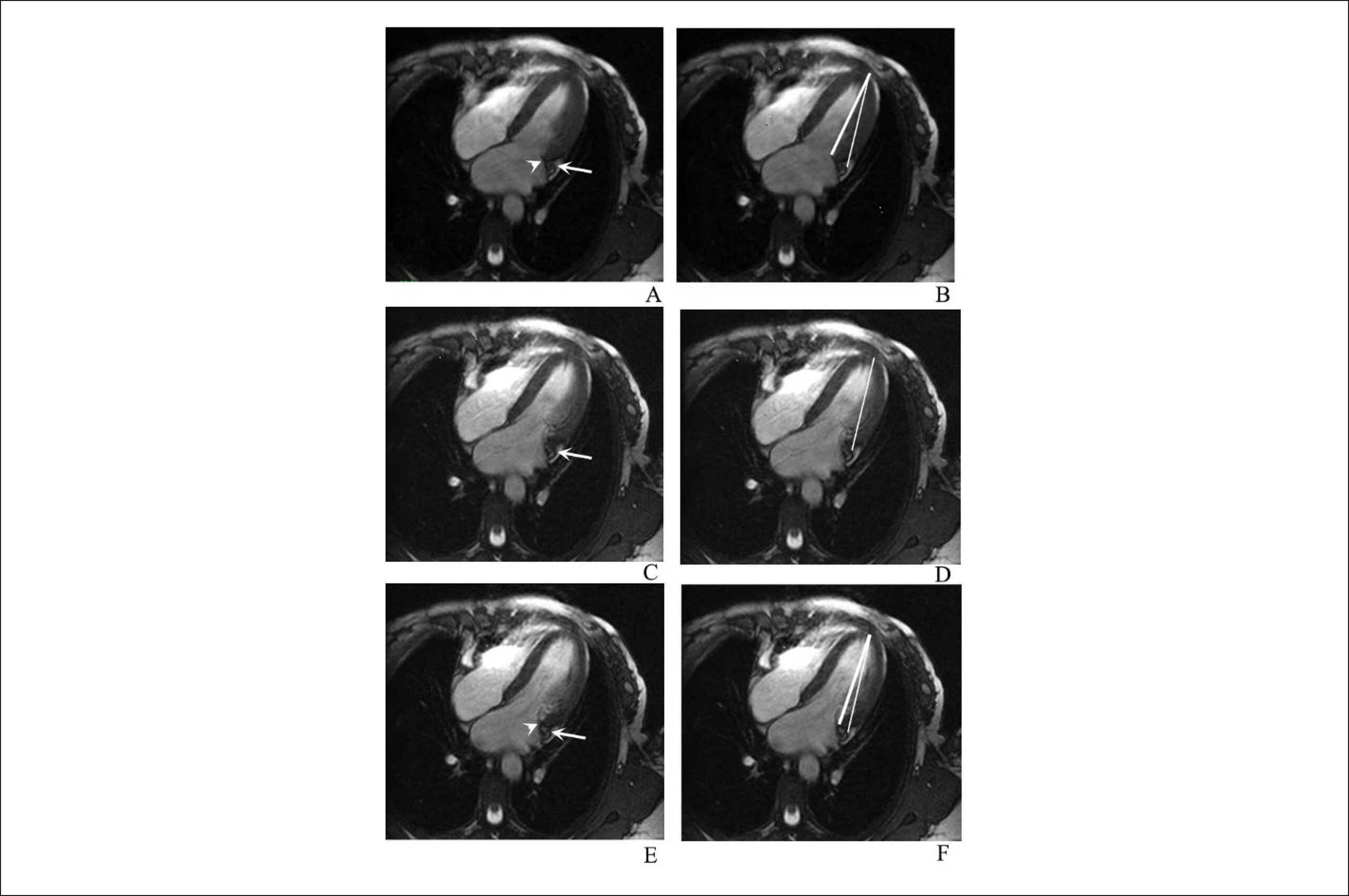
We compared 26 AVD patients with 19 normal controls. Diastolic function was evaluated as LV longitudinal displacement in 4-chamber view cine-CMR images using steady state free precession (SSFP) sequence during the entire cardiac cycle with temporal resolution < 50 ms. The resulting plot of atrioventricular junction (AVJ) position versus time generated variables of AVJ motion. Significance level of p < 0.05 was used.

Results:

Maximum longitudinal displacement (0.12 vs. 0.17 cm), maximum velocity during early diastole (MVED, 0.6 vs. 1.4s-1), slope of the best-fit line of displacement in diastasis (VDS, 0.22 vs. 0.03s-1), and VDS/MVED ratio (0.35 vs. 0.02) were significantly reduced in AVD patients compared with controls, respectively. Aortic regurgitation showed significantly worse longitudinal LV shortening compared with aortic stenosis. Higher LV mass indicated worse diastolic dysfunction.

Conclusions:

A simple linear measurement detected significant differences on LV diastolic function between AVD patients and controls. LV mass was the only independent predictor of diastolic dysfunction in these patients. This method can help in the evaluation of diastolic dysfunction, improving cardiomyopathy detection by CMR, without prolonging exam time or depending on expensive software.

  • Aortic Valve Insufficiency
  • Cardiomyopathy, Hypertrophic / complications
  • Cardiovascular Diseases, mortality
  • Diagnostic Imaging
  • Echocardiography
  • Heart Failure
  • Magnetic Resonance Spectroscopy
Read Full Article
Creative Commons Attribution (CC BY) Creative Commons license
23/09/2019 DOI: 10.5935/abc.20190193 Views 1
Figure 1
ABC
Original Article

Prevention of Sudden Cardiac Death in Hypertrophic Cardiomyopathy: What has Changed in The Guidelines?

Liliana Reis et al. Rogerio Teixeira Andreia Fernandes Inês Almeida … Marta Madeira Joana Silva Ana Botelho João Pais José Nascimento Lino Gonçalves

Abstract

Background:

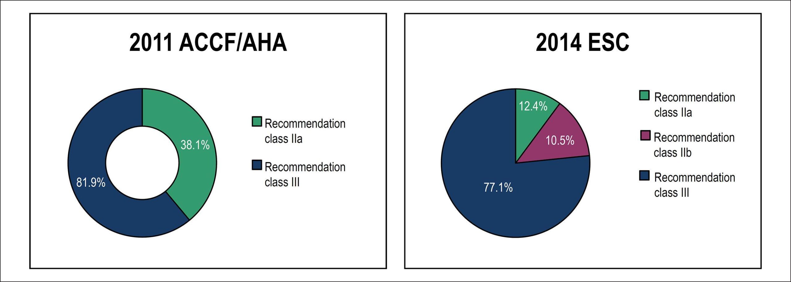
The new European Society of Cardiology guidelines for hypertrophic cardiomyopathy (HCM) define the estimation of sudden cardiac death (SCD) risk as an integral part of clinical management. An implantable cardioverter defibrillator (ICD) is recommended (class IIa) when the risk is ≥ 6%.

Objectives:

To compare the SCD risk stratification according to the 2011 and 2014 recommendations for ICD implantation in patients with HCM.

Methods:

Retrospective study including 105 patients diagnosed with HCM. The indication for ICD was assessed using the 2011 and 2014 guidelines. Statistical analysis was performed using SPSS software version 19.0.0.2®. The tests performed were bilateral, considering the significance level of 5% (p < 0.05).

Results:

Regarding primary prevention, according to the 2011 ACCF/AHA recommendations, 39.0% of the patients had indication for ICD implantation (level of evidence IIa). Using the 2014 guidelines, only 12.4% of the patients had an indication for ICD implantation. Comparing the two risk stratification models for patients with HCM, we detected a significant reduction in the number of indications for ICD implantation (p < 0.001). Of the 41 patients classified as IIa according to the 2011 recommendations, 68.3% received a different classification according to the 2014 guidelines.

Conclusion:

Significant differences were found when comparing the SCD risk stratification for ICD implantation in the two guidelines. The current SCD risk score seems to identify many low-risk patients who are not candidates for ICD implantation. The use of this new score results in a significant reduction in the number of ICD implanted.

  • Cardiomyopathy, Hypertrophic / complications
  • Death, Sudden, Cardiac/prevention & control
  • Defibrillators, Implantable / trends
  • Diagnostic Imaging
  • Syncope
Read Full Article
Creative Commons Attribution (CC BY) Creative Commons license
01/06/2018 DOI: 10.5935/abc.20180099 Views 1

How to cite this article

EndNote RIS

Electronic Document Format (Vancouver)

Oliveira I, Cruz I, Ana Neto , Bragança B, Abreu G, Azevedo J, Andrade A, et al. Mitral Valve Perforated Aneurysm: An Issue of Inflammation or Pressure Gradients?. Arquivos Brasileiros de Cardiologia 2022;119(5):809-12.

Electronic Document Format (ABNT)

Oliveira, Inês; Cruz, Isabel; Ana Neto, ; Bragança, Bruno; Abreu, Glória; Azevedo, João; Andrade, Aurora. Mitral Valve Perforated Aneurysm: An Issue of Inflammation or Pressure Gradients?. Arquivos Brasileiros de Cardiologia, v. 119, n. 5, p. 809-812, Sep. 2022.

Electronic Document Format (APA)

Oliveira, I., Cruz, I., Ana Neto, , Bragança, B., Abreu, G., Azevedo, J., & Andrade, A. (2022). Mitral Valve Perforated Aneurysm: An Issue of Inflammation or Pressure Gradients?. Arquivos Brasileiros de Cardiologia, 119(5), 809-812.

Electronic Document Format (ISO)

Oliveira, Inês and Cruz, Isabel and Ana Neto and Bragança, Bruno and Abreu, Glória and Azevedo, João and Andrade, Aurora. Mitral Valve Perforated Aneurysm: An Issue of Inflammation or Pressure Gradients?. Arquivos Brasileiros de Cardiologia [online]. 2022, vol. 119, n. 5, [cited 2026-10-14], pp. 809-812. Available from: <https://journals.cardiol.br/en/article/mitral-valve-perforated-aneurysm-an-issue-of-inflammation-or-pressure-gradients/>. ISSN 0066-782X

EndNote RIS

Electronic Document Format (Vancouver)

Ribeiro SM, Azevedo Filho CF, Sampaio R, Tarasoutchi F, Grinberg M, Kalil-Filho R, Rochitte CE, et al. Longitudinal Shortening of the Left Ventricle by Cine-CMR for Assessment of Diastolic Function in Patients with Aortic Valve Disease. Arquivos Brasileiros de Cardiologia 2019;114(2):284-92.

Electronic Document Format (ABNT)

Ribeiro, Sergio Marrone; Azevedo Filho, Clerio Francisco de; Sampaio, Roney; Tarasoutchi, Flávio; Grinberg, Max; Kalil-Filho, Roberto; Rochitte, Carlos Eduardo. Longitudinal Shortening of the Left Ventricle by Cine-CMR for Assessment of Diastolic Function in Patients with Aortic Valve Disease. Arquivos Brasileiros de Cardiologia, v. 114, n. 2, p. 284-292, Sep. 2019.

Electronic Document Format (APA)

Ribeiro, S. M., Azevedo Filho, C. F. , Sampaio, R., Tarasoutchi, F., Grinberg, M., Kalil-Filho, R., & Rochitte, C. E. (2019). Longitudinal Shortening of the Left Ventricle by Cine-CMR for Assessment of Diastolic Function in Patients with Aortic Valve Disease. Arquivos Brasileiros de Cardiologia, 114(2), 284-292.

Electronic Document Format (ISO)

Ribeiro, Sergio Marrone and Azevedo Filho, Clerio Francisco de and Sampaio, Roney and Tarasoutchi, Flávio and Grinberg, Max and Kalil-Filho, Roberto and Rochitte, Carlos Eduardo. Longitudinal Shortening of the Left Ventricle by Cine-CMR for Assessment of Diastolic Function in Patients with Aortic Valve Disease. Arquivos Brasileiros de Cardiologia [online]. 2019, vol. 114, n. 2, [cited 2026-10-14], pp. 284-292. Available from: <https://journals.cardiol.br/en/article/longitudinal-shortening-of-the-left-ventricle-by-cine-cmr-for-assessment-of-diastolic-function-in-patients-with-aortic-valve-disease/>. ISSN 0066-782X

EndNote RIS

Electronic Document Format (Vancouver)

Reis L, Teixeira R, Fernandes A, Almeida I, Madeira M, Silva J, Botelho A, et al. Prevention of Sudden Cardiac Death in Hypertrophic Cardiomyopathy: What has Changed in The Guidelines?. Arquivos Brasileiros de Cardiologia 2018;110(6):524-31.

Electronic Document Format (ABNT)

Reis, Liliana; Teixeira, Rogerio; Fernandes, Andreia; Almeida, Inês; Madeira, Marta; Silva, Joana; Botelho, Ana; Pais, João; Nascimento, José; Gonçalves, Lino. Prevention of Sudden Cardiac Death in Hypertrophic Cardiomyopathy: What has Changed in The Guidelines?. Arquivos Brasileiros de Cardiologia, v. 110, n. 6, p. 524-531, May. 2018.

Electronic Document Format (APA)

Reis, L., Teixeira, R., Fernandes, A., Almeida, I., Madeira, M., Silva, J., Botelho, A., Pais, J., Nascimento, J., & Gonçalves, L. (2018). Prevention of Sudden Cardiac Death in Hypertrophic Cardiomyopathy: What has Changed in The Guidelines?. Arquivos Brasileiros de Cardiologia, 110(6), 524-531.

Electronic Document Format (ISO)

Reis, Liliana and Teixeira, Rogerio and Fernandes, Andreia and Almeida, Inês and Madeira, Marta and Silva, Joana and Botelho, Ana and Pais, João and Nascimento, José and Gonçalves, Lino. Prevention of Sudden Cardiac Death in Hypertrophic Cardiomyopathy: What has Changed in The Guidelines?. Arquivos Brasileiros de Cardiologia [online]. 2018, vol. 110, n. 6, [cited 2026-10-14], pp. 524-531. Available from: <https://journals.cardiol.br/en/article/prevention-of-sudden-cardiac-death-in-hypertrophic-cardiomyopathy-what-has-changed-in-the-guidelines/>. ISSN 0066-782X

  • About ABC Family
  • Internal Regulation
  • Editorial Board
  • Brazilian Society of Cardiology (SBC)

Issues

SBC Guidelines

  • Videos
  • Podcasts
  • News