ABC Family Arquivos Brasileiros de Cardiologia International Journal of Cardiovascular Sciences ABC Heart Failure & Cardiomyopathy ABC Imagem Cardiovascular
Português English
ABC Family
Português English
  • About
    • About ABC Family
    • Internal Regulation
    • Editorial Board
    • Brazilian Society of Cardiology (SBC)
  • Issues
  • SBC Guidelines
  • Content
    • Videos
    • Podcasts
    • News
  • About
    • About ABC Family
    • Internal Regulation
    • Editorial Board
    • Brazilian Society of Cardiology (SBC)
  • Issues
  • SBC Guidelines
  • Content
    • Videos
    • Podcasts
    • News

ABC Family

  • ABC Family
  • Arquivos Brasileiros de Cardiologia
  • International Journal of Cardiovascular Sciences
  • ABC Heart Failure & Cardiomyopathy
  • ABC Imagem Cardiovascular
Search in

No content found.

Journals
Ordering
Article types
Others

  1. Home
  2. Keyword: Heart Neoplasms
Figure 1
ABCIC
Case Report

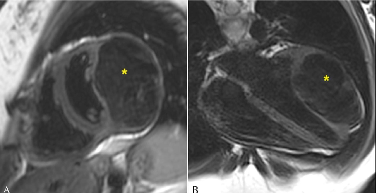
Intramyocardial Cardiac Tumor: A Case Report

Luisa Latado et al. Fabio Figueiredo Costa Carlos Frederico Lopes Benevides Jorge Andion Torreão … Adna Keyne Lima Adelina Sanches de Melo Adriana Lopes Latado

Introduction

Primary cardiac tumors are rare entities, approximately 75% of which are benign, with myxoma as the most common type., Only about 15% of primary cardiac tumors are malignant, predominantly sarcomas. In contrast, metastatic cardiac involvement is relatively frequent and has been identified in up to 9% of patients who died from cancer.

Clinical manifestations are more closely related to tumor location than to histopathological classification. They may result from embolic phenomena, intracardiac obstruction with signs of heart failure (HF), valvular involvement, myocardial invasion with alterations in contractile function and electrical conduction, and pericardial involvement with the risk of effusion and cardiac tamponade, among other presentations.

[…]
  • Cardiovascular Diseases
  • Case Reports
  • Heart Neoplasms
Read Full Article
Creative Commons Attribution (CC BY) Creative Commons license
03/02/2026 DOI: 10.36660/abcimg.20250051i
Figure 1
ABCIC
Case Report

Turmoil of Symptoms: The Devastating Impact of Cardiac Lymphoma – Multiple Clinical Manifestations and the Lethal Causes of Cardiac Involvement

Gustavo Carvalho et al. Maria Fernanda Miranda Carvalho Tiago Magalhães Alexandre Manoel Varela … Kamila Fernanda Staszko Mariana Rie Hayashida

Abstract

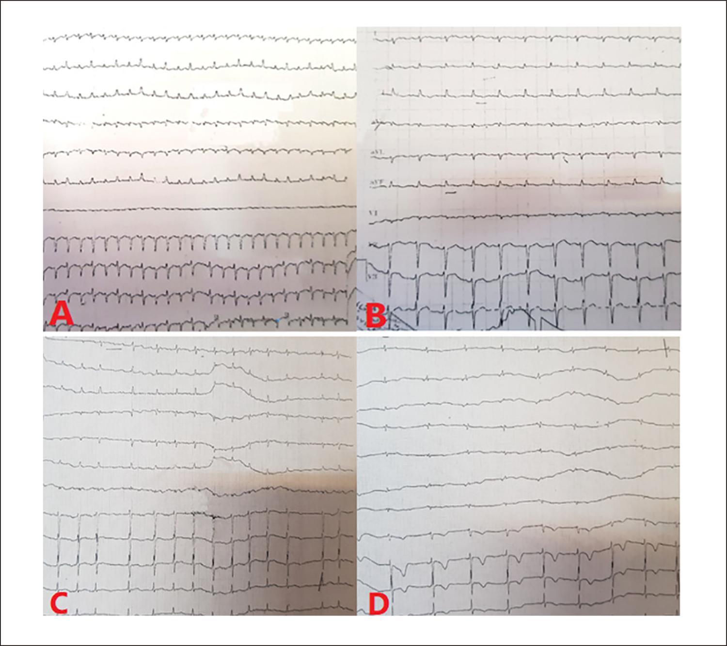
Primary cardiac lymphoma (PCL) is a rare and highly lethal neoplasm, whose nonspecific clinical presentation makes early diagnosis difficult. This article reports the case of an elderly patient, previously healthy, who experienced dizziness and syncope for four months before the discovery of a large tumor mass in the heart. Imaging exams, such as echocardiography and magnetic resonance imaging, were crucial for tumor identification, and the biopsy confirmed it to be a B-cell lymphoma with high replication. However, given the severity of the condition and the family’s refusal to proceed with treatment, the patient progressed to sudden death two weeks after diagnosis. Primary cardiac tumors (PCTs) are underdiagnosed, with varied symptoms that may include vascular obstructions, arrhythmias, and heart failure. PCLs account for only 1% to 2% of primary cardiac masses and are more common in elderly men. The diagnosis is challenging due to the absence of specific signs, making imaging exams and biopsies essential for disease confirmation. This case illustrates the complexity of managing PCLs, whose rapid progression limits therapeutic options. The lack of specific symptoms and silent progression highlight the importance of early diagnosis. The high mortality rate reinforces the need for greater medical attention to this condition, which frequently leads to fatal outcomes before an effective intervention can be implemented.

  • Biopsy
  • Cardiac Arrhythmias
  • Echocardiography
  • Heart Neoplasms
Read Full Article
Creative Commons Attribution (CC BY) Creative Commons license
15/08/2025 DOI: 10.36660/abcimg.20250033i Views 3
Figure 1
ABC
Research Letter

Primary Pericardial Angiosarcoma Complicated by Constrictive Pericarditis

Guilherme Augusto Reissig Pereira et al. Maiane Maria Pauletto Anna Paula Tscheika Fabrício Michalski Velho Luciano Folador

Introduction

Secondary malignant cardiac tumors are metastases from primary tumors and 22 to 132 times more common than primary cardiac tumors (PCTs).– PCTs are rare, with an estimated autopsy prevalence of 0.001-0.03%, and mostly (90%) benign.,

Among PCTs, cardiac angiosarcoma is a rare subtype of soft tissue sarcoma and the most common primary malignant tumor of the heart. Angiosarcomas consist of irregularly shaped vascular channels lined by anaplastic epithelial cells with substantial areas of necrosis and hemorrhage. Cardiac angiosarcoma is an aggressive tumor with a high rate of local recurrence and systemic dissemination. Without surgical resection, it has a mean overall survival of 3.8 (SD 2.5) months. The rapid infiltrating growth of this tumor can lead to emergency clinical presentations including myocardial rupture, pericardial effusions, cardiac tamponade, and constrictive pericarditis (CP).

[…]

  • Angiosarcoma
  • cardiac tumor
  • Constrictive pericarditis
  • Heart Neoplasms
  • Primary Cardiac Angiosarcoma
Read Full Article
Creative Commons Attribution (CC BY) Creative Commons license
09/05/2025 DOI: 10.36660/abc.20240683i Views 2
Figure 1
ABCIC
Case Report

A Common Primary Cardiac Tumor With an Uncommon Presentation

Juan Felipe Vasquez-Rodriguez et al. Carolina Paola Idrovo-Turbay Jaime Alberto Rodríguez Lorena Montes Villalobos

Abstract

Background

Cardiac tumors are rare, and most are secondary (metastatic) lesions. Primary tumors, though uncommon, are typically benign, with myxomas being the most frequent and occasionally displaying unique features. Diagnosis often requires multimodality imaging.

Case summary

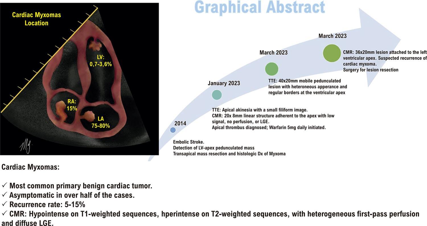
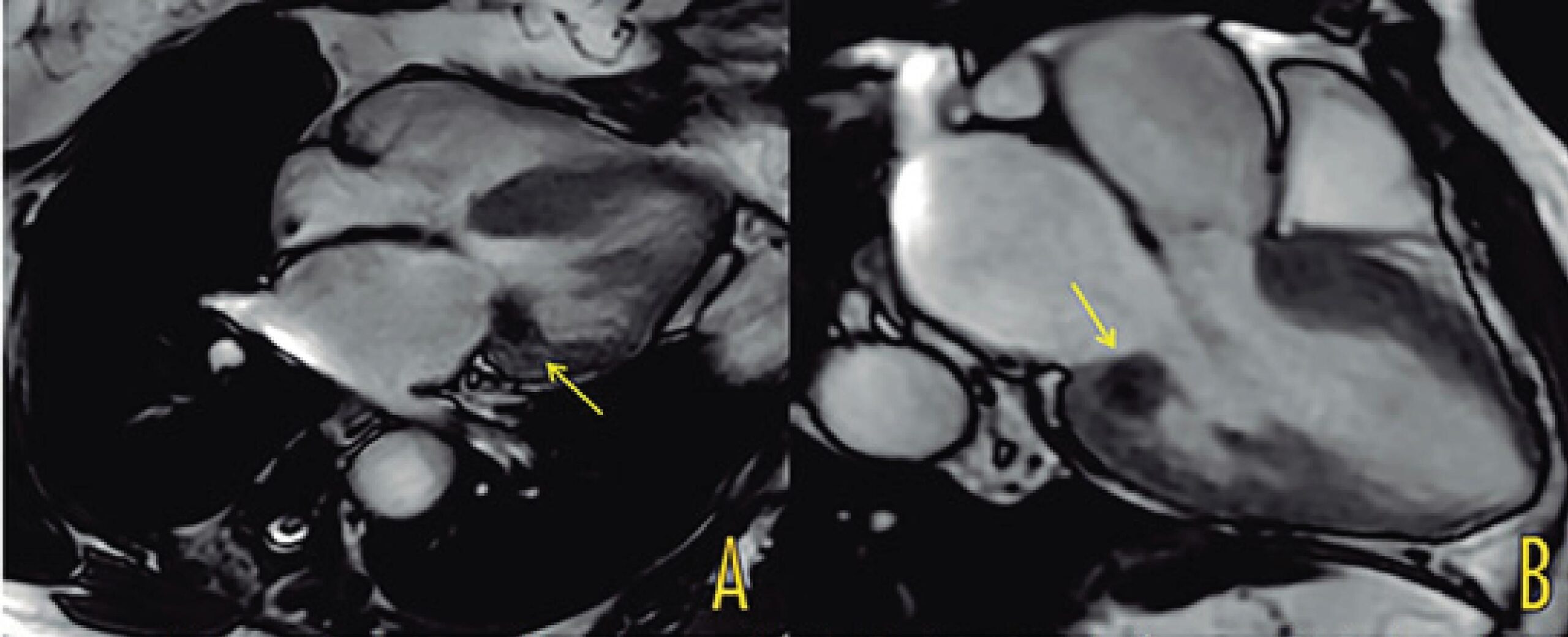
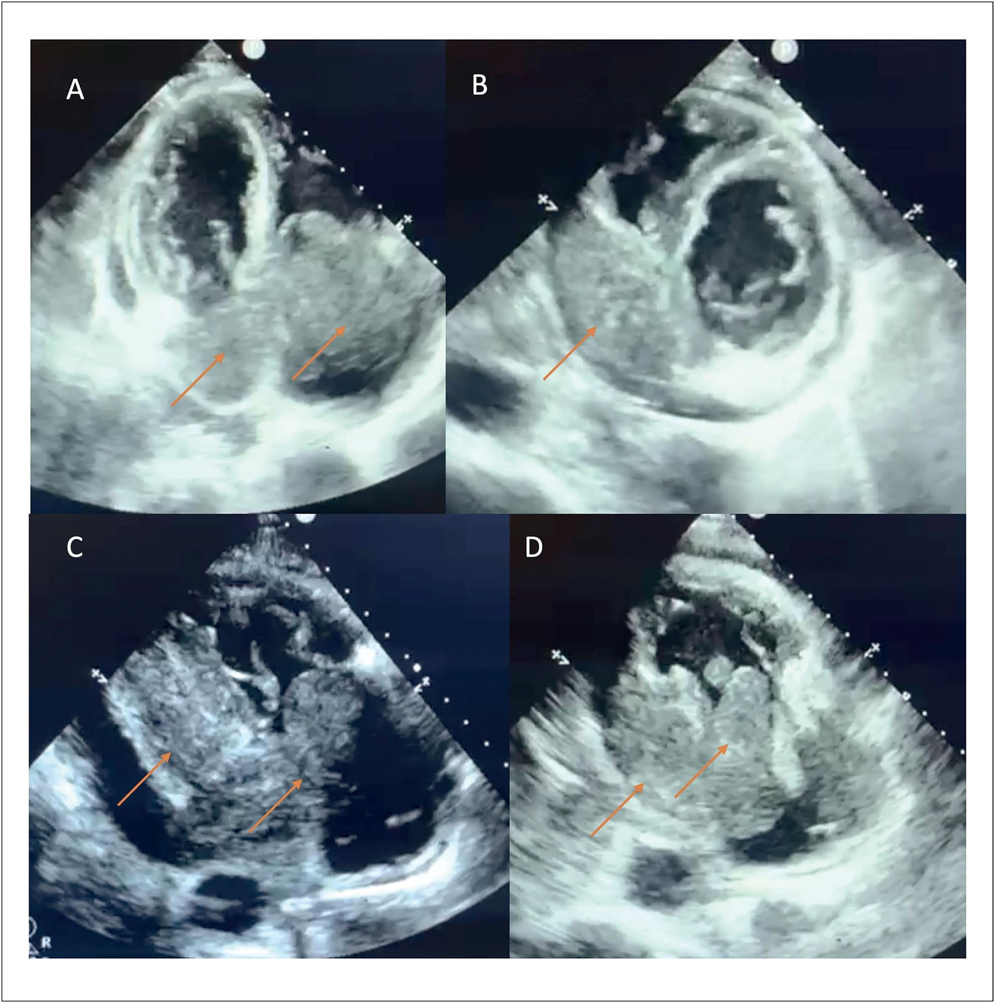
A 41-year-old man with a history of ischemic stroke and resection of an apical left ventricular myxoma presented for evaluation after a follow-up transthoracic echocardiogram (TTE) revealed a mobile, ovoid apical lesion with high embolic risk, despite being asymptomatic. Cardiac magnetic resonance (CMR) imaging suggested recurrence of the ventricular myxoma. Surgical resection was performed, and histopathology confirmed the diagnosis of cardiac myxoma (CM).

Discussion

CMs are the most common primary benign cardiac tumors, typically located in the left atrium and attached to the interatrial septum. They are less frequently found in the ventricular cavities. About half of cases are asymptomatic, and recurrence is possible after resection. This case highlights a rare instance of late recurrence in an atypical location.

  • Echocardiography
  • Heart Neoplasms
  • Magnetic Resonance Imaging
  • Myxoma
Read Full Article
Creative Commons Attribution (CC BY) Creative Commons license
27/03/2025 DOI: 10.36660/abcimg.20240123i Views 1
Figure 1
ABC
Case Report

Cardiac Lymphoma: A Case Report

Leticia Ferraz Pamplona et al. Kelly Barnabé Serpa de Oliveira João Ancelmo dos Reis Neto Luis Antelo Bustamante … Januário Manoel de Souza Flávia Fernandes Silva Zacchi Juliana Brenande de Oliveira Brito Salomon Soriano Ordinola Rojas

Abstract

Cardiac tumors are rare. Primary cardiac lymphoma is defined as non-Hodgkin lymphoma that involves only the heart and/or pericardium. It is an aggressive tumor with a poor prognosis and its symptoms may be nonspecific. A definitive diagnosis can only be obtained through histopathological study, and chemotherapy is the main treatment strategy.

This case is about a 71-year-old male patient, with a rare form of primary cardiac tumor. He had postoperative complications such as acute pulmonary edema and the need for a pacemaker, which occurred after tumor resection requiring reconstruction of the vena cava and suture of the right atrium.

Primary cardiac lymphoma is a tumor that is difficult to diagnose because it has a vague and non-specific symptomatic presentation. However, it should always be included as a differential diagnosis of cardiac masses. Early diagnosis can significantly improve the prognosis and increase the survival rate of patients by quickly referring them for specific treatment.

  • Heart
  • Heart Neoplasms
  • Lymphoma
Read Full Article
Creative Commons Attribution (CC BY) Creative Commons license
24/03/2025 DOI: 10.36660/abc.20240299i
Figure 1
ABCIC
Case Report

Atrial Myxoma as a Rare Cause of Stroke: Case Report

Maurício Ângelo de Almeida Junior et al. Ana Flavia Parreira de Morais Jamil Alli Murad Junior

Introduction

Atrial myxoma (AM) is the most common primary benign tumor affecting the heart. Its prevalence is rare, estimated at 0.03% in the general population. Surgical intervention should be introduced as soon as diagnosed due to the increased risk of cardiac complications, such as intracardiac obstructions or systemic embolizations.

Cardiac myxomas are commonly observed in the left atrium in >75% of cases, predominantly attached with a stalk to the fossa ovalis (>90%). However, they can be seen in other locations (left ventricle, right atrium, and, though quite rarely, the right ventricle).

[…]

  • Heart Neoplasms
  • Myxoma
  • Stroke
Read Full Article
Creative Commons Attribution (CC BY) Creative Commons license
30/01/2025 DOI: 10.36660/abcimg.20240099i Views 1
Figura 1
ABCIC
Case Report

Not Everything is as it Seems at First Glance: Multimodal Imaging in Heart Cyst Evaluation

Carlos Guaman-Valdivieso et al. Julia Aramburu Natalia Lluberas Natalia Huart Gabriel Parma

Abstract

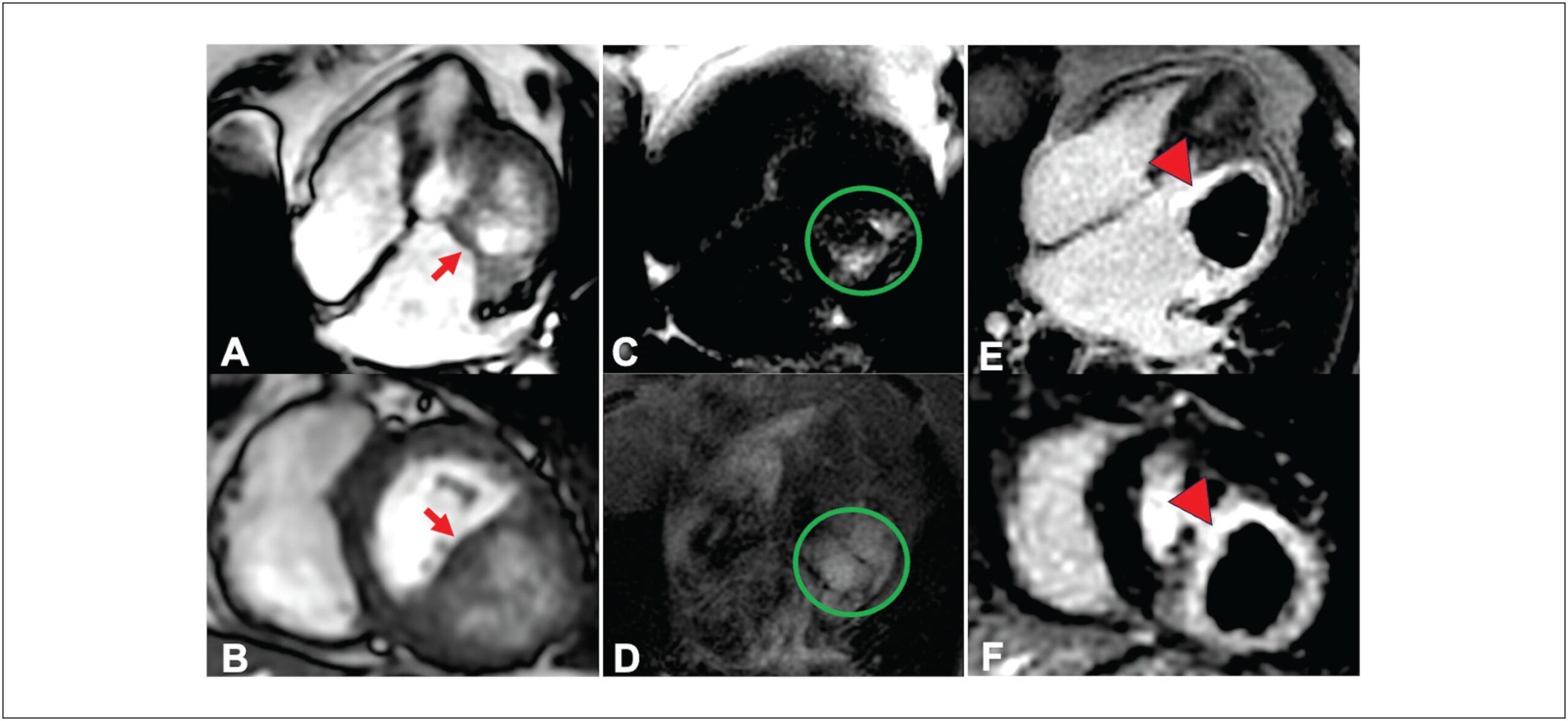
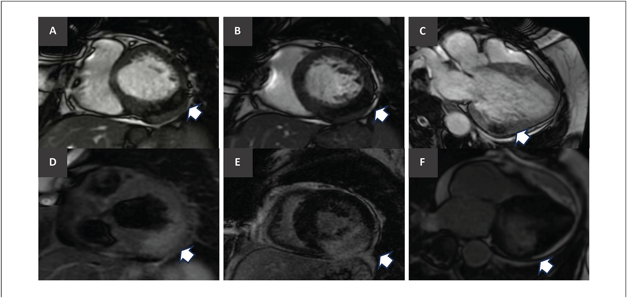
Cardiac masses, though rare, can lead to significant hemodynamic and arrhythmic issues, even if benign. Improved multimodal imaging has refined the diagnosis and understanding of these masses. We present an 85-year-old woman with New York Heart Association class II-III dyspnea for six months. Echocardiography showed a mass in the basal inferolateral wall of the left ventricle, along with severe left atrial dilation and moderate mitral regurgitation. Initially thought to be a pericardial cyst, further cardiac magnetic resonance (CMR) imaging identified an intramyocardial mass, and computed tomography (CT) suggested a hydatid cyst. Given the non-limiting symptoms and high surgical risk, the decision was made for expectant management. This case illustrates the rarity of cardiac cysts and the essential role of multimodal imaging in diagnosis and treatment planning.

  • Cardiac Imaging Techniques
  • Echinococcosis
  • Heart Neoplasms
Read Full Article
Creative Commons Attribution (CC BY) Creative Commons license
05/12/2024 DOI: 10.36660/abcimg.20240086i Views 1
Figure 1
ABCIC
Review Article

My Approach to Cardiovascular Computed Tomography and Magnetic Resonance Imaging in the Evaluation of Cardiac Pseudotumors: A Brief Review

Roberto Vitor Almeida Torres et al. Bruno Maeda Fuzissima Eduardo Kaiser Ururahy Nunes Fonseca Lucas de Pádua Gomes de Farias … Walther Yoshiharu Ishikawa José de Arimatéia Batista Araújo Filho

Abstract

Cardiac pseudotumoral lesions are non-neoplastic conditions that are often overlooked in the differential diagnosis of cardiac masses. They present a variable clinical picture, ranging from asymptomatic to causing complications such as ventricular filling restriction and outflow tract obstruction. Echocardiography is the first-line imaging method but has limitations, such as dependence on the acoustic window and operator variability. However, a multimodality approach, including CT and MRI, is essential for seeking an accurate diagnosis. CT, with its excellent spatial resolution, allows for anatomical detailing, assessment of intralesional calcifications and fat, and contributes to therapeutic planning. MRI is preferred for tissue characterization and differentiation between benign and malignant lesions. Normal anatomical structures, such as the Eustachian valve and Chiari network, can be confused with thrombi or tumors, requiring correct identification. Thrombi are common in patients with atrial fibrillation or mitral valve disease, with MRI being important for differentiating them from neoplasms. Other pseudotumoral conditions include vegetation, gossypibomas, caseous calcification of the mitral valve annulus, and lipomatous hypertrophy of the interatrial septum. The integration of advanced cardiovascular imaging modalities is fundamental for the diagnosis and management of these lesions, optimizing patient care.

  • Emission-Computed Tomography
  • Heart Neoplasms
  • Magnetic Resonance Spectroscopy
Read Full Article
Creative Commons Attribution (CC BY) Creative Commons license
29/08/2024 DOI: 10.36660/abcimg.20240044i Views 1
ABCHF
Image

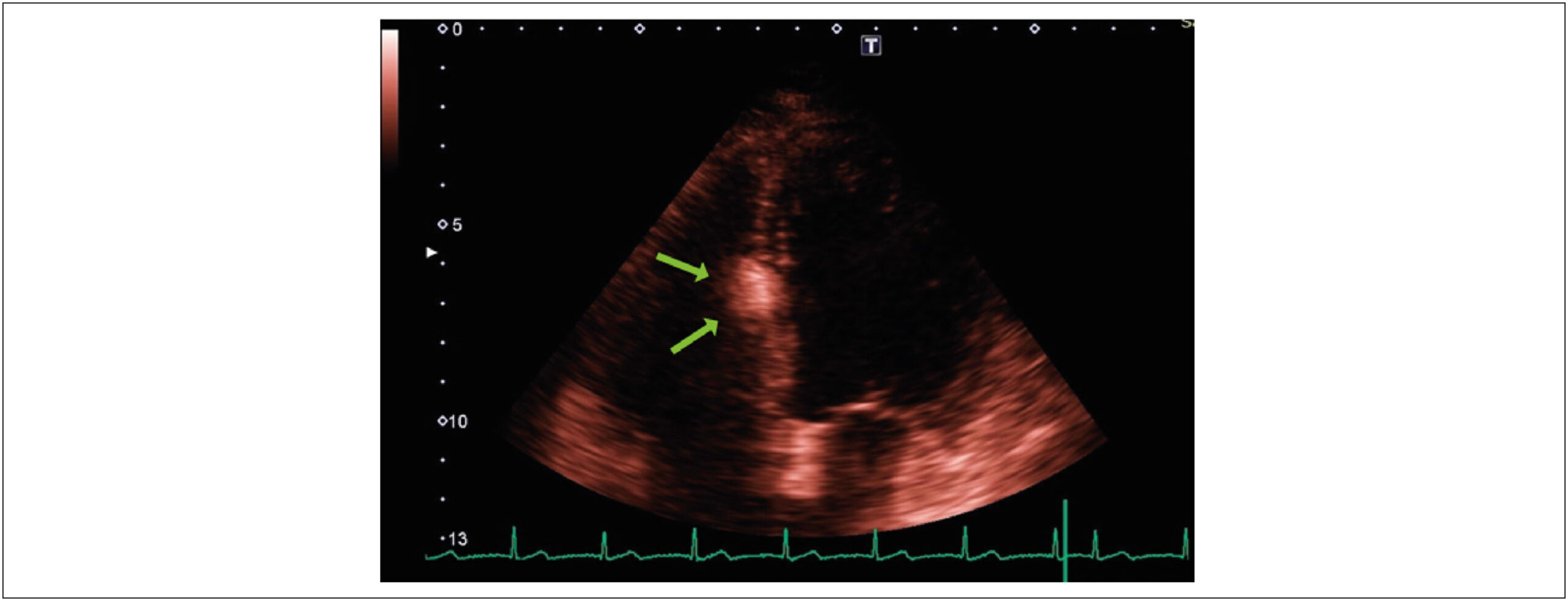
Cardiac Tumor in the Left Atrium

Henrique Iahnke Garbin et al. Matheus Piccoli Machado Klauberg Géris Mazzutti

A

[…]

  • Heart Atria
  • Heart Failure
  • Heart Neoplasms
  • Ultrasonography
Read Full Article
Creative Commons Attribution (CC BY) Creative Commons license
07/08/2024 DOI: 10.36660/abchf.20240004i
ABCIC
Review Article

Cardiac Tumors: Recognition Is Essential!

Eliza de Almeida Gripp et al. Bruna Zanforlin Jácome Anna Esther Araujo e Silva Fernanda Baptista Ribeiro Dias … Bruna Barbosa Chagas de Almeida Arnaldo Rabischoffsky

Abstract

Although cardiac tumors are rare and often found accidentally, they can be associated with constitutional, cardiac, or embolic symptoms and, thus, must always be considered in diagnostic hypotheses. Three-dimensional Doppler echocardiography and contrast enhancement are additional diagnostic resources and can help determine the most appropriate surgical approach, when necessary.

  • Echocardiography
  • Heart Neoplasms
  • Three-Dimensional Echocardiography
Read Full Article
Creative Commons Attribution (CC BY) Creative Commons license
26/06/2024 DOI: 10.36660/abcimg.20240032i Views 1
Perinatal Results and Long-Term Follow-Up of Fetal Cardiac Tumors: A 30-Year Historical Cohort Study
ABC
Original Article

Perinatal Results and Long-Term Follow-Up of Fetal Cardiac Tumors: A 30-Year Historical Cohort Study

Fabricio Marcondes Camargo et al. Maria de Lourdes Brizot Rossana Pulcineli Vieira Francisco Werther Brunow de Carvalho … Nana Miura Ikari Stella Verzinhasse Peres Marco Antônio Borges Lopes Lilian Maria Lopes

Abstract

Background:

This was a 30-year retrospective cohort study that approximates closely to the natural history of cardiac tumors diagnosed in the fetus, since there was no case of pregnancy interruption

Objective:

To assess morbidity and mortality in the perinatal period and at long term in fetuses diagnosed with cardiac tumor. Our secondary objective was to assess the evaluating factors of perinatal and postnatal results.

Methods:

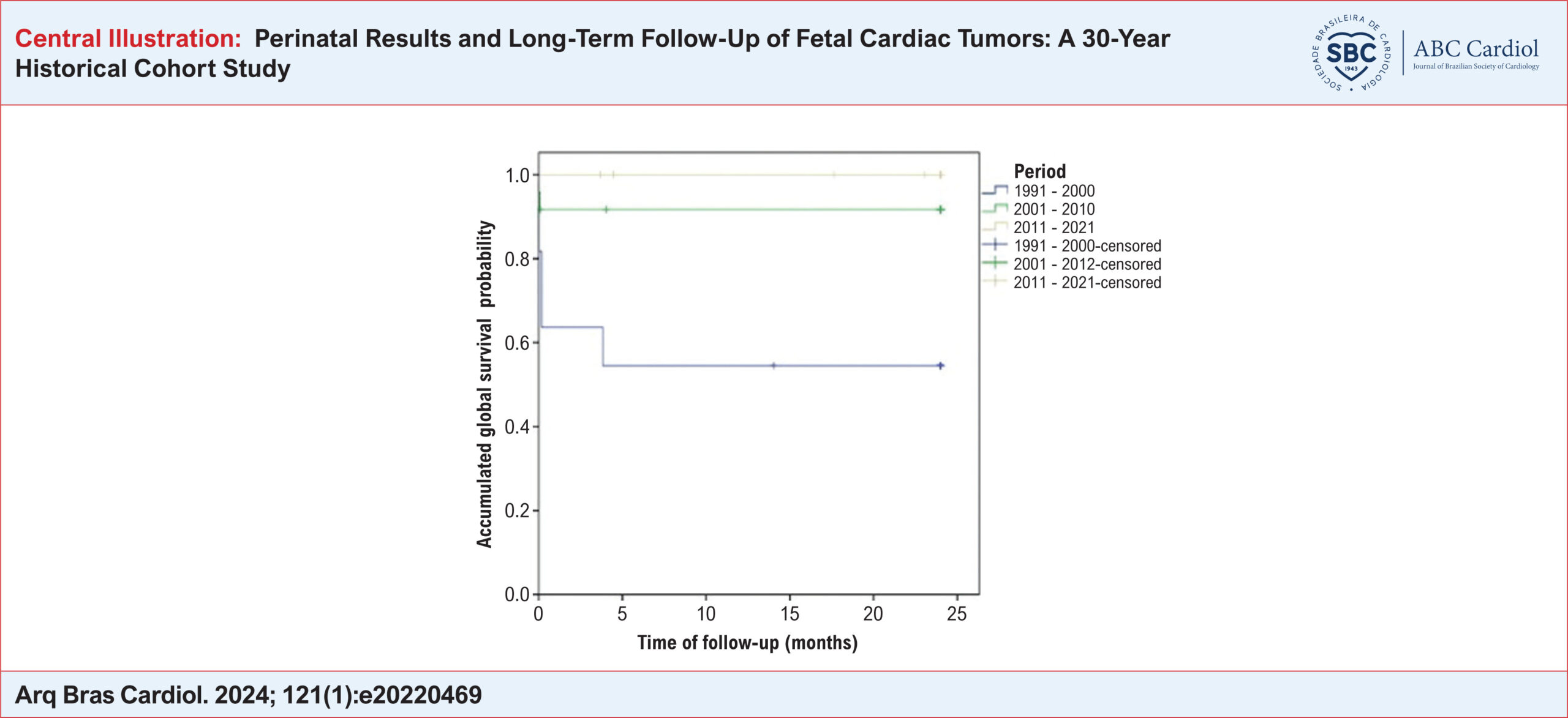
This was a retrospective cohort study with 74 pregnant women with an echocardiographic diagnosis of fetal cardiac tumor at two referral centers between May 1991 and November 2021. A descriptive analysis was performed, and data were expressed as absolute (n) and relative (%) frequencies, median and interquartile range. Fisher’s exact test was used to evaluate the association of echocardiographic characteristics and clinical manifestations with perinatal and postnatal results. Global survival was calculated using the Kaplan-Meier method and the curves were compared by the log-rank test. The time of follow-up, calculated in months, corresponded to the time elapsed from hospital discharge to current status (survived/ censoring or death). The level of significance was set at 5% (p<0.05).

Results:

Rhabdomyoma is the most common type of cardiac tumor (85%), with a high morbidity (79.3%) and overall mortality of 17.4%. The presence of fetal hydrops was a predictor of death.

Conclusion:

The presence of fetal hydrops had an impact on mortality, and hence is an important factor in counselling and determining the prognosis. Most deaths occurred before hospital discharge.

  • Echocardiography
  • Heart Neoplasms
  • Prenatal Diagnosis
  • Rhabdomyoma
Read Full Article
Creative Commons Attribution (CC BY) Creative Commons license
22/03/2024 DOI: 10.36660/abc.20220469 Views 2
ABCIC
Case Report

The Importance of Imaging Multimodality in the Diagnosis of a Rare Case of Papillary Fibroelastoma in the Left Ventricular Apex

Marcio Mendes Pereira et al. Vinícius José da Silva Nina José Xavier de Melo Filho Rodrigo de Jesus Louzeiro Melo … Marco Túlio Hercos Juliano Luma Sayonara Martins Pereira

Introduction

Papillary fibroelastoma (PF) is a rare, benign primary cardiac neoplasm, initially described as an incidental finding in autopsies or surgeries, and ranks third among the most common cardiac tumors. It mainly affects the heart valves, accounting for 75% of all tumors in this region and is often diagnosed incidentally.

Its clinical presentation can vary widely, ranging from asymptomatic cases to situations in which systemic embolization occurs, caused by adherent thrombi as well as by fragmentation of the tumor itself. Currently, PF is no longer a tumor diagnosed only in autopsies and can now detected with new cardiovascular imaging modalities, thus enabling early treatment and preventing complications.

[…]

  • Heart Neoplasms
  • Transient Ischemic Attack
Read Full Article
Creative Commons Attribution (CC BY) Creative Commons license
20/12/2023 DOI: 10.36660/abcimg.20230102i Views 2
Figure 1
ABC
Research Letter

Misdiagnosis of a Cardiac Angiosarcoma During the COVID-19 Pandemic

Arash Amin et al. Zohreh Taheri Mahshid Hesami Azin Alizahehasl … Zeinab Norouzi Nasibeh Mohammadi

Abstract

The final months of 2019 saw the emergence of a new pandemic termed “COVID-19”. Since then, this infection and its complications have been the priority of healthcare providers, with many symptoms attributed to its early and late presentations. Thus far, other diseases, even fatal situations, have been overlooked or misdiagnosed due to the attribution of patient symptoms to the presence of COVID-19 infection. We herein present a case of cardiac angiosarcoma in a young boy who had previously become infected with COVID-19 about two months earlier. Given the history of infection, the initial approach was post-COVID-19 myopericarditis management. However, the patient’s condition worsened, necessitating reevaluation via multimodalities with higher precision. Ultimately, the patient was diagnosed with a cardiac tumor. This article seeks to underscore the significance of taking heed of other diseases and fatal conditions during the COVID-19 pandemic with an emphasis on avoiding misdiagnosing other diseases.

  • Angiosarcoma/surgery
  • COVID-19/infection
  • Diagnostic Errors
  • Heart Neoplasms
  • Imaging/methods
  • Multimodal Imaging/diagnostic
Read Full Article
Creative Commons Attribution (CC BY) Creative Commons license
07/08/2023 DOI: 10.36660/abc.20220501
Figure 1
ABC
Research Letter

Intracardiac Metastasis of Colonic Adenocarcinoma 12 Years After Primary Tumor Control and Without Any Sign of Other Metastasis: A Case Report

Isabela Galizzi Faé et al. Gabriela Zamunaro Lopes Ruiz Gustavo Palmer Irffi Robson de Souza Almeida Junior … Pedro Anjos Conceição Eduardo Belisario Falchetto Luiz Guilherme Passaglia Geraldo Brasileiro Filho Cláudio Leo Gelape Clara Rodrigues Alves de Oliveira

Introduction

Thrombus, vegetation and tumor are the main diagnoses for intracardiac masses. Malignant cardiac tumors are rare and cardiac metastasis are around 20 times more frequent than primary tumors. Cardiac metastases originate from lymphatic or blood dissemination, mediastinum direct invasion or tumoral growth inside inferior vena cava or pulmonary veins– and can lead to obstruction of the right or left outflow or inflow tract or arrhythmias. Frequently, symptoms are similar to other prevalent cardiovascular diseases, such as dyspnea, chest pain, palpitations and edema, but sometimes a cardiac mass is found out incidentally during an image exam performed for an unrelated indication. We aim to describe an uncommon case of intracardiac metastasis from colonic adenocarcinoma 12 years after the end of the primary tumor treatment.

[…]

  • adenocarcinoma
  • Carcinoembryonic Antigen
  • Colonic Neoplasms
  • Diagnostic Imaging/methods
  • Genes, Tumor Suppresor
  • Heart Neoplasms
  • Neoplasm Metastasis
  • Thrombocytopenia
Read Full Article
Creative Commons Attribution (CC BY) Creative Commons license
19/12/2022 DOI: 10.36660/abc.20211014 Views 2
Figure 1
ABCHF
Review Article

Obstructive Cardiac Tumors

Sanderson Antonio Cauduro et al. João Pedro Passos Dutra Fabio Fernandes Marcely Bonatto … Maria Verônica Câmara Santos Letícia dos Santos de Oliveira Rocha Talita Ribeiro Mialski Ana Paula Konig da Nobrega Simone Cristina Soares Brandão Silvio Henrique Barberato

Abstract

Cardiac tumors are considered rare clinical entities and can affect any cardiac tissue. Metastatic (secondary) cardiac tumors are more frequently diagnosed than primary tumors (malignant or benign). Both types can cause valve and/or inflow and outflow tract obstructions in any cardiac chamber, leading to symptoms of heart failure, as well as embolization and arrhythmia. Treatment of benign tumors is usually surgical, and that of metastatic and primary malignant tumors will depend on their origin and type, with poor prognosis. Recurrence of benign tumors is frequent. The aim of this article is to provide the clinician with tools to optimize diagnosis, differential diagnosis, and treatment of tumors with obstructive features causing heart failure.

  • Diagnostic Imaging
  • Echocardiography
  • Heart Failure
  • Heart Neoplasms
  • Magnetic Resonance Spectroscopy
Read Full Article
Creative Commons Attribution (CC BY) Creative Commons license
18/12/2022 DOI: 10.36660/abchf.20230001 Views 1
Figure 1
ABC
Research Letter

An Incidental Finding of a Cardiac Sarcoma

Rita Reis Santos et al. João Abecasis Daniel A. Gomes Mariana Sousa Paiva … Bruno Rocha Regina Ribeiras Pedro Freitas Miguel Abecasis Marisa Trabulo

Introduction

Primary cardiac tumors are rare, and their clinical presentation ranges from incidental discovery on imaging tests to life-threatening presentations.–

We report a case of a young female with a history of atrial fibrillation (AF) who was found a left atrial mass in an elective cardiac computed tomography performed before AF-ablation and was qualified for surgical resection of the mass. A pathological exam revealed a primary undifferentiated pleomorphic cardiac sarcoma.

[…]

  • Atrial Fibrillation
  • Heart Neoplasms
  • Sarcoma
Read Full Article
Creative Commons Attribution (CC BY) Creative Commons license
05/08/2022 DOI: 10.36660/abc.20210703 Views 1
Figure 1
ABC
Image

Rare Presentation of Yolk Sac Tumor with Cardiac Involvement: Characteristics Detected by MRI

Cristhian Espinoza Romero et al. Williams Roberto Lata Guacho Kevin Rafael de Paula Robert Paladines Jimenez Eduardo Kaiser Ururahy Nunes Fonseca

Introduction

Primary cardiac tumors are extremely rare, with their incidence varying between 0.0017 and 0.28%, among which is the malignant germ cell yolk sac tumor (YST).

Although transthoracic echocardiography (TTE) is often the first line in the evaluation of cardiac tumors, currently, due to its good spatial resolution and tissue characterization, cardiac magnetic resonance (CMR) imaging is the technique of choice for the evaluation of these tumors., The intracardiac YST is a rare neoplasm, with few reported cases.–

[…]

  • Diagnostic Imaging
  • Drug Therapy
  • Echocardiography, Transthoracic/methods
  • Heart Failure
  • Heart Neoplasms
  • Magnetic Resonance Spectroscopy/ methods
  • Neoplasms, Germ Cell and Embryonal
Read Full Article
Creative Commons Attribution (CC BY) Creative Commons license
11/07/2022 DOI: 10.36660/abc.20210335 Views 1
Figure 1
ABCIC
Case Report

Cardiac Fibroma: Long-Term Follow-up by Cardiac Magnetic Resonance

Fernanda Sayuri Oshiro et al. Raul Serra Valério Alfredo Augusto Eyer Rodrigues Gilberto Szarf … Marly Uellendahl Maria Eduarda Menezes de Siqueira

Abstract

Background

Primary cardiac neoplasms are rare, and a correct diagnosis is essential to planning the most appropriate treatment.

Objective

To demonstrate the role of cardiac magnetic resonance imaging (CMR) in the assessment, diagnosis, and follow-up of cardiac fibroma.

Case report

Female 21-year-old patient with a myocardial mass detected on echocardiogram. CMR confirmed a diagnosis of cardiac fibroma. The patient chose to be followed up, and her condition remained stable after six years.

Discussion

Cardiac fibromas are the second most common neoplasm in children and young people. On CMR, it is characterized by intense and homogeneous delayed enhancement. CMR plays an important role in the assessment of cardiac masses.

  • Fibromas
  • Heart Neoplasms
  • Magnetic Resonance Imaging
Read Full Article
Creative Commons Attribution (CC BY) Creative Commons license
04/03/2022 DOI: 10.47593/2675-312X/20223501eabc263 Views 1
ABCIC
Case Report

Rare Cause of Chest Pain in Young Patient: Left Ventricular Myxoma

Gustavo Henrique Belarmino Góes et al. José Candido de Souza Ferraz Neto Ivaldo Pedrosa Calado Filho Ricardo de Carvalho Lima Dário Celestino Sobral Filho

[…]

  • Chest Pain
  • Dyspnea
  • Echocardiography/diagnostic Imaging
  • Heart Neoplasms
  • Myxoma/surgery
  • Syncope
  • Tachycardia
Read Full Article
Creative Commons Attribution (CC BY) Creative Commons license
20/01/2022 DOI: 10.5935/2318-8219.20190013
ABCIC
Image

Right Atrial Papillary Fibroelastoma

Irving Gabriel Araújo Bispo et al. Ricardo Volpatto Fernando Hideki Assakawa Diego Farias Costa … André Borges Paes Coelho Vera Márcia Lopes Gimenes

[…]

  • Echocardiography
  • Heart Neoplasms
  • Intraoperative Period
Read Full Article
Creative Commons Attribution (CC BY) Creative Commons license
14/01/2022 DOI: 10.5935/2318-8219.20190054 Views 1
ABCIC
Review Article

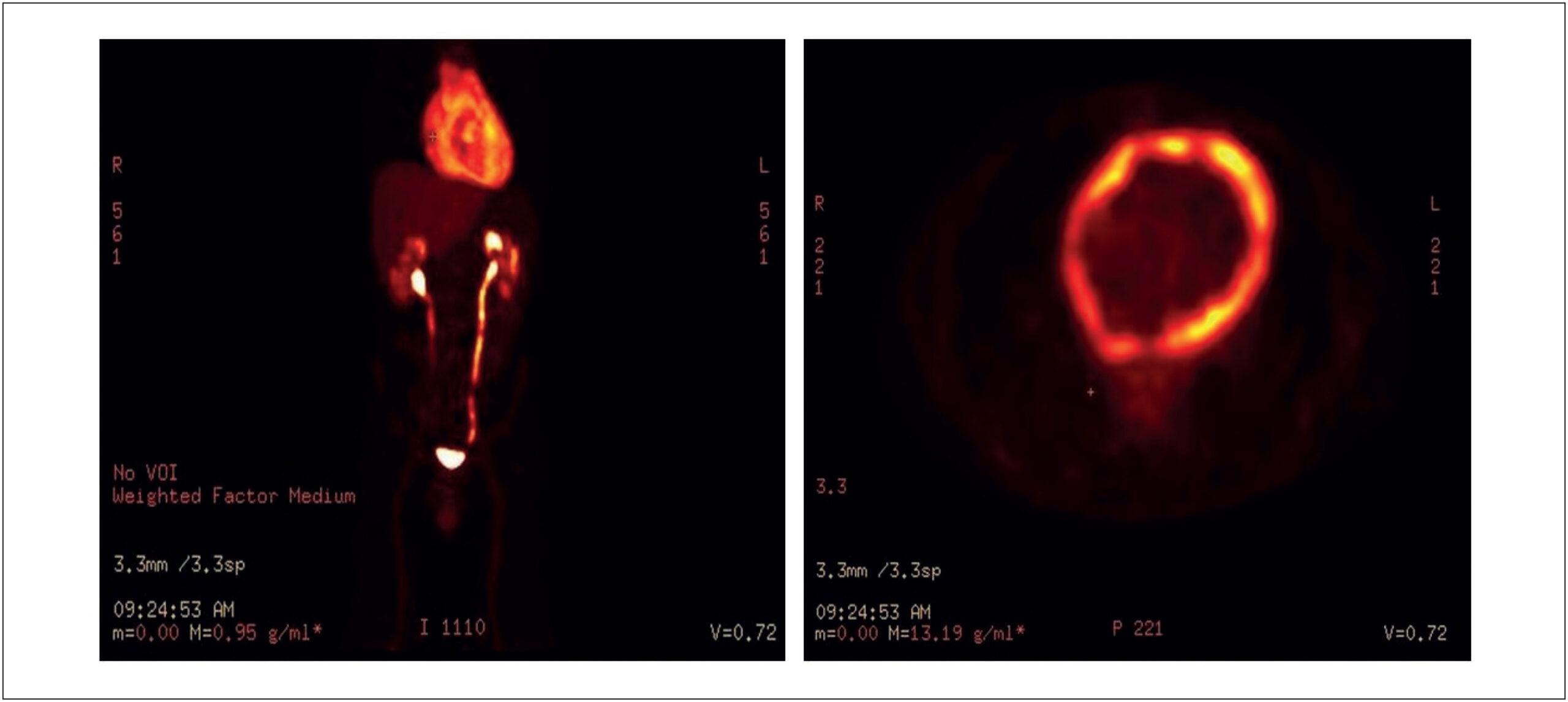
PET-CT 18F-FDG applications in cardiac tumors

Simone Cristina Soares Brandão et al. Luca Terracini Dompieri

ABSTRACT

Cardiac neoplasms are divided into primary and secondary. Secondary neoplasms are 20 to 40 times more common than the primary ones. Although rare, primary cardiac neoplasms may be benign or malignant. Benign neoplasms are responsible for 75% of the cases. The main objectives of cardiovascular imaging are to define tumor morphology and etiology, identify potential complications and assist in the establishment of treatment. For the diagnosis of cardiac neoplasms, positron emission tomography combined with computed tomography (PET-CT) with fluorodeoxyglucose-F18 (18F-FDG) is a technique that is still little used, especially in primary cardiac tumors. However, it can help differentiate between malignant and benign tumors, thus preventing cardiac biopsies and unnecessary invasive treatments. For this review, we searched the PubMed database, considering the publications on this topic in the past 10 years. PET-CT 18F-FDG is a useful test to differentiate benign from malignant heart masses according to the higher degree of glycolytic metabolism found in malignant neoplasms. Moreover, in malignant tumors, PET-CT 18F-FDG plays a central role in disease staging and may help assess treatment response.

  • Heart Neoplasms
  • Positron-Emission Tomography
  • Review
Read Full Article
Creative Commons Attribution (CC BY) Creative Commons license
14/01/2022 DOI: 10.5935/2318-8219.20190048 Views 1
Figure 1
ABCIC
Case Report

Lipoma in the Interventricular Septum – Case Report

Bruna Bonamigo Thomé et al. Laura Vilela Pazzini Nathalia Regina Pavan Estevan Vieira Cabeda Tobias Sato de Almeida

Abstract

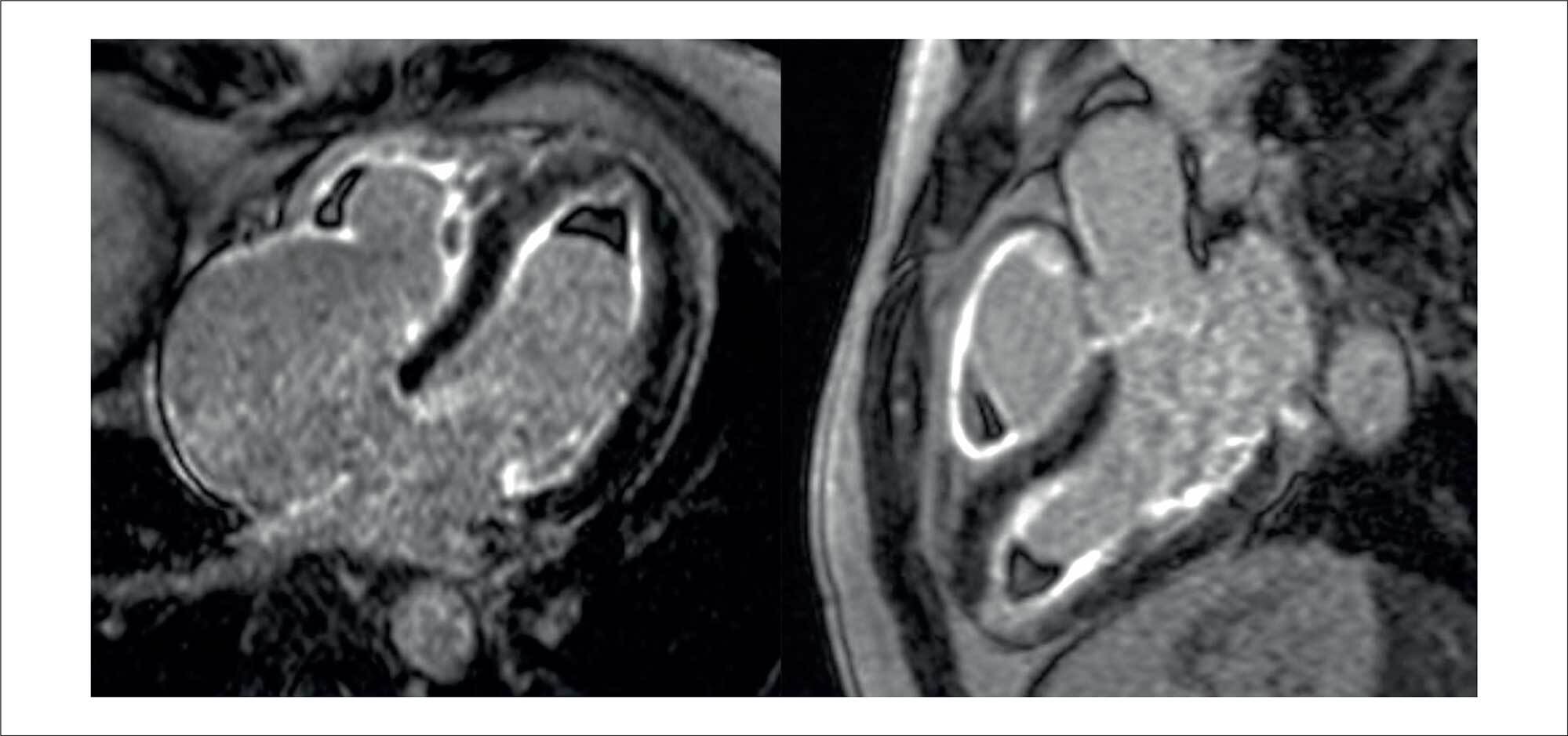
Primary cardiac tumors are a rare but mostly benign pathology found in up to 0.03% of autopsies. Among these benign tumors, with an incidence of 8.5%, lipomas are often located in the interatrial septum. When located in the interventricular septum, they are considered an even more unusual pathology with an unknown real prevalence. In most cases, the diagnosis is made incidentally on cardiovascular imaging and confirmed by histopathological examination. They are more commonly asymptomatic or feature nonspecific symptoms, but these lipomas can progress with arrhythmias, valve dysfunction, heart failure, and death, which highlights the importance of cardiovascular imaging methods in the differential diagnosis and to guide appropriate therapy. This report describes the case of a patient with an incidental echocardiographic finding of a cardiac mass located in the interventricular septum, followed by cardiac magnetic resonance imaging to better characterize the lesion and demonstrate its characteristics compatible with lipoma.

  • Diagnostic cardiovascular techniques
  • Echocardiography
  • Heart Neoplasms
  • Lipoma
Read Full Article
Creative Commons Attribution (CC BY) Creative Commons license
16/06/2021 DOI: 10.47593/2675-312X/20213401eabc152
Figure 1
IJCS
Case Report

Caseous Necrosis of the Mitral Valve: Imaging Methods Allow the Diagnosis and Prevent Surgery

Rodrigo de Moura Joaquim et al. Edileide de Barros Correia Suéllen Lacerda Bezerra Ibraim Masciarelli Francisco Pinto … Tiago Senra Garcia dos Santos Fabiano de Castro Albrecht

Introduction

Calcification of the mitral annulus (CMA) is defined as a chronic degeneration of the mitral annulus fibrous involving mainly the posterior annulus. It is a common condition in the elderly, especially in women, and is associated with hypertension. Caseous necrosis of the mitral valve (CNMV) is a rare and less known CMA variant with a prevalence of 0.64%, which can reach up to 2.7% in autopsy studies of patients with CMA.–

On echocardiography, CNMV appears as a large rounded or semilunar echodense mass, with a liquid filling and central echolucency, similar to a periannular tumor., The mechanism of liquefaction and caseification are still not well understood, but it is believed to occur due to changes in calcium metabolism.

[…]

  • Calcinosis
  • Diagnostic Imaging
  • Heart Neoplasms
  • Magnetic Resonance Imaging
  • Mitral Valve
  • Multidetector Computed Tomography
Read Full Article
Creative Commons Attribution (CC BY) Creative Commons license
01/01/2017 DOI: 10.5935/2359-4802.20170021 Views 1
Magnetic resonance imaging evaluation of cardiac masses
ABC
Artigo Original

Magnetic resonance imaging evaluation of cardiac masses

Maria Fernanda Braggion-Santos et al. Marcel Koenigkam-Santos Sara Reis Teixeira Gustavo Jardim Volpe … Henrique Simão Trad André Schmidt

BACKGROUND: Cardiac tumors are extremely rare; however, when there is clinical suspicion, proper diagnostic evaluation is necessary to plan the most appropriate treatment. In this context, cardiovascular magnetic resonance imaging (CMRI) plays an important role, allowing a comprehensive characterization of such lesions. OBJECTIVE: To review cases referred to a CMRI Department for investigation of cardiac and paracardiac masses. To describe the positive case series with a brief review of the literature for each type of lesion and the role of cardiovascular magnetic resonance imaging in evaluation. METHODS: Between August 2008 and December 2011, all cases referred for CMRI with suspicion of tumor involving the heart were reviewed. Cases with positive histopathological diagnosis, clinical evolution or therapeutic response compatible with the clinical suspicion and imaging findings were selected. RESULTS: Among the 13 cases included in our study, eight (62%) had histopathological confirmation. We describe five benign tumors (myxomas, rhabdomyoma and fibromas), five malignancies (sarcoma, lymphoma, Richter syndrome involving the heart and metastatic disease) and three non-neoplastic lesions (pericardial cyst, intracardiac thrombus and infectious vegetation). CONCLUSIONS: CMRI plays an important role in the evaluation of cardiac masses of non-neoplastic and neoplastic origin, contributing to a more accurate diagnosis in a noninvasive manner and assisting in treatment planning, allowing safe clinical follow-up with good reproducibility.

  • Heart Neoplasms
  • Magnetic Resonance Imaging
  • Myxoma
  • Sarcoma
Read Full Article
Creative Commons Attribution (CC BY) Creative Commons license
26/07/2013 DOI: 10.5935/abc.20130150 Views 3

Could not load more articles. Please try again.

How to cite this article

EndNote RIS

Electronic Document Format (Vancouver)

Latado L, Costa FF, Benevides CFL, Torreão JA, Lima AK, Melo AS, Latado AL, et al. Intramyocardial Cardiac Tumor: A Case Report. ABC Imagem Cardiovascular 2026;38(4):e20250051.

Electronic Document Format (ABNT)

Latado, Luisa; Costa, Fabio Figueiredo; Benevides, Carlos Frederico Lopes; Torreão, Jorge Andion; Lima, Adna Keyne; Melo, Adelina Sanches de; Latado, Adriana Lopes. Intramyocardial Cardiac Tumor: A Case Report. ABC Imagem Cardiovascular, v. 38, n. 4, e20250051, Feb. 2026.

Electronic Document Format (APA)

Latado, L., Costa, F. F., Benevides, C. F. L., Torreão, J. A., Lima, A. K., Melo, A. S. , & Latado, A. L. (2026). Intramyocardial Cardiac Tumor: A Case Report. ABC Imagem Cardiovascular, 38(4), e20250051.

Electronic Document Format (ISO)

Latado, Luisa and Costa, Fabio Figueiredo and Benevides, Carlos Frederico Lopes and Torreão, Jorge Andion and Lima, Adna Keyne and Melo, Adelina Sanches de and Latado, Adriana Lopes. Intramyocardial Cardiac Tumor: A Case Report. ABC Imagem Cardiovascular [online]. 2026, vol. 38, n. 4, [cited 2026-10-14], e20250051. Available from: <https://journals.cardiol.br/en/article/intramyocardial-cardiac-tumor-a-case-report/>. ISSN 2318-8219

EndNote RIS

Electronic Document Format (Vancouver)

Carvalho G, Carvalho MFM, Magalhães T, Varela AM, Staszko KF, Hayashida MR. Turmoil of Symptoms: The Devastating Impact of Cardiac Lymphoma – Multiple Clinical Manifestations and the Lethal Causes of Cardiac Involvement. ABC Imagem Cardiovascular 2025;38(3):e20250033.

Electronic Document Format (ABNT)

Carvalho, Gustavo; Carvalho, Maria Fernanda Miranda; Magalhães, Tiago; Varela, Alexandre Manoel; Staszko, Kamila Fernanda; Hayashida, Mariana Rie. Turmoil of Symptoms: The Devastating Impact of Cardiac Lymphoma – Multiple Clinical Manifestations and the Lethal Causes of Cardiac Involvement. ABC Imagem Cardiovascular, v. 38, n. 3, e20250033, Aug. 2025.

Electronic Document Format (APA)

Carvalho, G., Carvalho, M. F. M., Magalhães, T., Varela, A. M., Staszko, K. F., & Hayashida, M. R. (2025). Turmoil of Symptoms: The Devastating Impact of Cardiac Lymphoma – Multiple Clinical Manifestations and the Lethal Causes of Cardiac Involvement. ABC Imagem Cardiovascular, 38(3), e20250033.

Electronic Document Format (ISO)

Carvalho, Gustavo and Carvalho, Maria Fernanda Miranda and Magalhães, Tiago and Varela, Alexandre Manoel and Staszko, Kamila Fernanda and Hayashida, Mariana Rie. Turmoil of Symptoms: The Devastating Impact of Cardiac Lymphoma – Multiple Clinical Manifestations and the Lethal Causes of Cardiac Involvement. ABC Imagem Cardiovascular [online]. 2025, vol. 38, n. 3, [cited 2026-10-14], e20250033. Available from: <https://journals.cardiol.br/en/article/turmoil-of-symptoms-the-devastating-impact-of-cardiac-lymphoma-multiple-clinical-manifestations-and-the-lethal-causes-of-cardiac-involvement/>. ISSN 2318-8219

EndNote RIS

Electronic Document Format (Vancouver)

Pereira GAR, Pauletto MM, Tscheika AP, Velho FM, Folador L. Primary Pericardial Angiosarcoma Complicated by Constrictive Pericarditis. Arquivos Brasileiros de Cardiologia 2025;122(3):e20240683.

Electronic Document Format (ABNT)

Pereira, Guilherme Augusto Reissig; Pauletto, Maiane Maria; Tscheika, Anna Paula; Velho, Fabrício Michalski; Folador, Luciano. Primary Pericardial Angiosarcoma Complicated by Constrictive Pericarditis. Arquivos Brasileiros de Cardiologia, v. 122, n. 3, e20240683, May. 2025.

Electronic Document Format (APA)

Pereira, G. A. R., Pauletto, M. M., Tscheika, A. P., Velho, F. M., & Folador, L. (2025). Primary Pericardial Angiosarcoma Complicated by Constrictive Pericarditis. Arquivos Brasileiros de Cardiologia, 122(3), e20240683.

Electronic Document Format (ISO)

Pereira, Guilherme Augusto Reissig and Pauletto, Maiane Maria and Tscheika, Anna Paula and Velho, Fabrício Michalski and Folador, Luciano. Primary Pericardial Angiosarcoma Complicated by Constrictive Pericarditis. Arquivos Brasileiros de Cardiologia [online]. 2025, vol. 122, n. 3, [cited 2026-10-14], e20240683. Available from: <https://journals.cardiol.br/en/article/primary-pericardial-angiosarcoma-complicated-by-constrictive-pericarditis/>. ISSN 0066-782X

EndNote RIS

Electronic Document Format (Vancouver)

Vasquez-Rodriguez JF, Idrovo-Turbay CP, Rodríguez JA, Villalobos LM. A Common Primary Cardiac Tumor With an Uncommon Presentation. ABC Imagem Cardiovascular 2025;38(1):e20240123.

Electronic Document Format (ABNT)

Vasquez-Rodriguez, Juan Felipe; Idrovo-Turbay, Carolina Paola; Rodríguez, Jaime Alberto; Villalobos, Lorena Montes. A Common Primary Cardiac Tumor With an Uncommon Presentation. ABC Imagem Cardiovascular, v. 38, n. 1, e20240123, Mar. 2025.

Electronic Document Format (APA)

Vasquez-Rodriguez, J. F., Idrovo-Turbay, C. P., Rodríguez, J. A., & Villalobos, L. M. (2025). A Common Primary Cardiac Tumor With an Uncommon Presentation. ABC Imagem Cardiovascular, 38(1), e20240123.

Electronic Document Format (ISO)

Vasquez-Rodriguez, Juan Felipe and Idrovo-Turbay, Carolina Paola and Rodríguez, Jaime Alberto and Villalobos, Lorena Montes. A Common Primary Cardiac Tumor With an Uncommon Presentation. ABC Imagem Cardiovascular [online]. 2025, vol. 38, n. 1, [cited 2026-10-14], e20240123. Available from: <https://journals.cardiol.br/en/article/a-common-primary-cardiac-tumor-with-an-uncommon-presentation/>. ISSN 2318-8219

EndNote RIS

Electronic Document Format (Vancouver)

Pamplona LF, Oliveira KBS, Reis Neto JA, Bustamante LA, Souza JM, Zacchi FFS, Brito JBO, et al. Cardiac Lymphoma: A Case Report. Arquivos Brasileiros de Cardiologia 2025;122(2):e20240299.

Electronic Document Format (ABNT)

Pamplona, Leticia Ferraz; Oliveira, Kelly Barnabé Serpa de; Reis Neto, João Ancelmo dos; Bustamante, Luis Antelo; Souza, Januário Manoel de; Zacchi, Flávia Fernandes Silva; Brito, Juliana Brenande de Oliveira; Rojas, Salomon Soriano Ordinola. Cardiac Lymphoma: A Case Report. Arquivos Brasileiros de Cardiologia, v. 122, n. 2, e20240299, Mar. 2025.

Electronic Document Format (APA)

Pamplona, L. F., Oliveira, K. B. S. , Reis Neto, J. A. , Bustamante, L. A., Souza, J. M. , Zacchi, F. F. S., Brito, J. B. O., & Rojas, S. S. O. (2025). Cardiac Lymphoma: A Case Report. Arquivos Brasileiros de Cardiologia, 122(2), e20240299.

Electronic Document Format (ISO)

Pamplona, Leticia Ferraz and Oliveira, Kelly Barnabé Serpa de and Reis Neto, João Ancelmo dos and Bustamante, Luis Antelo and Souza, Januário Manoel de and Zacchi, Flávia Fernandes Silva and Brito, Juliana Brenande de Oliveira and Rojas, Salomon Soriano Ordinola. Cardiac Lymphoma: A Case Report. Arquivos Brasileiros de Cardiologia [online]. 2025, vol. 122, n. 2, [cited 2026-10-14], e20240299. Available from: <https://journals.cardiol.br/en/article/cardiac-lymphoma-a-case-report/>. ISSN 0066-782X

EndNote RIS

Electronic Document Format (Vancouver)

Almeida Junior M, Morais AFP, Murad Junior JA. Atrial Myxoma as a Rare Cause of Stroke: Case Report. ABC Imagem Cardiovascular 2025;38(1):e20240099.

Electronic Document Format (ABNT)

Almeida Junior, Maurício Ângelo de; Morais, Ana Flavia Parreira de; Murad Junior, Jamil Alli. Atrial Myxoma as a Rare Cause of Stroke: Case Report. ABC Imagem Cardiovascular, v. 38, n. 1, e20240099, Jan. 2025.

Electronic Document Format (APA)

Almeida Junior, M. , Morais, A. F. P. , & Murad Junior, J. A. (2025). Atrial Myxoma as a Rare Cause of Stroke: Case Report. ABC Imagem Cardiovascular, 38(1), e20240099.

Electronic Document Format (ISO)

Almeida Junior, Maurício Ângelo de and Morais, Ana Flavia Parreira de and Murad Junior, Jamil Alli. Atrial Myxoma as a Rare Cause of Stroke: Case Report. ABC Imagem Cardiovascular [online]. 2025, vol. 38, n. 1, [cited 2026-10-14], e20240099. Available from: <https://journals.cardiol.br/en/article/atrial-myxoma-as-a-rare-cause-of-stroke-case-report/>. ISSN 2318-8219

EndNote RIS

Electronic Document Format (Vancouver)

Guaman-Valdivieso C, Aramburu J, Lluberas N, Huart N, Parma G. Not Everything is as it Seems at First Glance: Multimodal Imaging in Heart Cyst Evaluation. ABC Imagem Cardiovascular 2024;37(4):e20240086.

Electronic Document Format (ABNT)

Guaman-Valdivieso, Carlos; Aramburu, Julia; Lluberas, Natalia; Huart, Natalia; Parma, Gabriel. Not Everything is as it Seems at First Glance: Multimodal Imaging in Heart Cyst Evaluation. ABC Imagem Cardiovascular, v. 37, n. 4, e20240086, Dec. 2024.

Electronic Document Format (APA)

Guaman-Valdivieso, C., Aramburu, J., Lluberas, N., Huart, N., & Parma, G. (2024). Not Everything is as it Seems at First Glance: Multimodal Imaging in Heart Cyst Evaluation. ABC Imagem Cardiovascular, 37(4), e20240086.

Electronic Document Format (ISO)

Guaman-Valdivieso, Carlos and Aramburu, Julia and Lluberas, Natalia and Huart, Natalia and Parma, Gabriel. Not Everything is as it Seems at First Glance: Multimodal Imaging in Heart Cyst Evaluation. ABC Imagem Cardiovascular [online]. 2024, vol. 37, n. 4, [cited 2026-10-14], e20240086. Available from: <https://journals.cardiol.br/en/article/not-everything-is-as-it-seems-at-first-glance-multimodal-imaging-in-heart-cyst-evaluation/>. ISSN 2318-8219

EndNote RIS

Electronic Document Format (Vancouver)

Torres RVA, Fuzissima BM, Fonseca EKUN, Farias LPG, Ishikawa WY, Araújo Filho JAB. My Approach to Cardiovascular Computed Tomography and Magnetic Resonance Imaging in the Evaluation of Cardiac Pseudotumors: A Brief Review. ABC Imagem Cardiovascular 2024;37(3):e20240044.

Electronic Document Format (ABNT)

Torres, Roberto Vitor Almeida; Fuzissima, Bruno Maeda; Fonseca, Eduardo Kaiser Ururahy Nunes; Farias, Lucas de Pádua Gomes de; Ishikawa, Walther Yoshiharu; Araújo Filho, José de Arimatéia Batista. My Approach to Cardiovascular Computed Tomography and Magnetic Resonance Imaging in the Evaluation of Cardiac Pseudotumors: A Brief Review. ABC Imagem Cardiovascular, v. 37, n. 3, e20240044, Aug. 2024.

Electronic Document Format (APA)

Torres, R. V. A., Fuzissima, B. M., Fonseca, E. K. U. N., Farias, L. P. G. , Ishikawa, W. Y., & Araújo Filho, J. A. B. (2024). My Approach to Cardiovascular Computed Tomography and Magnetic Resonance Imaging in the Evaluation of Cardiac Pseudotumors: A Brief Review. ABC Imagem Cardiovascular, 37(3), e20240044.

Electronic Document Format (ISO)

Torres, Roberto Vitor Almeida and Fuzissima, Bruno Maeda and Fonseca, Eduardo Kaiser Ururahy Nunes and Farias, Lucas de Pádua Gomes de and Ishikawa, Walther Yoshiharu and Araújo Filho, José de Arimatéia Batista. My Approach to Cardiovascular Computed Tomography and Magnetic Resonance Imaging in the Evaluation of Cardiac Pseudotumors: A Brief Review. ABC Imagem Cardiovascular [online]. 2024, vol. 37, n. 3, [cited 2026-10-14], e20240044. Available from: <https://journals.cardiol.br/en/article/my-approach-to-cardiovascular-computed-tomography-and-magnetic-resonance-imaging-in-the-evaluation-of-cardiac-pseudotumors-a-brief-review/>. ISSN 2318-8219

EndNote RIS

Electronic Document Format (Vancouver)

Garbin HI, Klauberg MPM, Mazzutti G. Cardiac Tumor in the Left Atrium. ABC Heart Fail Cardiomyop 2024;4(2):e20240004.

Electronic Document Format (ABNT)

Garbin, Henrique Iahnke; Klauberg, Matheus Piccoli Machado; Mazzutti, Géris. Cardiac Tumor in the Left Atrium. ABC Heart Fail Cardiomyop, v. 4, n. 2, e20240004, Aug. 2024.

Electronic Document Format (APA)

Garbin, H. I., Klauberg, M. P. M., & Mazzutti, G. (2024). Cardiac Tumor in the Left Atrium. ABC Heart Fail Cardiomyop, 4(2), e20240004.

Electronic Document Format (ISO)

Garbin, Henrique Iahnke and Klauberg, Matheus Piccoli Machado and Mazzutti, Géris. Cardiac Tumor in the Left Atrium. ABC Heart Fail Cardiomyop [online]. 2024, vol. 4, n. 2, [cited 2026-10-14], e20240004. Available from: <https://journals.cardiol.br/en/article/cardiac-tumor-in-the-left-atrium/>

EndNote RIS

Electronic Document Format (Vancouver)

Gripp EA, Jácome BZ, Silva AEA, Dias FBR, Almeida BBC, Rabischoffsky A. Cardiac Tumors: Recognition Is Essential!. ABC Imagem Cardiovascular 2024;37(2):e20240032.

Electronic Document Format (ABNT)

Gripp, Eliza de Almeida; Jácome, Bruna Zanforlin; Silva, Anna Esther Araujo e; Dias, Fernanda Baptista Ribeiro; Almeida, Bruna Barbosa Chagas de; Rabischoffsky, Arnaldo. Cardiac Tumors: Recognition Is Essential!. ABC Imagem Cardiovascular, v. 37, n. 2, e20240032, Jun. 2024.

Electronic Document Format (APA)

Gripp, E. A., Jácome, B. Z., Silva, A. E. A. , Dias, F. B. R., Almeida, B. B. C. , & Rabischoffsky, A. (2024). Cardiac Tumors: Recognition Is Essential!. ABC Imagem Cardiovascular, 37(2), e20240032.

Electronic Document Format (ISO)

Gripp, Eliza de Almeida and Jácome, Bruna Zanforlin and Silva, Anna Esther Araujo e and Dias, Fernanda Baptista Ribeiro and Almeida, Bruna Barbosa Chagas de and Rabischoffsky, Arnaldo. Cardiac Tumors: Recognition Is Essential!. ABC Imagem Cardiovascular [online]. 2024, vol. 37, n. 2, [cited 2026-10-14], e20240032. Available from: <https://journals.cardiol.br/en/article/cardiac-tumors-recognition-is-essential/>. ISSN 2318-8219

EndNote RIS

Electronic Document Format (Vancouver)

Camargo FM, Brizot ML, Francisco RPV, Carvalho WB, Ikari NM, Peres SV, Lopes MAB, et al. Perinatal Results and Long-Term Follow-Up of Fetal Cardiac Tumors: A 30-Year Historical Cohort Study. Arquivos Brasileiros de Cardiologia 2024;121(1):e20220469.

Electronic Document Format (ABNT)

Camargo, Fabricio Marcondes; Brizot, Maria de Lourdes; Francisco, Rossana Pulcineli Vieira; Carvalho, Werther Brunow de; Ikari, Nana Miura; Peres, Stella Verzinhasse; Lopes, Marco Antônio Borges; Lopes, Lilian Maria. Perinatal Results and Long-Term Follow-Up of Fetal Cardiac Tumors: A 30-Year Historical Cohort Study. Arquivos Brasileiros de Cardiologia, v. 121, n. 1, e20220469, Mar. 2024.

Electronic Document Format (APA)

Camargo, F. M., Brizot, M. L., Francisco, R. P. V., Carvalho, W. B. , Ikari, N. M., Peres, S. V., Lopes, M. A. B., & Lopes, L. M. (2024). Perinatal Results and Long-Term Follow-Up of Fetal Cardiac Tumors: A 30-Year Historical Cohort Study. Arquivos Brasileiros de Cardiologia, 121(1), e20220469.

Electronic Document Format (ISO)

Camargo, Fabricio Marcondes and Brizot, Maria de Lourdes and Francisco, Rossana Pulcineli Vieira and Carvalho, Werther Brunow de and Ikari, Nana Miura and Peres, Stella Verzinhasse and Lopes, Marco Antônio Borges and Lopes, Lilian Maria. Perinatal Results and Long-Term Follow-Up of Fetal Cardiac Tumors: A 30-Year Historical Cohort Study. Arquivos Brasileiros de Cardiologia [online]. 2024, vol. 121, n. 1, [cited 2026-10-14], e20220469. Available from: <https://journals.cardiol.br/en/article/perinatal-results-and-long-term-follow-up-of-fetal-cardiac-tumors-a-30-year-historical-cohort-study/>. ISSN 0066-782X

EndNote RIS

Electronic Document Format (Vancouver)

Pereira MM, Nina VJS, Melo Filho JX, Melo RJL, Juliano MTH, Pereira LSM. The Importance of Imaging Multimodality in the Diagnosis of a Rare Case of Papillary Fibroelastoma in the Left Ventricular Apex. ABC Imagem Cardiovascular 2023;36(4):e20230102.

Electronic Document Format (ABNT)

Pereira, Marcio Mendes; Nina, Vinícius José da Silva; Melo Filho, José Xavier de; Melo, Rodrigo de Jesus Louzeiro; Juliano, Marco Túlio Hercos; Pereira, Luma Sayonara Martins. The Importance of Imaging Multimodality in the Diagnosis of a Rare Case of Papillary Fibroelastoma in the Left Ventricular Apex. ABC Imagem Cardiovascular, v. 36, n. 4, e20230102, Dec. 2023.

Electronic Document Format (APA)

Pereira, M. M., Nina, V. J. S., Melo Filho, J. X. , Melo, R. J. L., Juliano, M. T. H., & Pereira, L. S. M. (2023). The Importance of Imaging Multimodality in the Diagnosis of a Rare Case of Papillary Fibroelastoma in the Left Ventricular Apex. ABC Imagem Cardiovascular, 36(4), e20230102.

Electronic Document Format (ISO)

Pereira, Marcio Mendes and Nina, Vinícius José da Silva and Melo Filho, José Xavier de and Melo, Rodrigo de Jesus Louzeiro and Juliano, Marco Túlio Hercos and Pereira, Luma Sayonara Martins. The Importance of Imaging Multimodality in the Diagnosis of a Rare Case of Papillary Fibroelastoma in the Left Ventricular Apex. ABC Imagem Cardiovascular [online]. 2023, vol. 36, n. 4, [cited 2026-10-14], e20230102. Available from: <https://journals.cardiol.br/en/article/the-importance-of-imaging-multimodality-in-the-diagnosis-of-a-rare-case-of-papillary-fibroelastoma-in-the-left-ventricular-apex/>. ISSN 2318-8219

EndNote RIS

Electronic Document Format (Vancouver)

Amin A, Taheri Z, Hesami M, Alizahehasl A, Norouzi Z, Mohammadi N. Misdiagnosis of a Cardiac Angiosarcoma During the COVID-19 Pandemic. Arquivos Brasileiros de Cardiologia 2023;120(7):e20220501.

Electronic Document Format (ABNT)

Amin, Arash; Taheri, Zohreh; Hesami, Mahshid; Alizahehasl, Azin; Norouzi, Zeinab; Mohammadi, Nasibeh. Misdiagnosis of a Cardiac Angiosarcoma During the COVID-19 Pandemic. Arquivos Brasileiros de Cardiologia, v. 120, n. 7, e20220501, Aug. 2023.

Electronic Document Format (APA)

Amin, A., Taheri, Z., Hesami, M., Alizahehasl, A., Norouzi, Z., & Mohammadi, N. (2023). Misdiagnosis of a Cardiac Angiosarcoma During the COVID-19 Pandemic. Arquivos Brasileiros de Cardiologia, 120(7), e20220501.

Electronic Document Format (ISO)

Amin, Arash and Taheri, Zohreh and Hesami, Mahshid and Alizahehasl, Azin and Norouzi, Zeinab and Mohammadi, Nasibeh. Misdiagnosis of a Cardiac Angiosarcoma During the COVID-19 Pandemic. Arquivos Brasileiros de Cardiologia [online]. 2023, vol. 120, n. 7, [cited 2026-10-14], e20220501. Available from: <https://journals.cardiol.br/en/article/misdiagnosis-of-a-cardiac-angiosarcoma-during-the-covid-19-pandemic/>. ISSN 0066-782X

EndNote RIS

Electronic Document Format (Vancouver)

Faé IG, Ruiz GZL, Irffi GP, Almeida Junior RS, Conceição PA, Falchetto EB, Passaglia LG, et al. Intracardiac Metastasis of Colonic Adenocarcinoma 12 Years After Primary Tumor Control and Without Any Sign of Other Metastasis: A Case Report. Arquivos Brasileiros de Cardiologia 2022;119(6):991-5.

Electronic Document Format (ABNT)

Faé, Isabela Galizzi; Ruiz, Gabriela Zamunaro Lopes; Irffi, Gustavo Palmer; Almeida Junior, Robson de Souza; Conceição, Pedro Anjos; Falchetto, Eduardo Belisario; Passaglia, Luiz Guilherme; Brasileiro Filho, Geraldo; Gelape, Cláudio Leo; Oliveira, Clara Rodrigues Alves de. Intracardiac Metastasis of Colonic Adenocarcinoma 12 Years After Primary Tumor Control and Without Any Sign of Other Metastasis: A Case Report. Arquivos Brasileiros de Cardiologia, v. 119, n. 6, p. 991-995, Dec. 2022.

Electronic Document Format (APA)

Faé, I. G., Ruiz, G. Z. L., Irffi, G. P., Almeida Junior, R. S., Conceição, P. A., Falchetto, E. B., Passaglia, L. G., Brasileiro Filho, G., Gelape, C. L., & Oliveira, C. R. A. (2022). Intracardiac Metastasis of Colonic Adenocarcinoma 12 Years After Primary Tumor Control and Without Any Sign of Other Metastasis: A Case Report. Arquivos Brasileiros de Cardiologia, 119(6), 991-995.

Electronic Document Format (ISO)

Faé, Isabela Galizzi and Ruiz, Gabriela Zamunaro Lopes and Irffi, Gustavo Palmer and Almeida Junior, Robson de Souza and Conceição, Pedro Anjos and Falchetto, Eduardo Belisario and Passaglia, Luiz Guilherme and Brasileiro Filho, Geraldo and Gelape, Cláudio Leo and Oliveira, Clara Rodrigues Alves de. Intracardiac Metastasis of Colonic Adenocarcinoma 12 Years After Primary Tumor Control and Without Any Sign of Other Metastasis: A Case Report. Arquivos Brasileiros de Cardiologia [online]. 2022, vol. 119, n. 6, [cited 2026-10-14], pp. 991-995. Available from: <https://journals.cardiol.br/en/article/intracardiac-metastasis-of-colonic-adenocarcinoma-12-years-after-primary-tumor-control-and-without-any-sign-of-other-metastasis-a-case-report/>. ISSN 0066-782X

EndNote RIS

Electronic Document Format (Vancouver)

Cauduro SA, Dutra JPP, Fernandes F, Bonatto M, Santos MVC, Rocha LSO, Mialski TR, et al. Obstructive Cardiac Tumors. ABC: Heart Failure & Cardiomyopathy 2022;2(4):367-73.

Electronic Document Format (ABNT)

Cauduro, Sanderson Antonio; Dutra, João Pedro Passos; Fernandes, Fabio; Bonatto, Marcely; Santos, Maria Verônica Câmara; Rocha, Letícia dos Santos de Oliveira; Mialski, Talita Ribeiro; Nobrega, Ana Paula Konig da; Brandão, Simone Cristina Soares; Barberato, Silvio Henrique. Obstructive Cardiac Tumors. ABC: Heart Failure & Cardiomyopathy, v. 2, n. 4, p. 367-373, Dec. 2022.

Electronic Document Format (APA)

Cauduro, S. A., Dutra, J. P. P., Fernandes, F., Bonatto, M., Santos, M. V. C., Rocha, L. S. O., Mialski, T. R., Nobrega, A. P. K. , Brandão, S. C. S., & Barberato, S. H. (2022). Obstructive Cardiac Tumors. ABC: Heart Failure & Cardiomyopathy, 2(4), 367-373.

Electronic Document Format (ISO)

Cauduro, Sanderson Antonio and Dutra, João Pedro Passos and Fernandes, Fabio and Bonatto, Marcely and Santos, Maria Verônica Câmara and Rocha, Letícia dos Santos de Oliveira and Mialski, Talita Ribeiro and Nobrega, Ana Paula Konig da and Brandão, Simone Cristina Soares and Barberato, Silvio Henrique. Obstructive Cardiac Tumors. ABC: Heart Failure & Cardiomyopathy [online]. 2022, vol. 2, n. 4, [cited 2026-10-14], pp. 367-373. Available from: <https://journals.cardiol.br/en/article/obstructive-cardiac-tumors/>

EndNote RIS

Electronic Document Format (Vancouver)

Santos RR, Abecasis J, Gomes DA, Paiva MS, Rocha B, Ribeiras R, Freitas P, et al. An Incidental Finding of a Cardiac Sarcoma. Arquivos Brasileiros de Cardiologia 2022;119(2):349-52.

Electronic Document Format (ABNT)

Santos, Rita Reis; Abecasis, João; Gomes, Daniel A.; Paiva, Mariana Sousa; Rocha, Bruno; Ribeiras, Regina; Freitas, Pedro; Abecasis, Miguel; Trabulo, Marisa. An Incidental Finding of a Cardiac Sarcoma. Arquivos Brasileiros de Cardiologia, v. 119, n. 2, p. 349-352, Aug. 2022.

Electronic Document Format (APA)

Santos, R. R., Abecasis, J., Gomes, D. A., Paiva, M. S., Rocha, B., Ribeiras, R., Freitas, P., Abecasis, M., & Trabulo, M. (2022). An Incidental Finding of a Cardiac Sarcoma. Arquivos Brasileiros de Cardiologia, 119(2), 349-352.

Electronic Document Format (ISO)

Santos, Rita Reis and Abecasis, João and Gomes, Daniel A. and Paiva, Mariana Sousa and Rocha, Bruno and Ribeiras, Regina and Freitas, Pedro and Abecasis, Miguel and Trabulo, Marisa. An Incidental Finding of a Cardiac Sarcoma. Arquivos Brasileiros de Cardiologia [online]. 2022, vol. 119, n. 2, [cited 2026-10-14], pp. 349-352. Available from: <https://journals.cardiol.br/en/article/an-incidental-finding-of-a-cardiac-sarcoma/>. ISSN 0066-782X

EndNote RIS

Electronic Document Format (Vancouver)

Romero CE, Guacho WRL, Paula KR, Jimenez RP, Fonseca EKUN. Rare Presentation of Yolk Sac Tumor with Cardiac Involvement: Characteristics Detected by MRI. Arquivos Brasileiros de Cardiologia 2022;119(1):136-8.

Electronic Document Format (ABNT)

Romero, Cristhian Espinoza; Guacho, Williams Roberto Lata; Paula, Kevin Rafael de; Jimenez, Robert Paladines; Fonseca, Eduardo Kaiser Ururahy Nunes. Rare Presentation of Yolk Sac Tumor with Cardiac Involvement: Characteristics Detected by MRI. Arquivos Brasileiros de Cardiologia, v. 119, n. 1, p. 136-138, Jul. 2022.

Electronic Document Format (APA)

Romero, C. E., Guacho, W. R. L., Paula, K. R. , Jimenez, R. P., & Fonseca, E. K. U. N. (2022). Rare Presentation of Yolk Sac Tumor with Cardiac Involvement: Characteristics Detected by MRI. Arquivos Brasileiros de Cardiologia, 119(1), 136-138.

Electronic Document Format (ISO)

Romero, Cristhian Espinoza and Guacho, Williams Roberto Lata and Paula, Kevin Rafael de and Jimenez, Robert Paladines and Fonseca, Eduardo Kaiser Ururahy Nunes. Rare Presentation of Yolk Sac Tumor with Cardiac Involvement: Characteristics Detected by MRI. Arquivos Brasileiros de Cardiologia [online]. 2022, vol. 119, n. 1, [cited 2026-10-14], pp. 136-138. Available from: <https://journals.cardiol.br/en/article/rare-presentation-of-yolk-sac-tumor-with-cardiac-involvement-characteristics-detected-by-mri/>. ISSN 0066-782X

EndNote RIS

Electronic Document Format (Vancouver)

Oshiro FS, Valério RS, Rodrigues AAE, Szarf G, Uellendahl M, Siqueira MEM. Cardiac Fibroma: Long-Term Follow-up by Cardiac Magnetic Resonance. Arquivos Brasileiros de Cardiologia Imagem cardiovascular 2022;35(1):eabc263.

Electronic Document Format (ABNT)

Oshiro, Fernanda Sayuri; Valério, Raul Serra; Rodrigues, Alfredo Augusto Eyer; Szarf, Gilberto; Uellendahl, Marly; Siqueira, Maria Eduarda Menezes de. Cardiac Fibroma: Long-Term Follow-up by Cardiac Magnetic Resonance. Arquivos Brasileiros de Cardiologia Imagem cardiovascular, v. 35, n. 1, eabc263, Mar. 2022.

Electronic Document Format (APA)

Oshiro, F. S., Valério, R. S., Rodrigues, A. A. E., Szarf, G., Uellendahl, M., & Siqueira, M. E. M. (2022). Cardiac Fibroma: Long-Term Follow-up by Cardiac Magnetic Resonance. Arquivos Brasileiros de Cardiologia Imagem cardiovascular, 35(1), eabc263.

Electronic Document Format (ISO)

Oshiro, Fernanda Sayuri and Valério, Raul Serra and Rodrigues, Alfredo Augusto Eyer and Szarf, Gilberto and Uellendahl, Marly and Siqueira, Maria Eduarda Menezes de. Cardiac Fibroma: Long-Term Follow-up by Cardiac Magnetic Resonance. Arquivos Brasileiros de Cardiologia Imagem cardiovascular [online]. 2022, vol. 35, n. 1, [cited 2026-10-14], eabc263. Available from: <https://journals.cardiol.br/en/article/cardiac-fibroma-long-term-follow-up-by-cardiac-magnetic-resonance/>. ISSN 2318-8219

EndNote RIS

Electronic Document Format (Vancouver)

Góes GHB, Ferraz Neto JCS, Calado Filho IP, Lima RC, Sobral Filho DC. Rare Cause of Chest Pain in Young Patient: Left Ventricular Myxoma. Arquivos Brasileiros de Cardiologia Imagem cardiovascular 2022;32(1):67-70.

Electronic Document Format (ABNT)

Góes, Gustavo Henrique Belarmino; Ferraz Neto, José Candido de Souza; Calado Filho, Ivaldo Pedrosa; Lima, Ricardo de Carvalho; Sobral Filho, Dário Celestino. Rare Cause of Chest Pain in Young Patient: Left Ventricular Myxoma. Arquivos Brasileiros de Cardiologia Imagem cardiovascular, v. 32, n. 1, p. 67-70, Jan. 2022.

Electronic Document Format (APA)

Góes, G. H. B., Ferraz Neto, J. C. S., Calado Filho, I. P., Lima, R. C., & Sobral Filho, D. C. (2022). Rare Cause of Chest Pain in Young Patient: Left Ventricular Myxoma. Arquivos Brasileiros de Cardiologia Imagem cardiovascular, 32(1), 67-70.

Electronic Document Format (ISO)

Góes, Gustavo Henrique Belarmino and Ferraz Neto, José Candido de Souza and Calado Filho, Ivaldo Pedrosa and Lima, Ricardo de Carvalho and Sobral Filho, Dário Celestino. Rare Cause of Chest Pain in Young Patient: Left Ventricular Myxoma. Arquivos Brasileiros de Cardiologia Imagem cardiovascular [online]. 2022, vol. 32, n. 1, [cited 2026-10-14], pp. 67-70. Available from: <https://journals.cardiol.br/en/article/rare-cause-of-chest-pain-in-young-patient-left-ventricular-myxoma/>. ISSN 2318-8219

EndNote RIS

Electronic Document Format (Vancouver)

Bispo IGA, Volpatto R, Assakawa FH, Costa DF, Coelho ABP, Gimenes VML. Right Atrial Papillary Fibroelastoma. Arquivos Brasileiros de Cardiologia Imagem cardiovascular 2022;32(4):344.

Electronic Document Format (ABNT)

Bispo, Irving Gabriel Araújo; Volpatto, Ricardo; Assakawa, Fernando Hideki; Costa, Diego Farias; Coelho, André Borges Paes; Gimenes, Vera Márcia Lopes. Right Atrial Papillary Fibroelastoma. Arquivos Brasileiros de Cardiologia Imagem cardiovascular, v. 32, n. 4, p. 344-344, Jan. 2022.

Electronic Document Format (APA)

Bispo, I. G. A., Volpatto, R., Assakawa, F. H., Costa, D. F., Coelho, A. B. P., & Gimenes, V. M. L. (2022). Right Atrial Papillary Fibroelastoma. Arquivos Brasileiros de Cardiologia Imagem cardiovascular, 32(4), 344-344.

Electronic Document Format (ISO)

Bispo, Irving Gabriel Araújo and Volpatto, Ricardo and Assakawa, Fernando Hideki and Costa, Diego Farias and Coelho, André Borges Paes and Gimenes, Vera Márcia Lopes. Right Atrial Papillary Fibroelastoma. Arquivos Brasileiros de Cardiologia Imagem cardiovascular [online]. 2022, vol. 32, n. 4, [cited 2026-10-14], pp. 344-344. Available from: <https://journals.cardiol.br/en/article/right-atrial-papillary-fibroelastoma/>. ISSN 2318-8219

EndNote RIS

Electronic Document Format (Vancouver)

Brandão SCS, Dompieri LT. PET-CT 18F-FDG applications in cardiac tumors. Arquivos Brasileiros de Cardiologia Imagem cardiovascular 2022;32(4):309-17.

Electronic Document Format (ABNT)

Brandão, Simone Cristina Soares; Dompieri, Luca Terracini. PET-CT 18F-FDG applications in cardiac tumors. Arquivos Brasileiros de Cardiologia Imagem cardiovascular, v. 32, n. 4, p. 309-317, Jan. 2022.

Electronic Document Format (APA)

Brandão, S. C. S., & Dompieri, L. T. (2022). PET-CT 18F-FDG applications in cardiac tumors. Arquivos Brasileiros de Cardiologia Imagem cardiovascular, 32(4), 309-317.

Electronic Document Format (ISO)

Brandão, Simone Cristina Soares and Dompieri, Luca Terracini. PET-CT 18F-FDG applications in cardiac tumors. Arquivos Brasileiros de Cardiologia Imagem cardiovascular [online]. 2022, vol. 32, n. 4, [cited 2026-10-14], pp. 309-317. Available from: <https://journals.cardiol.br/en/article/pet-ct-18f-fdg-applications-in-cardiac-tumors/>. ISSN 2318-8219

EndNote RIS

Electronic Document Format (Vancouver)

Thomé BB, Pazzini LV, Pavan NR, Cabeda EV, Almeida TS. Lipoma in the Interventricular Septum – Case Report. ABC Imagem Cardiovascular 2021;34(1):eabc152.

Electronic Document Format (ABNT)

Thomé, Bruna Bonamigo; Pazzini, Laura Vilela; Pavan, Nathalia Regina; Cabeda, Estevan Vieira; Almeida, Tobias Sato de. Lipoma in the Interventricular Septum – Case Report. ABC Imagem Cardiovascular, v. 34, n. 1, eabc152, Jun. 2021.

Electronic Document Format (APA)

Thomé, B. B., Pazzini, L. V., Pavan, N. R., Cabeda, E. V., & Almeida, T. S. (2021). Lipoma in the Interventricular Septum – Case Report. ABC Imagem Cardiovascular, 34(1), eabc152.

Electronic Document Format (ISO)

Thomé, Bruna Bonamigo and Pazzini, Laura Vilela and Pavan, Nathalia Regina and Cabeda, Estevan Vieira and Almeida, Tobias Sato de. Lipoma in the Interventricular Septum – Case Report. ABC Imagem Cardiovascular [online]. 2021, vol. 34, n. 1, [cited 2026-10-14], eabc152. Available from: <https://journals.cardiol.br/en/article/lipoma-in-the-interventricular-septum-case-report/>. ISSN 2318-8219

EndNote RIS

Electronic Document Format (Vancouver)

Joaquim RM, Correia EB, Bezerra SL, Pinto IMF, Santos TSG, Albrecht FC. Caseous Necrosis of the Mitral Valve: Imaging Methods Allow the Diagnosis and Prevent Surgery. International Journal of Cardiovascular Sciences 2017;30(2):177-80.

Electronic Document Format (ABNT)

Joaquim, Rodrigo de Moura; Correia, Edileide de Barros; Bezerra, Suéllen Lacerda; Pinto, Ibraim Masciarelli Francisco; Santos, Tiago Senra Garcia dos; Albrecht, Fabiano de Castro. Caseous Necrosis of the Mitral Valve: Imaging Methods Allow the Diagnosis and Prevent Surgery. International Journal of Cardiovascular Sciences, v. 30, n. 2, p. 177-180, Dec. 2016.

Electronic Document Format (APA)

Joaquim, R. M., Correia, E. B., Bezerra, S. L., Pinto, I. M. F., Santos, T. S. G. , & Albrecht, F. C. (2017). Caseous Necrosis of the Mitral Valve: Imaging Methods Allow the Diagnosis and Prevent Surgery. International Journal of Cardiovascular Sciences, 30(2), 177-180.

Electronic Document Format (ISO)

Joaquim, Rodrigo de Moura and Correia, Edileide de Barros and Bezerra, Suéllen Lacerda and Pinto, Ibraim Masciarelli Francisco and Santos, Tiago Senra Garcia dos and Albrecht, Fabiano de Castro. Caseous Necrosis of the Mitral Valve: Imaging Methods Allow the Diagnosis and Prevent Surgery. International Journal of Cardiovascular Sciences [online]. 2017, vol. 30, n. 2, [cited 2026-10-14], pp. 177-180. Available from: <https://journals.cardiol.br/en/article/caseous-necrosis-of-the-mitral-valve-imaging-methods-allow-the-diagnosis-and-prevent-surgery/>. ISSN 2359-4802

EndNote RIS

Electronic Document Format (Vancouver)

Braggion-Santos MF, Koenigkam-Santos M, Teixeira SR, Volpe GJ, Trad HS, Schmidt A. Magnetic resonance imaging evaluation of cardiac masses. Arquivos Brasileiros de Cardiologia 2013;101(3):263-72.

Electronic Document Format (ABNT)

Braggion-Santos, Maria Fernanda; Koenigkam-Santos, Marcel; Teixeira, Sara Reis; Volpe, Gustavo Jardim; Trad, Henrique Simão; Schmidt, André. Magnetic resonance imaging evaluation of cardiac masses. Arquivos Brasileiros de Cardiologia, v. 101, n. 3, p. 263-272, Jul. 2013.

Electronic Document Format (APA)

Braggion-Santos, M. F., Koenigkam-Santos, M., Teixeira, S. R., Volpe, G. J., Trad, H. S., & Schmidt, A. (2013). Magnetic resonance imaging evaluation of cardiac masses. Arquivos Brasileiros de Cardiologia, 101(3), 263-272.

Electronic Document Format (ISO)

Braggion-Santos, Maria Fernanda and Koenigkam-Santos, Marcel and Teixeira, Sara Reis and Volpe, Gustavo Jardim and Trad, Henrique Simão and Schmidt, André. Magnetic resonance imaging evaluation of cardiac masses. Arquivos Brasileiros de Cardiologia [online]. 2013, vol. 101, n. 3, [cited 2026-10-14], pp. 263-272. Available from: <https://journals.cardiol.br/en/article/magnetic-resonance-imaging-evaluation-of-cardiac-masses/>. ISSN 0066-782X

  • About ABC Family
  • Internal Regulation
  • Editorial Board
  • Brazilian Society of Cardiology (SBC)

Issues

SBC Guidelines

  • Videos
  • Podcasts
  • News