ABC Family Arquivos Brasileiros de Cardiologia International Journal of Cardiovascular Sciences ABC Heart Failure & Cardiomyopathy ABC Imagem Cardiovascular
Português English
ABC Family
Português English
  • About
    • About ABC Family
    • Internal Regulation
    • Editorial Board
    • Brazilian Society of Cardiology (SBC)
  • Issues
  • SBC Guidelines
  • Content
    • Videos
    • Podcasts
    • News
  • About
    • About ABC Family
    • Internal Regulation
    • Editorial Board
    • Brazilian Society of Cardiology (SBC)
  • Issues
  • SBC Guidelines
  • Content
    • Videos
    • Podcasts
    • News

ABC Family

  • ABC Family
  • Arquivos Brasileiros de Cardiologia
  • International Journal of Cardiovascular Sciences
  • ABC Heart Failure & Cardiomyopathy
  • ABC Imagem Cardiovascular
Search in

No content found.

Journals
Ordering
Article types
Others

  1. Home
  2. Keyword: Ventricular Dysfunction, Left
Figure 1
ABCIC
Case Report

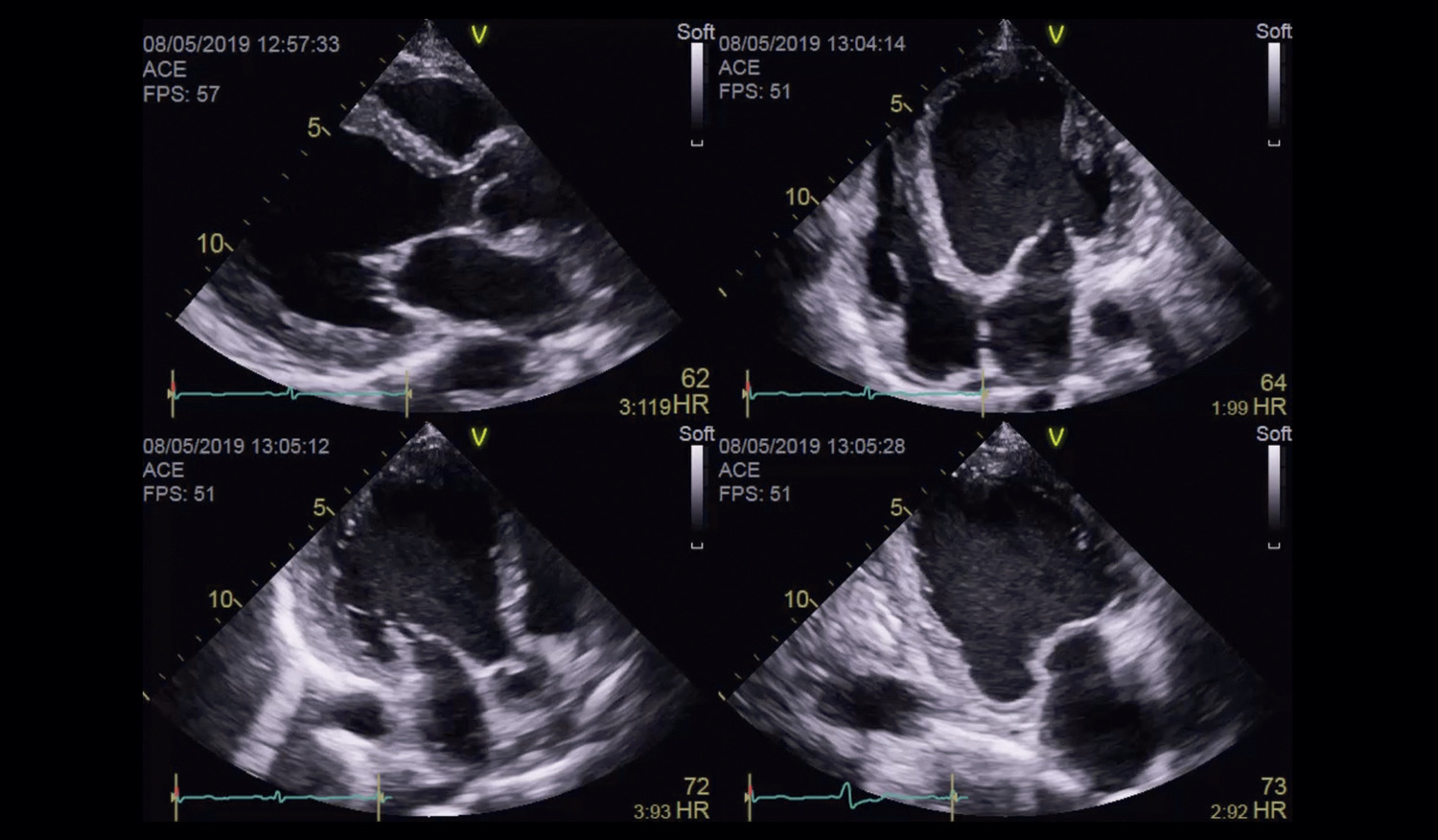
Comparative Analysis of Myocardial Work After Decongestion Therapy in a Patient With Acutely Decompensated HFrEF

Alexandre Costa Souza et al. Stephanie de Azevedo Drubi Bruna de Mattos Ivo Junqueira Ricardo André Sales Pereira Guedes … Adriano Chaves de Almeida Filho Carolina Thé Macêdo

Introduction

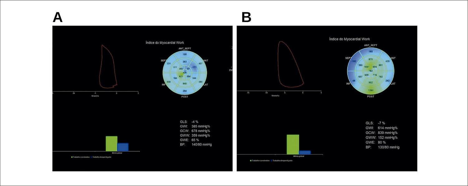
Acutely decompensated reduced ejection fraction heart failure (HFrEF) is one of the leading causes of hospitalization in our setting, with a 90-day readmission rate of 50%. The use of additional tools in the assessment of the left ventricular function, such as the measurement of myocardial deformation through the Global Longitudinal Strain of the left ventricle (GLS) has greater prognostic value than the evaluation of the left ventricular ejection fraction (LVEF) by the Simpson method. Despite the analysis of the myocardial deformity through the GLS being considered an independent predictor of mortality in patients with HFrEF, it is a parameter that experiences changes in pre- and after-load and, therefore, has limitations regarding the assessment of ventricular performance.– Myocardial work (MW) has stood out in recent years as a complementary tool to access parameters of myocardial function from the GLS-derived analysis, with the benefit of incorporating afterload information through the interpretation of the dynamic curve of deformity due to non-invasive left ventricular (LV) filling pressure. Hence, the analysis of MW has been encouraging in patients with HFrHF, and, when analyzed in combination with classic hemodynamic parameters, it can add information with greater accuracy and prognostic value in the cardiac decompensation scenario.–

[…]
  • Echocardiography
  • Heart Failure
  • Ventricular Dysfunction, Left
Read Full Article
Creative Commons Attribution (CC BY) Creative Commons license
27/06/2023 DOI: 10.36660/abcimg.20230008i
ABC
Editorial

Renin-Angiotensin System Inhibition and Beta Blockade Adrenergic May Be Useful to Attenuate Cardiotoxicity by Anthracyclines

Mario Fritsch Neves

Cardiotoxicity related to the use of anthracyclines is a major concern in oncology, as it can increase the morbidity of many patients receiving chemotherapy for various types of cancer. This toxicity can lead to left ventricular systolic dysfunction and, therefore, a higher incidence of heart failure, which can result in mortality in severe cases. A better understanding of the mechanisms involved and a better definition of diagnostic methods have provided significant advances in detecting this complication. However, there is still no consensus on the most accurate biomarkers for early identification of cardiotoxicity. Despite this, troponin measurement after each chemotherapy cycle can be a valid tool to indicate immediate intervention. ,

Manifestations of cardiotoxicity may remain restricted to myocardial cell damage detectable only by measuring circulating biomarkers or vary between silent ventricular dysfunction and clinically manifest heart failure. Strategies to avoid or at least attenuate anthracycline-induced cardiotoxicity are essential to improve the prognosis of patients using this class. The current recommendations include treating pre-existing cardiovascular risk factors, – choosing less cardiotoxic analogues, and administering low cumulative doses of anthracyclines. ,

[…]

  • Anthracyclines / toxicity
  • Drug Therapy
  • Heart Failure
  • Oncology
  • Ventricular Dysfunction, Left
Read Full Article
Creative Commons Attribution (CC BY) Creative Commons license
16/06/2023 DOI: 10.36660/abc.20230280
Figure 1
ABC
Research Letter

Left Ventricular Thrombosis and Pulmonary Thromboembolism in an Asymptomatic Covid-19 Patient

Natalia Lorenzo et al. Veronica Hernandez Alvaro Montes Fernando Rivero … Guillermo Reyes* Rio Aguilar

Research Letter

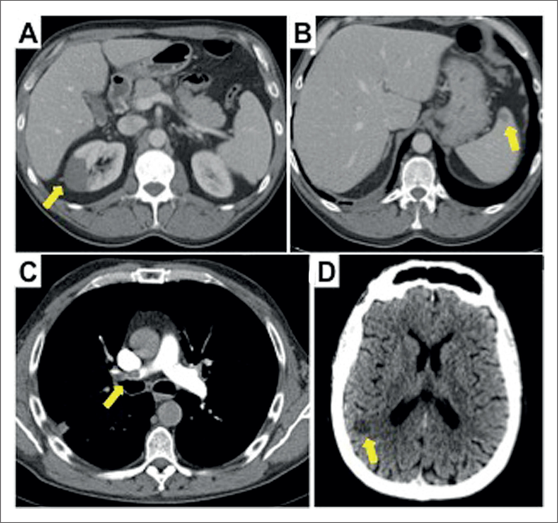
A 48-year-old-man presented to the emergency room with abdominal pain and vomiting of 12-hour duration. He had a history of porto-splenic venous thrombosis ten years before admission that was treated with 6 months of oral anticoagulation and felt to be due to moderate protein C deficiency, which was not confirmed in subsequent laboratory tests. Vital signs were normal, he was afebrile and no remarkable findings were found on physical examination except for pain on palpation in the left flank. The reverse transcriptase polymerase chain reaction (RT-PCR) test for COVID-19 was negative. Blood tests showed significant elevation of acute phase reactants (c-reactive protein: 138.3 mg/L, fibrinogen > 500mg/dL and leukocites 12.99 10^3/μL). Coagulation parameters where in normal range: phrotrombin time (PT) 12.1 seconds, partial thromboplastin time (PTT) 36.3 seconds, prothrombin activity (PA) 86%, international normalized ratio (INR) 1.08 and platelets 326.000/μL.

The abdominal ultrasound-scan did not show releant findings, so a computed tomography (CT) scan study was completed. CT abdominal scan found multiple infarcts in the right kidney () and isolated in spleen (), furthermore, a filling defect in the left ventricle was observed (). Transthoracic echocardiogram confirmed the presence of a pedunculated hyperechogenic and homogeneous mobile mass (3.1x2cm) with regular borders (, , ), anchored to the middle third of the septum of left ventricle (LV). Anatomy of LV was normal, with normal dimensions and normal ejection fraction without wall motion abnormalities.

[…]

  • Covid-19/complications
  • Diagnostic Imaging/methods
  • Infarction, Myocardial
  • Pulmonary embolism
  • SARS-CoV-2
  • Severe Acute Respiratory Syndrome/complications
  • Ventricular Dysfunction, Left
Read Full Article
Creative Commons Attribution (CC BY) Creative Commons license
19/12/2022 DOI: 10.36660/abc.20210590
Video 1
ABC
Research Letter

Non-Atherosclerotic Coronary and Vascular Disease Case Report: Searching for a Rare Clinical Entity

Gustavo Sá Mendes et al. António Epifânio Mesquita Bruno Rocha João Abecasis … Sancia Ramos Marisa Trabulo

Introduction

Here we report a challenging case of a rare systemic condition – immunoglobulin G4-related disease (IgG4-RD) – that presented with a rare cardiovascular manifestation. Aortitis due to IgG4-RD is well-documented in the literature, but rarely has it been related to the involvement of the coronary arterial tree. We documented IgG4-RD with diffuse coronary periarteritis, presenting as acute heart failure in this particular case. Despite the initial severity, multidisciplinary teamwork was the key to expeditious diagnosis and initiating tailored life-saving treatment targeting systemic inflammation and autoimmune organ involvement.

[…]

  • Aortitis
  • Coronary Artery Disease
  • Diagnostic,Imaging/methods
  • Heart Failure
  • Immunoglobulin G4-Related Disease
  • Inflammation
  • Multimodal Imaging/methods
  • Ventricular Dysfunction, Left
Read Full Article
Creative Commons Attribution (CC BY) Creative Commons license
05/09/2022 DOI: 10.36660/abc.20210722
Figure 1
ABC
Original Article

Resistance Exercise Training Mitigates Left Ventricular Dysfunctions in Pulmonary Artery Hypertension Model

Leôncio Lopes Soares et al. Luciano Bernardes Leite Luiz Otávio Guimarães Ervilha Bruna Aparecida Fonseca da Silva … Maíra Oliveira de Freitas Alexandre Martins Oliveira Portes Leonardo Mateus Teixeira Rezende Filipe Rios Drummond Miguel Araújo Carneiro-Júnior Mariana Machado Neves Emily Correna Carlo Reis Antônio José Natali

Abstract

Background

The right ventricular hypertrophy and dilation observed in pulmonary artery hypertension (PAH) damages the left ventricle (LV) dynamics by flattening the interventricular septum.

Objective

To investigate whether low- to moderate-intensity resistance exercise training (RT) is beneficial to LV and cardiomyocyte contractile functions in rats during the development of monocrotaline (MCT)-induced PAH.

Methods

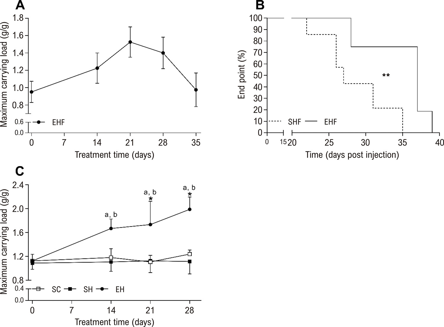
Male Wistar rats (Body weight: ~ 200 g) were used. To assess the time to potential heart failure onset (i.e., end point), rats were divided into sedentary hypertension until failure (SHF, n=6) and exercise hypertension until failure (EHF, n=6) groups. To test RT effects, rats were divided into sedentary control (SC, n = 7), sedentary hypertension (SH, n=7), and exercise hypertension (EH, n=7) groups. PAH was induced by two MCT injections (20 mg/kg, with 7 days interval). Exercise groups were submitted to an RT protocol (Ladder climbing; 55-65% of carrying maximal load), 5 times/week. Statistical significance was assumed at P < 0.05.

Results

RT prolonged the end point (~25 %), enhanced the physical effort tolerance (~ 55%), and mitigated the LV and cardiomyocyte contractility dysfunctions promoted by MCT by preserving the ejection fraction and fractional shortening, the amplitude of shortening, and the velocities of contraction and relaxation in cardiomyocytes. RT also prevented increases in left ventricle fibrosis and type I collagen caused by MCT, and maintained the type III collagen and myocyte dimensions reduced by MCT.

Conclusion

Low- to moderate-intensity RT benefits LV and cardiomyocyte contractile functions in rats during the development of MCT-induced PAH.

  • Exercise
  • Heart Failure
  • Myocytes, Cardiac
  • Physical Conditioning, Animal/methods
  • Pulmonary Hypertension
  • Rats
  • Ventricular Dysfunction, Left
Read Full Article
Creative Commons Attribution (CC BY) Creative Commons license
02/09/2022 DOI: 10.36660/abc.20210681 Views 1
Figure 1
ABCHF
Review Article

Long-Term Ventricular Assist Devices – Main Complications in Contemporary Clinical Practice

Dayanna Machado Pires Lemos et al. Gustavo Paes Silvano Kely Regina da Luz Marco Aurélio Lumertz Saffi … Marcus Vinicius Przepiorka Vieira Fernando Luis Scolari Lívia Adams Goldraich

Abstract

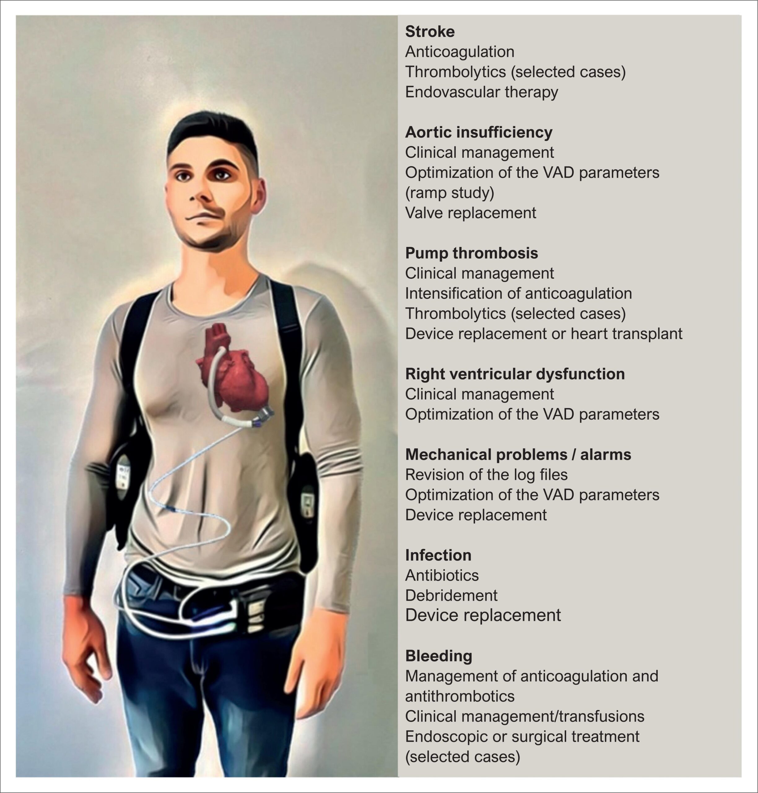
Advanced heart failure (HF) is associated with reduced quality of life and high hospitalization and mortality rates. Ventricular assist devices (VADs) promote an increase in cardiac output, and consequently improvements in body functions, functional capacity and patient survival. However, the use of VAD may be associated with complications and require systematic and specialized care. Ischemic and hemorrhagic stroke is among the most feared complications and its occurrence is related to thrombus formation in the pump. The connection between the driveline and the external power source is a potential source of infection that may extend to the mediastinum. Management of bleeding caused by anticoagulation therapy may be challenging, since discontinuation of the treatment may lead to thrombus formation. Aortic insufficiency and right ventricular dysfunction may occur, particularly in prolonged periods of support, requiring optimization of VAD parameters and clinical management. Although uncommon, mechanical failure of the VAD may occur and require replacement of the pump or even heart transplant. Thus, identification and management of the main complications of VAD in patients with advanced HF is needed, so that strategies for prevention and rigorous clinical follow-up can be implemented. This review aims to summarize the main adverse events in patients with long-term VAD.

  • Heart Failure
  • Heart-Assist Devices
  • Ventricular Dysfunction, Left
Read Full Article
Creative Commons Attribution (CC BY) Creative Commons license
29/06/2022 DOI: 10.36660/abchf.20220044 Views 1
ABCIC
Editorial

Cardiac Resynchronization Therapy 2018: An Echocardiographer’s Point of View

Luciano Belem

[…]

  • Atrial Fibrillation
  • Echocardiography
  • Heart Failure
  • Stroke Volume
  • Ventricular Dysfunction, Left
Read Full Article
Creative Commons Attribution (CC BY) Creative Commons license
04/02/2022 DOI: 10.5935/2318-8219.20180034
ABCIC
Image

Left Ventricular Hydatid Cyst

Paulo Yuri Santos de Andrade et al. Danilo Rocha Santos Caracas Magna Cristina da Silva Moreira

[…]

  • Echinoccosis
  • Echocardiography
  • Endocardium
  • Serology
  • Ventricular Dysfunction, Left
Read Full Article
Creative Commons Attribution (CC BY) Creative Commons license
04/02/2022 DOI: 10.5935/2318-8219.20170025
ABCIC
Case Report

Cardiac Amyloidosis: Restrictive Cardiomyopathy Prototype and Diastolic Dysfunction — Case Report

Thiago Augusto Vieira et al. Sandra de Barros Cobra Negreiros Diogo Wagner da Silva de Sousa Rodrigo de Freitas Garbero Carla de Oliveira Capanema

[…]

  • Amyloidosis
  • Cardiomyopathy, Restrictive
  • Drug Therapy
  • Echocardiography, Doppler
  • Ventricular Dysfunction, Left
Read Full Article
Creative Commons Attribution (CC BY) Creative Commons license
20/01/2022 DOI: 10.5935/2318-8219.20160035 Views 1
ABCIC
Original Article

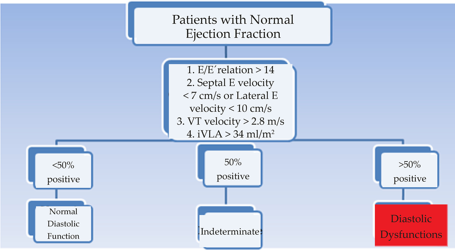
Diastolic Function Assessment with Doppler Echocardiography and Two-Dimensional Strain

José Maria Del Castillo et al. Eugenio Soares de Albuquerque Carlos Antônio da Mota Silveira Diana Patrícia Lamprea Antonia Dulcineide Medeiros Sena

Summary

Background

The evaluation of left ventricular (LV) diastolic dysfunction presents a significant number of indeterminate dysfunctions, especially when ejection fraction (EF) is preserved. Global longitudinal strain (GLS) and systolic strain rate (SSR) and early diastolic strain rate (EDSR) may be useful for reclassifying diagnosed patients.

Objective

To evaluate, using GLS, SSR and EDSR, patients with diastolic dysfunction, compare with healthy individuals, and determine the additive value of the method.

Methods

The study included 149 patients (age 62.2 ± 10.6) with diastolic dysfunction (49.7% grade 1; 15.4% grade 2; 18.1% grade 3 and 16.8% unspecified) and 189 healthy individuals (age 44.5 ± 13.3). Left ventricular (LV) and left atrial (LA) dimensions and function, mitral and tissue Doppler velocities and their ratios, GLS, SSR and EDSR have been determined. Data evaluation using the Kolmogorov-Smirnoff, Kruskal-Wallis tests, multiple regression analysis and area under the ROC curve. Data were considered significant when p < 0.05.

Results

In diastolic dysfunction, LV dimensions and thickness were increased and EF was lower. Mitral and tissue Doppler revealed abnormalities and LA volume and tricuspid regurgitation velocity were increased. GLS and EDSR were decreased in dysfunction grade 2 and 3 and EDSR was decreased in dysfunction grade 1, correlating better with diastolic dysfunction. The ROC cutoff value for the EDSR was 1.0 s-1.

Conclusion

Diastolic dysfunction supplemented with myocardial strain rate seems to add sensitivity and specificity where the diastolic function is indeterminate and may be used for reclassifying these patients. (Arq Bras Cardiol: Imagem cardiovasc. 2017;30(2):46-53)

  • Compliance/physiology
  • Coronary Artery Disease
  • Echocardiography, Doppler
  • Echocardiography, Stress
  • Ventricular Dysfunction, Left
Read Full Article
Creative Commons Attribution (CC BY) Creative Commons license
20/01/2022 DOI: 10.5935/2318-8219.20170012
ABCIC
Case Report

Type A Aortic Interruption: Case Report

Carlos José Mota de Lima et al. Danielli Oliveira da Costa Lino Acrísio Sales Valente Lúcia de Sousa Belém … Ana Carolina Brito de Alcantara Mayara Araújo Brilhante

[…]

  • Aortic Coarctation
  • Echocardiography
  • Heart Defects, Congenital
  • Ventricular Dysfunction, Left
Read Full Article
Creative Commons Attribution (CC BY) Creative Commons license
20/01/2022 DOI: 10.5935/2318-8219.20180014
ABC
Editorial

Electrocardiographic Diagnosis of Left Ventricular Hypertrophy

Claudio Leinig Pereira da Cunha

Left Ventricular Hypertrophy (LVH) is defined as an increase in left ventricular (LV) mass, which may be secondary to an increase in wall thickness (concentric LVH), increased cavity size (eccentric LVH), or both. The presentation of hypertrophied LV depends mainly on the underlying disease, with concentric LVH resulting in most cases from LV pressure overload (hypertension or aortic stenosis), while eccentric LVH mainly depends on LV volume overloads (mitral and aortic insufficiency) and dilated cardiomyopathies. Other causes of LVH include ventricular septal defects, hypertrophic cardiomyopathy, and physiological changes associated with athletic training.

The presence of LVH is clinically meaningful because it is associated with an increased incidence of heart failure, ventricular arrhythmias, peripheral vascular insufficiency, aortic dilatation, cerebrovascular events and sudden death or after myocardial infarction.

[…]

  • Cardiomyopathy, Hypertrophic
  • Diagnostic Imaging
  • Echocardiography/methods
  • Electrocardiography/methods
  • Hypertrophy, Left Ventriculat
  • Ventricular Dysfunction, Left
Read Full Article
Creative Commons Attribution (CC BY) Creative Commons license
22/11/2021 DOI: 10.36660/abc.20210868 Views 2
Figure 1
ABC
Original Article

Cardiac Fibrosis and Changes in Left Ventricle Function in Patients with Chronic Chagas Heart Disease

João Bosco de Figueiredo Santos et al. Ilan Gottlieb Eduardo Marinho Tassi Gabriel Cordeiro Camargo … Jacob Atié Sérgio Salles Xavier Roberto Coury Pedrosa Roberto Magalhães Saraiva

Abstract

Background

Chagas heart disease (CHD) is a slow progressing condition with fibrosis as the main histopathological finding.

Objectives

To study if cardiac fibrosis increases over time and correlates with increase in left ventricular (LV) size and reduction of ejection fraction (EF) in chronic CHD.

Methods

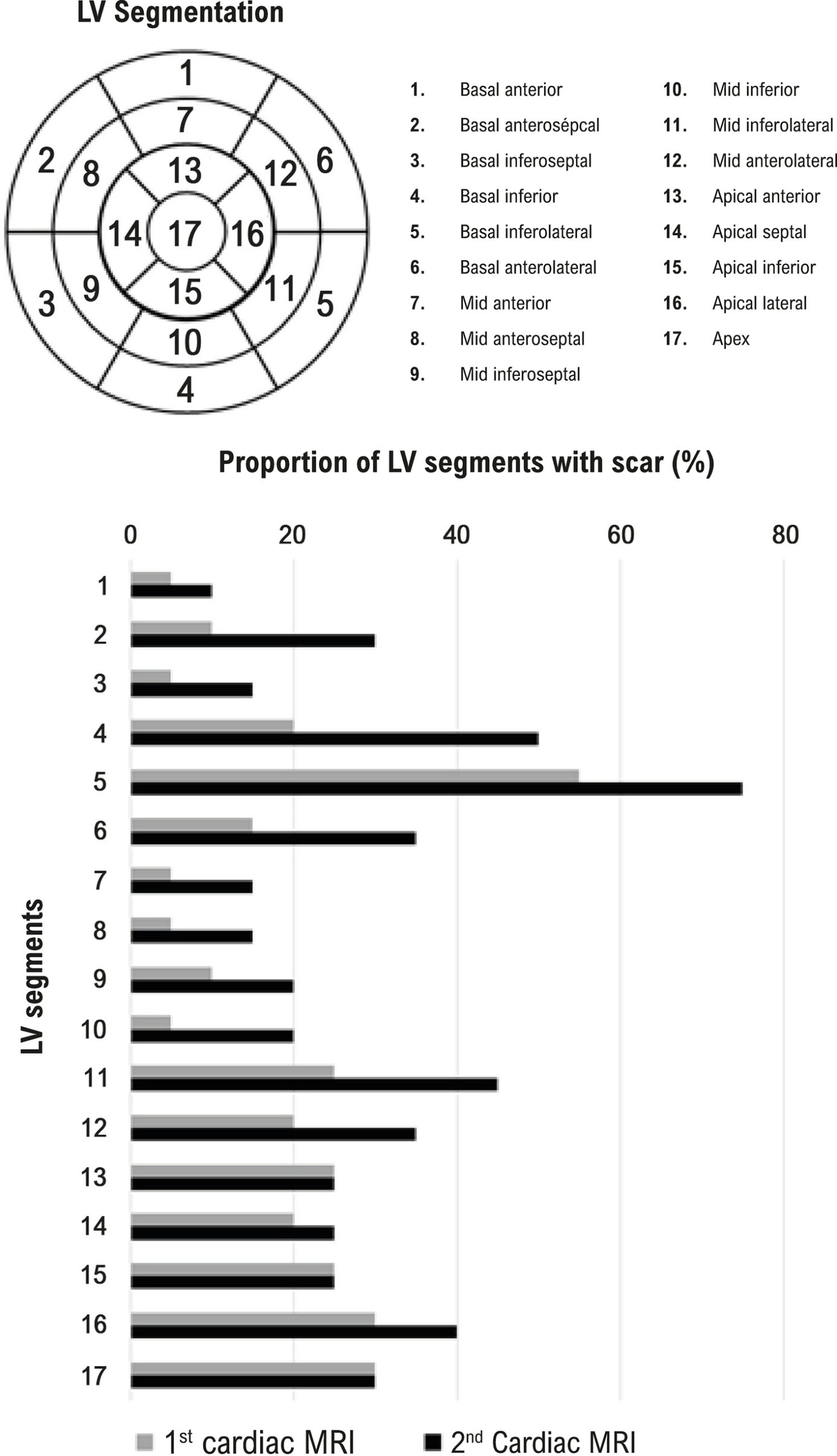
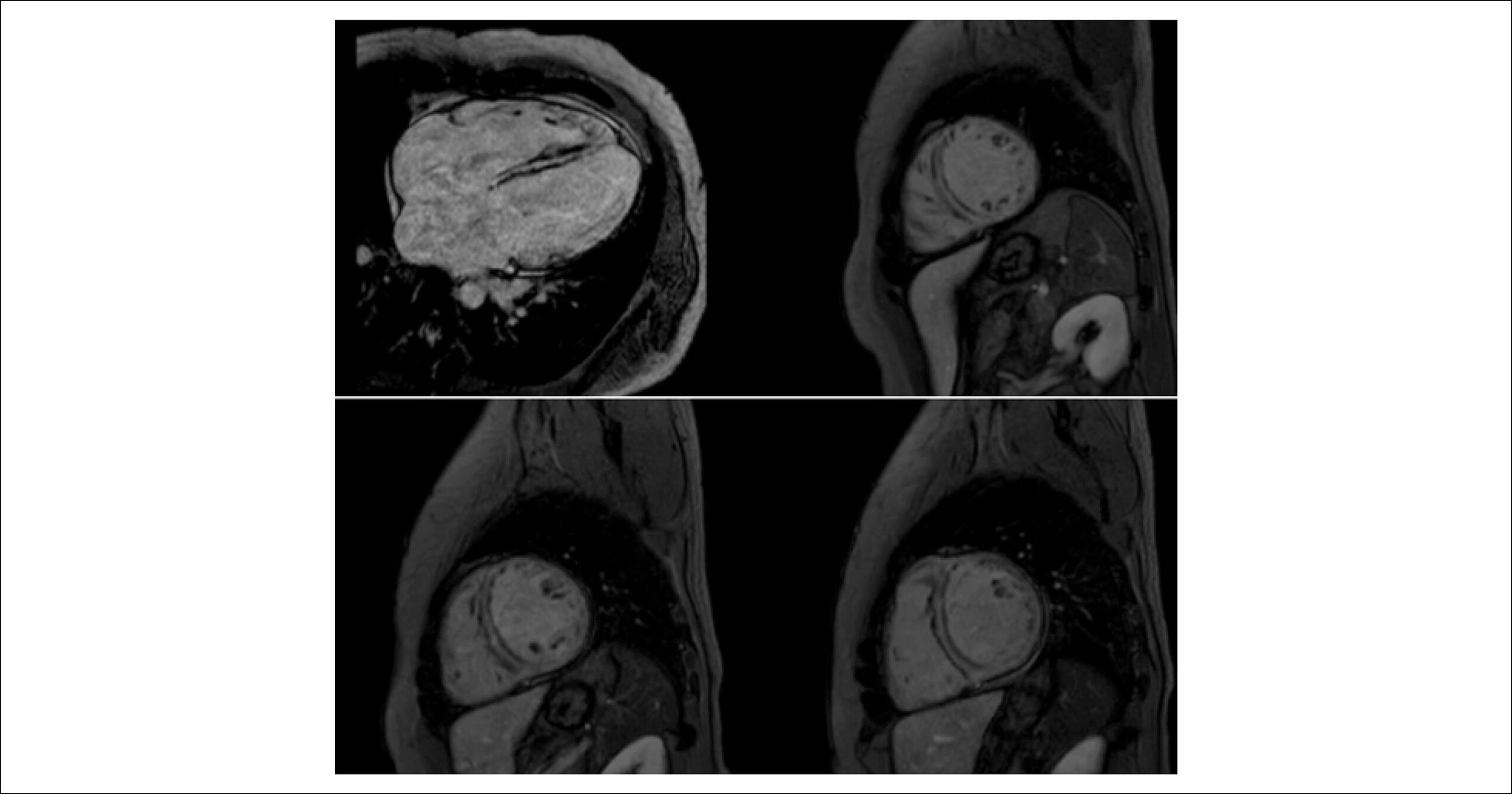
Retrospective study that included 20 individuals (50% men; 60±10 years) with chronic CHD who underwent two cardiac magnetic resonance imaging (MRI) with late gadolinium enhancement with a minimum interval of four years between tests. LV volume, EF, and fibrosis mass were determined by cardiac MRI. Associations of fibrosis mass at the first cardiac MRI and changes in LV volume and EF at the second cardiac MRI were tested using logistic regression analysis. P values <0.05 were considered significant.

Results

Patients were classified as follows: A (n=13; changes typical of CHD in the electrocardiogram and normal global and segmental LV systolic function) and B1 (n=7; LV wall motion abnormality and EF≥45%). Mean time between cardiac MRI studies was 5.4±0.5 years. LV fibrosis (in %LV mass) increased from 12.6±7.9% to 18.0±14.1% between MRI studies (p=0.02). Cardiac fibrosis mass at baseline was associated with decrease in >5 absolute units in LV EF from the first to the second MRI (OR 1.48, 95% CI 1.03-2.13, p=0.03). LV fibrosis mass was larger and increased between MRI studies in the group that presented decrease in LV EF between the tests.

Conclusions

Even patients at an initial stage of CHD show an increase in myocardial fibrosis over time, and the presence of LV fibrosis at baseline is associated with a decrease in LV systolic function.

  • Chagas Cardiomyopathy
  • Chagas disease
  • Diagnostic Imaging
  • Electrocardiography/methods
  • Endomyocardial Fibrosis
  • Magnetic Resonance Imaging/methods
  • Ventricular Dysfunction, Left
Read Full Article
Creative Commons Attribution (CC BY) Creative Commons license
08/10/2021 DOI: 10.36660/abc.20200597
ABC
Editorial

The Challenge of Assessing Sudden Cardiac Death Risk in Patients with Nonischemic Heart Failure

Pedro Pimenta de Mello Spineti et al. Rodrigo Souto da Silva Sá Bruno Reznik Wajsbrot

Sudden cardiac death (SCD) accounts for approximately 50% of deaths in patients with ischemic and nonischemic heart disease in the presence of severe left ventricular systolic dysfunction. Many clinical trials– have evaluated the effectiveness of implantable cardioverter defibrillators (ICDs) in primary prevention of SCD in the last 20 years.

The Multicenter Automatic Defibrillator Implantation Trial II (MADIT II) and the Sudden Cardiac Death in Heart Failure (SCD-HeFT) defined the principles for indication, of ICDs in primary prophylaxis of SCD of patients with heart failure (HF) of ischemic etiology. On the other hand, clinical trials evaluating patients with HF of nonischemic origin,– have shown heterogeneous results and, so far, there are uncertainties on the best candidates for this type of therapy.

[…]

  • Biomarkers
  • Death, Sudden, Cardiac
  • Endomyocardial Fibrosis
  • Galectin 3
  • Heart Failure/physiopathology
  • Mortality
  • Ventricular Dysfunction, Left
Read Full Article
Creative Commons Attribution (CC BY) Creative Commons license
20/09/2021 DOI: 10.36660/abc.20210633 Views 1
Figure 1
ABC
Original Article

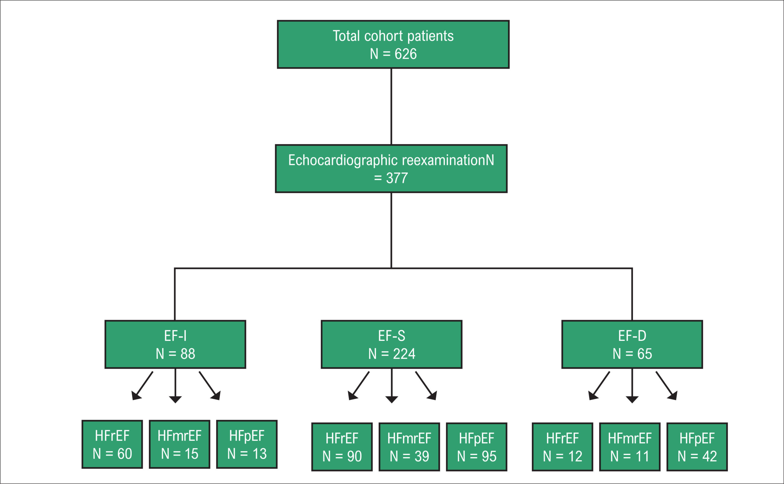
Increased, Decreased, or Stable Left Ventricle Ejection Fraction over Time in a Series of 626 Heart Failure Patients Receiving Medical Treatment

Meng-Meng Han et al. Wen-Shu Zhao Xiao-Rong Xu Xin Wang … Kui-Bao Li Cai-Jing Dang Juan Zhang Jia-Mei Liu Mu-Lei Chen Xin-Chun Yang Lin Xu Hua Wang

Abstract

Background:

Ejection fraction (EF) has been used in phenotype analyses and to make treatment decisions regarding heart failure (HF). Thus, EF has become a fundamental part of daily clinical practice.

Objective:

This study aims to investigate the characteristics, predictors, and outcomes associated with EF changes in patients with different types of severe HF.

Methods:

A total of 626 severe HF patients with New York Heart Association (NYHA) class III–IV were enrolled in this study. The patients were classified into three groups according to EF changes, namely, increased EF (EF-I), defined as an EF increase ≥10%, decreased EF (EF-D), defined as an EF decrease ≥10%, and stable EF (EF-S), defined as an EF change <10%. A p-value lower than 0.05 was considered significant.

Results:

Out of 377 severe HF patients, 23.3% presented EF-I, 59.5% presented EF-S, and 17.2% presented EF-D. The results further showed 68.2% of heart failure with reduced ejection fraction (HFrEF) in the EF-I group and 64.6% of heart failure with preserved ejection fraction (HFpEF) in the EF-D group. The predictors of EF-I included younger age, absence of diabetes, and lower left ventricular ejection fraction (LVEF). The predictors of EF-D were absence of atrial fibrillation, lower uric acid level, and higher LVEF. Within a median follow-up of 40 months, 44.8% of patients suffered from all-cause death.

Conclusion:

In severe HF, HFrEF presented the highest percentage in the EF-I group, and HFpEF was most common in the EF-D group.

  • Drug Utilization
  • Heart Failure / mortality
  • Prognosis
  • Stroke Volume
  • Ventricular Dysfunction, Left
Read Full Article
Creative Commons Attribution (CC BY) Creative Commons license
30/07/2021 DOI: 10.36660/abc.20200250
Figure 1
IJCS
Original Article

Analysis of Diastolic Left Ventricular Function in Adolescents with Juvenile Systemic Lupus Erythematosus

Talita Nolasco Loureiro et al. Cristina Ortiz Sobrinho Valete Marcia Bueno Castier Maria de Fatima Monteiro Leite Flavio Roberto Sztajnbok

Abstract

Background

Juvenile systemic lupus erythematosus (JSLE) is a chronic inflammatory disease that affects the heart in 50% of cases. The behavior of diastolic function in adolescents and the predictors of its occurrence by conventional echocardiography are poorly established.

Objectives

This study aimed to evaluate diastolic function in adolescents with JSLE and to identify possible predictors of its occurrence by conventional echocardiography.

Methods

Cross-sectional, observational, control group study in a tertiary hospital of 49 adolescents with JSLE and 49 controls, using the EACVI 2016 guideline classification. Statistical methods used were Fisher and Mann-Whitney tests. Multivariate logistic regression models were constructed. A significance level of 5% was adopted.

Results

Among 98 patients, the JSLE group had higher indexed left atrial volume (p <0.001), lower lateral E' value (p<0.001) and lower E/A ratio value (p<0.001). The diagnosis of JSLE was associated with a higher chance of increased left atrial index volume (OR 3.3; p value 0.03).

Conclusions

Based on the 2016 guideline, no diastolic dysfunction was found in JSLE. However, differences in the analyzed echocardiographic parameters were found in these adolescents.

  • Adolescent
  • Echocardiography/methods
  • Lupus Erythematosus, System
  • Ventricular Dysfunction, Left
Read Full Article
Creative Commons Attribution (CC BY) Creative Commons license
16/07/2021 DOI: 10.36660/ijcs.20200210 Views 1
Figure 1
ABC
Original Article

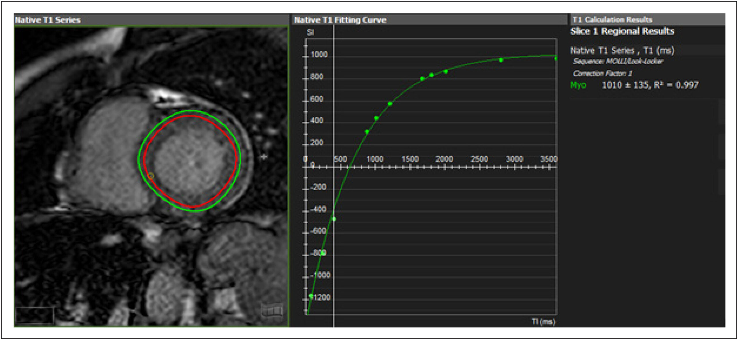
Clinical Heart Failure Stratification Through Native T1 Mapping: Experience of a Referral Service

Thiago dos Santos Silva Marques et al. André Maurício de Souza Fernandes Roberto Nery Dantas Júnior Robert W. Biederman … Ana Paula Marques de Oliveira Melo Roque Aras

Abstract

Background:

Diffuse cardiac fibrosis is an important factor in the prognostic assessment of patients with ventricular dysfunction. Cardiovascular magnetic resonance imaging (CMR) native T1 mapping is highly sensitive and considered an independent predictor of all-cause mortality and heart failure (HF) development in patients with cardiomyopathy.

Objectives:

To evaluate the feasibility of native T1 mapping assessment in patients with HF in a cardiology referral hospital and its association with structural parameters and functional profile.

Methods:

Cross-sectional study with adult patients with HF NYHA functional classes I and II, ischemic and non-ischemic, followed in a referral hospital, who underwent CMR. Native T1 values were analyzed for structural parameters, comorbidities, etiology, and categorization of HF by left ventricular ejection fraction (LVEF). Analyses were performed with a significance level of 5%.

Results:

Enrollment of 134 patients. Elevated native T1 values were found in patients with greater dilation (1004.9 vs 1042.7ms, p = 0.001), ventricular volumes (1021.3 vs 1050.3ms, p <0.01) and ventricular dysfunction (1010.1 vs 1053.4ms, p <0.001), also present when the non-ischemic group was analyzed separately. Patients classified as HF with reduced ejection fraction had higher T1 values than those with HF and preserved ejection fraction (HFPEF) (992.7 vs 1054.1ms, p <0.001). Of those with HFPEF, 55.2% had higher T1.

Conclusions:

CMR T1 mapping is feasible for clinical HF evaluation. There was a direct association between higher native T1 values and lower ejection fraction, and with larger LV diameters and volumes, regardless of the etiology of HF.

  • Cardiomyopathy, Dilated
  • Chagas Cardiomyopathy
  • Diagnosis Imaging
  • Fibrosis
  • Heart Failure
  • Magnetic Resonance Spectroscopy/ methods
  • Ventricular Dysfunction, Left
Read Full Article
Creative Commons Attribution (CC BY) Creative Commons license
17/05/2021 DOI: 10.36660/abc.20190782 Views 1
Figure 1
IJCS
Editorial

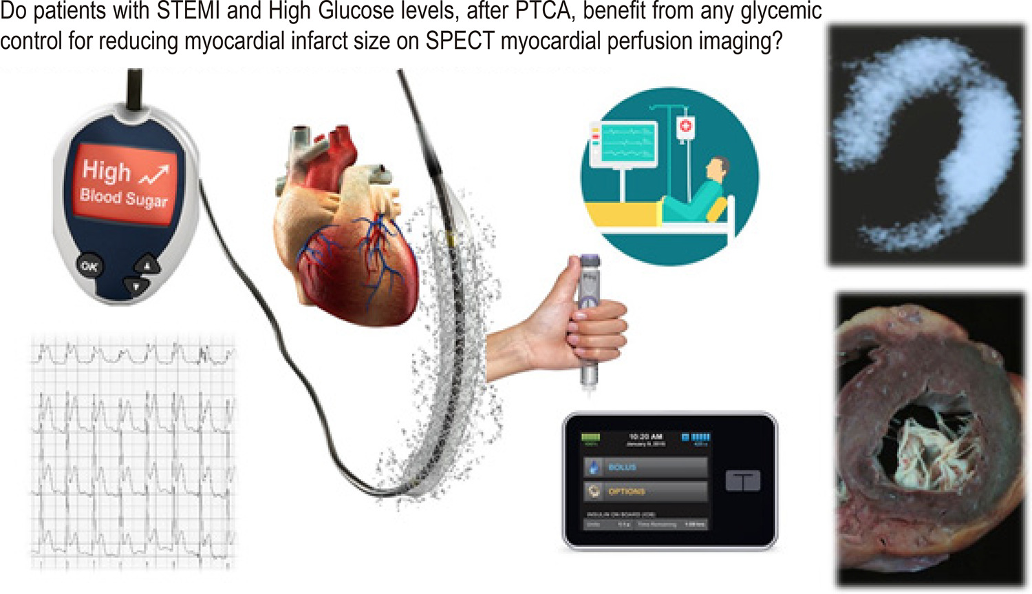
After a STEMI, is Less Sugar more Protective to Myocardium?

Rafael Willain Lopes et al. Alexandre Hohl

The infarct size reduction in acute myocardial infarction (AMI) has been studied for more than 50 years. Animal experiments were conducted in the 1970s, followed by the first clinical studies to reduce infarct size (IS), with fibrinolytic agents or with mechanical coronary angioplasty (PTCA). The clinical experience of coronary reperfusion indicated that left ventricle function was not normalized in 30% of the patients, and despite unblocking the epicardial vessel, demonstrated hemodynamically, there was no equivalent in myocardial perfusion. New concepts emerged such as reperfusion injury, microvascular dysfunction, “no-reflow” phenomenon, and stunned and hibernating myocardium, which became the focus of basic research and clinical investigation. To improve characterization, different technologies were used, such as contrast echocardiography, isotopic studies, including positron emission tomography, and magnetic resonance.

Cardioprotection (CP) aims to reduce IS and improve clinical outcomes. The translation of CP from preclinical and promising proof-of-concept studies to clinical benefit (CB) for patients has been quite unsatisfactory. Almost all these studies that did not translate into CB had infarct size reduction as the primary endpoint and used protocols selected to achieve IS reduction.

[…]

  • Angioplasty
  • Diagnostic Imaging
  • Fibrinolytic Agents
  • Myocardial Reperfusion
  • Myocardial Stunning
  • Percutaneous Coronary Intervention
  • ST Elevation Myocardial Infarction
  • Ventricular Dysfunction, Left
Read Full Article
Creative Commons Attribution (CC BY) Creative Commons license
09/10/2020 DOI: 10.36660/ijcs.20200173 Views 1
ABC
Editorial

Is Grace Risk Score the Holy Grail in Risk Stratification or Can We Improve it Even Further with Additional Biomarkers?

Ana Teresa Timóteo

B-type natriuretic peptide (BNP) has been recognized as a very useful marker for the detection of acute and chronic left ventricular dysfunction, both systolic and diastolic, that can be present in the context of sudden and prolonged myocardial ischemia. , These are the first steps in the ischemic cascade, leading to cell necrosis. For that reason, natriuretic peptides are usually elevated in the context of acute coronary syndromes.

Myocardial ischemia, even in the absence of left ventricular dysfunction, augments cardiac BNP gene expression, increasing plasma NT-proBNP concentrations. , BNP kinetics usually peaks at 16 hours of symptom’s onset in ST-elevation myocardial infarction and a second peak is usually observed by the fifth day. We can speculate that the first peak might be associated with ischemia and the second peak to left ventricular dysfunction associated with cell necrosis and early remodelling.

[…]

  • Acute coronary syndrome
  • Biomarkers
  • Brain
  • Factor, Natriuretic Atrial
  • Heart Failure
  • Mortality
  • Natriuretic Peptide
  • Ventricular Dysfunction, Left
Read Full Article
Creative Commons Attribution (CC BY) Creative Commons license
29/05/2020 DOI: 10.36660/abc.20200171 Views 2
Figure 1
ABC
Original Article

Prognostic Value of NT-proBNP versus Killip Classification in Patients with Acute Coronary Syndromes

Thiago M. B. Souza et al. Antônio Maurício S. Cerqueira Jr. Jessica G. Suerdieck Nicole C. de Sá … Gabriella S. Sodré Vitor C. A. Correia Yasmin F. Lacerda Leticia L. Fonseca Marcia M. Noya-Rabelo Luis C. L. Correia

Abstract

Background:

Plasma levels of brain natriuretic peptides have better diagnostic accuracy compared to clinical-radiologic judgment for acute heart failure. In acute coronary syndromes (ACS), the prognostic value of acute heart failure is incorporated into predictive models through Killip classification. It is not established whether NT-proBNP could increment prognostic prediction.

Objective:

To evaluate whether NT-proBNP, as a measure of left ventricular dysfunction, improves the in-hospital prognostic value of the GRACE score in ACS.

Methods:

Patients admitted due to acute chest pain, with electrocardiogram and/or troponin criteria for ACS were included in the study. The plasma level of NT-proBNP was measured at hospital admission and the primary endpoint was defined as cardiovascular death during hospitalization. P-value < 0.05 was considered as significant.

Results:

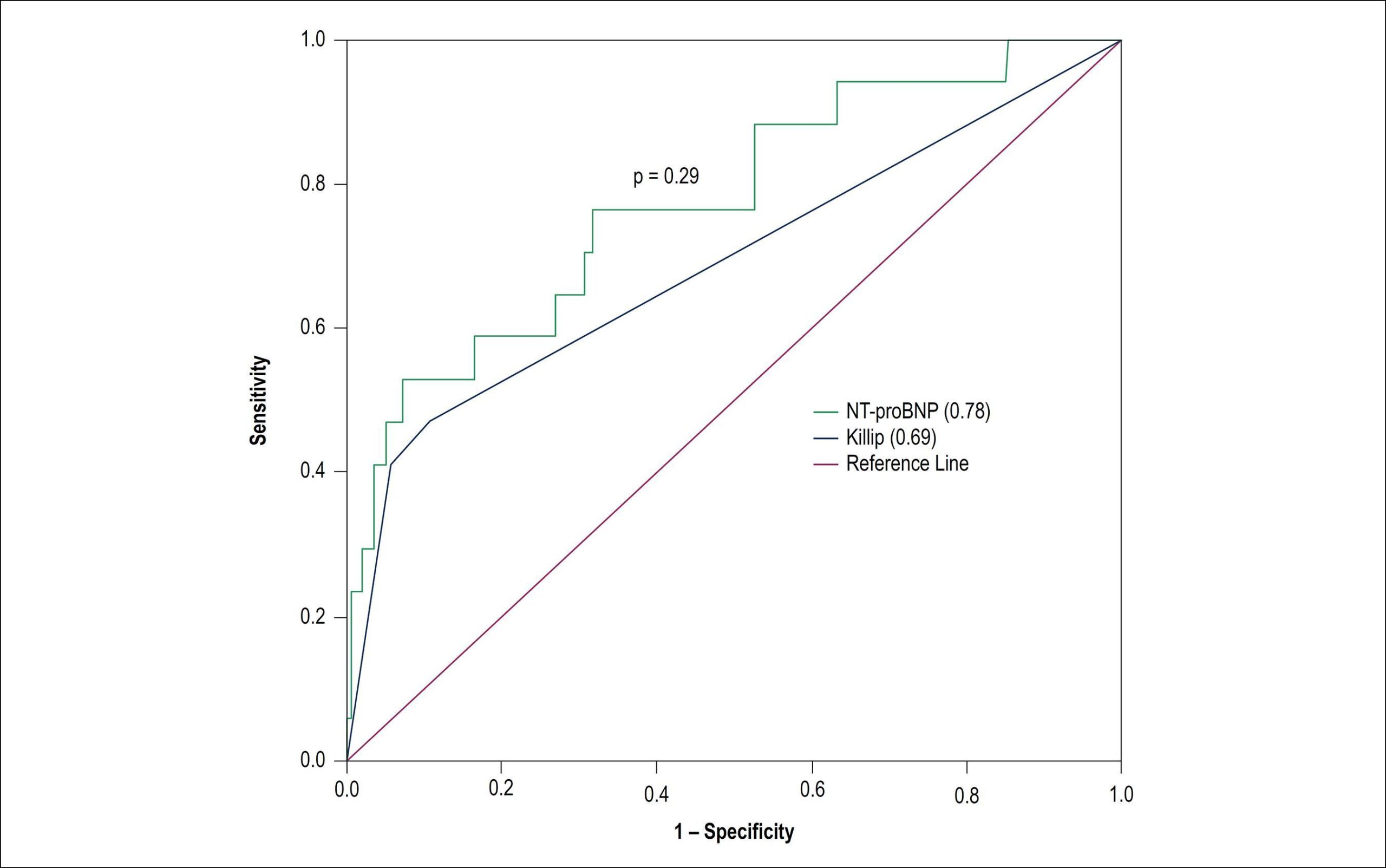
Among 352 patients studied, cardiovascular mortality was 4.8%. The predictive value of NT-proBNP for cardiovascular death was shown by a C-statistic of 0.78 (95% CI = 0.65-0.90). After adjustment for the GRACE model subtracted by Killip variable, NT-proBNP remained independently associated with cardiovascular death (p = 0.015). However, discrimination by the GRACE-BNP logistic model (C-statistics = 0.83; 95%CI = 0.69-0.97) was not superior to the traditional GRACE Score with Killip (C-statistic = 0.82; 95%CI = 0.68-0.97). The GRACE-BNP model did not provide improvement in the classification of patients to high risk by the GRACE Score (net reclassification index = – 0.15; p = 0.14).

Conclusion:

Despite the statistical association with cardiovascular death, there was no evidence that NT-proBNP increments the prognostic value of GRACE score in ACS.

  • Acute coronary syndrome
  • Biomarkers
  • Heart Failure
  • Mortality
  • Natriuretic Peptide, Brain
  • Ventricular Dysfunction, Left
Read Full Article
Creative Commons Attribution (CC BY) Creative Commons license
14/02/2020 DOI: 10.36660/abc.20180345 Views 1
ABC
Editorial

Admission NT-ProBNP in Myocardial Infarction: an Alert Sign?

Luís Beck-da-Silva

This issue of Arquivos Brasileiros de Cardiologia brings a paper entitled “The Usefulness of Admission Plasma NT-pro BNP Level to Predict Left Ventricular Aneurysm Formation after Acute ST-Segment Elevation Myocardial Infarction”. The authors bring a cohort of 1,519 post-acute ST-segment elevation myocardial infarction (STEMI) who were followed-uP for at least six months. Despite its observational and retrograde design, the authors were straightforward in looking for predictive variables that could foresee the occurrence of left ventricular aneurysms (LVA). Among other major clinical aspects such as previous coronary artery bypass graft, post-MI heart failure, younger age, smoking and no-reflow phenomenon; authors highlighted the importance of high NT-proBNP at admission as a predictor of LVA formation after acute STEMI.

I would probably highlight one weakness and a potentially positive aspect of their work.

[…]

  • Echocardiography/diagnostic Imaging
  • Heart Failure
  • Magnetic Resonance Spectroscopy/ methods
  • Myocardial Infarction
  • Natriuretic Peptide Brain/metabolism
  • Natriuretic Peptides/metabolismo
  • Ventricular Dysfunction, Left
Read Full Article
Creative Commons Attribution (CC BY) Creative Commons license
02/12/2019 DOI: 10.36660/abc.20190685 Views 1
ABC
Editorial

At Last, a Sensitive Method to Detect Incipient Systolic Dysfunction!

Carlos Eduardo Suaide Silva

Left ventricular systolic function evaluation has always been one of the main attributions of echocardiography. The degree of ventricular systolic dysfunction is an important predictor of outcome for a large number of diseases, including ischemic heart disease, cardiomyopathy, valvular heart disease, and congenital heart disease. In this area, the ejection fraction has been sovereign for many decades, but despite being a parameter that can, in most cases, inform the real state of global ventricular function, in many situations it may be normal in the presence of evident systolic or diastolic dysfunction.

Known clinical examples are the several cases of heart failure with preserved ejection fraction (HFpEF) or patients with hypertrophic cardiomyopathy, among others. For this reason, an index that can identify early ventricular dysfunction that is not connected with the ejection fraction has been sought for a long time.

[…]

  • Antiretroviral Therapy Highly Active
  • Cardiomyopathy, Hypertrophic
  • Echocardiography/methods
  • Heart Failure
  • HIV
  • Myocardial Contraction
  • Ventricular Dysfunction, Left
Read Full Article
Creative Commons Attribution (CC BY) Creative Commons license
04/11/2019 DOI: 10.5935/abc.20190198 Views 1
Figure 1
ABC
Review Article

Importance of Genetic Testing in Dilated Cardiomyopathy: Applications and Challenges in Clinical Practice

Arsonval Lamounier Júnior et al. Filipe Ferrari Renato Max Luiz Eduardo Fonteles Ritt Ricardo Stein

Abstract

Dilated cardiomyopathy (DCM) is a clinical syndrome characterized by left ventricular dilatation and contractile dysfunction. It is the most common cause of heart failure in young adults. The advent of next-generation sequencing has contributed to the discovery of a large amount of genomic data related to DCM. Mutations involving genes that encode cytoskeletal proteins, the sarcomere, and ion channels account for approximately 40% of cases previously classified as idiopathic DCM. In this scenario, geneticists and cardiovascular genetics specialists have begun to work together, building knowledge and establishing more accurate diagnoses. However, proper interpretation of genetic results is essential and multidisciplinary teams dedicated to the management and analysis of the obtained information should be considered. In this review, we approach genetic factors associated with DCM and their prognostic relevance and discuss how the use of genetic testing, when well recommended, can help cardiologists in the decision-making process.

  • Cardiomyopathy, Dilated/genetics
  • Genetic Testing/methods
  • Heart Failure
  • Heart Transplantation
  • Ventricular Dysfunction, Left
Read Full Article
Creative Commons Attribution (CC BY) Creative Commons license
02/09/2019 DOI: 10.5935/abc.20190144
Figure 1
ABC
Original Article

Global Longitudinal Strain Predicts Poor Functional Capacity in Patients with Systolic Heart Failure

Rafael José Coelho Maia et al. Simone Cristina Soares Brandão Jéssica Leite Giordano Bruno Parente … Filipe Pinheiro Bruna Thays Santana Araújo Maria Inês Remígio Aguiar Sílvia Marinho Martins Daniella Cunha Brandão Armele Dornelas de Andrade

Abstract

Background:

Left ventricular global longitudinal strain value (GLS) can predict functional capacity in patients with preserved left ventricular ejection fraction (LVEF) heart failure (HF) and to assess prognosis in reduced LVEF HF.

Objetive:

Correlate GLS with parameters of Cardiopulmonary Exercise Test (CPET) and to assess if they could predict systolic HF patients that are more appropriated to be referred to heart transplantation according to CPET criteria.

Methods:

Systolic HF patients with LVEF < 45%, NYHA functional class II and III, underwent prospectively CPET and echocardiography with strain analysis. LVEF and GLS were correlated with the following CPET variables: maxVO2, VE/VCO2 slope, heart rate reduction during the first minute of recovery (HRR) and time needed to reduce maxVO2 in 50% after physical exercise (T1/2VO2). ROC curve analysis of GLS to predict VO2 < 14 mL/kg/min and VE/VCO2 slope > 35 (heart transplantation’s criteria) was performed.

Results:

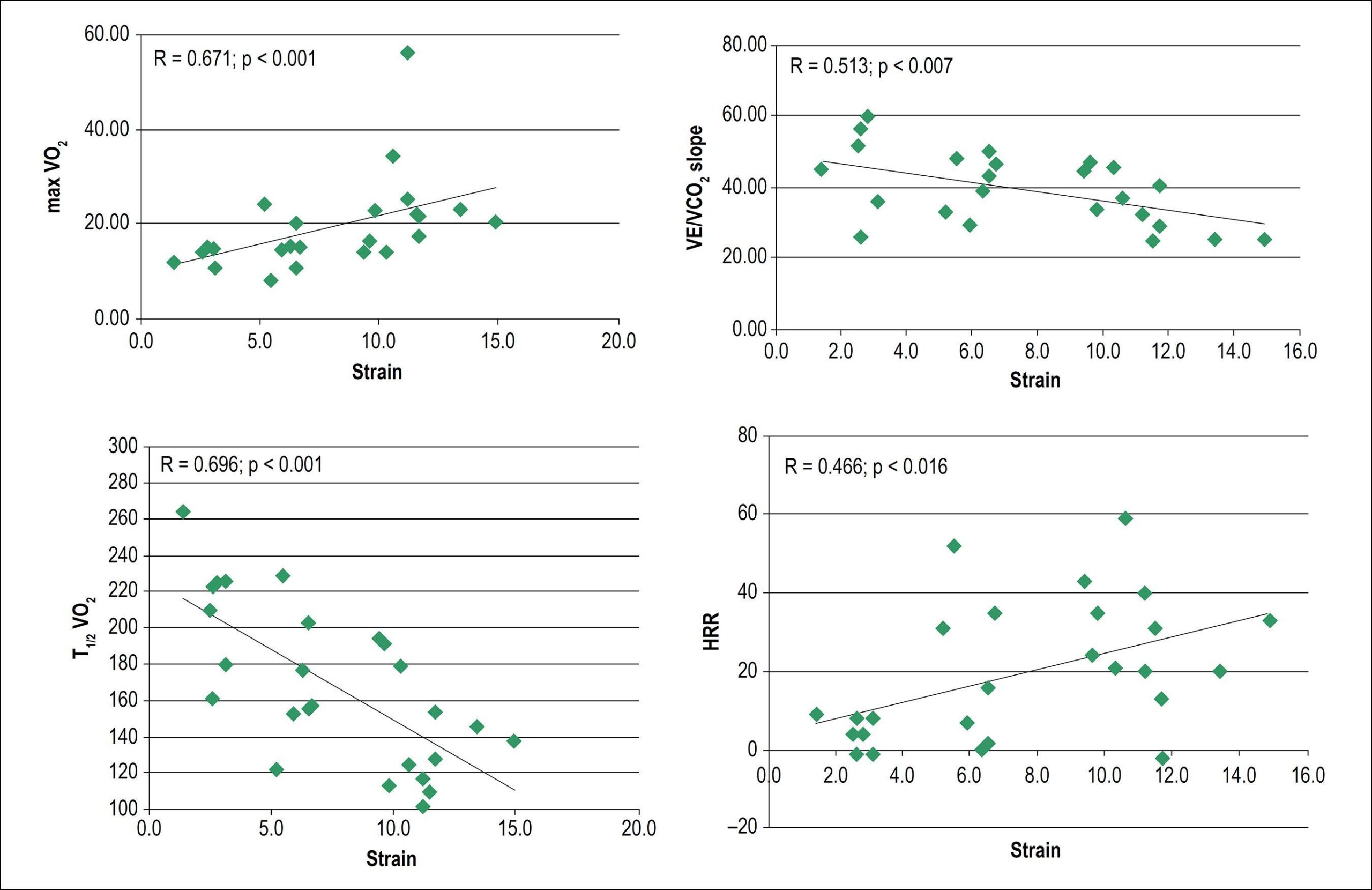
Twenty six patients were selected (age, 47 ± 12 years, 58% men, mean LVEF = 28 ± 8%). LVEF correlated only with maxVO2 and T1/2VO2. GLS correlated to all CPET variables (maxVO2: r = 0.671, p = 0.001; VE/VCO2 slope: r = -0.513, p = 0.007; HRR: r = 0.466, p = 0.016, and T1/2VO2: r = -0.696, p = 0.001). GLS area under the ROC curve to predict heart transplantation’s criteria was 0.88 (sensitivity 75%, specificity 83%) for a cut-off value of -5.7%, p = 0.03.

Conclusion:

GLS was significantly associated with all functional CPET parameters. It could classify HF patients according to the functional capacity and may stratify which patients have a poor prognosis and therefore to deserve more differentiated treatment, such as heart transplantation.

  • Echocardiography, Doppler/methods
  • Heart Failure
  • Longitudinal Strain
  • Torsion Abnormality
  • Torsion, Mechanical
  • Ventricular Dysfunction, Left
Read Full Article
Creative Commons Attribution (CC BY) Creative Commons license
18/07/2019 DOI: 10.5935/abc.20190119

Could not load more articles. Please try again.

How to cite this article

EndNote RIS

Electronic Document Format (Vancouver)

Souza AC, Drubi SA, Junqueira BMI, Guedes RASP, Almeida Filho AC, Macêdo CT. Comparative Analysis of Myocardial Work After Decongestion Therapy in a Patient With Acutely Decompensated HFrEF. ABC Imagem Cardiovascular 2023;36(2):e20230008.

Electronic Document Format (ABNT)

Souza, Alexandre Costa; Drubi, Stephanie de Azevedo; Junqueira, Bruna de Mattos Ivo; Guedes, Ricardo André Sales Pereira; Almeida Filho, Adriano Chaves de; Macêdo, Carolina Thé. Comparative Analysis of Myocardial Work After Decongestion Therapy in a Patient With Acutely Decompensated HFrEF. ABC Imagem Cardiovascular, v. 36, n. 2, e20230008, Jun. 2023.

Electronic Document Format (APA)

Souza, A. C., Drubi, S. A., Junqueira, B. M. I., Guedes, R. A. S. P., Almeida Filho, A. C. , & Macêdo, C. T. (2023). Comparative Analysis of Myocardial Work After Decongestion Therapy in a Patient With Acutely Decompensated HFrEF. ABC Imagem Cardiovascular, 36(2), e20230008.

Electronic Document Format (ISO)

Souza, Alexandre Costa and Drubi, Stephanie de Azevedo and Junqueira, Bruna de Mattos Ivo and Guedes, Ricardo André Sales Pereira and Almeida Filho, Adriano Chaves de and Macêdo, Carolina Thé. Comparative Analysis of Myocardial Work After Decongestion Therapy in a Patient With Acutely Decompensated HFrEF. ABC Imagem Cardiovascular [online]. 2023, vol. 36, n. 2, [cited 2026-10-14], e20230008. Available from: <https://journals.cardiol.br/en/article/comparative-analysis-of-myocardial-work-after-decongestion-therapy-in-a-patient-with-acutely-decompensated-hfref/>. ISSN 2318-8219

EndNote RIS

Electronic Document Format (Vancouver)

Neves MF. Renin-Angiotensin System Inhibition and Beta Blockade Adrenergic May Be Useful to Attenuate Cardiotoxicity by Anthracyclines. Arquivos Brasileiros de Cardiologia 2023;120(5):e20230280.

Electronic Document Format (ABNT)

Neves, Mario Fritsch. Renin-Angiotensin System Inhibition and Beta Blockade Adrenergic May Be Useful to Attenuate Cardiotoxicity by Anthracyclines. Arquivos Brasileiros de Cardiologia, v. 120, n. 5, e20230280, Jun. 2023.

Electronic Document Format (APA)

Neves, M. F. (2023). Renin-Angiotensin System Inhibition and Beta Blockade Adrenergic May Be Useful to Attenuate Cardiotoxicity by Anthracyclines. Arquivos Brasileiros de Cardiologia, 120(5), e20230280.

Electronic Document Format (ISO)

Neves, Mario Fritsch. Renin-Angiotensin System Inhibition and Beta Blockade Adrenergic May Be Useful to Attenuate Cardiotoxicity by Anthracyclines. Arquivos Brasileiros de Cardiologia [online]. 2023, vol. 120, n. 5, [cited 2026-10-14], e20230280. Available from: <https://journals.cardiol.br/en/article/renin-angiotensin-system-inhibition-and-beta-blockade-adrenergic-may-be-useful-to-attenuate-cardiotoxicity-by-anthracyclines/>. ISSN 0066-782X

EndNote RIS

Electronic Document Format (Vancouver)

Lorenzo N, Hernandez V, Montes A, Rivero F, Reyes* G, Aguilar R. Left Ventricular Thrombosis and Pulmonary Thromboembolism in an Asymptomatic Covid-19 Patient. Arquivos Brasileiros de Cardiologia 2022;119(6):996-8.

Electronic Document Format (ABNT)

Lorenzo, Natalia; Hernandez, Veronica; Montes, Alvaro; Rivero, Fernando; Reyes*, Guillermo; Aguilar, Rio. Left Ventricular Thrombosis and Pulmonary Thromboembolism in an Asymptomatic Covid-19 Patient. Arquivos Brasileiros de Cardiologia, v. 119, n. 6, p. 996-998, Dec. 2022.

Electronic Document Format (APA)

Lorenzo, N., Hernandez, V., Montes, A., Rivero, F., Reyes*, G., & Aguilar, R. (2022). Left Ventricular Thrombosis and Pulmonary Thromboembolism in an Asymptomatic Covid-19 Patient. Arquivos Brasileiros de Cardiologia, 119(6), 996-998.

Electronic Document Format (ISO)

Lorenzo, Natalia and Hernandez, Veronica and Montes, Alvaro and Rivero, Fernando and Reyes*, Guillermo and Aguilar, Rio. Left Ventricular Thrombosis and Pulmonary Thromboembolism in an Asymptomatic Covid-19 Patient. Arquivos Brasileiros de Cardiologia [online]. 2022, vol. 119, n. 6, [cited 2026-10-14], pp. 996-998. Available from: <https://journals.cardiol.br/en/article/left-ventricular-thrombosis-and-pulmonary-thromboembolism-in-an-asymptomatic-covid-19-patient/>. ISSN 0066-782X

EndNote RIS

Electronic Document Format (Vancouver)

Mendes GS, Mesquita AE, Rocha B, Abecasis J, Ramos S, Trabulo M. Non-Atherosclerotic Coronary and Vascular Disease Case Report: Searching for a Rare Clinical Entity. Arquivos Brasileiros de Cardiologia 2022;119(3):488-95.

Electronic Document Format (ABNT)

Mendes, Gustavo Sá; Mesquita, António Epifânio; Rocha, Bruno; Abecasis, João; Ramos, Sancia; Trabulo, Marisa. Non-Atherosclerotic Coronary and Vascular Disease Case Report: Searching for a Rare Clinical Entity. Arquivos Brasileiros de Cardiologia, v. 119, n. 3, p. 488-495, Sep. 2022.

Electronic Document Format (APA)

Mendes, G. S., Mesquita, A. E., Rocha, B., Abecasis, J., Ramos, S., & Trabulo, M. (2022). Non-Atherosclerotic Coronary and Vascular Disease Case Report: Searching for a Rare Clinical Entity. Arquivos Brasileiros de Cardiologia, 119(3), 488-495.

Electronic Document Format (ISO)

Mendes, Gustavo Sá and Mesquita, António Epifânio and Rocha, Bruno and Abecasis, João and Ramos, Sancia and Trabulo, Marisa. Non-Atherosclerotic Coronary and Vascular Disease Case Report: Searching for a Rare Clinical Entity. Arquivos Brasileiros de Cardiologia [online]. 2022, vol. 119, n. 3, [cited 2026-10-14], pp. 488-495. Available from: <https://journals.cardiol.br/en/article/non-atherosclerotic-coronary-and-vascular-disease-case-report-searching-for-a-rare-clinical-entity/>. ISSN 0066-782X

EndNote RIS

Electronic Document Format (Vancouver)

Soares LL, Leite LB, Ervilha LOG, Silva BAF, Freitas MO, Portes AMO, Rezende LMT, et al. Resistance Exercise Training Mitigates Left Ventricular Dysfunctions in Pulmonary Artery Hypertension Model. Arquivos Brasileiros de Cardiologia 2022;119(4):574-8.

Electronic Document Format (ABNT)

Soares, Leôncio Lopes; Leite, Luciano Bernardes; Ervilha, Luiz Otávio Guimarães; Silva, Bruna Aparecida Fonseca da; Freitas, Maíra Oliveira de; Portes, Alexandre Martins Oliveira; Rezende, Leonardo Mateus Teixeira; Drummond, Filipe Rios; Carneiro-Júnior, Miguel Araújo; Neves, Mariana Machado; Reis, Emily Correna Carlo; Natali, Antônio José. Resistance Exercise Training Mitigates Left Ventricular Dysfunctions in Pulmonary Artery Hypertension Model. Arquivos Brasileiros de Cardiologia, v. 119, n. 4, p. 574-584, Sep. 2022.

Electronic Document Format (APA)

Soares, L. L., Leite, L. B., Ervilha, L. O. G., Silva, B. A. F. , Freitas, M. O. , Portes, A. M. O., Rezende, L. M. T., Drummond, F. R., Carneiro-Júnior, M. A., Neves, M. M., Reis, E. C. C., & Natali, A. J. (2022). Resistance Exercise Training Mitigates Left Ventricular Dysfunctions in Pulmonary Artery Hypertension Model. Arquivos Brasileiros de Cardiologia, 119(4), 574-584.

Electronic Document Format (ISO)

Soares, Leôncio Lopes and Leite, Luciano Bernardes and Ervilha, Luiz Otávio Guimarães and Silva, Bruna Aparecida Fonseca da and Freitas, Maíra Oliveira de and Portes, Alexandre Martins Oliveira and Rezende, Leonardo Mateus Teixeira and Drummond, Filipe Rios and Carneiro-Júnior, Miguel Araújo and Neves, Mariana Machado and Reis, Emily Correna Carlo and Natali, Antônio José. Resistance Exercise Training Mitigates Left Ventricular Dysfunctions in Pulmonary Artery Hypertension Model. Arquivos Brasileiros de Cardiologia [online]. 2022, vol. 119, n. 4, [cited 2026-10-14], pp. 574-584. Available from: <https://journals.cardiol.br/en/article/resistance-exercise-training-mitigates-left-ventricular-dysfunctions-in-pulmonary-artery-hypertension-model/>. ISSN 0066-782X

EndNote RIS

Electronic Document Format (Vancouver)

Lemos DMP, Silvano GP, Luz KR, Saffi MAL, Vieira MVP, Scolari FL, Goldraich LA, et al. Long-Term Ventricular Assist Devices – Main Complications in Contemporary Clinical Practice. ABC: Heart Failure & Cardiomyopathy 2022;2(2):182-91.

Electronic Document Format (ABNT)

Lemos, Dayanna Machado Pires; Silvano, Gustavo Paes; Luz, Kely Regina da; Saffi, Marco Aurélio Lumertz; Vieira, Marcus Vinicius Przepiorka; Scolari, Fernando Luis; Goldraich, Lívia Adams. Long-Term Ventricular Assist Devices – Main Complications in Contemporary Clinical Practice. ABC: Heart Failure & Cardiomyopathy, v. 2, n. 2, p. 182-191, Jun. 2022.

Electronic Document Format (APA)

Lemos, D. M. P., Silvano, G. P., Luz, K. R. , Saffi, M. A. L., Vieira, M. V. P., Scolari, F. L., & Goldraich, L. A. (2022). Long-Term Ventricular Assist Devices – Main Complications in Contemporary Clinical Practice. ABC: Heart Failure & Cardiomyopathy, 2(2), 182-191.

Electronic Document Format (ISO)

Lemos, Dayanna Machado Pires and Silvano, Gustavo Paes and Luz, Kely Regina da and Saffi, Marco Aurélio Lumertz and Vieira, Marcus Vinicius Przepiorka and Scolari, Fernando Luis and Goldraich, Lívia Adams. Long-Term Ventricular Assist Devices – Main Complications in Contemporary Clinical Practice. ABC: Heart Failure & Cardiomyopathy [online]. 2022, vol. 2, n. 2, [cited 2026-10-14], pp. 182-191. Available from: <https://journals.cardiol.br/en/article/long-term-ventricular-assist-devices-main-complications-in-contemporary-clinical-practice/>

EndNote RIS

Electronic Document Format (Vancouver)

Belem L. Cardiac Resynchronization Therapy 2018: An Echocardiographer’s Point of View. Arquivos Brasileiros de Cardiologia Imagem cardiovascular 2022;31(4):222-4.

Electronic Document Format (ABNT)

Belem, Luciano. Cardiac Resynchronization Therapy 2018: An Echocardiographer’s Point of View. Arquivos Brasileiros de Cardiologia Imagem cardiovascular, v. 31, n. 4, p. 222-224, Feb. 2022.

Electronic Document Format (APA)

Belem, L. (2022). Cardiac Resynchronization Therapy 2018: An Echocardiographer’s Point of View. Arquivos Brasileiros de Cardiologia Imagem cardiovascular, 31(4), 222-224.

Electronic Document Format (ISO)

Belem, Luciano. Cardiac Resynchronization Therapy 2018: An Echocardiographer’s Point of View. Arquivos Brasileiros de Cardiologia Imagem cardiovascular [online]. 2022, vol. 31, n. 4, [cited 2026-10-14], pp. 222-224. Available from: <https://journals.cardiol.br/en/article/cardiac-resynchronization-therapy-2018-an-echocardiographers-point-of-view/>. ISSN 2318-8219

EndNote RIS

Electronic Document Format (Vancouver)

Andrade PYS, Caracas DRS, Moreira MCS. Left Ventricular Hydatid Cyst. Arquivos Brasileiros de Cardiologia Imagem cardiovascular 2022;30(3):116-7.

Electronic Document Format (ABNT)

Andrade, Paulo Yuri Santos de; Caracas, Danilo Rocha Santos; Moreira, Magna Cristina da Silva. Left Ventricular Hydatid Cyst. Arquivos Brasileiros de Cardiologia Imagem cardiovascular, v. 30, n. 3, p. 116-117, Feb. 2022.

Electronic Document Format (APA)

Andrade, P. Y. S. , Caracas, D. R. S., & Moreira, M. C. S. (2022). Left Ventricular Hydatid Cyst. Arquivos Brasileiros de Cardiologia Imagem cardiovascular, 30(3), 116-117.

Electronic Document Format (ISO)

Andrade, Paulo Yuri Santos de and Caracas, Danilo Rocha Santos and Moreira, Magna Cristina da Silva. Left Ventricular Hydatid Cyst. Arquivos Brasileiros de Cardiologia Imagem cardiovascular [online]. 2022, vol. 30, n. 3, [cited 2026-10-14], pp. 116-117. Available from: <https://journals.cardiol.br/en/article/left-ventricular-hydatid-cyst/>. ISSN 2318-8219

EndNote RIS

Electronic Document Format (Vancouver)

Vieira TA, Negreiros SBC, Sousa DWS, Garbero RF, Capanema CO. Cardiac Amyloidosis: Restrictive Cardiomyopathy Prototype and Diastolic Dysfunction — Case Report. Arquivos Brasileiros de Cardiologia Imagem cardiovascular 2022;30(1):13-7.

Electronic Document Format (ABNT)

Vieira, Thiago Augusto; Negreiros, Sandra de Barros Cobra; Sousa, Diogo Wagner da Silva de; Garbero, Rodrigo de Freitas; Capanema, Carla de Oliveira. Cardiac Amyloidosis: Restrictive Cardiomyopathy Prototype and Diastolic Dysfunction — Case Report. Arquivos Brasileiros de Cardiologia Imagem cardiovascular, v. 30, n. 1, p. 13-17, Jan. 2022.

Electronic Document Format (APA)

Vieira, T. A., Negreiros, S. B. C., Sousa, D. W. S. , Garbero, R. F., & Capanema, C. O. (2022). Cardiac Amyloidosis: Restrictive Cardiomyopathy Prototype and Diastolic Dysfunction — Case Report. Arquivos Brasileiros de Cardiologia Imagem cardiovascular, 30(1), 13-17.

Electronic Document Format (ISO)

Vieira, Thiago Augusto and Negreiros, Sandra de Barros Cobra and Sousa, Diogo Wagner da Silva de and Garbero, Rodrigo de Freitas and Capanema, Carla de Oliveira. Cardiac Amyloidosis: Restrictive Cardiomyopathy Prototype and Diastolic Dysfunction — Case Report. Arquivos Brasileiros de Cardiologia Imagem cardiovascular [online]. 2022, vol. 30, n. 1, [cited 2026-10-14], pp. 13-17. Available from: <https://journals.cardiol.br/en/article/cardiac-amyloidosis-restrictive-cardiomyopathy-prototype-and-diastolic-dysfunction-case-report/>. ISSN 2318-8219

EndNote RIS

Electronic Document Format (Vancouver)

Castillo JMD, Albuquerque ES, Silveira CAM, Lamprea DP, Sena ADM. Diastolic Function Assessment with Doppler Echocardiography and Two-Dimensional Strain. Arquivos Brasileiros de Cardiologia Imagem cardiovascular 2022;30(2):46-53.

Electronic Document Format (ABNT)

Castillo, José Maria Del; Albuquerque, Eugenio Soares de; Silveira, Carlos Antônio da Mota; Lamprea, Diana Patrícia; Sena, Antonia Dulcineide Medeiros. Diastolic Function Assessment with Doppler Echocardiography and Two-Dimensional Strain. Arquivos Brasileiros de Cardiologia Imagem cardiovascular, v. 30, n. 2, p. 46-53, Jan. 2022.

Electronic Document Format (APA)

Castillo, J. M. D., Albuquerque, E. S. , Silveira, C. A. M., Lamprea, D. P., & Sena, A. D. M. (2022). Diastolic Function Assessment with Doppler Echocardiography and Two-Dimensional Strain. Arquivos Brasileiros de Cardiologia Imagem cardiovascular, 30(2), 46-53.

Electronic Document Format (ISO)

Castillo, José Maria Del and Albuquerque, Eugenio Soares de and Silveira, Carlos Antônio da Mota and Lamprea, Diana Patrícia and Sena, Antonia Dulcineide Medeiros. Diastolic Function Assessment with Doppler Echocardiography and Two-Dimensional Strain. Arquivos Brasileiros de Cardiologia Imagem cardiovascular [online]. 2022, vol. 30, n. 2, [cited 2026-10-14], pp. 46-53. Available from: <https://journals.cardiol.br/en/article/diastolic-function-assessment-with-doppler-echocardiography-and-two-dimensional-strain/>. ISSN 2318-8219

EndNote RIS

Electronic Document Format (Vancouver)

Lima CJM, Lino DOC, Valente AS, Belém LS, Alcantara ACB, Brilhante MA. Type A Aortic Interruption: Case Report. Arquivos Brasileiros de Cardiologia Imagem cardiovascular 2022;31(2):99-102.

Electronic Document Format (ABNT)

Lima, Carlos José Mota de; Lino, Danielli Oliveira da Costa; Valente, Acrísio Sales; Belém, Lúcia de Sousa; Alcantara, Ana Carolina Brito de; Brilhante, Mayara Araújo. Type A Aortic Interruption: Case Report. Arquivos Brasileiros de Cardiologia Imagem cardiovascular, v. 31, n. 2, p. 99-102, Jan. 2022.

Electronic Document Format (APA)

Lima, C. J. M. , Lino, D. O. C., Valente, A. S., Belém, L. S., Alcantara, A. C. B. , & Brilhante, M. A. (2022). Type A Aortic Interruption: Case Report. Arquivos Brasileiros de Cardiologia Imagem cardiovascular, 31(2), 99-102.

Electronic Document Format (ISO)

Lima, Carlos José Mota de and Lino, Danielli Oliveira da Costa and Valente, Acrísio Sales and Belém, Lúcia de Sousa and Alcantara, Ana Carolina Brito de and Brilhante, Mayara Araújo. Type A Aortic Interruption: Case Report. Arquivos Brasileiros de Cardiologia Imagem cardiovascular [online]. 2022, vol. 31, n. 2, [cited 2026-10-14], pp. 99-102. Available from: <https://journals.cardiol.br/en/article/type-a-aortic-interruption-case-report/>. ISSN 2318-8219

EndNote RIS

Electronic Document Format (Vancouver)

Cunha CLP. Electrocardiographic Diagnosis of Left Ventricular Hypertrophy. Arquivos Brasileiros de Cardiologia 2021;117(5):932-3.

Electronic Document Format (ABNT)

Cunha, Claudio Leinig Pereira da. Electrocardiographic Diagnosis of Left Ventricular Hypertrophy. Arquivos Brasileiros de Cardiologia, v. 117, n. 5, p. 932-933, Nov. 2021.

Electronic Document Format (APA)

Cunha, C. L. P. (2021). Electrocardiographic Diagnosis of Left Ventricular Hypertrophy. Arquivos Brasileiros de Cardiologia, 117(5), 932-933.

Electronic Document Format (ISO)

Cunha, Claudio Leinig Pereira da. Electrocardiographic Diagnosis of Left Ventricular Hypertrophy. Arquivos Brasileiros de Cardiologia [online]. 2021, vol. 117, n. 5, [cited 2026-10-14], pp. 932-933. Available from: <https://journals.cardiol.br/en/article/electrocardiographic-diagnosis-of-left-ventricular-hypertrophy/>. ISSN 0066-782X

EndNote RIS

Electronic Document Format (Vancouver)

Santos JBF, Gottlieb I, Tassi EM, Camargo GC, Atié J, Xavier SS, Pedrosa RC, et al. Cardiac Fibrosis and Changes in Left Ventricle Function in Patients with Chronic Chagas Heart Disease. Arquivos Brasileiros de Cardiologia 2021;117(6):1081-90.

Electronic Document Format (ABNT)

Santos, João Bosco de Figueiredo; Gottlieb, Ilan; Tassi, Eduardo Marinho; Camargo, Gabriel Cordeiro; Atié, Jacob; Xavier, Sérgio Salles; Pedrosa, Roberto Coury; Saraiva, Roberto Magalhães. Cardiac Fibrosis and Changes in Left Ventricle Function in Patients with Chronic Chagas Heart Disease. Arquivos Brasileiros de Cardiologia, v. 117, n. 6, p. 1081-1090, Oct. 2021.

Electronic Document Format (APA)

Santos, J. B. F., Gottlieb, I., Tassi, E. M., Camargo, G. C., Atié, J., Xavier, S. S., Pedrosa, R. C., & Saraiva, R. M. (2021). Cardiac Fibrosis and Changes in Left Ventricle Function in Patients with Chronic Chagas Heart Disease. Arquivos Brasileiros de Cardiologia, 117(6), 1081-1090.

Electronic Document Format (ISO)

Santos, João Bosco de Figueiredo and Gottlieb, Ilan and Tassi, Eduardo Marinho and Camargo, Gabriel Cordeiro and Atié, Jacob and Xavier, Sérgio Salles and Pedrosa, Roberto Coury and Saraiva, Roberto Magalhães. Cardiac Fibrosis and Changes in Left Ventricle Function in Patients with Chronic Chagas Heart Disease. Arquivos Brasileiros de Cardiologia [online]. 2021, vol. 117, n. 6, [cited 2026-10-14], pp. 1081-1090. Available from: <https://journals.cardiol.br/en/article/cardiac-fibrosis-and-changes-in-left-ventricle-function-in-patients-with-chronic-chagas-heart-disease/>. ISSN 0066-782X

EndNote RIS

Electronic Document Format (Vancouver)

Spineti PPM, Sá RSS, Wajsbrot BR. The Challenge of Assessing Sudden Cardiac Death Risk in Patients with Nonischemic Heart Failure. Arquivos Brasileiros de Cardiologia 2021;117(3):542-3.

Electronic Document Format (ABNT)

Spineti, Pedro Pimenta de Mello; Sá, Rodrigo Souto da Silva; Wajsbrot, Bruno Reznik. The Challenge of Assessing Sudden Cardiac Death Risk in Patients with Nonischemic Heart Failure. Arquivos Brasileiros de Cardiologia, v. 117, n. 3, p. 542-543, Sep. 2021.

Electronic Document Format (APA)

Spineti, P. P. M., Sá, R. S. S., & Wajsbrot, B. R. (2021). The Challenge of Assessing Sudden Cardiac Death Risk in Patients with Nonischemic Heart Failure. Arquivos Brasileiros de Cardiologia, 117(3), 542-543.

Electronic Document Format (ISO)

Spineti, Pedro Pimenta de Mello and Sá, Rodrigo Souto da Silva and Wajsbrot, Bruno Reznik. The Challenge of Assessing Sudden Cardiac Death Risk in Patients with Nonischemic Heart Failure. Arquivos Brasileiros de Cardiologia [online]. 2021, vol. 117, n. 3, [cited 2026-10-14], pp. 542-543. Available from: <https://journals.cardiol.br/en/article/the-challenge-of-assessing-sudden-cardiac-death-risk-in-patients-with-nonischemic-heart-failure/>. ISSN 0066-782X

EndNote RIS

Electronic Document Format (Vancouver)

Han M, Zhao W, Xu X, Wang X, Li K, Dang C, Zhang J, et al. Increased, Decreased, or Stable Left Ventricle Ejection Fraction over Time in a Series of 626 Heart Failure Patients Receiving Medical Treatment. Arquivos Brasileiros de Cardiologia 2021;117(4):639-47.

Electronic Document Format (ABNT)

Han, Meng-Meng; Zhao, Wen-Shu; Xu, Xiao-Rong; Wang, Xin; Li, Kui-Bao; Dang, Cai-Jing; Zhang, Juan; Liu, Jia-Mei; Chen, Mu-Lei; Yang, Xin-Chun; Xu, Lin; Wang, Hua. Increased, Decreased, or Stable Left Ventricle Ejection Fraction over Time in a Series of 626 Heart Failure Patients Receiving Medical Treatment. Arquivos Brasileiros de Cardiologia, v. 117, n. 4, p. 639-647, Jul. 2021.

Electronic Document Format (APA)

Han, M., Zhao, W., Xu, X., Wang, X., Li, K., Dang, C., Zhang, J., Liu, J., Chen, M., Yang, X., Xu, L., & Wang, H. (2021). Increased, Decreased, or Stable Left Ventricle Ejection Fraction over Time in a Series of 626 Heart Failure Patients Receiving Medical Treatment. Arquivos Brasileiros de Cardiologia, 117(4), 639-647.

Electronic Document Format (ISO)

Han, Meng-Meng and Zhao, Wen-Shu and Xu, Xiao-Rong and Wang, Xin and Li, Kui-Bao and Dang, Cai-Jing and Zhang, Juan and Liu, Jia-Mei and Chen, Mu-Lei and Yang, Xin-Chun and Xu, Lin and Wang, Hua. Increased, Decreased, or Stable Left Ventricle Ejection Fraction over Time in a Series of 626 Heart Failure Patients Receiving Medical Treatment. Arquivos Brasileiros de Cardiologia [online]. 2021, vol. 117, n. 4, [cited 2026-10-14], pp. 639-647. Available from: <https://journals.cardiol.br/en/article/increased-decreased-or-stable-left-ventricle-ejection-fraction-over-time-in-a-series-of-626-heart-failure-patients-receiving-medical-treatment/>. ISSN 0066-782X

EndNote RIS

Electronic Document Format (Vancouver)

Loureiro TN, Valete COS, Castier MB, Leite MFM, Sztajnbok FR. Analysis of Diastolic Left Ventricular Function in Adolescents with Juvenile Systemic Lupus Erythematosus. International Journal of Cardiovascular Sciences 2021;34(6):675-84.

Electronic Document Format (ABNT)

Loureiro, Talita Nolasco; Valete, Cristina Ortiz Sobrinho; Castier, Marcia Bueno; Leite, Maria de Fatima Monteiro; Sztajnbok, Flavio Roberto. Analysis of Diastolic Left Ventricular Function in Adolescents with Juvenile Systemic Lupus Erythematosus. International Journal of Cardiovascular Sciences, v. 34, n. 6, p. 675-684, Jul. 2021.

Electronic Document Format (APA)

Loureiro, T. N., Valete, C. O. S., Castier, M. B., Leite, M. F. M., & Sztajnbok, F. R. (2021). Analysis of Diastolic Left Ventricular Function in Adolescents with Juvenile Systemic Lupus Erythematosus. International Journal of Cardiovascular Sciences, 34(6), 675-684.

Electronic Document Format (ISO)

Loureiro, Talita Nolasco and Valete, Cristina Ortiz Sobrinho and Castier, Marcia Bueno and Leite, Maria de Fatima Monteiro and Sztajnbok, Flavio Roberto. Analysis of Diastolic Left Ventricular Function in Adolescents with Juvenile Systemic Lupus Erythematosus. International Journal of Cardiovascular Sciences [online]. 2021, vol. 34, n. 6, [cited 2026-10-14], pp. 675-684. Available from: <https://journals.cardiol.br/en/article/analysis-of-diastolic-left-ventricular-function-in-adolescents-with-juvenile-systemic-lupus-erythematosus/>. ISSN 2359-4802

EndNote RIS

Electronic Document Format (Vancouver)

Marques TSS, Fernandes AMS, Dantas Júnior RN, Biederman RW, Melo APMO, Aras R. Clinical Heart Failure Stratification Through Native T1 Mapping: Experience of a Referral Service. Arquivos Brasileiros de Cardiologia 2021;116(5):919-25.

Electronic Document Format (ABNT)

Marques, Thiago dos Santos Silva; Fernandes, André Maurício de Souza; Dantas Júnior, Roberto Nery; Biederman, Robert W.; Melo, Ana Paula Marques de Oliveira; Aras, Roque. Clinical Heart Failure Stratification Through Native T1 Mapping: Experience of a Referral Service. Arquivos Brasileiros de Cardiologia, v. 116, n. 5, p. 919-925, May. 2021.

Electronic Document Format (APA)

Marques, T. S. S., Fernandes, A. M. S., Dantas Júnior, R. N., Biederman, R. W., Melo, A. P. M. O., & Aras, R. (2021). Clinical Heart Failure Stratification Through Native T1 Mapping: Experience of a Referral Service. Arquivos Brasileiros de Cardiologia, 116(5), 919-925.

Electronic Document Format (ISO)

Marques, Thiago dos Santos Silva and Fernandes, André Maurício de Souza and Dantas Júnior, Roberto Nery and Biederman, Robert W. and Melo, Ana Paula Marques de Oliveira and Aras, Roque. Clinical Heart Failure Stratification Through Native T1 Mapping: Experience of a Referral Service. Arquivos Brasileiros de Cardiologia [online]. 2021, vol. 116, n. 5, [cited 2026-10-14], pp. 919-925. Available from: <https://journals.cardiol.br/en/article/clinical-heart-failure-stratification-through-native-t1-mapping-experience-of-a-referral-service/>. ISSN 0066-782X

EndNote RIS

Electronic Document Format (Vancouver)

Lopes RW, Hohl A. After a STEMI, is Less Sugar more Protective to Myocardium?. International Journal of Cardiovascular Sciences 2020;33(5):506-8.

Electronic Document Format (ABNT)

Lopes, Rafael Willain; Hohl, Alexandre. After a STEMI, is Less Sugar more Protective to Myocardium?. International Journal of Cardiovascular Sciences, v. 33, n. 5, p. 506-508, Oct. 2020.

Electronic Document Format (APA)

Lopes, R. W., & Hohl, A. (2020). After a STEMI, is Less Sugar more Protective to Myocardium?. International Journal of Cardiovascular Sciences, 33(5), 506-508.

Electronic Document Format (ISO)

Lopes, Rafael Willain and Hohl, Alexandre. After a STEMI, is Less Sugar more Protective to Myocardium?. International Journal of Cardiovascular Sciences [online]. 2020, vol. 33, n. 5, [cited 2026-10-14], pp. 506-508. Available from: <https://journals.cardiol.br/en/article/after-a-stemi-is-less-sugar-more-protective-to-myocardium/>. ISSN 2359-4802

EndNote RIS

Electronic Document Format (Vancouver)

Timóteo AT. Is Grace Risk Score the Holy Grail in Risk Stratification or Can We Improve it Even Further with Additional Biomarkers?. Arquivos Brasileiros de Cardiologia 2020;114(4):673-4.

Electronic Document Format (ABNT)

Timóteo, Ana Teresa. Is Grace Risk Score the Holy Grail in Risk Stratification or Can We Improve it Even Further with Additional Biomarkers?. Arquivos Brasileiros de Cardiologia, v. 114, n. 4, p. 673-674, May. 2020.

Electronic Document Format (APA)

Timóteo, A. T. (2020). Is Grace Risk Score the Holy Grail in Risk Stratification or Can We Improve it Even Further with Additional Biomarkers?. Arquivos Brasileiros de Cardiologia, 114(4), 673-674.

Electronic Document Format (ISO)

Timóteo, Ana Teresa. Is Grace Risk Score the Holy Grail in Risk Stratification or Can We Improve it Even Further with Additional Biomarkers?. Arquivos Brasileiros de Cardiologia [online]. 2020, vol. 114, n. 4, [cited 2026-10-14], pp. 673-674. Available from: <https://journals.cardiol.br/en/article/is-grace-risk-score-the-holy-grail-in-risk-stratification-or-can-we-improve-it-even-further-with-additional-biomarkers/>. ISSN 0066-782X

EndNote RIS

Electronic Document Format (Vancouver)

Souza TMB, Cerqueira Jr. AMS, Suerdieck JG, Sá NC, Sodré GS, Correia VCA, Lacerda YF, et al. Prognostic Value of NT-proBNP versus Killip Classification in Patients with Acute Coronary Syndromes. Arquivos Brasileiros de Cardiologia 2020;114(4):666-72.

Electronic Document Format (ABNT)

Souza, Thiago M. B.; Cerqueira Jr., Antônio Maurício S.; Suerdieck, Jessica G.; Sá, Nicole C. de; Sodré, Gabriella S.; Correia, Vitor C. A.; Lacerda, Yasmin F.; Fonseca, Leticia L.; Noya-Rabelo, Marcia M.; Correia, Luis C. L.. Prognostic Value of NT-proBNP versus Killip Classification in Patients with Acute Coronary Syndromes. Arquivos Brasileiros de Cardiologia, v. 114, n. 4, p. 666-672, Feb. 2020.

Electronic Document Format (APA)

Souza, T. M. B., Cerqueira Jr., A. M. S., Suerdieck, J. G., Sá, N. C. , Sodré, G. S., Correia, V. C. A., Lacerda, Y. F., Fonseca, L. L., Noya-Rabelo, M. M., & Correia, L. C. L. (2020). Prognostic Value of NT-proBNP versus Killip Classification in Patients with Acute Coronary Syndromes. Arquivos Brasileiros de Cardiologia, 114(4), 666-672.

Electronic Document Format (ISO)

Souza, Thiago M. B. and Cerqueira Jr., Antônio Maurício S. and Suerdieck, Jessica G. and Sá, Nicole C. de and Sodré, Gabriella S. and Correia, Vitor C. A. and Lacerda, Yasmin F. and Fonseca, Leticia L. and Noya-Rabelo, Marcia M. and Correia, Luis C. L.. Prognostic Value of NT-proBNP versus Killip Classification in Patients with Acute Coronary Syndromes. Arquivos Brasileiros de Cardiologia [online]. 2020, vol. 114, n. 4, [cited 2026-10-14], pp. 666-672. Available from: <https://journals.cardiol.br/en/article/prognostic-value-of-nt-probnp-versus-killip-classification-in-patients-with-acute-coronary-syndromes/>. ISSN 0066-782X

EndNote RIS

Electronic Document Format (Vancouver)

Beck-da-Silva L. Admission NT-ProBNP in Myocardial Infarction: an Alert Sign?. Arquivos Brasileiros de Cardiologia 2019;113(6):1138.

Electronic Document Format (ABNT)

Beck-da-Silva, Luís. Admission NT-ProBNP in Myocardial Infarction: an Alert Sign?. Arquivos Brasileiros de Cardiologia, v. 113, n. 6, p. 1138-1138, Dec. 2019.

Electronic Document Format (APA)

Beck-da-Silva, L. (2019). Admission NT-ProBNP in Myocardial Infarction: an Alert Sign?. Arquivos Brasileiros de Cardiologia, 113(6), 1138-1138.

Electronic Document Format (ISO)

Beck-da-Silva, Luís. Admission NT-ProBNP in Myocardial Infarction: an Alert Sign?. Arquivos Brasileiros de Cardiologia [online]. 2019, vol. 113, n. 6, [cited 2026-10-14], pp. 1138-1138. Available from: <https://journals.cardiol.br/en/article/admission-nt-probnp-in-myocardial-infarction-an-alert-sign/>. ISSN 0066-782X

EndNote RIS

Electronic Document Format (Vancouver)

Silva CES. At Last, a Sensitive Method to Detect Incipient Systolic Dysfunction!. Arquivos Brasileiros de Cardiologia 2019;113(4):746-7.

Electronic Document Format (ABNT)

Silva, Carlos Eduardo Suaide. At Last, a Sensitive Method to Detect Incipient Systolic Dysfunction!. Arquivos Brasileiros de Cardiologia, v. 113, n. 4, p. 746-747, Nov. 2019.

Electronic Document Format (APA)

Silva, C. E. S. (2019). At Last, a Sensitive Method to Detect Incipient Systolic Dysfunction!. Arquivos Brasileiros de Cardiologia, 113(4), 746-747.

Electronic Document Format (ISO)

Silva, Carlos Eduardo Suaide. At Last, a Sensitive Method to Detect Incipient Systolic Dysfunction!. Arquivos Brasileiros de Cardiologia [online]. 2019, vol. 113, n. 4, [cited 2026-10-14], pp. 746-747. Available from: <https://journals.cardiol.br/en/article/at-last-a-sensitive-method-to-detect-incipient-systolic-dysfunction/>. ISSN 0066-782X

EndNote RIS

Electronic Document Format (Vancouver)

Lamounier Júnior A, Ferrari F, Max R, Ritt LEF, Stein R. Importance of Genetic Testing in Dilated Cardiomyopathy: Applications and Challenges in Clinical Practice. Arquivos Brasileiros de Cardiologia 2019;113(2):274-81.

Electronic Document Format (ABNT)

Lamounier Júnior, Arsonval; Ferrari, Filipe; Max, Renato; Ritt, Luiz Eduardo Fonteles; Stein, Ricardo. Importance of Genetic Testing in Dilated Cardiomyopathy: Applications and Challenges in Clinical Practice. Arquivos Brasileiros de Cardiologia, v. 113, n. 2, p. 274-281, Sep. 2019.

Electronic Document Format (APA)

Lamounier Júnior, A., Ferrari, F., Max, R., Ritt, L. E. F., & Stein, R. (2019). Importance of Genetic Testing in Dilated Cardiomyopathy: Applications and Challenges in Clinical Practice. Arquivos Brasileiros de Cardiologia, 113(2), 274-281.

Electronic Document Format (ISO)

Lamounier Júnior, Arsonval and Ferrari, Filipe and Max, Renato and Ritt, Luiz Eduardo Fonteles and Stein, Ricardo. Importance of Genetic Testing in Dilated Cardiomyopathy: Applications and Challenges in Clinical Practice. Arquivos Brasileiros de Cardiologia [online]. 2019, vol. 113, n. 2, [cited 2026-10-14], pp. 274-281. Available from: <https://journals.cardiol.br/en/article/importance-of-genetic-testing-in-dilated-cardiomyopathy-applications-and-challenges-in-clinical-practice/>. ISSN 0066-782X

EndNote RIS

Electronic Document Format (Vancouver)

Maia RJC, Brandão SCS, Leite J, Parente GB, Pinheiro F, Araújo BTS, Aguiar MIR, et al. Global Longitudinal Strain Predicts Poor Functional Capacity in Patients with Systolic Heart Failure. Arquivos Brasileiros de Cardiologia 2019;113(2):188-94.

Electronic Document Format (ABNT)

Maia, Rafael José Coelho; Brandão, Simone Cristina Soares; Leite, Jéssica; Parente, Giordano Bruno; Pinheiro, Filipe; Araújo, Bruna Thays Santana; Aguiar, Maria Inês Remígio; Martins, Sílvia Marinho; Brandão, Daniella Cunha; Andrade, Armele Dornelas de. Global Longitudinal Strain Predicts Poor Functional Capacity in Patients with Systolic Heart Failure. Arquivos Brasileiros de Cardiologia, v. 113, n. 2, p. 188-194, Jul. 2019.

Electronic Document Format (APA)

Maia, R. J. C., Brandão, S. C. S., Leite, J., Parente, G. B., Pinheiro, F., Araújo, B. T. S., Aguiar, M. I. R., Martins, S. M., Brandão, D. C., & Andrade, A. D. (2019). Global Longitudinal Strain Predicts Poor Functional Capacity in Patients with Systolic Heart Failure. Arquivos Brasileiros de Cardiologia, 113(2), 188-194.

Electronic Document Format (ISO)

Maia, Rafael José Coelho and Brandão, Simone Cristina Soares and Leite, Jéssica and Parente, Giordano Bruno and Pinheiro, Filipe and Araújo, Bruna Thays Santana and Aguiar, Maria Inês Remígio and Martins, Sílvia Marinho and Brandão, Daniella Cunha and Andrade, Armele Dornelas de. Global Longitudinal Strain Predicts Poor Functional Capacity in Patients with Systolic Heart Failure. Arquivos Brasileiros de Cardiologia [online]. 2019, vol. 113, n. 2, [cited 2026-10-14], pp. 188-194. Available from: <https://journals.cardiol.br/en/article/global-longitudinal-strain-predicts-poor-functional-capacity-in-patients-with-systolic-heart-failure/>. ISSN 0066-782X

  • About ABC Family
  • Internal Regulation
  • Editorial Board
  • Brazilian Society of Cardiology (SBC)

Issues

SBC Guidelines

  • Videos
  • Podcasts
  • News