ABC Family Arquivos Brasileiros de Cardiologia International Journal of Cardiovascular Sciences ABC Heart Failure & Cardiomyopathy ABC Imagem Cardiovascular
Português English
ABC Family
Português English
  • About
    • About ABC Family
    • Internal Regulation
    • Editorial Board
    • Brazilian Society of Cardiology (SBC)
  • Issues
  • SBC Guidelines
  • Content
    • Videos
    • Podcasts
    • News
  • About
    • About ABC Family
    • Internal Regulation
    • Editorial Board
    • Brazilian Society of Cardiology (SBC)
  • Issues
  • SBC Guidelines
  • Content
    • Videos
    • Podcasts
    • News

ABC Family

  • ABC Family
  • Arquivos Brasileiros de Cardiologia
  • International Journal of Cardiovascular Sciences
  • ABC Heart Failure & Cardiomyopathy
  • ABC Imagem Cardiovascular
Search in

No content found.

Journals
Ordering
Article types
Others

  1. Home
  2. Keyword: Endomyocardial Fibrosis
Figure 1
ABC
Research Letter

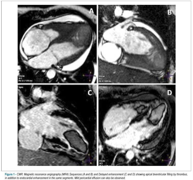
Surprising Cardiac Magnetic Resonance Imaging Findings: Rapidly Developing Intracardiac Thrombus with Endomyocardial Fibrosis

İrem Bilge Bulburu et al. Muhammet Gürdoğan Fethi Emre Ustabaşıoğlu Kenan Yalta

Introduction

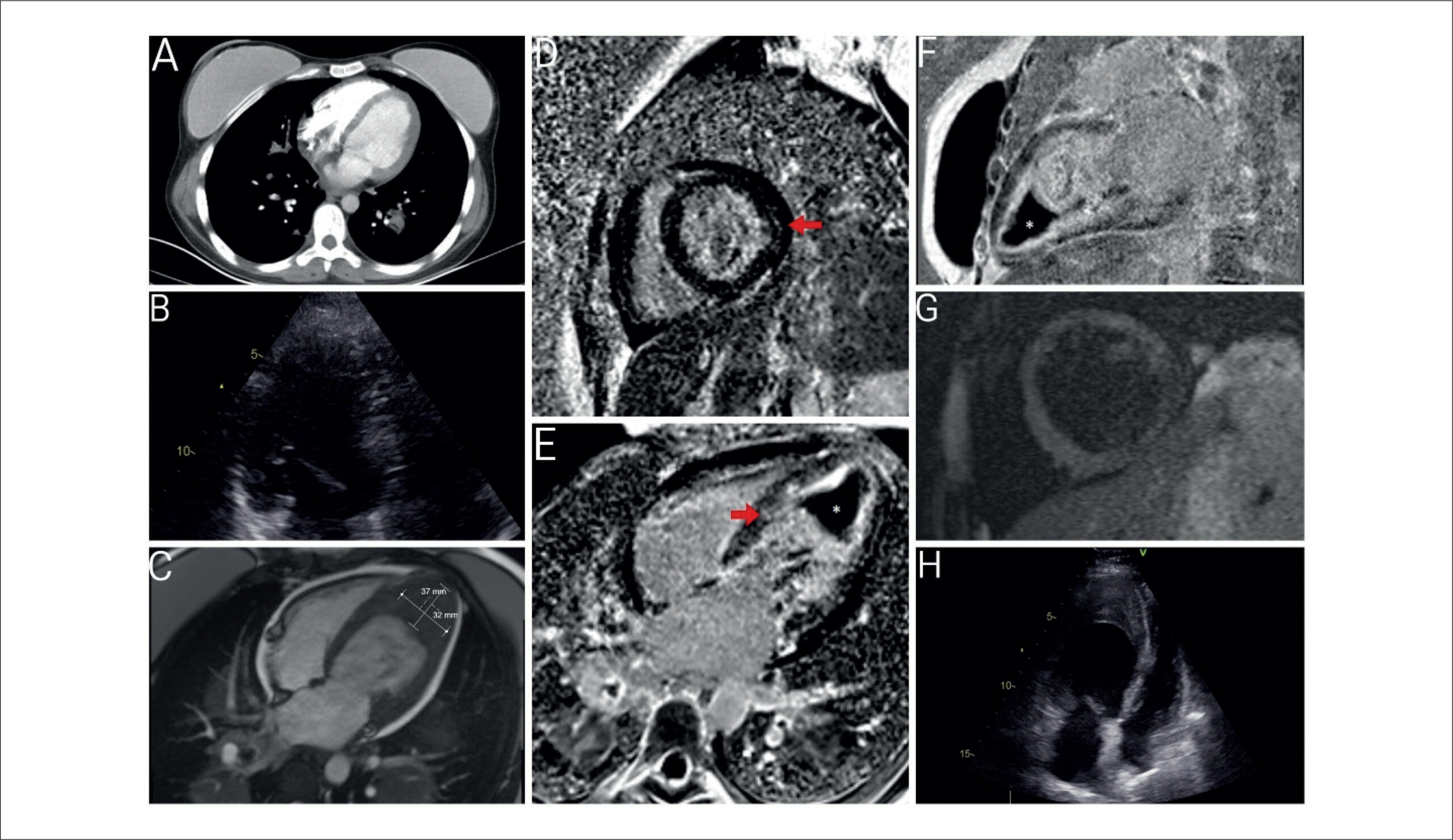
Endomyocardial fibrosis (EMF) is a rare form of restrictive cardiomyopathy, particularly prevalent in tropical regions. This condition is commonly associated with peripheral blood hypereosinophilia, though it may also occur in the absence of such finding. In this report, we describe an unusual case of EMF linked to non-Hodgkin’s lymphoma without hypereosinophilia, accompanied by a rapidly developing intracardiac thrombus.

[…]

  • Cardio-Oncology
  • Echocardiography
  • Endomyocardial Fibrosis
Read Full Article
Creative Commons Attribution (CC BY) Creative Commons license
04/08/2025 DOI: 10.36660/abc.20250030i Views 1
ABC
Editorial

Challenges and Perspectives of Cardiac Fibrosis Biomarkers: From Diagnosis to Clinical Application

Priscila Portugal dos Santos et al. Ricardo Luiz Damatto

As reported by Cheng-Mei et al. myocardial fibrosis is present in various cardiac diseases and can negatively impact the heart. It plays a central role in cardiac dysfunction and failure across different pathologies, serving as a strong predictor of poor clinical outcomes and mortality. However, it is essential to recognize that fibrosis is a complex and heterogeneous process, which can sometimes play a beneficial role, such as after a myocardial infarction. In this context, fibrosis acts as a reparative mechanism, replacing dead cardiomyocytes with scar tissue. This initial fibrotic response is crucial to prevent cardiac rupture, a severe and often fatal complication and one of the leading causes of death in patients with acute myocardial infarction.

Cardiac fibrosis is characterized by the excessive accumulation of extracellular matrix (ECM) components, especially collagens, within the myocardium. Understanding this process involves more than just collagen quantity; the characterization of fibrosis also considers collagen quality, the ratio of different fiber types, and the degree of collagen cross-linking. Type I collagen, which predominates in cardiac and vascular ECM, contributes to tissue strength and stiffness due to its thick, dense structure. Type III collagen, being more flexible, aids in ECM compliance. Thus, myocardial elasticity is determined by the ratio of type I to type III collagen fibers., Moreover, the degree of covalent cross-linking among the constituent microfibrils is critical to collagen fiber rigidity, increasing their resistance to degradation and significantly contributing to tissue stiffening., Therefore, a comprehensive understanding of cardiac fibrosis, aimed at developing new therapies and diagnostic methods, requires evaluating collagen quantity, quality, and cross-linking levels.

[…]

  • Biomarkers
  • collagen
  • Endomyocardial Fibrosis
  • Extracellular Matrix
Read Full Article
Creative Commons Attribution (CC BY) Creative Commons license
10/02/2025 DOI: 10.36660/abc.20240735i Views 2
ABCIC
Editorial

Quantification of Myocardial Fibrosis by Cardiac Magnetic Resonance Imaging: Advances, Impacts, and Perspectives

Isabela Bispo Santos da Silva Costa

Cardiac magnetic resonance imaging (CMR) has been established as a fundamental tool for the diagnosis and management of cardiomyopathies, especially due to its accuracy in tissue characterization, identification, and quantification of myocardial fibrosis., Late gadolinium enhancement (LGE) allows accurate assessment, identifying areas of myocardial fibrosis, and it serves to characterize, locate, and quantify them. In recent years, the role of LGE in the risk stratification of patients with hypertrophic cardiomyopathy (HCM) and other genetic cardiomyopathies has been the subject of several studies that aimed to correlate the presence and extent of myocardial fibrosis with the risk of sudden cardiac death (SCD) and major cardiovascular events.–

Different techniques are currently used to acquire, analyze, and quantify LGE by CMR. The presence of LGE is based on the differential tissue retention of gadolinium, which is preferentially absorbed in areas of fibrosis, where scar tissue is altered due to the presence of collagen. LGE acquisition techniques nowadays are fast, safe, and can be performed without requiring long respiratory pauses, as was the case in the past, with progressively better resolution and image definition. To quantify areas of fibrosis, different software programs that allow adequate quantification in a semi-automated manner are currently available.

[…]

  • Cardiomyopathies
  • Endomyocardial Fibrosis
  • Magnetic Resonance Imaging
Read Full Article
Creative Commons Attribution (CC BY) Creative Commons license
13/12/2024 DOI: 10.36660/abcimg.20240114i Views 1
Central Illustration
ABCIC
Review Article

How Do I Diagnose and Classify Endomyocardial Fibrosis in Under-Resourced Settings

Ana Mocumbi

Abstract

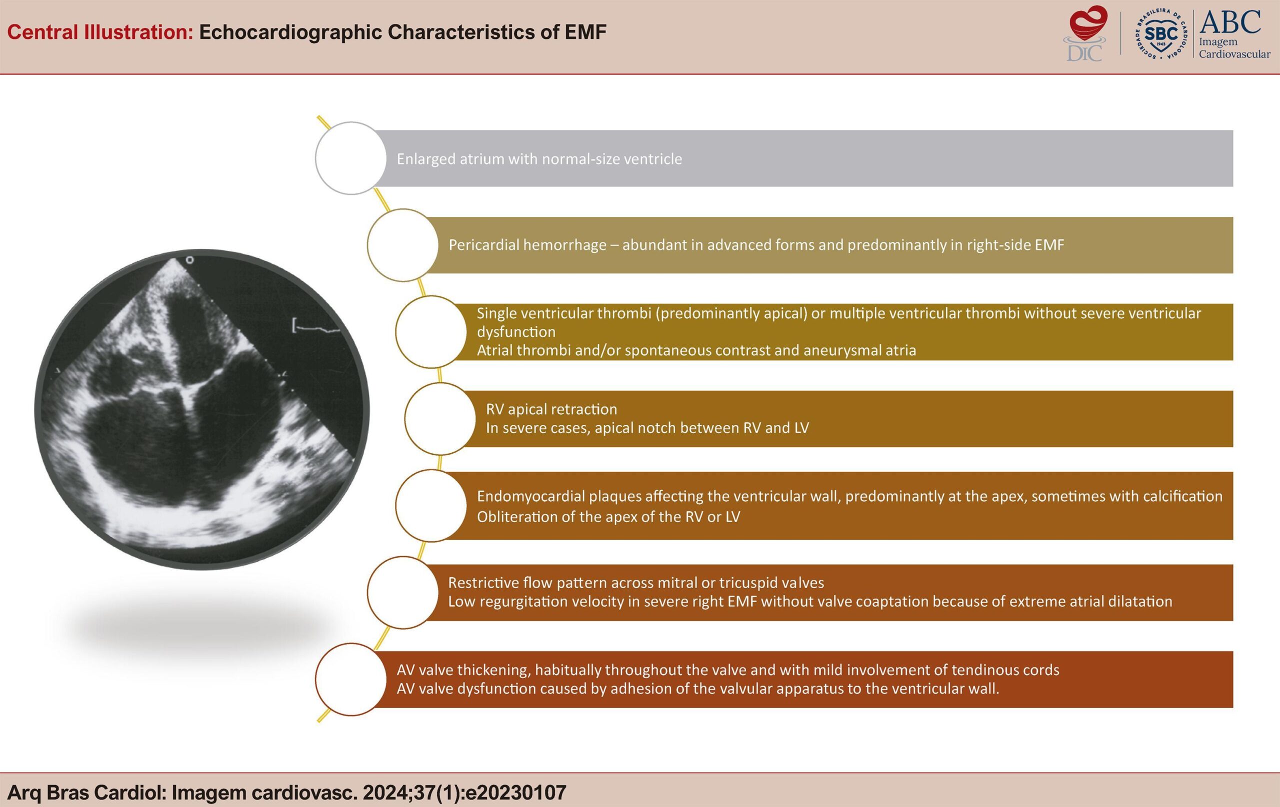
Endomyocardial fibrosis (EMF) is a disease of unclear etiology and pathogenesis that is characterized by fibrosis in the ventricular cavities and mainly affects children and adolescents from tropical regions of Africa, South America, and Asia. It is classified as a restrictive cardiomyopathy, with pathophysiology of restriction to diastolic filling associated with valve abnormalities, both caused by endocardial fibrosis. Typical echocardiographic features of this condition are endocardial thickening, severe atrioventricular (AV) valve regurgitation, aneurysmal atria, and heart distortion.

In endemic areas of Africa, in the absence of advanced imaging techniques, EMF diagnosis and management relies on transthoracic echocardiography for careful assessment of structural and hemodynamic abnormalities, aiming at planning medical or surgical management. This review highlights the key echocardiographic aspects of diagnosis and classification of EMF, as used in under-resourced settings in Africa. This is a standardized approach to screening populations with known occurrence of the disease and for informing medical and surgical management.

  • Diagnosis
  • Echocardiography
  • Endomyocardial Fibrosis
Read Full Article
Creative Commons Attribution (CC BY) Creative Commons license
28/02/2024 DOI: 10.36660/abcimg.20230107i Views 2
ABCIC
Editorial

Assessment of Myocardial Fibrosis by Myocardial Deformation Analysis via Echocardiography

Luiz Mário Baptista Martinelli et al. Fábio Fernandes Juliano Novaes Cardoso Viviane Tiemi Hotta

Abstract

Myocardial fibrosis (MF) is a pathological condition common to several primary and secondary heart diseases. Diffuse MF is a common feature in nearly all heart diseases, playing a crucial role in the progression of heart failure (HF). Several studies have indicated that the existence of MF serves as a prognostic factor for adverse outcomes across a range of heart conditions, underscoring the significance of this factor in clinical settings. Consequently, diagnostic approaches for MF, whether in subclinical phases or within established heart diseases, emerge as valuable instruments for early detection, risk stratification, and/or ongoing monitoring of disease developments. Despite being the most accurate non-invasive test for MF research, cardiovascular magnetic resonance (CMR) imaging is a test that is not widely available and is expensive. An alternative method with increased accessibility and cost-effectiveness for assessing heart diseases and researching MF involves echocardiography with the Speckle Tracking (STE) technique to evaluate myocardial deformation (“strain”). Studies show that diminished strain values (both global and segmental) in various heart conditions are associated with the presence and extent of MF, as determined by CMR or histological analysis. MF research using echocardiographic myocardial deformation analysis with STE has gained more space in clinical and research routines as it is an easy-to-perform and low-cost test. The results found by this technique can have a relevant diagnostic, therapeutic, and prognostic impact on clinical practice.

  • Cardiomyopathies
  • Echocardiography
  • Endomyocardial Fibrosis
  • Global Longitudinal Strain
Read Full Article
Creative Commons Attribution (CC BY) Creative Commons license
21/12/2023 DOI: 10.36660/abcimg.20230083i Views 1
ABC
Editorial

The Challenge of Making Cardiac Resonance a Global Reality

Hélder Jorge Andrade Gomes et al. Alcides Rocha de Figueredo Junior

Despite substantially reducing age-adjusted cardiovascular mortality rates in recent decades, cardiovascular disease remains the most common cause of death. Studies estimate that sudden cardiac death is responsible for approximately 50% of all deaths from cardiovascular causes, with ventricular tachycardia with degeneration to ventricular fibrillation and asystole being the most common fatal sequence of pathophysiological events.

The hard work done by authors of the article published in the Arquivos Brasileiros de Cardiologia illustrates the role of cardiac magnetic resonance (CMR) in the etiological elucidation of sudden death events encountered in clinical practice. In addition to the already expected Chagas and ischemic diseases, a wide variety of other diagnoses have been recognized: myocarditis, hypertrophic cardiomyopathy, dilated cardiomyopathy, arrhythmogenic heart disease, muscular dystrophy, hypertensive cardiomyopathy, metastases, non-compacted myocardium, and adrenergic cardiomyopathy (Takotsubo syndrome). The CMR was conclusive in more than 90% of cases and is a key tool for excluding structural heart disease and attributing the fatal event to extracardiac causes.

[…]

  • Cardiomyopathes
  • Cardiovascular Diseases, mortality
  • Death,Sudden
  • Diagnostic Imaging/trends
  • Endomyocardial Fibrosis
  • Magnetic Resonance Spectroscopy
  • Myocardial Ischemia
  • Stroke Volume
Read Full Article
Creative Commons Attribution (CC BY) Creative Commons license
21/04/2023 DOI: 10.36660/abc.20230187 Views 2
ABCIC
Review Manuscript

Current Role of Cardiac Magnetic Resonance Imaging in Hypertrophic Cardiomyopathy

Fábio Vieira Fernandes et al. Juliana Hiromi Silva Matsumoto Bello Afonso Akio Shiozaki Roberto Caldeira Cury

[…]

  • Cardiomyopathy, Hypertrophic, Familial
  • Echocardiography/methods
  • Electrocardiography/methods
  • Endomyocardial Fibrosis
  • Heart Failure
  • Magnetic Resonance Imaging/methods
Read Full Article
Creative Commons Attribution (CC BY) Creative Commons license
04/02/2022 DOI: 10.5935/2318-8219.20180048
ABC
Editorial

The Importance of Understanding the Progression of Myocardial Fibrosis in Chronic Chagas Cardiomyopathy

Carlos E. Rochitte

Myocardial fibrosis is one of the most important biological markers of Chagas’ cardiomyopathy and is directly related to the severity of the cardiomyopathy and the stage of the disease. In addition, myocardial fibrosis has prognostic value for major cardiovascular events, with a strong and close relationship with arrhythmic events in these patients.–

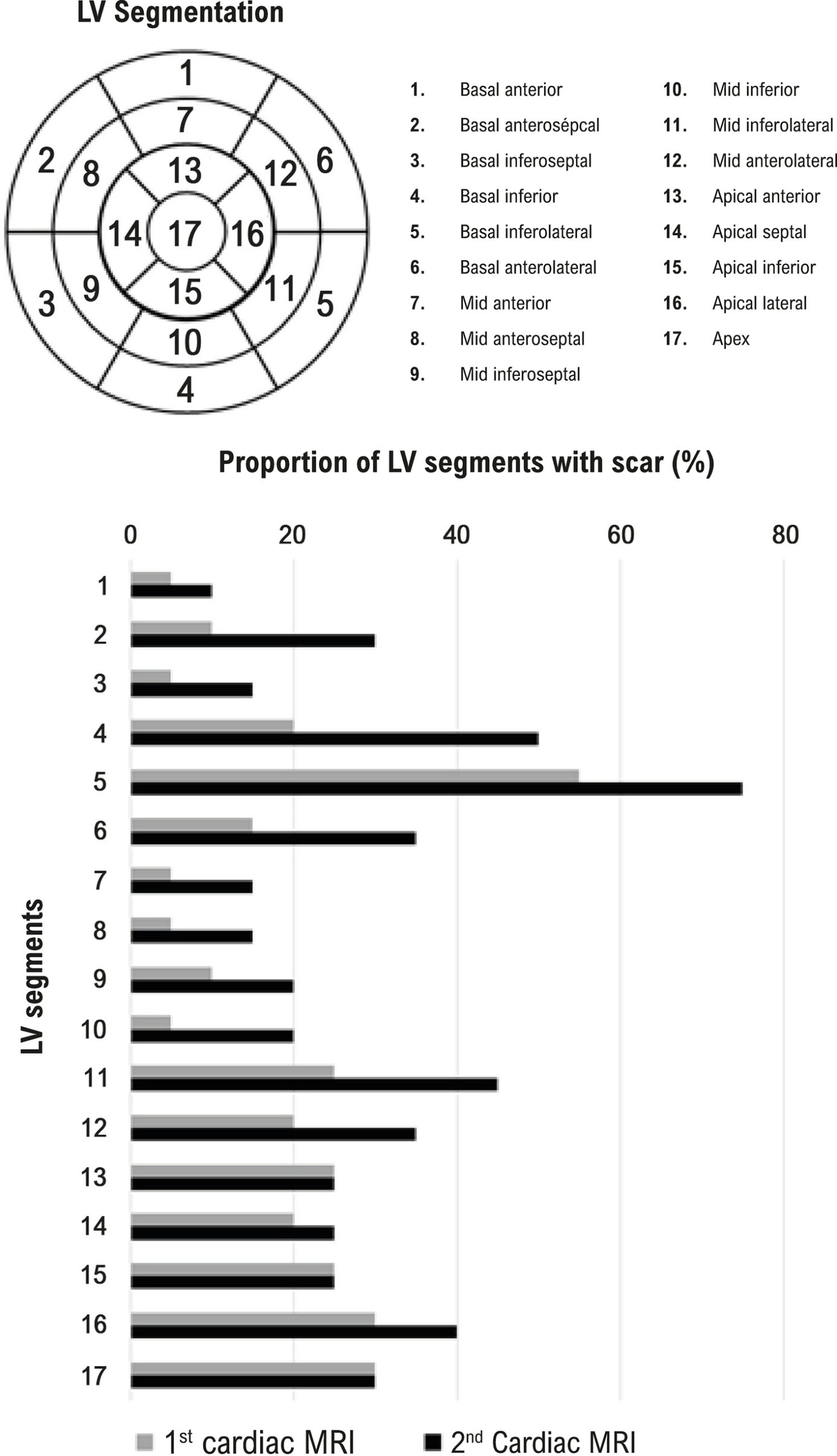
Cardiac magnetic resonance (CMR) is the reference imaging method for myocardial fibrosis detection and quantification in many cardiomyopathies, including Chagas cardiomyopathy. Initial studies included patients with Chagas cardiomyopathy at different stages of the disease and clearly demonstrated that the higher the disease severity the larger the amount of myocardial fibrosis. Based on these data and on the natural history of the disease, with progression of left ventricle dysfunction, one could safely infer that myocardial fibrosis progresses with time for any given patient. However, the longitudinal data using CMR in patients with Chagas disease was not available until the publication of the original article in this issue of ABC Cardiol by Santos et al. This is the first study to include data on myocardial fibrosis from two consecutive CMR of the same patient with a relatively long follow-up of 5.4 years. The authors demonstrated an impressive 43% increase of myocardial fibrosis over the follow-up period, which indicates a mean of 7.9% increase per year of myocardial fibrosis detected by CMR.

[…]

  • Chagas Diseases
  • chagasic cardiomyopathy
  • Diagnostic Imaging/methods
  • Endomyocardial Fibrosis
  • Heart Failure
  • Imaging Resonance Magnetic/methods
Read Full Article
Creative Commons Attribution (CC BY) Creative Commons license
03/12/2021 DOI: 10.36660/abc.20210897 Views 1
ABC
Editorial

Myocardial Scar and Ventricular Repolarization on the electrocardiogram: Insights from Cardiac Magnetic Resonance Imaging

Luciano C. Amado

Myocardial fibrosis is a final common feature of most inflammatory pathway and is frequently seen in patients with heart failure with reduced ejection fraction (HFrEF). , Myocardial scar, detected by contrast enhanced magnetic resonance imaging (ce-MRI), has been shown to be a predictor of prognosis and sudden cardiac death in patients with HFrEF. – Cardiac MRI has emerged as a non-invasive gold standard for the assessment of scar and risk prediction of ventricular arrhythmias. , , , Nevertheless, cardiac MRI can be time consuming, expensive, and not easily available.

The 12-lead surface electrocardiogram (ECG) is a simple and inexpensive diagnostic tool that is widely used in clinical practice for the diagnosis of cardiac arrhythmias. The ECG waveform is the expression of the transmembrane action potentials and is composed of two distinct processes: depolarization and repolarization. Ventricular repolarization is a complex electrical phenomenon which in clinical practice is measured from the beginning of the QRS complex to the end of the T-wave. Cardiac structural and electrical changes may cause action potential abnormalities, affecting the refractory period and thus favoring the onset of ventricular arrhythmias.

[…]

  • Death, Sudden, Cardiac
  • Electrocardiography/methods
  • Endomyocardial Fibrosis
  • Heart Failure
  • Magnetic Resonance Spectroscopy/ methods
  • Prognosis
  • Stroke Volume
Read Full Article
Creative Commons Attribution (CC BY) Creative Commons license
25/10/2021 DOI: 10.36660/abc.20210712 Views 2
Figure 1
ABC
Original Article

Cardiac Fibrosis and Changes in Left Ventricle Function in Patients with Chronic Chagas Heart Disease

João Bosco de Figueiredo Santos et al. Ilan Gottlieb Eduardo Marinho Tassi Gabriel Cordeiro Camargo … Jacob Atié Sérgio Salles Xavier Roberto Coury Pedrosa Roberto Magalhães Saraiva

Abstract

Background

Chagas heart disease (CHD) is a slow progressing condition with fibrosis as the main histopathological finding.

Objectives

To study if cardiac fibrosis increases over time and correlates with increase in left ventricular (LV) size and reduction of ejection fraction (EF) in chronic CHD.

Methods

Retrospective study that included 20 individuals (50% men; 60±10 years) with chronic CHD who underwent two cardiac magnetic resonance imaging (MRI) with late gadolinium enhancement with a minimum interval of four years between tests. LV volume, EF, and fibrosis mass were determined by cardiac MRI. Associations of fibrosis mass at the first cardiac MRI and changes in LV volume and EF at the second cardiac MRI were tested using logistic regression analysis. P values <0.05 were considered significant.

Results

Patients were classified as follows: A (n=13; changes typical of CHD in the electrocardiogram and normal global and segmental LV systolic function) and B1 (n=7; LV wall motion abnormality and EF≥45%). Mean time between cardiac MRI studies was 5.4±0.5 years. LV fibrosis (in %LV mass) increased from 12.6±7.9% to 18.0±14.1% between MRI studies (p=0.02). Cardiac fibrosis mass at baseline was associated with decrease in >5 absolute units in LV EF from the first to the second MRI (OR 1.48, 95% CI 1.03-2.13, p=0.03). LV fibrosis mass was larger and increased between MRI studies in the group that presented decrease in LV EF between the tests.

Conclusions

Even patients at an initial stage of CHD show an increase in myocardial fibrosis over time, and the presence of LV fibrosis at baseline is associated with a decrease in LV systolic function.

  • Chagas Cardiomyopathy
  • Chagas disease
  • Diagnostic Imaging
  • Electrocardiography/methods
  • Endomyocardial Fibrosis
  • Magnetic Resonance Imaging/methods
  • Ventricular Dysfunction, Left
Read Full Article
Creative Commons Attribution (CC BY) Creative Commons license
08/10/2021 DOI: 10.36660/abc.20200597
ABC
Editorial

The Challenge of Assessing Sudden Cardiac Death Risk in Patients with Nonischemic Heart Failure

Pedro Pimenta de Mello Spineti et al. Rodrigo Souto da Silva Sá Bruno Reznik Wajsbrot

Sudden cardiac death (SCD) accounts for approximately 50% of deaths in patients with ischemic and nonischemic heart disease in the presence of severe left ventricular systolic dysfunction. Many clinical trials– have evaluated the effectiveness of implantable cardioverter defibrillators (ICDs) in primary prevention of SCD in the last 20 years.

The Multicenter Automatic Defibrillator Implantation Trial II (MADIT II) and the Sudden Cardiac Death in Heart Failure (SCD-HeFT) defined the principles for indication, of ICDs in primary prophylaxis of SCD of patients with heart failure (HF) of ischemic etiology. On the other hand, clinical trials evaluating patients with HF of nonischemic origin,– have shown heterogeneous results and, so far, there are uncertainties on the best candidates for this type of therapy.

[…]

  • Biomarkers
  • Death, Sudden, Cardiac
  • Endomyocardial Fibrosis
  • Galectin 3
  • Heart Failure/physiopathology
  • Mortality
  • Ventricular Dysfunction, Left
Read Full Article
Creative Commons Attribution (CC BY) Creative Commons license
20/09/2021 DOI: 10.36660/abc.20210633 Views 1
ABC
Editorial

T1 Mapping in Heart Failure: Prognostic Implications

Sarah Moharem-Elgamal

Myocardial fibrosis leads to impaired diastolic and systolic function and is associated with increased major adverse cardiovascular events. It is a structural correlate that can be found in the different stages of heart failure. The two main types of myocardial fibrosis are interstitial fibrosis and replacement fibrosis. Interstitial fibrosis is a reversible process that occurs early in the disease process as increased collagen synthesis in diffuse microscopic distribution within the myocardium and sometimes by localized perivascular distribution. Replacement fibrosis typically occurs in the later stages of the disease after irreversible myocyte injury or death, in which cell apoptosis triggers fibroblasts and promotes macroscopic deposition of collagen fibrous tissue in the myocardium.

Cardiac magnetic resonance (CMR) has the capability to accurately quantify ventricular volumes and ejection fraction, as well as the non-invasive characterization of the myocardium. These unique features have led to the increased use of CMR in the assessment of patients with heart failure (HF). CMR can detect the presence and extent of replacement fibrosis through late gadolinium enhancement imaging and diffuse interstitial fibrosis through native T1 mapping. Interstitial fibrosis identified by native T1 mapping has been used as a marker of disease activity,– risk stratification and monitoring of the therapeutic management in heart failure patients.

[…]

  • Cellular Apoptose Susceptibility Protein
  • Endomyocardial Fibrosis
  • Fibroblasts
  • Heart Failure
  • Magnetic Resonance Spectroscopy/ methods
  • Mortality
Read Full Article
Creative Commons Attribution (CC BY) Creative Commons license
17/05/2021 DOI: 10.36660/abc.20210205
ABC
Editorial

Does Galectin-3 (Myocardial Fibrosis Biomarker) Predict Progression in Chagas Disease?

Roberto Coury Pedrosa

In recent decades, the epidemiology of Chagas disease (Chd) has changed significantly as a consequence of urbanization and migration. In Brazil, to date, it is the neglected disease with the highest burden, of which ~30% of individuals will progress to the symptomatic tissue disruptive stage within 20 – 30 years after being infected (around 2% per year progress from the indeterminate to the cardiac form, according to a contemporary study).

Persistent myocardial inflammation and fibrosis represent the main pathological characteristics of Chd that would be correlated with its progression., The use of myocardial fibrosis biomarkers to predict progression in patients with normal or near normal LV function is certainly important in Chd, where traditional risk factors, such as LVEF, may not be as useful. Nevertheless, studies have shown the value of myocardial fibrosis biomarkers in the progression of Chd pathogenesis.

[…]

  • Apoptosis
  • Chagas Disease Biomarkers
  • Endomyocardial Fibrosis
  • Galectin 3
Read Full Article
Creative Commons Attribution (CC BY) Creative Commons license
01/03/2021 DOI: 10.36660/abc.20201162
Figure 1
ABC
Case Report

Transthyretin Amyloidosis (ATTR) – The Role of Multimodality in the Definitive Diagnosis

Tonnison de Oliveira Silva et al. Eduardo Sahade Darze Luiz Eduardo Fonteles Ritt André Luiz Cerqueira Almeida Antônio Ximenes

Introduction

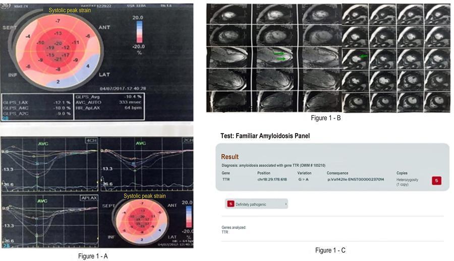
Transthyretin amyloidosis (ATTR) is a rare cause of restrictive cardiomyopathy and/or peripheral polyneuropathy, of a progressive, irreversible and fatal nature, underdiagnosed and with its definitive diagnosis performed late. Early diagnosis, characterization of the type of amyloidosis and subsequent establishment of specific therapy are fundamental for a better prognosis of this disease. We present a case of ATTR where clinical suspicion associated with multimodality diagnosis — nuclear medicine — was able to safely deliver diagnosis, without the need for biopsy.

[…]

  • Amyloidosis/complications
  • Cardiomyopathy,Restrictive
  • Diagnostic Imaging
  • Endomyocardial Fibrosis
  • Heart Failure
  • Hypertension
  • Prealbumin
Read Full Article
Creative Commons Attribution (CC BY) Creative Commons license
18/05/2020 DOI: 10.36660/abc.20180077
ABC
Editorial

Coronary Slow Flow Phenomenon – Adding Myocardial Fibrosis to the Equation

Filipe Penna de Carvalho et al. Clério Francisco de Azevedo

Initially described more than 40 years ago by Tambe et al., coronary slow flow phenomenon (CSFP) is characterized by delayed contrast medium progression in the absence of obstructive coronary epicardial disease during invasive coronary angiography. CSFP typically affects young male smokers, who often presents with acute coronary syndrome (ACS) or recurrent refractory resting angina requiring hospital admission.– Moreover, life-threatening arrhythmias and sudden cardiac death have also been associated with CSFP.

Despite increased awareness and research, CSFP remains an elusive and poorly understood condition, with many proposed pathogenic mechanisms including endothelial, vasomotor and microvascular dysfunction., Indeed, an abnormal regulation of microvascular tone that occurs only during resting conditions, while coronary flow reserve is within normal range, has been described in CSFP.

[…]

  • Cicatrix, Hypertrophic
  • Endomyocardial Fibrosis
  • Fractional Flow Reserve
  • Heart Failure
  • Magnetic Resonance Spectroscopy
  • Natriuretic-Peptide, C-Type
  • Prognosis
Read Full Article
Creative Commons Attribution (CC BY) Creative Commons license
06/04/2020 DOI: 10.36660/abc.20200187
Figure 1
IJCS
Original Article

Evaluation of Galectin-3 and Myocardial Fibrosis in Patients with Hypertrophic Cardiomyopathy

Murillo de Oliveira Antunes et al. Edmundo Arteaga-Fernández Fabio Fernandes Carla David Soffiatti … Paula de Cássia Buck Ester Cerdeira Sabino Carlos Henrique Valente Moreira Charles Mady

Abstract

Background:

Galectin-3 is the designation given to the protein that binds to ß-galactosides, expressed by activated macrophages and described as a cardiac fibrosis mediator. In hypertrophic cardiomyopathy (HCM), myocardial fibrosis is an independent predictor of adverse outcome; however, the association between Galectin-3 and myocardial fibrosis has not been studied in this cardiopathy.

Objective:

To evaluate the association of Galectin-3 and the presence of myocardial fibrosis in a patient with hypertrophic cardiomyopathy.

Methods:

Galectin-3 was measured in automated equipment using the Elisa technique in 100 participants divided into two groups: 50 patients with hypertrophic cardiomyopathy and 50 healthy control subjects. All patients with hypertrophic cardiomyopathy underwent magnetic nuclear resonance with the late enhancement technique to investigate myocardial fibrosis. For the statistical analysis, p values < 0.05 were considered statistically significant.

Results:

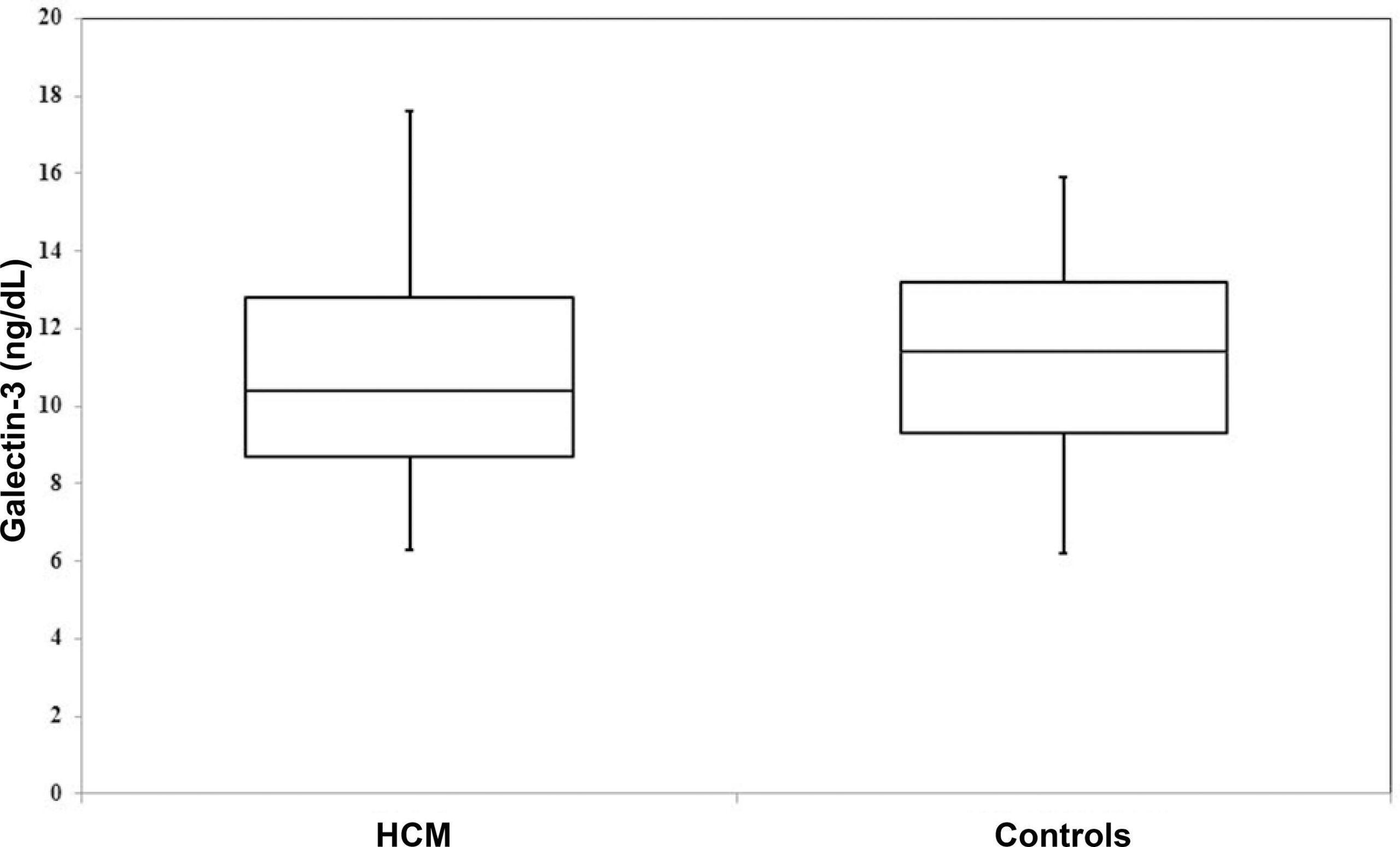
Galectin-3 levels were low and did not show significant differences between patients with hypertrophic cardiomyopathy and the control group, 10.3 ± 3.1 ng/dL and 11.3 ± 2.6 ng/dL (p = 0.12) respectively. Myocardial fibrosis was a common finding and was identified in 84% (42/50) of patients with HCM, but no differences were observed between Galectin-3 levels when comparing patients with and without fibrosis, 10.3 ± 2.4 ng/dL and 10.1 ± 2.1 ng/dL (p = 0.59).

Conclusion:

The results did not show an association between Galectin-3 and myocardial fibrosis in patients with hypertrophic cardiomyopathy, suggesting that non-inflammatory mechanisms of myocardial fibrosis formation and cardiac remodeling are involved in this cardiopathy.

  • Arrhythmias Cardiac
  • Cardiomyopathy, Hypertrophic
  • Diagnostic Imaging
  • Endomyocardial Fibrosis
  • Galectin 3
Read Full Article
Creative Commons Attribution (CC BY) Creative Commons license
18/02/2019 DOI: 10.5935/2359-4802.20180102 Views 1
Figure 1
ABC
Original Article

Diagnosis of Rejection by Analyzing Ventricular Late Potentials in Heart Transplant Patients

Vítor Nogueira Mendes et al. Telmo Santos Pereira Vítor Azevedo Matos

Background:

Heart transplant rejection originates slow and fragmented conduction. Signal-averaged ECG (SAECG) is a stratification method in the risk of rejection.

Objective:

To develop a risk score for rejection, using SAECG variables.

Methods:

We studied 28 transplant patients. First, we divided the sample into two groups based on the occurrence of acute rejection (5 with rejection and 23 without). In a second phase, we divided the sample considering the existence or not of rejection in at least one biopsy performed on the follow-up period (rejection pm1: 18 with rejection and 10 without).

Results:

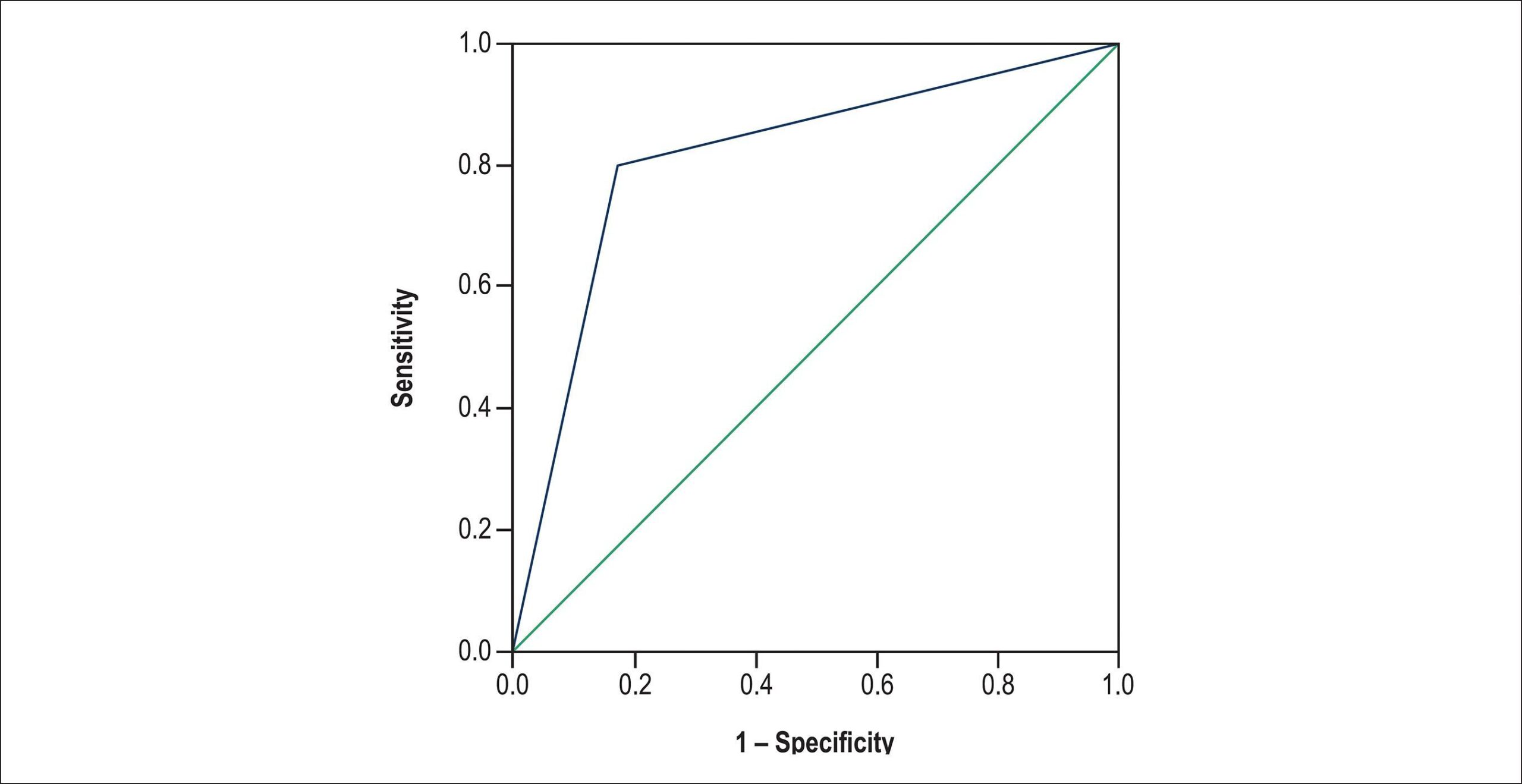
On conventional ECG, the presence of fibrosis was the only criterion associated with acute rejection (OR = 19; 95% CI = 1.65-218.47; p = 0.02). Considering the rejection pm1, an association was found with the SAECG variables, mainly with RMS40 (OR = 0.97; 95% CI = 0.87-0.99; p = 0.03) and LAS40 (OR = 1.06; 95% IC = 1.01-1.11; p = 0.03). We formulated a risk score including those variables, and evaluated its discriminative performance in our sample. The presence of fibrosis with increasing of LAS40 and decreasing of RMS40 showed a good ability to distinguish between patients with and without rejection (AUC = 0.82; p < 0.01), assuming a cutoff point of sensitivity = 83.3% and specificity = 60%.

Conclusion:

The SAECG distinguished between patients with and without rejection. The usefulness of the proposed risk score must be demonstrated in larger follow-up studies.

  • Electrocardiography
  • Endomyocardial Fibrosis
  • Graft Rejection
  • Heart Transplantation
Read Full Article
Creative Commons Attribution (CC BY) Creative Commons license
22/01/2016 DOI: 10.5935/abc.20160011
Pulmonary arterial hypertension: use of delayed contrast-enhanced cardiovascular magnetic resonance in risk assessment
ABC
Artigo Original

Pulmonary arterial hypertension: use of delayed contrast-enhanced cardiovascular magnetic resonance in risk assessment

Luiz Gustavo Pignataro Bessa et al. Flávia Pegado Junqueira Marcelo Luiz da Silva Bandeira Marcelo Iorio Garcia … Sérgio Salles Xavier Guilherme Lavall Diego Torres Daniel Waetge

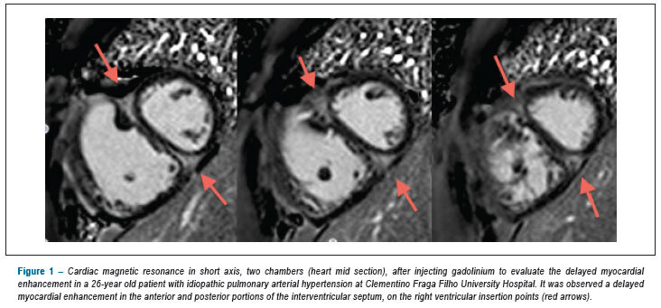
BACKGROUND: Pulmonary arterial hypertension is a severe and progressive disease. Its early diagnosis is the greatest clinical challenge. OBJECTIVE: To evaluate the presence and extension of the delayed myocardial contrast-enhanced cardiovascular magnetic resonance, as well as to verify if the percentage of the myocardial fibrosis mass is a severity predictor. METHODS: Cross-sectional study with 30 patients with pulmonary arterial hypertension of groups I and IV, subjected to clinical, functional and hemodynamic evaluation, and to cardiac magnetic resonance. RESULTS: The mean age of patients was 52 years old, with female predominance (77%). Among the patients, 53% had right ventricular failure at diagnosis, and 90% were in functional class II/III. The mean of the 6-minute walk test was 395m. In hemodynamic study with right catheterism, the mean average pulmonary arterial pressure was 53.3mmHg, of the cardiac index of 2.1L/ min.m², and median right atrial pressure was 13.5 mmHg. Delayed myocardial contrast-enhanced cardiovascular magnetic resonance was found in 28 patients. The mean fibrosis mass was 9.9 g and the median percentage of fibrosis mass was 6.17%. The presence of functional class IV, right ventricular failure at diagnosis, 6-minute walk test 15mmHg, with cardiac index < 2.0L/ min.m², there was a relevant association with the increased percentage of myocardial fibrosis. CONCLUSION: The percentage of the myocardial fibrosis mass indicates a non-invasive marker with promising perspectives in identifying patients with high risk factors for pulmonary hypertension.

  • Endomyocardial Fibrosis
  • Hypertension Pulmonary
  • Magnetic Resonance Imaging
  • Risk Assessment
Read Full Article
Creative Commons Attribution (CC BY) Creative Commons license
27/08/2013 DOI: 10.5935/abc.20130168 Views 1
Imatinib attenuates myocardial fibrosis in association with inhibition of the PDGFRα activity
ABC
Artigo Original

Imatinib attenuates myocardial fibrosis in association with inhibition of the PDGFRα activity

Li-kun Ma et al. Qian Li Li-feng He Jin-sheng Hua … Jun-ling Zhou Hua Yu Ke-fu Feng Hong-wu Chen Hao Hu Lin Wang

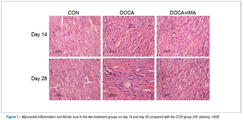
BACKGROUND: Imatinib is a tyrosine kinase receptor inhibitor that has been confirmed to exert inhibitory effect on the platelet derived growth factor PDGF receptor (PDGFRα and PDGFRβ) activity. OBJECTIVE: To investigate the protective effect of imatinib on the myocardial fibrosis in deoxycorticosterone-acetate (DOCA)/salt induced hypertensive rats. METHODS: Sixty male uninephrectomized Sprague-Dawley rats were assigned to three groups: control rats (CON group); deoxycorticosterone group (DOCA group); deoxycorticosterone and imatinib group (DOCA+IMA group). Systolic blood pressure (SBP) was measured biweekly. The apical portion of the left ventricle was studied. Sirius-Red staining, Hematoxylin-Eosin staining, immunohistochemistry and Western blot assay were employed. RESULTS: SBP in the DOCA group and DOCA+IMA group was higher than that in the CON group on day 14 and 28. Animals in the DOCA group showed severe interstitial and perivascular fibrosis on day 28, and the expressions of PI, PIII, tenascin-C and fibronectin were significantly higher than those in the DOCA+IMA group and CON group. When compared with the CON group, myocardial tissue inflammatory response and monocyte/macrophage infiltration of different degrees were observed in the DOCA group and DOCA+IMA group. Protein expressions of PDGF-A, PDGF-C and PDGFRα were signiflcantly higher in the DOCA and DOCA+IMA groups than those in the CON group, but the p-PDGFRα protein expression in the DOCA+IMA group was lower than that in the DOCA group. CONCLUSION: Imatinib can exert inhibitory effects on myocardial fibrosis in DOCA/salt induced hypertensive rats, which may be attributed to the inhibition of PDGFR-α activity.

  • Desoxycorticosterone
  • Endomyocardial Fibrosis
  • Receptor, Platelet-Derived Growth Factor Alpha
  • Receptors, Eph Family
Read Full Article
Creative Commons Attribution (CC BY) Creative Commons license
27/11/2012 DOI: 10.1590/S0066-782X2012005000109 Views 1
Biventricular thrombus and endomyocardial fibrosis in antiphospholipid syndrome
ABC
Case Report

Biventricular thrombus and endomyocardial fibrosis in antiphospholipid syndrome

Luiz Flávio Galvão Gonçalves et al. Fernanda Maria Silveira Souto Fernanda Nascimento Faro Joselina Luzia Menezes Oliveira … José Augusto Soares Barreto-Filho Antônio Carlos Sobral Sousa

CASE REPORT

Biventricular thrombus and endomyocardial fibrosis in antiphospholipid syndrome

[…]

  • Antibodies
  • Antiphospholipid
  • Antiphospholipid syndrome
  • Endomyocardial Fibrosis
  • Magnetic Resonance Spectroscopy
  • Thrombosis
Read Full Article
Creative Commons Attribution (CC BY) Creative Commons license
22/11/2012 DOI: 10.1590/S0066-782X2012001400017
Importance of the area of fibrosis at midterm evolution of patients submitted to ventricular reconstruction
ABC
Artigo Original

Importance of the area of fibrosis at midterm evolution of patients submitted to ventricular reconstruction

Gustavo Calado de Aguiar Ribeiro et al. Mauricio Lopes Fernando Antoniali Ana Nunes … Cledicyon Eloy Costa Juliano L. Fernandes

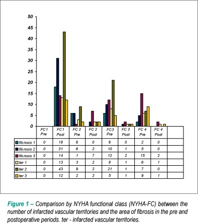
BACKGROUND: Although it is acknowledged that the ventricular reconstruction surgery (VRS) can promote reverse remodeling, new studies are necessary to define the influence of the left ventricular (LV) area of fibrosis. OBJECTIVE: To evaluate whether the extension of the area of fibrosis of the LV is important in the LV functional recovery after the surgery and correlate it with clinical factors. METHODS: Prospective analysis of 82 patients with ventricular dysfunction submitted to VRS. We analyzed the importance of the clinical characteristics and the amount of fibrosis was assessed, measured by cardiac magnetic resonance (CMR) as small, medium and large. RESULTS: All patients were followed for 36 months, with a mortality of 6%. The amount of medium fibrosis was 25.8% ± 13.6%. There was improvement in the left ventricular ejection fraction (LVEF), from 36.9% ± 6.8% to 48.2% ± 8.2% (p < 0.001). There was an inverse association between the amount of fibrosis and the increase in LVEF (r = -0.83, p < 0.0001). There was a decrease in the LV end-systolic volume of 43.3 ± 8.2ml/m² (p < 0.001). There was an improvement in heart failure symptoms, except in patients with large areas of fibrosis (p = 0.45). The independent predictors for events were: fibrotic area (p = 0.01), age (p = 0.01), LV end-systolic volume (p = 0.03) and LVEF (p = 0.02). The event-free follow-up was different in relation to the area of fibrosis (p < 0.01). CONCLUSION: In patients with ventricular dysfunction, the extension of the area of fibrosis was an independent predictor of the LV functional recovery after the VRS. The combination of cardiac MRI and clinical parameters can help in the indication for VRS.

  • Endomyocardial Fibrosis
  • Heart Failure
  • Stroke Volume
  • Ventricular Dysfunction, Left
Read Full Article
Creative Commons Attribution (CC BY) Creative Commons license
05/05/2010 DOI: 10.1590/S0066-782X2009001200009 Views 1
Myocardial fibrosis and ventricular remodeling in severe chronic aortic regurgitation
ABC
Artigo Original

Myocardial fibrosis and ventricular remodeling in severe chronic aortic regurgitation

Nelson Elias et al. Flávio Tarasoutchi Guilherme Sobreira Spina Roney O. Sampaio … Pablo M. A. Pomerantzeff Francisco Rafael Laurindo Max Grinberg

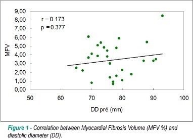
BACKGROUND: Significant symptomatic chronic aortic regurgitation (AR) leads to considerable left ventricular remodeling at the expense of myocyte hypertrophy and extracellular matrix remodeling. The relevance of interstitial fibrosis concentration in these patients is unknown. We analyzed the degree of fibrosis in the left ventricle (LV) in symptomatic patients with AR submitted to surgical treatment, and its relationship with functional and anatomical characteristics. OBJECTIVE: To evaluate myocardical fibrosis in chronic severe aortic regurgitation. METHODS: Twenty-eight patients with chronic symptomatic AR (16 with normal LV function and 12 with LV dysfunction) were selected and assessed pre- and postoperatively by echocardiography. Functional capacity was measured using maximal oxygen consumption (VO2max) through the cardiopulmonary test. Myocardial fibrosis volume fraction (MFV) was quantified through endomyocardial biopsy performed in all patients during surgery. We compared the histopathologic results with a nine-patient control group. RESULTS: The mean age was 39 ± 12 years, 75% of the patients were male, and the rheumatic etiology accounted for 84% of the cases. Twenty-five patients remained in FC l and ll at the end of the study, and there was a significant reduction of the LV diameters between the preoperative and late postoperative timepoints. Three deaths occurred but they were not related to postoperative ventricular dysfunction. The parameters of the cardiopulmonary test were similar between pre- and postoperative timepoints. MFV in patients with AR was significantly higher than in the control group (3.47 ± 1.9% vs 0.82 ± 0.96%, respectively, p=0.001). There was no statistical correlation among LV fibrosis and LV diameters, LVEF and MVO2. CONCLUSION: In patients with significant symptomatic AR, the presence of limited myocardial fibrosis was not associated with clinical, echocardiographic or functional complications.

  • Aortic Valve Insufficiency
  • Endomyocardial Fibrosis
  • Ventricular Function, Left
Read Full Article
Creative Commons Attribution (CC BY) Creative Commons license
04/02/2009 DOI: 10.1590/S0066-782X2009000100010
Figure 1B
ABC
Artigo Original

Nitric oxide synthesis blockade increases hypertrophy and cardiac fibrosis in rats submitted to aerobic training

Hugo Celso Dutra de Souza et al. Daniel Martins Dias Penteado Marli Cardoso Martin-Pinge Octávio Barbosa Neto … Vicente de Paula Antunes Teixeira João Henrique Dutra Blanco Valdo José Dias da Silva

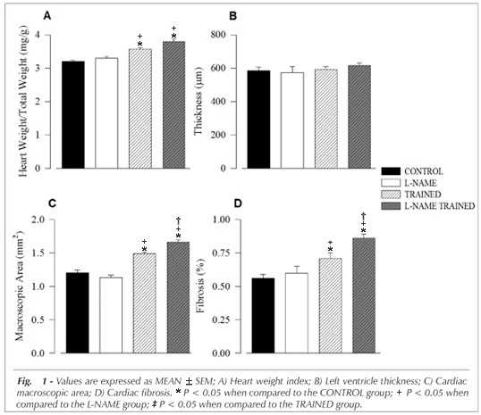
OBJECTIVE: The objective of the present study was to evaluate cardiac tissue adaptations in rats submitted to aerobic training after nitric oxide (NO) synthesis blockade. METHODS: The animals (n=48) were divided into four groups: sedentary (CONTROL group); hypertensive after administration of NG-nitro-L-arginine methyl ester for 7 days (L-NAME Group); trained for 8 weeks through swimming exercises (TRAINED Group);trained and treated with L-NAME during the last week (L-NAME TRAINED Group). All the animals were submitted to the experiment procedures for blood pressure (BP) readings and cardiac morphometric evaluation. RESULTS: In comparison to the other groups, the L-NAME and L-NAME TRAINED groups were hypertensive (p<0.05); however, BP elevation in the L-NAME TRAINED group was significantly lower than the L-NAME group (p<0.05). The heart weight indexes for the TRAINED and L-NAME TRAINED groups were higher than the CONTROL and L-NAME groups (p<0.05). Also they had presented higher rates of macroscopic cardiac area and cardiac fibrosis in relation to the rest (p<0.05); comparisons revealed that the values for the L-NAME TRAINED group were significantly higher (p<0.05) than the others. CONCLUSION: Short term NO synthesis blockade in sedentary animals induced hypertension but did not cause cardiac hypertrophy. In the trained animals, the inhibition of NO synthesis attenuated hypertension, induced cardiac hypertrophy and significantly increased myocardial fibrosis, indicating that NO plays an important role in cardiac tissue adaptations caused by aerobic exercise.

  • Cardiomegaly
  • Endomyocardial Fibrosis
  • Exercise
  • Hypertension
  • Nitric Oxide
Read Full Article
Creative Commons Attribution (CC BY) Creative Commons license
05/09/2007 DOI: 10.1590/S0066-782X2007001400005 Views 1
Clinical Meaning of Ascites in Patients with Endomyocardial Fibrosis
ABC
Original Article

Clinical Meaning of Ascites in Patients with Endomyocardial Fibrosis

Antonio Carlos Pereira Barretto et al. Charles Mady Sergio Almeida Oliveira Edmundo Arteaga … Creusa Dal Bó José Antonio Franchini Ramires

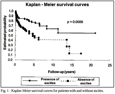
OBJECTIVE: To evaluate the clinical meaning of ascites and the main features of patients with ascites and endomyocardial fibrosis. METHODS: We studied 166 patients with endomyocardial fibrosis (mean age 37 years, 114 women) treated over the last 20 years. Ventriculography findings, surgery or necropsy confirmed the diagnosis in all patients. Most patients belonged to New York Heart Association Functional Class III/IV (134, 83.7%). Eighty-one (50.6%) had biventricular, 28 (17.5%) had right ventricular, and 51 (31.8%) had left ventricular involvement. During follow-up, 56 patients died. RESULTS: Ascites was present in 67 (41.8%) patients, and right ventricular involvement was present in 59 (88%). In the comparison between patients with or without ascites, those with ascites had higher mortality (49.2% and 24.7%, respectively). Patients with ascites had a higher incidence of edema (95% vs. 43%), hepatomegaly (5.8cm vs. 4.1cm), mean right atrium pressure (19.3 vs. 12mmHg), and final right ventricle diastolic pressure (18.7 vs. 12.9mmHg). Also, patients with ascites had a longer history of illness (5.1 and 3.9 years, respectively) and had atrial fibrillation more frequently (44.7% vs. 30.1%). CONCLUSION: Ascites was observed in less than 50% of cases of endomyocardial fibrosis and was associated with greater involvement of the right ventricle and with a longer duration of the disease, thus being a characteristic of a worse prognosis.

  • ascites
  • Endomyocardial Fibrosis
  • restrictive cardiomiopathy
Read Full Article
Creative Commons Attribution (CC BY) Creative Commons license
31/01/2007 DOI: 10.1590/S0066-782X2002000200008

Could not load more articles. Please try again.

How to cite this article

EndNote RIS

Electronic Document Format (Vancouver)

Bulburu B, Gürdoğan M, Ustabaşıoğlu FE, Yalta K. Surprising Cardiac Magnetic Resonance Imaging Findings: Rapidly Developing Intracardiac Thrombus with Endomyocardial Fibrosis. Arquivos Brasileiros de Cardiologia 2025;122(6):e20250030.

Electronic Document Format (ABNT)

Bulburu, İrem Bilge; Gürdoğan, Muhammet; Ustabaşıoğlu, Fethi Emre; Yalta, Kenan. Surprising Cardiac Magnetic Resonance Imaging Findings: Rapidly Developing Intracardiac Thrombus with Endomyocardial Fibrosis. Arquivos Brasileiros de Cardiologia, v. 122, n. 6, e20250030, Aug. 2025.

Electronic Document Format (APA)

Bulburu, B., Gürdoğan, M., Ustabaşıoğlu, F. E., & Yalta, K. (2025). Surprising Cardiac Magnetic Resonance Imaging Findings: Rapidly Developing Intracardiac Thrombus with Endomyocardial Fibrosis. Arquivos Brasileiros de Cardiologia, 122(6), e20250030.

Electronic Document Format (ISO)

Bulburu, İrem Bilge and Gürdoğan, Muhammet and Ustabaşıoğlu, Fethi Emre and Yalta, Kenan. Surprising Cardiac Magnetic Resonance Imaging Findings: Rapidly Developing Intracardiac Thrombus with Endomyocardial Fibrosis. Arquivos Brasileiros de Cardiologia [online]. 2025, vol. 122, n. 6, [cited 2026-10-14], e20250030. Available from: <https://journals.cardiol.br/en/article/surprising-cardiac-magnetic-resonance-imaging-findings-rapidly-developing-intracardiac-thrombus-with-endomyocardial-fibrosis/>. ISSN 0066-782X

EndNote RIS

Electronic Document Format (Vancouver)

Santos PP, Damatto RL. Challenges and Perspectives of Cardiac Fibrosis Biomarkers: From Diagnosis to Clinical Application. Arquivos Brasileiros de Cardiologia 2025;121(12):e20240735.

Electronic Document Format (ABNT)

Santos, Priscila Portugal dos; Damatto, Ricardo Luiz. Challenges and Perspectives of Cardiac Fibrosis Biomarkers: From Diagnosis to Clinical Application. Arquivos Brasileiros de Cardiologia, v. 121, n. 12, e20240735, Feb. 2025.

Electronic Document Format (APA)

Santos, P. P. , & Damatto, R. L. (2025). Challenges and Perspectives of Cardiac Fibrosis Biomarkers: From Diagnosis to Clinical Application. Arquivos Brasileiros de Cardiologia, 121(12), e20240735.

Electronic Document Format (ISO)

Santos, Priscila Portugal dos and Damatto, Ricardo Luiz. Challenges and Perspectives of Cardiac Fibrosis Biomarkers: From Diagnosis to Clinical Application. Arquivos Brasileiros de Cardiologia [online]. 2025, vol. 121, n. 12, [cited 2026-10-14], e20240735. Available from: <https://journals.cardiol.br/en/article/challenges-and-perspectives-of-cardiac-fibrosis-biomarkers-from-diagnosis-to-clinical-application/>. ISSN 0066-782X

EndNote RIS

Electronic Document Format (Vancouver)

Costa IBSS. Quantification of Myocardial Fibrosis by Cardiac Magnetic Resonance Imaging: Advances, Impacts, and Perspectives. ABC Imagem Cardiovascular 2024;37(4):e20240114.

Electronic Document Format (ABNT)

Costa, Isabela Bispo Santos da Silva. Quantification of Myocardial Fibrosis by Cardiac Magnetic Resonance Imaging: Advances, Impacts, and Perspectives. ABC Imagem Cardiovascular, v. 37, n. 4, e20240114, Dec. 2024.

Electronic Document Format (APA)

Costa, I. B. S. S. (2024). Quantification of Myocardial Fibrosis by Cardiac Magnetic Resonance Imaging: Advances, Impacts, and Perspectives. ABC Imagem Cardiovascular, 37(4), e20240114.

Electronic Document Format (ISO)

Costa, Isabela Bispo Santos da Silva. Quantification of Myocardial Fibrosis by Cardiac Magnetic Resonance Imaging: Advances, Impacts, and Perspectives. ABC Imagem Cardiovascular [online]. 2024, vol. 37, n. 4, [cited 2026-10-14], e20240114. Available from: <https://journals.cardiol.br/en/article/quantification-of-myocardial-fibrosis-by-cardiac-magnetic-resonance-imaging-advances-impacts-and-perspectives/>. ISSN 2318-8219

EndNote RIS

Electronic Document Format (Vancouver)

Mocumbi A. How Do I Diagnose and Classify Endomyocardial Fibrosis in Under-Resourced Settings. ABC Imagem Cardiovascular 2024;37(1):e20230107.

Electronic Document Format (ABNT)

Mocumbi, Ana. How Do I Diagnose and Classify Endomyocardial Fibrosis in Under-Resourced Settings. ABC Imagem Cardiovascular, v. 37, n. 1, e20230107, Feb. 2024.

Electronic Document Format (APA)

Mocumbi, A. (2024). How Do I Diagnose and Classify Endomyocardial Fibrosis in Under-Resourced Settings. ABC Imagem Cardiovascular, 37(1), e20230107.

Electronic Document Format (ISO)

Mocumbi, Ana. How Do I Diagnose and Classify Endomyocardial Fibrosis in Under-Resourced Settings. ABC Imagem Cardiovascular [online]. 2024, vol. 37, n. 1, [cited 2026-10-14], e20230107. Available from: <https://journals.cardiol.br/en/article/how-do-i-diagnose-and-classify-endomyocardial-fibrosis-in-under-resourced-settings/>. ISSN 2318-8219

EndNote RIS

Electronic Document Format (Vancouver)

Martinelli LMB, Fernandes F, Cardoso JN, Hotta VT. Assessment of Myocardial Fibrosis by Myocardial Deformation Analysis via Echocardiography. ABC Imagem Cardiovascular 2023;36(4):e20230083.

Electronic Document Format (ABNT)

Martinelli, Luiz Mário Baptista; Fernandes, Fábio; Cardoso, Juliano Novaes; Hotta, Viviane Tiemi. Assessment of Myocardial Fibrosis by Myocardial Deformation Analysis via Echocardiography. ABC Imagem Cardiovascular, v. 36, n. 4, e20230083, Dec. 2023.

Electronic Document Format (APA)

Martinelli, L. M. B., Fernandes, F., Cardoso, J. N., & Hotta, V. T. (2023). Assessment of Myocardial Fibrosis by Myocardial Deformation Analysis via Echocardiography. ABC Imagem Cardiovascular, 36(4), e20230083.

Electronic Document Format (ISO)

Martinelli, Luiz Mário Baptista and Fernandes, Fábio and Cardoso, Juliano Novaes and Hotta, Viviane Tiemi. Assessment of Myocardial Fibrosis by Myocardial Deformation Analysis via Echocardiography. ABC Imagem Cardiovascular [online]. 2023, vol. 36, n. 4, [cited 2026-10-14], e20230083. Available from: <https://journals.cardiol.br/en/article/assessment-of-myocardial-fibrosis-by-myocardial-deformation-analysis-via-echocardiography/>. ISSN 2318-8219

EndNote RIS

Electronic Document Format (Vancouver)

Gomes HJA, Figueredo Junior AR. The Challenge of Making Cardiac Resonance a Global Reality. Arquivos Brasileiros de Cardiologia 2023;120(4):e20230187.

Electronic Document Format (ABNT)

Gomes, Hélder Jorge Andrade; Figueredo Junior, Alcides Rocha de. The Challenge of Making Cardiac Resonance a Global Reality. Arquivos Brasileiros de Cardiologia, v. 120, n. 4, e20230187, Apr. 2023.

Electronic Document Format (APA)

Gomes, H. J. A., & Figueredo Junior, A. R. (2023). The Challenge of Making Cardiac Resonance a Global Reality. Arquivos Brasileiros de Cardiologia, 120(4), e20230187.

Electronic Document Format (ISO)

Gomes, Hélder Jorge Andrade and Figueredo Junior, Alcides Rocha de. The Challenge of Making Cardiac Resonance a Global Reality. Arquivos Brasileiros de Cardiologia [online]. 2023, vol. 120, n. 4, [cited 2026-10-14], e20230187. Available from: <https://journals.cardiol.br/en/article/the-challenge-of-making-cardiac-resonance-a-global-reality/>. ISSN 0066-782X

EndNote RIS

Electronic Document Format (Vancouver)

Fernandes FV, Bello JHSM, Shiozaki AA, Cury RC. Current Role of Cardiac Magnetic Resonance Imaging in Hypertrophic Cardiomyopathy. Arquivos Brasileiros de Cardiologia Imagem cardiovascular 2022;31(4):277-83.

Electronic Document Format (ABNT)

Fernandes, Fábio Vieira; Bello, Juliana Hiromi Silva Matsumoto; Shiozaki, Afonso Akio; Cury, Roberto Caldeira. Current Role of Cardiac Magnetic Resonance Imaging in Hypertrophic Cardiomyopathy. Arquivos Brasileiros de Cardiologia Imagem cardiovascular, v. 31, n. 4, p. 277-283, Feb. 2022.

Electronic Document Format (APA)

Fernandes, F. V., Bello, J. H. S. M., Shiozaki, A. A., & Cury, R. C. (2022). Current Role of Cardiac Magnetic Resonance Imaging in Hypertrophic Cardiomyopathy. Arquivos Brasileiros de Cardiologia Imagem cardiovascular, 31(4), 277-283.

Electronic Document Format (ISO)

Fernandes, Fábio Vieira and Bello, Juliana Hiromi Silva Matsumoto and Shiozaki, Afonso Akio and Cury, Roberto Caldeira. Current Role of Cardiac Magnetic Resonance Imaging in Hypertrophic Cardiomyopathy. Arquivos Brasileiros de Cardiologia Imagem cardiovascular [online]. 2022, vol. 31, n. 4, [cited 2026-10-14], pp. 277-283. Available from: <https://journals.cardiol.br/en/article/current-role-of-cardiac-magnetic-resonance-imaging-in-hypertrophic-cardiomyopathy/>. ISSN 2318-8219

EndNote RIS

Electronic Document Format (Vancouver)

Rochitte CE. The Importance of Understanding the Progression of Myocardial Fibrosis in Chronic Chagas Cardiomyopathy. Arquivos Brasileiros de Cardiologia 2021;117(6):1091-2.

Electronic Document Format (ABNT)

Rochitte, Carlos E.. The Importance of Understanding the Progression of Myocardial Fibrosis in Chronic Chagas Cardiomyopathy. Arquivos Brasileiros de Cardiologia, v. 117, n. 6, p. 1091-1092, Dec. 2021.

Electronic Document Format (APA)

Rochitte, C. E. (2021). The Importance of Understanding the Progression of Myocardial Fibrosis in Chronic Chagas Cardiomyopathy. Arquivos Brasileiros de Cardiologia, 117(6), 1091-1092.

Electronic Document Format (ISO)

Rochitte, Carlos E.. The Importance of Understanding the Progression of Myocardial Fibrosis in Chronic Chagas Cardiomyopathy. Arquivos Brasileiros de Cardiologia [online]. 2021, vol. 117, n. 6, [cited 2026-10-14], pp. 1091-1092. Available from: <https://journals.cardiol.br/en/article/the-importance-of-understanding-the-progression-of-myocardial-fibrosis-in-chronic-chagas-cardiomyopathy/>. ISSN 0066-782X

EndNote RIS

Electronic Document Format (Vancouver)

Amado LC. Myocardial Scar and Ventricular Repolarization on the electrocardiogram: Insights from Cardiac Magnetic Resonance Imaging. Arquivos Brasileiros de Cardiologia 2021;117(4):688-9.

Electronic Document Format (ABNT)

Amado, Luciano C.. Myocardial Scar and Ventricular Repolarization on the electrocardiogram: Insights from Cardiac Magnetic Resonance Imaging. Arquivos Brasileiros de Cardiologia, v. 117, n. 4, p. 688-689, Oct. 2021.

Electronic Document Format (APA)

Amado, L. C. (2021). Myocardial Scar and Ventricular Repolarization on the electrocardiogram: Insights from Cardiac Magnetic Resonance Imaging. Arquivos Brasileiros de Cardiologia, 117(4), 688-689.

Electronic Document Format (ISO)

Amado, Luciano C.. Myocardial Scar and Ventricular Repolarization on the electrocardiogram: Insights from Cardiac Magnetic Resonance Imaging. Arquivos Brasileiros de Cardiologia [online]. 2021, vol. 117, n. 4, [cited 2026-10-14], pp. 688-689. Available from: <https://journals.cardiol.br/en/article/myocardial-scar-and-ventricular-repolarization-on-the-electrocardiogram-insights-from-cardiac-magnetic-resonance-imaging/>. ISSN 0066-782X

EndNote RIS

Electronic Document Format (Vancouver)

Santos JBF, Gottlieb I, Tassi EM, Camargo GC, Atié J, Xavier SS, Pedrosa RC, et al. Cardiac Fibrosis and Changes in Left Ventricle Function in Patients with Chronic Chagas Heart Disease. Arquivos Brasileiros de Cardiologia 2021;117(6):1081-90.

Electronic Document Format (ABNT)

Santos, João Bosco de Figueiredo; Gottlieb, Ilan; Tassi, Eduardo Marinho; Camargo, Gabriel Cordeiro; Atié, Jacob; Xavier, Sérgio Salles; Pedrosa, Roberto Coury; Saraiva, Roberto Magalhães. Cardiac Fibrosis and Changes in Left Ventricle Function in Patients with Chronic Chagas Heart Disease. Arquivos Brasileiros de Cardiologia, v. 117, n. 6, p. 1081-1090, Oct. 2021.

Electronic Document Format (APA)

Santos, J. B. F., Gottlieb, I., Tassi, E. M., Camargo, G. C., Atié, J., Xavier, S. S., Pedrosa, R. C., & Saraiva, R. M. (2021). Cardiac Fibrosis and Changes in Left Ventricle Function in Patients with Chronic Chagas Heart Disease. Arquivos Brasileiros de Cardiologia, 117(6), 1081-1090.

Electronic Document Format (ISO)

Santos, João Bosco de Figueiredo and Gottlieb, Ilan and Tassi, Eduardo Marinho and Camargo, Gabriel Cordeiro and Atié, Jacob and Xavier, Sérgio Salles and Pedrosa, Roberto Coury and Saraiva, Roberto Magalhães. Cardiac Fibrosis and Changes in Left Ventricle Function in Patients with Chronic Chagas Heart Disease. Arquivos Brasileiros de Cardiologia [online]. 2021, vol. 117, n. 6, [cited 2026-10-14], pp. 1081-1090. Available from: <https://journals.cardiol.br/en/article/cardiac-fibrosis-and-changes-in-left-ventricle-function-in-patients-with-chronic-chagas-heart-disease/>. ISSN 0066-782X

EndNote RIS

Electronic Document Format (Vancouver)

Spineti PPM, Sá RSS, Wajsbrot BR. The Challenge of Assessing Sudden Cardiac Death Risk in Patients with Nonischemic Heart Failure. Arquivos Brasileiros de Cardiologia 2021;117(3):542-3.

Electronic Document Format (ABNT)

Spineti, Pedro Pimenta de Mello; Sá, Rodrigo Souto da Silva; Wajsbrot, Bruno Reznik. The Challenge of Assessing Sudden Cardiac Death Risk in Patients with Nonischemic Heart Failure. Arquivos Brasileiros de Cardiologia, v. 117, n. 3, p. 542-543, Sep. 2021.

Electronic Document Format (APA)

Spineti, P. P. M., Sá, R. S. S., & Wajsbrot, B. R. (2021). The Challenge of Assessing Sudden Cardiac Death Risk in Patients with Nonischemic Heart Failure. Arquivos Brasileiros de Cardiologia, 117(3), 542-543.

Electronic Document Format (ISO)

Spineti, Pedro Pimenta de Mello and Sá, Rodrigo Souto da Silva and Wajsbrot, Bruno Reznik. The Challenge of Assessing Sudden Cardiac Death Risk in Patients with Nonischemic Heart Failure. Arquivos Brasileiros de Cardiologia [online]. 2021, vol. 117, n. 3, [cited 2026-10-14], pp. 542-543. Available from: <https://journals.cardiol.br/en/article/the-challenge-of-assessing-sudden-cardiac-death-risk-in-patients-with-nonischemic-heart-failure/>. ISSN 0066-782X

EndNote RIS

Electronic Document Format (Vancouver)

Moharem-Elgamal S. T1 Mapping in Heart Failure: Prognostic Implications. Arquivos Brasileiros de Cardiologia 2021;116(5):926-7.

Electronic Document Format (ABNT)

Moharem-Elgamal, Sarah. T1 Mapping in Heart Failure: Prognostic Implications. Arquivos Brasileiros de Cardiologia, v. 116, n. 5, p. 926-927, May. 2021.

Electronic Document Format (APA)

Moharem-Elgamal, S. (2021). T1 Mapping in Heart Failure: Prognostic Implications. Arquivos Brasileiros de Cardiologia, 116(5), 926-927.

Electronic Document Format (ISO)

Moharem-Elgamal, Sarah. T1 Mapping in Heart Failure: Prognostic Implications. Arquivos Brasileiros de Cardiologia [online]. 2021, vol. 116, n. 5, [cited 2026-10-14], pp. 926-927. Available from: <https://journals.cardiol.br/en/article/t1-mapping-in-heart-failure-prognostic-implications/>. ISSN 0066-782X

EndNote RIS

Electronic Document Format (Vancouver)

Pedrosa RC. Does Galectin-3 (Myocardial Fibrosis Biomarker) Predict Progression in Chagas Disease?. Arquivos Brasileiros de Cardiologia 2021;116(2):257-8.

Electronic Document Format (ABNT)

Pedrosa, Roberto Coury. Does Galectin-3 (Myocardial Fibrosis Biomarker) Predict Progression in Chagas Disease?. Arquivos Brasileiros de Cardiologia, v. 116, n. 2, p. 257-258, Feb. 2021.

Electronic Document Format (APA)

Pedrosa, R. C. (2021). Does Galectin-3 (Myocardial Fibrosis Biomarker) Predict Progression in Chagas Disease?. Arquivos Brasileiros de Cardiologia, 116(2), 257-258.

Electronic Document Format (ISO)

Pedrosa, Roberto Coury. Does Galectin-3 (Myocardial Fibrosis Biomarker) Predict Progression in Chagas Disease?. Arquivos Brasileiros de Cardiologia [online]. 2021, vol. 116, n. 2, [cited 2026-10-14], pp. 257-258. Available from: <https://journals.cardiol.br/en/article/does-galectin-3-myocardial-fibrosis-biomarker-predict-progression-in-chagas-disease/>. ISSN 0066-782X

EndNote RIS

Electronic Document Format (Vancouver)

Silva TO, Darze ES, Ritt LEF, Almeida ALC, Ximenes A. Transthyretin Amyloidosis (ATTR) – The Role of Multimodality in the Definitive Diagnosis. Arquivos Brasileiros de Cardiologia 2020;114(4 suppl 1):8-12.

Electronic Document Format (ABNT)

Silva, Tonnison de Oliveira; Darze, Eduardo Sahade; Ritt, Luiz Eduardo Fonteles; Almeida, André Luiz Cerqueira; Ximenes, Antônio. Transthyretin Amyloidosis (ATTR) – The Role of Multimodality in the Definitive Diagnosis. Arquivos Brasileiros de Cardiologia, v. 114, n. 4 suppl 1, p. 8-12, May. 2020.

Electronic Document Format (APA)

Silva, T. O., Darze, E. S., Ritt, L. E. F., Almeida, A. L. C., & Ximenes, A. (2020). Transthyretin Amyloidosis (ATTR) – The Role of Multimodality in the Definitive Diagnosis. Arquivos Brasileiros de Cardiologia, 114(4 suppl 1), 8-12.

Electronic Document Format (ISO)

Silva, Tonnison de Oliveira and Darze, Eduardo Sahade and Ritt, Luiz Eduardo Fonteles and Almeida, André Luiz Cerqueira and Ximenes, Antônio. Transthyretin Amyloidosis (ATTR) – The Role of Multimodality in the Definitive Diagnosis. Arquivos Brasileiros de Cardiologia [online]. 2020, vol. 114, n. 4 suppl 1, [cited 2026-10-14], pp. 8-12. Available from: <https://journals.cardiol.br/en/article/transthyretin-amyloidosis-attr-the-role-of-multimodality-in-the-definitive-diagnosis/>. ISSN 0066-782X

EndNote RIS

Electronic Document Format (Vancouver)

Carvalho FP, Azevedo CF. Coronary Slow Flow Phenomenon – Adding Myocardial Fibrosis to the Equation. Arquivos Brasileiros de Cardiologia 2020;114(3):552-3.

Electronic Document Format (ABNT)

Carvalho, Filipe Penna de; Azevedo, Clério Francisco de. Coronary Slow Flow Phenomenon – Adding Myocardial Fibrosis to the Equation. Arquivos Brasileiros de Cardiologia, v. 114, n. 3, p. 552-553, Apr. 2020.

Electronic Document Format (APA)

Carvalho, F. P. , & Azevedo, C. F. (2020). Coronary Slow Flow Phenomenon – Adding Myocardial Fibrosis to the Equation. Arquivos Brasileiros de Cardiologia, 114(3), 552-553.

Electronic Document Format (ISO)

Carvalho, Filipe Penna de and Azevedo, Clério Francisco de. Coronary Slow Flow Phenomenon – Adding Myocardial Fibrosis to the Equation. Arquivos Brasileiros de Cardiologia [online]. 2020, vol. 114, n. 3, [cited 2026-10-14], pp. 552-553. Available from: <https://journals.cardiol.br/en/article/coronary-slow-flow-phenomenon-adding-myocardial-fibrosis-to-the-equation/>. ISSN 0066-782X

EndNote RIS

Electronic Document Format (Vancouver)

Antunes MO, Arteaga-Fernández E, Fernandes F, Soffiatti CD, Buck PC, Sabino EC, Moreira CHV, et al. Evaluation of Galectin-3 and Myocardial Fibrosis in Patients with Hypertrophic Cardiomyopathy. International Journal of Cardiovascular Sciences 2019;32(2):152-7.

Electronic Document Format (ABNT)

Antunes, Murillo de Oliveira; Arteaga-Fernández, Edmundo; Fernandes, Fabio; Soffiatti, Carla David; Buck, Paula de Cássia; Sabino, Ester Cerdeira; Moreira, Carlos Henrique Valente; Mady, Charles. Evaluation of Galectin-3 and Myocardial Fibrosis in Patients with Hypertrophic Cardiomyopathy. International Journal of Cardiovascular Sciences, v. 32, n. 2, p. 152-157, Feb. 2019.

Electronic Document Format (APA)

Antunes, M. O., Arteaga-Fernández, E., Fernandes, F., Soffiatti, C. D., Buck, P. C., Sabino, E. C., Moreira, C. H. V., & Mady, C. (2019). Evaluation of Galectin-3 and Myocardial Fibrosis in Patients with Hypertrophic Cardiomyopathy. International Journal of Cardiovascular Sciences, 32(2), 152-157.

Electronic Document Format (ISO)

Antunes, Murillo de Oliveira and Arteaga-Fernández, Edmundo and Fernandes, Fabio and Soffiatti, Carla David and Buck, Paula de Cássia and Sabino, Ester Cerdeira and Moreira, Carlos Henrique Valente and Mady, Charles. Evaluation of Galectin-3 and Myocardial Fibrosis in Patients with Hypertrophic Cardiomyopathy. International Journal of Cardiovascular Sciences [online]. 2019, vol. 32, n. 2, [cited 2026-10-14], pp. 152-157. Available from: <https://journals.cardiol.br/en/article/evaluation-of-galectin-3-and-myocardial-fibrosis-in-patients-with-hypertrophic-cardiomyopathy/>. ISSN 2359-4802

EndNote RIS

Electronic Document Format (Vancouver)

Mendes VN, Pereira TS, Matos VA. Diagnosis of Rejection by Analyzing Ventricular Late Potentials in Heart Transplant Patients. Arquivos Brasileiros de Cardiologia 2016;106(2):136-44.

Electronic Document Format (ABNT)

Mendes, Vítor Nogueira; Pereira, Telmo Santos; Matos, Vítor Azevedo. Diagnosis of Rejection by Analyzing Ventricular Late Potentials in Heart Transplant Patients. Arquivos Brasileiros de Cardiologia, v. 106, n. 2, p. 136-144, Jan. 2016.

Electronic Document Format (APA)

Mendes, V. N., Pereira, T. S., & Matos, V. A. (2016). Diagnosis of Rejection by Analyzing Ventricular Late Potentials in Heart Transplant Patients. Arquivos Brasileiros de Cardiologia, 106(2), 136-144.

Electronic Document Format (ISO)

Mendes, Vítor Nogueira and Pereira, Telmo Santos and Matos, Vítor Azevedo. Diagnosis of Rejection by Analyzing Ventricular Late Potentials in Heart Transplant Patients. Arquivos Brasileiros de Cardiologia [online]. 2016, vol. 106, n. 2, [cited 2026-10-14], pp. 136-144. Available from: <https://journals.cardiol.br/en/article/diagnosis-of-rejection-by-analyzing-ventricular-late-potentials-in-heart-transplant-patients/>. ISSN 0066-782X

EndNote RIS

Electronic Document Format (Vancouver)

Bessa LGP, Junqueira FP, Bandeira MLS, Garcia MI, Xavier SS, Lavall G, Torres D, et al. Pulmonary arterial hypertension: use of delayed contrast-enhanced cardiovascular magnetic resonance in risk assessment. Arquivos Brasileiros de Cardiologia 2013;101(4):336-43.

Electronic Document Format (ABNT)

Bessa, Luiz Gustavo Pignataro; Junqueira, Flávia Pegado; Bandeira, Marcelo Luiz da Silva; Garcia, Marcelo Iorio; Xavier, Sérgio Salles; Lavall, Guilherme; Torres, Diego; Waetge, Daniel. Pulmonary arterial hypertension: use of delayed contrast-enhanced cardiovascular magnetic resonance in risk assessment. Arquivos Brasileiros de Cardiologia, v. 101, n. 4, p. 336-343, Aug. 2013.

Electronic Document Format (APA)

Bessa, L. G. P., Junqueira, F. P., Bandeira, M. L. S., Garcia, M. I., Xavier, S. S., Lavall, G., Torres, D., & Waetge, D. (2013). Pulmonary arterial hypertension: use of delayed contrast-enhanced cardiovascular magnetic resonance in risk assessment. Arquivos Brasileiros de Cardiologia, 101(4), 336-343.

Electronic Document Format (ISO)

Bessa, Luiz Gustavo Pignataro and Junqueira, Flávia Pegado and Bandeira, Marcelo Luiz da Silva and Garcia, Marcelo Iorio and Xavier, Sérgio Salles and Lavall, Guilherme and Torres, Diego and Waetge, Daniel. Pulmonary arterial hypertension: use of delayed contrast-enhanced cardiovascular magnetic resonance in risk assessment. Arquivos Brasileiros de Cardiologia [online]. 2013, vol. 101, n. 4, [cited 2026-10-14], pp. 336-343. Available from: <https://journals.cardiol.br/en/article/pulmonary-arterial-hypertension-use-of-delayed-contrast-enhanced-cardiovascular-magnetic-resonance-in-risk-assessment/>. ISSN 0066-782X

EndNote RIS

Electronic Document Format (Vancouver)

Ma L, Li Q, He L, Hua J, Zhou J, Yu H, Feng K, et al. Imatinib attenuates myocardial fibrosis in association with inhibition of the PDGFRα activity. Arquivos Brasileiros de Cardiologia 2012;99(6):1082-91.

Electronic Document Format (ABNT)

Ma, Li-kun; Li, Qian; He, Li-feng; Hua, Jin-sheng; Zhou, Jun-ling; Yu, Hua; Feng, Ke-fu; Chen, Hong-wu; Hu, Hao; Wang, Lin. Imatinib attenuates myocardial fibrosis in association with inhibition of the PDGFRα activity. Arquivos Brasileiros de Cardiologia, v. 99, n. 6, p. 1082-1091, Nov. 2012.

Electronic Document Format (APA)

Ma, L., Li, Q., He, L., Hua, J., Zhou, J., Yu, H., Feng, K., Chen, H., Hu, H., & Wang, L. (2012). Imatinib attenuates myocardial fibrosis in association with inhibition of the PDGFRα activity. Arquivos Brasileiros de Cardiologia, 99(6), 1082-1091.

Electronic Document Format (ISO)

Ma, Li-kun and Li, Qian and He, Li-feng and Hua, Jin-sheng and Zhou, Jun-ling and Yu, Hua and Feng, Ke-fu and Chen, Hong-wu and Hu, Hao and Wang, Lin. Imatinib attenuates myocardial fibrosis in association with inhibition of the PDGFRα activity. Arquivos Brasileiros de Cardiologia [online]. 2012, vol. 99, n. 6, [cited 2026-10-14], pp. 1082-1091. Available from: <https://journals.cardiol.br/en/article/imatinib-attenuates-myocardial-fibrosis-in-association-with-inhibition-of-the-pdgfr%ce%b1-activity/>. ISSN 0066-782X

EndNote RIS

Electronic Document Format (Vancouver)

Gonçalves LFG, Souto FMS, Faro FN, Oliveira JLM, Barreto-Filho JAS, Sousa ACS. Biventricular thrombus and endomyocardial fibrosis in antiphospholipid syndrome. Arquivos Brasileiros de Cardiologia 2012;99(5):e162-5.

Electronic Document Format (ABNT)

Gonçalves, Luiz Flávio Galvão; Souto, Fernanda Maria Silveira; Faro, Fernanda Nascimento; Oliveira, Joselina Luzia Menezes; Barreto-Filho, José Augusto Soares; Sousa, Antônio Carlos Sobral. Biventricular thrombus and endomyocardial fibrosis in antiphospholipid syndrome. Arquivos Brasileiros de Cardiologia, v. 99, n. 5, p. e162-e165, Nov. 2012.

Electronic Document Format (APA)

Gonçalves, L. F. G., Souto, F. M. S., Faro, F. N., Oliveira, J. L. M., Barreto-Filho, J. A. S., & Sousa, A. C. S. (2012). Biventricular thrombus and endomyocardial fibrosis in antiphospholipid syndrome. Arquivos Brasileiros de Cardiologia, 99(5), e162-e165.

Electronic Document Format (ISO)

Gonçalves, Luiz Flávio Galvão and Souto, Fernanda Maria Silveira and Faro, Fernanda Nascimento and Oliveira, Joselina Luzia Menezes and Barreto-Filho, José Augusto Soares and Sousa, Antônio Carlos Sobral. Biventricular thrombus and endomyocardial fibrosis in antiphospholipid syndrome. Arquivos Brasileiros de Cardiologia [online]. 2012, vol. 99, n. 5, [cited 2026-10-14], pp. e162-e165. Available from: <https://journals.cardiol.br/en/article/biventricular-thrombus-and-endomyocardial-fibrosis-in-antiphospholipid-syndrome/>. ISSN 0066-782X

EndNote RIS

Electronic Document Format (Vancouver)

Ribeiro GCA, Lopes M, Antoniali F, Nunes A, Costa CE, Fernandes JL. Importance of the area of fibrosis at midterm evolution of patients submitted to ventricular reconstruction. Arquivos Brasileiros de Cardiologia 2010;93(6):610-6.

Electronic Document Format (ABNT)

Ribeiro, Gustavo Calado de Aguiar; Lopes, Mauricio; Antoniali, Fernando; Nunes, Ana; Costa, Cledicyon Eloy; Fernandes, Juliano L.. Importance of the area of fibrosis at midterm evolution of patients submitted to ventricular reconstruction. Arquivos Brasileiros de Cardiologia, v. 93, n. 6, p. 610-616, May. 2010.

Electronic Document Format (APA)

Ribeiro, G. C. A., Lopes, M., Antoniali, F., Nunes, A., Costa, C. E., & Fernandes, J. L. (2010). Importance of the area of fibrosis at midterm evolution of patients submitted to ventricular reconstruction. Arquivos Brasileiros de Cardiologia, 93(6), 610-616.

Electronic Document Format (ISO)

Ribeiro, Gustavo Calado de Aguiar and Lopes, Mauricio and Antoniali, Fernando and Nunes, Ana and Costa, Cledicyon Eloy and Fernandes, Juliano L.. Importance of the area of fibrosis at midterm evolution of patients submitted to ventricular reconstruction. Arquivos Brasileiros de Cardiologia [online]. 2010, vol. 93, n. 6, [cited 2026-10-14], pp. 610-616. Available from: <https://journals.cardiol.br/en/article/importance-of-the-area-of-fibrosis-at-midterm-evolution-of-patients-submitted-to-ventricular-reconstruction/>. ISSN 0066-782X

EndNote RIS

Electronic Document Format (Vancouver)

Elias N, Tarasoutchi F, Spina GS, Sampaio RO, Pomerantzeff PMA, Laurindo FR, Grinberg M, et al. Myocardial fibrosis and ventricular remodeling in severe chronic aortic regurgitation. Arquivos Brasileiros de Cardiologia 2009;92(1):63-7.

Electronic Document Format (ABNT)

Elias, Nelson; Tarasoutchi, Flávio; Spina, Guilherme Sobreira; Sampaio, Roney O.; Pomerantzeff, Pablo M. A.; Laurindo, Francisco Rafael; Grinberg, Max. Myocardial fibrosis and ventricular remodeling in severe chronic aortic regurgitation. Arquivos Brasileiros de Cardiologia, v. 92, n. 1, p. 63-67, Feb. 2009.

Electronic Document Format (APA)

Elias, N., Tarasoutchi, F., Spina, G. S., Sampaio, R. O., Pomerantzeff, P. M. A., Laurindo, F. R., & Grinberg, M. (2009). Myocardial fibrosis and ventricular remodeling in severe chronic aortic regurgitation. Arquivos Brasileiros de Cardiologia, 92(1), 63-67.

Electronic Document Format (ISO)

Elias, Nelson and Tarasoutchi, Flávio and Spina, Guilherme Sobreira and Sampaio, Roney O. and Pomerantzeff, Pablo M. A. and Laurindo, Francisco Rafael and Grinberg, Max. Myocardial fibrosis and ventricular remodeling in severe chronic aortic regurgitation. Arquivos Brasileiros de Cardiologia [online]. 2009, vol. 92, n. 1, [cited 2026-10-14], pp. 63-67. Available from: <https://journals.cardiol.br/en/article/myocardial-fibrosis-and-ventricular-remodeling-in-severe-chronic-aortic-regurgitation/>. ISSN 0066-782X

EndNote RIS

Electronic Document Format (Vancouver)

Souza HCD, Penteado DMD, Martin-Pinge MC, Barbosa Neto O, Teixeira VPA, Blanco JHD, Silva VJD, et al. Nitric oxide synthesis blockade increases hypertrophy and cardiac fibrosis in rats submitted to aerobic training. Arquivos Brasileiros de Cardiologia 2007;89(2):99-104.

Electronic Document Format (ABNT)

Souza, Hugo Celso Dutra de; Penteado, Daniel Martins Dias; Martin-Pinge, Marli Cardoso; Barbosa Neto, Octávio; Teixeira, Vicente de Paula Antunes; Blanco, João Henrique Dutra; Silva, Valdo José Dias da. Nitric oxide synthesis blockade increases hypertrophy and cardiac fibrosis in rats submitted to aerobic training. Arquivos Brasileiros de Cardiologia, v. 89, n. 2, p. 99-104, Sep. 2007.

Electronic Document Format (APA)

Souza, H. C. D. , Penteado, D. M. D., Martin-Pinge, M. C., Barbosa Neto, O., Teixeira, V. P. A., Blanco, J. H. D., & Silva, V. J. D. (2007). Nitric oxide synthesis blockade increases hypertrophy and cardiac fibrosis in rats submitted to aerobic training. Arquivos Brasileiros de Cardiologia, 89(2), 99-104.

Electronic Document Format (ISO)

Souza, Hugo Celso Dutra de and Penteado, Daniel Martins Dias and Martin-Pinge, Marli Cardoso and Barbosa Neto, Octávio and Teixeira, Vicente de Paula Antunes and Blanco, João Henrique Dutra and Silva, Valdo José Dias da. Nitric oxide synthesis blockade increases hypertrophy and cardiac fibrosis in rats submitted to aerobic training. Arquivos Brasileiros de Cardiologia [online]. 2007, vol. 89, n. 2, [cited 2026-10-14], pp. 99-104. Available from: <https://journals.cardiol.br/en/article/nitric-oxide-synthesis-blockade-increases-hypertrophy-and-cardiac-fibrosis-in-rats-submitted-to-aerobic-training/>. ISSN 0066-782X

EndNote RIS

Electronic Document Format (Vancouver)

Barretto ACP, Mady C, Oliveira SA, Arteaga E, Bó CD, Ramires JAF. Clinical Meaning of Ascites in Patients with Endomyocardial Fibrosis. Arquivos Brasileiros de Cardiologia 2007;78(2):196-9.

Electronic Document Format (ABNT)

Barretto, Antonio Carlos Pereira; Mady, Charles; Oliveira, Sergio Almeida; Arteaga, Edmundo; Bó, Creusa Dal; Ramires, José Antonio Franchini. Clinical Meaning of Ascites in Patients with Endomyocardial Fibrosis. Arquivos Brasileiros de Cardiologia, v. 78, n. 2, p. 196-199, Jan. 2007.

Electronic Document Format (APA)

Barretto, A. C. P., Mady, C., Oliveira, S. A., Arteaga, E., Bó, C. D., & Ramires, J. A. F. (2007). Clinical Meaning of Ascites in Patients with Endomyocardial Fibrosis. Arquivos Brasileiros de Cardiologia, 78(2), 196-199.

Electronic Document Format (ISO)

Barretto, Antonio Carlos Pereira and Mady, Charles and Oliveira, Sergio Almeida and Arteaga, Edmundo and Bó, Creusa Dal and Ramires, José Antonio Franchini. Clinical Meaning of Ascites in Patients with Endomyocardial Fibrosis. Arquivos Brasileiros de Cardiologia [online]. 2007, vol. 78, n. 2, [cited 2026-10-14], pp. 196-199. Available from: <https://journals.cardiol.br/en/article/clinical-meaning-of-ascites-in-patients-with-endomyocardial-fibrosis/>. ISSN 0066-782X

  • About ABC Family
  • Internal Regulation
  • Editorial Board
  • Brazilian Society of Cardiology (SBC)

Issues

SBC Guidelines

  • Videos
  • Podcasts
  • News