ABC Family Arquivos Brasileiros de Cardiologia International Journal of Cardiovascular Sciences ABC Heart Failure & Cardiomyopathy ABC Imagem Cardiovascular
Português English
ABC Family
Português English
  • About
    • About ABC Family
    • Internal Regulation
    • Editorial Board
    • Brazilian Society of Cardiology (SBC)
  • Issues
  • SBC Guidelines
  • Content
    • Videos
    • Podcasts
    • News
  • About
    • About ABC Family
    • Internal Regulation
    • Editorial Board
    • Brazilian Society of Cardiology (SBC)
  • Issues
  • SBC Guidelines
  • Content
    • Videos
    • Podcasts
    • News

ABC Family

  • ABC Family
  • Arquivos Brasileiros de Cardiologia
  • International Journal of Cardiovascular Sciences
  • ABC Heart Failure & Cardiomyopathy
  • ABC Imagem Cardiovascular
Search in

No content found.

Journals
Ordering
Article types
Others

  1. Home
  2. Keyword: Echocardiography, Doppler/methods
ABC
Editorial

The Right Way to Identify Bad Cardiorespiratory Fitness: Look to the Right

Willian R. Menegazzo et al. Anderson Donelli da Silveira

The right ventricle (RV), previously known as the forgotten ventricle, is progressively gaining attention as a strong indicator of poorer prognosis in heart disease., Besides prognostic implications, RV dysfunction is associated with worse cardiorespiratory fitness (CRF), independently of other anatomic and functional variables., Unfortunately, the current treatment strategies for RV failure do not confer the same clinical and prognostic benefits of the left ventricle (LV) failure,, which limits its management and warrants new treatment strategies.

Ramirez et al. accessed this important topic by studying the RV function on 109 patients with ST-elevation myocardial (STEMI), its association with CRF, and its improvement after cardiac rehabilitation (CR) in a single center in Monterrey, Mexico. Their primary goal was to evaluate the association between RV function and CRF in this population, and the secondary objectives were to analyze the CRF before and after a program of CR in patients with or without RV dysfunction. They evaluated exercise tolerance through cardiopulmonary exercise testing and RV longitudinal function through echocardiographic measurement of the S’ Doppler wave of the tricuspid annulus and the tricuspid annular posterior systolic excursion and RV radial function through RV fractional area change. The population was mainly male gender (83%), with anterior STEMI (55%), median LV ejection fraction of 49%, and normal RV function (90%). They have shown an association and correlation between RV radial and longitudinal dysfunction and CRF before and after CR. The main finding is that RV dysfunction evaluated after STEMI and before CR, being radial or longitudinal, was associated with a lack of improvement of at least 1 Metabolic Equivalent Task after a program of CR (p = 0.028 for RV radial dysfunction and p = 0.008 for RV longitudinal dysfunction). The authors concluded that RV dysfunction in STEMI patients is associated and correlated with poorer exercise capacity and functional class before CR and that CR improved CRF. This helps to identify a subgroup of patients with a higher risk of being symptomatic, with a worse prognosis and possibly better candidates for CR.

[…]
  • Cardiac Rehabilitation
  • Cardiorespiratory Fitness
  • Echocardiography, Doppler/methods
  • Exercise Test/methods
  • Myocardial Infarction
  • Ventricular Dysfunction Right
Read Full Article
Creative Commons Attribution (CC BY) Creative Commons license
27/10/2023 DOI: 10.36660/abc.20230630
ABCIC
Case Report

Spasm of the Internal Thoracic Artery. Value of Echocardiography and Doppler in Long-term Follow-up

José Sebastião de Abreu et al. Tereza Cristina Pinheiro Diogenes Ana Gardenia L. Ponte Farias Marilia Esther Benevides Abreu … Renan Abreu Freire José Erirtônio Façanha Barreto

[…]

  • Echocardiography, Doppler/methods
  • Heart Aneurysm
  • Internal Mammary-Coronary Artery Anastomosis
  • Mammary Arteries
  • Myocardial Reperfusion
  • Spasm
Read Full Article
Creative Commons Attribution (CC BY) Creative Commons license
04/02/2022 DOI: 10.5935/2318-8219.20180045 Views 1
ABCIC
Original Article

Comparison of Block-Matching and Optical Flow in Strain Echocardiography Analysis

Renata Rejane Linhares et al. Rodrigo Bellio de Mattos Barreto Carlos Eduardo Suaide Silva David Costa de Souza Le Bihan … Luciana Braz Peixoto Andresa Paes da Cruz Luara Piovan Garcia Claudia Gianini Monaco Manuel Adán Gil

Abstract

Introduction

Different technologies have been used to evaluate left ventricular systolic function. Of particular interest is the two-dimensional strain echocardiography (2DSTE). Two different methods have been used to quantify myocardial strain by 2DSTE: block matching and optical flow. Both are present in commercially available echocardiographs. However, there is no consensus as to whether the longitudinal strain measures using these methods are overlapping.

Objective

To compare the longitudinal myocardial strain peak values obtained through 2DSTE using two different methods (block matching x optical flow).

Method

Strain measurements in 16 left ventricular myocardial segments were taken as recommended by the American Society of Echocardiography using block matching (Vivid 7, GE, Horten, Norway) and, immediately after, using optical flow (My Lab 60, Esaote, Florence, Italy) in a randomized way and their values were compared.

Results

There were 28 individuals aged 27.9 ± 7.7, of whom 50% were male and all with an ejection fraction greater than 55%. Global longitudinal strain was higher using block matching (p = 0.02). In the analysis of the 16 segments, 10 segments had different values, especially the apical segments.

Conclusion

Longitudinal myocardial strain values obtained by different methods are not overlapping and must be used with caution. Normality values also vary according to the manufacturer and the method used. (Arq Bras Cardiol: Imagem cardiovasc. 2017;30(3):87-91)

  • Diagnostic Imaging
  • Echocardiography, Doppler/methods
  • Heart Diseases
  • Image Processing Computer-Assisted/methods
Read Full Article
Creative Commons Attribution (CC BY) Creative Commons license
04/02/2022 DOI: 10.5935/2318-8219.20170019
ABCIC
Original Article

Ligation of the Branches of the Anastomosed Internal Thoracic Artery in the Anterior Descending Coronary Artery and the Effect on Flow Velocities and Functional Status of the Graft

José Sebastião de Abreu et al. Tereza Cristina D Pinheiro José Acácio Feitosa Marilia Esther B Abreu … Ana Gardenia L. P Farias Marcia Maria Carneiro Sandra Nívea R Falcão

Summary

Introduction

Modification of internal thoracic artery flow occurs after its anastomosis in the left anterior descending coronary artery.

Objective

To evaluate the effect of ligation of the proximal branches of anastomosed internal thoracic artery flow on the left anterior descending in relation to velocities and coronary flow velocity reserve in patients with left ventricular ejection fraction preserved (> 50%).

Methods

Prospective study of patients with left ventricular ejection fraction > 50% and revascularized. Group I was composed of 25 patients with ligation of the major branches of the internal thoracic artery flow before its anastomosis in the left anterior descending coronary artery, and Group II was composed of 28 patients without ligation. Doppler was recorded at the proximal level of internal thoracic artery flow in the preoperative, early postoperative and 6 months later. The systolic peak velocity and diastolic, and systolic mean velocity and diastolic were measured. Coronary flow velocity reserve was obtained during dobutamine stress echocardiography in postoperative period 6 months later.

Results

In the postoperative, the systolic peak velocity and the systolic mean velocity decreased while increasing the diastolic peak velocity and the diastolic mean velocity in the groups (p < 0.05). From the postoperative to the postoperative period 6 months later, only the diastolic peak velocity modified, occurring its decrease in the groups (p < 0.05). During the dobutamine stress echocardiography, diastolic peak velocity and the diastolic mean velocity increased (p < 0.05) and groups did not differ, however, the systolic peak velocity and the systolic mean velocity increased only in Group II (p < 0.05). The coronary flow velocity reserve of the groups calculated by diastolic peak velocity (Group I = 2.17 ± 0.64 and Group II = 2.28 ± 0.63) and diastolic mean velocity (Group I = 2.27 ± 0.54 and Group II = 2.5 ± 0.79) did not differ.

Conclusion

In patients with preserved left ventricular ejection fraction, the ligation of the large branches of the anastomosed internal thoracic artery flow into the left anterior descending coronary artery does not compromise the coronary flow velocity reserve, but determines limitation in the increase of the systolic velocities. (Arq Bras Cardiol: Imagem cardiovasc. 2018;31(4):231-240)

  • Echocardiography, Doppler/methods
  • Fractional Flow Reserve
  • Internal Mammary-Coronary Artery Anastomosis
  • Mammary Arteries/surgery
  • Myocardial
  • Myocardial Revascularization
  • Stroke Volume
Read Full Article
Creative Commons Attribution (CC BY) Creative Commons license
04/02/2022 DOI: 10.5935/2318-8219.20180049 Views 2
Figure 1
ABC
Original Article

Diastolic Function and Biomarkers of Long-Distance Walking Participants

Maicon Borges Euzebio et al. Priscila Valverde de O. Vitorino Watila Moura Sousa Milena Andrade Melo … Sérgio Henrique Nascente Costa Ana Luiza Lima Sousa Thiago de Souza Veiga Jardim Ana Carolina Arantes Paulo Cesar B. Veiga Jardim Weimar Kunz Sebba Barroso

Abstract

Background:

The effects of long-distance walking on the cardiovascular system have been little studied.

Objectives:

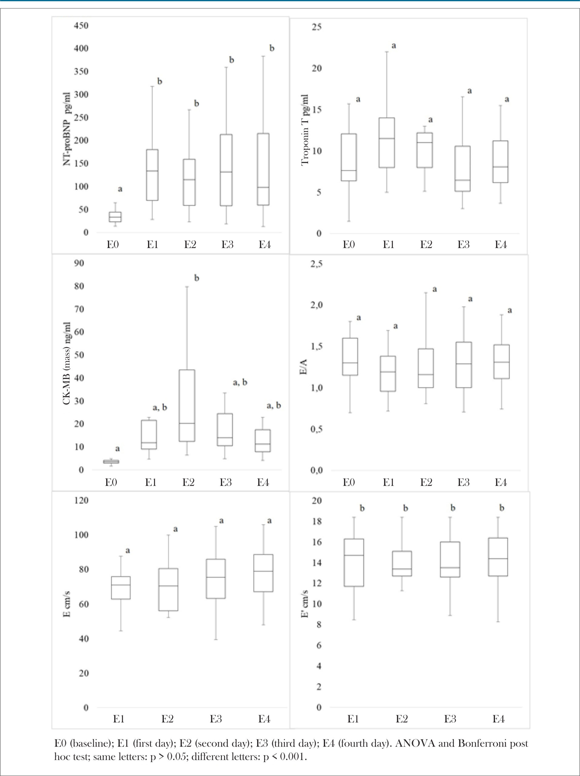
The general objective of this study was to verify these effects on the behavior of diastolic function and the cardiac biomarkers CK-MB (mass), troponin T, and NT-proBNP, in amateur athletes.

Method:

This longitudinal study, conducted in 2015, evaluated participants during the following 5 stages: E0 (baseline) before starting the trajectory and the others, E1, E2, E3, and E4, at the end of each day, totaling 244.7 km. At all stages, the biomarkers NT-proBNP, CK-MB (mass), and troponin T were measured. Echocardiogram was performed to analyze the E, A and E’ waves. P < 0.05 was adopted as significant.

Results:

The study evaluated 25 participants, with an average age of 46 ± 10.5 years and body mass index of 20.2 ± 2.3 kg/m2. Increased values were found for NT-proBNP from E0 to E1, E2, E3, and E4 (p < 0.001), CK-MB (mass) from E0 to E2 (p < 0.001), and E' wave from E0 to E1, E2, E3, and E4 (p < 0.001). Positive correlations were identified between the following: CK-MB (mass) and troponin T (E1: r = 0.524, p = 0.010; E4: r = 0.413, p = 0.044); CK-MB (mass) and NT-proBNP (E4: r = 0.539, p = 0.006); and E/A and E' (E0: r = 0.603, p < 0.001; E1: r = 0.639, p < 0.001; E4: r = 0.593, p = 0.002). A negative correlation was found between CK-MB (mass) and E/A (E1: r = −0.428, p = 0.041).

Conclusion:

The effects of intense, prolonged, and interspersed physical activity were verified based on significant variations in the behavior of CK-MB (mass), NT-proBNP, and the E’ wave. Notwithstanding the alterations found, there were no criteria suggestive of myocardial damage

  • Athletes
  • Biomarkers, Biological
  • Blood Pressure
  • Echocardiography, Doppler/methods
  • Natriuretic Peptide, Brain
  • Troponin T
  • walking
Read Full Article
Creative Commons Attribution (CC BY) Creative Commons license
23/10/2020 DOI: 10.36660/abc.20190271 Views 1
Figure 1
ABC
Original Article

Velocity-Time Integral of Aortic Regurgitation: A Novel Echocardiographic Marker in the Evaluation of Aortic Regurgitation Severity

José Abellán-Huerta et al. Juan Carlos Bonaque-González Ramón Rubio-Patón José García-Gómez … Santiago Egea-Beneyto Federico Soria-Arcos Luciano Consuegra-Sánchez Rosa María Soto-Ruiz José Luis Ramos-Martín Juan Antonio Castillo-Moreno

Abstract

Background

Echocardiography is essential for the diagnosis and quantification of aortic regurgitation (AR). Velocity-time integral (VTI) of AR flow could be related to AR severity.

Objective

This study aims to assess whether VTI is an echocardiographic marker of AR severity.

Methods

We included all patients with moderate or severe native AR and sinus rhythm who visited our imaging laboratory from January to October 2016. All individuals underwent a complete echocardiogram with AR VTI measurement. The association between VTI and AR severity was analyzed by logistic regression and multivariate regression models. A p-value<0,05 was considered statistically significant.

Results

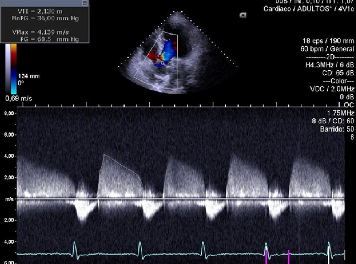
Among the 62 patients included (68.5±14.9 years old; 64.5%: moderate AR; 35.5%: severe AR), VTI was higher in individuals with moderate AR compared to those with severe AR (2.2±0.5 m vs. 1.9±0.5 m, p=0.01). Patients with severe AR presented greater values of left ventricular end-diastolic diameter (LVEDD) (56.1±7.1 mm vs. 47.3±9.6 mm, p=0.001), left ventricular end-diastolic volume (LVEDV) (171±36.5 mL vs. 106±46.6 mL, p<0.001), effective regurgitant orifice (0.44±0.1 cm2vs. 0.18±0.1 cm2, p=0.002), and regurgitant volume (71.3±25.7 mL vs. 42.5±10.9 mL, p=0.05), as well as lower left ventricular ejection fraction (LVEF) (54.1±11.2% vs. 63.2±13.3%, p=0.012). The VTI proved to be a marker of AR severity, irrespective of LVEDD, LVEDV, and LVEF (odds ratio 0.160, p=0.032) and of heart rate and diastolic blood pressure (DBP) (odds ratio 0.232, p=0.044).

Conclusions

The VTI of AR flow was inversely associated with AR severity regardless of left ventricular diameter and volume, heart rate, DBP, and LVEF. VTI could be a marker of AR severity in patients with native AR and sinus rhythm. (Arq Bras Cardiol. 2020; [online].ahead print, PP.0-0)

  • Aortic Valve Insufficiency/diagnosis,imaging
  • Echocardiography, Doppler/methods
  • Heart Failure
Read Full Article
Creative Commons Attribution (CC BY) Creative Commons license
15/07/2020 DOI: 10.36660/abc.20190243
Figure 1
ABC
Image

Abdominal Pain: an Uncommon Presentation of Myocardial Rupture

Daniel Seabra et al. Ana Neto Inês Oliveira Rui Pontes dos Santos … João Azevedo Paula Pinto

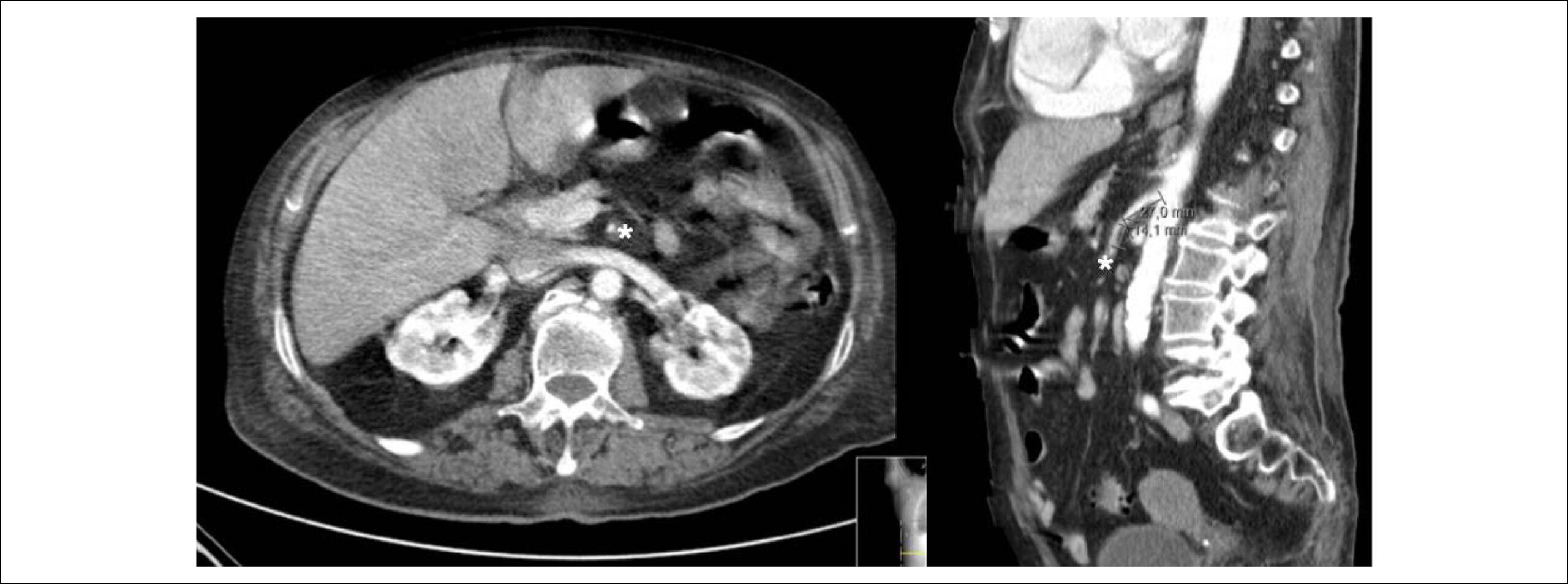
An 84-years-old woman with hypertension and dyslipidemia was admitted in the emergency room with acute abdominal pain; patient complained of chest pain three weeks before hospital admission. On physical examination, she was hypotensive, tachycardic (with arrhythmic pulse), tachypneic and with diffuse abdominal pain. Electrocardiogram showed atrial fibrillation with rapid ventricular response, complete left bundle branch block and inferior Q waves. Abdominal computed tomography (CT) revealed a thrombus in the superior mesenteric artery (, white asterisk). The patient showed a clinical course with congestive heart failure and low cardiac output. Transthoracic echocardiogram (-) showed mild left ventricular dilatation with a mild dysfunction and a pseudoaneurysm of the basal half of the posterior and inferior walls with left-to-right shunt, confirmed by color Doppler imaging (-). Cardiac CT () revealed contained myocardial rupture, located at the basal segments of the inferior and posterior septal walls, extending to the free wall of the right ventricle, forming a pseudo-cavity, which communicates with the true cavity of the right ventricle (). Despite vasopressor and inotropic support and proposal for cardiac surgery, the patient had an unfavorable course.

[…]

  • Abdominal Pain
  • Dyslipidemias
  • Echocardiography, Doppler/methods
  • Hypertension
  • Myocardial Infarction/complications
  • Thrombosis/surgery
  • Tomography, X Ray-Computed/methods
Read Full Article
Creative Commons Attribution (CC BY) Creative Commons license
20/03/2020 DOI: 10.36660/abc.20190495
Figure 1
ABC
Image

Double-Inlet Single Ventricle with Malposed Great Arteries

Paulo Andrade et al. Danilo Santos Magna Moreira Adail Almeida

A 62-year-old man came to the Echocardiography Service with a history of arterial hypertension and systolic murmur in the mitral area. At the subsequent evaluation, the patient reported dyspnea and fatigue on moderate exertion, but without an impact on social life. Peripheral oxygen saturation at rest ranged from 95% to 98%; extremities were warm and perfused, with no signs of peripheral hypoperfusion; cyanosis and digital clubbing were absent.

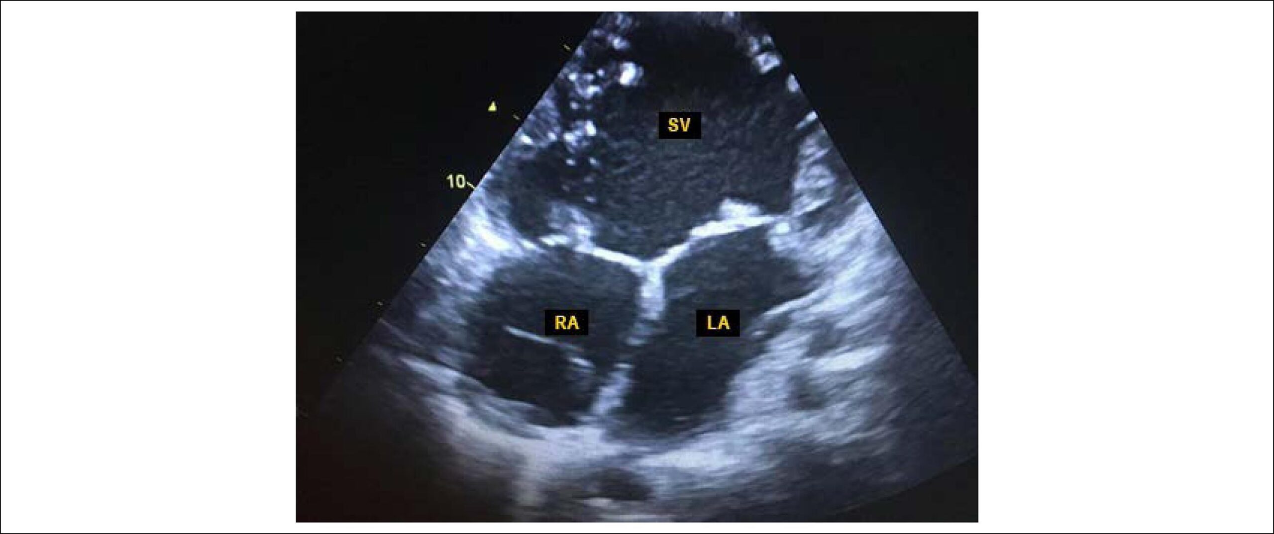
The echocardiogram disclosed a case of levocardia, with the presence of a double-inlet single ventricle with transposition of the great arteries (, and ), with situs solitus, enlargement of the atrial chambers associated with significant mitral regurgitation due to annulus dilatation.

[…]

  • Aged
  • Diagnosis Imaging
  • Echocardiography, Doppler/methods
  • Mitral Valve Insufficiency
  • Transposition of great vessels/ surgery
Read Full Article
Creative Commons Attribution (CC BY) Creative Commons license
10/10/2019 DOI: 10.5935/abc.20190160
Figure 1
ABC
Original Article

Global Longitudinal Strain Predicts Poor Functional Capacity in Patients with Systolic Heart Failure

Rafael José Coelho Maia et al. Simone Cristina Soares Brandão Jéssica Leite Giordano Bruno Parente … Filipe Pinheiro Bruna Thays Santana Araújo Maria Inês Remígio Aguiar Sílvia Marinho Martins Daniella Cunha Brandão Armele Dornelas de Andrade

Abstract

Background:

Left ventricular global longitudinal strain value (GLS) can predict functional capacity in patients with preserved left ventricular ejection fraction (LVEF) heart failure (HF) and to assess prognosis in reduced LVEF HF.

Objetive:

Correlate GLS with parameters of Cardiopulmonary Exercise Test (CPET) and to assess if they could predict systolic HF patients that are more appropriated to be referred to heart transplantation according to CPET criteria.

Methods:

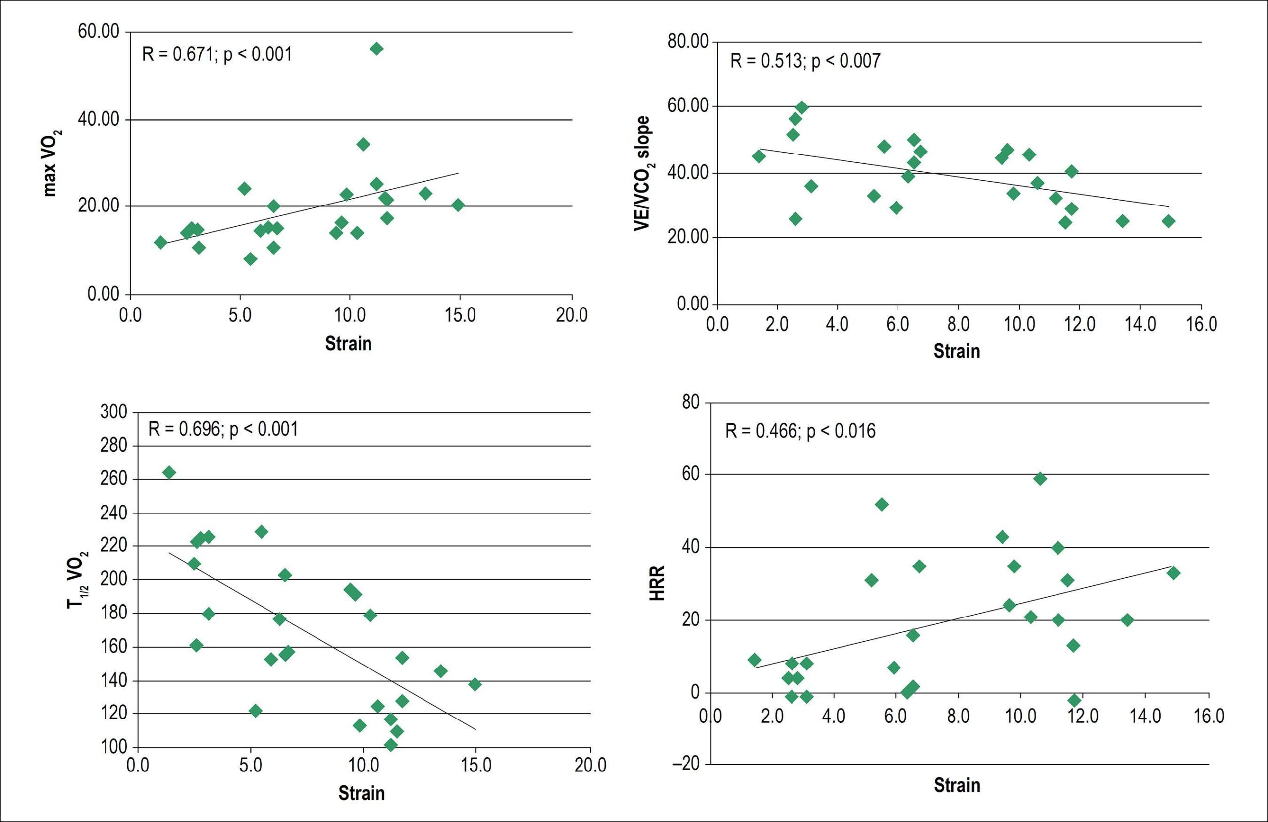
Systolic HF patients with LVEF < 45%, NYHA functional class II and III, underwent prospectively CPET and echocardiography with strain analysis. LVEF and GLS were correlated with the following CPET variables: maxVO2, VE/VCO2 slope, heart rate reduction during the first minute of recovery (HRR) and time needed to reduce maxVO2 in 50% after physical exercise (T1/2VO2). ROC curve analysis of GLS to predict VO2 < 14 mL/kg/min and VE/VCO2 slope > 35 (heart transplantation’s criteria) was performed.

Results:

Twenty six patients were selected (age, 47 ± 12 years, 58% men, mean LVEF = 28 ± 8%). LVEF correlated only with maxVO2 and T1/2VO2. GLS correlated to all CPET variables (maxVO2: r = 0.671, p = 0.001; VE/VCO2 slope: r = -0.513, p = 0.007; HRR: r = 0.466, p = 0.016, and T1/2VO2: r = -0.696, p = 0.001). GLS area under the ROC curve to predict heart transplantation’s criteria was 0.88 (sensitivity 75%, specificity 83%) for a cut-off value of -5.7%, p = 0.03.

Conclusion:

GLS was significantly associated with all functional CPET parameters. It could classify HF patients according to the functional capacity and may stratify which patients have a poor prognosis and therefore to deserve more differentiated treatment, such as heart transplantation.

  • Echocardiography, Doppler/methods
  • Heart Failure
  • Longitudinal Strain
  • Torsion Abnormality
  • Torsion, Mechanical
  • Ventricular Dysfunction, Left
Read Full Article
Creative Commons Attribution (CC BY) Creative Commons license
18/07/2019 DOI: 10.5935/abc.20190119

How to cite this article

EndNote RIS

Electronic Document Format (Vancouver)

Menegazzo WR, Silveira AD. The Right Way to Identify Bad Cardiorespiratory Fitness: Look to the Right. Arquivos Brasileiros de Cardiologia 2023;120(9):e20230630.

Electronic Document Format (ABNT)

Menegazzo, Willian R.; Silveira, Anderson Donelli da. The Right Way to Identify Bad Cardiorespiratory Fitness: Look to the Right. Arquivos Brasileiros de Cardiologia, v. 120, n. 9, e20230630, Oct. 2023.

Electronic Document Format (APA)

Menegazzo, W. R., & Silveira, A. D. (2023). The Right Way to Identify Bad Cardiorespiratory Fitness: Look to the Right. Arquivos Brasileiros de Cardiologia, 120(9), e20230630.

Electronic Document Format (ISO)

Menegazzo, Willian R. and Silveira, Anderson Donelli da. The Right Way to Identify Bad Cardiorespiratory Fitness: Look to the Right. Arquivos Brasileiros de Cardiologia [online]. 2023, vol. 120, n. 9, [cited 2026-10-14], e20230630. Available from: <https://journals.cardiol.br/en/article/the-right-way-to-identify-bad-cardiorespiratory-fitness-look-to-the-right/>. ISSN 0066-782X

EndNote RIS

Electronic Document Format (Vancouver)

Abreu JS, Diogenes TCP, Farias AGLP, Abreu MEB, Freire RA, Barreto JEF. Spasm of the Internal Thoracic Artery. Value of Echocardiography and Doppler in Long-term Follow-up. Arquivos Brasileiros de Cardiologia Imagem cardiovascular 2022;31(4):314-7.

Electronic Document Format (ABNT)

Abreu, José Sebastião de; Diogenes, Tereza Cristina Pinheiro; Farias, Ana Gardenia L. Ponte; Abreu, Marilia Esther Benevides; Freire, Renan Abreu; Barreto, José Erirtônio Façanha. Spasm of the Internal Thoracic Artery. Value of Echocardiography and Doppler in Long-term Follow-up. Arquivos Brasileiros de Cardiologia Imagem cardiovascular, v. 31, n. 4, p. 314-317, Feb. 2022.

Electronic Document Format (APA)

Abreu, J. S. , Diogenes, T. C. P., Farias, A. G. L. P., Abreu, M. E. B., Freire, R. A., & Barreto, J. E. F. (2022). Spasm of the Internal Thoracic Artery. Value of Echocardiography and Doppler in Long-term Follow-up. Arquivos Brasileiros de Cardiologia Imagem cardiovascular, 31(4), 314-317.

Electronic Document Format (ISO)

Abreu, José Sebastião de and Diogenes, Tereza Cristina Pinheiro and Farias, Ana Gardenia L. Ponte and Abreu, Marilia Esther Benevides and Freire, Renan Abreu and Barreto, José Erirtônio Façanha. Spasm of the Internal Thoracic Artery. Value of Echocardiography and Doppler in Long-term Follow-up. Arquivos Brasileiros de Cardiologia Imagem cardiovascular [online]. 2022, vol. 31, n. 4, [cited 2026-10-14], pp. 314-317. Available from: <https://journals.cardiol.br/en/article/spasm-of-the-internal-thoracic-artery-value-of-echocardiography-and-doppler-in-long-term-follow-up/>. ISSN 2318-8219

EndNote RIS

Electronic Document Format (Vancouver)

Linhares RR, Barreto RBM, Silva CES, Bihan DCSL, Peixoto LB, Cruz AP, Garcia LP, et al. Comparison of Block-Matching and Optical Flow in Strain Echocardiography Analysis. Arquivos Brasileiros de Cardiologia Imagem cardiovascular 2022;30(3):87-91.

Electronic Document Format (ABNT)

Linhares, Renata Rejane; Barreto, Rodrigo Bellio de Mattos; Silva, Carlos Eduardo Suaide; Bihan, David Costa de Souza Le; Peixoto, Luciana Braz; Cruz, Andresa Paes da; Garcia, Luara Piovan; Monaco, Claudia Gianini; Gil, Manuel Adán. Comparison of Block-Matching and Optical Flow in Strain Echocardiography Analysis. Arquivos Brasileiros de Cardiologia Imagem cardiovascular, v. 30, n. 3, p. 87-91, Feb. 2022.

Electronic Document Format (APA)

Linhares, R. R., Barreto, R. B. M., Silva, C. E. S., Bihan, D. C. S. L., Peixoto, L. B., Cruz, A. P. , Garcia, L. P., Monaco, C. G., & Gil, M. A. (2022). Comparison of Block-Matching and Optical Flow in Strain Echocardiography Analysis. Arquivos Brasileiros de Cardiologia Imagem cardiovascular, 30(3), 87-91.

Electronic Document Format (ISO)

Linhares, Renata Rejane and Barreto, Rodrigo Bellio de Mattos and Silva, Carlos Eduardo Suaide and Bihan, David Costa de Souza Le and Peixoto, Luciana Braz and Cruz, Andresa Paes da and Garcia, Luara Piovan and Monaco, Claudia Gianini and Gil, Manuel Adán. Comparison of Block-Matching and Optical Flow in Strain Echocardiography Analysis. Arquivos Brasileiros de Cardiologia Imagem cardiovascular [online]. 2022, vol. 30, n. 3, [cited 2026-10-14], pp. 87-91. Available from: <https://journals.cardiol.br/en/article/comparison-of-block-matching-and-optical-flow-in-strain-echocardiography-analysis/>. ISSN 2318-8219

EndNote RIS

Electronic Document Format (Vancouver)

Abreu JS, Pinheiro TCD, Feitosa JA, Abreu MEB, Farias AGLP, Carneiro MM, Falcão SNR, et al. Ligation of the Branches of the Anastomosed Internal Thoracic Artery in the Anterior Descending Coronary Artery and the Effect on Flow Velocities and Functional Status of the Graft. Arquivos Brasileiros de Cardiologia Imagem cardiovascular 2022;31(4):258-69.

Electronic Document Format (ABNT)

Abreu, José Sebastião de; Pinheiro, Tereza Cristina D; Feitosa, José Acácio; Abreu, Marilia Esther B; Farias, Ana Gardenia L. P; Carneiro, Marcia Maria; Falcão, Sandra Nívea R. Ligation of the Branches of the Anastomosed Internal Thoracic Artery in the Anterior Descending Coronary Artery and the Effect on Flow Velocities and Functional Status of the Graft. Arquivos Brasileiros de Cardiologia Imagem cardiovascular, v. 31, n. 4, p. 258-269, Feb. 2022.

Electronic Document Format (APA)

Abreu, J. S. , Pinheiro, T. C. D., Feitosa, J. A., Abreu, M. E. B., Farias, A. G. L. P., Carneiro, M. M., & Falcão, S. N. R. (2022). Ligation of the Branches of the Anastomosed Internal Thoracic Artery in the Anterior Descending Coronary Artery and the Effect on Flow Velocities and Functional Status of the Graft. Arquivos Brasileiros de Cardiologia Imagem cardiovascular, 31(4), 258-269.

Electronic Document Format (ISO)

Abreu, José Sebastião de and Pinheiro, Tereza Cristina D and Feitosa, José Acácio and Abreu, Marilia Esther B and Farias, Ana Gardenia L. P and Carneiro, Marcia Maria and Falcão, Sandra Nívea R. Ligation of the Branches of the Anastomosed Internal Thoracic Artery in the Anterior Descending Coronary Artery and the Effect on Flow Velocities and Functional Status of the Graft. Arquivos Brasileiros de Cardiologia Imagem cardiovascular [online]. 2022, vol. 31, n. 4, [cited 2026-10-14], pp. 258-269. Available from: <https://journals.cardiol.br/en/article/ligation-of-the-branches-of-the-anastomosed-internal-thoracic-artery-in-the-anterior-descending-coronary-artery-and-the-effect-on-flow-velocities-and-functional-status-of-the-graft/>. ISSN 2318-8219

EndNote RIS

Electronic Document Format (Vancouver)

Euzebio MB, Vitorino PVO, Sousa WM, Melo MA, Costa SHN, Sousa ALL, Jardim TSV, et al. Diastolic Function and Biomarkers of Long-Distance Walking Participants. Arquivos Brasileiros de Cardiologia 2020;115(4):620-7.

Electronic Document Format (ABNT)

Euzebio, Maicon Borges; Vitorino, Priscila Valverde de O.; Sousa, Watila Moura; Melo, Milena Andrade; Costa, Sérgio Henrique Nascente; Sousa, Ana Luiza Lima; Jardim, Thiago de Souza Veiga; Arantes, Ana Carolina; Jardim, Paulo Cesar B. Veiga; Barroso, Weimar Kunz Sebba. Diastolic Function and Biomarkers of Long-Distance Walking Participants. Arquivos Brasileiros de Cardiologia, v. 115, n. 4, p. 620-627, Oct. 2020.

Electronic Document Format (APA)

Euzebio, M. B., Vitorino, P. V. O., Sousa, W. M., Melo, M. A., Costa, S. H. N., Sousa, A. L. L., Jardim, T. S. V., Arantes, A. C., Jardim, P. C. B. V., & Barroso, W. K. S. (2020). Diastolic Function and Biomarkers of Long-Distance Walking Participants. Arquivos Brasileiros de Cardiologia, 115(4), 620-627.

Electronic Document Format (ISO)

Euzebio, Maicon Borges and Vitorino, Priscila Valverde de O. and Sousa, Watila Moura and Melo, Milena Andrade and Costa, Sérgio Henrique Nascente and Sousa, Ana Luiza Lima and Jardim, Thiago de Souza Veiga and Arantes, Ana Carolina and Jardim, Paulo Cesar B. Veiga and Barroso, Weimar Kunz Sebba. Diastolic Function and Biomarkers of Long-Distance Walking Participants. Arquivos Brasileiros de Cardiologia [online]. 2020, vol. 115, n. 4, [cited 2026-10-14], pp. 620-627. Available from: <https://journals.cardiol.br/en/article/diastolic-function-and-biomarkers-of-long-distance-walking-participants/>. ISSN 0066-782X

EndNote RIS

Electronic Document Format (Vancouver)

Abellán-Huerta J, Bonaque-González JC, Rubio-Patón R, García-Gómez J, Egea-Beneyto S, Soria-Arcos F, Consuegra-Sánchez L, et al. Velocity-Time Integral of Aortic Regurgitation: A Novel Echocardiographic Marker in the Evaluation of Aortic Regurgitation Severity. Arquivos Brasileiros de Cardiologia 2020;115(2):253-60.

Electronic Document Format (ABNT)

Abellán-Huerta, José; Bonaque-González, Juan Carlos; Rubio-Patón, Ramón; García-Gómez, José; Egea-Beneyto, Santiago; Soria-Arcos, Federico; Consuegra-Sánchez, Luciano; Soto-Ruiz, Rosa María; Ramos-Martín, José Luis; Castillo-Moreno, Juan Antonio. Velocity-Time Integral of Aortic Regurgitation: A Novel Echocardiographic Marker in the Evaluation of Aortic Regurgitation Severity. Arquivos Brasileiros de Cardiologia, v. 115, n. 2, p. 253-260, Jul. 2020.

Electronic Document Format (APA)

Abellán-Huerta, J., Bonaque-González, J. C., Rubio-Patón, R., García-Gómez, J., Egea-Beneyto, S., Soria-Arcos, F., Consuegra-Sánchez, L., Soto-Ruiz, R. M., Ramos-Martín, J. L., & Castillo-Moreno, J. A. (2020). Velocity-Time Integral of Aortic Regurgitation: A Novel Echocardiographic Marker in the Evaluation of Aortic Regurgitation Severity. Arquivos Brasileiros de Cardiologia, 115(2), 253-260.

Electronic Document Format (ISO)

Abellán-Huerta, José and Bonaque-González, Juan Carlos and Rubio-Patón, Ramón and García-Gómez, José and Egea-Beneyto, Santiago and Soria-Arcos, Federico and Consuegra-Sánchez, Luciano and Soto-Ruiz, Rosa María and Ramos-Martín, José Luis and Castillo-Moreno, Juan Antonio. Velocity-Time Integral of Aortic Regurgitation: A Novel Echocardiographic Marker in the Evaluation of Aortic Regurgitation Severity. Arquivos Brasileiros de Cardiologia [online]. 2020, vol. 115, n. 2, [cited 2026-10-14], pp. 253-260. Available from: <https://journals.cardiol.br/en/article/velocity-time-integral-of-aortic-regurgitation-a-novel-echocardiographic-marker-in-the-evaluation-of-aortic-regurgitation-severity/>. ISSN 0066-782X

EndNote RIS

Electronic Document Format (Vancouver)

Seabra D, Ana Neto , Oliveira I, Santos RP, Azevedo J, Pinto P. Abdominal Pain: an Uncommon Presentation of Myocardial Rupture. Arquivos Brasileiros de Cardiologia 2020;114(2):319-22.

Electronic Document Format (ABNT)

Seabra, Daniel; Ana Neto, ; Oliveira, Inês; Santos, Rui Pontes dos; Azevedo, João; Pinto, Paula. Abdominal Pain: an Uncommon Presentation of Myocardial Rupture. Arquivos Brasileiros de Cardiologia, v. 114, n. 2, p. 319-322, Mar. 2020.

Electronic Document Format (APA)

Seabra, D., Ana Neto, , Oliveira, I., Santos, R. P. , Azevedo, J., & Pinto, P. (2020). Abdominal Pain: an Uncommon Presentation of Myocardial Rupture. Arquivos Brasileiros de Cardiologia, 114(2), 319-322.

Electronic Document Format (ISO)

Seabra, Daniel and Ana Neto and Oliveira, Inês and Santos, Rui Pontes dos and Azevedo, João and Pinto, Paula. Abdominal Pain: an Uncommon Presentation of Myocardial Rupture. Arquivos Brasileiros de Cardiologia [online]. 2020, vol. 114, n. 2, [cited 2026-10-14], pp. 319-322. Available from: <https://journals.cardiol.br/en/article/abdominal-pain-an-uncommon-presentation-of-myocardial-rupture/>. ISSN 0066-782X

EndNote RIS

Electronic Document Format (Vancouver)

Andrade P, Santos D, Moreira M, Almeida A. Double-Inlet Single Ventricle with Malposed Great Arteries. Arquivos Brasileiros de Cardiologia 2019;113(3):444-6.

Electronic Document Format (ABNT)

Andrade, Paulo; Santos, Danilo; Moreira, Magna; Almeida, Adail. Double-Inlet Single Ventricle with Malposed Great Arteries. Arquivos Brasileiros de Cardiologia, v. 113, n. 3, p. 444-446, Oct. 2019.

Electronic Document Format (APA)

Andrade, P., Santos, D., Moreira, M., & Almeida, A. (2019). Double-Inlet Single Ventricle with Malposed Great Arteries. Arquivos Brasileiros de Cardiologia, 113(3), 444-446.

Electronic Document Format (ISO)

Andrade, Paulo and Santos, Danilo and Moreira, Magna and Almeida, Adail. Double-Inlet Single Ventricle with Malposed Great Arteries. Arquivos Brasileiros de Cardiologia [online]. 2019, vol. 113, n. 3, [cited 2026-10-14], pp. 444-446. Available from: <https://journals.cardiol.br/en/article/double-inlet-single-ventricle-with-malposed-great-arteries/>. ISSN 0066-782X

EndNote RIS

Electronic Document Format (Vancouver)

Maia RJC, Brandão SCS, Leite J, Parente GB, Pinheiro F, Araújo BTS, Aguiar MIR, et al. Global Longitudinal Strain Predicts Poor Functional Capacity in Patients with Systolic Heart Failure. Arquivos Brasileiros de Cardiologia 2019;113(2):188-94.

Electronic Document Format (ABNT)

Maia, Rafael José Coelho; Brandão, Simone Cristina Soares; Leite, Jéssica; Parente, Giordano Bruno; Pinheiro, Filipe; Araújo, Bruna Thays Santana; Aguiar, Maria Inês Remígio; Martins, Sílvia Marinho; Brandão, Daniella Cunha; Andrade, Armele Dornelas de. Global Longitudinal Strain Predicts Poor Functional Capacity in Patients with Systolic Heart Failure. Arquivos Brasileiros de Cardiologia, v. 113, n. 2, p. 188-194, Jul. 2019.

Electronic Document Format (APA)

Maia, R. J. C., Brandão, S. C. S., Leite, J., Parente, G. B., Pinheiro, F., Araújo, B. T. S., Aguiar, M. I. R., Martins, S. M., Brandão, D. C., & Andrade, A. D. (2019). Global Longitudinal Strain Predicts Poor Functional Capacity in Patients with Systolic Heart Failure. Arquivos Brasileiros de Cardiologia, 113(2), 188-194.

Electronic Document Format (ISO)

Maia, Rafael José Coelho and Brandão, Simone Cristina Soares and Leite, Jéssica and Parente, Giordano Bruno and Pinheiro, Filipe and Araújo, Bruna Thays Santana and Aguiar, Maria Inês Remígio and Martins, Sílvia Marinho and Brandão, Daniella Cunha and Andrade, Armele Dornelas de. Global Longitudinal Strain Predicts Poor Functional Capacity in Patients with Systolic Heart Failure. Arquivos Brasileiros de Cardiologia [online]. 2019, vol. 113, n. 2, [cited 2026-10-14], pp. 188-194. Available from: <https://journals.cardiol.br/en/article/global-longitudinal-strain-predicts-poor-functional-capacity-in-patients-with-systolic-heart-failure/>. ISSN 0066-782X

  • About ABC Family
  • Internal Regulation
  • Editorial Board
  • Brazilian Society of Cardiology (SBC)

Issues

SBC Guidelines

  • Videos
  • Podcasts
  • News