ABC Family Arquivos Brasileiros de Cardiologia International Journal of Cardiovascular Sciences ABC Heart Failure & Cardiomyopathy ABC Imagem Cardiovascular
Português English
ABC Family
Português English
  • About
    • About ABC Family
    • Internal Regulation
    • Editorial Board
    • Brazilian Society of Cardiology (SBC)
  • Issues
  • SBC Guidelines
  • Content
    • Videos
    • Podcasts
    • News
  • About
    • About ABC Family
    • Internal Regulation
    • Editorial Board
    • Brazilian Society of Cardiology (SBC)
  • Issues
  • SBC Guidelines
  • Content
    • Videos
    • Podcasts
    • News

ABC Family

  • ABC Family
  • Arquivos Brasileiros de Cardiologia
  • International Journal of Cardiovascular Sciences
  • ABC Heart Failure & Cardiomyopathy
  • ABC Imagem Cardiovascular
Search in

No content found.

Journals
Ordering
Article types
Others

  1. Home
  2. Keyword: Myocytes, Cardiac
Figure 1
ABC
Original Article

Electrocardiographic Evaluation of Normal Newborns in the First Week of Life – Observational Study

Marina de Souza Pimenta et al. Nelson Samesima Carlos Alberto Pastore Vera Lucia Jornada Krebs … Gabriela Nunes Leal Werther Brunow de Carvalho

Abstract

Background

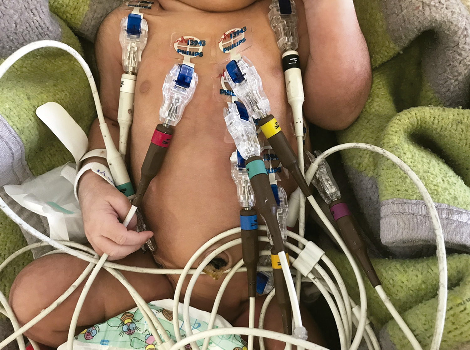
The neonatal period is marked by major changes in the cardiovascular system, especially in the first week of life. Unlike the adult population, studies on electrocardiogram (ECG) data in the neonatal period are scarce. This is the first study to describe electrocardiographic changes in a cohort of newborns with normal echocardiograms.

Objectives

To analyze the electrocardiographic patterns of a population of full-term NB, without any cardiac morphological or functional anomalies, and compare the results with the literature.

Methods

In this observational study, echocardiograms and ECG results from 94 newborns divided in three age groups (up to 24 hours, between 25 and 72 hours, and between 73 and 168 hours of life) were evaluated and compared with those reported by Davignon et al. A p-value <0.05 was considered statistically significant.

Results

There were significant differences in T-wave direction in leads V1 (p= 0.04), V2 (p= 0.02), V3 (p= 0.008) and V4 (p= 0.005) between the three age groups. There were differences between our findings and the current literature in most of the parameters.

Conclusion

Term newborns within 24 hours of life showed significantly more positive T waves than older ones. Many differences from the Davignon’s ECG parameters were found, particularly in the P, Q, R, S amplitudes, QRS duration, R/S and R+S. These findings indicate that more studies are needed for a definitive interpretation of the ECG in newborns.
  • Electrocardiography
  • Infant, Newborn
  • Myocytes, Cardiac
Read Full Article
Creative Commons Attribution (CC BY) Creative Commons license
02/09/2022 DOI: 10.36660/abc.20210843 Views 1
Figure 1
ABC
Original Article

Resistance Exercise Training Mitigates Left Ventricular Dysfunctions in Pulmonary Artery Hypertension Model

Leôncio Lopes Soares et al. Luciano Bernardes Leite Luiz Otávio Guimarães Ervilha Bruna Aparecida Fonseca da Silva … Maíra Oliveira de Freitas Alexandre Martins Oliveira Portes Leonardo Mateus Teixeira Rezende Filipe Rios Drummond Miguel Araújo Carneiro-Júnior Mariana Machado Neves Emily Correna Carlo Reis Antônio José Natali

Abstract

Background

The right ventricular hypertrophy and dilation observed in pulmonary artery hypertension (PAH) damages the left ventricle (LV) dynamics by flattening the interventricular septum.

Objective

To investigate whether low- to moderate-intensity resistance exercise training (RT) is beneficial to LV and cardiomyocyte contractile functions in rats during the development of monocrotaline (MCT)-induced PAH.

Methods

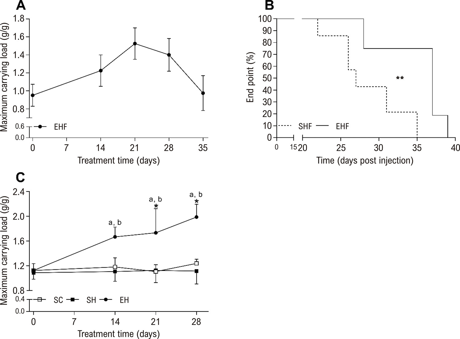
Male Wistar rats (Body weight: ~ 200 g) were used. To assess the time to potential heart failure onset (i.e., end point), rats were divided into sedentary hypertension until failure (SHF, n=6) and exercise hypertension until failure (EHF, n=6) groups. To test RT effects, rats were divided into sedentary control (SC, n = 7), sedentary hypertension (SH, n=7), and exercise hypertension (EH, n=7) groups. PAH was induced by two MCT injections (20 mg/kg, with 7 days interval). Exercise groups were submitted to an RT protocol (Ladder climbing; 55-65% of carrying maximal load), 5 times/week. Statistical significance was assumed at P < 0.05.

Results

RT prolonged the end point (~25 %), enhanced the physical effort tolerance (~ 55%), and mitigated the LV and cardiomyocyte contractility dysfunctions promoted by MCT by preserving the ejection fraction and fractional shortening, the amplitude of shortening, and the velocities of contraction and relaxation in cardiomyocytes. RT also prevented increases in left ventricle fibrosis and type I collagen caused by MCT, and maintained the type III collagen and myocyte dimensions reduced by MCT.

Conclusion

Low- to moderate-intensity RT benefits LV and cardiomyocyte contractile functions in rats during the development of MCT-induced PAH.

  • Exercise
  • Heart Failure
  • Myocytes, Cardiac
  • Physical Conditioning, Animal/methods
  • Pulmonary Hypertension
  • Rats
  • Ventricular Dysfunction, Left
Read Full Article
Creative Commons Attribution (CC BY) Creative Commons license
02/09/2022 DOI: 10.36660/abc.20210681 Views 1
ABC
Editorial

A New Pathway to Coronary Occlusion with Elabela?

Ricardo Wang et al. Estevão Lanna Figueiredo Fernando Carvalho Neuschwander

Total coronary occlusion (TCO) is characterized by an obstruction with an estimated duration of more than 3 months and is present in approximately 15–18% of all coronary angiographies performed. Clinical manifestations can range from asymptomatic, through stable angina, to varying degrees of ventricular dysfunction. This mainly depends on the degree of collateral circulation. If it is poor, it may compromise the viability of myocytes and associate with various degrees of necrosis and/or hibernating myocardium. In the presence of well-developed collaterals (Rentrop grade III), the patient may be asymptomatic and have a preserved contractile function. Therefore, the formation of a well-developed collateral circulation, especially before vessel occlusion, is essential for the preservation of myocytes.

Despite advances in the clinical treatment of stable angina, many patients with TCO undergo interventional procedures. Of these, most are referred for coronary artery bypass grafting surgery or percutaneous coronary intervention (PCI) with more advanced techniques, but also with a higher risk of complications. The study of mechanisms potentially stimulating arteriogenesis and angiogenesis (collateral circulation formation) opens up opportunities for the development of drugs that, in turn, would avoid unnecessary interventions and/or could benefit patients with symptoms refractory to optimized conventional clinical treatment and who are poor candidates for interventions (e.g., thin distal bed, diffuse coronary disease).

[…]

  • Biomarkers (Elabela)
  • Collateral Circulation
  • Coronary Occlusion
  • Microvascular Angina
  • Myocytes, Cardiac
Read Full Article
Creative Commons Attribution (CC BY) Creative Commons license
20/09/2021 DOI: 10.36660/abc.20210662 Views 1
ABC
Editorial

Influence of Doxorubicin Treatment on Heme Metabolism in Cardiomyoblasts: An In Vitro Study

Mariana Gatto et al. Gustavo Augusto Ferreira Mota

The last decades have been marked with potential advances in cancer diagnosis and therapy, leading to a decrease in mortality and increased patient survival. Doxorubicin is a drug that belongs to the anthracycline family, a class of anticancer drugs extracted from streptomycin. Although the mechanism of action of this drug in cancer is complex, it is known that it interferes with the synthesis of DNA and RNA in addition to inducing the production of free radicals that damage the cell membrane and DNA. However, many of the chemotherapeutic agents used in the neoplasms treatment protocols induce several side effects, including cardiac toxicity and negative repercussions in the vascular system such as thrombolytic ischemia, arterial hypertension, ventricular dysfunction and heart failure., The cardiotoxic effect of doxorubicin is dose-dependent, and the primary mechanism of toxicity is the induction of oxidative stress in the myocardium, with high production of reactive oxygen (ROS) and nitrogen (RNS) species that trigger DNA damage and deregulation of various processes intracellular.

The heme molecule mediates the iron availability and is essential for numerous biological processes in aerobic organisms. In the cardiovascular system, it plays a crucial role in antioxidant defense, signal transduction, oxygen transport, hemoglobin storage, and mitochondrial electron transport. Four mitochondrial and four cytoplasmic enzymes participate in heme metabolism. For heme synthesis, glycine and succinyl coenzyme A are condensed into δ-aminolevulinic acid (ALA) by the enzymes aminolevulinic acid synthases (ALAS), known as ALAS1 and ALAS2. The degradation of heme is carried out by enzymes called heme oxygenase (HOX), which produces carbon monoxide, biliverdin, and iron. HOX-1 isoenzyme regulates normal physiological conditions while HOX-2 isoenzyme protects the cells from oxidative stress. On the other hand, experimental studies have shown that elevated heme protein levels are related to increased oxidative stress and toxicity in cardiomyocytes., However, few studies in the literature verified the influence of anthracyclines on heme protein metabolism in cardiomyocytes.

[…]

  • Anthracyclines
  • Doxorubicin/therapeutic use
  • Heme
  • Hemoglobin E/metabolism
  • Myocytes, Cardiac
Read Full Article
Creative Commons Attribution (CC BY) Creative Commons license
01/03/2021 DOI: 10.36660/abc.20200662 Views 1
Figure 1
ABC
Original Article

Pioglitazone Induces Cardiomyocyte Apoptosis and Inhibits Cardiomyocyte Hypertrophy Via VEGFR-2 Signaling Pathway

Wenliang Zhong et al. Wen Jin Shanghua Xu Yanqing Wu … Shunxiang Luo Minlie Liang Lianglong Chen

Abstract

Background:

Pioglitazone has been widely used as an insulin-sensitizing agent for improving glycemic control in patients with type 2 diabetes mellitus. However, cardiovascular risk and protective effects of pioglitazone remain controversial.

Objectives:

In this study, we investigated whether pioglitazone affects cardiomyocyte apoptosis and hypertrophy by regulating the VEGFR-2 signaling pathway.

Methods:

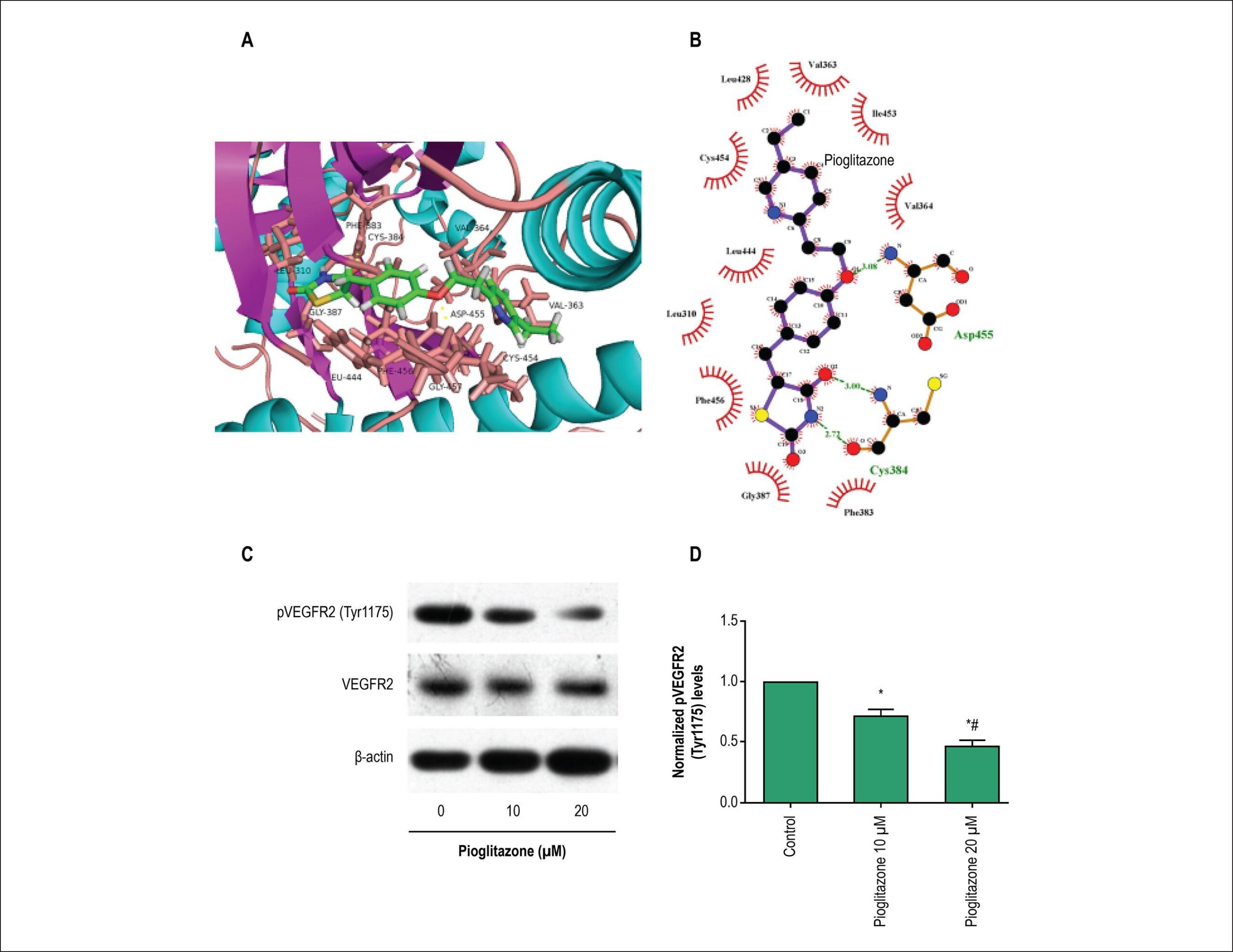
Cardiomyocytes were enzymatically isolated from 1- to 3-day-old Sprague-Dawley rat ventricles. Effects of pioglitazone and the VEGFR-2-selective inhibitor apatinib on cardiomyocyte apoptotic rate was determined using flow cytometry, and hypertrophy was evaluated using [3H]-leucine incorporation. The protein expressions of unphosphorylated and phosphorylated VEGFR-2, Akt, P53, and mTOR were determined by Western-Blotting. Analysis of variance (ANOVA) was used to assess the differences between groups.

Results:

Pioglitazone and VEGFR-2-selective inhibitor apatinib reduced rat cardiomyocyte viability and cardiomyocyte hypertrophy induced by angiotensin II in vitro. Furthermore, in the same in vitro model, pioglitazone and apatinib significantly increased the expression of Bax and phosphorylated P53 and decreased the expression of phosphorylated VEGFR-2, Akt, and mTOR, which promote cardiomyocyte hypertrophy.

Conclusions:

These findings indicate that pioglitazone induces cardiomyocyte apoptosis and inhibits cardiomyocyte hypertrophy by modulating the VEGFR-2 signaling pathway.

  • antihypertensive agents
  • Apoptosis
  • Cardiomegaly
  • Heart Failure/physiopathology
  • Insulin Resistance
  • Myocytes, Cardiac
  • Thiazolidinediones
Read Full Article
Creative Commons Attribution (CC BY) Creative Commons license
02/07/2018 DOI: 10.5935/abc.20180108
Figure 1
ABC
Original Article

Moderate Continuous Aerobic Exercise Training Improves Cardiomyocyte Contractility in Β1 Adrenergic Receptor Knockout Mice

Aurora Corrêa Rodrigues et al. Antônio José Natali Daise Nunes Queiroz da Cunha Alexandre Jayme Lopes Dantas Costa … Anselmo Gomes de Moura Miguel Araújo Carneiro-Júnior Leonardo Bonato Félix Patrícia Chakur Brum Thales Nicolau Prímola-Gomes

Abstract

Background:

The lack of cardiac β1-adrenergic receptors (β1-AR) negatively affects the regulation of both cardiac inotropy and lusitropy, leading, in the long term, to heart failure (HF). Moderate-intensity aerobic exercise (MCAE) is recommended as an adjunctive therapy for patients with HF.

Objective:

We tested the effects of MCAE on the contractile properties of left ventricular (LV) myocytes from β1 adrenergic receptor knockout (β1ARKO) mice.

Methods:

Four- to five-month-old male wild type (WT) and β1ARKO mice were divided into groups: WT control (WTc) and trained (WTt); and β1ARKO control (β1ARKOc) and trained (β1ARKOt). Animals from trained groups were submitted to a MCAE regimen (60 min/day; 60% of maximal speed, 5 days/week) on a treadmill, for 8 weeks. P ≤ 0.05 was considered significant in all comparisons.

Results:

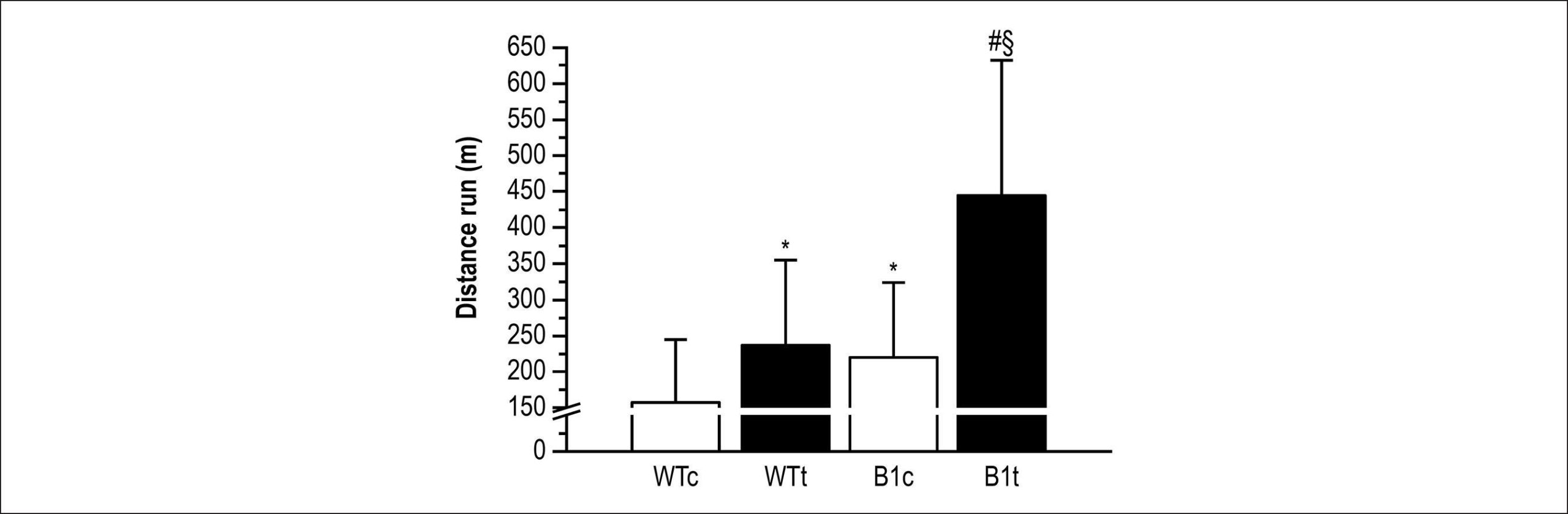
The β1ARKO and exercised mice exhibited a higher (p < 0.05) running capacity than WT and sedentary ones, respectively. The β1ARKO mice showed higher body (BW), heart (HW) and left ventricle (LVW) weights, as well as the HW/BW and LVW/BW than WT mice. However, the MCAE did not affect these parameters. Left ventricular myocytes from β1ARKO mice showed increased (p < 0.05) amplitude and velocities of contraction and relaxation than those from WT. In addition, MCAE increased (p < 0.05) amplitude and velocities of contraction and relaxation in β1ARKO mice.

Conclusion:

MCAE improves myocyte contractility in the left ventricle of β1ARKO mice. This is evidence to support the therapeutic value of this type of exercise training in the treatment of heart diseases involving β1-AR desensitization or reduction.

  • Adrenergic beta 1 Receptor Antagonists
  • Exercise
  • Heart Failure
  • Mice
  • Myocardial Contraction
  • Myocytes, Cardiac
Read Full Article
Creative Commons Attribution (CC BY) Creative Commons license
19/02/2018 DOI: 10.5935/abc.20180025 Views 1
Figure 1
ABC
Brief Communication

Electrical Properties of Isolated Cardiomyocytes in a Rat Model of Thiamine Deficiency

Artur Santos-Miranda et al. Jader Santos Cruz Danilo Roman-Campos

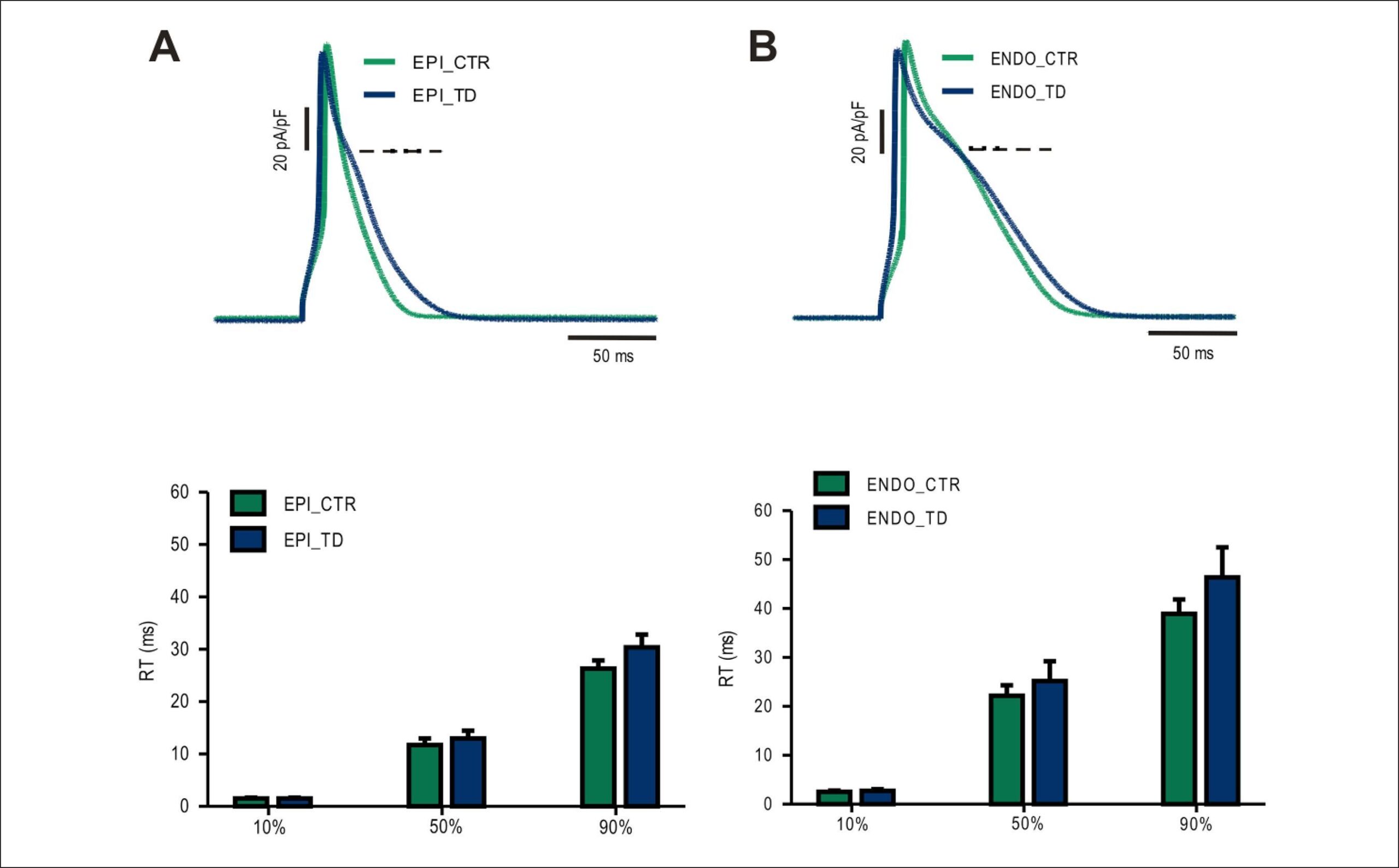
In modern society, thiamine deficiency (TD) remains an important medical condition linked to altered cardiac function. There have been contradictory reports about the impact of TD on heart physiology, especially in the context of cardiac excitability. In order to address this particular question, we used a TD rat model and patch-clamp technique to investigate the electrical properties of isolated cardiomyocytes from epicardium and endocardium. Neither cell type showed substantial differences on the action potential waveform and transient outward potassium current. Based on our results we can conclude that TD does not induce major electrical remodeling in isolated cardiac myocytes in either endocardium or epicardium cells.

  • Action Potentials
  • Electric Stimulation
  • Myocytes, Cardiac
  • Rats, Heart Failure
  • Thiamine Deficiency/ physiopathology
Read Full Article
Creative Commons Attribution (CC BY) Creative Commons license
01/03/2015 DOI: 10.5935/abc.20150010 Views 1
Influence of long-term obesity on myocardial gene expression
ABC
Artigo Original

Influence of long-term obesity on myocardial gene expression

Ana Paula Lima-Leopoldo et al. André Soares Leopoldo Danielle Cristina Tomaz Silva André Ferreira do Nascimento … Dijon Henrique Salomé de Campos Renata de Azevedo Melo Luvizotto Silvio Assis de Oliveira Júnior Carlos Roberto Padovani Célia Regina Nogueira Antonio Carlos Cicogna

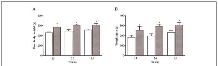
BACKGROUND: Several authors have shown that deterioration of cardiac function is associated with the degree and duration of obesity. It is necessary to establish the gene expression patterns after prolonged periods of obesity. OBJECTIVE: This study tested the hypothesis that increased duration of exposure to obesity leads to a reduction in the mRNA levels of proteins involved in regulation of myocardial Ca2+ homeostasis. In addition, this study verified whether the decrease in mRNA expression was caused by a reduction in thyroid hormone. METHODS: Thirty-day-old male Wistar rats were distributed in two groups: control (C) and obese (Ob). The C group was fed a standard diet and the Ob was fed with high-fat diets for 15, 30 and 45 weeks. Obesity was defined by adiposity index. The gene expression was assessed by quantitative real-time PCR. RESULTS: The adiposity index was higher in the Ob compared to the C after all periods. While obesity at 15 and 45 weeks resulted in a reduction in mRNA of sarcoplasmic reticulum Ca2+- ATPase (SERCA2a), Na+/Ca2+ exchanger (NCX), and calsequestrin (CSQ), L-type Ca2+ channels, ryanodine receptor, SERCA2a, phospholamban (PLB), NCX, and CSQ expression were increased compared to the C after 30 weeks. There was no significant association between T3 levels and mRNA expression. CONCLUSIONS: Our data indicate that obesity over the short and long periods of time may promote alteration in gene expression of Ca2+ homeostasis regulatory proteins without influence by thyroid hormone.

  • Gene Expression
  • Myocardium
  • Myocytes, Cardiac
  • Obesity
  • Thyroid Hormones
Read Full Article
Creative Commons Attribution (CC BY) Creative Commons license
15/04/2013 DOI: 10.5935/abc.20130045 Views 1
ABC
Artigo Original

Immunohistochemical expression of cell differentiation and growth in neonate cardiomyocytes

Tarcísio Fulgêncio Alves da Silva et al. Greyce Kelly de Souza Mona Adalgisa Simões Francisco Cesar Pabis Lucia de Noronha

BACKGROUND: The cardiac alterations during the fetal heart transition to extrauterine life have been explored by several animal studies and the cell mechanisms responsible for these modifications are not well documented in humans. OBJECTIVE: To evaluate the mechanism of cell differentiation into cardiomyocytes that occur in the first days of life, through immunohistochemical analysis of proteins involved in proliferation and muscle contraction processes, in samples of human neonate myocardium. METHODS: Cross-sectional study of paraffin-sample sections of myocardium from an autopsy database of human neonates, divided into two sample groups: full-term neonates who died after a maximum of two days of life (NEO1) with 10 cases, and full-term infants who died between 3 and 10 days of life (NEO2) with 14 cases, in order to follow a temporal line that would contemplate the transition from fetal circulation to extrauterine life. The samples were studied in tissue microarray and the antibodies used were Ki67, PCNA, PTEN, Bcl2 (proliferation), HHF35 and sarcomeric actin (contractile proteins). RESULTS: Difference was observed regarding Ki67, p = 0.02; HHF35, p <0.01 and sarcomeric actin, p = 0.02, with Ki67 expression being higher in NEO1 group, whereas HHF35 and sarcomeric actin expression was higher in the NEO2 group. CONCLUSION: The results suggest that cardiomyocytes have a proliferation characteristic (Ki67) in NEO1 which, following a temporal line, will be replaced by a differentiation characteristic (HHF35 and sarcomeric actin) in NEO2.

  • Immunohistochemistry
  • Infant, Newborn
  • Myocytes, Cardiac
  • phase transition
  • placental circulation
Read Full Article
Creative Commons Attribution (CC BY) Creative Commons license
09/08/2012 DOI: 10.1590/S0066-782X2012005000074
Use of fluorescence in a modified disector method to estimate the number of myocytes in cardiac tissue
ABC
Artigo Original

Use of fluorescence in a modified disector method to estimate the number of myocytes in cardiac tissue

Rômulo Dias Novaes et al. Arlete Rita Penitente André Talvani Antônio José Natali … Clóvis Andrade Neves Izabel Regina Santos Costa Maldonado

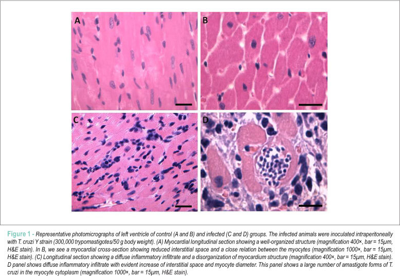
BACKGROUND: Conventional disector methods currently require considerable financial, technical and operational costs to estimate the number of cells, including cardyomyocytes, in a 3D area. OBJECTIVE: To use fluorescence microscopy in a modified disector method to determine the number of myocytes in cardiac tissue in normal and pathological conditions. METHODS: The study employed four-month-old male Wistar rats with weight of 366.25 ± 88.21g randomized in control (CG, n=8) and infected (IG, n=8) groups. IG animals were inoculated with T. cruzi Y strain (300,000 trypomastigotes/50g wt). After eight weeks, the animals were weighted and euthanized. The left ventricles (LV) were removed for stereological analysis of numerical density of cardiomyocytes (Nv[c]) and total number of these cells in the LV (N[c]). These parameters were estimated using a fluorescent disector (FD) and compared with the conventional optical (OD) and physical (PD) disector methods. RESULTS: In both disector methods, IG animals presented significant decrease of Nv[c] and N[c] compared to CG animals (P 0.05). A strong correlation, equal or above 96%, was obtained between FD, OD and PD. CONCLUSION: The FD method seems to be equally reliable to determine Nv[c] and N[c] in normal and pathological conditions and presents some advantages compared to conventional disector methods: reduction of histological slices and images in the stereological analysis, reduction of time to analyze the images, construction of FD in simple microscopes using the epifluorescence mode, distinction of disector planes in lower magnifications.

  • Cell Separation
  • flow cytometry
  • Myocytes, Cardiac
Read Full Article
Creative Commons Attribution (CC BY) Creative Commons license
16/02/2012 DOI: 10.1590/S0066-782X2012005000014
Inhibition of L-type calcium current by tramadol and enantiomers in cardiac myocytes from rats
ABC
Artigo Original

Inhibition of L-type calcium current by tramadol and enantiomers in cardiac myocytes from rats

Emiliano Medei et al. Juliana M. Raimundo José Hamilton M. Nascimento Margarete M. Trachez … Roberto T. Sudo Gisele Zapata-Sudo

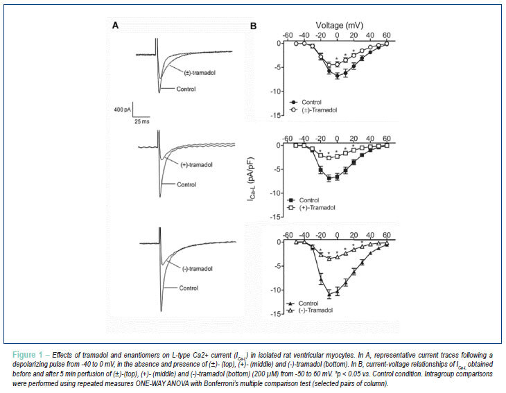
BACKGROUND: Tramadol is a centrally acting analgesic, whose mechanism of action involves opioid-receptor activation. Previously, we have shown that tramadol and its enantiomers had a negative inotropic effect on the papillary muscle in which the (+)-enantiomer is more potent than (-)- and (±)-tramadol. OBJECTIVE: In this study, we investigated the effects of tramadol and its enantiomers on L-type calcium current (I Ca-L). RESULTS: Tramadol (200 µM) reduced the peak amplitude of I Ca-L at potentials from 0 to +50 mV. At 0 mV, I Ca-L was reduced by 33.7 ± 7.2%. (+)- and (-)-tramadol (200 µM) produced a similar inhibition of I Ca-L, in which the peak amplitude was reduced by 64.4 ± 2.8% and 68.9 ± 5.8%, respectively at 0 mV (p > 0.05). Tramadol, (+)- and (-)-tramadol shifted the steady-state inactivation of I Ca-L to more negative membrane potentials. Also, tramadol and (+)-tramadol markedly shifted the time-dependent recovery curve of I Ca-L to the right and slowed down the recovery of I Ca-L from inactivation. The time constant was increased from 175.6 ± 18.6 to 305.0 ± 32.9 ms (p < 0.01) for tramadol and from 248.1 ± 28.1 ms to 359.0 ± 23.8 ms (p < 0.05) for (+)-tramadol. The agonist of µ-opioid receptor DAMGO had no effect on the I Ca-L. CONCLUSION: The inhibition of I Ca-L induced by tramadol and its enantiomers was unrelated to the activation of opioid receptors and could explain, at least in part, their negative cardiac inotropic effect.

  • calcium channels, L-type
  • Myocytes, Cardiac
  • Rats
  • Tramadol
Read Full Article
Creative Commons Attribution (CC BY) Creative Commons license
30/09/2011 DOI: 10.1590/S0066-782X2011005000091 Views 1
Swimming training attenuates contractile dysfunction in diabetic rat cardiomyocytes
ABC
Artigo Original

Swimming training attenuates contractile dysfunction in diabetic rat cardiomyocytes

Márcia Ferreira da Silva et al. Maria do Carmo Gouveia Pelúzio Paulo Roberto dos Santos Amorim Vitor Neiva Lavorato … Natália Pereira do Santos Luiz Henrique Marchesi Bozi Arlete Rita Penitente Daniel Luciano Falkoski Felipe Gomes Berfort Antonio Jose Natali

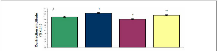
BACKGROUND: Experimental diabetes promotes contractile dysfunction in cardiomyocytes, but the effects of swimming in this disorder are not known. OBJECTIVE: To test the effects of a swimming training program (STP) on cardiomyocyte contractile dysfunction in rats with experimental diabetes. METHODS: Wistar rats (age: 30 days; mean body weight: 84.19 g) with diabetes induced by streptozotocin (60 mg/kg body weight; glucose > 300 mg/dl) were divided into sedentary diabetic rats (SD, n = 10) and exercised diabetic rats (ED, n = 13). Animals of same age and weight served as sedentary controls (SC, n = 10) and exercised controls (EC, n = 06). Animals and ED and EC underwent a STP (05 days/week, 90 min/day) for 08 weeks. Left ventricular (LV) myocytes were isolated and electrically stimulated at 3.0 Hz at room temperature (∼ 25º C). RESULTS: Diabetes reduced contractile function in cardiomyocytes of animals compared to controls (i.e., lower amplitude of contraction, longer duration of contraction and relaxation). The STP attenuated the reduced amplitude of contraction (SC, 11 ± 0.2% vs ED, 11.6 ± 0.2%), time to peak contraction (SC, 319 ± 5.8 ms vs ED, 333 ± 4.8 ms) and time to 50.0% of relaxation (SC, 619 ± 22.2 ms vs ED 698 ± 18.6 ms) of cardiomyocytes of diabetic rats. Diabetes reduced the size of cardiomyocytes, however, the STP minimized the reduction of cell volume and width, without changing length. CONCLUSION: The swimming training program attenuated the contractile dysfunction of the LV myocytes of rats with experimental diabetes.

  • Diabetes Mellitus
  • Myocytes, Cardiac
  • Physical Exertion
  • Rats
  • Swimming
Read Full Article
Creative Commons Attribution (CC BY) Creative Commons license
06/05/2011 DOI: 10.1590/S0066-782X2011005000055 Views 1
Factors and mechanisms involved in left ventricular hypertrophy and the anti-hypertrophic role of nitric oxide
ABC
Review Article

Factors and mechanisms involved in left ventricular hypertrophy and the anti-hypertrophic role of nitric oxide

José Antonio Dias Garcia et al. Erika Kristina Incerpi

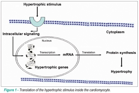
The left ventricular hypertrophy (LVH) occurs in response to the hemodynamic overload in some physiological and pathological conditions. However, it has not been completely elucidated whether the primary stimulation for the hypertrophy is the mechanical stretching of the heart, neurohumoral factors, or even the interaction of both. These factors are translated inside the cell as biochemical alterations that lead to the activation of second (cytosolic) and third (nuclear) messengers that will act in the cell nucleus, regulating transcription, and will finally determine the genic expression that induces LVH. The LVH is characterized by structural alterations due to the increase in the cardiomyocyte dimensions, the proliferation of the interstitial connective tissue and the rarefaction of the coronary microcirculation. Recently, nitric oxide (•NO) has appeared as an important regulator of cardiac remodeling, specifically recognized as an anti-hypertrophic mediator. Some studies have demonstrated the cellular targets, the anti-hypertrophic signaling pathways and the functional role of •NO. Thus, the LVH seems to develop as a result of the loss of the balance between the pro and the anti-hypertrophic signaling pathways. This new knowledge about the pro and anti-hypertrophic signaling pathways will allow the development of new strategies in the treatment of pathological LVH.

  • Hypertrophy Left Ventricular
  • Myocytes, Cardiac
  • Nitric Oxide
Read Full Article
Creative Commons Attribution (CC BY) Creative Commons license
25/06/2008 DOI: 10.1590/S0066-782X2008000600010

How to cite this article

EndNote RIS

Electronic Document Format (Vancouver)

Pimenta MS, Samesima N, Pastore CA, Krebs VLJ, Leal GN, Carvalho WB. Electrocardiographic Evaluation of Normal Newborns in the First Week of Life – Observational Study. Arquivos Brasileiros de Cardiologia 2022;119(4):587-92.

Electronic Document Format (ABNT)

Pimenta, Marina de Souza; Samesima, Nelson; Pastore, Carlos Alberto; Krebs, Vera Lucia Jornada; Leal, Gabriela Nunes; Carvalho, Werther Brunow de. Electrocardiographic Evaluation of Normal Newborns in the First Week of Life – Observational Study. Arquivos Brasileiros de Cardiologia, v. 119, n. 4, p. 587-592, Sep. 2022.

Electronic Document Format (APA)

Pimenta, M. S., Samesima, N., Pastore, C. A., Krebs, V. L. J., Leal, G. N., & Carvalho, W. B. (2022). Electrocardiographic Evaluation of Normal Newborns in the First Week of Life – Observational Study. Arquivos Brasileiros de Cardiologia, 119(4), 587-592.

Electronic Document Format (ISO)

Pimenta, Marina de Souza and Samesima, Nelson and Pastore, Carlos Alberto and Krebs, Vera Lucia Jornada and Leal, Gabriela Nunes and Carvalho, Werther Brunow de. Electrocardiographic Evaluation of Normal Newborns in the First Week of Life – Observational Study. Arquivos Brasileiros de Cardiologia [online]. 2022, vol. 119, n. 4, [cited 2026-10-14], pp. 587-592. Available from: <https://journals.cardiol.br/en/article/electrocardiographic-evaluation-of-normal-newborns-in-the-first-week-of-life-observational-study/>. ISSN 0066-782X

EndNote RIS

Electronic Document Format (Vancouver)

Soares LL, Leite LB, Ervilha LOG, Silva BAF, Freitas MO, Portes AMO, Rezende LMT, et al. Resistance Exercise Training Mitigates Left Ventricular Dysfunctions in Pulmonary Artery Hypertension Model. Arquivos Brasileiros de Cardiologia 2022;119(4):574-8.

Electronic Document Format (ABNT)

Soares, Leôncio Lopes; Leite, Luciano Bernardes; Ervilha, Luiz Otávio Guimarães; Silva, Bruna Aparecida Fonseca da; Freitas, Maíra Oliveira de; Portes, Alexandre Martins Oliveira; Rezende, Leonardo Mateus Teixeira; Drummond, Filipe Rios; Carneiro-Júnior, Miguel Araújo; Neves, Mariana Machado; Reis, Emily Correna Carlo; Natali, Antônio José. Resistance Exercise Training Mitigates Left Ventricular Dysfunctions in Pulmonary Artery Hypertension Model. Arquivos Brasileiros de Cardiologia, v. 119, n. 4, p. 574-584, Sep. 2022.

Electronic Document Format (APA)

Soares, L. L., Leite, L. B., Ervilha, L. O. G., Silva, B. A. F. , Freitas, M. O. , Portes, A. M. O., Rezende, L. M. T., Drummond, F. R., Carneiro-Júnior, M. A., Neves, M. M., Reis, E. C. C., & Natali, A. J. (2022). Resistance Exercise Training Mitigates Left Ventricular Dysfunctions in Pulmonary Artery Hypertension Model. Arquivos Brasileiros de Cardiologia, 119(4), 574-584.

Electronic Document Format (ISO)

Soares, Leôncio Lopes and Leite, Luciano Bernardes and Ervilha, Luiz Otávio Guimarães and Silva, Bruna Aparecida Fonseca da and Freitas, Maíra Oliveira de and Portes, Alexandre Martins Oliveira and Rezende, Leonardo Mateus Teixeira and Drummond, Filipe Rios and Carneiro-Júnior, Miguel Araújo and Neves, Mariana Machado and Reis, Emily Correna Carlo and Natali, Antônio José. Resistance Exercise Training Mitigates Left Ventricular Dysfunctions in Pulmonary Artery Hypertension Model. Arquivos Brasileiros de Cardiologia [online]. 2022, vol. 119, n. 4, [cited 2026-10-14], pp. 574-584. Available from: <https://journals.cardiol.br/en/article/resistance-exercise-training-mitigates-left-ventricular-dysfunctions-in-pulmonary-artery-hypertension-model/>. ISSN 0066-782X

EndNote RIS

Electronic Document Format (Vancouver)

Wang R, Figueiredo EL, Neuschwander FC. A New Pathway to Coronary Occlusion with Elabela?. Arquivos Brasileiros de Cardiologia 2021;117(3):511.

Electronic Document Format (ABNT)

Wang, Ricardo; Figueiredo, Estevão Lanna; Neuschwander, Fernando Carvalho. A New Pathway to Coronary Occlusion with Elabela?. Arquivos Brasileiros de Cardiologia, v. 117, n. 3, p. 511-511, Sep. 2021.

Electronic Document Format (APA)

Wang, R., Figueiredo, E. L., & Neuschwander, F. C. (2021). A New Pathway to Coronary Occlusion with Elabela?. Arquivos Brasileiros de Cardiologia, 117(3), 511-511.

Electronic Document Format (ISO)

Wang, Ricardo and Figueiredo, Estevão Lanna and Neuschwander, Fernando Carvalho. A New Pathway to Coronary Occlusion with Elabela?. Arquivos Brasileiros de Cardiologia [online]. 2021, vol. 117, n. 3, [cited 2026-10-14], pp. 511-511. Available from: <https://journals.cardiol.br/en/article/a-new-pathway-to-coronary-occlusion-with-elabela/>. ISSN 0066-782X

EndNote RIS

Electronic Document Format (Vancouver)

Gatto M, Mota GAF. Influence of Doxorubicin Treatment on Heme Metabolism in Cardiomyoblasts: An In Vitro Study. Arquivos Brasileiros de Cardiologia 2021;116(2):323-4.

Electronic Document Format (ABNT)

Gatto, Mariana; Mota, Gustavo Augusto Ferreira. Influence of Doxorubicin Treatment on Heme Metabolism in Cardiomyoblasts: An In Vitro Study. Arquivos Brasileiros de Cardiologia, v. 116, n. 2, p. 323-324, Feb. 2021.

Electronic Document Format (APA)

Gatto, M., & Mota, G. A. F. (2021). Influence of Doxorubicin Treatment on Heme Metabolism in Cardiomyoblasts: An In Vitro Study. Arquivos Brasileiros de Cardiologia, 116(2), 323-324.

Electronic Document Format (ISO)

Gatto, Mariana and Mota, Gustavo Augusto Ferreira. Influence of Doxorubicin Treatment on Heme Metabolism in Cardiomyoblasts: An In Vitro Study. Arquivos Brasileiros de Cardiologia [online]. 2021, vol. 116, n. 2, [cited 2026-10-14], pp. 323-324. Available from: <https://journals.cardiol.br/en/article/influence-of-doxorubicin-treatment-on-heme-metabolism-in-cardiomyoblasts-an-in-vitro-study/>. ISSN 0066-782X

EndNote RIS

Electronic Document Format (Vancouver)

Zhong W, Jin W, Xu S, Wu Y, Luo S, Liang M, Chen L, et al. Pioglitazone Induces Cardiomyocyte Apoptosis and Inhibits Cardiomyocyte Hypertrophy Via VEGFR-2 Signaling Pathway. Arquivos Brasileiros de Cardiologia 2018;111(2):162-9.

Electronic Document Format (ABNT)

Zhong, Wenliang; Jin, Wen; Xu, Shanghua; Wu, Yanqing; Luo, Shunxiang; Liang, Minlie; Chen, Lianglong. Pioglitazone Induces Cardiomyocyte Apoptosis and Inhibits Cardiomyocyte Hypertrophy Via VEGFR-2 Signaling Pathway. Arquivos Brasileiros de Cardiologia, v. 111, n. 2, p. 162-169, Jul. 2018.

Electronic Document Format (APA)

Zhong, W., Jin, W., Xu, S., Wu, Y., Luo, S., Liang, M., & Chen, L. (2018). Pioglitazone Induces Cardiomyocyte Apoptosis and Inhibits Cardiomyocyte Hypertrophy Via VEGFR-2 Signaling Pathway. Arquivos Brasileiros de Cardiologia, 111(2), 162-169.

Electronic Document Format (ISO)

Zhong, Wenliang and Jin, Wen and Xu, Shanghua and Wu, Yanqing and Luo, Shunxiang and Liang, Minlie and Chen, Lianglong. Pioglitazone Induces Cardiomyocyte Apoptosis and Inhibits Cardiomyocyte Hypertrophy Via VEGFR-2 Signaling Pathway. Arquivos Brasileiros de Cardiologia [online]. 2018, vol. 111, n. 2, [cited 2026-10-14], pp. 162-169. Available from: <https://journals.cardiol.br/en/article/pioglitazone-induces-cardiomyocyte-apoptosis-and-inhibits-cardiomyocyte-hypertrophy-via-vegfr-2-signaling-pathway/>. ISSN 0066-782X

EndNote RIS

Electronic Document Format (Vancouver)

Rodrigues AC, Natali AJ, Cunha DNQ, Costa AJLD, Moura AG, Carneiro-Júnior MA, Félix LB, et al. Moderate Continuous Aerobic Exercise Training Improves Cardiomyocyte Contractility in Β1 Adrenergic Receptor Knockout Mice. Arquivos Brasileiros de Cardiologia 2018;110(3):256-62.

Electronic Document Format (ABNT)

Rodrigues, Aurora Corrêa; Natali, Antônio José; Cunha, Daise Nunes Queiroz da; Costa, Alexandre Jayme Lopes Dantas; Moura, Anselmo Gomes de; Carneiro-Júnior, Miguel Araújo; Félix, Leonardo Bonato; Brum, Patrícia Chakur; Prímola-Gomes, Thales Nicolau. Moderate Continuous Aerobic Exercise Training Improves Cardiomyocyte Contractility in Β1 Adrenergic Receptor Knockout Mice. Arquivos Brasileiros de Cardiologia, v. 110, n. 3, p. 256-262, Feb. 2018.

Electronic Document Format (APA)

Rodrigues, A. C., Natali, A. J., Cunha, D. N. Q. , Costa, A. J. L. D., Moura, A. G. , Carneiro-Júnior, M. A., Félix, L. B., Brum, P. C., & Prímola-Gomes, T. N. (2018). Moderate Continuous Aerobic Exercise Training Improves Cardiomyocyte Contractility in Β1 Adrenergic Receptor Knockout Mice. Arquivos Brasileiros de Cardiologia, 110(3), 256-262.

Electronic Document Format (ISO)

Rodrigues, Aurora Corrêa and Natali, Antônio José and Cunha, Daise Nunes Queiroz da and Costa, Alexandre Jayme Lopes Dantas and Moura, Anselmo Gomes de and Carneiro-Júnior, Miguel Araújo and Félix, Leonardo Bonato and Brum, Patrícia Chakur and Prímola-Gomes, Thales Nicolau. Moderate Continuous Aerobic Exercise Training Improves Cardiomyocyte Contractility in Β1 Adrenergic Receptor Knockout Mice. Arquivos Brasileiros de Cardiologia [online]. 2018, vol. 110, n. 3, [cited 2026-10-14], pp. 256-262. Available from: <https://journals.cardiol.br/en/article/moderate-continuous-aerobic-exercise-training-improves-cardiomyocyte-contractility-in-%ce%b21-adrenergic-receptor-knockout-mice/>. ISSN 0066-782X

EndNote RIS

Electronic Document Format (Vancouver)

Santos-Miranda A, Cruz JS, Roman-Campos D. Electrical Properties of Isolated Cardiomyocytes in a Rat Model of Thiamine Deficiency. Arquivos Brasileiros de Cardiologia 2015;104(3):242-5.

Electronic Document Format (ABNT)

Santos-Miranda, Artur; Cruz, Jader Santos; Roman-Campos, Danilo. Electrical Properties of Isolated Cardiomyocytes in a Rat Model of Thiamine Deficiency. Arquivos Brasileiros de Cardiologia, v. 104, n. 3, p. 242-245, Feb. 2015.

Electronic Document Format (APA)

Santos-Miranda, A., Cruz, J. S., & Roman-Campos, D. (2015). Electrical Properties of Isolated Cardiomyocytes in a Rat Model of Thiamine Deficiency. Arquivos Brasileiros de Cardiologia, 104(3), 242-245.

Electronic Document Format (ISO)

Santos-Miranda, Artur and Cruz, Jader Santos and Roman-Campos, Danilo. Electrical Properties of Isolated Cardiomyocytes in a Rat Model of Thiamine Deficiency. Arquivos Brasileiros de Cardiologia [online]. 2015, vol. 104, n. 3, [cited 2026-10-14], pp. 242-245. Available from: <https://journals.cardiol.br/en/article/electrical-properties-of-isolated-cardiomyocytes-in-a-rat-model-of-thiamine-deficiency/>. ISSN 0066-782X

EndNote RIS

Electronic Document Format (Vancouver)

Lima-Leopoldo AP, Leopoldo AS, Silva DCT, Nascimento AF, Campos DHS, Luvizotto RAM, Oliveira Júnior SA, et al. Influence of long-term obesity on myocardial gene expression. Arquivos Brasileiros de Cardiologia 2013;100(3):229-37.

Electronic Document Format (ABNT)

Lima-Leopoldo, Ana Paula; Leopoldo, André Soares; Silva, Danielle Cristina Tomaz; Nascimento, André Ferreira do; Campos, Dijon Henrique Salomé de; Luvizotto, Renata de Azevedo Melo; Oliveira Júnior, Silvio Assis de; Padovani, Carlos Roberto; Nogueira, Célia Regina; Cicogna, Antonio Carlos. Influence of long-term obesity on myocardial gene expression. Arquivos Brasileiros de Cardiologia, v. 100, n. 3, p. 229-237, Apr. 2013.

Electronic Document Format (APA)

Lima-Leopoldo, A. P., Leopoldo, A. S., Silva, D. C. T., Nascimento, A. F. , Campos, D. H. S. , Luvizotto, R. A. M., Oliveira Júnior, S. A. , Padovani, C. R., Nogueira, C. R., & Cicogna, A. C. (2013). Influence of long-term obesity on myocardial gene expression. Arquivos Brasileiros de Cardiologia, 100(3), 229-237.

Electronic Document Format (ISO)

Lima-Leopoldo, Ana Paula and Leopoldo, André Soares and Silva, Danielle Cristina Tomaz and Nascimento, André Ferreira do and Campos, Dijon Henrique Salomé de and Luvizotto, Renata de Azevedo Melo and Oliveira Júnior, Silvio Assis de and Padovani, Carlos Roberto and Nogueira, Célia Regina and Cicogna, Antonio Carlos. Influence of long-term obesity on myocardial gene expression. Arquivos Brasileiros de Cardiologia [online]. 2013, vol. 100, n. 3, [cited 2026-10-14], pp. 229-237. Available from: <https://journals.cardiol.br/en/article/influence-of-long-term-obesity-on-myocardial-gene-expression/>. ISSN 0066-782X

EndNote RIS

Electronic Document Format (Vancouver)

Silva TFA, Souza GK, Simões MA, Pabis FC, Noronha L. Immunohistochemical expression of cell differentiation and growth in neonate cardiomyocytes. Arquivos Brasileiros de Cardiologia 2012;99(3):797-801.

Electronic Document Format (ABNT)

Silva, Tarcísio Fulgêncio Alves da; Souza, Greyce Kelly de; Simões, Mona Adalgisa; Pabis, Francisco Cesar; Noronha, Lucia de. Immunohistochemical expression of cell differentiation and growth in neonate cardiomyocytes. Arquivos Brasileiros de Cardiologia, v. 99, n. 3, p. 797-801, Aug. 2012.

Electronic Document Format (APA)

Silva, T. F. A. , Souza, G. K. , Simões, M. A., Pabis, F. C., & Noronha, L. (2012). Immunohistochemical expression of cell differentiation and growth in neonate cardiomyocytes. Arquivos Brasileiros de Cardiologia, 99(3), 797-801.

Electronic Document Format (ISO)

Silva, Tarcísio Fulgêncio Alves da and Souza, Greyce Kelly de and Simões, Mona Adalgisa and Pabis, Francisco Cesar and Noronha, Lucia de. Immunohistochemical expression of cell differentiation and growth in neonate cardiomyocytes. Arquivos Brasileiros de Cardiologia [online]. 2012, vol. 99, n. 3, [cited 2026-10-14], pp. 797-801. Available from: <https://journals.cardiol.br/en/article/immunohistochemical-expression-of-cell-differentiation-and-growth-in-neonate-cardiomyocytes/>. ISSN 0066-782X

EndNote RIS

Electronic Document Format (Vancouver)

Novaes RD, Penitente AR, Talvani A, Natali AJ, Neves CA, Maldonado IRSC. Use of fluorescence in a modified disector method to estimate the number of myocytes in cardiac tissue. Arquivos Brasileiros de Cardiologia 2012;98(3):252-8.

Electronic Document Format (ABNT)

Novaes, Rômulo Dias; Penitente, Arlete Rita; Talvani, André; Natali, Antônio José; Neves, Clóvis Andrade; Maldonado, Izabel Regina Santos Costa. Use of fluorescence in a modified disector method to estimate the number of myocytes in cardiac tissue. Arquivos Brasileiros de Cardiologia, v. 98, n. 3, p. 252-258, Feb. 2012.

Electronic Document Format (APA)

Novaes, R. D., Penitente, A. R., Talvani, A., Natali, A. J., Neves, C. A., & Maldonado, I. R. S. C. (2012). Use of fluorescence in a modified disector method to estimate the number of myocytes in cardiac tissue. Arquivos Brasileiros de Cardiologia, 98(3), 252-258.

Electronic Document Format (ISO)

Novaes, Rômulo Dias and Penitente, Arlete Rita and Talvani, André and Natali, Antônio José and Neves, Clóvis Andrade and Maldonado, Izabel Regina Santos Costa. Use of fluorescence in a modified disector method to estimate the number of myocytes in cardiac tissue. Arquivos Brasileiros de Cardiologia [online]. 2012, vol. 98, n. 3, [cited 2026-10-14], pp. 252-258. Available from: <https://journals.cardiol.br/en/article/use-of-fluorescence-in-a-modified-disector-method-to-estimate-the-number-of-myocytes-in-cardiac-tissue/>. ISSN 0066-782X

EndNote RIS

Electronic Document Format (Vancouver)

Medei E, Raimundo JM, Nascimento JHM, Trachez MM, Sudo RT, Zapata-Sudo G. Inhibition of L-type calcium current by tramadol and enantiomers in cardiac myocytes from rats. Arquivos Brasileiros de Cardiologia 2011;97(4):324-31.

Electronic Document Format (ABNT)

Medei, Emiliano; Raimundo, Juliana M.; Nascimento, José Hamilton M.; Trachez, Margarete M.; Sudo, Roberto T.; Zapata-Sudo, Gisele. Inhibition of L-type calcium current by tramadol and enantiomers in cardiac myocytes from rats. Arquivos Brasileiros de Cardiologia, v. 97, n. 4, p. 324-331, Sep. 2011.

Electronic Document Format (APA)

Medei, E., Raimundo, J. M., Nascimento, J. H. M., Trachez, M. M., Sudo, R. T., & Zapata-Sudo, G. (2011). Inhibition of L-type calcium current by tramadol and enantiomers in cardiac myocytes from rats. Arquivos Brasileiros de Cardiologia, 97(4), 324-331.

Electronic Document Format (ISO)

Medei, Emiliano and Raimundo, Juliana M. and Nascimento, José Hamilton M. and Trachez, Margarete M. and Sudo, Roberto T. and Zapata-Sudo, Gisele. Inhibition of L-type calcium current by tramadol and enantiomers in cardiac myocytes from rats. Arquivos Brasileiros de Cardiologia [online]. 2011, vol. 97, n. 4, [cited 2026-10-14], pp. 324-331. Available from: <https://journals.cardiol.br/en/article/inhibition-of-l-type-calcium-current-by-tramadol-and-enantiomers-in-cardiac-myocytes-from-rats/>. ISSN 0066-782X

EndNote RIS

Electronic Document Format (Vancouver)

Silva MF, Pelúzio MCG, Amorim PRS, Lavorato VN, Santos NP, Bozi LHM, Penitente AR, et al. Swimming training attenuates contractile dysfunction in diabetic rat cardiomyocytes. Arquivos Brasileiros de Cardiologia 2011;97(1):33-9.

Electronic Document Format (ABNT)

Silva, Márcia Ferreira da; Pelúzio, Maria do Carmo Gouveia; Amorim, Paulo Roberto dos Santos; Lavorato, Vitor Neiva; Santos, Natália Pereira do; Bozi, Luiz Henrique Marchesi; Penitente, Arlete Rita; Falkoski, Daniel Luciano; Berfort, Felipe Gomes; Natali, Antonio Jose. Swimming training attenuates contractile dysfunction in diabetic rat cardiomyocytes. Arquivos Brasileiros de Cardiologia, v. 97, n. 1, p. 33-39, May. 2011.

Electronic Document Format (APA)

Silva, M. F. , Pelúzio, M. C. G., Amorim, P. R. S., Lavorato, V. N., Santos, N. P. , Bozi, L. H. M., Penitente, A. R., Falkoski, D. L., Berfort, F. G., & Natali, A. J. (2011). Swimming training attenuates contractile dysfunction in diabetic rat cardiomyocytes. Arquivos Brasileiros de Cardiologia, 97(1), 33-39.

Electronic Document Format (ISO)

Silva, Márcia Ferreira da and Pelúzio, Maria do Carmo Gouveia and Amorim, Paulo Roberto dos Santos and Lavorato, Vitor Neiva and Santos, Natália Pereira do and Bozi, Luiz Henrique Marchesi and Penitente, Arlete Rita and Falkoski, Daniel Luciano and Berfort, Felipe Gomes and Natali, Antonio Jose. Swimming training attenuates contractile dysfunction in diabetic rat cardiomyocytes. Arquivos Brasileiros de Cardiologia [online]. 2011, vol. 97, n. 1, [cited 2026-10-14], pp. 33-39. Available from: <https://journals.cardiol.br/en/article/swimming-training-attenuates-contractile-dysfunction-in-diabetic-rat-cardiomyocytes/>. ISSN 0066-782X

EndNote RIS

Electronic Document Format (Vancouver)

Garcia JAD, Incerpi EK. Factors and mechanisms involved in left ventricular hypertrophy and the anti-hypertrophic role of nitric oxide. Arquivos Brasileiros de Cardiologia 2008;90(6):443-50.

Electronic Document Format (ABNT)

Garcia, José Antonio Dias; Incerpi, Erika Kristina. Factors and mechanisms involved in left ventricular hypertrophy and the anti-hypertrophic role of nitric oxide. Arquivos Brasileiros de Cardiologia, v. 90, n. 6, p. 443-450, Jun. 2008.

Electronic Document Format (APA)

Garcia, J. A. D., & Incerpi, E. K. (2008). Factors and mechanisms involved in left ventricular hypertrophy and the anti-hypertrophic role of nitric oxide. Arquivos Brasileiros de Cardiologia, 90(6), 443-450.

Electronic Document Format (ISO)

Garcia, José Antonio Dias and Incerpi, Erika Kristina. Factors and mechanisms involved in left ventricular hypertrophy and the anti-hypertrophic role of nitric oxide. Arquivos Brasileiros de Cardiologia [online]. 2008, vol. 90, n. 6, [cited 2026-10-14], pp. 443-450. Available from: <https://journals.cardiol.br/en/article/factors-and-mechanisms-involved-in-left-ventricular-hypertrophy-and-the-anti-hypertrophic-role-of-nitric-oxide/>. ISSN 0066-782X

  • About ABC Family
  • Internal Regulation
  • Editorial Board
  • Brazilian Society of Cardiology (SBC)

Issues

SBC Guidelines

  • Videos
  • Podcasts
  • News