ABC Family Arquivos Brasileiros de Cardiologia International Journal of Cardiovascular Sciences ABC Heart Failure & Cardiomyopathy ABC Imagem Cardiovascular
Português English
ABC Family
Português English
  • About
    • About ABC Family
    • Internal Regulation
    • Editorial Board
    • Brazilian Society of Cardiology (SBC)
  • Issues
  • SBC Guidelines
  • Content
    • Videos
    • Podcasts
    • News
  • About
    • About ABC Family
    • Internal Regulation
    • Editorial Board
    • Brazilian Society of Cardiology (SBC)
  • Issues
  • SBC Guidelines
  • Content
    • Videos
    • Podcasts
    • News

ABC Family

  • ABC Family
  • Arquivos Brasileiros de Cardiologia
  • International Journal of Cardiovascular Sciences
  • ABC Heart Failure & Cardiomyopathy
  • ABC Imagem Cardiovascular
Search in

No content found.

Journals
Ordering
Article types
Others

  1. Home
  2. Keyword: Diagnostic Imaging/methods
Figure 1
ABC
Image

Multiple Intracardiac Metastases – A Chaplet Heart

Mariana Tinoco et al. Margarida Castro Hans Dabó Filipa Cordeiro … Pedro von Hafe António Lourenço

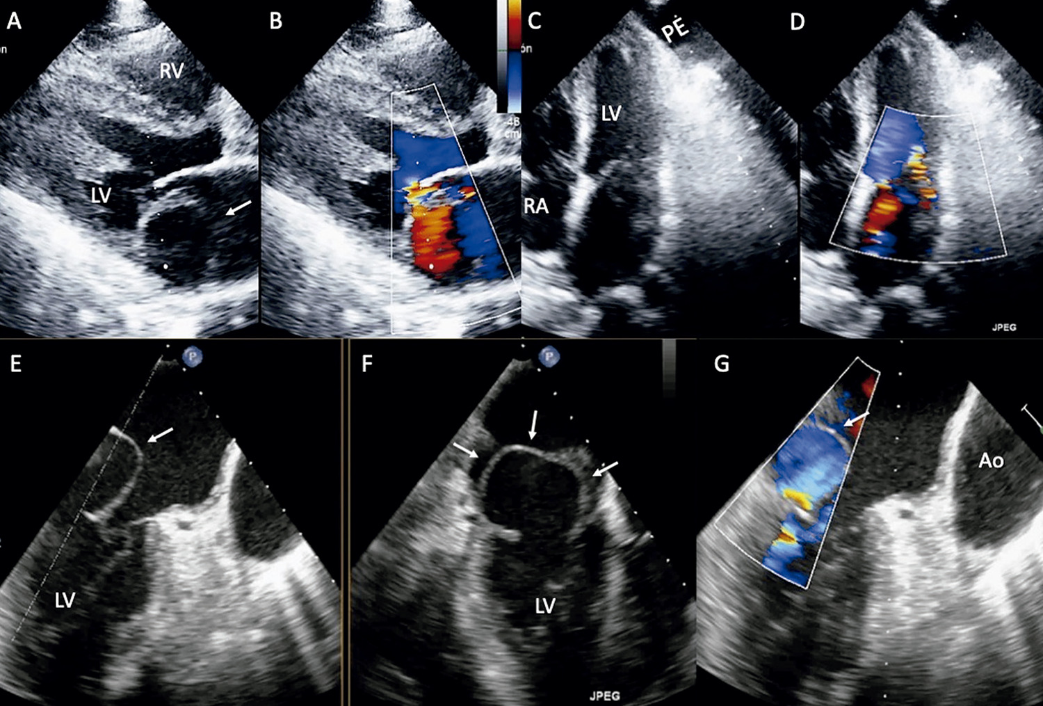
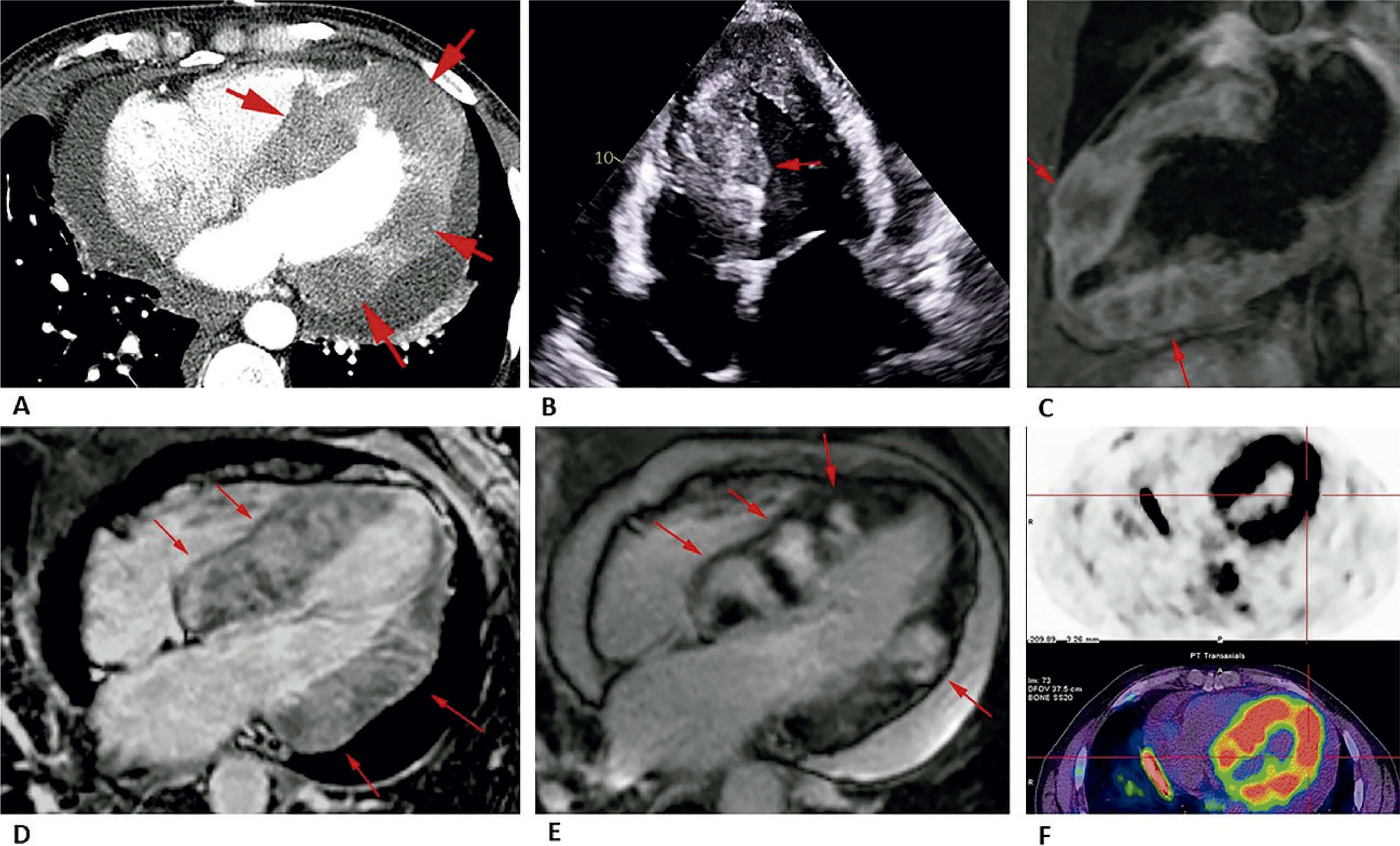
A 52-year-old male was undergoing first-line chemotherapy with pemetrexed due to a stage IV right hilar lung adenocarcinoma with likely pleural, pericardial, liver, and bone metastases. During follow-up, no evidence of disease progression was found. Four years later a follow-up chest CT scan documented a nodular heterogeneous enhancement of the left ventricular (LV) myocardium (), the remaining lesions were overlapping. He had no cardiovascular symptoms and clinical examination was unremarkable. Transthoracic echocardiography () and cardiac MRI (Figures 1C-E) revealed several well-rounded endo-myocardial masses, some of which multilobulated sometimes with almost transmural extension. The lesions are predominantly distributed along the LV inferoseptal wall, inferolateral wall, anterolateral wall, anterior wall, and right side of the interventricular septum. These lesions had heterogeneous signal characteristics, being predominantly hyperintense on T1-weighted images, hypointense on T2-weighted images with a peripheral halo of hyperintensity, showing early heterogeneous and late intense gadolinium enhancement; and abnormal uptake of fluorodeoxyglucose in a 18F-FDG PET/CT ().

[…]
  • Cardioncology
  • Diagnostic Imaging/methods
  • Echocardiography/methods
  • Multimodal Imaging
  • Neoplasm Metastasis
Read Full Article
Creative Commons Attribution (CC BY) Creative Commons license
15/07/2024 DOI: 10.36660/abc.20230732i
Figure 1
ABC
Research Letter

Interpreting Acute Myocardial Infarction 40 Years Later. Evolution of Knowledge: What is the Best Explanation?

Vitor Coutinho Andrade et al. Paulo Cury Rezende Whady Hueb José Antonio Franchini Ramires

Introduction

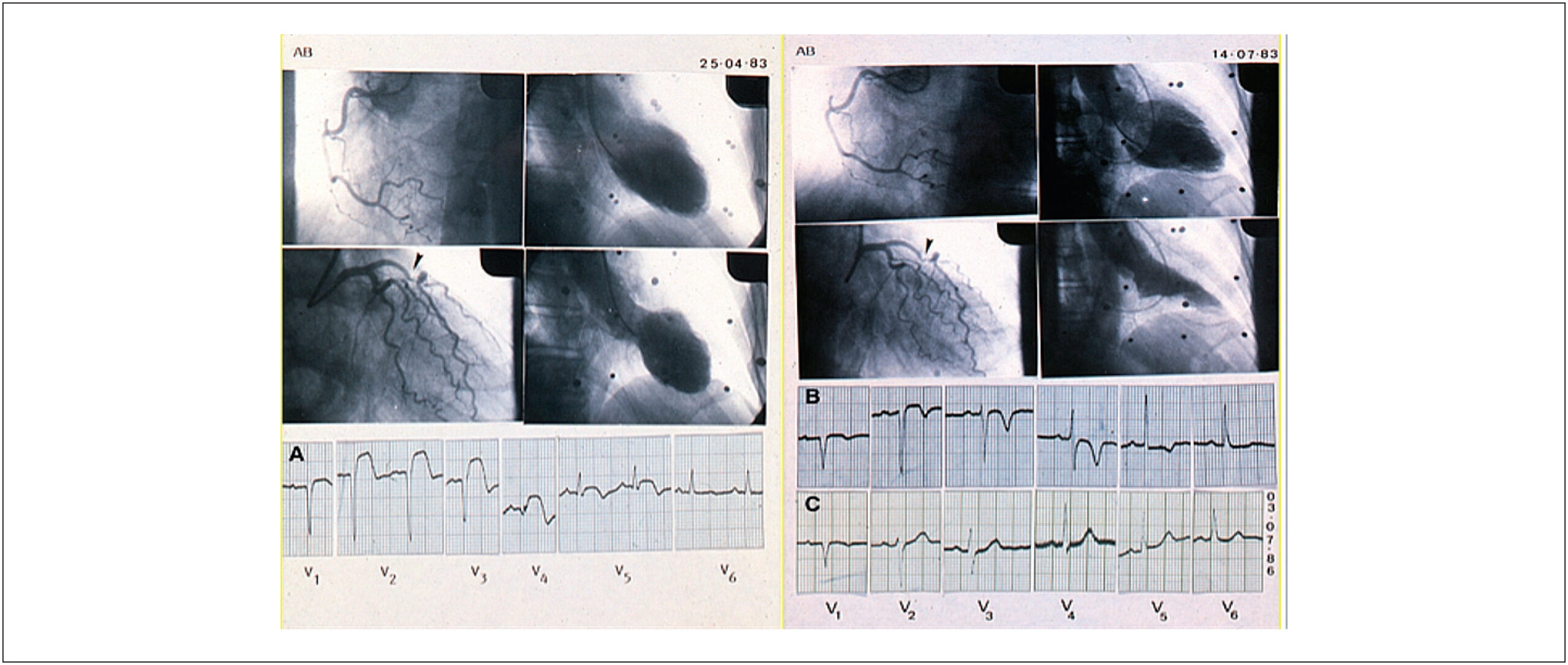
Plaque rupture and erosion are the most common immediate pathologies associated with acute myocardial infarction. However, other mechanisms must be considered. The reported case occurred at a time when percutaneous or pharmacological coronary revascularization were not the standard treatment, which sparked discussion about these other mechanisms, as well as the possible advantages associated with the myocardial revascularization procedures.

[…]

  • Coronary Vasoespasm
  • Diagnostic Imaging/methods
  • Dyslipidemias
  • Myocardial Infarction
  • Myocardial Stunning
Read Full Article
Creative Commons Attribution (CC BY) Creative Commons license
29/04/2024 DOI: 10.36660/abc.2023-0757i Views 1
Figure 1
ABC
Case Report

Acute ST-Elevation Myocardial Infarction in a Young Adult: Rare Presentation of Giant Atrial Myxoma

Rodrigo Rufino Pereira Silva et al. Carolina Jerônimo Magalhães Rafael Silvestre Vieira da Silva Giulia Antoni Ferreira Rocha … Paulo Ernando Ferraz Cavalcanti Sérgio Tavares Montenegro

Abstract

Cardiac tumors are rare entities, among which atrial myxoma (AM) stands as the most frequent, accounting for approximately half of all reported cases. The incidence of AM is estimated to range from 0.001% to 0.3% within the general population, yet only about 0.06% of these cases present with coronary embolic events.

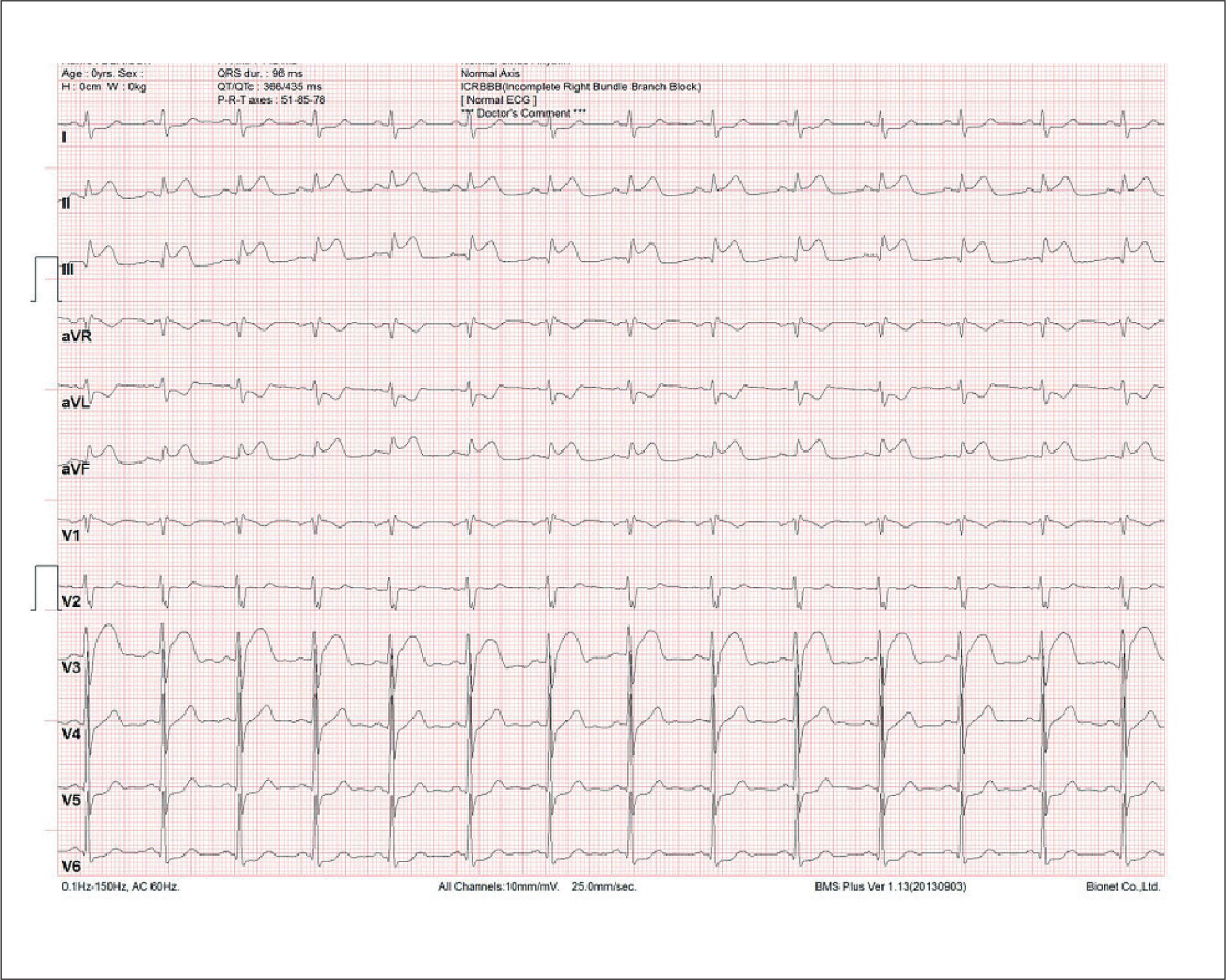
We report on a 33-year-old male smoker who experienced acute, severe precordial pain radiating to the left upper limb, lasting for one hour. The electrocardiographic evaluation demonstrated ST-segment elevation in leads D2, D3, and aVF, alongside significantly elevated serum troponin levels, confirming a diagnosis of ST-segment elevation myocardial infarction (STEMI). Subsequent coronary angiography revealed proximal occlusion of the right coronary artery due to thrombus. An initial attempt of thrombus aspiration was unsuccessful, followed by primary angioplasty with balloon inflation without stent placement. Further diagnostic exploration through transthoracic echocardiography identified a homogenous, smooth-surfaced mass measuring 5.2 cm x 2.3 cm attached to the interatrial septum. This mass, characterized by lobulations, prolapsed into the mitral valve and left ventricle during diastole, consistent with AM. Surgical resection of the mass was successfully performed, with the patient being discharged asymptomatic.

In the reported case, the patient’s profile, notably his age, and gender, diverges from the typical epidemiological characteristics associated with AM. This case adds to the limited number of reports where the inferior wall is affected by the right coronary artery being occluded. This report emphasizes the significance of differential diagnoses in younger patients presenting with STEMI.

  • Diagnostic Imaging/methods
  • Myxoma
  • ST Elevation Myocardial Infarction
  • Young Adult
Read Full Article
Creative Commons Attribution (CC BY) Creative Commons license
22/04/2024 DOI: 10.36660/abc.20230538i Views 1
Figure 1
ABC
Research Letter

Marked Concentric Myocardial Hypertrophy with Good Postoperative Evolution in a 4 Years Old Child

Edmar Atik et al. Gabriela Nunes Leal Marcelo B. Jatene

Introduction

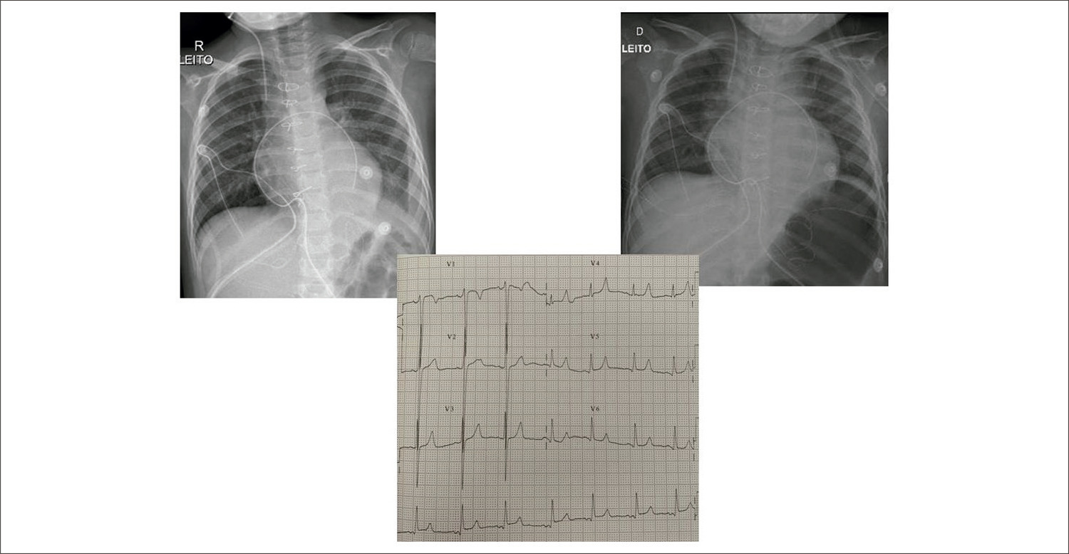
Hypertrophic cardiomyopathy (HCM), characterized by hypertrophy of the left ventricle, has no etiological explanation under conditions of the usual diseases that cause cardiac overloads like systemic arterial hypertension, obstructive congenital heart diseases, and other pathologies. – It results from cardiovascular genetic pathology causing a complex disease, from gene diversity (more than 1400 mutations identified in 11 different genes) to phenotypic expression, histological characteristics, and manifested symptoms. Beta myosins (MYH7) genes occur in 35% of the cases, and myosin is linked to protein C (MYBPC3) in 49%, corresponding to three-quarters of the pathogenic mutations. Higher evolutionary risk is observed in the first group of changes, mainly to sudden death. The breakdown of myofibrils with bizarre nuclei and increased extracellular connective tissue characterize the histological findings.

Genetic diagnosis is achieved in 80% of these cases in children, which is important for risk stratification, therapeutic planning, and genetic counseling.

[…]

  • Abnormalities Congenital/surgery
  • Cardiomyopathy, Hypertrophic/surgery
  • Diagnostic Imaging/methods
  • Hypertrophy, Left Ventricular/surgery
  • Risk Factors
  • Sudden Cardiac Death
Read Full Article
Creative Commons Attribution (CC BY) Creative Commons license
19/06/2023 DOI: 10.36660/abc.20220869 Views 1
ABC
Editorial

Brazilian Interventional Cardiology and Chronic Coronary Occlusions: Where Are We?

José Mariani Junior

The coronary atherosclerotic disease has several facets of clinical presentation; chronic coronary occlusion (CCO) is one that produces total obstruction of the lumen of the vessel, known or presumed for three months or more. It is present in 16-18% of patients with significant coronary disease.

Percutaneous treatment of this type of lesion is almost as old as the first coronary angioplasty performed by Andreas Gruntzig in 1979. Martin Kaltenbach first performed it in Frankfurt in the late 1970s and by Geoffrey Hartzler in Kansas City in the following decades., Since then, the growing and spectacular incorporation of technology into medicine, especially in Interventional Cardiology, associated with the knowledge and expertise of new techniques dedicated to complex coronary interventions, which include CCO, have allowed great, safe, and effective advances in the approach to these lesions.

[…]

  • Coronary Angiography/methods
  • Coronary Occlusion/mortality
  • Coronary Occlusion/therapy
  • Diagnostic Imaging/methods
  • Drug-Eluting Stents
  • Ultrasonography,Interventional/methods
Read Full Article
Creative Commons Attribution (CC BY) Creative Commons license
01/05/2023 DOI: 10.36660/abc.20230172
ABC
Editorial

Left Ventricular Mechanics: Untwisting the Pathways of the Cardiovascular Response to Exercise

Eduardo M. Vilela et al. Ricardo Fontes-Carvalho

Cardiovascular (CV) disease is a major cause of morbidity and mortality, with coronary artery disease (CAD) being one of its most challenging presentations. Over the years, several strategies have allowed marked improvements in its management. Physical exercise (a core component of cardiac rehabilitation programs) is currently a cornerstone of comprehensive contemporary secondary prevention strategies. While the benefits of physical exercise have been extensively described in the presence of CAD, the myriad mechanistic pathways by which this intervention exerts its various effects have yet to be fully ascertained., Indeed, while its positive impact on functional capacity has been highlighted, the relative role of exercise training in systolic function has shown some variability across different studies., While considering that differences in program designs and populations under study could, at least partially, help explain some of these divergences, the study of the mechanisms by which exercise could impact cardiac function (namely in the setting of CAD) remains a topic of considerable interest.

Advances in imaging modalities have allowed for an ever more precise understanding of CV physiology and the impact of different pathological processes., While ejection fraction (EF) remains the most ubiquitous parameter to assess left ventricular (LV) systolic function, other parameters, such as those derived from deformation imaging, have increasingly come under the spotlight to try to address some of the pitfalls related to EF.,, In this regard, the notion that different fiber orientations and their complex and dynamic interplay could be key to LV function has progressively led to the concept that the heart’s structural design has a particularly central role in its biomechanics and, thus, its overall function., These have also underscored the possible relevance of parameters such as global longitudinal strain (GLS) and ancillary measures such as LV twist and torsion., As previously detailed, these could allow insights into the coupling mechanisms between systole and diastole and global cardiac function.,,

[…]

  • Cardiovascuar Diseases/prevention and control
  • Coronary Artery Disease
  • Diagnostic Imaging/methods
  • Echocardiography/methods
Read Full Article
Creative Commons Attribution (CC BY) Creative Commons license
21/04/2023 DOI: 10.36660/abc.20230181 Views 1
ABC
Editorial

Nuclear Cardiology in the Optimization of Resynchronization Therapy: Quo Vadis?

Gabriel Blacher Grossman

Myocardial Viability and Resynchronization Therapy

Several studies in the literature have shown that the absence of myocardial viability at the site where the electrode was positioned reduces the efficacy of CRT. Ypenburg et al. evaluated the presence of fibrosis using GATED-SPECT with Tc-99m-tetrofosmin before CRT. Patients without fibrosis at the electrode implantation site showed improvement in NYHA class, quality of life, 6-minute walk test, ventricular volumes, and ejection fraction at a 6-month follow-up compared to patients with fibrosis at the electrode implantation site for CRT RT. The extent of the viable myocardial area was related to the reduction of ventricular volumes and ejection fraction. Bose et al. also demonstrated in a retrospective study involving 160 patients with ischemic cardiomyopathy that those patients in which electrode placement was in a site with fibrosis or fibrosis and ischemia more often had the primary outcome (hospitalization for HF and death) at 3 years compared to those patients with normal myocardium. Therefore, the fibrosis and myocardial viability evaluation at the electrode implementation site should be considered before CRT.

[…]

  • Cardiac Resynchronization Therapy
  • Diagnostic Imaging/methods
  • Heart Failure
  • Myocardial Ischemia Myocardial Perfusion Imaging
  • Nuclear Medicine, Cardiology
  • Radioisotopes
Read Full Article
Creative Commons Attribution (CC BY) Creative Commons license
07/04/2023 DOI: 10.36660/abc.20230136 Views 1
Figure 1
ABC
Original Article

Predicting Significant Coronary Obstruction in a Population with Suspected Coronary Disease and Absence of Coronary Calcium: CORE-64 / CORE320 Studies

Anderson C. Armstrong et al. Rodrigo Cerci Matthew B. Matheson Tiago Magalhães … Satoru Kishi Jeff Brinker Melvin E. Clouse Carlos E. Rochitte Christopher Cox João A. C. Lima Armin Arbab-Zadeh

Abstract

Background

Coronary artery calcium (CAC) scanning can be performed using non-contrast computed tomography to predict cardiovascular events, but has less value for risk stratification in symptomatic patients.

Objective

To identify and validate predictors of significant coronary obstruction (SCO) in symptomatic patients without coronary artery calcification.

Methods

A total of 4,258 participants were screened from the CORE64 and CORE320 studies that enrolled patients referred for invasive angiography, and from the Quanta Registry that included patients referred for coronary computed tomography angiography (CTA). Logistic regression models evaluated associations between cardiovascular risk factors, CAC, and SCO. An algorithm to assess the risk of SCO was proposed for patients without CAC. Significance level of 5% was used in the analyses.

Results

Of the 509 participants of the CORE study, 117 (23%) had zero coronary calcium score; 13 (11%) patients without CAC had SCO. Zero calcium score was related to younger age, female gender, lower body mass index, no diabetes, and no dyslipidemia. Being a current smoker increased ~3.5 fold the probability of SCO and other CV risk factors were not significantly associated. Considering the clinical findings, an algorithm to further stratify zero calcium score patients was proposed and had a limited performance in the validation cohort (AUC 58; 95%CI 43, 72).

Conclusion

A lower cardiovascular risk profile is associated with zero calcium score in a setting of high-risk patients. Smoking is the strongest predictor of SCO in patients without CAC.

  • Chest Pain
  • Coronary Angiography
  • Coronary Artery Disease / complications
  • Diagnostic Imaging/methods
  • Indice Calcio
  • Tomography
  • X-Ray Computed/methods
Read Full Article
Creative Commons Attribution (CC BY) Creative Commons license
20/03/2023 DOI: 10.36660/abc.20220183
Figure 1
ABC
Image

Tricky Diagnosis: An Aberrant Mitral Valve Chord

Marina Santos et al. Mariana Paiva Joana Ferreira Sara Guerreiro

Case

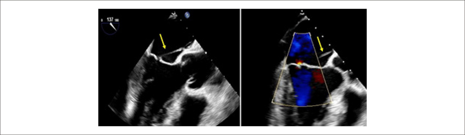
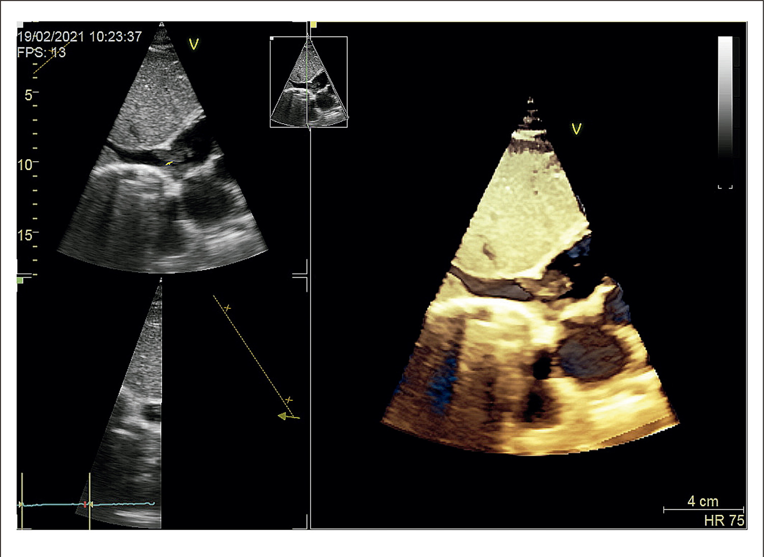
We present the case of a 52-year-old man with hepatitis C and a history of intravenous drug abuse, admitted to the infectology department with spondylodiscitis and psoas muscle abscess. Staphylococcus aureus was isolated from blood, urine, and spinal fluid cultures, and the transthoracic echocardiogram was negative for vegetation, abscess, or fistula. The transesophageal echocardiogram (TEE) showed a filamentous and mobile structure attached to the atrial surface of the mitral valve (MV), and a diagnosis of infective endocarditis was presumed. Seven days later, the patient was referred to our tertiary institution for a detailed TEE. In 2D images, we found a thin, well-delineated strand connecting the interatrial septum (IAS) to the tip of the anterior mitral leaflet without significant regurgitation ( , supplementary video 1). 3D imaging confirmed the presence of an anomalous chord connecting the A2 scallop of MV to the middle of the interatrial septum ( , supplementary video 2). There was no evidence of infective endocarditis, and this strand was found to be compatible with an anomalous left atrial MV chord.

[…]

  • Diagnostic Imaging/methods
  • Endocarditis, bacterial
  • Heart Defects, Congenital Atrial Septum
  • Mitral Valve Insufficiency
  • Staphylococcus Aureus
Read Full Article
Creative Commons Attribution (CC BY) Creative Commons license
10/03/2023 DOI: 10.36660/abc.20220573
Figure 1
ABC
Image

A Rare Subtype of a Rare Tumor

Paulo Medeiros et al. Ana Rita Coelho João Magalhães Nuno Salomé Vítor Pereira

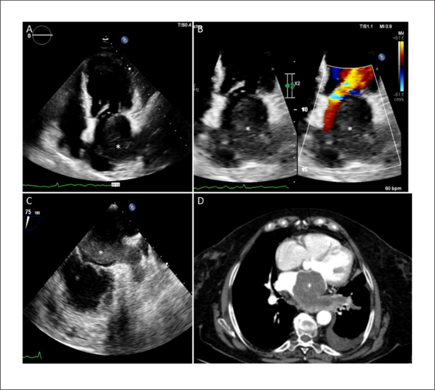
A 71-year-old woman was admitted to our center, complaining of fatigue, dyspnea, and anorexia. Physical examination revealed a grade III low-pitched diastolic murmur at the mitral focus. Transthoracic echocardiogram (TTE) showed an oval mass inside the left atrium (LA) extending from its roof to the mitral valve ( ), causing significant transmitral pressure gradient ( ). Transesophageal echocardiogram (TEE) ( ) raised the suspicion of left superior pulmonary vein involvement, which was confirmed by computed tomography (CT) angiography ( ). Cardiac magnetic resonance imaging characterized the mass as isointense on T1 and hyperintense on T2-weightened images; there was a contrast uptake, in first-passage sequences, and a heterogeneous late gadolinium enhancement ( ).

[…]

  • Diagnostic Imaging/methods
  • Heart Failure/complications
  • Heart Neoplasms/surgery
  • Mitral Valve/surgery
  • Multimodal Imaging/methods, Leiomyosarcoma/surgery
Read Full Article
Creative Commons Attribution (CC BY) Creative Commons license
27/02/2023 DOI: 10.36660/abc.20220486
Figure 1
ABC
Viewpoint

The “Five Malignant Waves” of the Electrocardiography

Mitermayer Reis Brito

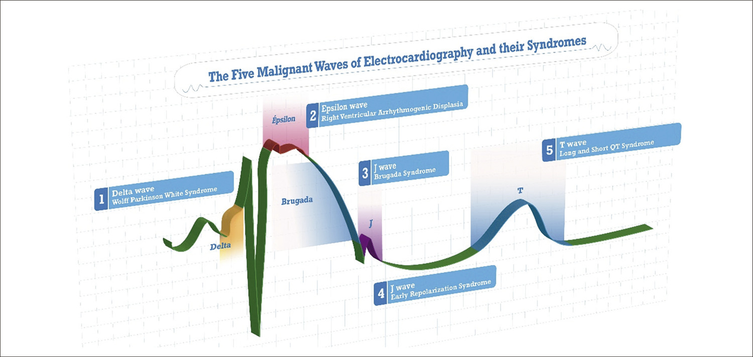
The main ECG features of the five malignant waves and their syndromes

The Main ECG features of the “Five Malignant Waves” and their syndromes are presented in .

[…]

  • Arrhythmias Cardiac
  • Death, Sudden
  • Diagnostic Imaging/methods
  • Electrocardiography/methods
  • Syncope
  • Tachycardia, Ventricular
Read Full Article
Creative Commons Attribution (CC BY) Creative Commons license
23/02/2023 DOI: 10.36660/abc.20220649 Views 1
Figure 1
ABC
Research Letter

Intracardiac Metastasis of Colonic Adenocarcinoma 12 Years After Primary Tumor Control and Without Any Sign of Other Metastasis: A Case Report

Isabela Galizzi Faé et al. Gabriela Zamunaro Lopes Ruiz Gustavo Palmer Irffi Robson de Souza Almeida Junior … Pedro Anjos Conceição Eduardo Belisario Falchetto Luiz Guilherme Passaglia Geraldo Brasileiro Filho Cláudio Leo Gelape Clara Rodrigues Alves de Oliveira

Introduction

Thrombus, vegetation and tumor are the main diagnoses for intracardiac masses. Malignant cardiac tumors are rare and cardiac metastasis are around 20 times more frequent than primary tumors. Cardiac metastases originate from lymphatic or blood dissemination, mediastinum direct invasion or tumoral growth inside inferior vena cava or pulmonary veins– and can lead to obstruction of the right or left outflow or inflow tract or arrhythmias. Frequently, symptoms are similar to other prevalent cardiovascular diseases, such as dyspnea, chest pain, palpitations and edema, but sometimes a cardiac mass is found out incidentally during an image exam performed for an unrelated indication. We aim to describe an uncommon case of intracardiac metastasis from colonic adenocarcinoma 12 years after the end of the primary tumor treatment.

[…]

  • adenocarcinoma
  • Carcinoembryonic Antigen
  • Colonic Neoplasms
  • Diagnostic Imaging/methods
  • Genes, Tumor Suppresor
  • Heart Neoplasms
  • Neoplasm Metastasis
  • Thrombocytopenia
Read Full Article
Creative Commons Attribution (CC BY) Creative Commons license
19/12/2022 DOI: 10.36660/abc.20211014 Views 2
Figure 1
ABC
Research Letter

Caseous Calcification of the Mitral Annulus: A Post-Heart Transplant Diagnosis

Bruno Jordão Chaves et al. Matheus Bitencourt Duarte Luiz Guilherme Passaglia Claudio Gelape … Paulo Hernane Rabelo Azevedo Geraldo Brasileiro Filho

Introduction

Caseous calcified mitral annulus is a non-neoplastic cardiac lesion considered to be a variant of mitral annular calcification that should be suspected when heart masses are detected by echocardiogram, chest x-ray or other radiologic studies.– The majority of the patients is asymptomatic, but signs and symptoms of mitral regurgitation, systemic embolization and atrioventricular blocks have been described.³ The diagnosis is confirmed by anatomopathologic examination. The prognosis is good. Here, we present a caseous calcification of the mitral annulus case diagnosed after the pathologic analysis of explanted heart.

[…]

  • Calcinosis
  • Cardiomyopathies
  • Diagnostic Imaging/methods
  • Heart Neoplasms/surgery
  • Heart Transplantation
  • Mitral Valve Insufficiency
Read Full Article
Creative Commons Attribution (CC BY) Creative Commons license
19/12/2022 DOI: 10.36660/abc.20210906
Figure 1
ABC
Research Letter

Left Ventricular Thrombosis and Pulmonary Thromboembolism in an Asymptomatic Covid-19 Patient

Natalia Lorenzo et al. Veronica Hernandez Alvaro Montes Fernando Rivero … Guillermo Reyes* Rio Aguilar

Research Letter

A 48-year-old-man presented to the emergency room with abdominal pain and vomiting of 12-hour duration. He had a history of porto-splenic venous thrombosis ten years before admission that was treated with 6 months of oral anticoagulation and felt to be due to moderate protein C deficiency, which was not confirmed in subsequent laboratory tests. Vital signs were normal, he was afebrile and no remarkable findings were found on physical examination except for pain on palpation in the left flank. The reverse transcriptase polymerase chain reaction (RT-PCR) test for COVID-19 was negative. Blood tests showed significant elevation of acute phase reactants (c-reactive protein: 138.3 mg/L, fibrinogen > 500mg/dL and leukocites 12.99 10^3/μL). Coagulation parameters where in normal range: phrotrombin time (PT) 12.1 seconds, partial thromboplastin time (PTT) 36.3 seconds, prothrombin activity (PA) 86%, international normalized ratio (INR) 1.08 and platelets 326.000/μL.

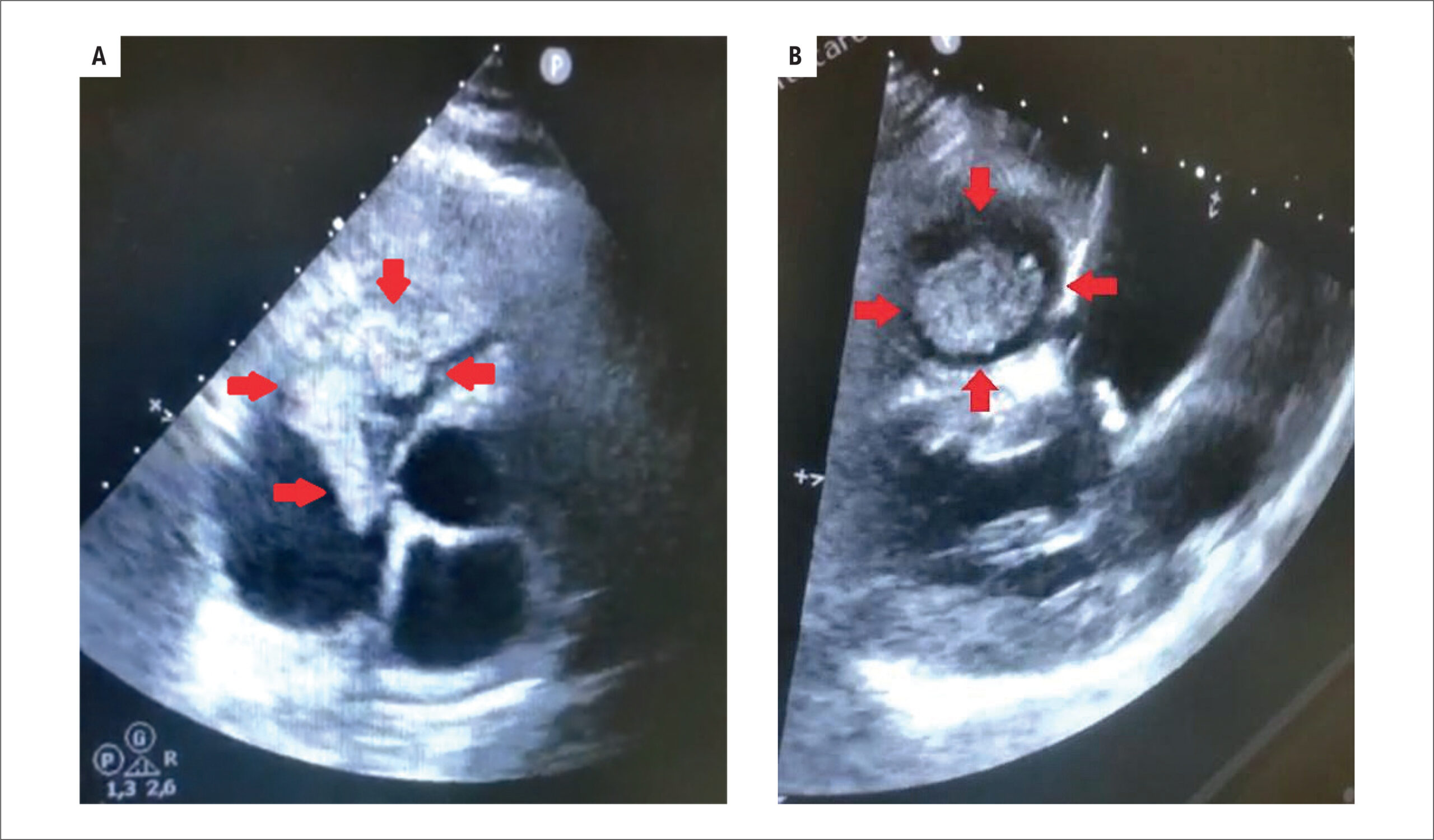
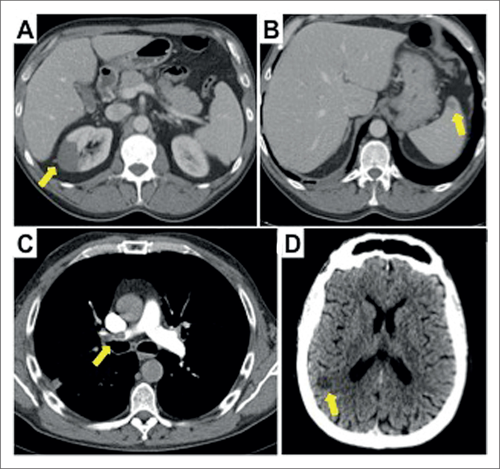
The abdominal ultrasound-scan did not show releant findings, so a computed tomography (CT) scan study was completed. CT abdominal scan found multiple infarcts in the right kidney () and isolated in spleen (), furthermore, a filling defect in the left ventricle was observed (). Transthoracic echocardiogram confirmed the presence of a pedunculated hyperechogenic and homogeneous mobile mass (3.1x2cm) with regular borders (, , ), anchored to the middle third of the septum of left ventricle (LV). Anatomy of LV was normal, with normal dimensions and normal ejection fraction without wall motion abnormalities.

[…]

  • Covid-19/complications
  • Diagnostic Imaging/methods
  • Infarction, Myocardial
  • Pulmonary embolism
  • SARS-CoV-2
  • Severe Acute Respiratory Syndrome/complications
  • Ventricular Dysfunction, Left
Read Full Article
Creative Commons Attribution (CC BY) Creative Commons license
19/12/2022 DOI: 10.36660/abc.20210590
ABC
Editorial

Macrophages and Neovascularization in In-Stent Neoatherosclerosis: An Accelerated Inflammatory Phenotype by OCT with Therapeutic Implications

Maria Cristina Oliveira Izar et al. Francisco A. H. Fonseca

In this edition of Arquivos Brasileiros de Cardiologia, Pinheiro et al. show optical coherence tomography (OCT) data with clear differences in inflammation and neovascularization among de novo atherosclerosis, in-stent restenosis due to intimal hyperplasia, and in-stent neoatherosclerosis.

Patients undergoing percutaneous coronary intervention with stent implantation may have recurrent symptoms of coronary heart disease due to in-stent restenosis due to vascular injury that triggers an intimal proliferative response, minimized by current techniques and stents of new generation. However, the neointimal lesion secondary to a delay in neoendothelialization can lead to symptom recurrence, usually during the first year of coronary intervention.

[…]

  • Atherosclerosis
  • Coronary Restenosis
  • Diagnostic Imaging/methods
  • Percutaneous Coronary Intervention
  • Phenotype/inflammation
Read Full Article
Creative Commons Attribution (CC BY) Creative Commons license
19/12/2022 DOI: 10.36660/abc.20220732 Views 1
ABC
Editorial

Coronary Tortuosity as a New Phenotype for Ischemia without Coronary Artery Disease

Mohammad Eltahlawi

Coronary artery disease constitutes a great burden in many countries. In many cases, the detection of coronary ischemia by non-invasive imaging may not correlate with the presence of significant coronary stenosis. Therefore, the term “Ischemia with non-obstructive coronary artery (INOCA)” has emerged. Many theories have been proposed for such a phenomenon. Coronary tortuosity (CorT) is one of these etiologies that was found to be associated with subclinical atherosclerosis and increased coronary artery calcium score. Besides, CorT is associated with the same risk factors of ischemia, such as smoking, old age, arterial hypertension and dyslipidemia. CorT was thought to be a phenomenon; however, the association of this phenomenon with multiple cardiovascular disorders augments its clinical impact. Li et al. found that Hypertensive patients with CorT have more incidence of lacunar infarct. Turgut et al. concluded that CorT might indicate impaired left ventricular relaxation. Dogdus et al. proved that CorT negatively affects left ventricular function evaluated by 3D strain parameters with considerable depression of longitudinal deformation of the myocardium.

In this study by Estrada et al., the investigators found a highly significant association between CorT and ischemia. The presence of ischemia in territories with CorT was more frequent than those without CorT (67% versus 28% (p<0.0001)) detected by myocardial perfusion imaging (MPI). This study analyzed the specific features of tortuosity concerning the presence of myocardial ischemia. They found that the number of bend angles detected in systole during coronary angiography is associated with a high risk of myocardial ischemia (p = 0.021). Some previous studies considered the geometry of CorT and suggested indices for grading such tortuosity depending mainly on the degree of angulation or bending.–

[…]

  • Atherosclerosis
  • Calcium Metabolism Disorders/complications
  • Coronary Artery Disease / complications
  • Diagnostic Imaging/methods
  • Dyslipidemias
  • Myocardial Ischemia/complications
  • Risk Factors
Read Full Article
Creative Commons Attribution (CC BY) Creative Commons license
19/12/2022 DOI: 10.36660/abc.20220826
Figure 1
ABC
Image

Peek-A-Boo What are You? The Diagnostic Challenge of a Cardiac Mass

Mariana Tinoco et al. Filipa Castro Sérgio Leite Francisco Sousa António Lourenço

A 49-year-old female was found to have an incidental right atrial mass on a routine transthoracic echocardiography (TTE) ( ). She reported no previous significant medical history, besides a mammoplasty in 2017 complicated with implant rupture in 2019. She did not take any medication, was fit and well, and showed full exercise tolerance. There were no risk factors for venous thromboembolism, and she had no significant family medical history. The patient is a former smoker of 12 pack-years. She had 2 previous pregnancies with no miscarriages. The physical examination was unremarkable.

[…]

  • Cardiac Mass
  • Diagnostic Imaging/methods
  • Diagnostic Techniques and Procedures
  • Echocardiography, Transesophageal/methods
  • Heart Atrial
  • Resonance Magnetic/methods
  • Trombosis
Read Full Article
Creative Commons Attribution (CC BY) Creative Commons license
21/10/2022 DOI: 10.36660/abc.20211033
Figure 1
ABC
Image

Transesophageal Two- and Three-Dimensional Echocardiographic Assessment of Spontaneous Left Atrial Dissection

Javier Ivan Armenta-Moreno et al. Joaquin Berarducci Abel Mauricio Garcia-Cardenas José Carlos Armendariz-Ferrari … Jorge Luis Bermudez-Gonzalez Juan Ignacio Straface Jose Antonio Luna-Alvarez-Amezquita Nilda Espinola-Zavaleta

A 41-year-old woman was admitted to the emergency room with an onset of acute dyspnea, jugular ingurgitation, and with the first heart sound diminished, followed by a grade III/IV holosystolic murmur that was better heard at the apex and edema of the lower limbs.

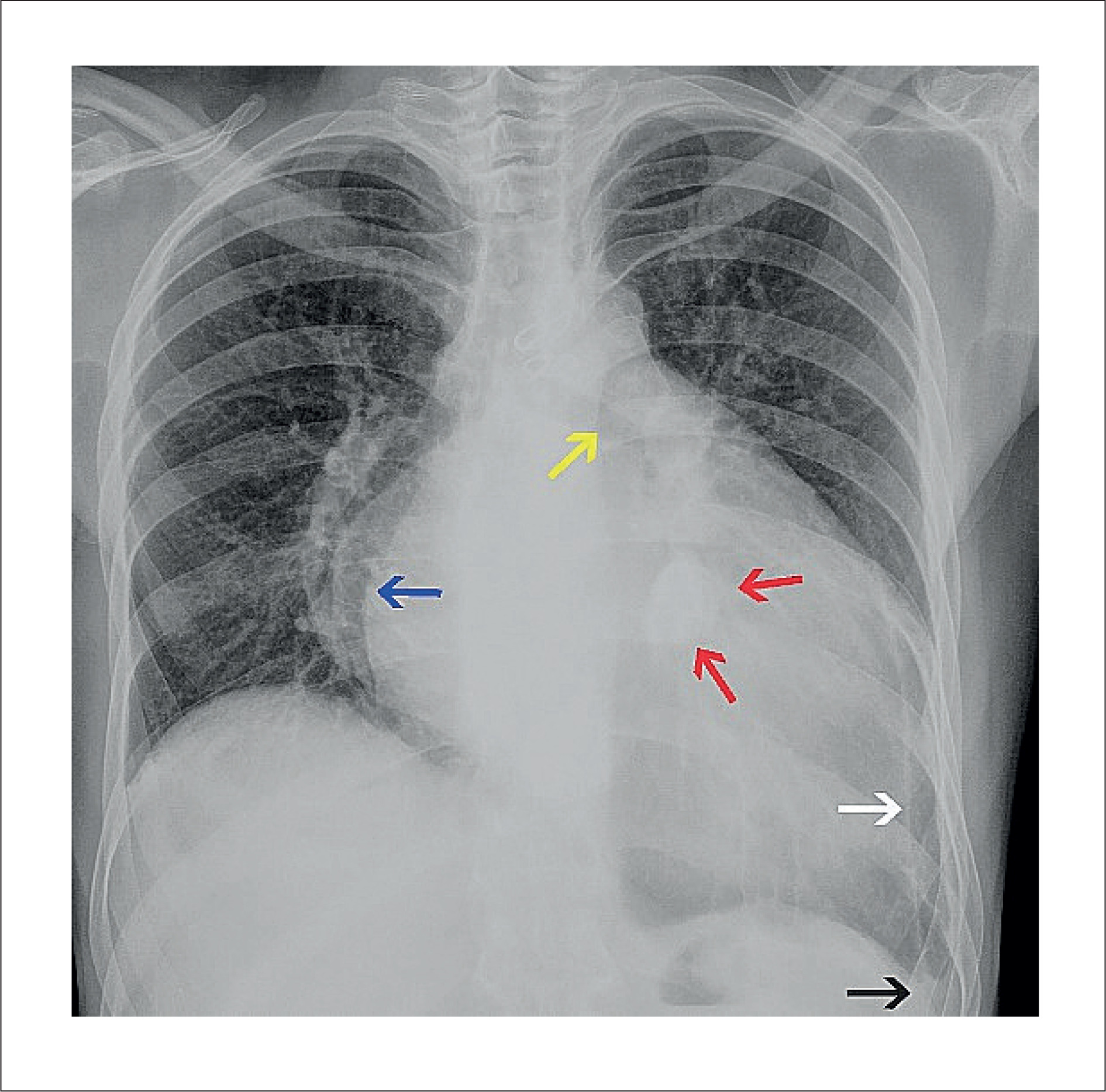
The vital signs were heart rate 91 bpm, respiratory rate 21 rpm, blood pressure 110/60 mmHg, and oxygen saturation 91%. The chest x-ray showed cardiomegaly with a cardiothoracic index of 0.62 and pulmonary venocapillary hypertension.

[…]

  • Atrial Function, Left/physiology
  • Diagnostic Imaging/methods
  • Echocardiography, Three Dimensional / methods
  • Heart Failure/physiopathology
  • Shock, Cardiogenic
Read Full Article
Creative Commons Attribution (CC BY) Creative Commons license
05/09/2022 DOI: 10.36660/abc.20210740
Figure 1
IJCS
Original Article

Strain Magnitude Assessed at Rest and During Stress Echocardiography in Patients with Normal Coronary Flow Reserve

José Sebastião de Abreu et al. Tereza Cristina Pinheiro Diógenes Marília Esther Benevides Abreu Henrique Maia Costa … Ana Gardenia Liberato Ponte Farias Marcia Maria Carneiro

Abstract

Background

Coronary flow and myocardial contractile performance assessed by strain magnitude increase during a dobutamine stress echocardiogram (DSE). Normal coronary flow reserve (CFR) can be attained upon completion of a DSE at age-predicted maximum heart rate (HR) (HRmax = 220 – age)] or submaximal HR [(0.85) HRmax] or before completion (early CFR).

Objective

To ascertain the association between delta strain and HR in patients with early normal CFR.

Methods

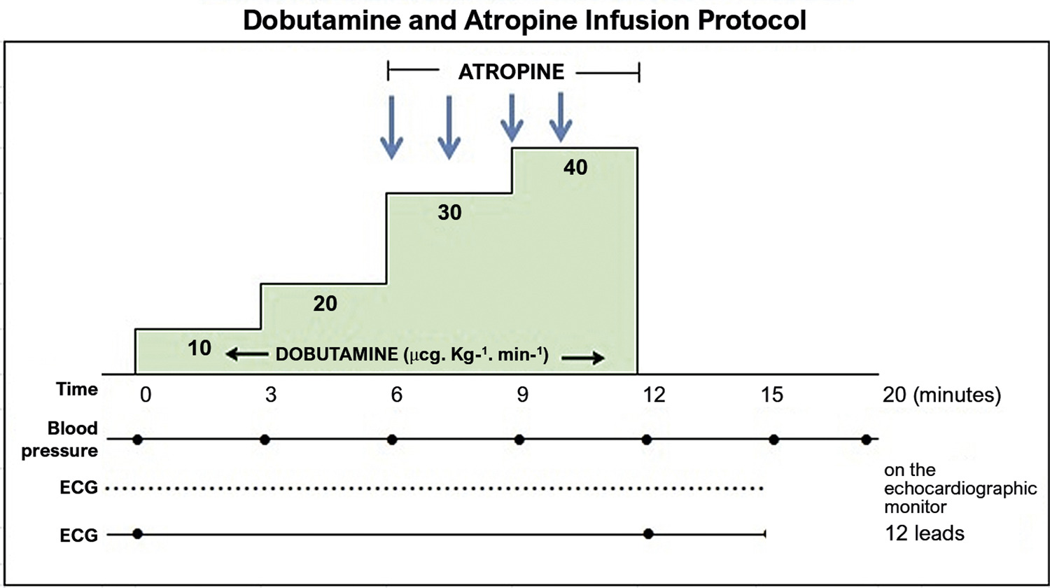
This prospective study included patients whose normal CFR was obtained before the DSE was completed. Percentage of resting HR (%HRrest) = [(HRrest ÷ HRmax) 100]% and %HR CFR = [(HR at the time of CFR attainment) ÷ (HRmax) 100]% were recorded. Strain was assessed in the left ventricular region of interest, and delta strain was calculated as the difference between the measures obtained at HRrest and after the DSE was completed. Strain agreement analysis for HRrest, %HRrest, and %HR CFR was performed using the kappa coefficient. The Shapiro-Wilk test was used to assess data normality, and the Mann-Whitney test was used to compare the groups. A p-value < 0.05 was considered statistically significant.

Results

Strain measured -23.3% ± 4.3% at baseline and -31.1% ± 4.9% during the DSE. In delta strain > 8 absolute points, the ROC curves showed an area under the curve of 0.874 ± 0.07 for %HRrest (p = 0.001) and an area under the curve of 0.862 ± 0.07 for %HR CFR (p = 0.001). In delta strain > 8 points, %HRrest ≤ 42.6% of HRmax and %HR CFR ≤ 62.5% of HRmax showed an accuracy of 82.9% and 79.8%, respectively.

Conclusion

In this study, lower HRrest and HR at the time of CFR attainment had a good association with better myocardial contractile performance, according to the change in strain magnitude.

  • Blood Flow
  • Blood flow velocity
  • Cardiotonic Agents
  • Coronary Artery Disease
  • Diagnostic Imaging/methods
  • Echocardiography, Stress / methods
  • Myocardial Contraction Strain
  • Vasodilatador Agents
Read Full Article
Creative Commons Attribution (CC BY) Creative Commons license
01/08/2022 DOI: 10.36660/ijcs.20210244 Views 1
ABC
Editorial

Chagas Heart Disease: The Evolution of the Disease and its Complementary Exams

Claudio Leinig Pereira da Cunha

As in many areas of Medicine, the scenario that harbors Chagas Disease(CD) has changed substantially. For approximately 50 years, as a Resident Physician, I was on duty at the Emergency Department of the Hospital de Clínicas of the Universidade Federal do Paraná and routinely attended, on each shift, one or more cases of patients with clear Congestive Heart Failure, anasarca, electrocardiogram with conduction disorders and multiple arrhythmias, chest X-ray with cardiomegaly and laboratory diagnosis of CD. There was no echocardiogram. Today this is an unusual situation; when it occurs, students are invited to see an exuberant picture of Chagas’ heart disease (CHD), which appears every semester.

In epidemiological surveys, similar findings are observed. In 1984, the prevalence of CD in the State of Paraná, Brazil, was 4% of the population. In 2020, estimates of the prevalence of infections by Trypanosoma cruzi ranged from 1.02% to 2.4% in Brazil. In the period 1975/83, among 291 municipalities in Paraná, 90 (30.9%) had triatomine insects infected by T. cruzi, while in 1990, these were found in only 4 municipalities (1.4%), with subsequent eradication of vector contamination. Vector control strategies have led to a substantial decline in the global prevalence of the disease, estimated at 18 million in 1990 and 6 million in 2018.

[…]

  • Chagas Cardiomyopathy/complications
  • Chagas disease
  • Clinical Evolution
  • Diagnostic Imaging/methods
  • Epidemiology
  • Heart Failure
Read Full Article
Creative Commons Attribution (CC BY) Creative Commons license
11/07/2022 DOI: 10.36660/abc.20220418
ABC
Original Article

Evaluation of Endothelial Dysfunction in COVID-19 With Flow-Mediated Dilatation

Asli Kurtar Mansiroglu et al. Hande Seymen Isa Sincer Yilmaz Gunes

Abstract

Background:

Inflammation is known to play a crucial role in many diseases, including COVID-19.

Objective:

Using flow-mediated dilatation (FMD), we aimed to assess the effects of inflammation on endothelial function in COVID-19 patients.

Methods:

This study was conducted with a total of 161 subjects, of whom 80 were diagnosed with COVID-19 within the last six months (comprising 48 women and 32 men with a mean age of 32.10 ± 5.87 years) and 81 were healthy controls (comprising 45 women and 36 men with a mean age of 30.51 ± 7.33 years). We analyzed the findings of transthoracic echocardiography and FMD in all subjects. All results were considered statistically significant at the level of p < 0.05.

Results:

The echocardiography and FMD of the COVID-19 group were performed 35 days (range: 25-178) after diagnosis. There was no statistically significant difference in echocardiographic parameters. Differently, FMD (%) was significantly higher in the control group (9.52 ± 5.98 vs. 12.01 ± 6.18, p=0.01). In multivariate analysis with the forward stepwise model, FMD was significantly different in the control group compared to the COVID-19 group (1.086 (1.026 – 1.149), p=0.04). A Spearman’s correlation test indicated that FMD (r=0.27, p=0.006) had a weak positive correlation with the presence of COVID-19.

Conclusion:

Our findings point to COVID-19-induced endothelial dysfunction, as assessed by FMD, in the early recovery phase.

  • Covid-19/complications
  • Diagnostic Imaging/methods
  • Echocardiography/methods
  • Endothelial, Cells/infection
  • Endothelium Vascular/injuries
  • Flow Dilatation
  • Myalgia
  • Olfaction Disorders
  • Taste Disorders
  • Ultrasonography/methods
Read Full Article
Creative Commons Attribution (CC BY) Creative Commons license
03/06/2022 DOI: 10.36660/abc.20210561 Views 1
ABC
Editorial

It is Time for Coronary Computed Tomography Angiography to be Incorporated into the SUS

Nilson Tavares Poppi

In the evaluation of patients with stable coronary artery disease (CAD), also called chronic coronary syndrome (CCS), complementary tests are used both for diagnostic and prognostic purposes. , Anatomical (coronary angiography and coronary CT angiography) and functional (exercise testing, stress echocardiography, rest and stress myocardial perfusion imaging by scintigraphy, magnetic resonance, and positron emission tomography) tests are available. Coronary angiography, the gold standard, is invasive and therefore, indicated for clinically more severe patients, or those with poor prognostic findings in non-invasive testing, when myocardial revascularization is considered or planned.

The choice for the most appropriate diagnostic test is an important and challenging issue for the cardiologist in the clinical evaluation of CCS. The first step in this decision-making process is the assessment of the pre-test probability (PTP) of CAD. As recommended by the current SCC guidelines, , patients classified as having high PTP should receive medical therapy and undergo testing for prognostic information. The patients with low PTP should be assessed for an alternative diagnosis more likely than CAD. Patients with PTP calculated between 15-85% are in the intermediate range, where the complementary tests are more useful and important for CAD diagnosis. In addition to diagnostic accuracy and PTP, the selection of a non-invasive test depends on the clinical characteristics of patients, local expertise, and the availability of tests. In Brazil, it is estimated that up to 80% of the population depends exclusively on medical care provided by the public health system (SUS). In this context of managing economic resources, physicians and health managers should focus on the most cost-effective options for the diagnosis of CAD.

[…]

  • Computed Tomography Angiography
  • Coronary Artery Disease
  • Cost-Benefit Analysis
  • Diagnostic Imaging/methods
  • Stable Angina
  • Unified Health System
Read Full Article
Creative Commons Attribution (CC BY) Creative Commons license
18/03/2022 DOI: 10.36660/abc.20220033 Views 1
ABCIC
Viewpoint

Non-compaction cardiomyopathy: How Can Imaging Contribute?

José de Arimateia Batista Araújo Filho et al. Antonildes Nascimento Assunção-Jr José Rodrigues Parga

[…]

  • Arrhythmias Cardiac
  • Cardiomyopathies/physiopathology
  • Diagnostic Imaging/methods
  • Heart Failure
  • Magnetic Resonance Spectroscopy/ methods
  • Thromboembolism
Read Full Article
Creative Commons Attribution (CC BY) Creative Commons license
20/01/2022 DOI: 10.5935/2318-8219.20190004
ABC
Editorial

The Importance of Understanding the Progression of Myocardial Fibrosis in Chronic Chagas Cardiomyopathy

Carlos E. Rochitte

Myocardial fibrosis is one of the most important biological markers of Chagas’ cardiomyopathy and is directly related to the severity of the cardiomyopathy and the stage of the disease. In addition, myocardial fibrosis has prognostic value for major cardiovascular events, with a strong and close relationship with arrhythmic events in these patients.–

Cardiac magnetic resonance (CMR) is the reference imaging method for myocardial fibrosis detection and quantification in many cardiomyopathies, including Chagas cardiomyopathy. Initial studies included patients with Chagas cardiomyopathy at different stages of the disease and clearly demonstrated that the higher the disease severity the larger the amount of myocardial fibrosis. Based on these data and on the natural history of the disease, with progression of left ventricle dysfunction, one could safely infer that myocardial fibrosis progresses with time for any given patient. However, the longitudinal data using CMR in patients with Chagas disease was not available until the publication of the original article in this issue of ABC Cardiol by Santos et al. This is the first study to include data on myocardial fibrosis from two consecutive CMR of the same patient with a relatively long follow-up of 5.4 years. The authors demonstrated an impressive 43% increase of myocardial fibrosis over the follow-up period, which indicates a mean of 7.9% increase per year of myocardial fibrosis detected by CMR.

[…]

  • Chagas Diseases
  • chagasic cardiomyopathy
  • Diagnostic Imaging/methods
  • Endomyocardial Fibrosis
  • Heart Failure
  • Imaging Resonance Magnetic/methods
Read Full Article
Creative Commons Attribution (CC BY) Creative Commons license
03/12/2021 DOI: 10.36660/abc.20210897 Views 1

Could not load more articles. Please try again.

How to cite this article

EndNote RIS

Electronic Document Format (Vancouver)

Tinoco M, Castro M, Dabó H, Cordeiro F, Hafe P, Lourenço A. Multiple Intracardiac Metastases – A Chaplet Heart. Arquivos Brasileiros de Cardiologia 2024;121(6):e20230732.

Electronic Document Format (ABNT)

Tinoco, Mariana; Castro, Margarida; Dabó, Hans; Cordeiro, Filipa; Hafe, Pedro von; Lourenço, António. Multiple Intracardiac Metastases – A Chaplet Heart. Arquivos Brasileiros de Cardiologia, v. 121, n. 6, e20230732, Jul. 2024.

Electronic Document Format (APA)

Tinoco, M., Castro, M., Dabó, H., Cordeiro, F., Hafe, P. , & Lourenço, A. (2024). Multiple Intracardiac Metastases – A Chaplet Heart. Arquivos Brasileiros de Cardiologia, 121(6), e20230732.

Electronic Document Format (ISO)

Tinoco, Mariana and Castro, Margarida and Dabó, Hans and Cordeiro, Filipa and Hafe, Pedro von and Lourenço, António. Multiple Intracardiac Metastases – A Chaplet Heart. Arquivos Brasileiros de Cardiologia [online]. 2024, vol. 121, n. 6, [cited 2026-10-14], e20230732. Available from: <https://journals.cardiol.br/en/article/multiple-intracardiac-metastases-a-chaplet-heart/>. ISSN 0066-782X

EndNote RIS

Electronic Document Format (Vancouver)

Andrade VC, Rezende PC, Hueb W, Ramires JAF. Interpreting Acute Myocardial Infarction 40 Years Later. Evolution of Knowledge: What is the Best Explanation?. Arquivos Brasileiros de Cardiologia 2024;121(3):e20230757.

Electronic Document Format (ABNT)

Andrade, Vitor Coutinho; Rezende, Paulo Cury; Hueb, Whady; Ramires, José Antonio Franchini. Interpreting Acute Myocardial Infarction 40 Years Later. Evolution of Knowledge: What is the Best Explanation?. Arquivos Brasileiros de Cardiologia, v. 121, n. 3, e20230757, Apr. 2024.

Electronic Document Format (APA)

Andrade, V. C., Rezende, P. C., Hueb, W., & Ramires, J. A. F. (2024). Interpreting Acute Myocardial Infarction 40 Years Later. Evolution of Knowledge: What is the Best Explanation?. Arquivos Brasileiros de Cardiologia, 121(3), e20230757.

Electronic Document Format (ISO)

Andrade, Vitor Coutinho and Rezende, Paulo Cury and Hueb, Whady and Ramires, José Antonio Franchini. Interpreting Acute Myocardial Infarction 40 Years Later. Evolution of Knowledge: What is the Best Explanation?. Arquivos Brasileiros de Cardiologia [online]. 2024, vol. 121, n. 3, [cited 2026-10-14], e20230757. Available from: <https://journals.cardiol.br/en/article/interpreting-acute-myocardial-infarction-40-years-later-evolution-of-knowledge-what-is-the-best-explanation/>. ISSN 0066-782X

EndNote RIS

Electronic Document Format (Vancouver)

Silva RRP, Magalhães CJ, Silva RSV, Rocha GAF, Cavalcanti PEF, Montenegro ST. Acute ST-Elevation Myocardial Infarction in a Young Adult: Rare Presentation of Giant Atrial Myxoma. Arquivos Brasileiros de Cardiologia 2024;121(3):e20230538.

Electronic Document Format (ABNT)

Silva, Rodrigo Rufino Pereira; Magalhães, Carolina Jerônimo; Silva, Rafael Silvestre Vieira da; Rocha, Giulia Antoni Ferreira; Cavalcanti, Paulo Ernando Ferraz; Montenegro, Sérgio Tavares. Acute ST-Elevation Myocardial Infarction in a Young Adult: Rare Presentation of Giant Atrial Myxoma. Arquivos Brasileiros de Cardiologia, v. 121, n. 3, e20230538, Apr. 2024.

Electronic Document Format (APA)

Silva, R. R. P., Magalhães, C. J., Silva, R. S. V. , Rocha, G. A. F., Cavalcanti, P. E. F., & Montenegro, S. T. (2024). Acute ST-Elevation Myocardial Infarction in a Young Adult: Rare Presentation of Giant Atrial Myxoma. Arquivos Brasileiros de Cardiologia, 121(3), e20230538.

Electronic Document Format (ISO)

Silva, Rodrigo Rufino Pereira and Magalhães, Carolina Jerônimo and Silva, Rafael Silvestre Vieira da and Rocha, Giulia Antoni Ferreira and Cavalcanti, Paulo Ernando Ferraz and Montenegro, Sérgio Tavares. Acute ST-Elevation Myocardial Infarction in a Young Adult: Rare Presentation of Giant Atrial Myxoma. Arquivos Brasileiros de Cardiologia [online]. 2024, vol. 121, n. 3, [cited 2026-10-14], e20230538. Available from: <https://journals.cardiol.br/en/article/acute-st-elevation-myocardial-infarction-in-a-young-adult-rare-presentation-of-giant-atrial-myxoma/>. ISSN 0066-782X

EndNote RIS

Electronic Document Format (Vancouver)

Atik E, Leal GN, Jatene MB. Marked Concentric Myocardial Hypertrophy with Good Postoperative Evolution in a 4 Years Old Child. Arquivos Brasileiros de Cardiologia 2023;120(5):e20220869.

Electronic Document Format (ABNT)

Atik, Edmar; Leal, Gabriela Nunes; Jatene, Marcelo B.. Marked Concentric Myocardial Hypertrophy with Good Postoperative Evolution in a 4 Years Old Child. Arquivos Brasileiros de Cardiologia, v. 120, n. 5, e20220869, Jun. 2023.

Electronic Document Format (APA)

Atik, E., Leal, G. N., & Jatene, M. B. (2023). Marked Concentric Myocardial Hypertrophy with Good Postoperative Evolution in a 4 Years Old Child. Arquivos Brasileiros de Cardiologia, 120(5), e20220869.

Electronic Document Format (ISO)

Atik, Edmar and Leal, Gabriela Nunes and Jatene, Marcelo B.. Marked Concentric Myocardial Hypertrophy with Good Postoperative Evolution in a 4 Years Old Child. Arquivos Brasileiros de Cardiologia [online]. 2023, vol. 120, n. 5, [cited 2026-10-14], e20220869. Available from: <https://journals.cardiol.br/en/article/marked-concentric-myocardial-hypertrophy-with-good-postoperative-evolution-in-a-4-years-old-child/>. ISSN 0066-782X

EndNote RIS

Electronic Document Format (Vancouver)

Mariani Junior J. Brazilian Interventional Cardiology and Chronic Coronary Occlusions: Where Are We?. Arquivos Brasileiros de Cardiologia 2023;120(4):e20230172.

Electronic Document Format (ABNT)

Mariani Junior, José. Brazilian Interventional Cardiology and Chronic Coronary Occlusions: Where Are We?. Arquivos Brasileiros de Cardiologia, v. 120, n. 4, e20230172, Apr. 2023.

Electronic Document Format (APA)

Mariani Junior, J. (2023). Brazilian Interventional Cardiology and Chronic Coronary Occlusions: Where Are We?. Arquivos Brasileiros de Cardiologia, 120(4), e20230172.

Electronic Document Format (ISO)

Mariani Junior, José. Brazilian Interventional Cardiology and Chronic Coronary Occlusions: Where Are We?. Arquivos Brasileiros de Cardiologia [online]. 2023, vol. 120, n. 4, [cited 2026-10-14], e20230172. Available from: <https://journals.cardiol.br/en/article/brazilian-interventional-cardiology-and-chronic-coronary-occlusions-where-are-we/>. ISSN 0066-782X

EndNote RIS

Electronic Document Format (Vancouver)

Vilela EM, Fontes-Carvalho R. Left Ventricular Mechanics: Untwisting the Pathways of the Cardiovascular Response to Exercise. Arquivos Brasileiros de Cardiologia 2023;120(4):e20230181.

Electronic Document Format (ABNT)

Vilela, Eduardo M.; Fontes-Carvalho, Ricardo. Left Ventricular Mechanics: Untwisting the Pathways of the Cardiovascular Response to Exercise. Arquivos Brasileiros de Cardiologia, v. 120, n. 4, e20230181, Apr. 2023.

Electronic Document Format (APA)

Vilela, E. M., & Fontes-Carvalho, R. (2023). Left Ventricular Mechanics: Untwisting the Pathways of the Cardiovascular Response to Exercise. Arquivos Brasileiros de Cardiologia, 120(4), e20230181.

Electronic Document Format (ISO)

Vilela, Eduardo M. and Fontes-Carvalho, Ricardo. Left Ventricular Mechanics: Untwisting the Pathways of the Cardiovascular Response to Exercise. Arquivos Brasileiros de Cardiologia [online]. 2023, vol. 120, n. 4, [cited 2026-10-14], e20230181. Available from: <https://journals.cardiol.br/en/article/left-ventricular-mechanics-untwisting-the-pathways-of-the-cardiovascular-response-to-exercise/>. ISSN 0066-782X

EndNote RIS

Electronic Document Format (Vancouver)

Grossman GB. Nuclear Cardiology in the Optimization of Resynchronization Therapy: Quo Vadis?. Arquivos Brasileiros de Cardiologia 2023;120(3):e20230136.

Electronic Document Format (ABNT)

Grossman, Gabriel Blacher. Nuclear Cardiology in the Optimization of Resynchronization Therapy: Quo Vadis?. Arquivos Brasileiros de Cardiologia, v. 120, n. 3, e20230136, Apr. 2023.

Electronic Document Format (APA)

Grossman, G. B. (2023). Nuclear Cardiology in the Optimization of Resynchronization Therapy: Quo Vadis?. Arquivos Brasileiros de Cardiologia, 120(3), e20230136.

Electronic Document Format (ISO)

Grossman, Gabriel Blacher. Nuclear Cardiology in the Optimization of Resynchronization Therapy: Quo Vadis?. Arquivos Brasileiros de Cardiologia [online]. 2023, vol. 120, n. 3, [cited 2026-10-14], e20230136. Available from: <https://journals.cardiol.br/en/article/nuclear-cardiology-in-the-optimization-of-resynchronization-therapy-quo-vadis/>. ISSN 0066-782X

EndNote RIS

Electronic Document Format (Vancouver)

Armstrong AC, Cerci R, Matheson MB, Magalhães T, Kishi S, Brinker J, Clouse ME, et al. Predicting Significant Coronary Obstruction in a Population with Suspected Coronary Disease and Absence of Coronary Calcium: CORE-64 / CORE320 Studies. Arquivos Brasileiros de Cardiologia 2023;120(3):e20220183.

Electronic Document Format (ABNT)

Armstrong, Anderson C.; Cerci, Rodrigo; Matheson, Matthew B.; Magalhães, Tiago; Kishi, Satoru; Brinker, Jeff; Clouse, Melvin E.; Rochitte, Carlos E.; Cox, Christopher; Lima, João A. C.; Arbab-Zadeh, Armin. Predicting Significant Coronary Obstruction in a Population with Suspected Coronary Disease and Absence of Coronary Calcium: CORE-64 / CORE320 Studies. Arquivos Brasileiros de Cardiologia, v. 120, n. 3, e20220183, Mar. 2023.

Electronic Document Format (APA)

Armstrong, A. C., Cerci, R., Matheson, M. B., Magalhães, T., Kishi, S., Brinker, J., Clouse, M. E., Rochitte, C. E., Cox, C., Lima, J. A. C., & Arbab-Zadeh, A. (2023). Predicting Significant Coronary Obstruction in a Population with Suspected Coronary Disease and Absence of Coronary Calcium: CORE-64 / CORE320 Studies. Arquivos Brasileiros de Cardiologia, 120(3), e20220183.

Electronic Document Format (ISO)

Armstrong, Anderson C. and Cerci, Rodrigo and Matheson, Matthew B. and Magalhães, Tiago and Kishi, Satoru and Brinker, Jeff and Clouse, Melvin E. and Rochitte, Carlos E. and Cox, Christopher and Lima, João A. C. and Arbab-Zadeh, Armin. Predicting Significant Coronary Obstruction in a Population with Suspected Coronary Disease and Absence of Coronary Calcium: CORE-64 / CORE320 Studies. Arquivos Brasileiros de Cardiologia [online]. 2023, vol. 120, n. 3, [cited 2026-10-14], e20220183. Available from: <https://journals.cardiol.br/en/article/predicting-significant-coronary-obstruction-in-a-population-with-suspected-coronary-disease-and-absence-of-coronary-calcium-core-64-core320-studies/>. ISSN 0066-782X

EndNote RIS

Electronic Document Format (Vancouver)

Santos M, Paiva M, Ferreira J, Guerreiro S. Tricky Diagnosis: An Aberrant Mitral Valve Chord. Arquivos Brasileiros de Cardiologia 2023;120(3):e20220573.

Electronic Document Format (ABNT)

Santos, Marina; Paiva, Mariana; Ferreira, Joana; Guerreiro, Sara. Tricky Diagnosis: An Aberrant Mitral Valve Chord. Arquivos Brasileiros de Cardiologia, v. 120, n. 3, e20220573, Mar. 2023.

Electronic Document Format (APA)

Santos, M., Paiva, M., Ferreira, J., & Guerreiro, S. (2023). Tricky Diagnosis: An Aberrant Mitral Valve Chord. Arquivos Brasileiros de Cardiologia, 120(3), e20220573.

Electronic Document Format (ISO)

Santos, Marina and Paiva, Mariana and Ferreira, Joana and Guerreiro, Sara. Tricky Diagnosis: An Aberrant Mitral Valve Chord. Arquivos Brasileiros de Cardiologia [online]. 2023, vol. 120, n. 3, [cited 2026-10-14], e20220573. Available from: <https://journals.cardiol.br/en/article/tricky-diagnosis-an-aberrant-mitral-valve-chord/>. ISSN 0066-782X

EndNote RIS

Electronic Document Format (Vancouver)

Medeiros P, Coelho AR, Magalhães J, Salomé N, Pereira V. A Rare Subtype of a Rare Tumor. Arquivos Brasileiros de Cardiologia 2023;120(2):e20220486.

Electronic Document Format (ABNT)

Medeiros, Paulo; Coelho, Ana Rita; Magalhães, João; Salomé, Nuno; Pereira, Vítor. A Rare Subtype of a Rare Tumor. Arquivos Brasileiros de Cardiologia, v. 120, n. 2, e20220486, Feb. 2023.

Electronic Document Format (APA)

Medeiros, P., Coelho, A. R., Magalhães, J., Salomé, N., & Pereira, V. (2023). A Rare Subtype of a Rare Tumor. Arquivos Brasileiros de Cardiologia, 120(2), e20220486.

Electronic Document Format (ISO)

Medeiros, Paulo and Coelho, Ana Rita and Magalhães, João and Salomé, Nuno and Pereira, Vítor. A Rare Subtype of a Rare Tumor. Arquivos Brasileiros de Cardiologia [online]. 2023, vol. 120, n. 2, [cited 2026-10-14], e20220486. Available from: <https://journals.cardiol.br/en/article/a-rare-subtype-of-a-rare-tumor/>. ISSN 0066-782X

EndNote RIS

Electronic Document Format (Vancouver)

Brito MR. The “Five Malignant Waves” of the Electrocardiography. Arquivos Brasileiros de Cardiologia 2023;120(3):e20220649.

Electronic Document Format (ABNT)

Brito, Mitermayer Reis. The “Five Malignant Waves” of the Electrocardiography. Arquivos Brasileiros de Cardiologia, v. 120, n. 3, e20220649, Feb. 2023.

Electronic Document Format (APA)

Brito, M. R. (2023). The “Five Malignant Waves” of the Electrocardiography. Arquivos Brasileiros de Cardiologia, 120(3), e20220649.

Electronic Document Format (ISO)

Brito, Mitermayer Reis. The “Five Malignant Waves” of the Electrocardiography. Arquivos Brasileiros de Cardiologia [online]. 2023, vol. 120, n. 3, [cited 2026-10-14], e20220649. Available from: <https://journals.cardiol.br/en/article/the-five-malignant-waves-of-the-electrocardiography/>. ISSN 0066-782X

EndNote RIS

Electronic Document Format (Vancouver)

Faé IG, Ruiz GZL, Irffi GP, Almeida Junior RS, Conceição PA, Falchetto EB, Passaglia LG, et al. Intracardiac Metastasis of Colonic Adenocarcinoma 12 Years After Primary Tumor Control and Without Any Sign of Other Metastasis: A Case Report. Arquivos Brasileiros de Cardiologia 2022;119(6):991-5.

Electronic Document Format (ABNT)

Faé, Isabela Galizzi; Ruiz, Gabriela Zamunaro Lopes; Irffi, Gustavo Palmer; Almeida Junior, Robson de Souza; Conceição, Pedro Anjos; Falchetto, Eduardo Belisario; Passaglia, Luiz Guilherme; Brasileiro Filho, Geraldo; Gelape, Cláudio Leo; Oliveira, Clara Rodrigues Alves de. Intracardiac Metastasis of Colonic Adenocarcinoma 12 Years After Primary Tumor Control and Without Any Sign of Other Metastasis: A Case Report. Arquivos Brasileiros de Cardiologia, v. 119, n. 6, p. 991-995, Dec. 2022.

Electronic Document Format (APA)

Faé, I. G., Ruiz, G. Z. L., Irffi, G. P., Almeida Junior, R. S., Conceição, P. A., Falchetto, E. B., Passaglia, L. G., Brasileiro Filho, G., Gelape, C. L., & Oliveira, C. R. A. (2022). Intracardiac Metastasis of Colonic Adenocarcinoma 12 Years After Primary Tumor Control and Without Any Sign of Other Metastasis: A Case Report. Arquivos Brasileiros de Cardiologia, 119(6), 991-995.

Electronic Document Format (ISO)

Faé, Isabela Galizzi and Ruiz, Gabriela Zamunaro Lopes and Irffi, Gustavo Palmer and Almeida Junior, Robson de Souza and Conceição, Pedro Anjos and Falchetto, Eduardo Belisario and Passaglia, Luiz Guilherme and Brasileiro Filho, Geraldo and Gelape, Cláudio Leo and Oliveira, Clara Rodrigues Alves de. Intracardiac Metastasis of Colonic Adenocarcinoma 12 Years After Primary Tumor Control and Without Any Sign of Other Metastasis: A Case Report. Arquivos Brasileiros de Cardiologia [online]. 2022, vol. 119, n. 6, [cited 2026-10-14], pp. 991-995. Available from: <https://journals.cardiol.br/en/article/intracardiac-metastasis-of-colonic-adenocarcinoma-12-years-after-primary-tumor-control-and-without-any-sign-of-other-metastasis-a-case-report/>. ISSN 0066-782X

EndNote RIS

Electronic Document Format (Vancouver)

Chaves BJ, Duarte MB, Passaglia LG, Gelape C, Azevedo PHR, Brasileiro Filho G. Caseous Calcification of the Mitral Annulus: A Post-Heart Transplant Diagnosis. Arquivos Brasileiros de Cardiologia 2022;119(6):999-1001.

Electronic Document Format (ABNT)

Chaves, Bruno Jordão; Duarte, Matheus Bitencourt; Passaglia, Luiz Guilherme; Gelape, Claudio; Azevedo, Paulo Hernane Rabelo; Brasileiro Filho, Geraldo. Caseous Calcification of the Mitral Annulus: A Post-Heart Transplant Diagnosis. Arquivos Brasileiros de Cardiologia, v. 119, n. 6, p. 999-1001, Dec. 2022.

Electronic Document Format (APA)

Chaves, B. J., Duarte, M. B., Passaglia, L. G., Gelape, C., Azevedo, P. H. R., & Brasileiro Filho, G. (2022). Caseous Calcification of the Mitral Annulus: A Post-Heart Transplant Diagnosis. Arquivos Brasileiros de Cardiologia, 119(6), 999-1001.

Electronic Document Format (ISO)

Chaves, Bruno Jordão and Duarte, Matheus Bitencourt and Passaglia, Luiz Guilherme and Gelape, Claudio and Azevedo, Paulo Hernane Rabelo and Brasileiro Filho, Geraldo. Caseous Calcification of the Mitral Annulus: A Post-Heart Transplant Diagnosis. Arquivos Brasileiros de Cardiologia [online]. 2022, vol. 119, n. 6, [cited 2026-10-14], pp. 999-1001. Available from: <https://journals.cardiol.br/en/article/caseous-calcification-of-the-mitral-annulus-a-post-heart-transplant-diagnosis/>. ISSN 0066-782X

EndNote RIS

Electronic Document Format (Vancouver)

Lorenzo N, Hernandez V, Montes A, Rivero F, Reyes* G, Aguilar R. Left Ventricular Thrombosis and Pulmonary Thromboembolism in an Asymptomatic Covid-19 Patient. Arquivos Brasileiros de Cardiologia 2022;119(6):996-8.

Electronic Document Format (ABNT)

Lorenzo, Natalia; Hernandez, Veronica; Montes, Alvaro; Rivero, Fernando; Reyes*, Guillermo; Aguilar, Rio. Left Ventricular Thrombosis and Pulmonary Thromboembolism in an Asymptomatic Covid-19 Patient. Arquivos Brasileiros de Cardiologia, v. 119, n. 6, p. 996-998, Dec. 2022.

Electronic Document Format (APA)

Lorenzo, N., Hernandez, V., Montes, A., Rivero, F., Reyes*, G., & Aguilar, R. (2022). Left Ventricular Thrombosis and Pulmonary Thromboembolism in an Asymptomatic Covid-19 Patient. Arquivos Brasileiros de Cardiologia, 119(6), 996-998.

Electronic Document Format (ISO)

Lorenzo, Natalia and Hernandez, Veronica and Montes, Alvaro and Rivero, Fernando and Reyes*, Guillermo and Aguilar, Rio. Left Ventricular Thrombosis and Pulmonary Thromboembolism in an Asymptomatic Covid-19 Patient. Arquivos Brasileiros de Cardiologia [online]. 2022, vol. 119, n. 6, [cited 2026-10-14], pp. 996-998. Available from: <https://journals.cardiol.br/en/article/left-ventricular-thrombosis-and-pulmonary-thromboembolism-in-an-asymptomatic-covid-19-patient/>. ISSN 0066-782X

EndNote RIS

Electronic Document Format (Vancouver)

Izar MCO, Fonseca FAH. Macrophages and Neovascularization in In-Stent Neoatherosclerosis: An Accelerated Inflammatory Phenotype by OCT with Therapeutic Implications. Arquivos Brasileiros de Cardiologia 2022;119(6):938-9.

Electronic Document Format (ABNT)

Izar, Maria Cristina Oliveira; Fonseca, Francisco A. H.. Macrophages and Neovascularization in In-Stent Neoatherosclerosis: An Accelerated Inflammatory Phenotype by OCT with Therapeutic Implications. Arquivos Brasileiros de Cardiologia, v. 119, n. 6, p. 938-939, Dec. 2022.

Electronic Document Format (APA)

Izar, M. C. O., & Fonseca, F. A. H. (2022). Macrophages and Neovascularization in In-Stent Neoatherosclerosis: An Accelerated Inflammatory Phenotype by OCT with Therapeutic Implications. Arquivos Brasileiros de Cardiologia, 119(6), 938-939.

Electronic Document Format (ISO)

Izar, Maria Cristina Oliveira and Fonseca, Francisco A. H.. Macrophages and Neovascularization in In-Stent Neoatherosclerosis: An Accelerated Inflammatory Phenotype by OCT with Therapeutic Implications. Arquivos Brasileiros de Cardiologia [online]. 2022, vol. 119, n. 6, [cited 2026-10-14], pp. 938-939. Available from: <https://journals.cardiol.br/en/article/macrophages-and-neovascularization-in-in-stent-neoatherosclerosis-an-accelerated-inflammatory-phenotype-by-oct-with-therapeutic-implications/>. ISSN 0066-782X

EndNote RIS

Electronic Document Format (Vancouver)

Eltahlawi M. Coronary Tortuosity as a New Phenotype for Ischemia without Coronary Artery Disease. Arquivos Brasileiros de Cardiologia 2022;119(6):891-2.

Electronic Document Format (ABNT)

Eltahlawi, Mohammad. Coronary Tortuosity as a New Phenotype for Ischemia without Coronary Artery Disease. Arquivos Brasileiros de Cardiologia, v. 119, n. 6, p. 891-892, Dec. 2022.

Electronic Document Format (APA)

Eltahlawi, M. (2022). Coronary Tortuosity as a New Phenotype for Ischemia without Coronary Artery Disease. Arquivos Brasileiros de Cardiologia, 119(6), 891-892.

Electronic Document Format (ISO)

Eltahlawi, Mohammad. Coronary Tortuosity as a New Phenotype for Ischemia without Coronary Artery Disease. Arquivos Brasileiros de Cardiologia [online]. 2022, vol. 119, n. 6, [cited 2026-10-14], pp. 891-892. Available from: <https://journals.cardiol.br/en/article/coronary-tortuosity-as-a-new-phenotype-for-ischemia-without-coronary-artery-disease/>. ISSN 0066-782X

EndNote RIS

Electronic Document Format (Vancouver)

Tinoco M, Castro F, Leite S, Sousa F, Lourenço A. Peek-A-Boo What are You? The Diagnostic Challenge of a Cardiac Mass. Arquivos Brasileiros de Cardiologia 2022;119(4):616-8.

Electronic Document Format (ABNT)

Tinoco, Mariana; Castro, Filipa; Leite, Sérgio; Sousa, Francisco; Lourenço, António. Peek-A-Boo What are You? The Diagnostic Challenge of a Cardiac Mass. Arquivos Brasileiros de Cardiologia, v. 119, n. 4, p. 616-618, Oct. 2022.

Electronic Document Format (APA)

Tinoco, M., Castro, F., Leite, S., Sousa, F., & Lourenço, A. (2022). Peek-A-Boo What are You? The Diagnostic Challenge of a Cardiac Mass. Arquivos Brasileiros de Cardiologia, 119(4), 616-618.

Electronic Document Format (ISO)

Tinoco, Mariana and Castro, Filipa and Leite, Sérgio and Sousa, Francisco and Lourenço, António. Peek-A-Boo What are You? The Diagnostic Challenge of a Cardiac Mass. Arquivos Brasileiros de Cardiologia [online]. 2022, vol. 119, n. 4, [cited 2026-10-14], pp. 616-618. Available from: <https://journals.cardiol.br/en/article/peek-a-boo-what-are-you-the-diagnostic-challenge-of-a-cardiac-mass/>. ISSN 0066-782X

EndNote RIS

Electronic Document Format (Vancouver)

Armenta-Moreno JI, Berarducci J, Garcia-Cardenas AM, Armendariz-Ferrari JC, Bermudez-Gonzalez JL, Straface JI, Luna-Alvarez-Amezquita JA, et al. Transesophageal Two- and Three-Dimensional Echocardiographic Assessment of Spontaneous Left Atrial Dissection. Arquivos Brasileiros de Cardiologia 2022;119(3):499-501.

Electronic Document Format (ABNT)

Armenta-Moreno, Javier Ivan; Berarducci, Joaquin; Garcia-Cardenas, Abel Mauricio; Armendariz-Ferrari, José Carlos; Bermudez-Gonzalez, Jorge Luis; Straface, Juan Ignacio; Luna-Alvarez-Amezquita, Jose Antonio; Espinola-Zavaleta, Nilda. Transesophageal Two- and Three-Dimensional Echocardiographic Assessment of Spontaneous Left Atrial Dissection. Arquivos Brasileiros de Cardiologia, v. 119, n. 3, p. 499-501, Sep. 2022.

Electronic Document Format (APA)

Armenta-Moreno, J. I., Berarducci, J., Garcia-Cardenas, A. M., Armendariz-Ferrari, J. C., Bermudez-Gonzalez, J. L., Straface, J. I., Luna-Alvarez-Amezquita, J. A., & Espinola-Zavaleta, N. (2022). Transesophageal Two- and Three-Dimensional Echocardiographic Assessment of Spontaneous Left Atrial Dissection. Arquivos Brasileiros de Cardiologia, 119(3), 499-501.

Electronic Document Format (ISO)

Armenta-Moreno, Javier Ivan and Berarducci, Joaquin and Garcia-Cardenas, Abel Mauricio and Armendariz-Ferrari, José Carlos and Bermudez-Gonzalez, Jorge Luis and Straface, Juan Ignacio and Luna-Alvarez-Amezquita, Jose Antonio and Espinola-Zavaleta, Nilda. Transesophageal Two- and Three-Dimensional Echocardiographic Assessment of Spontaneous Left Atrial Dissection. Arquivos Brasileiros de Cardiologia [online]. 2022, vol. 119, n. 3, [cited 2026-10-14], pp. 499-501. Available from: <https://journals.cardiol.br/en/article/transesophageal-two-and-three-dimensional-echocardiographic-assessment-of-spontaneous-left-atrial-dissection/>. ISSN 0066-782X

EndNote RIS

Electronic Document Format (Vancouver)

Abreu JS, Diógenes TCP, Abreu MEB, Costa HM, Farias AGLP, Carneiro MM. Strain Magnitude Assessed at Rest and During Stress Echocardiography in Patients with Normal Coronary Flow Reserve. International Journal of Cardiovascular Sciences 2022;35(6):794-802.

Electronic Document Format (ABNT)

Abreu, José Sebastião de; Diógenes, Tereza Cristina Pinheiro; Abreu, Marília Esther Benevides; Costa, Henrique Maia; Farias, Ana Gardenia Liberato Ponte; Carneiro, Marcia Maria. Strain Magnitude Assessed at Rest and During Stress Echocardiography in Patients with Normal Coronary Flow Reserve. International Journal of Cardiovascular Sciences, v. 35, n. 6, p. 794-802, Jul. 2022.

Electronic Document Format (APA)

Abreu, J. S. , Diógenes, T. C. P., Abreu, M. E. B., Costa, H. M., Farias, A. G. L. P., & Carneiro, M. M. (2022). Strain Magnitude Assessed at Rest and During Stress Echocardiography in Patients with Normal Coronary Flow Reserve. International Journal of Cardiovascular Sciences, 35(6), 794-802.

Electronic Document Format (ISO)

Abreu, José Sebastião de and Diógenes, Tereza Cristina Pinheiro and Abreu, Marília Esther Benevides and Costa, Henrique Maia and Farias, Ana Gardenia Liberato Ponte and Carneiro, Marcia Maria. Strain Magnitude Assessed at Rest and During Stress Echocardiography in Patients with Normal Coronary Flow Reserve. International Journal of Cardiovascular Sciences [online]. 2022, vol. 35, n. 6, [cited 2026-10-14], pp. 794-802. Available from: <https://journals.cardiol.br/en/article/strain-magnitude-assessed-at-rest-and-during-stress-echocardiography-in-patients-with-normal-coronary-flow-reserve/>. ISSN 2359-4802

EndNote RIS

Electronic Document Format (Vancouver)

Cunha CLP. Chagas Heart Disease: The Evolution of the Disease and its Complementary Exams. Arquivos Brasileiros de Cardiologia 2022;119(1):12-3.

Electronic Document Format (ABNT)

Cunha, Claudio Leinig Pereira da. Chagas Heart Disease: The Evolution of the Disease and its Complementary Exams. Arquivos Brasileiros de Cardiologia, v. 119, n. 1, p. 12-13, Jul. 2022.

Electronic Document Format (APA)

Cunha, C. L. P. (2022). Chagas Heart Disease: The Evolution of the Disease and its Complementary Exams. Arquivos Brasileiros de Cardiologia, 119(1), 12-13.

Electronic Document Format (ISO)

Cunha, Claudio Leinig Pereira da. Chagas Heart Disease: The Evolution of the Disease and its Complementary Exams. Arquivos Brasileiros de Cardiologia [online]. 2022, vol. 119, n. 1, [cited 2026-10-14], pp. 12-13. Available from: <https://journals.cardiol.br/en/article/chagas-heart-disease-the-evolution-of-the-disease-and-its-complementary-exams/>. ISSN 0066-782X

EndNote RIS

Electronic Document Format (Vancouver)

Mansiroglu AK, Seymen H, Sincer I, Gunes Y. Evaluation of Endothelial Dysfunction in COVID-19 With Flow-Mediated Dilatation. Arquivos Brasileiros de Cardiologia 2022;119(2):319-25.

Electronic Document Format (ABNT)

Mansiroglu, Asli Kurtar; Seymen, Hande; Sincer, Isa; Gunes, Yilmaz. Evaluation of Endothelial Dysfunction in COVID-19 With Flow-Mediated Dilatation. Arquivos Brasileiros de Cardiologia, v. 119, n. 2, p. 319-325, Jun. 2022.

Electronic Document Format (APA)

Mansiroglu, A. K., Seymen, H., Sincer, I., & Gunes, Y. (2022). Evaluation of Endothelial Dysfunction in COVID-19 With Flow-Mediated Dilatation. Arquivos Brasileiros de Cardiologia, 119(2), 319-325.

Electronic Document Format (ISO)

Mansiroglu, Asli Kurtar and Seymen, Hande and Sincer, Isa and Gunes, Yilmaz. Evaluation of Endothelial Dysfunction in COVID-19 With Flow-Mediated Dilatation. Arquivos Brasileiros de Cardiologia [online]. 2022, vol. 119, n. 2, [cited 2026-10-14], pp. 319-325. Available from: <https://journals.cardiol.br/en/article/evaluation-of-endothelial-dysfunction-in-covid-19-with-flow-mediated-dilatation/>. ISSN 0066-782X

EndNote RIS

Electronic Document Format (Vancouver)

Poppi NT. It is Time for Coronary Computed Tomography Angiography to be Incorporated into the SUS. Arquivos Brasileiros de Cardiologia 2022;118(3):586-7.

Electronic Document Format (ABNT)

Poppi, Nilson Tavares. It is Time for Coronary Computed Tomography Angiography to be Incorporated into the SUS. Arquivos Brasileiros de Cardiologia, v. 118, n. 3, p. 586-587, Mar. 2022.

Electronic Document Format (APA)

Poppi, N. T. (2022). It is Time for Coronary Computed Tomography Angiography to be Incorporated into the SUS. Arquivos Brasileiros de Cardiologia, 118(3), 586-587.

Electronic Document Format (ISO)

Poppi, Nilson Tavares. It is Time for Coronary Computed Tomography Angiography to be Incorporated into the SUS. Arquivos Brasileiros de Cardiologia [online]. 2022, vol. 118, n. 3, [cited 2026-10-14], pp. 586-587. Available from: <https://journals.cardiol.br/en/article/it-is-time-for-coronary-computed-tomography-angiography-to-be-incorporated-into-the-sus/>. ISSN 0066-782X

EndNote RIS

Electronic Document Format (Vancouver)

Araújo Filho JAB, Assunção-Jr AN, Parga JR. Non-compaction cardiomyopathy: How Can Imaging Contribute?. Arquivos Brasileiros de Cardiologia Imagem cardiovascular 2022;32(1):83-4.

Electronic Document Format (ABNT)

Araújo Filho, José de Arimateia Batista; Assunção-Jr, Antonildes Nascimento; Parga, José Rodrigues. Non-compaction cardiomyopathy: How Can Imaging Contribute?. Arquivos Brasileiros de Cardiologia Imagem cardiovascular, v. 32, n. 1, p. 83-84, Jan. 2022.

Electronic Document Format (APA)

Araújo Filho, J. A. B., Assunção-Jr, A. N., & Parga, J. R. (2022). Non-compaction cardiomyopathy: How Can Imaging Contribute?. Arquivos Brasileiros de Cardiologia Imagem cardiovascular, 32(1), 83-84.

Electronic Document Format (ISO)

Araújo Filho, José de Arimateia Batista and Assunção-Jr, Antonildes Nascimento and Parga, José Rodrigues. Non-compaction cardiomyopathy: How Can Imaging Contribute?. Arquivos Brasileiros de Cardiologia Imagem cardiovascular [online]. 2022, vol. 32, n. 1, [cited 2026-10-14], pp. 83-84. Available from: <https://journals.cardiol.br/en/article/non-compaction-cardiomyopathy-how-can-imaging-contribute/>. ISSN 2318-8219

EndNote RIS

Electronic Document Format (Vancouver)

Rochitte CE. The Importance of Understanding the Progression of Myocardial Fibrosis in Chronic Chagas Cardiomyopathy. Arquivos Brasileiros de Cardiologia 2021;117(6):1091-2.

Electronic Document Format (ABNT)

Rochitte, Carlos E.. The Importance of Understanding the Progression of Myocardial Fibrosis in Chronic Chagas Cardiomyopathy. Arquivos Brasileiros de Cardiologia, v. 117, n. 6, p. 1091-1092, Dec. 2021.

Electronic Document Format (APA)

Rochitte, C. E. (2021). The Importance of Understanding the Progression of Myocardial Fibrosis in Chronic Chagas Cardiomyopathy. Arquivos Brasileiros de Cardiologia, 117(6), 1091-1092.

Electronic Document Format (ISO)

Rochitte, Carlos E.. The Importance of Understanding the Progression of Myocardial Fibrosis in Chronic Chagas Cardiomyopathy. Arquivos Brasileiros de Cardiologia [online]. 2021, vol. 117, n. 6, [cited 2026-10-14], pp. 1091-1092. Available from: <https://journals.cardiol.br/en/article/the-importance-of-understanding-the-progression-of-myocardial-fibrosis-in-chronic-chagas-cardiomyopathy/>. ISSN 0066-782X

  • About ABC Family
  • Internal Regulation
  • Editorial Board
  • Brazilian Society of Cardiology (SBC)

Issues

SBC Guidelines

  • Videos
  • Podcasts
  • News