ABC Family Arquivos Brasileiros de Cardiologia International Journal of Cardiovascular Sciences ABC Heart Failure & Cardiomyopathy ABC Imagem Cardiovascular
Português English
ABC Family
Português English
  • About
    • About ABC Family
    • Internal Regulation
    • Editorial Board
    • Brazilian Society of Cardiology (SBC)
  • Issues
  • SBC Guidelines
  • Content
    • Videos
    • Podcasts
    • News
  • About
    • About ABC Family
    • Internal Regulation
    • Editorial Board
    • Brazilian Society of Cardiology (SBC)
  • Issues
  • SBC Guidelines
  • Content
    • Videos
    • Podcasts
    • News

ABC Family

  • ABC Family
  • Arquivos Brasileiros de Cardiologia
  • International Journal of Cardiovascular Sciences
  • ABC Heart Failure & Cardiomyopathy
  • ABC Imagem Cardiovascular
Search in

No content found.

Journals
Ordering
Article types
Others

  1. Home
  2. Keyword: Stroke Volume
ABCIC
Editorial

Beyond Ejection Fraction: Integrating Myocardial Strain and Cardiac Magnetic Resonance into the Contemporary Assessment of Left Ventricular Function

Marly Uellendahl

The assessment of left ventricular function is undergoing a profound transformation. For decades, left ventricular ejection fraction (LVEF) has occupied a central role in clinical practice, guiding diagnosis, risk stratification, and therapeutic decision-making across a broad spectrum of cardiovascular diseases. Its extensive clinical validation, ease of acquisition, and reproducibility have established two-dimensional echocardiography as the first-line modality for evaluating left ventricular systolic function.,

However, advances in cardiovascular imaging have demonstrated that myocardial function cannot be fully characterized by a single volumetric parameter. Although LVEF remains a robust marker of global left ventricular performance, its dependence on loading conditions and its limited sensitivity for detecting early contractile abnormalities underscore the need for a more comprehensive functional assessment.

[…]

  • Left Ventricular Dysfunction
  • Magnetic Resonance Imaging
  • Stroke Volume
Read Full Article
Creative Commons Attribution (CC BY) Creative Commons license
27/07/2026 DOI: 10.36660/abcimg.20260087i Views 1
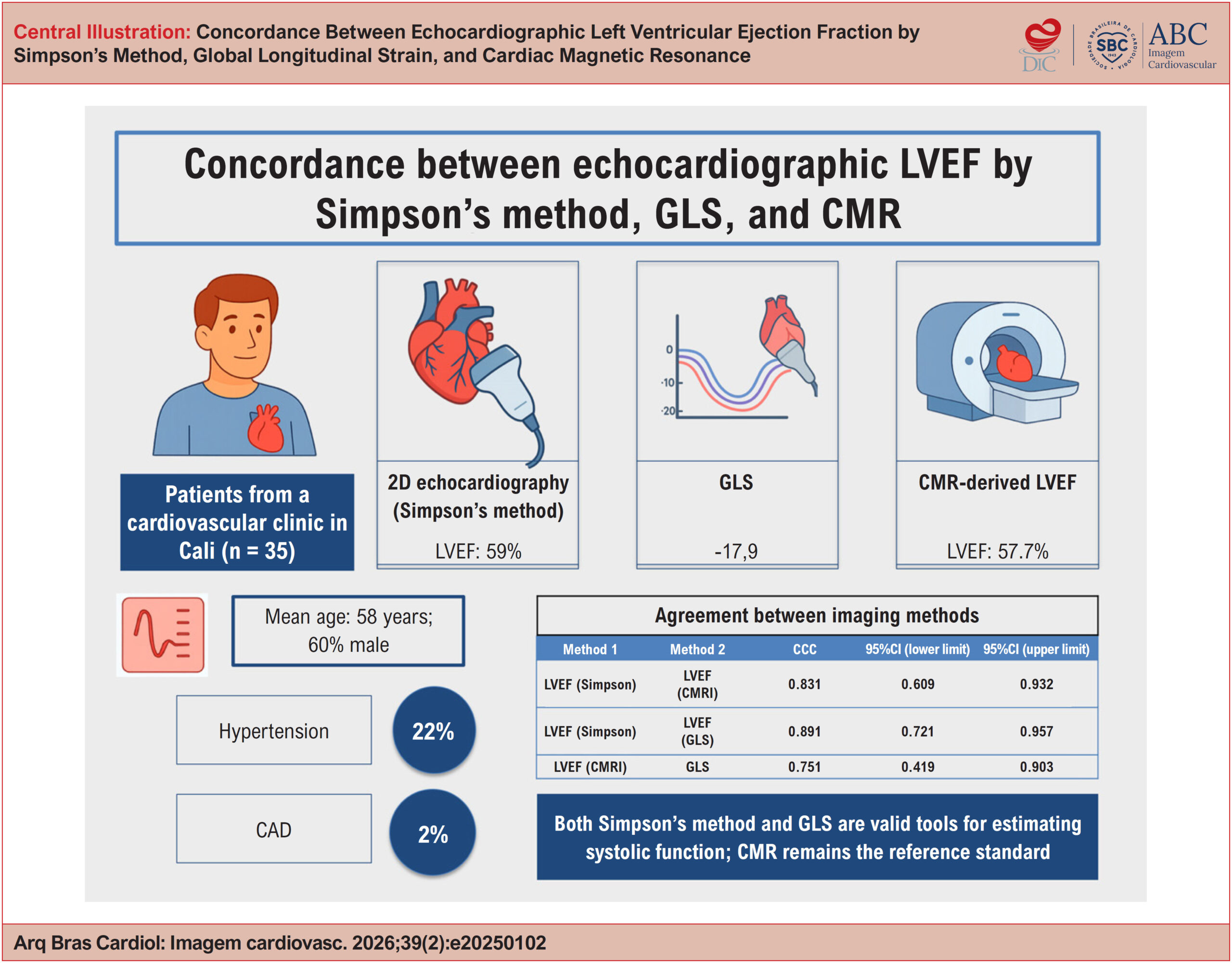
Concordance Between Echocardiographic Left Ventricular Ejection Fraction by Simpson's Method, Global Longitudinal Strain, and Cardiac Magnetic Resonance
ABCIC
Original Article

Concordance Between Echocardiographic Left Ventricular Ejection Fraction by Simpson's Method, Global Longitudinal Strain, and Cardiac Magnetic Resonance

Álvaro Herrera-Escandón et al. Juan Pablo Morales-Grisales Sebastián Ayala-Zapata Stephany Barbosa-Balaguera … Álvaro José Muriel-Ruiz Juan Felipe Bravo-Rueda José Eduardo Citelli-Ramírez Luis Fernando Osío-Jimenez Luis Miguel Benitez-Gómez Carlos Javier Ramírez-Estupiñán

Abstract

Background:

Left ventricular ejection fraction (LVEF) measured by echocardiography is a widely used parameter in clinical practice for the assessment of ventricular function. More recently, global longitudinal strain (GLS) has emerged as a complementary method, as has the assessment of LVEF by cardiac magnetic resonance (CMR). However, regional evidence evaluating the concordance among these three techniques remains limited.

Objectives:

To assess the concordance between echocardiographic and CMR measurements in patients treated at a cardiovascular clinic in the city of Cali, Colombia.

Methods:

This cross-sectional, analytical, descriptive study included 35 patients with confirmed or suspected heart disease, in whom all three methods were performed consecutively. Concordance was evaluated using Lin’s concordance correlation coefficient (CCC), Bland-Altman plots for LVEF, and linear and quadratic weighted κ coefficients for agreement between LVEF classifications.

Results:

The mean age was 58 years, and 60% of participants were male. The most common comorbidities were hypertension (22%) and dyslipidemia (11%). The mean LVEF was 59% by Simpson’s method and 57.7% by CMR, while the mean GLS was −17.7%. Concordance was as follows: Simpson’s LVEF vs. CMR (CCC, 0.831; 95%CI, 0.609-0.932); GLS vs. CMR-derived LVEF (CCC, 0.751; 95%CI, 0.419-0.903); and Simpson’s LVEF vs. GLS (CCC, 0.891; 95%CI, 0.721-0.957).

Conclusions:

Both Simpson’s method and GLS are valid tools for estimating systolic function. CMR remains the reference standard.

  • Echocardiography
  • Magnetic Resonance Spectroscopy
  • Stroke Volume
Read Full Article
Creative Commons Attribution (CC BY) Creative Commons license
24/07/2026 DOI: 10.36660/abcimg.20250102i Views 1
Echocardiographic Assessment of Diastolic Dysfunction in Special Situations
ABCIC
Review Article

Echocardiographic Assessment of Diastolic Dysfunction in Special Situations

Antonio Amador Calvilho Júnior

Abstract

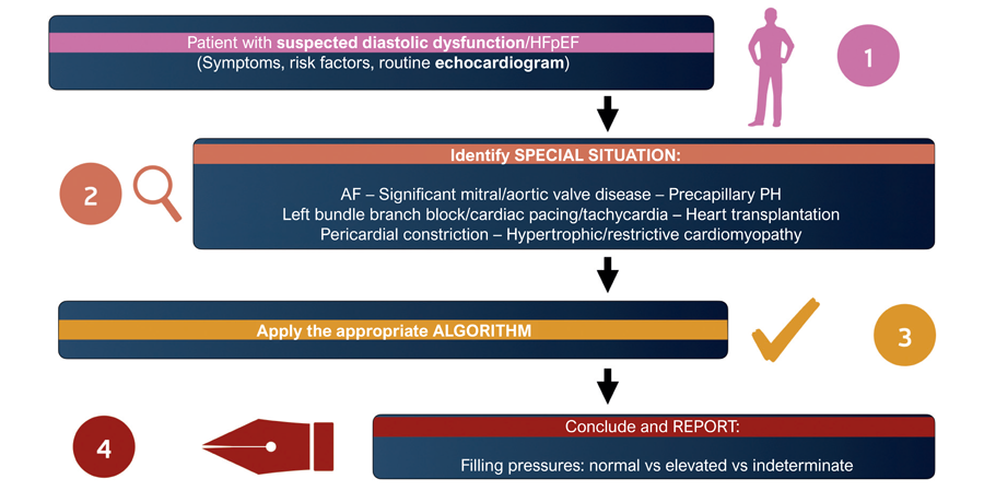
Echocardiographic assessment of diastolic function and left ventricular (LV) filling pressures is fundamental to the evaluation of dyspnea and the management of heart failure. However, conventional algorithms have limitations in several special clinical settings in which rhythm disturbances, valvular heart disease, pulmonary hypertension (PH), or structural cardiac abnormalities interfere with the interpretation of Doppler parameters. This article presents a practical approach to assessing diastolic function in conditions such as atrial fibrillation (AF), PH, mitral valve disease, mitral annular calcification, aortic valve disease, conduction disturbances, ventricular pacing, and restrictive cardiomyopathies. The main parameters applicable to each context, the technical aspects of image acquisition, and strategies for preparing a clear and clinically useful echocardiographic report are discussed.

  • Diastolic Heart Failure
  • Doppler Echocardiography
  • Stroke Volume
Read Full Article
Creative Commons Attribution (CC BY) Creative Commons license
17/07/2026 DOI: 10.36660/abcimg.20260029i Views 1
Figure 1
ABC
Original Article

Clinical and Echocardiographic Particularities of Hypertrophic Cardiomyopathy in a Brazilian Population and its Prognostic Impact

Georgina Del Cisne Jadán Luzuriaga et al. Edmundo Arteaga-Fernandez Viviane Tiemi Hotta Barbara Ianni … Luciano Nastari Felix Ramires Guilherme Wesley Peixoto da Fonseca Charles Mady Fábio Fernandes Juliano Novaes Cardoso

Abstract

Background

Hypertrophic cardiomyopathy (HCM) presents echocardiographic abnormalities that are important for diagnosis and prognosis. Data are scarce in the Brazilian literature.

Objective

To assess clinical and echocardiographic characteristics and disease progression in a Brazilian cohort of patients with HCM.

Methods

This retrospective cohort included patients with HCM aged ≥ 18 years. Patients with moderate or severe aortic stenosis and those undergoing septal reduction were excluded. The significance level adopted in the statistical analysis was 5%.

Results

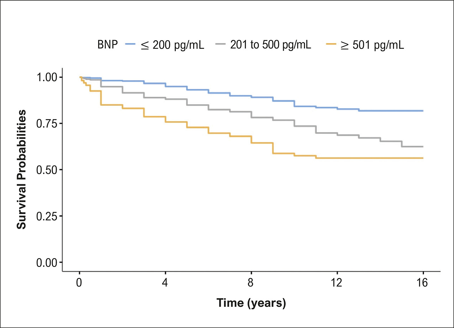
The study included 1244 patients, between 2010 and 2022, with a mean follow-up time of 7.7 ± 4.5 years; 53.6% of patients were men. Mean age was 54.6 ± 16.5 years, and mean left ventricular ejection fraction (LVEF) was 65.8% ± 7.6. We observed LVEF ≤ 50% in 5.8% of patients, asymmetric form in 88.7%, and septal hypertrophy in 85.4%. We found systolic anterior motion of the mitral valve in 30.1% of patients, left ventricular outflow tract obstruction in 30.7%, and septum ≥ 28 mm in 7.2%. Only 1 patient had ventricular aneurysm. Atrial fibrillation/flutter occurred in 9.6% of patients. Overall mortality occurred in 232 patients (1.3%/year). Patients with B-type natriuretic peptide (BNP) > 200 pg/ml, left atrium ≥ 45 mm, and LVEF ≤ 50% had higher mortality (p < 0.001). Age and atrial fibrillation/flutter were also associated with mortality.

Conclusions

The majority of patients had LVEF > 50%, asymmetric hypertrophy, and septal predominance. BNP, LA diameter, LVEF ≤ 50%, age, and atrial fibrillation/flutter were associated with worse prognosis.

  • Echocardiography
  • Hypertrophic Cardiomyopathy
  • Mortality
  • Stroke Volume
Read Full Article
Creative Commons Attribution (CC BY) Creative Commons license
12/05/2025 DOI: 10.36660/abc.20240640i Views 1
Figure 1
ABCHF
Original Article

Characteristics and Prognosis of Patients with Acute Heart Failure According to the Universal Ejection Fraction Classification

Michelle Bozko Collini et al. Gustavo Sarot Pereira da Cunha Leonardo Henrique dos Santos de Melo Matheus Bissa Duarte Ferreira … Jorge Tadashi Daikubara Neto Rafael Moretti Carolina Ruschel Senger Karoline Cordeiro Vercka Jessica Tamires Reichert Lucas Muller Prado Jamilly Giuriatti Anziliero Eduardo Leal Adam Raphael Henrique Déa Cirino Miguel Morita Fernandes-Silva

Abstract

Background:

The universal classification of heart failure (HF) defined four categories based on ejection fraction (EF). Although it has been shown these EF-based categories have distinct prognoses and responses to treatment, most of the data has been focused on chronic HF.

Objectives:

We compared the prognosis and characteristics of patients with acute HF according to the universal EF classification.

Methods:

We performed a prospective cohort study of patients admitted to a tertiary hospital for acute HF. Patients were classified into the four categories of the universal EF classification: reduced (HFrEF), mildly reduced (HFmrEF), preserved (HFpEF), and improved (HFimpEF), based on an echocardiogram conducted during the hospitalization. The primary outcome was all-cause death in six months of follow-up.

Results:

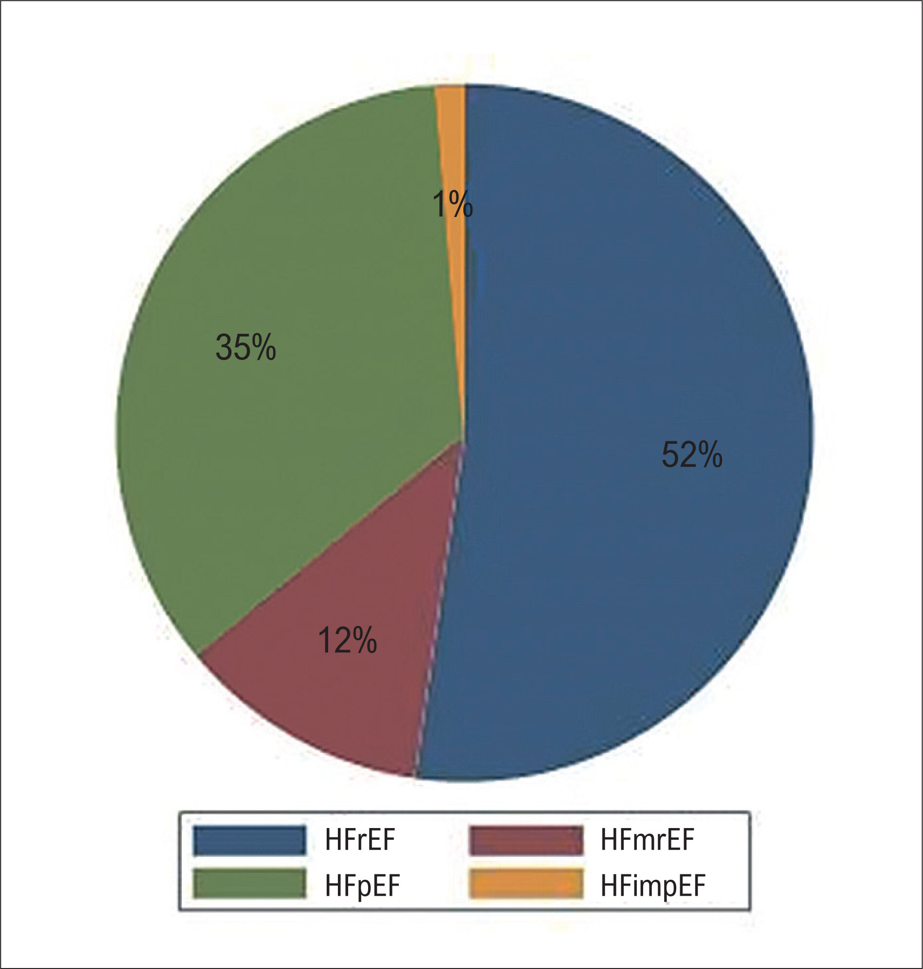
153 patients hospitalized for acute HF (67.2 ± 14.9 years, 50.3% female, EF = 43.8 ± 17.6%) were included, being 52% HFrEF, 35% HFpEF, 12% HFmrEF, and 1% HFimpEF. HFrEF patients were more likely to have an ischemic etiology (42.5%), while HFpEF patients were more likely female (67.3%), had hypertension (90.9%) and atrial fibrillation (49.1%). Six-month mortality was similar among HFrEF, HFpEF, and HFmrEF categories (15% vs 20% vs 11%, respectively, Log-rank p = 0.75), and change in EF from a previous echocardiogram was not associated with outcomes.

Conclusion:

In patients with acute HF, the EF categories from the universal classification had similar mortality rates. The proportion of patients with improved EF was very small in patients with acute HF and improvement of EF was not associated with better outcomes.

  • Heart Failure
  • Prognosis
  • Stroke Volume
Read Full Article
Creative Commons Attribution (CC BY) Creative Commons license
18/10/2024 DOI: 10.36660/abchf.20240035i Views 2
Figure 1
ABC
Editorial

Advanced Heart Failure and Onset of New Prognostic Markers: Where are We?

Meliza Goi Roscani

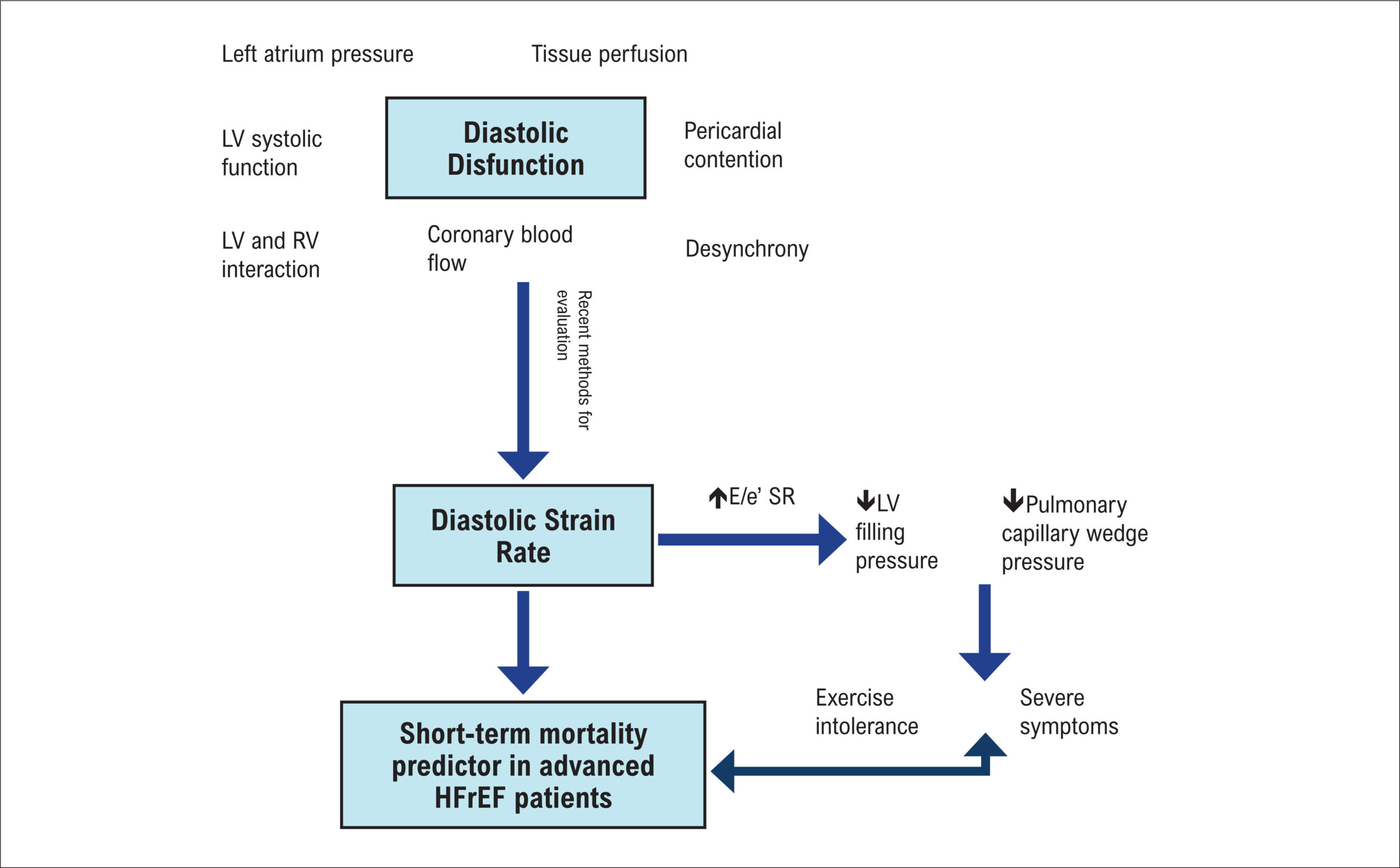
Advanced heart failure (HF) is a complex syndrome characterized by the presence of severe and persistent symptoms of HF, important cardiac dysfunction, episodes of pulmonary or systemic congestion requiring high-dose of intravenous diuretics, episodes of low output requiring inotropes or vasoactive drugs, malignant arrhythmias, hospitalization in the past 12 months or severe impairment of exercise capacity. The utility of multiple markers that have good accuracy in the detection of worse prognosis are increasingly being explored in the literature. Brain natriuretic peptide (BNP) and NT-proBNP are considered biomarkers prognostic indicators in HF and can help to identify patients with more severity of disease.

Recently, the onset of new echocardiographic markers appears to play an important role in detecting patients with more severe progression of this syndrome. Various parameters derived from the innovative speckle tracking echocardiography (STE) technique are of great value in the diagnostic and prognostic of several pathologies.–

[…]

  • Biomarkers
  • Heart Failure
  • Prognosis
  • Stroke Volume
Read Full Article
Creative Commons Attribution (CC BY) Creative Commons license
23/09/2024 DOI: 10.36660/abc.20240453i
Figure 1
ABCIC
Review Article

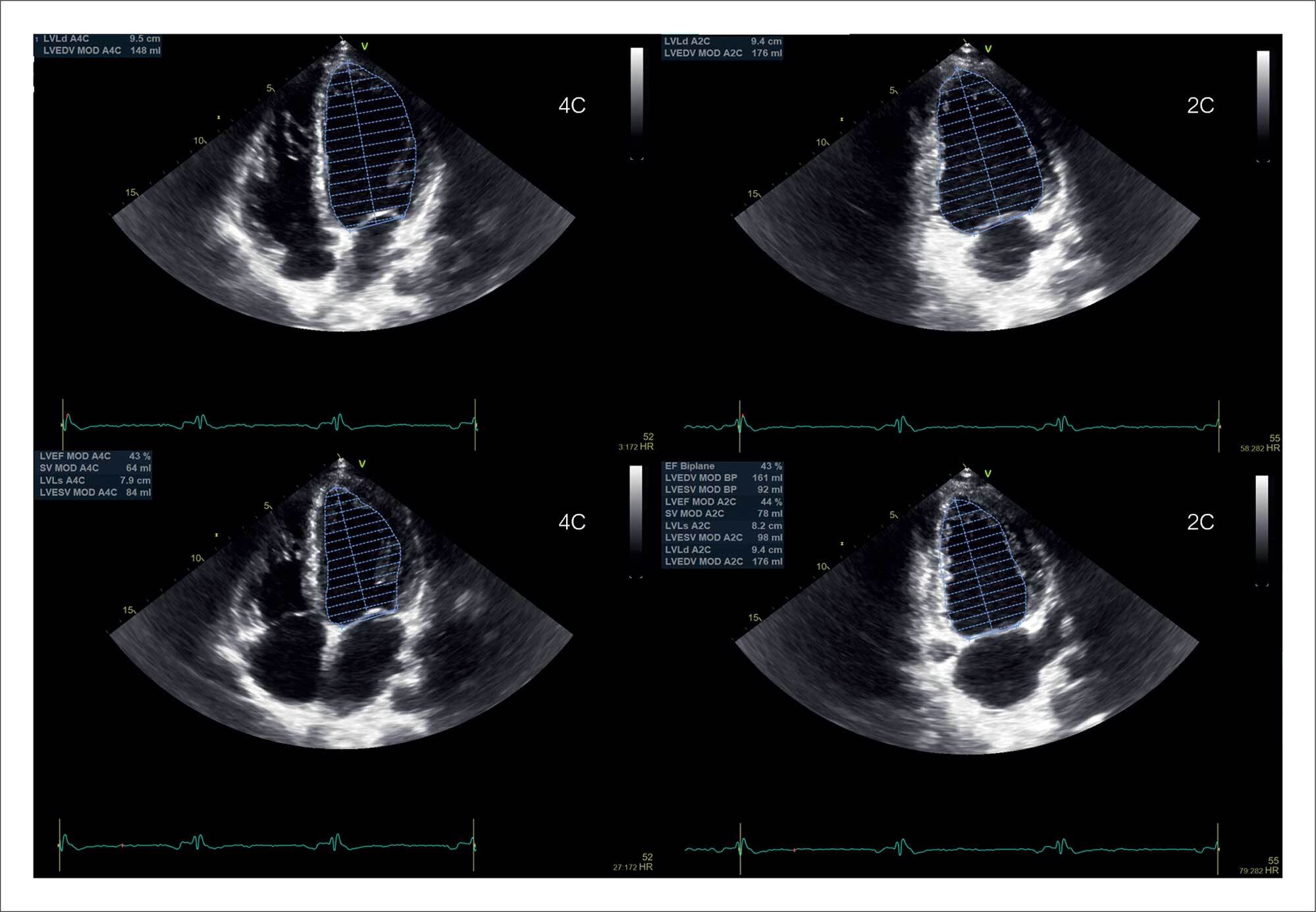
My Approach to Calculate the Left Ventricular Ejection Fraction: From a Two-Dimensional to a Three-Dimensional Technique

Lucas Arraes de França et al. Danilo Bora Moleta

Abstract

Echocardiography is the most used diagnostic method for measuring left ventricular systolic function indices. It is possible to estimate, in a quick, available, and non-invasive way, parameters such as ventricular volumes, stroke volume, and ejection fraction. Being an expert in the technique is quite important to estimate these parameters accurately. Being able to refine the technique constantly is also important, and this is closely associated with the examiner’s experience and knowledge of the current software solutions.

In this review article, we will discuss how to accurately measure left ventricular volumes and their ejection fraction using two-dimensional (2D) and three-dimensional (3D) echocardiographic techniques. We will also outline tips and suggestions based on the main North American and European guidelines for quantifying heart chambers and the most recent articles in the literature.

  • Echocardiography
  • Stroke Volume
  • Three-Dimensional Echocardiography
Read Full Article
Creative Commons Attribution (CC BY) Creative Commons license
29/08/2024 DOI: 10.36660/abcimg.20240059i
IJCS
Editorial

HFrEF, HFmrEF, and HFpEF: Are They a Continuum or Different Faces of a Syndrome?

Even Edilce Mol et al. Camila Hartmann Lídia Ana Zytynski Moura

Heart failure (HF) is a significant and growing public health problem. Recent projections have shown that the prevalence of HF in the United States will increase by 46% between 2012 and 2030. This is driven by an increase in the life expectancy of the population combined with a reduction in mortality from the disease resulting from advances in therapies in recent years.

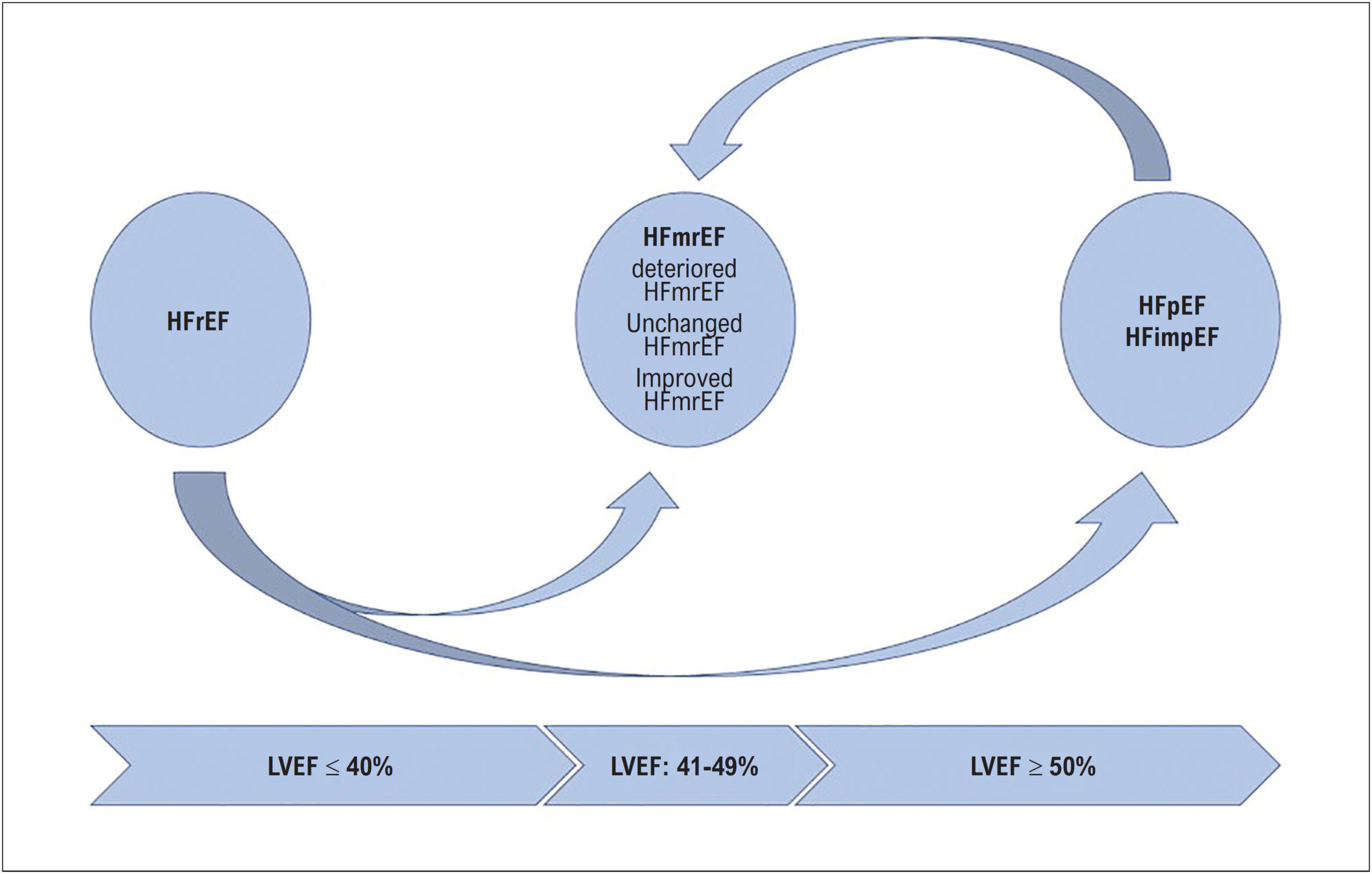
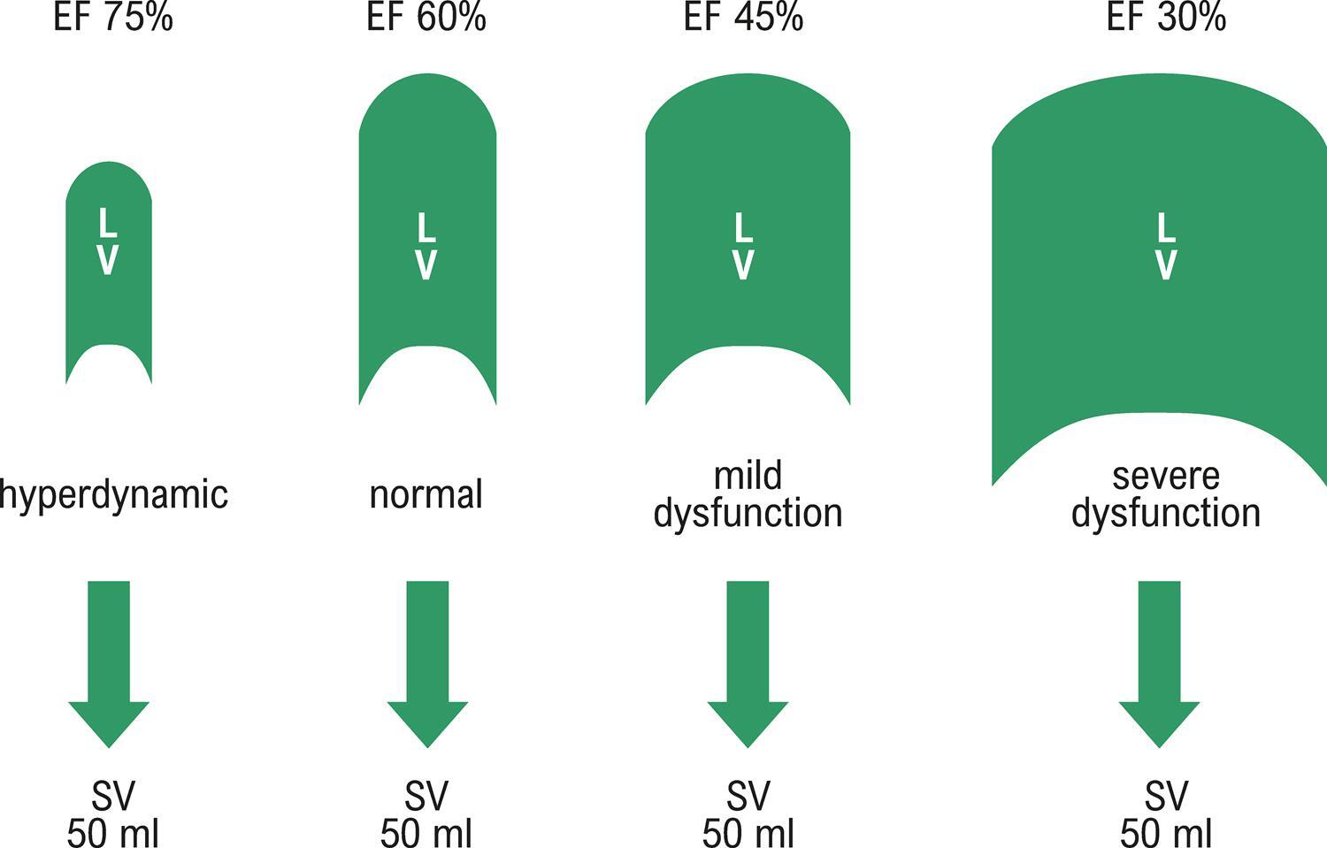
Of all the classifications of HF, the most used one is based on left ventricular ejection fraction (LVEF), which is categorized into three groups: HF with preserved ejection fraction (HFpEF) (LVEF ≥50%), HF with reduced ejection fraction (HFrEF) (LVEF ≤40%), and HF with moderate or mildly reduced ejection fraction (HFmrEF) (LVEF 41–49%). Although the prognosis appears to be similar among HF subtypes based on ejection fraction, , there are differences between the groups in terms of clinical-epidemiologic profile and functional capacity.

[…]

  • Cardiac Rehabilitation
  • Heart Failure
  • Stroke Volume
Read Full Article
Creative Commons Attribution (CC BY) Creative Commons license
07/06/2024 DOI: 10.36660/ijcs.20240048
Selvester QRS Score is a Predictor of Mortality in Heart Failure with Preserved Ejection Fraction
ABC
Original Article

Selvester QRS Score is a Predictor of Mortality in Heart Failure with Preserved Ejection Fraction

Fatih Sivri et al. Yahya Kemal Icen Hasan Koca Mükremin Coşkun … Mustafa Ardınç Orshan Deniz Fatih Necip Arici Mevlüt Koc Hasan Güngör

Abstract

Background

The Selvester QRS (S-QRS) score on a 12-lead electrocardiogram (ECG) is associated with both the amount of myocardial scar and poor prognosis in myocardial infarction patients. However, its prognostic value in heart failure (HF) with preserved ejection fraction (HFpEF) is unknown.

Objective

This study aims to investigate the predictive value of the S-QRS score for mortality in HFpEF.

Methods

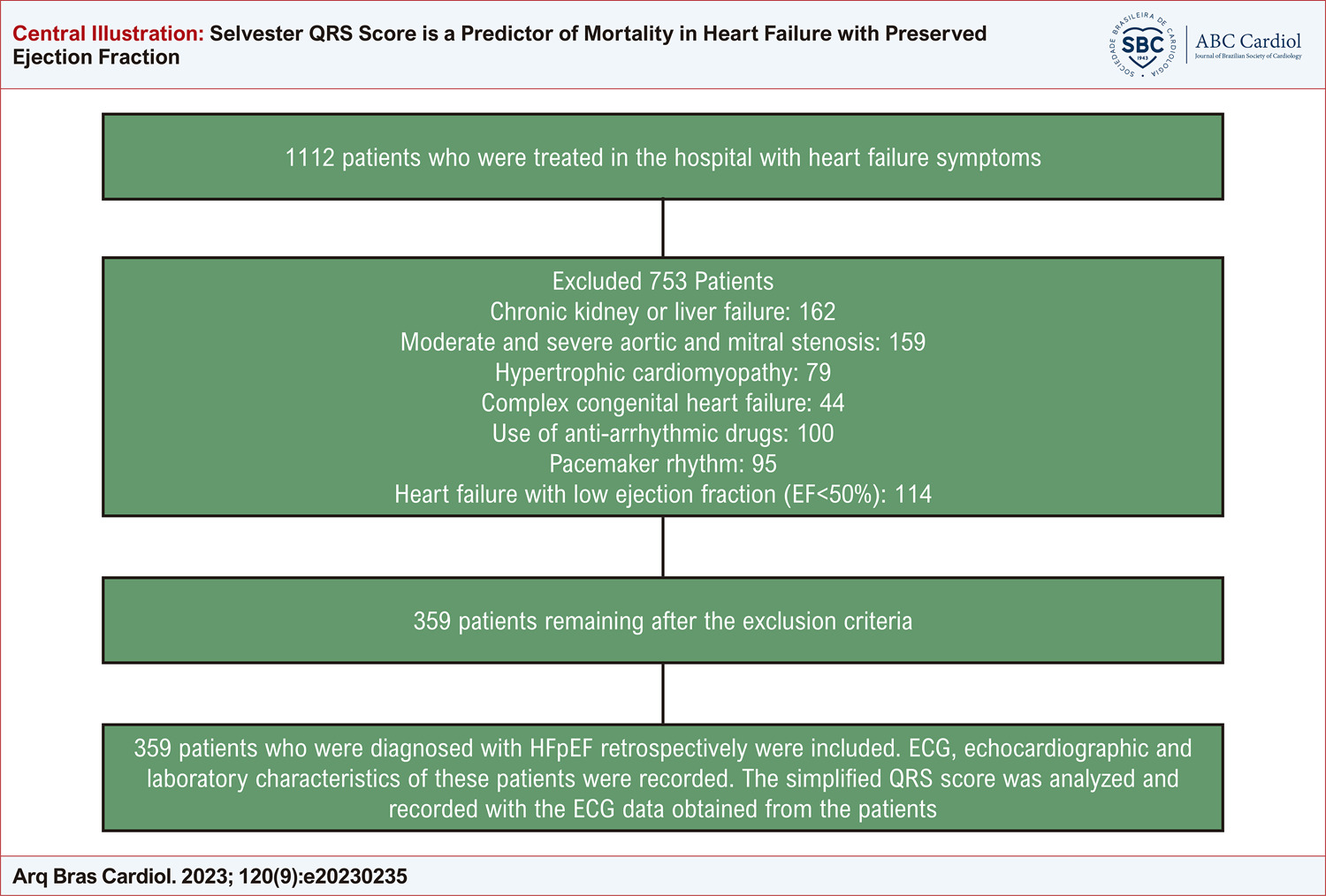
359 patients were retrospectively enrolled in this study. Electrocardiographic, echocardiographic, and laboratory features of the patients were recorded. The simplified S-QRS score was measured and recorded. The mean follow-up time of the patients was 38.1±9.5 months. Statistical significance was set at p < 0.05.

Results

Of 359 patients, 270 were in the survivor group, and 89 were in the deceased group. Age, Hs-CRP, troponin, pro-BNP, left atrial (LA) diameter, LA volume index, QRS duration, Tpe, and S-QRS score were statistically high in the deceased group. In multivariate logistic regression analysis, age, Hs-CRP, NT-proBNP, LA diameter, LA volume index, Tpe, and S-QRS score were shown to be independent risk factors for mortality. In the receiver-operating characteristic (ROC) analysis, the cut-off value of the S-QRS score was 5.5, the sensitivity was 80.8%, and the specificity was 77.2% (AUC:0.880, p:0.00). In Kaplan-Meier analysis, it was found that mortality was higher in the group with S-QRS score ≥ 5.5 than in the group with S-QRS score < 5.5. (Long-rank, p:0.00)

Conclusions

We think that the S-QRS score can be used as a prognostic indicator of long-term mortality in patients with HFpEF.

  • Echocardiography/methods
  • Electrocardiography/methods
  • Heart Failure
  • Mortality
  • Myocardial Infarction
  • Selvester QRS
  • Stroke Volume
Read Full Article
Creative Commons Attribution (CC BY) Creative Commons license
09/10/2023 DOI: 10.36660/abc.20230235 Views 1
ABC
Editorial

The Challenge of Making Cardiac Resonance a Global Reality

Hélder Jorge Andrade Gomes et al. Alcides Rocha de Figueredo Junior

Despite substantially reducing age-adjusted cardiovascular mortality rates in recent decades, cardiovascular disease remains the most common cause of death. Studies estimate that sudden cardiac death is responsible for approximately 50% of all deaths from cardiovascular causes, with ventricular tachycardia with degeneration to ventricular fibrillation and asystole being the most common fatal sequence of pathophysiological events.

The hard work done by authors of the article published in the Arquivos Brasileiros de Cardiologia illustrates the role of cardiac magnetic resonance (CMR) in the etiological elucidation of sudden death events encountered in clinical practice. In addition to the already expected Chagas and ischemic diseases, a wide variety of other diagnoses have been recognized: myocarditis, hypertrophic cardiomyopathy, dilated cardiomyopathy, arrhythmogenic heart disease, muscular dystrophy, hypertensive cardiomyopathy, metastases, non-compacted myocardium, and adrenergic cardiomyopathy (Takotsubo syndrome). The CMR was conclusive in more than 90% of cases and is a key tool for excluding structural heart disease and attributing the fatal event to extracardiac causes.

[…]

  • Cardiomyopathes
  • Cardiovascular Diseases, mortality
  • Death,Sudden
  • Diagnostic Imaging/trends
  • Endomyocardial Fibrosis
  • Magnetic Resonance Spectroscopy
  • Myocardial Ischemia
  • Stroke Volume
Read Full Article
Creative Commons Attribution (CC BY) Creative Commons license
21/04/2023 DOI: 10.36660/abc.20230187 Views 2
Figure 1
ABC
Research Letter

Ten-Year Survival of Heart Failure Patients with Left Ventricular Ejection Fraction of 40-59%: A Potential Phenotypic Classification?

Luiz Claudio Danzmann et al. Luiz Carlos Bodanese Aline Petracco Petzold Anna Paula Tscheika … Ellen Hettwer Magedanz Lucas Celia Petersen Evgeny Belyavskiy Fernanda Lourega Chieza

Abstract

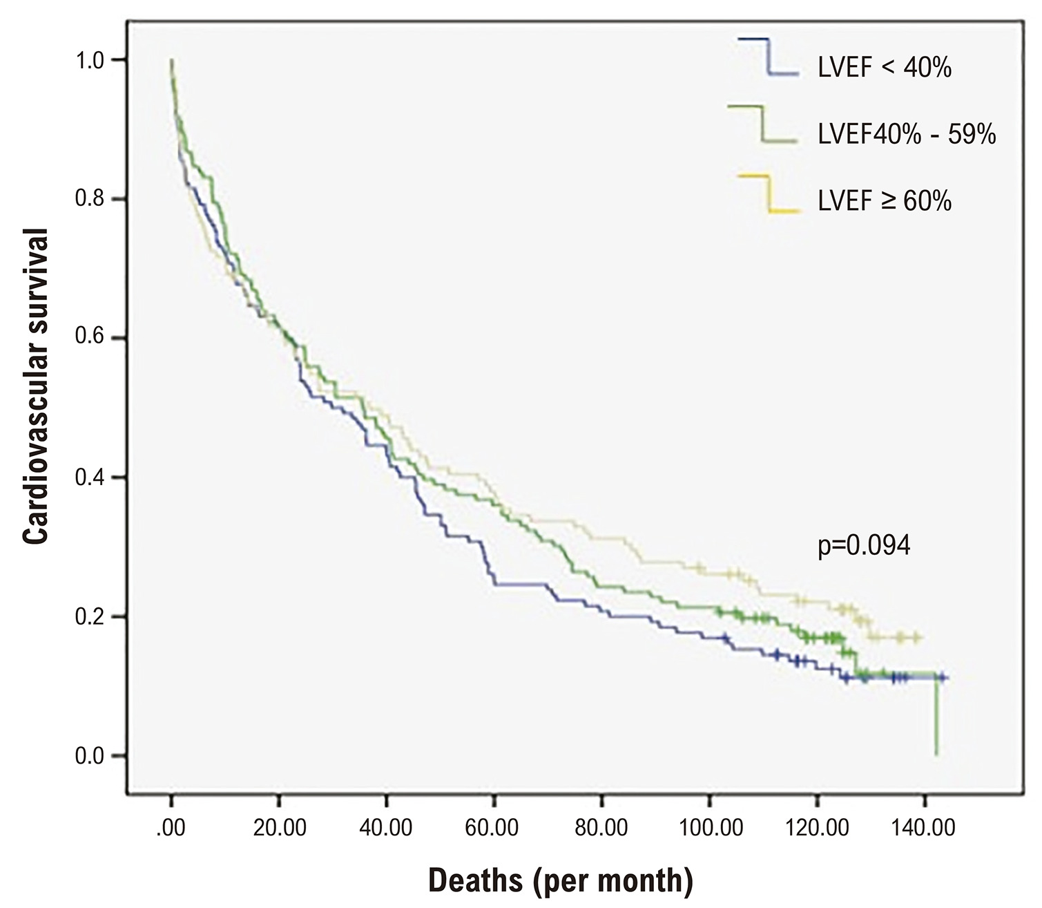
Definitions of left ventricular ejection fraction (LVEF) cut-off values for HF with mildly reduced LVEF (HFmrEF) have been a subject of debate, in the face of evidence that some drugs used in the treatment of HF with LFEV < 40% (HFrEF) are also effective in patients with LVEF < 60%. The aim of this study was to compare overall survival and cardiovascular survival in HF patients with LVEF of 40-59% in patients with HFrEF and HF with LVEF ≥ 60%. Patients with decompensated HF who met the Framingham diagnostic criteria at hospital admission between 2009 and 2011 were included. Patients were divided into HFrEF, HF with LVEF 40-59%, and HF with LVEF ≥ 60%. The Kaplan-Meier was used to determine ten-year overall survival and cardiovascular survival. The statistical significance was established at p<0.05. A total of 400 patients were included, with a mean age of 69 ± 14 years. Cardiovascular survival in patients with HF and LVEF of 40-59% was not significantly different than in patients with HFrEF (adjusted Hazard Ratio [HR] 0.86; 95% Confidence Interval [CI] 0.61-1.22, Ptrend = NS), but was statistically different compared with patients with LVEF ≥ 60% (adjusted HR of 0.64; 95% CI 0.44-0.94, Ptrend = 0.023). No difference was found in 10-year survival between the LVEF groups. Patients with HF and LVEF ≥ 60% had significantly higher cardiovascular survival compared with the other groups.

  • Heart Failure
  • Mortality
  • Stroke Volume
  • Survival
Read Full Article
Creative Commons Attribution (CC BY) Creative Commons license
13/02/2023 DOI: 10.36660/abc.20210772
ABC
Editorial

Focusing on the Right Ventricle in PRKAG2 Syndrome

Luís Rocha Lopes

The PRKAG2 gene encodes the adenosine monophosphate-activated protein kinase (AMPK) gamma 2 regulatory subunit. AMPK has a central role in cellular energy homeostasis. Pathogenic variation in PRKAG2 causes an autosomal dominant syndrome comprising ventricular hypertrophy, supraventricular arrhythmias, electrocardiographic pre-excitation and conduction system abnormalities.– The genetic background of this syndromic presentation was discovered in 2001.. Histologically there is myocyte glycogen accumulation, and the typical pattern of hypertrophy is usually described as biventricular and concentric, similar to other metabolic phenocopies of hypertrophic cardiomyopathy (HCM), with systolic dysfunction as another possible “red-flag” feature. The more frequent pathogenic variants are p.Arg302Gln and p.Asn488Ile. Due to the condition’s rarity, most publications consist of small case series or case reports, with few exceptions.. None of the previous publications specifically report right ventricular imaging findings in this condition.

Expanding on their previous work, where 3D echocardiography and strain imaging findings of the left ventricle were described in a cohort of 30 patients with PRKAG2 syndrome, Pena et al. provide a short report focusing on the right ventricle (RV), using the same cohort, in the current edition of this Journal.

[…]

  • Echocardiography/methods
  • Hypertrophy
  • Myocardial Deformability
  • PRKAG2 Syndrome
  • Right ventricle
  • Stroke Volume
Read Full Article
Creative Commons Attribution (CC BY) Creative Commons license
19/12/2022 DOI: 10.36660/abc.20220795 Views 1
ABCHF
Viewpoint

HFpEF: Evidence from Recent Clinical Trials and New Perspectives

José Francisco Kerr Saraiva et al. Nathalia dos Reis de Moraes

Heart failure (HF) is a highly prevalent clinical syndrome affecting more than 10% of the population older than 70 years of age, with a great impact on morbidity and mortality., HF is traditionally divided into three categories based on the left ventricular ejection fraction (EF): HF with reduced EF (EF50%; HFpEF).

HFpEF represents nearly half of HF cases in developed countries; its pathophysiology is complex and still little understood. The great variety of HFpEF phenotypes, makes its diagnosis and treatment challenging. Unlike HFrEF, before the emergence of SGLT-2 inhibitors, no treatment had been shown effective in reducing the hard outcomes in HFpEF. Also, the wide variability in diagnostic criteria of HFpEF adopted in different clinical trials may have contributed to the difficulty in showing therapeutic effectiveness in reducing mortality.

[…]

  • Heart Failure
  • Mortality
  • Stroke Volume
Read Full Article
Creative Commons Attribution (CC BY) Creative Commons license
01/12/2022 DOI: 10.36660/abchf.20220071 Views 3
Figure 1
ABC
Original Article

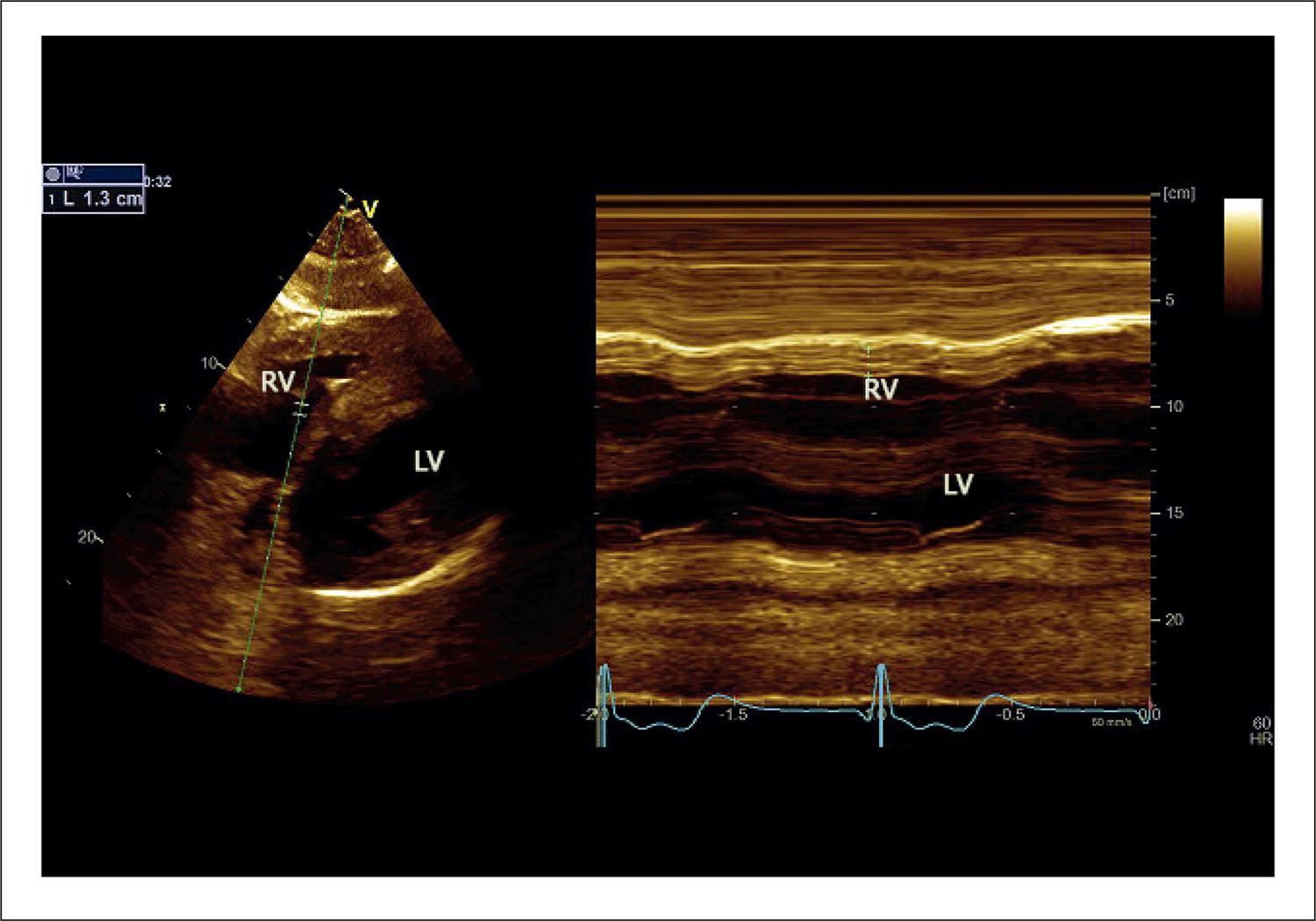
Right Ventricle Involvement by Glycogen Storage Cardiomyopathy (PRKAG2): Standard and Advanced Echocardiography Analyses

José Luiz Barros Pena et al. Fabricio Junqueira de Melo Wander Costa Santos Isabel Cristina Gomes Moura … Gabriela Pansanato Nakashima Natalia Costa Freitas Eduardo Back Sternick

Abstract

Background

PRKAG2 syndrome is a rare, early-onset autosomal dominant inherited disease. We aimed to describe the right ventricle (RV) echocardiographic findings using two and three-dimensional (2D and 3D) modalities including myocardial deformation indices in this cardiomyopathy. We also aimed to demonstrate whether this technique could identify changes in RV function that could distinguish any particular findings.

Methods

Thirty patients with genetically proven PRKAG2 (R302Q and H401Q), 16 (53.3%) males, mean age 39.1 ± 15.4 years, underwent complete echocardiography examination. RV-focused, 4-chamber view was acquired for 2D and 3D measurements. Student’s t or Wilcoxon-Mann-Whitney tests were used to compare numerical variables between 2 groups, and p < 0.05 was considered significant.

Results

Twelve patients (40%) had a pacemaker implanted for 12.4 ± 9.9 years. RV free wall mean diastolic thickness was 7.9 ± 2.9 mm. RV 4-chamber longitudinal strain (RV4LS), including the free wall and interventricular septum, was –17.3% ± 6.7%, and RV free wall longitudinal strain (RVFWLS) was −19.1% ± 8.5%. The RVFWLS apical ratio measured 0.63 ± 0.15. Mean RV 3D ejection fraction (EF) was 42.6% ± 10.9% and below normal limits in 56.7% of patients. Positive correlation occurred between RV 3DEF, RV4LS, and RVFWLS, especially for patients without a pacemaker (p = 0.006).

Conclusion

RV involvement in PRKAG2 syndrome is frequent, occurring in different degrees. Echocardiography is a valuable tool in detecting RV myocardial abnormalities in this condition. The use of 2D RV4LS, RVFWLS, and 3DEF offers reliable indicators of RV systolic dysfunction in this rare, challenging cardiomyopathy.

  • Cardiomyopathy, Hypertrophic, Familial
  • Echocardiography/methods
  • Glycogen Storage Disease/complications
  • Hypertrophy, Right Ventricular
  • Pacemaker, Artificial
  • PRKAG2 Syndrome/genetics
  • Stroke Volume
Read Full Article
Creative Commons Attribution (CC BY) Creative Commons license
21/11/2022 DOI: 10.36660/abc.20210801 Views 1
ABC
Editorial

Measurement of PETCO2 at Anaerobic Threshold: A Best Prognostic Marker in Patients with Cardiac Resynchronization Therapy?

Anderson Donelli da Silveira et al. Maurício Pimentel

The cardiopulmonary exercise test (CPET) is a consolidated tool in the functional and prognostic assessment of patients with heart failure with reduced ejection fraction (HFrRF), being a cornerstone in the evaluation for the indication of advanced therapies in HFrEF. , Cardiac resynchronization therapy (CRT), in addition to reducing mortality, can improve cardiorespiratory fitness, leading to an increase in peak oxygen consumption (VO2peak) and a reduction in the slope of the respiratory equivalent CO2 ratio (VE /VCO2 slope). – In recent years, with the evolution of treatments, general mortality and the risk of the sudden death of patients with HFpEF have been reduced. , In this context, reviewing the prognosis and the values associated with a higher risk among the CPET variables in patients undergoing CRT becomes important.

The measurement of end-tidal carbon dioxide pressure (PETCO2) during CPET, both at rest, and at the first ventilatory threshold or anaerobic threshold (PETCO2LA), has a well-established prognostic value in heart failure. , The increase of dead space ventilation, caused by the impairment of the ventilation/perfusion (V/Q) ratio, for example, in patients with left ventricular dysfunction, leads to a reduction in alveolar CO2 and, consequently, in PETCO2. It is expected that there will be an increase in its measurement up to the anaerobic threshold, which is correlated with an increase in cardiac output.

[…]

  • Cardiac Output, High
  • Cardiac resynchronization Therapy/methods
  • Ergoespyrometry/methods
  • Heart Failure
  • Prognosis
  • Stroke Volume
Read Full Article
Creative Commons Attribution (CC BY) Creative Commons license
05/09/2022 DOI: 10.36660/abc.20220477 Views 1
ABC
Editorial

Utility of Biomarkers in Suspected Cardiac Amyloidosis: Opportunity for More Frequent and Early Diagnosis

João Marcos Barbosa-Ferreira et al. Andreza Araújo de Oliveira

The meta-analysis entitled “Diagnostic Role of NT-proBNP in Patients with Cardiac Amyloidosis Involvement: A Meta-Analysis” brings us an important review of the usefulness of NT-proBNP measurement in patients with cardiac involvement by amyloidosis. The demonstration of good sensitivity and specificity of this biomarker reinforces its usefulness in the diagnosis of cardiac amyloidosis (CA).

Cardiac amyloidosis has been increasingly diagnosed, especially in patients with the heart failure phenotype of preserved ejection fraction. Just over half of the patients with symptoms of heart failure have preserved ejection fraction, especially elderly individuals. This finding is usually considered only as age-related diastolic dysfunction and associated comorbidities. However, this factor should be one of the warning signs for diagnosing CA, especially when associated with high levels of biomarkers. A great variability in the frequency of diagnosis of CA in the general population has been described, ranging from 5 to 74% between the various studies. This variability may be related to factors such as low clinical suspicion or difficulties accessing complementary exams necessary for diagnosing cardiac involvement in amyloidosis. The diagnostic flowchart in patients with suspected cardiac involvement by amyloidosis is mainly based on imaging tests.– These tests can be expensive, such as myocardial scintigraphy, strain echocardiography and cardiac magnetic resonance, and, often, they are only available at cardiology referral centers, making the diagnosis of CA more difficult and delayed. In addition, it is important to note that the late diagnosis of these patients can directly influence the prognosis by delaying the start of treatment, leading to a median of 6 months of survival after the development of symptoms in the AL form of amyloidosis for example.

[…]

  • Amyloidosis, Cardiac
  • Biomarkers
  • diagnostic
  • Heart Failure
  • Imaging/methods
  • NT-proBNP
  • Stroke Volume
  • Ventricular Dysfunction
Read Full Article
Creative Commons Attribution (CC BY) Creative Commons license
05/08/2022 DOI: 10.36660/abc.20220437
Figure 1
ABC
Research Letter

Heart Failure with Mildly Reduced Ejection Fraction: Therapeutical Considerations and Reasons for This Renaming

Eduardo Thadeu de Oliveira Correia et al. Evandro Tinoco Mesquita

Introduction

Heart failure (HF) has been classically divided into HF with reduced ejection fraction (HFrEF) and HF with preserved ejection fraction (HFpEF). However, to better classify HF patients with a left ventricular ejection fraction (LVEF) between 41 and 49%, previous guidelines have introduced the term HF with mid-range ejection fraction. Nonetheless, shortly after its formal introduction, HF with mid-range ejection fraction is now called HF with mildly reduced ejection fraction (HFmrEF). In this letter, we explore the reasons behind this renaming and why this change is more important than it may seem.

[…]

  • Angiotensin-Converting Enzyme Inhibitors
  • Cardiovascular Diseases
  • Digoxin
  • Heart Failure
  • Mineralocortical Receptor Antagonists
  • Prognosis
  • Stroke Volume
Read Full Article
Creative Commons Attribution (CC BY) Creative Commons license
11/07/2022 DOI: 10.36660/abc.20210752 Views 4
Figure 1
ABCIC
What do Cardiologists Expect

What do Cardiologists Expect from the Echocardiogram in Heart Failure with Reduced Ejection Fraction?

Ciro Mancilha Murad et al. Fabiana Goulart Marcondes-Braga Danilo Bora Moleta

Introduction

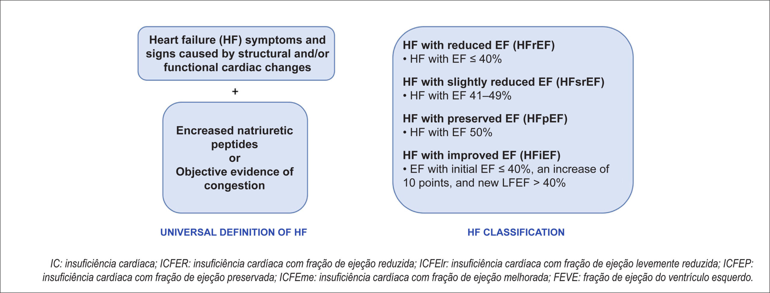
The echocardiogram plays a central role in assessments of patients with heart failure (HF), contributing to its classification, etiology definition, severity stratification, hemodynamic assessment, and clinical follow-up. HF has been historically divided according to the echocardiographic parameter left ventricular ejection fraction (LVEF). The most recent classification divided it into preserved (≥50%), reduced (≤40%), and slightly reduced LVEF (41–49%)., Different societies have recently proposed a universal definition of HF that involves the presence of symptoms and signs caused by structural and/or functional cardiac changes associated with increased natriuretic peptide levels or objective evidence of congestion. The authors also highlight the importance of classifying HF according to LVEF. () This article will describe the main parameters which the clinical cardiologist should pay attention on the echocardiogram of patients with HF with reduced ejection fraction (HFrEF) and how best to use them in clinical practice.

[…]

  • Echocardiography
  • Heart Failure
  • Stroke Volume
Read Full Article
Creative Commons Attribution (CC BY) Creative Commons license
13/06/2022 DOI: 10.47593/2675-312X/20223502ecard06 Views 2
ABC
Editorial

Is it Possible to Non-Invasively Study the Hemodynamic Adaptations of Chagas Cardiomyopathy by the Volume-Time Curve Using 3D Echocardiography?

José Luiz Barros Pena

Three-dimensional echocardiography (3DE) represents a great innovation in cardiovascular ultrasound. Increased computer processing power and advances in the development of transducers have allowed acquiring of cardiac structures from any spatial point of view, without assumptions about their shape. Recent studies have demonstrated that when cardiac chamber sizes are quantified using 3DE, their volumes are similar to those obtained using cardiac magnetic resonance imaging compared to two-dimensional echocardiography (2DE)., The usefulness of 3DE has particularly been demonstrated primarily in realistic anatomical heart valve images and in guiding and monitoring cardiac procedures.

3DE allows volume calculation of the left ventricle (LV) throughout the cardiac cycle, making it possible to construct a volume-time curve. This method is more accurate than 2DE because the left ventricular volume is constructed by analyzing hundreds of points at the edge of the endocardium. No specific plane or geometric model is necessary to describe the complex LV structure. In this paper, Pinto et al. tested the hypothesis of studying the hemodynamic adaptations of non-invasive Chagas cardiomyopathy using the volume-time curve generated by 3DE. They generated a polynomial adjusted to the LV volume curve using specific software. Their objective was to present a cross-sectional study evaluating LV function, comparing volume curves in 20 patients with Chagas cardiomyopathy (CC) and 15 gender- and age-matched healthy controls.

[…]

  • Blood Pressure
  • Chagas Caddiomyopathy
  • Echocardiography, Three Dimensional / methods
  • Heart Failure
  • Stroke Volume
Read Full Article
Creative Commons Attribution (CC BY) Creative Commons license
10/06/2022 DOI: 10.36660/abc.20220284 Views 1
Figure 1
ABC
Original Article

The Volume-Time Curve by Three-Dimensional Echocardiography in Chagas Cardiomyopathy: Insights into the Mechanism of Hemodynamic Adaptations

Airandes de Sousa Pinto et al. Maria Carmo Pereira Nunes Carlos Alberto Rodrigues Bráulio Muzzi Ribeiro de Oliveira … João da Rocha Medrado Neto Timothy C. Tan Manoel Otavio da Costa Rocha

Abstract

Background

Three-dimensional echocardiography (3D ECHO) allows the generation of a volume-time curve representative of changes in the left ventricular (LV) volume throughout the entire cardiac cycle.

Objective

This study aims to demonstrate the hemodynamic adaptations present in Chagas cardiomyopathy (CC) by means of the volume and flow measurements obtained by the volume-time curve by 3D ECHO.

Methods

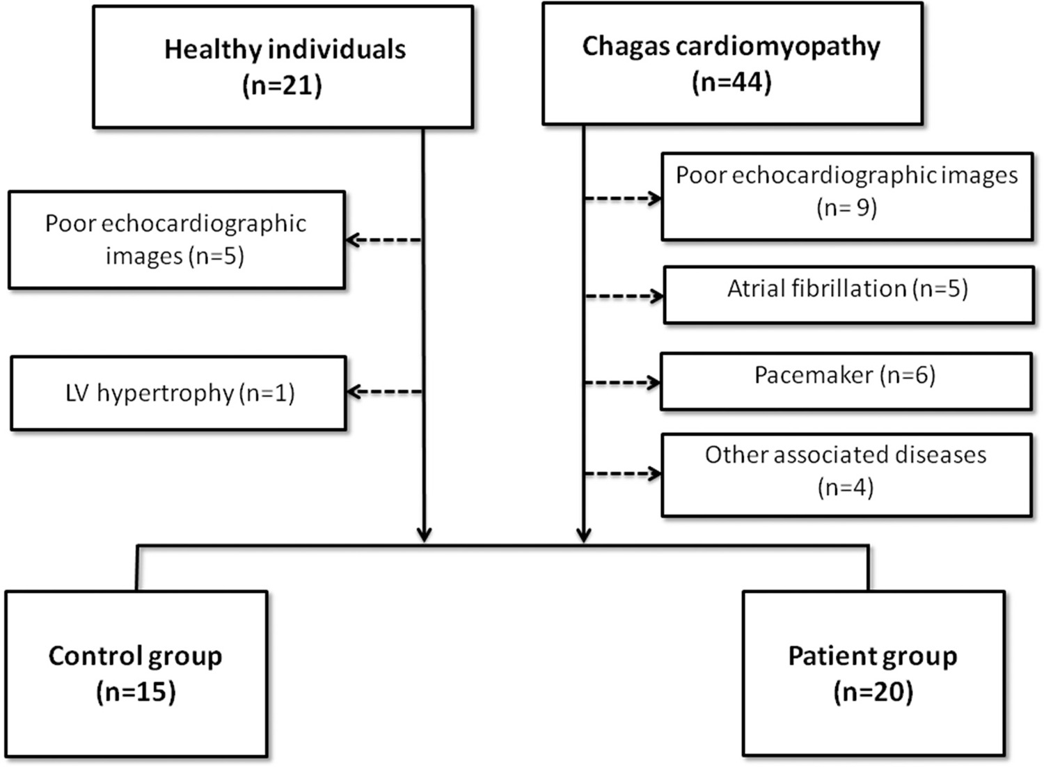
Twenty patients with CC and 15 healthy subjects were prospectively enrolled in a cross-sectional design study. 3D ECHO was performed in all subjects and the volume over time curves of the LV was generated. The flow was obtained by the first derivative of the volume-time curve using the software MATLAB. Statistical significance was set at p<0.05.

Results

Although CC patients had lower LV ejection fraction compared to the control group (29.8±7.5 vs. 57.7±6.1, p<0.001), stroke volume (61.5±25.2 vs. 53.8±21.0, p=0.364) and maximum ejection flow during systole (-360.3±147.5 vs. -305.6±126.0, p=0.231) were similar between the groups. Likewise, the maximum flow in the early diastolic filling phase and during atrial contraction was similar between groups. An increase in preload expressed by LV end diastolic volume (204.8±79.4 vs. 93.0±32.6), p<0.001) may maintain the flow and stroke volumes similar to the controls.

Conclusion

Using a non-invasive tool, we demonstrated that an increase in LV end-diastolic volume may be the main adaptation mechanism that maintains the flow and stroke volumes in the setting of severe LV systolic dysfunction.

  • Atrial Fibrillation
  • Chagas Cardiomyopathy
  • Echocardiography, Three Dimensional Echocardiogram
  • Frank-Starling Law
  • Stroke Volume
Read Full Article
Creative Commons Attribution (CC BY) Creative Commons license
02/05/2022 DOI: 10.36660/abc.20201308 Views 2
ABC
Editorial

Microalbuminuria and the Risk of Mortality in Patients with Acute Heart Failure

Jerzy Beltowski

Heart failure (HF) is a chronic and progressive clinical syndrome resulting from structural or functional abnormalities of the heart. The most common symptoms and signs of HF are dyspnea, fatigue, pulmonary and peripheral congestion or edema and jugular vein distension. The prevalence of HF continuously increases due to improved treatment and reduced short-term mortality in patients with acute coronary syndromes, congenital heart disease, population aging and improved survival of patients with already developed heart failure by the widespread application of modern disease-modifying medications and devices.

Acute heart failure (AHF) is recognized when symptoms of HF appear in the patient without the history of previous HF (de novo HF) or when symptoms and signs are rapidly exacerbating in a patient with previously recognized HF (decompensated HF). Acute heart failure is the most common cause of unplanned hospital admissions in older patients. The pathophysiology of both conditions is similar, but de novo HF requires a more detailed diagnostic approach to find the underlying pathology. The initial treatment of AHF includes intravenous diuretics and short-acting vasodilators. The minority of patients with AHF present with cardiogenic shock associated with low blood pressure and severely compromised perfusion of peripheral tissues; the cardiogenic shock is associated with much higher mortality than AHF without shock. Whereas the treatment of chronic HF has substantially improved survival rates, the outcome of AHF is still poor, with high mortality and hospital readmission rates. Currently used therapy is directed to reduce the pre and afterload of the heart and does not target the specific underlying pathology in a given patient, which may explain unsatisfactory progress of clinical outcomes. Therefore, individualized therapy is highly appreciated, requiring establishing specific markers.,

[…]

  • Albuminuria/physiopathology
  • Diuretics/therapeutic use
  • Heart Failure/physiopathology
  • Natriuretic Peptides/therapeutic use
  • Stroke Volume
Read Full Article
Creative Commons Attribution (CC BY) Creative Commons license
29/04/2022 DOI: 10.36660/abc.20220172
Figure 1
ABCHF
Viewpoint

Rethinking the Definition of Heart Failure Based on Ejection Fraction: Reflections with Impact on Therapy

Marcelo Iorio Garcia

The universal definition of heart failure (HF) has been in a state of constant change in the last decades. It is necessary for the definition to be simple, comprehensible, and easily applied in clinically, as well as capable of differentiating patients’ stages and severity, allowing stratification of levels of care, especially regarding candidates for specific therapies.

In relation to ejection fraction (EF), which is the subject of our reflection, the most recent document proposed the following subdivisions according to left ventricular ejection fraction (LVEF): HF with reduced ejection fraction (HFrEF), EF ≤ 40%; HF with midrange ejection fraction (HFmrEF), EF 41% to 49%; HF with preserved ejection fraction (HFpEF), EF ≥ 50%; and HF with improved EF (HFiEF), HF with EF ≤ 40% that has increased by at least 10 points, with the second measurement reaching > 40%.

[…]

  • Heart Failure
  • Stroke Volume
  • Therapeutics
Read Full Article
Creative Commons Attribution (CC BY) Creative Commons license
12/04/2022 DOI: 10.36660/abchf.20220016 Views 2
Figure 1
ABCHF
Review Article

Essential Therapy for Heart Failure with Preserved Ejection Fraction in 2022

Luiz Cláudio Danzmann et al. Joana Carolina Junqueira de Brum Leticia Kunst Priscila Braun Eduardo Lima Garcia

Abstract

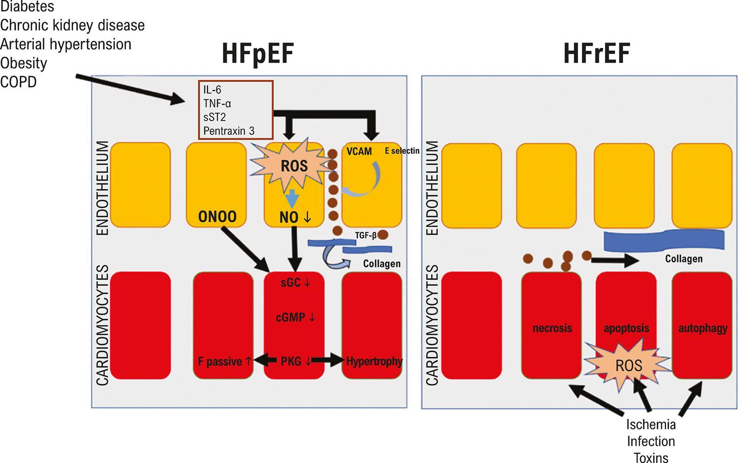
Heart failure with preserved ejection fraction (HFpEF) is a syndrome in which there is clinical evidence of heart failure (HF), left ventricle ejection fraction (LVEF) ≥50%, and evidence of diastolic dysfunction and/or structural cardiac changes. The pathophysiology of HF with preserved LVEF is related to the primary morbidities responsible for cardiac and vascular aggression via a chronic proinflammatory state involving the endothelium. Currently, the foundation of management of HFpEF rests on 5 pillars: control of circulatory congestion, management of primary morbidities or etiologies, use of medications with proven clinical benefit, identification and management of secondary etiologies, and cardiopulmonary rehabilitation. Essential therapy for HFpEF is founded on precise diagnosis, definition of etiology, estimation of severity, and use of medications with cardiovascular action of proven efficacy.

  • Functional Residual Capacity
  • Heart Failure
  • Stroke Volume
Read Full Article
Creative Commons Attribution (CC BY) Creative Commons license
12/04/2022 DOI: 10.36660/abchf.20220011 Views 2
IJCS
Original Article

Cognitive Alterations in Elderly Patients with Heart Failure’

Fernanda Guedes Afiune et al. Salvador Rassi Abrahão Afiune Neto

Abstract

Background:

Among the various pathologies that affect the elderly, Heart Failure (HF) stands out. Recently, an attempt has been made to verify the existence of cognitive impairment associated with HF.

Objectives:

To compare the cognitive performance of elderly people with heart failure with that of age-matched individuals without this pathology. Check the existence of marked impairment in some cognitive functions in the clinical group.

Methods:

The sample consisted of 78 elderly people, whose inclusion criterion was the presence of HF and no HF (control group); age over 60 years, both sexes, and any level of education. The control group consisted of 37 individuals (with a median age of 68 years – Interquartile range of 12) and the HF group, with 41 individuals (with a median age of 67 years – Interquartile range of 11). The subjects were matched in terms of education level, with a predominance of elderly people with 0 to 4 years of education (65.9% in the Clinical Group and 59.5% in the Control Group). Eleven neuropsychological tests covering cognitive functions were used: attention, language, memory, mood, and executive function. Statistical analysis was performed using SPSS software, version 23, with a significance level of 5%. The Chi-square test and the Mann-Whitney test were applied.

Results:

The results showed significant differences between the groups, mainly in executive functions, which include the ability to plan, switch, and recall previously stored information.

Conclusion:

Our study showed differences between the cognitive performance of elderly people with HF and elderly people without HF. The main alteration was found in the so-called executive functions, attention, and memory.

  • Aged
  • Anxiety
  • Cardiovascular Diseases
  • Cognition Disorders
  • Depression
  • Heart Failure
  • Neurological Diseases
  • Neuropsychology
  • Stroke Volume
Read Full Article
Creative Commons Attribution (CC BY) Creative Commons license
25/02/2022 DOI: 10.36660/ijcs.20200144 Views 1

Could not load more articles. Please try again.

How to cite this article

EndNote RIS

Electronic Document Format (Vancouver)

Uellendahl M. Beyond Ejection Fraction: Integrating Myocardial Strain and Cardiac Magnetic Resonance into the Contemporary Assessment of Left Ventricular Function. ABC Imagem Cardiovascular 2026;39(2):e20260087.

Electronic Document Format (ABNT)

Uellendahl, Marly. Beyond Ejection Fraction: Integrating Myocardial Strain and Cardiac Magnetic Resonance into the Contemporary Assessment of Left Ventricular Function. ABC Imagem Cardiovascular, v. 39, n. 2, e20260087, Jul. 2026.

Electronic Document Format (APA)

Uellendahl, M. (2026). Beyond Ejection Fraction: Integrating Myocardial Strain and Cardiac Magnetic Resonance into the Contemporary Assessment of Left Ventricular Function. ABC Imagem Cardiovascular, 39(2), e20260087.

Electronic Document Format (ISO)

Uellendahl, Marly. Beyond Ejection Fraction: Integrating Myocardial Strain and Cardiac Magnetic Resonance into the Contemporary Assessment of Left Ventricular Function. ABC Imagem Cardiovascular [online]. 2026, vol. 39, n. 2, [cited 2026-10-15], e20260087. Available from: <https://journals.cardiol.br/en/article/beyond-ejection-fraction-integrating-myocardial-strain-and-cardiac-magnetic-resonance-into-the-contemporary-assessment-of-left-ventricular-function/>. ISSN 2318-8219

EndNote RIS

Electronic Document Format (Vancouver)

Herrera-Escandón , Morales-Grisales JP, Ayala-Zapata S, Barbosa-Balaguera S, Muriel-Ruiz J, Bravo-Rueda JF, Citelli-Ramírez JE, et al. Concordance Between Echocardiographic Left Ventricular Ejection Fraction by Simpson’s Method, Global Longitudinal Strain, and Cardiac Magnetic Resonance. ABC Imagem Cardiovascular 2026;39(2):e20250102.

Electronic Document Format (ABNT)

Herrera-Escandón, Álvaro; Morales-Grisales, Juan Pablo; Ayala-Zapata, Sebastián; Barbosa-Balaguera, Stephany; Muriel-Ruiz, Álvaro José; Bravo-Rueda, Juan Felipe; Citelli-Ramírez, José Eduardo; Osío-Jimenez, Luis Fernando; Benitez-Gómez, Luis Miguel; Ramírez-Estupiñán, Carlos Javier. Concordance Between Echocardiographic Left Ventricular Ejection Fraction by Simpson’s Method, Global Longitudinal Strain, and Cardiac Magnetic Resonance. ABC Imagem Cardiovascular, v. 39, n. 2, e20250102, Jul. 2026.

Electronic Document Format (APA)

Herrera-Escandón, , Morales-Grisales, J. P., Ayala-Zapata, S., Barbosa-Balaguera, S., Muriel-Ruiz, J., Bravo-Rueda, J. F., Citelli-Ramírez, J. E., Osío-Jimenez, L. F., Benitez-Gómez, L. M., & Ramírez-Estupiñán, C. J. (2026). Concordance Between Echocardiographic Left Ventricular Ejection Fraction by Simpson’s Method, Global Longitudinal Strain, and Cardiac Magnetic Resonance. ABC Imagem Cardiovascular, 39(2), e20250102.

Electronic Document Format (ISO)

Herrera-Escandón, Álvaro and Morales-Grisales, Juan Pablo and Ayala-Zapata, Sebastián and Barbosa-Balaguera, Stephany and Muriel-Ruiz, Álvaro José and Bravo-Rueda, Juan Felipe and Citelli-Ramírez, José Eduardo and Osío-Jimenez, Luis Fernando and Benitez-Gómez, Luis Miguel and Ramírez-Estupiñán, Carlos Javier. Concordance Between Echocardiographic Left Ventricular Ejection Fraction by Simpson’s Method, Global Longitudinal Strain, and Cardiac Magnetic Resonance. ABC Imagem Cardiovascular [online]. 2026, vol. 39, n. 2, [cited 2026-10-15], e20250102. Available from: <https://journals.cardiol.br/en/article/concordance-between-echocardiographic-left-ventricular-ejection-fraction-by-simpsons-method-global-longitudinal-strain-and-cardiac-magnetic-resonance/>. ISSN 2318-8219

EndNote RIS

Electronic Document Format (Vancouver)

Calvilho Júnior AA. Echocardiographic Assessment of Diastolic Dysfunction in Special Situations. ABC Imagem Cardiovascular 2026;39(2):e20260029.

Electronic Document Format (ABNT)

Calvilho Júnior, Antonio Amador. Echocardiographic Assessment of Diastolic Dysfunction in Special Situations. ABC Imagem Cardiovascular, v. 39, n. 2, e20260029, Jul. 2026.

Electronic Document Format (APA)

Calvilho Júnior, A. A. (2026). Echocardiographic Assessment of Diastolic Dysfunction in Special Situations. ABC Imagem Cardiovascular, 39(2), e20260029.

Electronic Document Format (ISO)

Calvilho Júnior, Antonio Amador. Echocardiographic Assessment of Diastolic Dysfunction in Special Situations. ABC Imagem Cardiovascular [online]. 2026, vol. 39, n. 2, [cited 2026-10-15], e20260029. Available from: <https://journals.cardiol.br/en/article/echocardiographic-assessment-of-diastolic-dysfunction-in-special-situations/>. ISSN 2318-8219

EndNote RIS

Electronic Document Format (Vancouver)

Luzuriaga GDCJ, Arteaga-Fernandez E, Hotta VT, Ianni B, Nastari L, Ramires F, Fonseca GWP, et al. Clinical and Echocardiographic Particularities of Hypertrophic Cardiomyopathy in a Brazilian Population and its Prognostic Impact. Arquivos Brasileiros de Cardiologia 2025;122(4):e20240640.

Electronic Document Format (ABNT)

Luzuriaga, Georgina Del Cisne Jadán; Arteaga-Fernandez, Edmundo; Hotta, Viviane Tiemi; Ianni, Barbara; Nastari, Luciano; Ramires, Felix; Fonseca, Guilherme Wesley Peixoto da; Mady, Charles; Fernandes, Fábio; Cardoso, Juliano Novaes. Clinical and Echocardiographic Particularities of Hypertrophic Cardiomyopathy in a Brazilian Population and its Prognostic Impact. Arquivos Brasileiros de Cardiologia, v. 122, n. 4, e20240640, May. 2025.

Electronic Document Format (APA)

Luzuriaga, G. D. C. J., Arteaga-Fernandez, E., Hotta, V. T., Ianni, B., Nastari, L., Ramires, F., Fonseca, G. W. P. , Mady, C., Fernandes, F., & Cardoso, J. N. (2025). Clinical and Echocardiographic Particularities of Hypertrophic Cardiomyopathy in a Brazilian Population and its Prognostic Impact. Arquivos Brasileiros de Cardiologia, 122(4), e20240640.

Electronic Document Format (ISO)

Luzuriaga, Georgina Del Cisne Jadán and Arteaga-Fernandez, Edmundo and Hotta, Viviane Tiemi and Ianni, Barbara and Nastari, Luciano and Ramires, Felix and Fonseca, Guilherme Wesley Peixoto da and Mady, Charles and Fernandes, Fábio and Cardoso, Juliano Novaes. Clinical and Echocardiographic Particularities of Hypertrophic Cardiomyopathy in a Brazilian Population and its Prognostic Impact. Arquivos Brasileiros de Cardiologia [online]. 2025, vol. 122, n. 4, [cited 2026-10-15], e20240640. Available from: <https://journals.cardiol.br/en/article/clinical-and-echocardiographic-particularities-of-hypertrophic-cardiomyopathy-in-a-brazilian-population-and-its-prognostic-impact/>. ISSN 0066-782X

EndNote RIS

Electronic Document Format (Vancouver)

Collini MB, Cunha GSP, Melo LHS, Ferreira MBD, Daikubara Neto JT, Moretti R, Senger CR, et al. Characteristics and Prognosis of Patients with Acute Heart Failure According to the Universal Ejection Fraction Classification. ABC Heart Fail Cardiomyop 2024;4(2):e20240035.

Electronic Document Format (ABNT)

Collini, Michelle Bozko; Cunha, Gustavo Sarot Pereira da; Melo, Leonardo Henrique dos Santos de; Ferreira, Matheus Bissa Duarte; Daikubara Neto, Jorge Tadashi; Moretti, Rafael; Senger, Carolina Ruschel; Vercka, Karoline Cordeiro; Reichert, Jessica Tamires; Prado, Lucas Muller; Anziliero, Jamilly Giuriatti; Adam, Eduardo Leal; Cirino, Raphael Henrique Déa; Fernandes-Silva, Miguel Morita. Characteristics and Prognosis of Patients with Acute Heart Failure According to the Universal Ejection Fraction Classification. ABC Heart Fail Cardiomyop, v. 4, n. 2, e20240035, Oct. 2024.

Electronic Document Format (APA)

Collini, M. B., Cunha, G. S. P. , Melo, L. H. S. , Ferreira, M. B. D., Daikubara Neto, J. T., Moretti, R., Senger, C. R., Vercka, K. C., Reichert, J. T., Prado, L. M., Anziliero, J. G., Adam, E. L., Cirino, R. H. D., & Fernandes-Silva, M. M. (2024). Characteristics and Prognosis of Patients with Acute Heart Failure According to the Universal Ejection Fraction Classification. ABC Heart Fail Cardiomyop, 4(2), e20240035.

Electronic Document Format (ISO)

Collini, Michelle Bozko and Cunha, Gustavo Sarot Pereira da and Melo, Leonardo Henrique dos Santos de and Ferreira, Matheus Bissa Duarte and Daikubara Neto, Jorge Tadashi and Moretti, Rafael and Senger, Carolina Ruschel and Vercka, Karoline Cordeiro and Reichert, Jessica Tamires and Prado, Lucas Muller and Anziliero, Jamilly Giuriatti and Adam, Eduardo Leal and Cirino, Raphael Henrique Déa and Fernandes-Silva, Miguel Morita. Characteristics and Prognosis of Patients with Acute Heart Failure According to the Universal Ejection Fraction Classification. ABC Heart Fail Cardiomyop [online]. 2024, vol. 4, n. 2, [cited 2026-10-15], e20240035. Available from: <https://journals.cardiol.br/en/article/characteristics-and-prognosis-of-patients-with-acute-heart-failure-according-to-the-universal-ejection-fraction-classification/>

EndNote RIS

Electronic Document Format (Vancouver)

Roscani MG. Advanced Heart Failure and Onset of New Prognostic Markers: Where are We?. Arquivos Brasileiros de Cardiologia 2024;121(8):e20240453.

Electronic Document Format (ABNT)

Roscani, Meliza Goi. Advanced Heart Failure and Onset of New Prognostic Markers: Where are We?. Arquivos Brasileiros de Cardiologia, v. 121, n. 8, e20240453, Sep. 2024.

Electronic Document Format (APA)

Roscani, M. G. (2024). Advanced Heart Failure and Onset of New Prognostic Markers: Where are We?. Arquivos Brasileiros de Cardiologia, 121(8), e20240453.

Electronic Document Format (ISO)

Roscani, Meliza Goi. Advanced Heart Failure and Onset of New Prognostic Markers: Where are We?. Arquivos Brasileiros de Cardiologia [online]. 2024, vol. 121, n. 8, [cited 2026-10-15], e20240453. Available from: <https://journals.cardiol.br/en/article/advanced-heart-failure-and-onset-of-new-prognostic-markers-where-are-we/>. ISSN 0066-782X

EndNote RIS

Electronic Document Format (Vancouver)

França LA, Moleta DB. My Approach to Calculate the Left Ventricular Ejection Fraction: From a Two-Dimensional to a Three-Dimensional Technique. ABC Imagem Cardiovascular 2024;37(3):e20240059.

Electronic Document Format (ABNT)

França, Lucas Arraes de; Moleta, Danilo Bora. My Approach to Calculate the Left Ventricular Ejection Fraction: From a Two-Dimensional to a Three-Dimensional Technique. ABC Imagem Cardiovascular, v. 37, n. 3, e20240059, Aug. 2024.

Electronic Document Format (APA)

França, L. A. , & Moleta, D. B. (2024). My Approach to Calculate the Left Ventricular Ejection Fraction: From a Two-Dimensional to a Three-Dimensional Technique. ABC Imagem Cardiovascular, 37(3), e20240059.

Electronic Document Format (ISO)

França, Lucas Arraes de and Moleta, Danilo Bora. My Approach to Calculate the Left Ventricular Ejection Fraction: From a Two-Dimensional to a Three-Dimensional Technique. ABC Imagem Cardiovascular [online]. 2024, vol. 37, n. 3, [cited 2026-10-15], e20240059. Available from: <https://journals.cardiol.br/en/article/my-approach-to-calculate-the-left-ventricular-ejection-fraction-from-a-two-dimensional-to-a-three-dimensional-technique/>. ISSN 2318-8219

EndNote RIS

Electronic Document Format (Vancouver)

Mol EE, Hartmann C, Moura LAZ. HFrEF, HFmrEF, and HFpEF: Are They a Continuum or Different Faces of a Syndrome?. International Journal of Cardiovascular Sciences 2024;37:e20240048.

Electronic Document Format (ABNT)

Mol, Even Edilce; Hartmann, Camila; Moura, Lídia Ana Zytynski. HFrEF, HFmrEF, and HFpEF: Are They a Continuum or Different Faces of a Syndrome?. International Journal of Cardiovascular Sciences, v. 37, e20240048, Jun. 2024.

Electronic Document Format (APA)

Mol, E. E., Hartmann, C., & Moura, L. A. Z. (2024). HFrEF, HFmrEF, and HFpEF: Are They a Continuum or Different Faces of a Syndrome?. International Journal of Cardiovascular Sciences, 37, e20240048.

Electronic Document Format (ISO)

Mol, Even Edilce and Hartmann, Camila and Moura, Lídia Ana Zytynski. HFrEF, HFmrEF, and HFpEF: Are They a Continuum or Different Faces of a Syndrome?. International Journal of Cardiovascular Sciences [online]. 2024, vol. 37, [cited 2026-10-15], e20240048. Available from: <https://journals.cardiol.br/en/article/hfref-hfmref-and-hfpef-are-they-a-continuum-or-different-faces-of-a-syndrome/>. ISSN 2359-4802

EndNote RIS

Electronic Document Format (Vancouver)

Sivri F, Icen YK, Koca H, Coşkun M, Ardınç M, Deniz O, Arici FN, et al. Selvester QRS Score is a Predictor of Mortality in Heart Failure with Preserved Ejection Fraction. Arquivos Brasileiros de Cardiologia 2023;120(9):e20230235.

Electronic Document Format (ABNT)

Sivri, Fatih; Icen, Yahya Kemal; Koca, Hasan; Coşkun, Mükremin; Ardınç, Mustafa; Deniz, Orshan; Arici, Fatih Necip; Koc, Mevlüt; Güngör, Hasan. Selvester QRS Score is a Predictor of Mortality in Heart Failure with Preserved Ejection Fraction. Arquivos Brasileiros de Cardiologia, v. 120, n. 9, e20230235, Oct. 2023.

Electronic Document Format (APA)

Sivri, F., Icen, Y. K., Koca, H., Coşkun, M., Ardınç, M., Deniz, O., Arici, F. N., Koc, M., & Güngör, H. (2023). Selvester QRS Score is a Predictor of Mortality in Heart Failure with Preserved Ejection Fraction. Arquivos Brasileiros de Cardiologia, 120(9), e20230235.

Electronic Document Format (ISO)

Sivri, Fatih and Icen, Yahya Kemal and Koca, Hasan and Coşkun, Mükremin and Ardınç, Mustafa and Deniz, Orshan and Arici, Fatih Necip and Koc, Mevlüt and Güngör, Hasan. Selvester QRS Score is a Predictor of Mortality in Heart Failure with Preserved Ejection Fraction. Arquivos Brasileiros de Cardiologia [online]. 2023, vol. 120, n. 9, [cited 2026-10-15], e20230235. Available from: <https://journals.cardiol.br/en/article/selvester-qrs-score-is-a-predictor-of-mortality-in-heart-failure-with-preserved-ejection-fraction/>. ISSN 0066-782X

EndNote RIS

Electronic Document Format (Vancouver)

Gomes HJA, Figueredo Junior AR. The Challenge of Making Cardiac Resonance a Global Reality. Arquivos Brasileiros de Cardiologia 2023;120(4):e20230187.

Electronic Document Format (ABNT)

Gomes, Hélder Jorge Andrade; Figueredo Junior, Alcides Rocha de. The Challenge of Making Cardiac Resonance a Global Reality. Arquivos Brasileiros de Cardiologia, v. 120, n. 4, e20230187, Apr. 2023.

Electronic Document Format (APA)

Gomes, H. J. A., & Figueredo Junior, A. R. (2023). The Challenge of Making Cardiac Resonance a Global Reality. Arquivos Brasileiros de Cardiologia, 120(4), e20230187.

Electronic Document Format (ISO)

Gomes, Hélder Jorge Andrade and Figueredo Junior, Alcides Rocha de. The Challenge of Making Cardiac Resonance a Global Reality. Arquivos Brasileiros de Cardiologia [online]. 2023, vol. 120, n. 4, [cited 2026-10-15], e20230187. Available from: <https://journals.cardiol.br/en/article/the-challenge-of-making-cardiac-resonance-a-global-reality/>. ISSN 0066-782X

EndNote RIS

Electronic Document Format (Vancouver)

Danzmann LC, Bodanese LC, Petzold AP, Tscheika AP, Magedanz EH, Petersen LC, Belyavskiy E, et al. Ten-Year Survival of Heart Failure Patients with Left Ventricular Ejection Fraction of 40-59%: A Potential Phenotypic Classification?. Arquivos Brasileiros de Cardiologia 2023;120(1):e20210772.

Electronic Document Format (ABNT)

Danzmann, Luiz Claudio; Bodanese, Luiz Carlos; Petzold, Aline Petracco; Tscheika, Anna Paula; Magedanz, Ellen Hettwer; Petersen, Lucas Celia; Belyavskiy, Evgeny; Chieza, Fernanda Lourega. Ten-Year Survival of Heart Failure Patients with Left Ventricular Ejection Fraction of 40-59%: A Potential Phenotypic Classification?. Arquivos Brasileiros de Cardiologia, v. 120, n. 1, e20210772, Feb. 2023.

Electronic Document Format (APA)

Danzmann, L. C., Bodanese, L. C., Petzold, A. P., Tscheika, A. P., Magedanz, E. H., Petersen, L. C., Belyavskiy, E., & Chieza, F. L. (2023). Ten-Year Survival of Heart Failure Patients with Left Ventricular Ejection Fraction of 40-59%: A Potential Phenotypic Classification?. Arquivos Brasileiros de Cardiologia, 120(1), e20210772.

Electronic Document Format (ISO)

Danzmann, Luiz Claudio and Bodanese, Luiz Carlos and Petzold, Aline Petracco and Tscheika, Anna Paula and Magedanz, Ellen Hettwer and Petersen, Lucas Celia and Belyavskiy, Evgeny and Chieza, Fernanda Lourega. Ten-Year Survival of Heart Failure Patients with Left Ventricular Ejection Fraction of 40-59%: A Potential Phenotypic Classification?. Arquivos Brasileiros de Cardiologia [online]. 2023, vol. 120, n. 1, [cited 2026-10-15], e20210772. Available from: <https://journals.cardiol.br/en/article/ten-year-survival-of-heart-failure-patients-with-left-ventricular-ejection-fraction-of-40-59-a-potential-phenotypic-classification/>. ISSN 0066-782X

EndNote RIS

Electronic Document Format (Vancouver)

Lopes LR. Focusing on the Right Ventricle in PRKAG2 Syndrome. Arquivos Brasileiros de Cardiologia 2022;119(6):910-1.

Electronic Document Format (ABNT)

Lopes, Luís Rocha. Focusing on the Right Ventricle in PRKAG2 Syndrome. Arquivos Brasileiros de Cardiologia, v. 119, n. 6, p. 910-911, Dec. 2022.

Electronic Document Format (APA)

Lopes, L. R. (2022). Focusing on the Right Ventricle in PRKAG2 Syndrome. Arquivos Brasileiros de Cardiologia, 119(6), 910-911.

Electronic Document Format (ISO)

Lopes, Luís Rocha. Focusing on the Right Ventricle in PRKAG2 Syndrome. Arquivos Brasileiros de Cardiologia [online]. 2022, vol. 119, n. 6, [cited 2026-10-15], pp. 910-911. Available from: <https://journals.cardiol.br/en/article/focusing-on-the-right-ventricle-in-prkag2-syndrome/>. ISSN 0066-782X

EndNote RIS

Electronic Document Format (Vancouver)

Saraiva JFK, Moraes NR. HFpEF: Evidence from Recent Clinical Trials and New Perspectives. ABC: Heart Failure & Cardiomyopathy 2022;2(3):326-8.

Electronic Document Format (ABNT)

Saraiva, José Francisco Kerr; Moraes, Nathalia dos Reis de. HFpEF: Evidence from Recent Clinical Trials and New Perspectives. ABC: Heart Failure & Cardiomyopathy, v. 2, n. 3, p. 326-328, Nov. 2022.

Electronic Document Format (APA)

Saraiva, J. F. K., & Moraes, N. R. (2022). HFpEF: Evidence from Recent Clinical Trials and New Perspectives. ABC: Heart Failure & Cardiomyopathy, 2(3), 326-328.

Electronic Document Format (ISO)

Saraiva, José Francisco Kerr and Moraes, Nathalia dos Reis de. HFpEF: Evidence from Recent Clinical Trials and New Perspectives. ABC: Heart Failure & Cardiomyopathy [online]. 2022, vol. 2, n. 3, [cited 2026-10-15], pp. 326-328. Available from: <https://journals.cardiol.br/en/article/hfpef-evidence-from-recent-clinical-trials-and-new-perspectives/>

EndNote RIS

Electronic Document Format (Vancouver)

Pena JLB, Melo FJ, Santos WC, Moura ICG, Nakashima GP, Freitas NC, Sternick EB, et al. Right Ventricle Involvement by Glycogen Storage Cardiomyopathy (PRKAG2): Standard and Advanced Echocardiography Analyses. Arquivos Brasileiros de Cardiologia 2022;119(6):902-9.

Electronic Document Format (ABNT)

Pena, José Luiz Barros; Melo, Fabricio Junqueira de; Santos, Wander Costa; Moura, Isabel Cristina Gomes; Nakashima, Gabriela Pansanato; Freitas, Natalia Costa; Sternick, Eduardo Back. Right Ventricle Involvement by Glycogen Storage Cardiomyopathy (PRKAG2): Standard and Advanced Echocardiography Analyses. Arquivos Brasileiros de Cardiologia, v. 119, n. 6, p. 902-909, Nov. 2022.

Electronic Document Format (APA)

Pena, J. L. B., Melo, F. J. , Santos, W. C., Moura, I. C. G., Nakashima, G. P., Freitas, N. C., & Sternick, E. B. (2022). Right Ventricle Involvement by Glycogen Storage Cardiomyopathy (PRKAG2): Standard and Advanced Echocardiography Analyses. Arquivos Brasileiros de Cardiologia, 119(6), 902-909.

Electronic Document Format (ISO)

Pena, José Luiz Barros and Melo, Fabricio Junqueira de and Santos, Wander Costa and Moura, Isabel Cristina Gomes and Nakashima, Gabriela Pansanato and Freitas, Natalia Costa and Sternick, Eduardo Back. Right Ventricle Involvement by Glycogen Storage Cardiomyopathy (PRKAG2): Standard and Advanced Echocardiography Analyses. Arquivos Brasileiros de Cardiologia [online]. 2022, vol. 119, n. 6, [cited 2026-10-15], pp. 902-909. Available from: <https://journals.cardiol.br/en/article/right-ventricle-involvement-by-glycogen-storage-cardiomyopathy-prkag2-standard-and-advanced-echocardiography-analyses/>. ISSN 0066-782X

EndNote RIS

Electronic Document Format (Vancouver)

Silveira AD, Pimentel M. Measurement of PETCO2 at Anaerobic Threshold: A Best Prognostic Marker in Patients with Cardiac Resynchronization Therapy?. Arquivos Brasileiros de Cardiologia 2022;119(3):424-5.

Electronic Document Format (ABNT)

Silveira, Anderson Donelli da; Pimentel, Maurício. Measurement of PETCO2 at Anaerobic Threshold: A Best Prognostic Marker in Patients with Cardiac Resynchronization Therapy?. Arquivos Brasileiros de Cardiologia, v. 119, n. 3, p. 424-425, Sep. 2022.

Electronic Document Format (APA)

Silveira, A. D. , & Pimentel, M. (2022). Measurement of PETCO2 at Anaerobic Threshold: A Best Prognostic Marker in Patients with Cardiac Resynchronization Therapy?. Arquivos Brasileiros de Cardiologia, 119(3), 424-425.

Electronic Document Format (ISO)

Silveira, Anderson Donelli da and Pimentel, Maurício. Measurement of PETCO2 at Anaerobic Threshold: A Best Prognostic Marker in Patients with Cardiac Resynchronization Therapy?. Arquivos Brasileiros de Cardiologia [online]. 2022, vol. 119, n. 3, [cited 2026-10-15], pp. 424-425. Available from: <https://journals.cardiol.br/en/article/measurement-of-petco2-at-anaerobic-threshold-a-best-prognostic-marker-in-patients-with-cardiac-resynchronization-therapy/>. ISSN 0066-782X

EndNote RIS

Electronic Document Format (Vancouver)

Barbosa-Ferreira JM, Oliveira AA. Utility of Biomarkers in Suspected Cardiac Amyloidosis: Opportunity for More Frequent and Early Diagnosis. Arquivos Brasileiros de Cardiologia 2022;119(2):223-4.

Electronic Document Format (ABNT)

Barbosa-Ferreira, João Marcos; Oliveira, Andreza Araújo de. Utility of Biomarkers in Suspected Cardiac Amyloidosis: Opportunity for More Frequent and Early Diagnosis. Arquivos Brasileiros de Cardiologia, v. 119, n. 2, p. 223-224, Aug. 2022.

Electronic Document Format (APA)

Barbosa-Ferreira, J. M., & Oliveira, A. A. (2022). Utility of Biomarkers in Suspected Cardiac Amyloidosis: Opportunity for More Frequent and Early Diagnosis. Arquivos Brasileiros de Cardiologia, 119(2), 223-224.

Electronic Document Format (ISO)

Barbosa-Ferreira, João Marcos and Oliveira, Andreza Araújo de. Utility of Biomarkers in Suspected Cardiac Amyloidosis: Opportunity for More Frequent and Early Diagnosis. Arquivos Brasileiros de Cardiologia [online]. 2022, vol. 119, n. 2, [cited 2026-10-15], pp. 223-224. Available from: <https://journals.cardiol.br/en/article/utility-of-biomarkers-in-suspected-cardiac-amyloidosis-opportunity-for-more-frequent-and-early-diagnosis/>. ISSN 0066-782X

EndNote RIS

Electronic Document Format (Vancouver)

Correia ETO, Mesquita ET. Heart Failure with Mildly Reduced Ejection Fraction: Therapeutical Considerations and Reasons for This Renaming. Arquivos Brasileiros de Cardiologia 2022;119(1):124-7.

Electronic Document Format (ABNT)

Correia, Eduardo Thadeu de Oliveira; Mesquita, Evandro Tinoco. Heart Failure with Mildly Reduced Ejection Fraction: Therapeutical Considerations and Reasons for This Renaming. Arquivos Brasileiros de Cardiologia, v. 119, n. 1, p. 124-127, Jul. 2022.

Electronic Document Format (APA)

Correia, E. T. O., & Mesquita, E. T. (2022). Heart Failure with Mildly Reduced Ejection Fraction: Therapeutical Considerations and Reasons for This Renaming. Arquivos Brasileiros de Cardiologia, 119(1), 124-127.

Electronic Document Format (ISO)

Correia, Eduardo Thadeu de Oliveira and Mesquita, Evandro Tinoco. Heart Failure with Mildly Reduced Ejection Fraction: Therapeutical Considerations and Reasons for This Renaming. Arquivos Brasileiros de Cardiologia [online]. 2022, vol. 119, n. 1, [cited 2026-10-15], pp. 124-127. Available from: <https://journals.cardiol.br/en/article/heart-failure-with-mildly-reduced-ejection-fraction-therapeutical-considerations-and-reasons-for-this-renaming/>. ISSN 0066-782X

EndNote RIS

Electronic Document Format (Vancouver)

Murad CM, Marcondes-Braga FG, Moleta DB. What do Cardiologists Expect from the Echocardiogram in Heart Failure with Reduced Ejection Fraction?. ABC Imagem Cardiovascular 2022;35(2):ecard06.

Electronic Document Format (ABNT)

Murad, Ciro Mancilha; Marcondes-Braga, Fabiana Goulart; Moleta, Danilo Bora. What do Cardiologists Expect from the Echocardiogram in Heart Failure with Reduced Ejection Fraction?. ABC Imagem Cardiovascular, v. 35, n. 2, ecard06, Jun. 2022.

Electronic Document Format (APA)

Murad, C. M., Marcondes-Braga, F. G., & Moleta, D. B. (2022). What do Cardiologists Expect from the Echocardiogram in Heart Failure with Reduced Ejection Fraction?. ABC Imagem Cardiovascular, 35(2), ecard06.

Electronic Document Format (ISO)

Murad, Ciro Mancilha and Marcondes-Braga, Fabiana Goulart and Moleta, Danilo Bora. What do Cardiologists Expect from the Echocardiogram in Heart Failure with Reduced Ejection Fraction?. ABC Imagem Cardiovascular [online]. 2022, vol. 35, n. 2, [cited 2026-10-15], ecard06. Available from: <https://journals.cardiol.br/en/article/what-do-cardiologists-expect-from-the-echocardiogram-in-heart-failure-with-reduced-ejection-fraction/>. ISSN 2318-8219

EndNote RIS

Electronic Document Format (Vancouver)

Pena JLB. Is it Possible to Non-Invasively Study the Hemodynamic Adaptations of Chagas Cardiomyopathy by the Volume-Time Curve Using 3D Echocardiography?. Arquivos Brasileiros de Cardiologia 2022;118(6):1106-7.

Electronic Document Format (ABNT)

Pena, José Luiz Barros. Is it Possible to Non-Invasively Study the Hemodynamic Adaptations of Chagas Cardiomyopathy by the Volume-Time Curve Using 3D Echocardiography?. Arquivos Brasileiros de Cardiologia, v. 118, n. 6, p. 1106-1107, Jun. 2022.

Electronic Document Format (APA)

Pena, J. L. B. (2022). Is it Possible to Non-Invasively Study the Hemodynamic Adaptations of Chagas Cardiomyopathy by the Volume-Time Curve Using 3D Echocardiography?. Arquivos Brasileiros de Cardiologia, 118(6), 1106-1107.

Electronic Document Format (ISO)

Pena, José Luiz Barros. Is it Possible to Non-Invasively Study the Hemodynamic Adaptations of Chagas Cardiomyopathy by the Volume-Time Curve Using 3D Echocardiography?. Arquivos Brasileiros de Cardiologia [online]. 2022, vol. 118, n. 6, [cited 2026-10-15], pp. 1106-1107. Available from: <https://journals.cardiol.br/en/article/is-it-possible-to-non-invasively-study-the-hemodynamic-adaptations-of-chagas-cardiomyopathy-by-the-volume-time-curve-using-3d-echocardiography/>. ISSN 0066-782X

EndNote RIS

Electronic Document Format (Vancouver)

Pinto AS, Nunes MCP, Rodrigues CA, Oliveira BMR, Medrado Neto JR, Tan TC, Rocha MOC, et al. The Volume-Time Curve by Three-Dimensional Echocardiography in Chagas Cardiomyopathy: Insights into the Mechanism of Hemodynamic Adaptations. Arquivos Brasileiros de Cardiologia 2022;118(6):1099-105.

Electronic Document Format (ABNT)

Pinto, Airandes de Sousa; Nunes, Maria Carmo Pereira; Rodrigues, Carlos Alberto; Oliveira, Bráulio Muzzi Ribeiro de; Medrado Neto, João da Rocha; Tan, Timothy C.; Rocha, Manoel Otavio da Costa. The Volume-Time Curve by Three-Dimensional Echocardiography in Chagas Cardiomyopathy: Insights into the Mechanism of Hemodynamic Adaptations. Arquivos Brasileiros de Cardiologia, v. 118, n. 6, p. 1099-1105, May. 2022.

Electronic Document Format (APA)

Pinto, A. S., Nunes, M. C. P., Rodrigues, C. A., Oliveira, B. M. R. , Medrado Neto, J. R., Tan, T. C., & Rocha, M. O. C. (2022). The Volume-Time Curve by Three-Dimensional Echocardiography in Chagas Cardiomyopathy: Insights into the Mechanism of Hemodynamic Adaptations. Arquivos Brasileiros de Cardiologia, 118(6), 1099-1105.

Electronic Document Format (ISO)

Pinto, Airandes de Sousa and Nunes, Maria Carmo Pereira and Rodrigues, Carlos Alberto and Oliveira, Bráulio Muzzi Ribeiro de and Medrado Neto, João da Rocha and Tan, Timothy C. and Rocha, Manoel Otavio da Costa. The Volume-Time Curve by Three-Dimensional Echocardiography in Chagas Cardiomyopathy: Insights into the Mechanism of Hemodynamic Adaptations. Arquivos Brasileiros de Cardiologia [online]. 2022, vol. 118, n. 6, [cited 2026-10-15], pp. 1099-1105. Available from: <https://journals.cardiol.br/en/article/the-volume-time-curve-by-three-dimensional-echocardiography-in-chagas-cardiomyopathy-insights-into-the-mechanism-of-hemodynamic-adaptations/>. ISSN 0066-782X

EndNote RIS

Electronic Document Format (Vancouver)

Beltowski J. Microalbuminuria and the Risk of Mortality in Patients with Acute Heart Failure. Arquivos Brasileiros de Cardiologia 2022;118(4):710-1.

Electronic Document Format (ABNT)

Beltowski, Jerzy. Microalbuminuria and the Risk of Mortality in Patients with Acute Heart Failure. Arquivos Brasileiros de Cardiologia, v. 118, n. 4, p. 710-711, Apr. 2022.

Electronic Document Format (APA)

Beltowski, J. (2022). Microalbuminuria and the Risk of Mortality in Patients with Acute Heart Failure. Arquivos Brasileiros de Cardiologia, 118(4), 710-711.

Electronic Document Format (ISO)

Beltowski, Jerzy. Microalbuminuria and the Risk of Mortality in Patients with Acute Heart Failure. Arquivos Brasileiros de Cardiologia [online]. 2022, vol. 118, n. 4, [cited 2026-10-15], pp. 710-711. Available from: <https://journals.cardiol.br/en/article/microalbuminuria-and-the-risk-of-mortality-in-patients-with-acute-heart-failure/>. ISSN 0066-782X

EndNote RIS

Electronic Document Format (Vancouver)

Garcia MI. Rethinking the Definition of Heart Failure Based on Ejection Fraction: Reflections with Impact on Therapy. ABC Heart Fail Cardiomyop 2022;2(1):76-9.

Electronic Document Format (ABNT)

Garcia, Marcelo Iorio. Rethinking the Definition of Heart Failure Based on Ejection Fraction: Reflections with Impact on Therapy. ABC Heart Fail Cardiomyop, v. 2, n. 1, p. 76-79, Apr. 2022.

Electronic Document Format (APA)

Garcia, M. I. (2022). Rethinking the Definition of Heart Failure Based on Ejection Fraction: Reflections with Impact on Therapy. ABC Heart Fail Cardiomyop, 2(1), 76-79.

Electronic Document Format (ISO)

Garcia, Marcelo Iorio. Rethinking the Definition of Heart Failure Based on Ejection Fraction: Reflections with Impact on Therapy. ABC Heart Fail Cardiomyop [online]. 2022, vol. 2, n. 1, [cited 2026-10-15], pp. 76-79. Available from: <https://journals.cardiol.br/en/article/rethinking-the-definition-of-heart-failure-based-on-ejection-fraction-reflections-with-impact-on-therapy/>

EndNote RIS

Electronic Document Format (Vancouver)

Danzmann LC, Brum JCJ, Kunst L, Braun P, Garcia EL. Essential Therapy for Heart Failure with Preserved Ejection Fraction in 2022. ABC Heart Fail Cardiomyop 2022;2(1):55-63.

Electronic Document Format (ABNT)

Danzmann, Luiz Cláudio; Brum, Joana Carolina Junqueira de; Kunst, Leticia; Braun, Priscila; Garcia, Eduardo Lima. Essential Therapy for Heart Failure with Preserved Ejection Fraction in 2022. ABC Heart Fail Cardiomyop, v. 2, n. 1, p. 55-63, Apr. 2022.

Electronic Document Format (APA)

Danzmann, L. C., Brum, J. C. J. , Kunst, L., Braun, P., & Garcia, E. L. (2022). Essential Therapy for Heart Failure with Preserved Ejection Fraction in 2022. ABC Heart Fail Cardiomyop, 2(1), 55-63.

Electronic Document Format (ISO)

Danzmann, Luiz Cláudio and Brum, Joana Carolina Junqueira de and Kunst, Leticia and Braun, Priscila and Garcia, Eduardo Lima. Essential Therapy for Heart Failure with Preserved Ejection Fraction in 2022. ABC Heart Fail Cardiomyop [online]. 2022, vol. 2, n. 1, [cited 2026-10-15], pp. 55-63. Available from: <https://journals.cardiol.br/en/article/essential-therapy-for-heart-failure-with-preserved-ejection-fraction-in-2022/>

EndNote RIS

Electronic Document Format (Vancouver)

Afiune FG, Rassi S, Afiune Neto A. Cognitive Alterations in Elderly Patients with Heart Failure’. International Journal of Cardiovascular Sciences 2022;35(3):364-72.

Electronic Document Format (ABNT)

Afiune, Fernanda Guedes; Rassi, Salvador; Afiune Neto, Abrahão. Cognitive Alterations in Elderly Patients with Heart Failure’. International Journal of Cardiovascular Sciences, v. 35, n. 3, p. 364-372, Feb. 2022.

Electronic Document Format (APA)

Afiune, F. G., Rassi, S., & Afiune Neto, A. (2022). Cognitive Alterations in Elderly Patients with Heart Failure’. International Journal of Cardiovascular Sciences, 35(3), 364-372.

Electronic Document Format (ISO)

Afiune, Fernanda Guedes and Rassi, Salvador and Afiune Neto, Abrahão. Cognitive Alterations in Elderly Patients with Heart Failure’. International Journal of Cardiovascular Sciences [online]. 2022, vol. 35, n. 3, [cited 2026-10-15], pp. 364-372. Available from: <https://journals.cardiol.br/en/article/cognitive-alterations-in-elderly-patients-with-heart-failure/>. ISSN 2359-4802

  • About ABC Family
  • Internal Regulation
  • Editorial Board
  • Brazilian Society of Cardiology (SBC)

Issues

SBC Guidelines

  • Videos
  • Podcasts
  • News