ABC Family Arquivos Brasileiros de Cardiologia International Journal of Cardiovascular Sciences ABC Heart Failure & Cardiomyopathy ABC Imagem Cardiovascular
Português English
ABC Family
Português English
  • About
    • About ABC Family
    • Internal Regulation
    • Editorial Board
    • Brazilian Society of Cardiology (SBC)
  • Issues
  • SBC Guidelines
  • Content
    • Videos
    • Podcasts
    • News
  • About
    • About ABC Family
    • Internal Regulation
    • Editorial Board
    • Brazilian Society of Cardiology (SBC)
  • Issues
  • SBC Guidelines
  • Content
    • Videos
    • Podcasts
    • News

ABC Family

  • ABC Family
  • Arquivos Brasileiros de Cardiologia
  • International Journal of Cardiovascular Sciences
  • ABC Heart Failure & Cardiomyopathy
  • ABC Imagem Cardiovascular
Search in

No content found.

Journals
Ordering
Article types
Others

  1. Home
  2. Keyword: Fibrosis
ABC
Editorial

Cardiac Magnetic Resonance Imaging and Histology in Aortic Valve Disease: Convergences and Limitations

Helder Jorge Andrade Gomes et al. Tiago Senra

Myocardial fibrosis is a central element of ventricular remodeling in severe aortic valve disease and is associated with ventricular dysfunction and worse outcomes, even after valve intervention.– Although the collagen volume fraction obtained by histology represents the gold standard for quantifying interstitial fibrosis, myocardial biopsy is not part of routine clinical practice due to its invasive nature and sample limitations. In this context, cardiac magnetic resonance imaging (CMR) has become established as a non-invasive method for tissue characterization, allowing the identification of regional fibrosis through late enhancement and the estimation of interstitial expansion through T1 mapping and calculation of extracellular volume (ECV).–

The study published in this issue of the Arquivos Brasileiros de Cardiologia makes a significant contribution by directly comparing CMR parameters with the collagen volume fraction obtained by myocardial biopsy in 108 patients with severe aortic valve disease who underwent surgery. Unlike many previous series composed exclusively of aortic stenosis, this cohort also included patients with aortic insufficiency, broadening the applicability of the findings and incorporating a more comprehensive pathophysiological spectrum of ventricular overload.

[…]

  • Aortic Valve Disease
  • Fibrosis
  • Magnetic Resonance Imaging
Read Full Article
Creative Commons Attribution (CC BY) Creative Commons license
22/05/2026 DOI: 10.36660/abc.20260140i Views 1
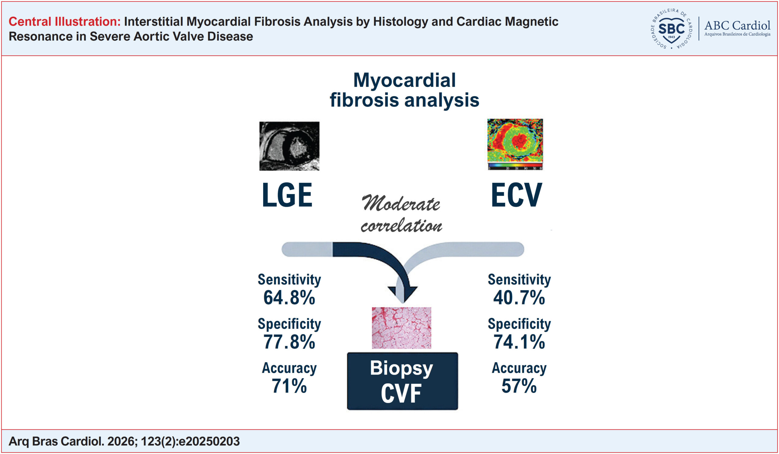
Interstitial Myocardial Fibrosis Analysis by Histology and Cardiac Magnetic Resonance in Severe Aortic Valve Disease
ABC
Original Article

Interstitial Myocardial Fibrosis Analysis by Histology and Cardiac Magnetic Resonance in Severe Aortic Valve Disease

Juliana Hiromi Silva Matsumoto Bello et al. Lea M.M.F. Demarchi Rafael Almeida Fonseca Gabriele N. Lima … Lucas José Tachotti Pires Vitor E. E. Rosa Roney Orismar Sampaio Paulo S. Gutierrez Vera D. Aiello Flávio Tarasoutchi Carlos E. Rochitte

Abstract

Background:

Aortic valve disease may lead to the accumulation of myocardial fibrosis (MF). Cardiac magnetic resonance (CMR) with late gadolinium enhancement (LGE) evaluates regional fibrosis but does not detect interstitial fibrosis, which can be estimated by extracellular volume (ECV) using native (pre-contrast) and post-contrast T1 mapping.

Objectives:

To compare MF measured by collagen volume fraction (CVF) in myocardial biopsy with LGE and ECV in patients with severe aortic valve disease.

Methods:

A total of 108 patients undergoing aortic valve replacement with myocardial biopsy and preoperative CMR were included. LGE and ECV were compared with CVF using Pearson or Spearman correlation coefficients, diagnostic performance analysis, and agreement testing. Statistical significance was set at p < 0.05.

Results:

Increased CVF was observed in 96% of patients (21.9 ± 5.5%), LGE in 44% (1.3 ± 3%), and increased ECV in 33.3% (29.0 ± 4.0%). Moderate correlations were found between LGE and CVF and between ECV and CVF (r = 0.44 and r = 0.36, respectively; p = 0.001). LGE and ECV demonstrated sensitivities of 64.8% and 40.7%, specificities of 77.8% and 74.1%, and accuracies of 71% and 57%, respectively, using CVF above the median as the reference for MF. Agreement between CVF and ECV revealed a mean difference of −0.167 and a Cohen’s kappa value of 0.171.

Conclusions:

CMR detects MF in patients with severe aortic valve disease confirmed by biopsy. LGE and ECV show moderate correlation with CVF. Combined analysis of LGE and ECV may provide complementary information; however, additional studies are needed to determine their prognostic impact.

  • Aortic Valve Disease
  • Fibrosis
  • Histology
  • Magnetic Resonance Imaging
  • Miocardio
Read Full Article
Creative Commons Attribution (CC BY) Creative Commons license
15/05/2026 DOI: 10.36660/abc.20250203i Views 1
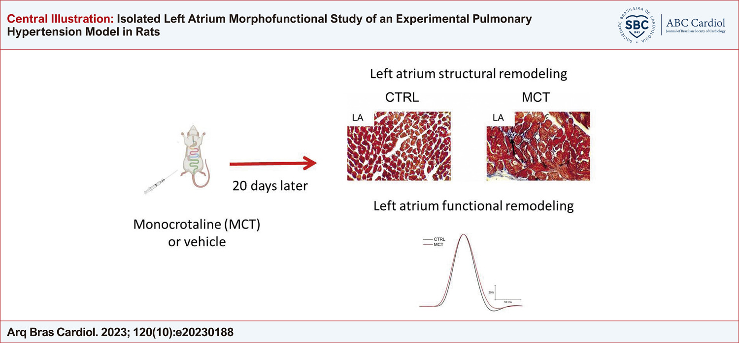
Isolated Left Atrium Morphofunctional Study of an Experimental Pulmonary Hypertension Model in Rats
ABC
Original Article

Isolated Left Atrium Morphofunctional Study of an Experimental Pulmonary Hypertension Model in Rats

Jorge Lucas Teixeira-Fonseca et al. Julliane Vasconcelos Joviano-Santos Fabiana da Silva Alcântara Polyana Leal da Silva … Michael Ramon Lima Conceição Danilo Roman-Campos

Abstract

Background

The high incidence of atrial arrhythmias in pulmonary hypertension (PH) might be associated with poor prognosis, and the left atrium (LA) may play a role in this. An important finding in PH studies is that LA remodeling is underestimated.

Objective

This study investigated LA morphology and mechanical function, as well as the susceptibility to develop arrhythmias in a monocrotaline-induced PH (MCT-PH) model.

Methods

Wistar rats aged 4 weeks received 50 mg/kg of MCT. Electrocardiography and histology analysis were performed to evaluate the establishment of the MCT-PH model. The tissue was mounted in an isolated organ bath to characterize the LA mechanical function

Results

Compared with the control group (CTRL), the MCT-PH model presented LA hypertrophy and changes in cardiac electrical activity, as evidenced by increased P wave duration, PR and QT interval in MCT-PH rats. In LA isolated from MCT-PH rats, no alteration in inotropism was observed; however, the time to peak contraction was delayed in the experimental MCT-PH group. Finally, there was no difference in arrhythmia susceptibility of LA from MCT-PH animals after the burst pacing protocol.

Conclusion

The morphofunctional remodeling of the LA did not lead to increased susceptibility to ex vivo arrhythmia after application of the burst pacing protocol.

  • Cardiac Arrhythmias
  • Fibrosis
  • Heart Atria
  • Monocrotaline
  • Pulmonary Hypertension
Read Full Article
Creative Commons Attribution (CC BY) Creative Commons license
20/10/2023 DOI: 10.36660/abc.20230188 Views 2
Figure 1
ABCHF
Viewpoint

Endomyocardial Fibrosis: Viewpoint

Luma Ornelas Sousa Rêgo et al. Gustavo Sampaio Vilas-Boas André Rodrigues Durães

Abstract

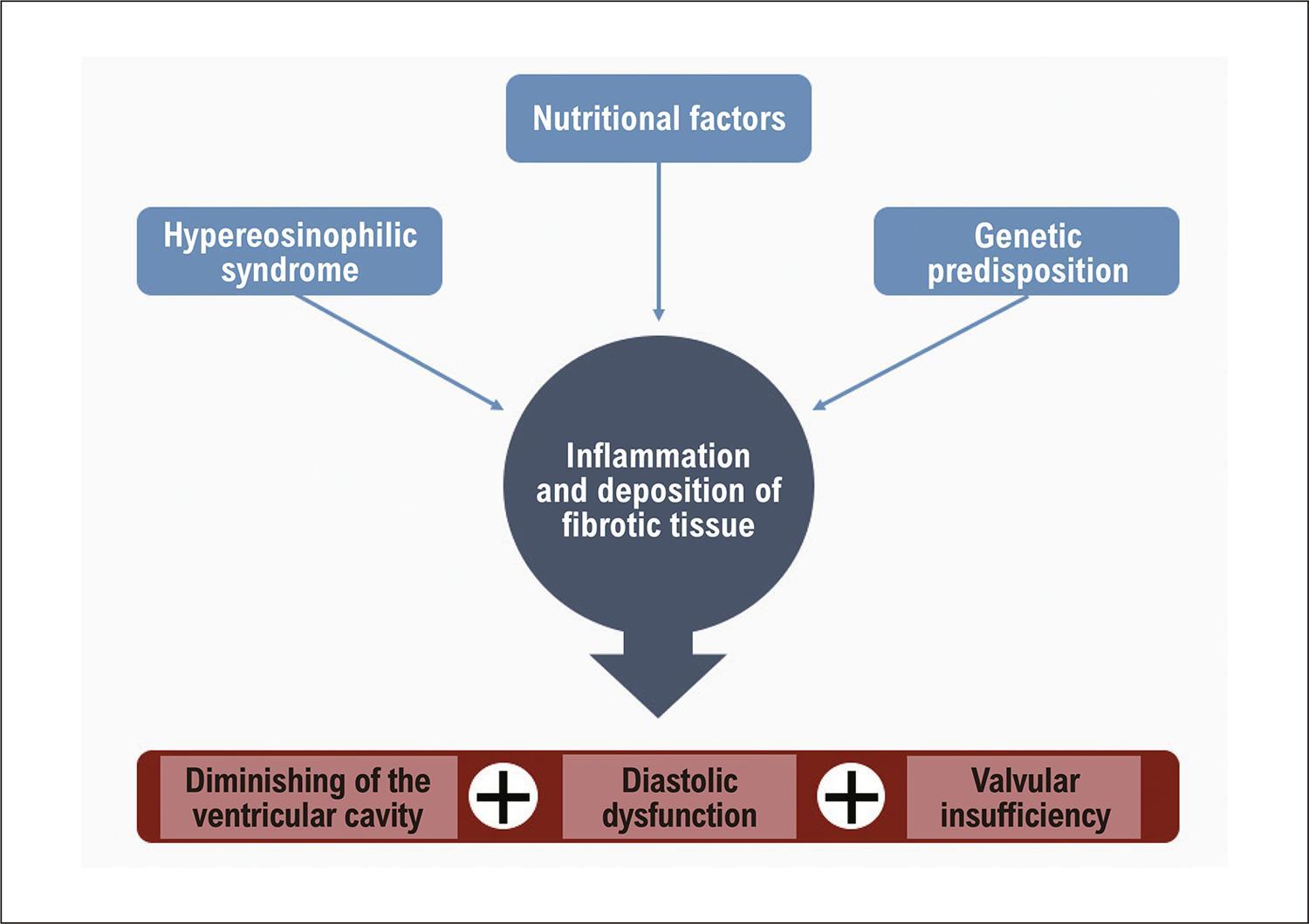
Endomyocardial fibrosis (EMF) is a restrictive cardiomyopathy of unknown origin, affecting mainly young people from tropical countries. EMF is considered to be the most restrictive cardiomyopathy in the world. The main risk factors for EMF appear to be age, gender, ethnicity, social vulnerability, and eosinophilia. The deposition of fibrotic tissue in the endocardium, myocardium, and heart valves leads to a decrease in the ventricular cavity and dysfunction in the diastolic filling of the ventricles. In this article we will express our point of view on this pathology, which is relatively unknown and with limited treatment options.

  • cardiomyopathy
  • Endomyocardial
  • Fibrosis
  • Ventricles
Read Full Article
Creative Commons Attribution (CC BY) Creative Commons license
10/07/2023 DOI: 10.36660/abchf.20230028
Figure 1
ABC
Original Article

Clinical Heart Failure Stratification Through Native T1 Mapping: Experience of a Referral Service

Thiago dos Santos Silva Marques et al. André Maurício de Souza Fernandes Roberto Nery Dantas Júnior Robert W. Biederman … Ana Paula Marques de Oliveira Melo Roque Aras

Abstract

Background:

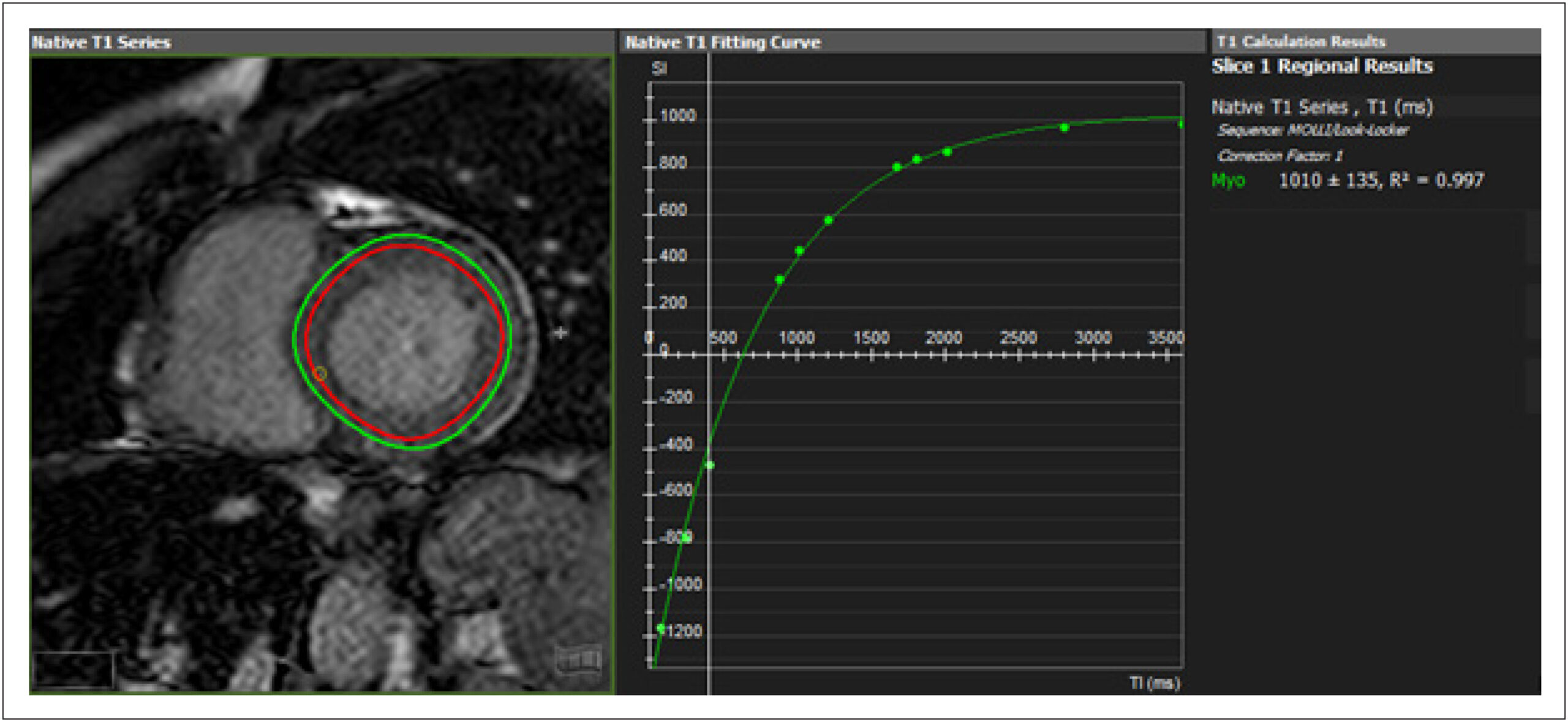
Diffuse cardiac fibrosis is an important factor in the prognostic assessment of patients with ventricular dysfunction. Cardiovascular magnetic resonance imaging (CMR) native T1 mapping is highly sensitive and considered an independent predictor of all-cause mortality and heart failure (HF) development in patients with cardiomyopathy.

Objectives:

To evaluate the feasibility of native T1 mapping assessment in patients with HF in a cardiology referral hospital and its association with structural parameters and functional profile.

Methods:

Cross-sectional study with adult patients with HF NYHA functional classes I and II, ischemic and non-ischemic, followed in a referral hospital, who underwent CMR. Native T1 values were analyzed for structural parameters, comorbidities, etiology, and categorization of HF by left ventricular ejection fraction (LVEF). Analyses were performed with a significance level of 5%.

Results:

Enrollment of 134 patients. Elevated native T1 values were found in patients with greater dilation (1004.9 vs 1042.7ms, p = 0.001), ventricular volumes (1021.3 vs 1050.3ms, p <0.01) and ventricular dysfunction (1010.1 vs 1053.4ms, p <0.001), also present when the non-ischemic group was analyzed separately. Patients classified as HF with reduced ejection fraction had higher T1 values than those with HF and preserved ejection fraction (HFPEF) (992.7 vs 1054.1ms, p <0.001). Of those with HFPEF, 55.2% had higher T1.

Conclusions:

CMR T1 mapping is feasible for clinical HF evaluation. There was a direct association between higher native T1 values and lower ejection fraction, and with larger LV diameters and volumes, regardless of the etiology of HF.

  • Cardiomyopathy, Dilated
  • Chagas Cardiomyopathy
  • Diagnosis Imaging
  • Fibrosis
  • Heart Failure
  • Magnetic Resonance Spectroscopy/ methods
  • Ventricular Dysfunction, Left
Read Full Article
Creative Commons Attribution (CC BY) Creative Commons license
17/05/2021 DOI: 10.36660/abc.20190782 Views 1
ABC
Editorial

Histopathological Characterization of Mitral Valvular Lesions in Patients With Rheumatic Heart Disease: Is Inflammation Also to Blame for Chronic Valvular Heart Disease Progression?

Vitor Emer Egypto Rosa

Rheumatic fever (RF) and rheumatic heart disease (RHD) remain highly prevalent, affecting approximately 40 million people, mostly in low- and middle-income countries, but also in select marginalized populations in higher-income countries., Brazilian data regarding RF prevalence is scarce mainly due to (a) difficulties in acute RF diagnosis, (b) cost of RHD screening and (c) the fact that reported data on surgery or death may represent RF incidence from 2 decades ago. The most feared consequence of RF is valvular heart disease, which leads to a worsening in the quality of life, hospitalizations and need for surgical procedure, primarily in young people., Therefore, there is an urgent need to better understand the factors that influence valvular heart disease (VHD) progression. In this context, Gomes et al. studied the histopathological changes in mitral valves of patients with RHD undergoing mitral valve replacement.

Inflammation in one of the key components of VHD in patients with RF. In the acute stage of infection, the histopathological findings are the presence of dense valvular inflammatory infiltrates and Aschoff nodules, characterized as collagen fibrinoid degeneration surrounded by lymphocytes, macrophages, giant cells, plasma cells, and palisades of fibroblasts.– Chronic RHD usually presents with cuspid calcification, fibrosis and commissural fusion. However, the role of inflammation in chronic VHD is still under study.

[…]

  • Epidemiology
  • Fibrosis
  • Inflammation
  • Mitral Valve Insufficiency / surgery
  • Rheumatic Heart Disease/diagnosis
Read Full Article
Creative Commons Attribution (CC BY) Creative Commons license
23/04/2021 DOI: 10.36660/abc.20201244 Views 1
Figure 1
ABC
Original Article

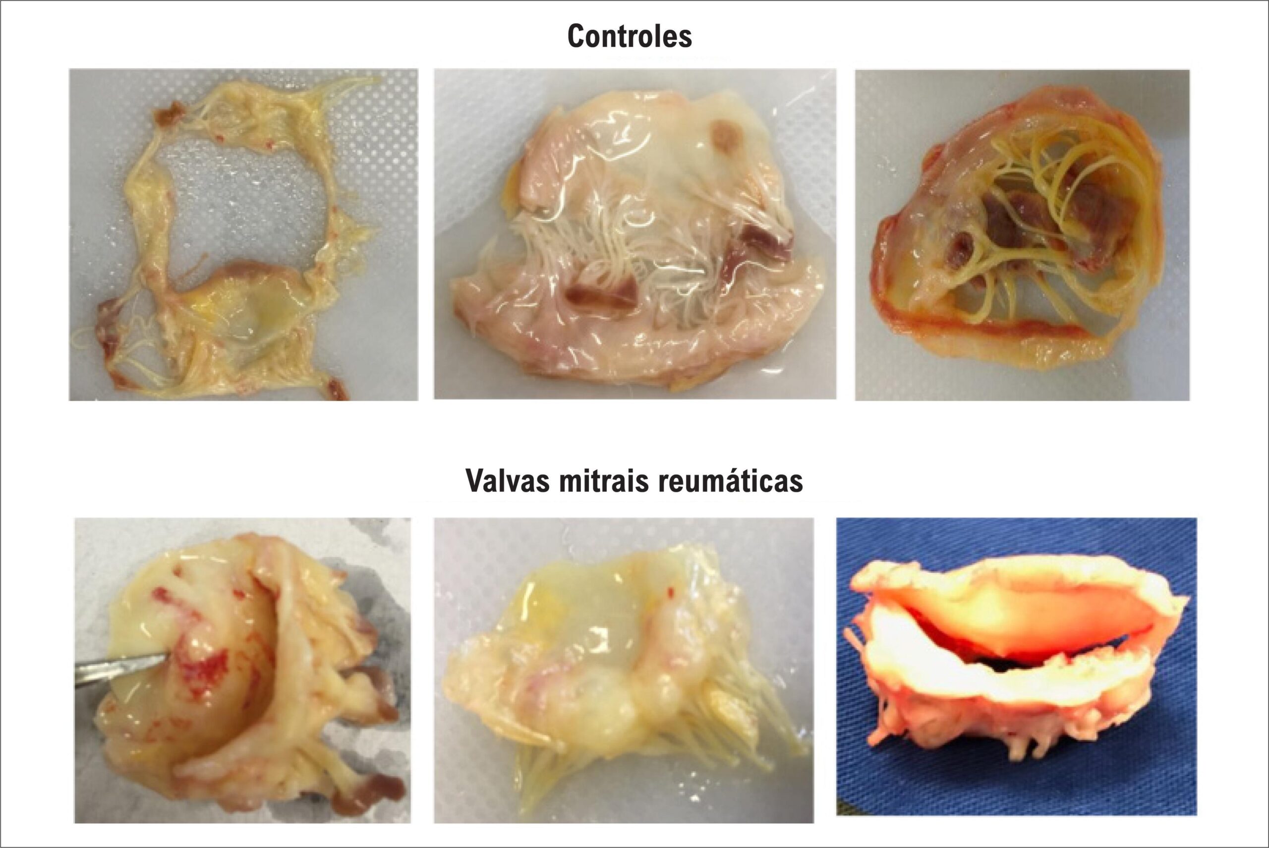
Histopathological Characterization of Mitral Valvular Lesions from Patients with Rheumatic Heart Disease

Nayana F. A. Gomes et al. Marcelo A. Pascoal-Xavier Livia S. A. Passos Thiago Mendonça Nunes Paula … João Marcelo de Souza Aguiar Felipe Vieira Guarçoni Maria Cecília Landim Nassif Claudio Leo Gelape Renato Braulio Paulo Henrique N. Costa Luiz Guilherme Passaglia Raquel Braga Martins Walderez O. Dutra Maria Carmo P. Nunes

Abstract

Background:

The underlying mechanisms by which rheumatic heart disease (RHD) lead to severe valve dysfunction are not completely understood.

Objective:

The present study evaluated the histopathological changes in mitral valves (MV) seeking an association between the pattern of predominant valvular dysfunction and histopathological findings.

Methods:

In 40 patients who underwent MV replacement due to RHD, and in 20 controls that underwent heart transplant, histological aspects of the excised MV were analyzed. Clinical and echocardiographic data were also collected. Histological analyses were performed using hematoxylin-eosin staining. Inflammation, fibrosis, neoangiogenesis, calcification and adipose metaplasia were determined. A p value<0.05 was considered to be statistically significant.

Results:

The mean age of RHD patients was 53±13 years, 36 (90%) were female, whereas the mean age of controls was 50±12 years, similar to the cases, with the majority of males (70%). The rheumatic valve endocardium presented greater thickness than the controls (1.3±0.5 mm versus 0.90±0.4 mm, p=0.003, respectively), and a more intense inflammatory infiltrate in the endocardium (78% versus 36%; p=0.004), with predominance of mononuclear cells. Moderate to marked fibrosis occurred more frequently in rheumatic valves than in control valves (100% vs. 29%; p<0.001). Calcification occurred in 35% of rheumatic valves, especially among stenotic valves, which was associated with the mitral valve area (p=0.003).

Conclusions:

Despite intense degree of fibrosis, the inflammatory process remains active in the rheumatic mitral valve, even at late disease with valve dysfunction. Calcification predominated in stenotic valves and in patients with right ventricular dysfunction.

  • calcification
  • Echocardiography/methods
  • Fibrosis
  • Histology
  • Inflammation
  • Mitral Valve Stenosis
  • Myocarditis
  • Rheumatic Disease
  • Rheumatic Fever
Read Full Article
Creative Commons Attribution (CC BY) Creative Commons license
23/04/2021 DOI: 10.36660/abc.20200154 Views 2
Figure 1
ABC
Image

Non-Progressive Hepatic Form of Andersen Disease as a Mimic of Hypertrophic Cardiomyopathy

Fabio Mastrocola et al. William Santos de Oliveira Adalberto Atsushi Porto Roberto Moreno Mendonça Nestor Rodrigues de Oliveira Neto

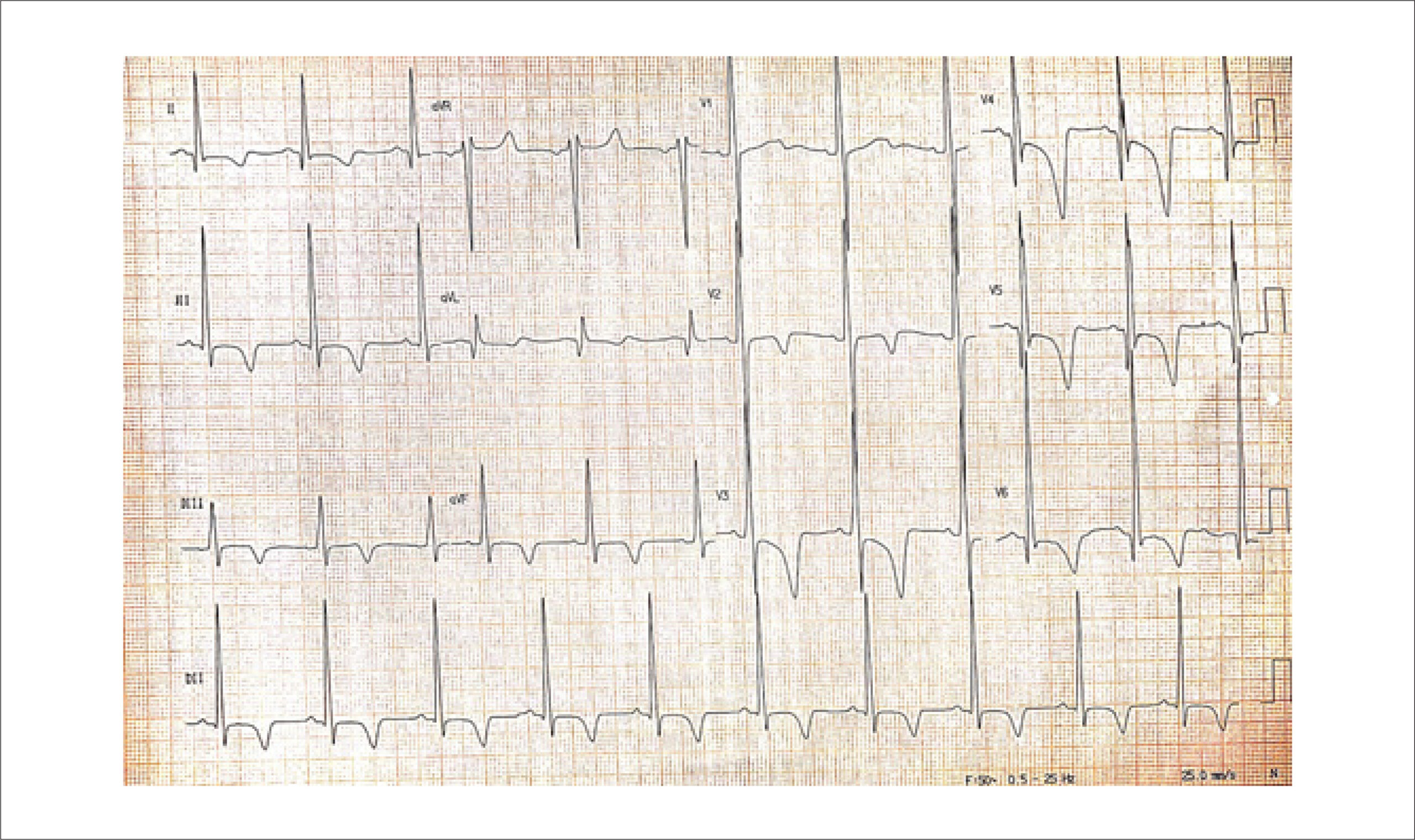
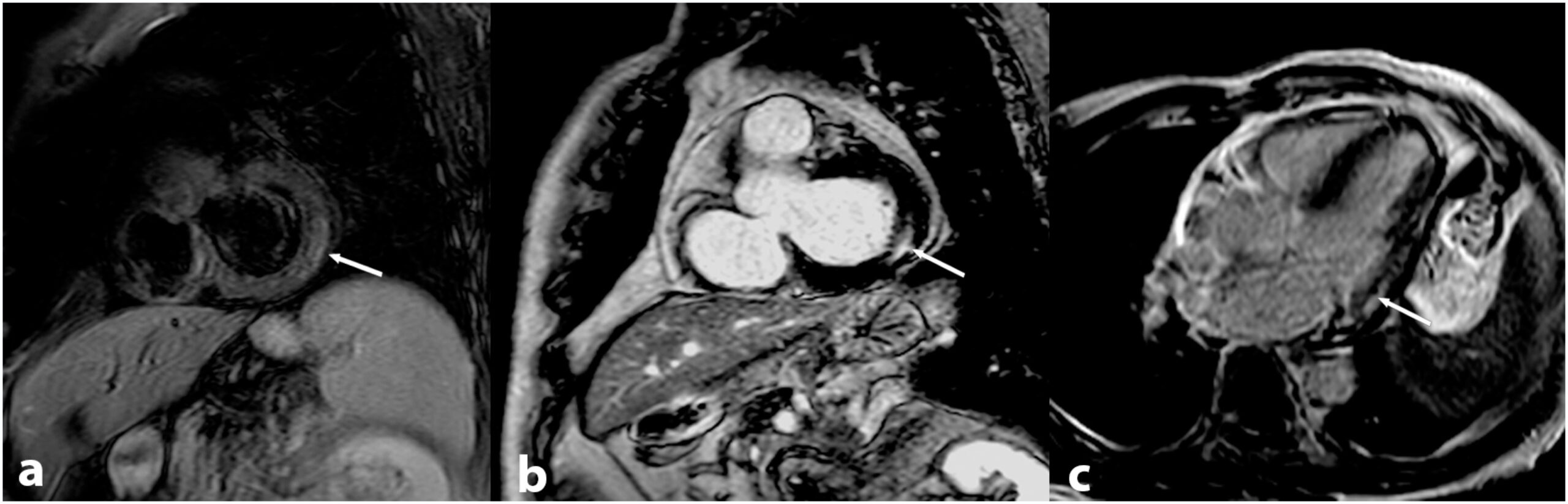
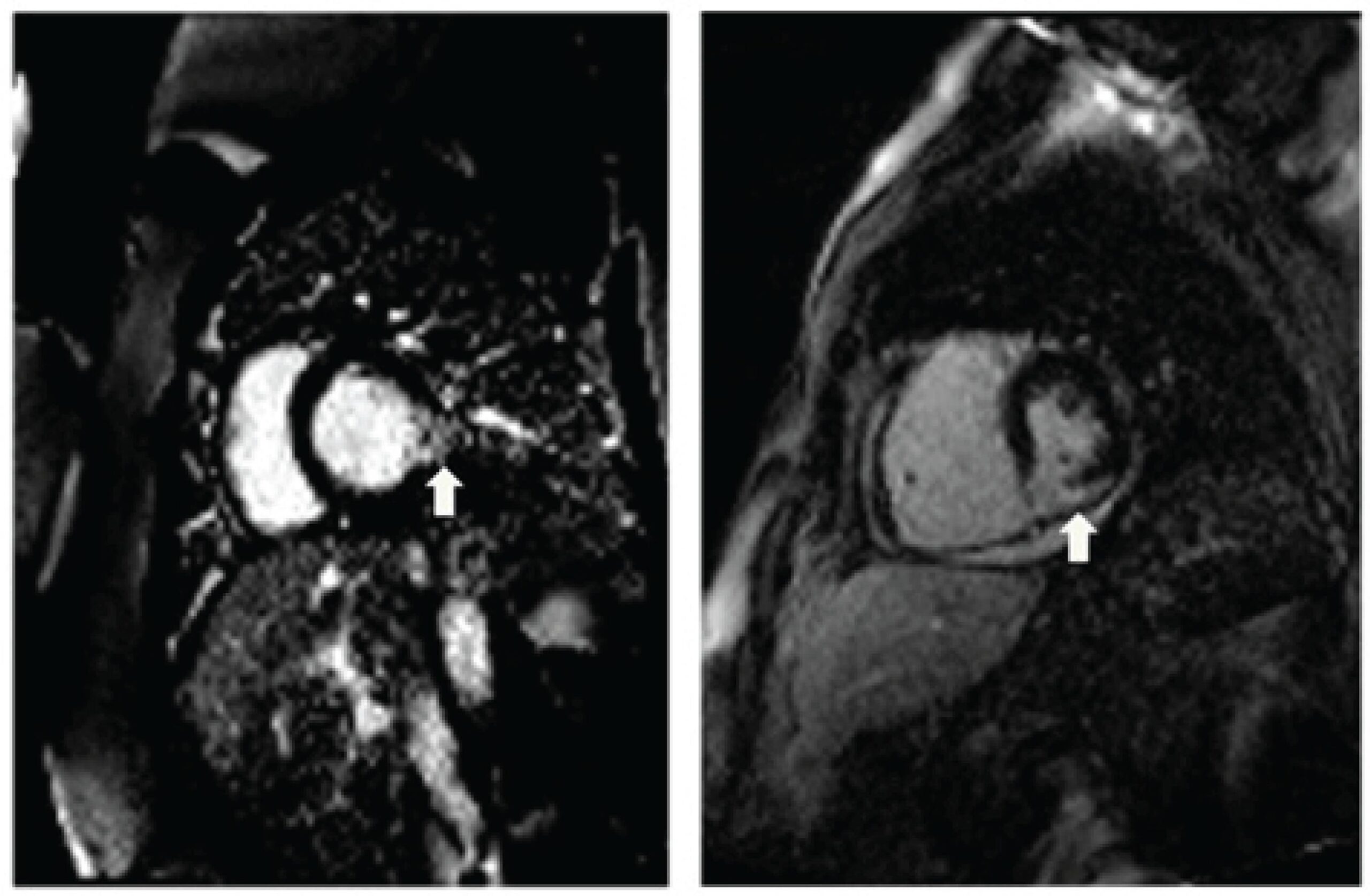
An 18-year-old male patient presented to our ambulatory services due to a 3-month course of progressive dyspnea and non-productive cough. Symptoms were present even at rest, and he exhibited severe limitation at physical activity. He had a diagnosis of glycogen storage disease (GSD) type IV attained by liver biopsy since three years of age, when he developed hepatomegaly and mild hepatic dysfunction. At the time, histopathological examination showed grade 2 fibrosis along with numerous intracytoplasmic PAS-positive deposits that were resistant to diastase. Thenceforth, his liver function remained stable, and he persisted otherwise asymptomatic. His physical examination was unremarkable, and routine laboratory evaluation was within the normal ranges. However, his basal ECG () showed signs of left ventricular hypertrophy. He was submitted to a cardiovascular magnetic resonance (CMR), which showed asymmetrical hypertrophy with predominance in the interventricular septum (). Late gadolinium enhancement (LGE) was present in a patchy, multifocal pattern (). Medical therapy for heart failure was initiated; the patient is now asymptomatic and maintains regular follow-up in our ambulatory.

Patients with classic GSD type IV have unrelenting liver disease with fast progression to cirrhosis during childhood; however, a small subset of affected individuals may present with milder hepatic dysfunction that does not advance to end-stage liver disease., Accordingly, our patient had the diagnosis of GSD type IV by three years of age, but did not develop cirrhosis afterwards.

[…]

  • Cardiomyopathy, Hypertrophic
  • Fibrosis
  • Glycogen Storage Disease Type IV
  • Heart Failure
  • Magnetic Resonance Spectroscopy/ methods
  • PRKAG2 Syndrome
  • Prognosis
Read Full Article
Creative Commons Attribution (CC BY) Creative Commons license
07/12/2020 DOI: 10.36660/abc.20200218
Figure 1
IJCS
Review Article

Cardiac Magnetic Resonance in the Assessment of Chagas Disease and its Complications

Ariane Binoti Pacheco et al. Rodrigo de Jesus Louzeiro Melo Carlos Eduardo Rochitte

Abstract

The well-known occurrence of Chagas disease in endemic areas has become a worldwide problem, and cardiac magnetic resonance allows the early detection of cardiac involvement and complications of this disease. Cardiac magnetic resonance is a useful tool in all phases of Chagas disease, and new promising techniques using T1 mapping and extracellular volume measurements are able to detect cardiac involvement even earlier than conventional techniques.

  • Chagas Cardiomyopathy/complications
  • Chagas Diseases/complications
  • Endemic Diseases
  • Extracellular Volume
  • Fibrosis
  • Gadolinium /radiation effects
  • Heart Failure
  • Magnetic Resonance Imaging/methods
  • T1 Mapping
Read Full Article
Creative Commons Attribution (CC BY) Creative Commons license
25/11/2020 DOI: 10.36660/ijcs.20200250
Figure 1
ABC
Original Article

Galectin-3 Levels in Patients with Chronic Constrictive Pericarditis

Fábio Fernandes et al. Dirceu Thiago Pessoa de Melo Felix José Alvarez Ramires Ester Cerdeira Sabino … Carlos Henrique Valente Moreira Luiz Alberto Benvenutti Viviane Tiemi Hotta Ana Luiza Carrari Sayegh Francis Ribeiro de Souza Ricardo Ribeiro Dias Charles Mady

Abstract

Background

Galectin-3 (Gal-3) is a proinflammatory, profibrotic molecule implicated in the pathogenesis of heart failure. The role of Gal-3 in patients with chronic constrictive pericarditis (CCP) is not clear.

Objective

The aim of this study was to assess plasma Gal-3 in patients with CCP and correlate it with clinical, functional and histologic parameters.

Methods

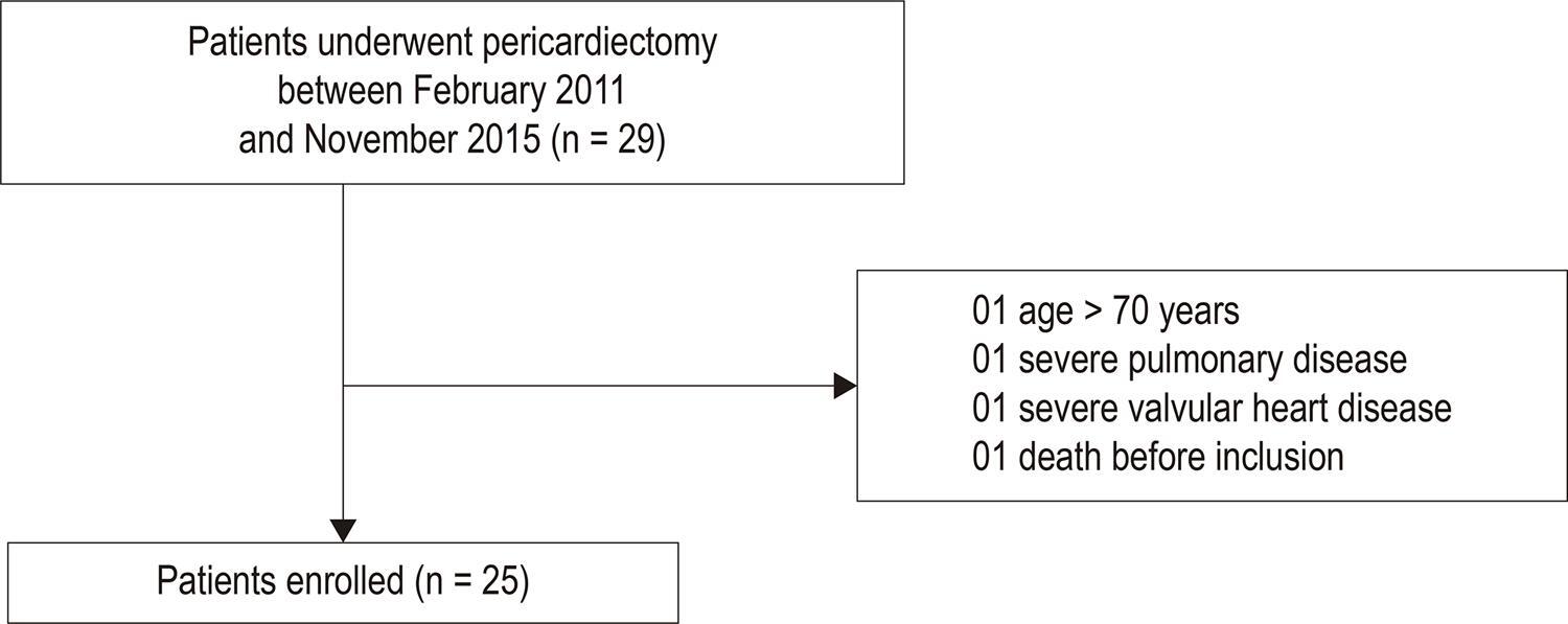
We prospectively evaluated 25 symptomatic patients with CCP referred for pericardiectomy and 21 healthy controls. Patients underwent clinical assessment, Gal-3 and B-type natriuretic peptide (BNP) measurements, echocardiography, cardiac magnetic resonance imaging and cardiopulmonary exercise test (CPET) at baseline. Six months after pericardiectomy CPET was repeated. An alpha error < 5% was considered statistically significant, with a confidence interval of 95%.

Results

Twenty-five patients with a median age of 45 years were included. Etiology was mainly idiopathic (n = 19, 76%); and 14 (56%) patients had NYHA functional class III/IV. Median BNP and Gal-3 were 143 (89-209) pg/dL and 14.8 (9.7-17.2) ng/mL, respectively. Gal-3 levels were not significantly higher in CCP patients than in control (p = 0.22). There were no significant correlations of Gal-3 with BNP, echocardiographic and cardiac magnetic resonance measures and histological findings. After pericardiectomy, it was found a statistically significant correlation between Gal-3 and the CPTE measures test duration (r = –0.79; p < 0.001) and exercise time (r = –0.79; p < 0.001).

Conclusions

Patients with CCP had normal levels of Gal-3 as compared to the controls. Gal-3 did not correlate with morphological and functional measures before pericardiectomy. However, the associations between Gal-3 and exercise intolerance after pericardiectomy may suggest a role of Gal-3 in prognosis prediction after pericardiectomy. (Arq Bras Cardiol. 2020; 114(4):683-689)

  • Cell Diferentiation
  • Fibrosis
  • Galectin 3
  • Pericardiectomy/methods
  • Pericardite Constrictive/surgery
Read Full Article
Creative Commons Attribution (CC BY) Creative Commons license
29/05/2020 DOI: 10.36660/abc.20190152
Figure 1
ABC
Original Article

The Presence and Extension of Myocardial Fibrosis in the Undetermined Form of Chagas’ Disease: A Study Using Magnetic Resonance

Marcia Maria Noya-Rabelo et al. Carolina The Macedo Ticiana Larocca Admilson Machado … Thais Pacheco Jorge Torreão Bruno Solano de Freitas Souza Milena B. P. Soares Ricardo Ribeiro-dos-Santos Luis Claudio Lemos Correia

Abstract

Background:

Previous data has shown that patients in the indeterminate form of Chagas disease may present myocardial fibrosis as shown on through magnetic resonance imaging (MRI). However, there is little information available regarding the degree of severity of myocardial fibrosis in these individuals. This variable has the potential to predict the evolution of Chagas’ disease into its cardiac form.

Objectives:

To describe the frequency and extent of myocardial fibrosis evaluated using an MRI in patients in the indeterminate form, and to compare it with other forms of the disease.

Methods:

Patients were admitted one after another. Their clinical history was collected and they were submitted to laboratory exams and an MRI.

Results:

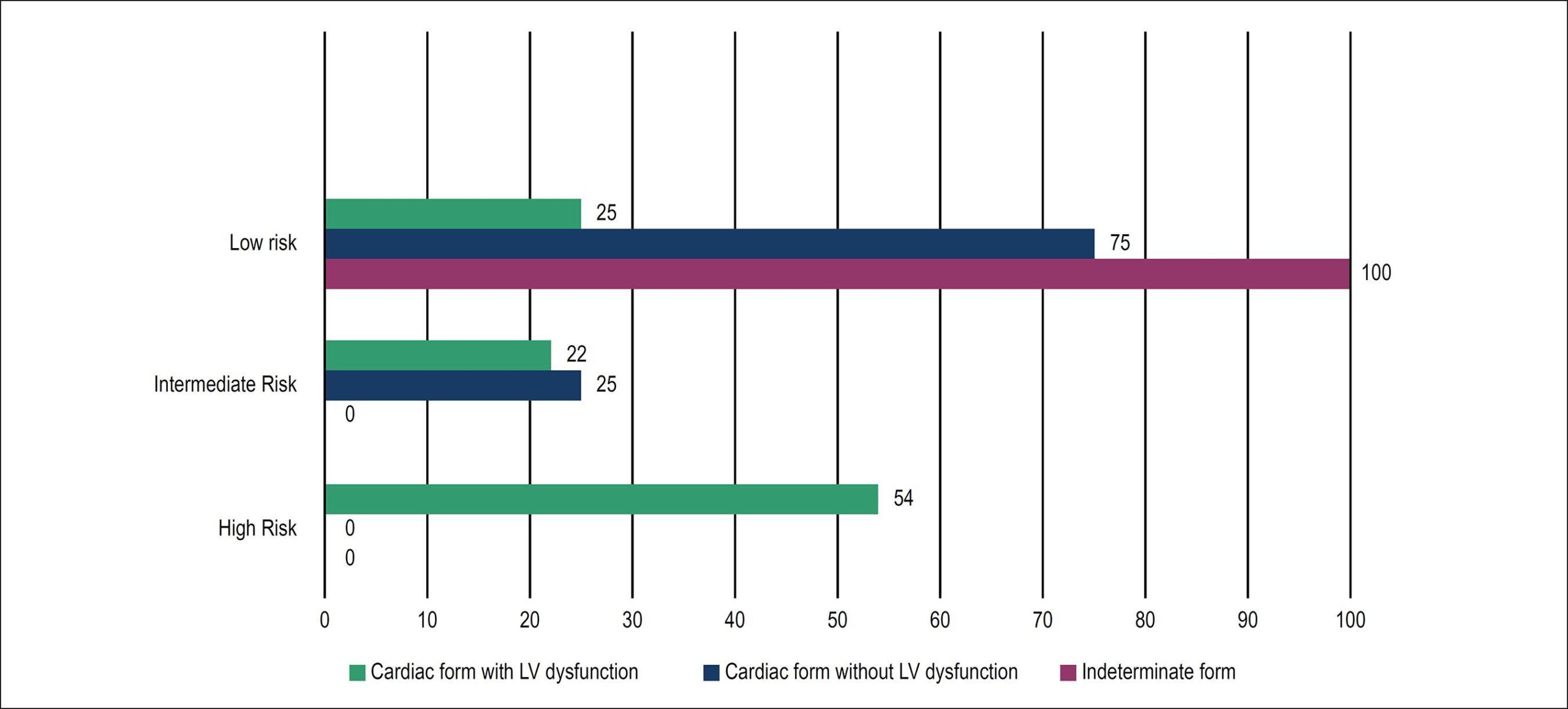
Sixty-one patients with Chagas’ disease, with an average age of 58 ± 9 years old, 17 patients in the indeterminate form, 16 in the cardiac form without left ventricular (LV) dysfunction and 28 in the cardiac form with LV dysfunction were studied. P <0.05 was considered to be statistically significant. Late enhancement was detected in 37 patients (64%). Myocardial fibrosis was identified in 6 individuals in indeterminate form (41%; 95% CI 23-66) in a proportion similar to that observed in cardiac form without LV dysfunction (44%); p = 1.0. Among the individuals with fibrosis, the total area of the affected myocardium was 4.1% (IIQ: 2.1 – 10.7) in the indeterminate form versus 2.3% (IIQ: 1-5) in the cardiac form without LV (p = 0.18). The left ventricular fraction ejection in subjects in the indeterminate form was similar to that of the individuals in the cardiac form without ventricular dysfunction (p = 0.09).

Conclusion:

The presence of fibrosis in the indeterminate form of Chagas’ disease has a frequency and extension similar to that of in the cardiac form without dysfunction, suggesting that the former is part of a subclinical disease spectrum, rather than lacking cardiac involvement.

  • Chagas Cardiomyopathy
  • Chagas disease
  • Fibrosis
  • Magnetic Resonance Imaging
Read Full Article
Creative Commons Attribution (CC BY) Creative Commons license
19/02/2018 DOI: 10.5935/abc.20180016
Figure 1
ABC
Original Article

Large Accumulation of Collagen and Increased Activation of Mast Cells in Hearts of Mice with Hyperlipidemia

Yunpeng Cheng et al. Yanqiu Zhu Jiashu Zhang Xingping Duan Ying Zhang

Abstract

Background:

Hyperlipidemia, which is characterized by an elevation of lipids in the bloodstream, is a major risk factor for cardiac disease.

Objectives:

The present study investigated the role of fibrosis in the progression of hyperlipidemia in the mice heart, and whether mast cell activation was associated with the fibrosis process.

Methods:

Hyperlipidemia was produced in C57BL / 6 mice by feeding them on a high-fat diet for 8 weeks.To assess tissue fibrosis, picrosirius red staining was performed. Hematoxylin & eosin (H&E) staining was performed to identify the histopathological changes in the hearts. Immunohistochemistry was also accomplished to determine the localization of transforming growth factor (TGF)-β and α-smooth muscle actin (α-SMA). Western blotting was performed to analyze the expression of chymase, tryptase, TGF-β, α-SMA and activity of Wnt/β-catenin pathway. At the end, serum total cholesterol (TC) and triglycerides (TG) levels were measured. All the values were expressed as means ± SD, the statistical significance level adopted was 5%.

Results:

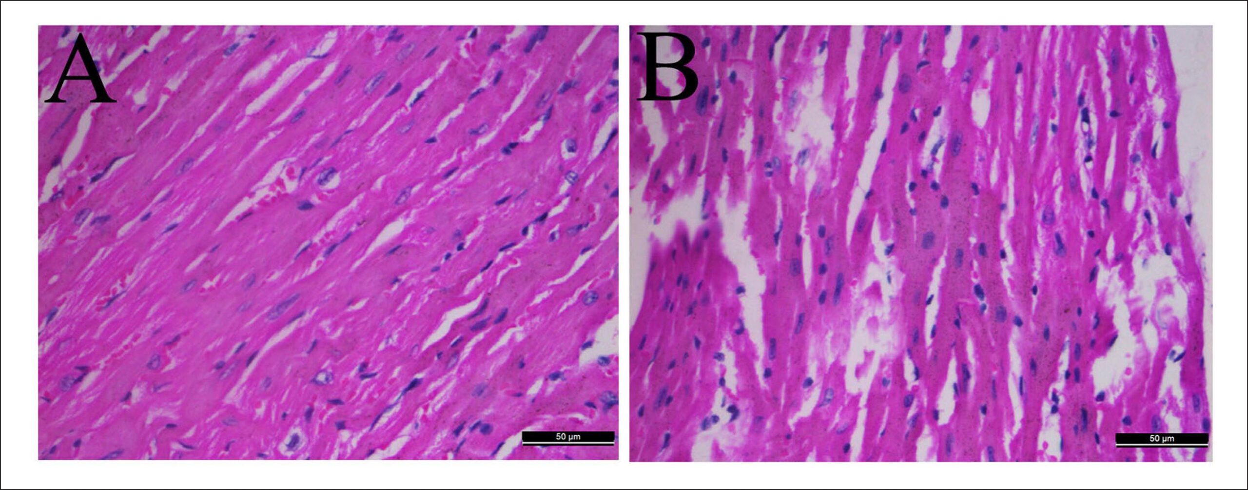
Hyperlipidemia mice showed significantly increased collagen deposition in the hearts compared with normal mice. In addition, H&E staining showed significant cellular degeneration. Cardiac muscle was arranged in disorder with fracture in mice of the model group. Immunohistochemistry and western blot analysis revealed that expression levels of tryptase, chymase, β-catenin, TGF-β and α-SMA were significantly increased in the hyperlipidemia mice compared with the control group.

Conclusions:

The results indicated that mast cell activation might induce cardiac fibrosis by tryptase and chymase in hyperlipidemia, which had a close relationship with the increased activity of TGF-β/Wnt/β-catenin pathway.

  • Fibrosis
  • Heart
  • Hyperlipidemias / physiopathology
  • Leukemia, Mast-Cell
  • Rats
Read Full Article
Creative Commons Attribution (CC BY) Creative Commons license
28/09/2017 DOI: 10.5935/abc.20170143 Views 1
Figure 1
IJCS
Original Article

Assessment of Right Ventricle Function and Myocardial Fibrosis by Cardiovascular Magnetic Resonance in Patients with Inferior Wall Myocardial Infarction

Priscila Neri Lacerda et al. Rafael Fernandes Almeida Fernanda Gabriella Figueiredo Pinto Adilson Machado Gomes Júnior … Jéssica Mendes Santos Cristiano Ricardo Bastos de Macêdo André Maurício Souza Fernandes Roque Aras Júnior

Abstract

Background:

Right ventricular dysfunction (RVD) can be found in 30-50% of patients with inferior wall myocardial infarction (I-MI) and predicts early mortality. Myocardial fibrosis is associated with progressive ventricular dysfunction and severe prognosis. In these patients, cardiovascular magnetic resonance (CMR) is an important risk stratification method.

Objectives:

This study sought to evaluate the association between RVD and myocardial fibrosis in patients with I-MI, using CMR.

Methods:

Cohort study conducted in a prominent center of cardiology. Forty individuals with I-MI were included in the study. CMR was performed during hospitalization to estimate parameters of right ventricle function and to quantify myocardial fibrosis through late gadolinium enhancement (LGE) technique. Patients were stratified by ventricular function, and clinical characteristics were compared between study groups.

Results:

Forty patients were included in the study, 75% were male and 43% elderly (age ≥ 60 years). Hypertension (45%) and smoking (33%) were the most prevalent cardiovascular risk factors. RVD was found in 33% of patients. Mean fibrosis mass was 22 ± 12 g in patients with RVD compared with 15 ± 8 g in patients with preserved ventricular function (p = 0.051).

Conclusions:

The findings of our study indicate a possible association between RVD and myocardial fibrosis in patients with I-MI. However, further studies with larger series are needed to confirm our findings.

  • Fibrosis
  • Magnetic Resonance Imaging
  • Myocardial Infarction
  • spectroscopy
Read Full Article
Creative Commons Attribution (CC BY) Creative Commons license
01/01/2017 DOI: 10.5935/2359-4802.20170037
ABC
Artigo Original

Blockade of renin-angiotensin system attenuates cardiac remodeling in rats undergoing aortic stenosis

Giancarlo Gonçalves et al. Leonardo A. M. Zornoff Henrique B. Ribeiro Marina P. Okoshi … Fernando R. S. Cordaro Katashi Okoshi Carlos R. Padovani Flávio F. Aragon Antonio C. Cicogna

OBJECTIVE: To assess the role of the AT1 receptor blocker and the angiotensin-converting enzyme inhibitor in cardiac remodeling induced by aortic stenosis in rats. METHODS: Wistar rats were divided into the following 4 groups: 1) C – control (n=13); 2) AoS – aortic stenosis (n=11); 3) LIS – AoS treated with lisinopril, 20 mg/kg/day (n=11); and 4) LOS – AoS treated with losartan, 40 mg/kg/day (n=9). The treatments were initiated 3 days before surgery. After 6 weeks, the animals underwent echocardiographic study, and quantification of the hydroxyproline (HOP) concentration and the left ventricular (LV) myocyte cross-sectional area (CSA). RESULTS: Aortic stenosis induced an increase in left ventricular wall thickness. The LIS and LOS groups showed no difference as compared with the control group. The AoS and LIS rats had greater left atrial diameters than the control rats did, while no difference was observed in the LOS animals. The AoS animals had greater values of shortening percentage than control animals did. This fact was modified with neither LIS nor LOS. The cross-sectional area of the animals in the AoS group was greater than that in the control group. However, treatment with LOS and LIS attenuated the AoS-induced increase in area. Aortic stenosis caused an increase in HOP concentration, while the LOS group showed no difference as compared with the control group. CONCLUSION: Blockade of the renin-angiotensin system with AT1 blocker and ACEI may attenuate the development of heart hypertrophy, but only the blockade of AT1 receptors attenuates left ventricular interstitial fibrosis.

  • Echocardiogram
  • Fibrosis
  • Hypertrophy
  • Ventricular Function
Read Full Article
Creative Commons Attribution (CC BY) Creative Commons license
03/05/2005 DOI: 10.1590/S0066-782X2005000400006 Views 1
Effects of losartan on ventricular remodeling in experimental infarction in rats
ABC
Original Article

Effects of losartan on ventricular remodeling in experimental infarction in rats

Leonardo A. M. Zornoff et al. Luiz S. Matsubara Beatriz B. Matsubara Sergio A. R. Paiva Joel Spadaro

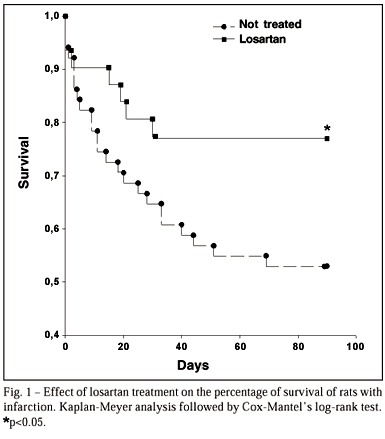
OBJECTIVE: To evaluate the effects of losartan on ventricular remodeling and on survival after myocardial infarction in rats. METHODS: After surgical occlusion of left coronary artery, 84 surviving male Wistar rats were divided into two groups: LO treated with losartan (20mg/kg/day, n=33) and NT (n=51), without medication. After 3 months, we analyzed mortality; ventricular to body mass ratio (VM /BM); myocardial hydroxyproline concentration (HOP); isovolumetric pressure, +dp/dt, -dp/dt, and diastolic volume/left ventricle mass ratio (VO/LV). RESULTS: Mortality was: LO = 22%, and NT = 47% (p<0.05). Ventricular mass,(VM/BM, mg/g) was 4.14 ± 0.76 and 3.54±0.48, in the NT and LO groups, respectively (p0.05). The V0/LV values (median) were 0.24 mL/g in group LO and 0.31 mL/g in group NT (p<0.05) compared to NT group. There were no differences between the groups for +dp/dt and -dp/dt parameters. CONCLUSION: 1- The use of losartan myocardial infarction causes an attenuation of ventricular remodeling, bringing about an increased survival, an attenuation of ventricular hypertrophy and dilation, and an improvement of the isovolumetric pressure; 2- the treatment does not modify the myocardial collagen concentration.

  • angiotensin II
  • Fibrosis
  • Ventricular Function
Read Full Article
Creative Commons Attribution (CC BY) Creative Commons license
08/01/2002 DOI: 10.1590/S0066-782X2000001200001

How to cite this article

EndNote RIS

Electronic Document Format (Vancouver)

Gomes HJA, Senra T. Cardiac Magnetic Resonance Imaging and Histology in Aortic Valve Disease: Convergences and Limitations. Arquivos Brasileiros de Cardiologia 2026;123(2):e20260140.

Electronic Document Format (ABNT)

Gomes, Helder Jorge Andrade; Senra, Tiago. Cardiac Magnetic Resonance Imaging and Histology in Aortic Valve Disease: Convergences and Limitations. Arquivos Brasileiros de Cardiologia, v. 123, n. 2, e20260140, May. 2026.

Electronic Document Format (APA)

Gomes, H. J. A., & Senra, T. (2026). Cardiac Magnetic Resonance Imaging and Histology in Aortic Valve Disease: Convergences and Limitations. Arquivos Brasileiros de Cardiologia, 123(2), e20260140.

Electronic Document Format (ISO)

Gomes, Helder Jorge Andrade and Senra, Tiago. Cardiac Magnetic Resonance Imaging and Histology in Aortic Valve Disease: Convergences and Limitations. Arquivos Brasileiros de Cardiologia [online]. 2026, vol. 123, n. 2, [cited 2026-10-14], e20260140. Available from: <https://journals.cardiol.br/en/article/cardiac-magnetic-resonance-imaging-and-histology-in-aortic-valve-disease-convergences-and-limitations/>. ISSN 0066-782X

EndNote RIS

Electronic Document Format (Vancouver)

Bello JHSM, Demarchi LM, Fonseca RA, Lima GN, Pires LJT, Rosa VEE, Sampaio RO, et al. Interstitial Myocardial Fibrosis Analysis by Histology and Cardiac Magnetic Resonance in Severe Aortic Valve Disease. Arquivos Brasileiros de Cardiologia 2026;123(2):e20250203.

Electronic Document Format (ABNT)

Bello, Juliana Hiromi Silva Matsumoto; Demarchi, Lea M.M.F.; Fonseca, Rafael Almeida; Lima, Gabriele N.; Pires, Lucas José Tachotti; Rosa, Vitor E. E.; Sampaio, Roney Orismar; Gutierrez, Paulo S.; Aiello, Vera D.; Tarasoutchi, Flávio; Rochitte, Carlos E.. Interstitial Myocardial Fibrosis Analysis by Histology and Cardiac Magnetic Resonance in Severe Aortic Valve Disease. Arquivos Brasileiros de Cardiologia, v. 123, n. 2, e20250203, May. 2026.

Electronic Document Format (APA)

Bello, J. H. S. M., Demarchi, L. M., Fonseca, R. A., Lima, G. N., Pires, L. J. T., Rosa, V. E. E., Sampaio, R. O., Gutierrez, P. S., Aiello, V. D., Tarasoutchi, F., & Rochitte, C. E. (2026). Interstitial Myocardial Fibrosis Analysis by Histology and Cardiac Magnetic Resonance in Severe Aortic Valve Disease. Arquivos Brasileiros de Cardiologia, 123(2), e20250203.

Electronic Document Format (ISO)

Bello, Juliana Hiromi Silva Matsumoto and Demarchi, Lea M.M.F. and Fonseca, Rafael Almeida and Lima, Gabriele N. and Pires, Lucas José Tachotti and Rosa, Vitor E. E. and Sampaio, Roney Orismar and Gutierrez, Paulo S. and Aiello, Vera D. and Tarasoutchi, Flávio and Rochitte, Carlos E.. Interstitial Myocardial Fibrosis Analysis by Histology and Cardiac Magnetic Resonance in Severe Aortic Valve Disease. Arquivos Brasileiros de Cardiologia [online]. 2026, vol. 123, n. 2, [cited 2026-10-14], e20250203. Available from: <https://journals.cardiol.br/en/article/interstitial-myocardial-fibrosis-analysis-by-histology-and-cardiac-magnetic-resonance-in-severe-aortic-valve-disease/>. ISSN 0066-782X

EndNote RIS

Electronic Document Format (Vancouver)

Teixeira-Fonseca JL, Joviano-Santos JV, Alcântara FS, Silva PL, Conceição MRL, Roman-Campos D. Isolated Left Atrium Morphofunctional Study of an Experimental Pulmonary Hypertension Model in Rats. Arquivos Brasileiros de Cardiologia 2023;120(10):e20230188.

Electronic Document Format (ABNT)

Teixeira-Fonseca, Jorge Lucas; Joviano-Santos, Julliane Vasconcelos; Alcântara, Fabiana da Silva; Silva, Polyana Leal da; Conceição, Michael Ramon Lima; Roman-Campos, Danilo. Isolated Left Atrium Morphofunctional Study of an Experimental Pulmonary Hypertension Model in Rats. Arquivos Brasileiros de Cardiologia, v. 120, n. 10, e20230188, Oct. 2023.

Electronic Document Format (APA)

Teixeira-Fonseca, J. L., Joviano-Santos, J. V., Alcântara, F. S., Silva, P. L. , Conceição, M. R. L., & Roman-Campos, D. (2023). Isolated Left Atrium Morphofunctional Study of an Experimental Pulmonary Hypertension Model in Rats. Arquivos Brasileiros de Cardiologia, 120(10), e20230188.

Electronic Document Format (ISO)

Teixeira-Fonseca, Jorge Lucas and Joviano-Santos, Julliane Vasconcelos and Alcântara, Fabiana da Silva and Silva, Polyana Leal da and Conceição, Michael Ramon Lima and Roman-Campos, Danilo. Isolated Left Atrium Morphofunctional Study of an Experimental Pulmonary Hypertension Model in Rats. Arquivos Brasileiros de Cardiologia [online]. 2023, vol. 120, n. 10, [cited 2026-10-14], e20230188. Available from: <https://journals.cardiol.br/en/article/isolated-left-atrium-morphofunctional-study-of-an-experimental-pulmonary-hypertension-model-in-rats/>. ISSN 0066-782X

EndNote RIS

Electronic Document Format (Vancouver)

Rêgo LOS, Vilas-Boas GS, Durães AR. Endomyocardial Fibrosis: Viewpoint. ABC Heart Fail Cardiomyop 2023;3(1):e20230028.

Electronic Document Format (ABNT)

Rêgo, Luma Ornelas Sousa; Vilas-Boas, Gustavo Sampaio; Durães, André Rodrigues. Endomyocardial Fibrosis: Viewpoint. ABC Heart Fail Cardiomyop, v. 3, n. 1, e20230028, Jul. 2023.

Electronic Document Format (APA)

Rêgo, L. O. S., Vilas-Boas, G. S., & Durães, A. R. (2023). Endomyocardial Fibrosis: Viewpoint. ABC Heart Fail Cardiomyop, 3(1), e20230028.

Electronic Document Format (ISO)

Rêgo, Luma Ornelas Sousa and Vilas-Boas, Gustavo Sampaio and Durães, André Rodrigues. Endomyocardial Fibrosis: Viewpoint. ABC Heart Fail Cardiomyop [online]. 2023, vol. 3, n. 1, [cited 2026-10-14], e20230028. Available from: <https://journals.cardiol.br/en/article/endomyocardial-fibrosis-viewpoint/>

EndNote RIS

Electronic Document Format (Vancouver)

Marques TSS, Fernandes AMS, Dantas Júnior RN, Biederman RW, Melo APMO, Aras R. Clinical Heart Failure Stratification Through Native T1 Mapping: Experience of a Referral Service. Arquivos Brasileiros de Cardiologia 2021;116(5):919-25.

Electronic Document Format (ABNT)

Marques, Thiago dos Santos Silva; Fernandes, André Maurício de Souza; Dantas Júnior, Roberto Nery; Biederman, Robert W.; Melo, Ana Paula Marques de Oliveira; Aras, Roque. Clinical Heart Failure Stratification Through Native T1 Mapping: Experience of a Referral Service. Arquivos Brasileiros de Cardiologia, v. 116, n. 5, p. 919-925, May. 2021.

Electronic Document Format (APA)

Marques, T. S. S., Fernandes, A. M. S., Dantas Júnior, R. N., Biederman, R. W., Melo, A. P. M. O., & Aras, R. (2021). Clinical Heart Failure Stratification Through Native T1 Mapping: Experience of a Referral Service. Arquivos Brasileiros de Cardiologia, 116(5), 919-925.

Electronic Document Format (ISO)

Marques, Thiago dos Santos Silva and Fernandes, André Maurício de Souza and Dantas Júnior, Roberto Nery and Biederman, Robert W. and Melo, Ana Paula Marques de Oliveira and Aras, Roque. Clinical Heart Failure Stratification Through Native T1 Mapping: Experience of a Referral Service. Arquivos Brasileiros de Cardiologia [online]. 2021, vol. 116, n. 5, [cited 2026-10-14], pp. 919-925. Available from: <https://journals.cardiol.br/en/article/clinical-heart-failure-stratification-through-native-t1-mapping-experience-of-a-referral-service/>. ISSN 0066-782X

EndNote RIS

Electronic Document Format (Vancouver)

Rosa VEE. Histopathological Characterization of Mitral Valvular Lesions in Patients With Rheumatic Heart Disease: Is Inflammation Also to Blame for Chronic Valvular Heart Disease Progression?. Arquivos Brasileiros de Cardiologia 2021;116(3):413-4.

Electronic Document Format (ABNT)

Rosa, Vitor Emer Egypto. Histopathological Characterization of Mitral Valvular Lesions in Patients With Rheumatic Heart Disease: Is Inflammation Also to Blame for Chronic Valvular Heart Disease Progression?. Arquivos Brasileiros de Cardiologia, v. 116, n. 3, p. 413-414, Apr. 2021.

Electronic Document Format (APA)

Rosa, V. E. E. (2021). Histopathological Characterization of Mitral Valvular Lesions in Patients With Rheumatic Heart Disease: Is Inflammation Also to Blame for Chronic Valvular Heart Disease Progression?. Arquivos Brasileiros de Cardiologia, 116(3), 413-414.

Electronic Document Format (ISO)

Rosa, Vitor Emer Egypto. Histopathological Characterization of Mitral Valvular Lesions in Patients With Rheumatic Heart Disease: Is Inflammation Also to Blame for Chronic Valvular Heart Disease Progression?. Arquivos Brasileiros de Cardiologia [online]. 2021, vol. 116, n. 3, [cited 2026-10-14], pp. 413-414. Available from: <https://journals.cardiol.br/en/article/histopathological-characterization-of-mitral-valvular-lesions-in-patients-with-rheumatic-heart-disease-is-inflammation-also-to-blame-for-chronic-valvular-heart-disease-progression/>. ISSN 0066-782X

EndNote RIS

Electronic Document Format (Vancouver)

Gomes NFA, Pascoal-Xavier MA, Passos LSA, Paula TMN, Aguiar JMS, Guarçoni FV, Nassif MCL, et al. Histopathological Characterization of Mitral Valvular Lesions from Patients with Rheumatic Heart Disease. Arquivos Brasileiros de Cardiologia 2021;116(3):404-12.

Electronic Document Format (ABNT)

Gomes, Nayana F. A.; Pascoal-Xavier, Marcelo A.; Passos, Livia S. A.; Paula, Thiago Mendonça Nunes; Aguiar, João Marcelo de Souza; Guarçoni, Felipe Vieira; Nassif, Maria Cecília Landim; Gelape, Claudio Leo; Braulio, Renato; Costa, Paulo Henrique N.; Passaglia, Luiz Guilherme; Martins, Raquel Braga; Dutra, Walderez O.; Nunes, Maria Carmo P.. Histopathological Characterization of Mitral Valvular Lesions from Patients with Rheumatic Heart Disease. Arquivos Brasileiros de Cardiologia, v. 116, n. 3, p. 404-412, Apr. 2021.

Electronic Document Format (APA)

Gomes, N. F. A., Pascoal-Xavier, M. A., Passos, L. S. A., Paula, T. M. N., Aguiar, J. M. S., Guarçoni, F. V., Nassif, M. C. L., Gelape, C. L., Braulio, R., Costa, P. H. N., Passaglia, L. G., Martins, R. B., Dutra, W. O., & Nunes, M. C. P. (2021). Histopathological Characterization of Mitral Valvular Lesions from Patients with Rheumatic Heart Disease. Arquivos Brasileiros de Cardiologia, 116(3), 404-412.

Electronic Document Format (ISO)

Gomes, Nayana F. A. and Pascoal-Xavier, Marcelo A. and Passos, Livia S. A. and Paula, Thiago Mendonça Nunes and Aguiar, João Marcelo de Souza and Guarçoni, Felipe Vieira and Nassif, Maria Cecília Landim and Gelape, Claudio Leo and Braulio, Renato and Costa, Paulo Henrique N. and Passaglia, Luiz Guilherme and Martins, Raquel Braga and Dutra, Walderez O. and Nunes, Maria Carmo P.. Histopathological Characterization of Mitral Valvular Lesions from Patients with Rheumatic Heart Disease. Arquivos Brasileiros de Cardiologia [online]. 2021, vol. 116, n. 3, [cited 2026-10-14], pp. 404-412. Available from: <https://journals.cardiol.br/en/article/histopathological-characterization-of-mitral-valvular-lesions-from-patients-with-rheumatic-heart-disease/>. ISSN 0066-782X

EndNote RIS

Electronic Document Format (Vancouver)

Mastrocola F, Oliveira WS, Porto AA, Mendonça RM, Oliveira Neto NR. Non-Progressive Hepatic Form of Andersen Disease as a Mimic of Hypertrophic Cardiomyopathy. Arquivos Brasileiros de Cardiologia 2020;115(5):964-6.

Electronic Document Format (ABNT)

Mastrocola, Fabio; Oliveira, William Santos de; Porto, Adalberto Atsushi; Mendonça, Roberto Moreno; Oliveira Neto, Nestor Rodrigues de. Non-Progressive Hepatic Form of Andersen Disease as a Mimic of Hypertrophic Cardiomyopathy. Arquivos Brasileiros de Cardiologia, v. 115, n. 5, p. 964-966, Dec. 2020.

Electronic Document Format (APA)

Mastrocola, F., Oliveira, W. S. , Porto, A. A., Mendonça, R. M., & Oliveira Neto, N. R. (2020). Non-Progressive Hepatic Form of Andersen Disease as a Mimic of Hypertrophic Cardiomyopathy. Arquivos Brasileiros de Cardiologia, 115(5), 964-966.

Electronic Document Format (ISO)

Mastrocola, Fabio and Oliveira, William Santos de and Porto, Adalberto Atsushi and Mendonça, Roberto Moreno and Oliveira Neto, Nestor Rodrigues de. Non-Progressive Hepatic Form of Andersen Disease as a Mimic of Hypertrophic Cardiomyopathy. Arquivos Brasileiros de Cardiologia [online]. 2020, vol. 115, n. 5, [cited 2026-10-14], pp. 964-966. Available from: <https://journals.cardiol.br/en/article/non-progressive-hepatic-form-of-andersen-disease-as-a-mimic-of-hypertrophic-cardiomyopathy/>. ISSN 0066-782X

EndNote RIS

Electronic Document Format (Vancouver)

Pacheco AB, Melo RJL, Rochitte CE. Cardiac Magnetic Resonance in the Assessment of Chagas Disease and its Complications. International Journal of Cardiovascular Sciences 2020;33(6):705-12.

Electronic Document Format (ABNT)

Pacheco, Ariane Binoti; Melo, Rodrigo de Jesus Louzeiro; Rochitte, Carlos Eduardo. Cardiac Magnetic Resonance in the Assessment of Chagas Disease and its Complications. International Journal of Cardiovascular Sciences, v. 33, n. 6, p. 705-712, Nov. 2020.

Electronic Document Format (APA)

Pacheco, A. B., Melo, R. J. L., & Rochitte, C. E. (2020). Cardiac Magnetic Resonance in the Assessment of Chagas Disease and its Complications. International Journal of Cardiovascular Sciences, 33(6), 705-712.

Electronic Document Format (ISO)

Pacheco, Ariane Binoti and Melo, Rodrigo de Jesus Louzeiro and Rochitte, Carlos Eduardo. Cardiac Magnetic Resonance in the Assessment of Chagas Disease and its Complications. International Journal of Cardiovascular Sciences [online]. 2020, vol. 33, n. 6, [cited 2026-10-14], pp. 705-712. Available from: <https://journals.cardiol.br/en/article/cardiac-magnetic-resonance-in-the-assessment-of-chagas-disease-and-its-complications/>. ISSN 2359-4802

EndNote RIS

Electronic Document Format (Vancouver)

Fernandes F, Melo DTP, Ramires FJA, Sabino EC, Moreira CHV, Benvenutti LA, Hotta VT, et al. Galectin-3 Levels in Patients with Chronic Constrictive Pericarditis. Arquivos Brasileiros de Cardiologia 2020;114(4):683-9.

Electronic Document Format (ABNT)

Fernandes, Fábio; Melo, Dirceu Thiago Pessoa de; Ramires, Felix José Alvarez; Sabino, Ester Cerdeira; Moreira, Carlos Henrique Valente; Benvenutti, Luiz Alberto; Hotta, Viviane Tiemi; Sayegh, Ana Luiza Carrari; Souza, Francis Ribeiro de; Dias, Ricardo Ribeiro; Mady, Charles. Galectin-3 Levels in Patients with Chronic Constrictive Pericarditis. Arquivos Brasileiros de Cardiologia, v. 114, n. 4, p. 683-689, May. 2020.

Electronic Document Format (APA)

Fernandes, F., Melo, D. T. P. , Ramires, F. J. A., Sabino, E. C., Moreira, C. H. V., Benvenutti, L. A., Hotta, V. T., Sayegh, A. L. C., Souza, F. R. , Dias, R. R., & Mady, C. (2020). Galectin-3 Levels in Patients with Chronic Constrictive Pericarditis. Arquivos Brasileiros de Cardiologia, 114(4), 683-689.

Electronic Document Format (ISO)

Fernandes, Fábio and Melo, Dirceu Thiago Pessoa de and Ramires, Felix José Alvarez and Sabino, Ester Cerdeira and Moreira, Carlos Henrique Valente and Benvenutti, Luiz Alberto and Hotta, Viviane Tiemi and Sayegh, Ana Luiza Carrari and Souza, Francis Ribeiro de and Dias, Ricardo Ribeiro and Mady, Charles. Galectin-3 Levels in Patients with Chronic Constrictive Pericarditis. Arquivos Brasileiros de Cardiologia [online]. 2020, vol. 114, n. 4, [cited 2026-10-14], pp. 683-689. Available from: <https://journals.cardiol.br/en/article/galectin-3-levels-in-patients-with-chronic-constrictive-pericarditis/>. ISSN 0066-782X

EndNote RIS

Electronic Document Format (Vancouver)

Noya-Rabelo MM, Macedo CT, Larocca T, Machado A, Pacheco T, Torreão J, Souza BSF, et al. The Presence and Extension of Myocardial Fibrosis in the Undetermined Form of Chagas’ Disease: A Study Using Magnetic Resonance. Arquivos Brasileiros de Cardiologia 2018;110(2):124-31.

Electronic Document Format (ABNT)

Noya-Rabelo, Marcia Maria; Macedo, Carolina The; Larocca, Ticiana; Machado, Admilson; Pacheco, Thais; Torreão, Jorge; Souza, Bruno Solano de Freitas; Soares, Milena B. P.; Ribeiro-dos-Santos, Ricardo; Correia, Luis Claudio Lemos. The Presence and Extension of Myocardial Fibrosis in the Undetermined Form of Chagas’ Disease: A Study Using Magnetic Resonance. Arquivos Brasileiros de Cardiologia, v. 110, n. 2, p. 124-131, Feb. 2018.

Electronic Document Format (APA)

Noya-Rabelo, M. M., Macedo, C. T., Larocca, T., Machado, A., Pacheco, T., Torreão, J., Souza, B. S. F., Soares, M. B. P., Ribeiro-dos-Santos, R., & Correia, L. C. L. (2018). The Presence and Extension of Myocardial Fibrosis in the Undetermined Form of Chagas’ Disease: A Study Using Magnetic Resonance. Arquivos Brasileiros de Cardiologia, 110(2), 124-131.

Electronic Document Format (ISO)

Noya-Rabelo, Marcia Maria and Macedo, Carolina The and Larocca, Ticiana and Machado, Admilson and Pacheco, Thais and Torreão, Jorge and Souza, Bruno Solano de Freitas and Soares, Milena B. P. and Ribeiro-dos-Santos, Ricardo and Correia, Luis Claudio Lemos. The Presence and Extension of Myocardial Fibrosis in the Undetermined Form of Chagas’ Disease: A Study Using Magnetic Resonance. Arquivos Brasileiros de Cardiologia [online]. 2018, vol. 110, n. 2, [cited 2026-10-14], pp. 124-131. Available from: <https://journals.cardiol.br/en/article/the-presence-and-extension-of-myocardial-fibrosis-in-the-undetermined-form-of-chagas-disease-a-study-using-magnetic-resonance/>. ISSN 0066-782X

EndNote RIS

Electronic Document Format (Vancouver)

Cheng Y, Zhu Y, Zhang J, Duan X, Zhang Y. Large Accumulation of Collagen and Increased Activation of Mast Cells in Hearts of Mice with Hyperlipidemia. Arquivos Brasileiros de Cardiologia 2017;109(5):404-9.

Electronic Document Format (ABNT)

Cheng, Yunpeng; Zhu, Yanqiu; Zhang, Jiashu; Duan, Xingping; Zhang, Ying. Large Accumulation of Collagen and Increased Activation of Mast Cells in Hearts of Mice with Hyperlipidemia. Arquivos Brasileiros de Cardiologia, v. 109, n. 5, p. 404-409, Sep. 2017.

Electronic Document Format (APA)

Cheng, Y., Zhu, Y., Zhang, J., Duan, X., & Zhang, Y. (2017). Large Accumulation of Collagen and Increased Activation of Mast Cells in Hearts of Mice with Hyperlipidemia. Arquivos Brasileiros de Cardiologia, 109(5), 404-409.

Electronic Document Format (ISO)

Cheng, Yunpeng and Zhu, Yanqiu and Zhang, Jiashu and Duan, Xingping and Zhang, Ying. Large Accumulation of Collagen and Increased Activation of Mast Cells in Hearts of Mice with Hyperlipidemia. Arquivos Brasileiros de Cardiologia [online]. 2017, vol. 109, n. 5, [cited 2026-10-14], pp. 404-409. Available from: <https://journals.cardiol.br/en/article/large-accumulation-of-collagen-and-increased-activation-of-mast-cells-in-hearts-of-mice-with-hyperlipidemia/>. ISSN 0066-782X

EndNote RIS

Electronic Document Format (Vancouver)

Lacerda PN, Almeida RF, Pinto FGF, Gomes Júnior AM, Santos JM, Macêdo CRB, Fernandes AMS, et al. Assessment of Right Ventricle Function and Myocardial Fibrosis by Cardiovascular Magnetic Resonance in Patients with Inferior Wall Myocardial Infarction. International Journal of Cardiovascular Sciences 2017;30(2):109-16.

Electronic Document Format (ABNT)

Lacerda, Priscila Neri; Almeida, Rafael Fernandes; Pinto, Fernanda Gabriella Figueiredo; Gomes Júnior, Adilson Machado; Santos, Jéssica Mendes; Macêdo, Cristiano Ricardo Bastos de; Fernandes, André Maurício Souza; Aras Júnior, Roque. Assessment of Right Ventricle Function and Myocardial Fibrosis by Cardiovascular Magnetic Resonance in Patients with Inferior Wall Myocardial Infarction. International Journal of Cardiovascular Sciences, v. 30, n. 2, p. 109-116, Dec. 2016.

Electronic Document Format (APA)

Lacerda, P. N., Almeida, R. F., Pinto, F. G. F., Gomes Júnior, A. M., Santos, J. M., Macêdo, C. R. B. , Fernandes, A. M. S., & Aras Júnior, R. (2017). Assessment of Right Ventricle Function and Myocardial Fibrosis by Cardiovascular Magnetic Resonance in Patients with Inferior Wall Myocardial Infarction. International Journal of Cardiovascular Sciences, 30(2), 109-116.

Electronic Document Format (ISO)

Lacerda, Priscila Neri and Almeida, Rafael Fernandes and Pinto, Fernanda Gabriella Figueiredo and Gomes Júnior, Adilson Machado and Santos, Jéssica Mendes and Macêdo, Cristiano Ricardo Bastos de and Fernandes, André Maurício Souza and Aras Júnior, Roque. Assessment of Right Ventricle Function and Myocardial Fibrosis by Cardiovascular Magnetic Resonance in Patients with Inferior Wall Myocardial Infarction. International Journal of Cardiovascular Sciences [online]. 2017, vol. 30, n. 2, [cited 2026-10-14], pp. 109-116. Available from: <https://journals.cardiol.br/en/article/assessment-of-right-ventricle-function-and-myocardial-fibrosis-by-cardiovascular-magnetic-resonance-in-patients-with-inferior-wall-myocardial-infarction/>. ISSN 2359-4802

EndNote RIS

Electronic Document Format (Vancouver)

Gonçalves G, Zornoff LAM, Ribeiro HB, Okoshi MP, Cordaro FRS, Okoshi K, Padovani CR, et al. Blockade of renin-angiotensin system attenuates cardiac remodeling in rats undergoing aortic stenosis. Arquivos Brasileiros de Cardiologia 2005;84(4):304-8.

Electronic Document Format (ABNT)

Gonçalves, Giancarlo; Zornoff, Leonardo A. M.; Ribeiro, Henrique B.; Okoshi, Marina P.; Cordaro, Fernando R. S.; Okoshi, Katashi; Padovani, Carlos R.; Aragon, Flávio F.; Cicogna, Antonio C.. Blockade of renin-angiotensin system attenuates cardiac remodeling in rats undergoing aortic stenosis. Arquivos Brasileiros de Cardiologia, v. 84, n. 4, p. 304-308, May. 2005.

Electronic Document Format (APA)

Gonçalves, G., Zornoff, L. A. M., Ribeiro, H. B., Okoshi, M. P., Cordaro, F. R. S., Okoshi, K., Padovani, C. R., Aragon, F. F., & Cicogna, A. C. (2005). Blockade of renin-angiotensin system attenuates cardiac remodeling in rats undergoing aortic stenosis. Arquivos Brasileiros de Cardiologia, 84(4), 304-308.

Electronic Document Format (ISO)

Gonçalves, Giancarlo and Zornoff, Leonardo A. M. and Ribeiro, Henrique B. and Okoshi, Marina P. and Cordaro, Fernando R. S. and Okoshi, Katashi and Padovani, Carlos R. and Aragon, Flávio F. and Cicogna, Antonio C.. Blockade of renin-angiotensin system attenuates cardiac remodeling in rats undergoing aortic stenosis. Arquivos Brasileiros de Cardiologia [online]. 2005, vol. 84, n. 4, [cited 2026-10-14], pp. 304-308. Available from: <https://journals.cardiol.br/en/article/blockade-of-renin-angiotensin-system-attenuates-cardiac-remodeling-in-rats-undergoing-aortic-stenosis/>. ISSN 0066-782X

EndNote RIS

Electronic Document Format (Vancouver)

Zornoff LAM, Matsubara LS, Matsubara BB, Paiva SAR, Spadaro J. Effects of losartan on ventricular remodeling in experimental infarction in rats. Arquivos Brasileiros de Cardiologia 2002;75(6):465-70.

Electronic Document Format (ABNT)

Zornoff, Leonardo A. M.; Matsubara, Luiz S.; Matsubara, Beatriz B.; Paiva, Sergio A. R.; Spadaro, Joel. Effects of losartan on ventricular remodeling in experimental infarction in rats. Arquivos Brasileiros de Cardiologia, v. 75, n. 6, p. 465-470, Jan. 2002.

Electronic Document Format (APA)

Zornoff, L. A. M., Matsubara, L. S., Matsubara, B. B., Paiva, S. A. R., & Spadaro, J. (2002). Effects of losartan on ventricular remodeling in experimental infarction in rats. Arquivos Brasileiros de Cardiologia, 75(6), 465-470.

Electronic Document Format (ISO)

Zornoff, Leonardo A. M. and Matsubara, Luiz S. and Matsubara, Beatriz B. and Paiva, Sergio A. R. and Spadaro, Joel. Effects of losartan on ventricular remodeling in experimental infarction in rats. Arquivos Brasileiros de Cardiologia [online]. 2002, vol. 75, n. 6, [cited 2026-10-14], pp. 465-470. Available from: <https://journals.cardiol.br/en/article/effects-of-losartan-on-ventricular-remodeling-in-experimental-infarction-in-rats/>. ISSN 0066-782X

  • About ABC Family
  • Internal Regulation
  • Editorial Board
  • Brazilian Society of Cardiology (SBC)

Issues

SBC Guidelines

  • Videos
  • Podcasts
  • News