ABC Family Arquivos Brasileiros de Cardiologia International Journal of Cardiovascular Sciences ABC Heart Failure & Cardiomyopathy ABC Imagem Cardiovascular
Português English
ABC Family
Português English
  • About
    • About ABC Family
    • Internal Regulation
    • Editorial Board
    • Brazilian Society of Cardiology (SBC)
  • Issues
  • SBC Guidelines
  • Content
    • Videos
    • Podcasts
    • News
  • About
    • About ABC Family
    • Internal Regulation
    • Editorial Board
    • Brazilian Society of Cardiology (SBC)
  • Issues
  • SBC Guidelines
  • Content
    • Videos
    • Podcasts
    • News

ABC Family

  • ABC Family
  • Arquivos Brasileiros de Cardiologia
  • International Journal of Cardiovascular Sciences
  • ABC Heart Failure & Cardiomyopathy
  • ABC Imagem Cardiovascular
Search in

No content found.

Journals
Ordering
Article types
Others

  1. Home
  2. Keyword: Ventricular Function, Left
Figure 1
IJCS
Original Article

Effects of Physical Exercise on Left Ventricular Function in Type 2 Diabetes Mellitus: A Systematic Review

Ariane Petronilho et al. Mariana de Oliveira Gois Camila Sakaguchi Maria Cecília Moraes Frade … Meliza Goi Roscani Aparecida Maria Catai

Abstract

Background

The incidence of diabetes mellitus in younger adults is rising over the years. The diabetic population has an increased risk of developing heart failure, and diabetic individuals with heart failure have four times greater mortality rate. Studies results about exercise effect on left ventricular function in type 2 diabetes mellitus are heterogenous.

Objective

This review aimed to analyze the effects of physical exercise on left ventricular dysfunction in type 2 diabetes mellitus (T2DM).

Methods

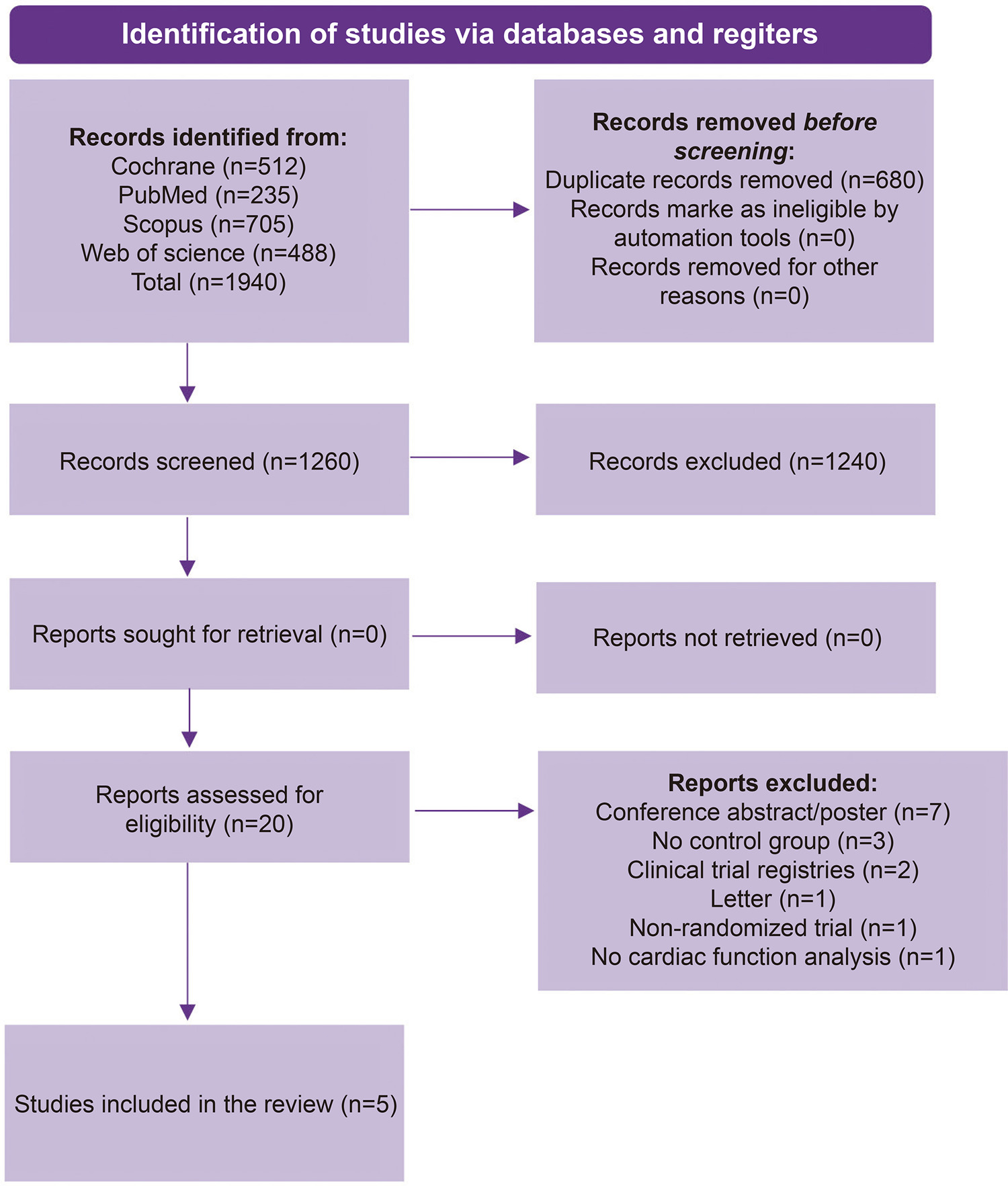
Only randomized clinical trials with humans published in English were included. Inclusion criteria were studies with type 2 diabetes patients, physical exercise, control group and left ventricular function. Exclusion criteria were studies with animals, children, teenagers, elderly individuals and athletes, presence of diet intervention, and patients with type 1 diabetes, cancer, cardiac, pulmonary, or neurological diseases. Electronic databases PubMed, Web of Science, Cochrane, and Scopus were last searched in September 2021. Risk of bias was assessed by the Physiotherapy Evidence Database (PEDro) scale.

Results

Five studies were included, representing 314 diabetic individuals submitted to resistance and aerobic exercise training. Of the variables analyzed, physical exercise improved peak torsion (PTo), global longitudinal strain, global strain rate (GSR), time to peak untwist rate (PUTR), early diastolic filling rate (EDFR) and peak early diastolic strain rate (PEDSR).

Conclusion

To our knowledge, this is the first systematic review on the effects of exercise on left ventricular function in T2DM including only randomized clinical trials with humans. Physical exercise seems to improve systolic and diastolic strain, twist, and torsion. High intensity exercise was reported to be superior to moderate intensity exercise in one study. This review was limited by the small number of studies and their heterogeneity regarding exercise protocols, follow-up period, exercise supervision and left ventricular function variables analyzed. This review was registered in PROSPERO (CRD42021234964).
  • Diabetes Mellitus
  • Echocardiography
  • Exercise
  • Ventricular Function, Left
Read Full Article
Creative Commons Attribution (CC BY) Creative Commons license
03/03/2023 DOI: 10.36660/ijcs.20220020
Figure 1
ABC
Original Article

Abnormal Echocardiographic Findings in Hospitalized Patients with Covid-19: A Systematic Review and Meta-analysis

Silvio Henrique Barberato et al. Eduardo G. Bruneto Gabriel S. Reis Paula Rauen Franco de Oliveira … Alexandre F. Possamai Odilson Silvestre Miguel M. Fernandes Silva

Abstract

Background

Coronavirus disease 2019 (Covid-19) can lead to severe respiratory distress and acute cardiac injury, but it is unclear how often it can cause cardiac dysfunction.

Objective

In this systematic review, we aimed to summarize the main echocardiographic findings in patients with Covid-19.

Methods

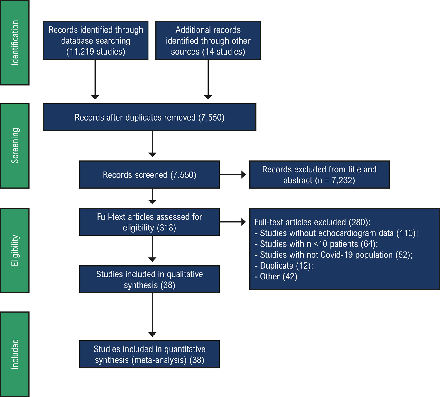
We systematically searched in PUBMED, EMBASE, LILACS and Cochrane databases, in addition MedRxiv and Scielo preprints from inception to July 21st, 2021. Studies reporting echocardiographic data in patients with Covid-19 were included. Demographic characteristics, previous cardiovascular disease (CVD), and echocardiographic findings were extracted. We performed a meta-analysis of proportions to estimate the main echocardiographic findings. The level of significance was p < 0.05.

Results

From 11,233 studies, 38 fulfilled inclusion criteria and were included in the meta-analysis. The estimated proportions of left ventricular (LV) systolic dysfunction were 25% (95%CI: 19, 31; I293%), abnormal global longitudinal strain 34% (95% CI 23, 45; I290%), righ ventricular (RV) systolic dysfunction 17% (95%CI 13, 21; I290%), pericardial effusion 17% (95%CI: 9, 26; I297%), and pulmonary hypertension 23% (95%CI: 15, 33, I2 96%). LV systolic dysfunction was directly associated with study-specific prevalence of previous abnormal echocardiogram (p<0.001). The proportion of patients in mechanical ventilation, indicating severity of disease, did not explain the heterogeneity in the proportions of LV dysfunction (p=0.37).

Conclusion

Among hospitalized patients with Covid-19, LV dysfunction has been reported in one quarter, with smaller proportions of right ventricular dysfunction, pericardial effusion and pulmonary hypertension. However, there was a higher proportion of LV dysfunction among studies reporting the presence of prior heart disease, which suggests that cardiac dysfunction was mostly pre-existing.

  • COVID-19
  • Echocardiography
  • Ventricular Function, Left
Read Full Article
Creative Commons Attribution (CC BY) Creative Commons license
11/07/2022 DOI: 10.36660/abc.20210485
Figure 1
ABC
Original Article

Sonothrombolysis Promotes Improvement in Left Ventricular Wall Motion and Perfusion Scores after Acute Myocardial Infarction

Bruno G. Tavares et al. Miguel Osman Aguiar Jeane Tsutsui Mucio Oliveira … Alexandre de Matos Soeiro José Nicolau Henrique Ribeiro Hsu PoChiang João Sbano Carlos Eduardo Rochitte Bernardo Lopes José Ramirez Roberto Kalil Filho Wilson Mathias

Abstract

Background

It has recently been demonstrated that the application of high-energy ultrasound and microbubbles, in a technique known as sonothrombolysis, dissolves intravascular thrombi and increases the angiographic recanalization rate in patients with ST-segment–elevation myocardial infarction (STEMI).

Objective

To evaluate the effects of sonothrombolysis on left ventricular wall motion and myocardial perfusion in patients with STEMI, using real-time myocardial perfusion echocardiography (RTMPE).

Methods

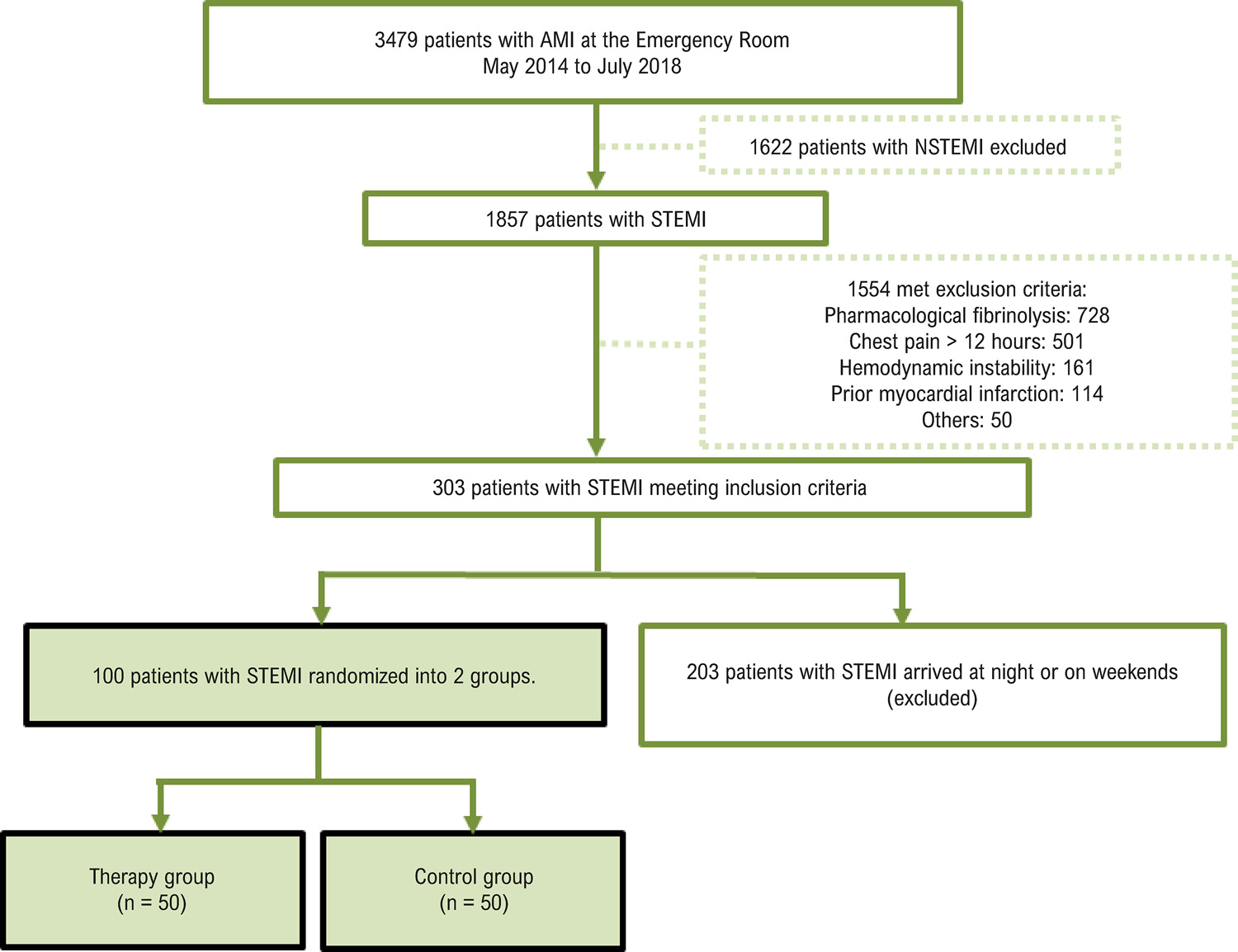
One hundred patients with STEMI were randomized into the following 2 groups: therapy (50 patients treated with sonothrombolysis and primary coronary angioplasty) and control (50 patients treated with primary coronary angioplasty). The patients underwent RTMPE for analysis of left ventricular ejection fraction (LVEF), wall motion score index (WMSI), and number of segments with myocardial perfusion defects 72 hours after STEMI and at 6 months of follow-up. P < 0.05 was considered statistically significant.

Results

Patients treated with sonothrombolysis had higher LVEF than the control group at 72 hours (50% ± 10% versus 44% ± 10%; p = 0.006), and this difference was maintained at 6 months of follow-up (53% ± 10% versus 48% ± 12%; p = 0.008). The WMSI was similar in the therapy and control groups at 72 hours (1.62 ± 0.39 versus 1.75 ± 0.40; p = 0.09), but it was lower in the therapy group at 6 months (1.46 ± 0.36 versus 1.64 ± 0.44; p = 0.02). The number of segments with perfusion defects on RTMPE was similar in therapy and control group at 72 hours (5.92 ± 3.47 versus 6.94 ± 3.39; p = 0.15), but it was lower in the therapy group at 6 months (4.64 ± 3.31 versus 6.57 ± 4.29; p = 0.01).

Conclusion

Sonothrombolysis in patients with STEMI resulted in improved wall motion and ventricular perfusion scores over time.

  • Contrast Media
  • Microbubbles
  • Myocardial Infarction
  • Pulmonary embolism
  • Sonothrombolysis
  • Ventricular Function, Left
Read Full Article
Creative Commons Attribution (CC BY) Creative Commons license
29/04/2022 DOI: 10.36660/abc.20200651 Views 1
ABC
Original Article

Factors That Impact the Decision to Perform Left Ventriculography in Coronary Artery Disease

Claudia de Castro Lima Santos et al. Ricardo Peixoto Oliveira Joberto Sena Adriano Dourado Oliveira … Marcelo Gottschald Ferreira Ademar Santos Filho Heitor Guissoni José Carlos Brito Gilson Soares Feitosa Gilson Soares Feitosa-Filho

Abstract

Background

Left ventriculography is an invasive method for assessment of left ventricular systolic function. Since the advent of noninvasive methods, its use has been questioned, as it carries some risk to the patient.

Objective

To assess which factors are independently associated with the decision to perform ventriculography in patients with coronary artery disease.

Methods

Analytical, retrospective, database review study of electronic medical records comparing 21 predefined variables of interest among patients undergoing coronary angiography. P-values <0.05 were considered significant.

Results

We evaluated 600 consecutive patients undergoing coronary angiography. Left ventriculography was performed in the majority of cases (54%). After multivariate analysis, patients with chronic coronary syndrome (OR 1.72; 95% CI: 1.20–2.46; p < 0.01) were more likely to undergo the procedure. Patients with known ventricular function (OR 0.58; 95% CI: 0.40–0.85; p < 0.01); those with a history of CABG (OR 0.31; 95% CI: 0.14–0.69; p < 0.01) or hypertension (OR 0.58; 95% CI: 0.36–0.94; p = 0.02); and those with higher creatinine levels (OR 0.42; 95% CI: 0.26–0.69; p < 0.01) had greater odds of not undergoing ventriculography.

Conclusions

In patients undergoing coronary angiography, a diagnosis of chronic coronary syndrome was independently associated with greater likelihood of left ventriculography, while having previously determined ventricular function, a history of hypertension or CABG, and higher creatinine levels were associated with a decreased likelihood of undergoing this procedure.

  • Cardiovascular Diseases
  • Coronary Angiography/methods
  • Coronary Artery Disease
  • Hypertension
  • Myocardial Revascularization/surgery
  • Ventricular Function, Left
  • Ventriculography/methods
Read Full Article
Creative Commons Attribution (CC BY) Creative Commons license
18/03/2022 DOI: 10.36660/abc.20200217 Views 2
ABCIC
Original Article

Semiautomatic Quantification of Left Ventricular Ejection Fraction and Volumes by Two-Dimensional Echocardiography: Comparison with Automatic Three-Dimensional Echocardiography

Rafael Borsoi et al. Miguel Morita Fernandes da Silva Silvio Henrique Barberato

Abstract

Background

Left ventricular ejection fraction is one of the most used echocardiographic parameters in clinical practice. Its estimation by two-dimensional manual method (Simpson method) has limited reproducibility and accuracy, and semi-automatic methods have been proposed. It becomes necessary to compare the semi-automatic two-dimensional method with more accurate methods of assessing left ventricular ejection fraction, such as measurement by automatic three-dimensional echocardiography.

Objective

To compare the left ventricular ejection fraction, and left ventricular end-diastolic and end-systolic volumes estimates by the semi-automatic two-dimensional method with those obtained using the automatic three-dimensional method.

Method

Observational cross-sectional study, including patients in sinus rhythm, left ventricular ejection fraction > 50% and without significant structural heart disease, submitted to transthoracic echocardiography. Student’s t test, Pearson’s coefficient and Bland-Altman analysis were used in the statistical analysis.

Results

Forty patients were included, 53% women, 35% with arterial hypertension, 25% with dyslipidemia, 10% diabetic, 10% smokers and 13% with previous angioplasty. The mean values of left ventricular ejection fraction by three-dimensional and two-dimensional were 62.1 ± 5.8% and 61.7 ± 5.9% (p = 0.50), respectively. There was a strong correlation between left ventricular ejection fraction determined by two-dimensional and three-dimensional (r = 0.75; p <0.001), as well as with left ventricular end-diastolic (r = 0.75; p <0.001) and end-systolic (r = 0.76; p < 0.001). There was good agreement between two-dimensional and three-dimensional left ventricular ejection fraction (mean difference: -0.39; 95% confidence interval -1.7-0.9).

Conclusion

Left ventricular ejection fraction estimated by the two-dimensional semi-automatic method showed good agreement with the automatic three-dimensional method. The findings suggest the two-dimensional semi-automatic method as a reliable alternative for assessing volumes and left ventricular ejection fraction.

  • Echocardiography, Doppler
  • Echocardiography, Three-Dimensional
  • Stroke Volume
  • Ventricular Function, Left
Read Full Article
Creative Commons Attribution (CC BY) Creative Commons license
17/12/2021 DOI: 10.47593/2675-312X/20203304eabc98
ABC
Original Article

Arterial Stiffness and Left Ventricular Myocardial Function in Children with a Well-Functioning Bicuspid Aortic Valve

Pelin Kosger et al. Tugcem Akin Hikmet Kiztanir Birsen Ucar

Abstract

Background:

Arterial stiffness is an important predictor factor of aortopathy and myocardial remodeling in patients with a bicuspid aortic valve and it might be increased in childhood.

Objective:

To assess the arterial stiffness and left ventricular myocardial function in children with a well-functioning bicuspid aortic valve.

Methods:

Forty-four children with a bicuspid aortic valve and 41 healthy peers with a tricuspid aortic valve were included in this case-control study. Diameters and the related z-scores of the aortic root and ascending aorta were obtained. As for the left ventricular myocardial function, along with the mitral inflow velocities and M-Mode parameters, myocardial velocities and time intervals were assessed with tissue Doppler imaging. A pulse wave analysis was performed by oscillometric device (Mobil-o-Graph). A p value <0.05 was considered significant.

Results:

The left ventricular mass index, mitral inflow A velocity, diameter and z-score of the ascending aorta, and myocardial performance index were significantly higher in patients (p=0.04, p=0.02,p=0.04, p<0.001,and p0.05).

Conclusion:

According to the oscillometric pulse wave analysis, the children with a well-functioning bicuspid aortic valve had similar arterial stiffness to that of the healthy peers. The ascending aorta diameter was established as an independent predictor of left ventricular myocardial function. Arterial stiffness may not be a severe risk factor in pediatric patients without marked ascending aorta dilation.

  • Aortic Stiffness
  • Child
  • Dilatation, Pathologic
  • Myocardial
  • Pulse Wave Analysis
  • Ventricular Function, Left
Read Full Article
Creative Commons Attribution (CC BY) Creative Commons license
20/09/2021 DOI: 10.36660/abc.20200657 Views 1
Figure 1
IJCS
Brief Communication

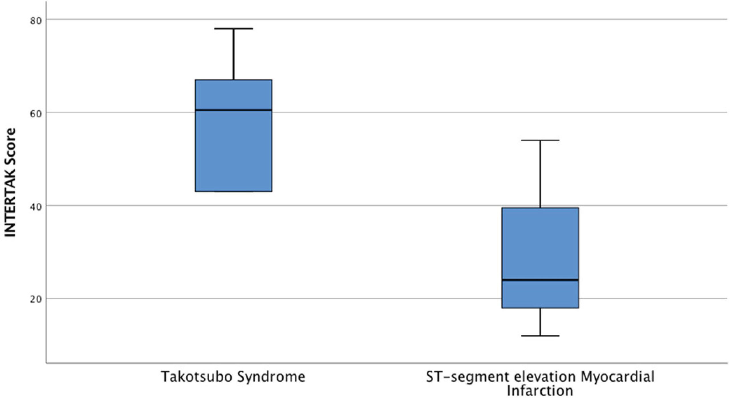
Takotsubo Syndrome in the Context of Transmural Acute Myocardial Infarction: Prevalence and How to Differentiate?

João Paulo Soares Costa et al. Luiz Eduardo Fonteles Ritt Rafael Modesto Fernandes Felipe Alvarenga Duarte Campos … Queila Borges Eduardo Sahade Darzé

Introduction

Takotsubo syndrome (TTS) is characterized by transient left ventricular dysfunction culminating in acute cardiac dysfunction. It is frequently preceded by emotional or physical stress.,

The most common symptoms at presentation are chest pain and dyspnea. Because the presentation of TTS is similar to that of acute coronary syndrome (ACS), the differential diagnosis is difficult, especially from ST-elevation myocardial infarction (STEMI). Absence of coronary artery disease in the affected area must be confirmed to establish the diagnosis of TTS. To facilitate the diagnosis of TTS, the European Society of Cardiology has recently developed the InterTAK diagnostic score, a promising tool that estimates the probability of a TTS event.,

[…]

  • Acute Coronary Syndrome / diagnosis
  • ST Elevation Myocardial Infarction
  • Stress, Physiological
  • Stress, Psychological
  • Takotsubo Cardiomyopathy
  • Ventricular Function, Left
Read Full Article
Creative Commons Attribution (CC BY) Creative Commons license
17/02/2020 DOI: 10.5935/2359-4802.20190080 Views 2
ABC
Letter to the Editor

Left Ventricle Mass Index, a Confounding Variable of Global Longitudinal Strain to be Noticed

Eduardo Thadeu de Oliveira Correia et al. Letícia Mara dos Santos Barbetta

Dear Editor,

We read with great interest the article entitled as “Strain Analysis of Left Ventricular Function in the Association of Hypertrophic Cardiomyopathy and Systemic Arterial Hypertension”. In this paper, the authors evaluated the global longitudinal strain (GLS) of the left ventricle (LV) in two distinct groups: patients with hypertrophic cardiomyopathy (HCM) and patients with HCM and systemic arterial hypertension (SAH) and demonstrated that GLS was lower in the second group. This important finding may indicate greater impairment of LV function in patients with SAH and HCM.

[…]

  • Cardiomyopathy, Hypertrophic
  • Heart Failure
  • Hypertension
  • Hypertrophy Left Ventricular
  • Strain
  • Ventricular Function, Left
Read Full Article
Creative Commons Attribution (CC BY) Creative Commons license
10/02/2020 DOI: 10.36660/abc.20190708 Views 1
ABC
Editorial

Aortic Arch Calcification on routine Chest Radiography is Strongly and Independently Associated with Non-Dipper Blood Pressure Pattern

Rui Póvoa

Arterial hypertension is the most prevalent chronic disease worldwide, and it is estimated that in Brazil, according to the Vigitel study of 2018, the Brazilian population has 24.7% of hypertensive individuals. It is the most important risk factor for all cardiovascular diseases, being one of the main factors responsible for stroke, coronary artery disease, heart failure, renal failure, and so on.

Blood pressure is not simply a biological phenomenon characterized by two numbers expressed in mmHg that reflect systolic and diastolic pressure. It is a multiform event resulting from the hydrodynamic action of a complex fluid that exerts force on a compliant vascular wall, with elastic properties governed by multiple forces arising from vascular structures (muscle cells, extracellular matrix, elastic fibers, etc.). This vector wave of blood follows the laws of sympathetic and parasympathetic interaction, varying more or less depending on the conditions of each patient. Therefore, the simple measurement of blood pressure in the office does not reflect the reality of the patient’s true daily life.

[…]

  • antihypertensive agents
  • Blood Pressure Monitoring, Ambulatory
  • calcification
  • Calcinosis
  • Cardiomyopathies
  • Heart Rate
  • Hypertension
  • Thoracic, Aorta/physiopathology
  • Ventricular Function, Left
Read Full Article
Creative Commons Attribution (CC BY) Creative Commons license
10/02/2020 DOI: 10.36660/abc.20190790 Views 1
ABC
Editorial

Real-Time Three-Dimensional Echocardiography and Myocardial Strain: Ready for Use in Clinical Practice

Frederico Jose Neves Mancuso

The ability of echocardiography to quantify volumes and to evaluate the contractile function of cardiac chambers has evolved greatly in recent years, particularly due to the development of real-time three-dimensional echocardiography (echo3D) and the possibility myocardial deformation (Strain) analysis by the speckle tracking technique.–

Unlike two-dimensional echocardiography, where geometric inferences and mathematical calculations are required, the echo3D allows direct measurement of ventricular and atrial volumes, from which function data such as left and right ventricular ejection fraction, and left atrial function, including total emptying fraction and active emptying fraction parameters.–

[…]

  • Echocardiography, Three-Dimensional
  • Reference Values
  • Speckle Tracking
  • Strain
  • Ventricular Function, Left
Read Full Article
Creative Commons Attribution (CC BY) Creative Commons license
02/12/2019 DOI: 10.5935/abc.20190179
Figure 1
ABC
Original Article

Strain Analysis of Left Ventricular Function in the Association of Hypertrophic Cardiomyopathy and Systemic Arterial Hypertension

Thereza Cristina Pereira Gil et al. Marcia Bueno Castier Alyne Freitas Pereira Gondar Ana Ferreira Sales … Marceli de Oliveira Santos Fernanda Cristina da Silva de Lima Ricardo Mourilhe-Rocha

Abstract

Background:

Hypertrophic cardiomyopathy (HCM) is the most common heart disease of genetic origin in the world population, with a prevalence of at least 1/500. The association with systemic arterial hypertension (SAH) is not uncommon, as it affects approximately 25% of the world population. Most studies aim at the differential diagnosis between these diseases, but little is known about the magnitude of this association.

Objective:

To compare left ventricular global longitudinal strain (GLS) in HCM patients with and without associated SAH.

Methods:

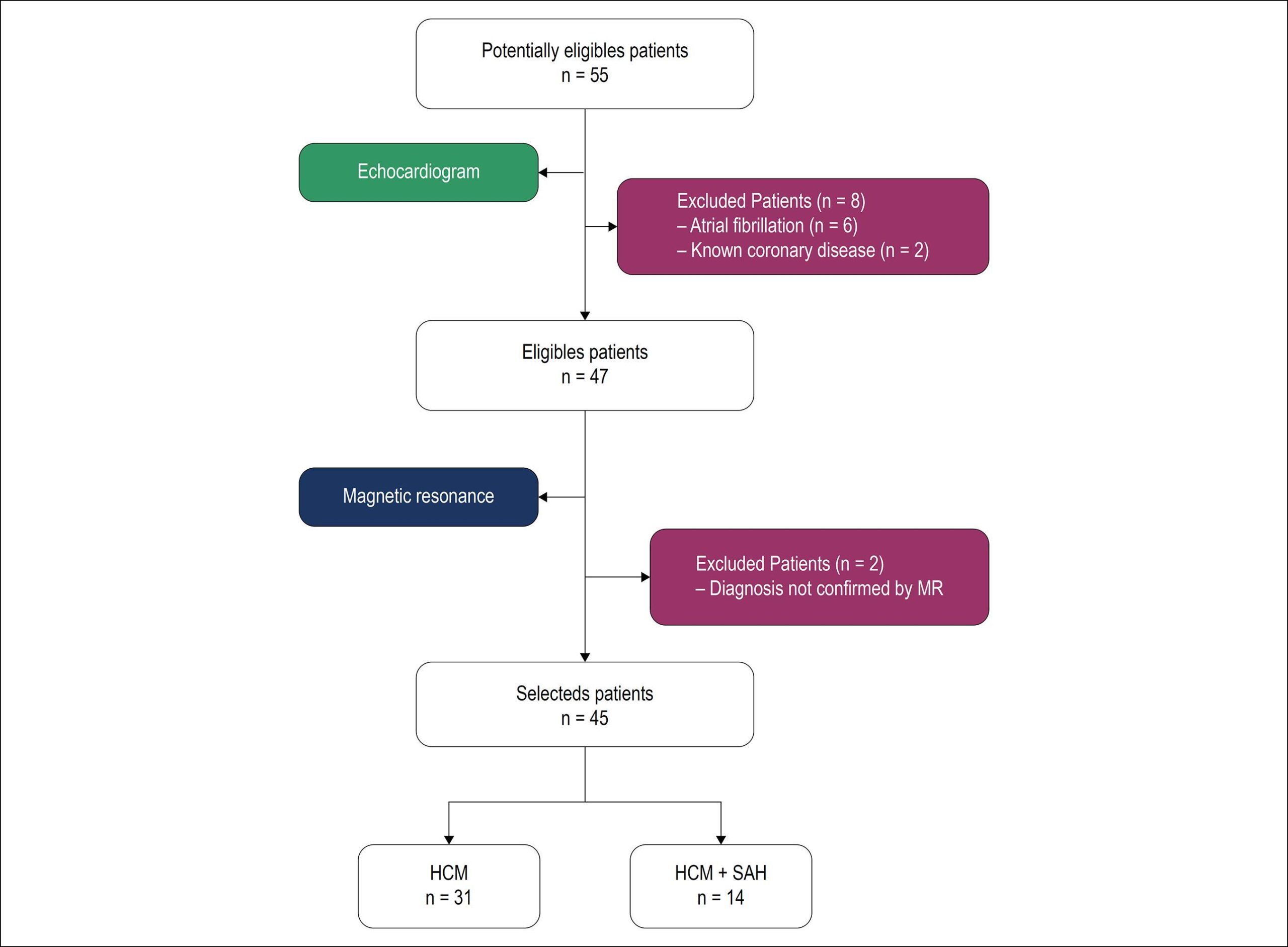
Retrospective cross-sectional study that included 45 patients with HCM and preserved ejection fraction, with diagnosis confirmed by magnetic resonance imaging, including 14 hypertensive patients. Transthoracic echocardiography was performed, with emphasis on left ventricular myocardial strain analysis using GLS. In this study, p < 0.05 was considered statistically significant.

Results:

Left ventricular strain was significantly lower in hypertensive individuals compared to normotensive individuals (-10.29 ± 2.46 vs. -12.35% ± 3.55%, p = 0.0303), indicating greater impairment of ventricular function in that group. Mean age was also significantly higher in hypertensive patients (56.1 ± 13.9 vs. 40.2 ± 12.7 years, p = 0.0001). Diastolic dysfunction was better characterized in hypertensive patients (p = 0.0242).

Conclusion:

Myocardial strain was significantly lower in the group of patients with HCM and SAH, suggesting greater impairment of ventricular function. This finding may be related to a worse prognosis with early evolution to heart failure. Prospective studies are required to confirm this hypothesis.

  • Cardiomyopathy, Hypertrophic
  • Heart Failure
  • Hypertension
  • Strain
  • Ventricular Function, Left
Read Full Article
Creative Commons Attribution (CC BY) Creative Commons license
02/09/2019 DOI: 10.5935/abc.20190176
Figure 1
ABC
Original Article

Adropin and Irisin in Patients with Cardiac Cachexia

Ali Kemal Kalkan et al. Huseyin Altug Cakmak Mehmet Erturk Kübra Erol Kalkan … Fatih Uzun Omer Tasbulak Vesile Ornek Diker Suleyman Aydin Ahmet Celik

Abstract

Background:

Cardiac cachexia is an important predictive factor of the reduction in survival of patients with heart failure with reduced ejection fraction.

Objectives:

The aims of the present study were to evaluate adropin and irisin levels in cachectic and non-cachectic subjects and the relationships between the levels of these proteins and clinical and laboratory parameters in patients with HFrEF.

Methods:

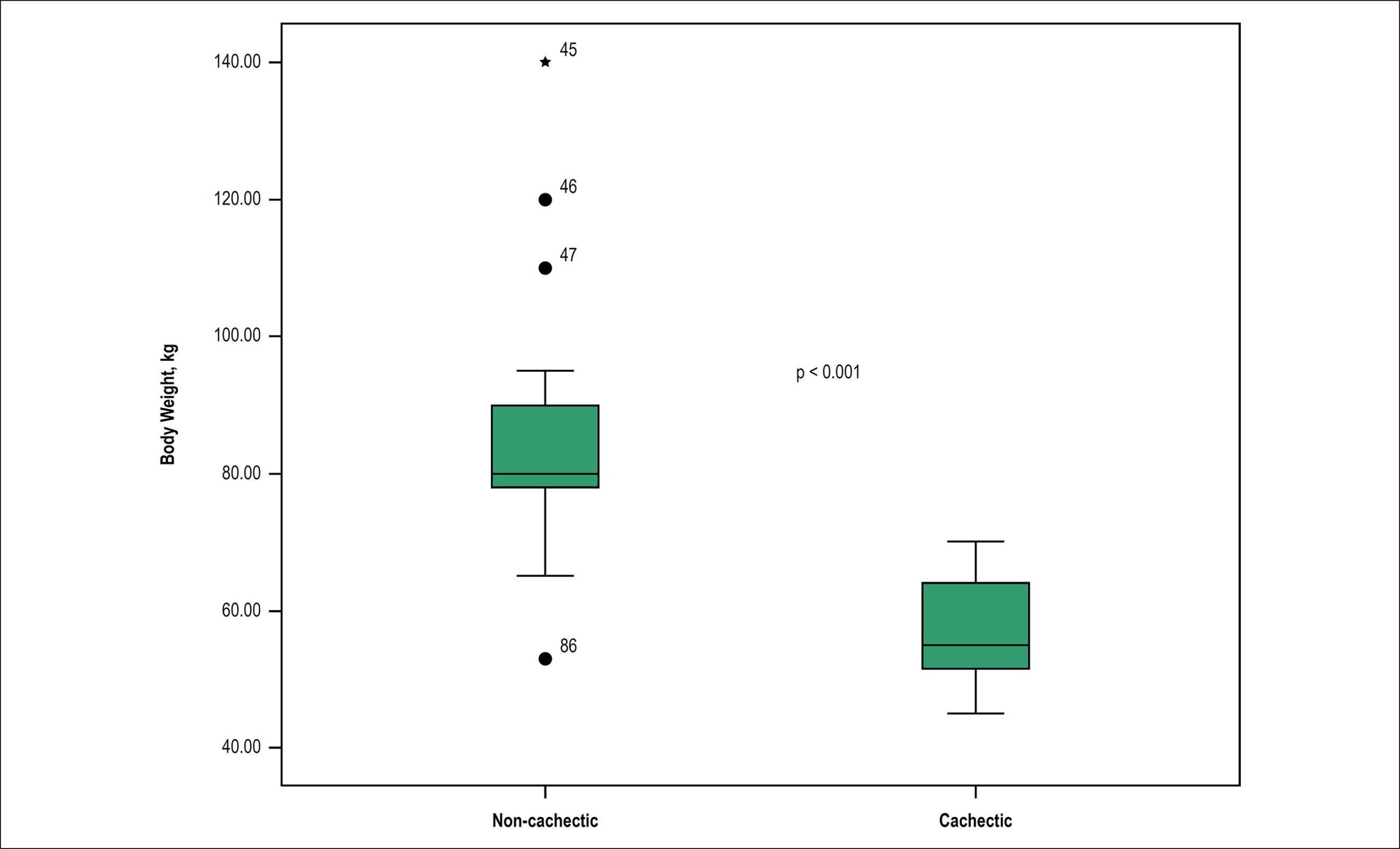
The clinical records of patients who were admitted to the cardiology outpatient clinic for heart failure with reduced ejection fraction were screened. Cachectic patients were identified and assigned to the study group (n = 44, mean age, 65.4 ± 11.2 y; 61.4% men). Heart failure with reduced ejection fraction patients without weight loss were enrolled as the control group (n = 42, mean age, 61.0 ± 16.5 y; 64.3% men). The serum adropin and irisin levels of all patients were measured. A p-value < 0.05 was considered significant.

Results:

Serum adropin and irisin levels were significantly higher in the cachexia group than in the controls (Adropin (ng/L); 286.1 (231.3-404.0) vs 213.7 (203.1-251.3); p < 0.001, Irisin (µg/mL); 2.6 (2.2-4.4) vs 2.1 (1.8-2.4); p = 0.001). Serum adropin and irisin levels were positively correlated with brain natriuretic peptide (BNP) levels and New York Heart Association (NYHA) class and negatively correlated with body mass index (BMI) and serum albumin levels (all p values: < 0.001). In a multivariate analysis, adropin was the only independent predictor of cachexia in the heart failure with reduced ejection fraction patients (OR: 1.021; 95% CI: 1.004−1.038; p = 0.017).

Conclusions:

The results suggest that adropin and irisin may be novel markers of cardiac cachexia in heart failure with reduced ejection fraction patients. Adropin and irisin are related with the severity of heart failure.

  • Adropin
  • Cachexia / complications
  • Heart Failure / physiopathology, Hypertrophy, Left Ventricular
  • Hormones
  • Peptides
  • Ventricular Function, Left
Read Full Article
Creative Commons Attribution (CC BY) Creative Commons license
02/07/2018 DOI: 10.5935/abc.20180109
Graph 1
ABC
Original Article

Impact of a High-Intensity Training on Ventricular Function in Rats After Acute Myocardial Infarction

Simone de Campos Neitzke Winter et al. Rafael Michel de Macedo Júlio Cesar Francisco Paula Costa Santos … Ana Paula Sarraff Lopes Leanderson Franco de Meira Katherine A. Teixeira de Carvalho José Rocha Faria Neto Ana Carolina Brandt de Macedo Luiz César Guarita-Souza

Abstract

Background:

Physical exercise should be part of the treatment of post-acute myocardial infarction (AMI) patients.

Objective:

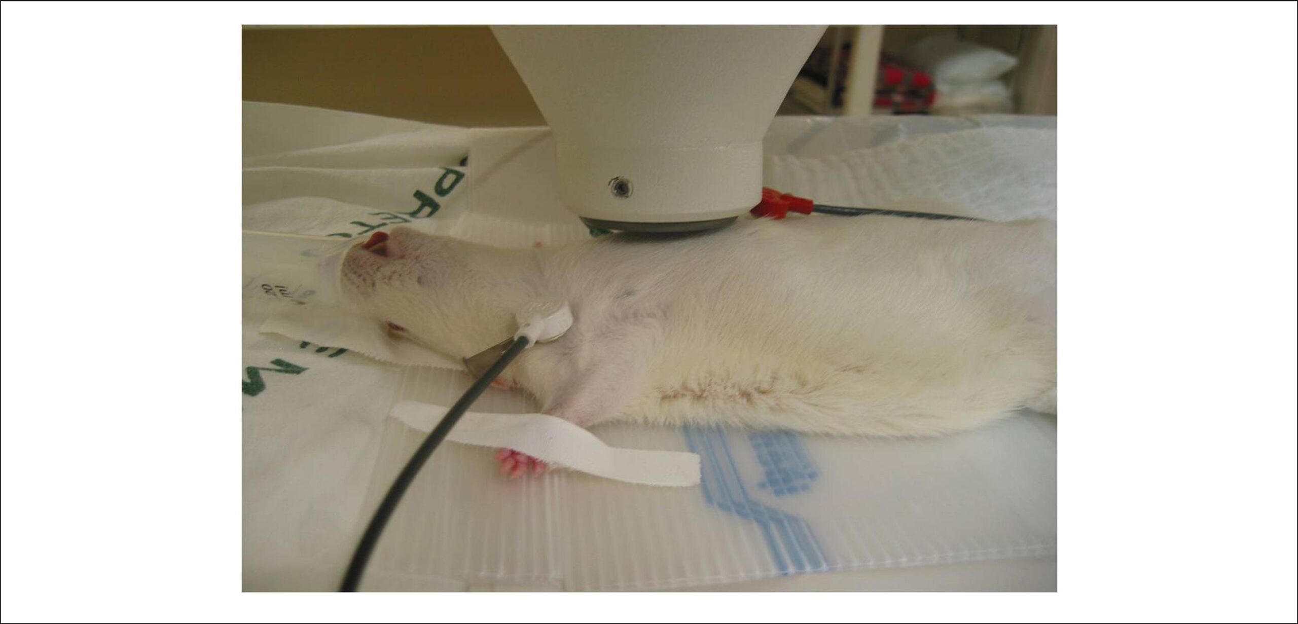
To evaluate the effects of two training prescription models (continuous x interval) and its impact on ventricular function in rats after AMI with normal ventricular function.

Methods:

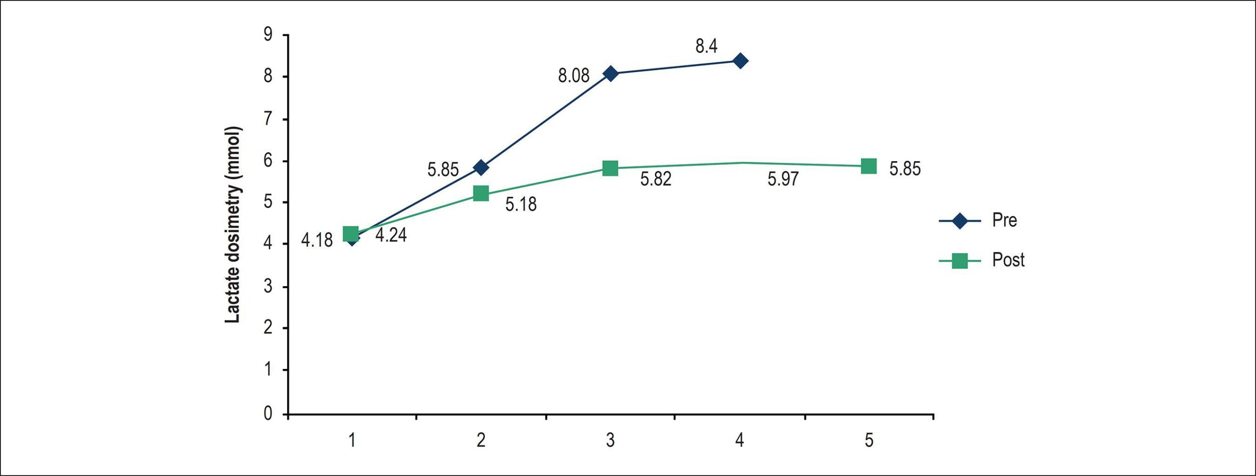
Forty Wistar rats were evaluated by echocardiography 21 days after the AMI. Those with LVEF = 50% (n = 29) were included in the study and randomized to control group (CG n = 10), continuous training group (CTG n = 9) or interval training group (ITG, n = 10). Then, a swimming test with control of lactate production was performed. Based on its result, the lactate threshold (LT) was established to define the training intensities. After six weeks, the animals were reassessed by echocardiography and lactate production. Outcome measures were end-diastolic diameter (EDD), end-systolic diameter (ESD), left ventricular ejection fraction (LVEF, %) lactate at rest, lactate without overload, and lactate with 12g and 13.5g of additional load. Group comparisons of quantitative variables of the study were performed by one-factor analysis of variance (ANOVA). The Newman-Keuls test was used for multiple comparisons of the groups. Within-group comparisons of dependent variables between the two training protocols were performed by Student’s t-test. Normality of the variables was tested by the Shapiro-Wilks test. Values of p < 0.05 indicated statistical significance.

Results:

EDD, ESD, and LVEF before and after the training period were similar in within-group comparisons. However, EDD was significantly different (p=0.008) in the CG. Significant differences were found for L12g (p=0.002) and L13.5g (p = 0.032) in the ITG, and for L12g (p = 0.014) in the CG. No differences were found in the echocardiographic parameters between the groups. Significant differences were found in lactate without overload (p = 0.016) and L12 (p = 0.031) in the second assessment compared with the first, and between the groups – ITG vs. CG (p = 0.019) and CTG vs. CG (p = 0.035).

Conclusion:

Both methods produced a training effect without altering ventricular function.

  • Anaerobic Threshold
  • Exercise
  • Myocardial Infarction
  • Rats
  • Ventricular Function, Left
Read Full Article
Creative Commons Attribution (CC BY) Creative Commons license
12/03/2018 DOI: 10.5935/abc.20180036 Views 1
Figure 1
ABC
Original Article

Natriuretic Peptide and Clinical Evaluation in the Diagnosis of Heart Failure Hemodynamic Profile: Comparison with Tissue Doppler Echocardiography

Gustavo Luiz Gouvêa de Almeida Junior et al. Nadine Clausell Marcelo Iorio Garcia Roberto Esporcatte … Fernando Oswaldo Dias Rangel Ricardo Mourilhe Rocha Luis Beck-da-Silva Fabricio Braga da Silva Paula de Castro Carvalho Gorgulho Sergio Salles Xavier

Abstract

Background:

Physical examination and B-type natriuretic peptide (BNP) have been used to estimate hemodynamics and tailor therapy of acute decompensated heart failure (ADHF) patients. However, correlation between these parameters and left ventricular filling pressures is controversial.

Objective:

This study was designed to evaluate the diagnostic accuracy of physical examination, chest radiography (CR) and BNP in estimating left atrial pressure (LAP) as assessed by tissue Doppler echocardiogram.

Methods:

Patients admitted with ADHF were prospectively assessed. Diagnostic characteristics of physical signs of heart failure, CR and BNP in predicting elevation (> 15 mm Hg) of LAP, alone or combined, were calculated. Spearman test was used to analyze the correlation between non-normal distribution variables. The level of significance was 5%.

Results:

Forty-three patients were included, with mean age of 69.9 ± 11.1years, left ventricular ejection fraction of 25 ± 8.0%, and BNP of 1057 ± 1024.21 pg/mL. Individually, all clinical, CR or BNP parameters had a poor performance in predicting LAP ≥ 15 mm Hg. A clinical score of congestion had the poorest performance [area under the receiver operating characteristic curve (AUC) 0.53], followed by clinical score + CR (AUC 0.60), clinical score + CR + BNP > 400 pg/mL (AUC 0.62), and clinical score + CR + BNP > 1000 pg/mL (AUC 0.66).

Conclusion:

Physical examination, CR and BNP had a poor performance in predicting a LAP ≥ 15 mm Hg. Using these parameters alone or in combination may lead to inaccurate estimation of hemodynamics.

  • Echocardiography, Doppler
  • Heart Failure
  • Hemodynamics
  • Natriuretic Peptide, Brain
  • Ventricular Function, Left
Read Full Article
Creative Commons Attribution (CC BY) Creative Commons license
01/03/2018 DOI: 10.5935/abc.20180046 Views 1
Figure 1
ABC
Original Article

Comparison between Radionuclide Ventriculography and Echocardiography for Quantification of Left Ventricular Systolic Function in Rats Exposed to Doxorubicin

Luciano Fonseca Lemos de Oliveira et al. João Lucas O'Connell Eduardo Elias Vieira de Carvalho Érica Carolina Campos Pulici … Minna Moreira Dias Romano Benedito Carlos Maciel Marcus Vinicius Simões

Abstract

Background:

Radionuclide ventriculography (RV) is a validated method to evaluate the left ventricular systolic function (LVSF) in small rodents. However, no prior study has compared the results of RV with those obtained by other imaging methods in this context.

Objectives:

To compare the results of LVSF obtained by RV and echocardiography (ECHO) in an experimental model of cardiotoxicity due to doxorubicin (DXR) in rats.

Methods:

Adult male Wistar rats serving as controls (n = 7) or receiving DXR (n = 22) in accumulated doses of 8, 12, and 16 mg/kg were evaluated with ECHO performed with a Sonos 5500 Philips equipment (12-MHz transducer) and RV obtained with an Orbiter-Siemens gamma camera using a pinhole collimator with a 4-mm aperture. Histopathological quantification of myocardial fibrosis was performed after euthanasia.

Results:

The control animals showed comparable results in the LVSF analysis obtained with ECHO and RV (83.5 ± 5% and 82.8 ± 2.8%, respectively, p > 0.05). The animals that received DXR presented lower LVSF values when compared with controls (p < 0.05); however, the LVSF values obtained by RV (60.6 ± 12.5%) were lower than those obtained by ECHO (71.8 ± 10.1%, p = 0.0004) in this group. An analysis of the correlation between the LVSF and myocardial fibrosis showed a moderate correlation when the LVSF was assessed by ECHO (r = -0.69, p = 0.0002) and a stronger correlation when it was assessed by RV (r = -0.79, p < 0.0001). On multiple regression analysis, only RV correlated independently with myocardial fibrosis.

Conclusion:

RV is an alternative method to assess the left ventricular function in small rodents in vivo. When compared with ECHO, RV showed a better correlation with the degree of myocardial injury in a model of DXR-induced cardiotoxicity.

  • Comparative Study
  • Doxorubicin
  • Echocardiography/methods
  • Radionuclide Ventriculography / methods
  • Rats
  • Ventricular Function, Left
Read Full Article
Creative Commons Attribution (CC BY) Creative Commons license
01/01/2017 DOI: 10.5935/abc.20160194 Views 1
Figure 1
ABC
Brief Communication

Diastolic Function in Paced Children with Cardiac Defects: Septum vs Apex

Michel Cabrera Ortega et al. Adel Eladio Gonzalez Morejon Giselle Ricardo Serrano Dunia Barbara Benitez Ramos

Abstract

In children with structural congenital heart disease (CHD), the effects of chronic ventricular pacing on diastolic function are not well known. On the other hand, the beneficial effect of septal pacing over apical pacing is still controversial.

The aim of this study was to evaluate the influence of different right ventricular (RV) pacing site on left ventricular (LV) diastolic function in children with cardiac defects.

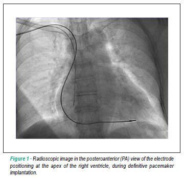
Twenty-nine pediatric patients with complete atrioventricular block (CAVB) and CHD undergoing permanent pacing were prospectively studied. Pacing sites were RV apex (n = 16) and RV septum (n = 13). Echocardiographic assessment was performed before pacemaker implantation and after it, during a mean follow‑up of 4.9 years.

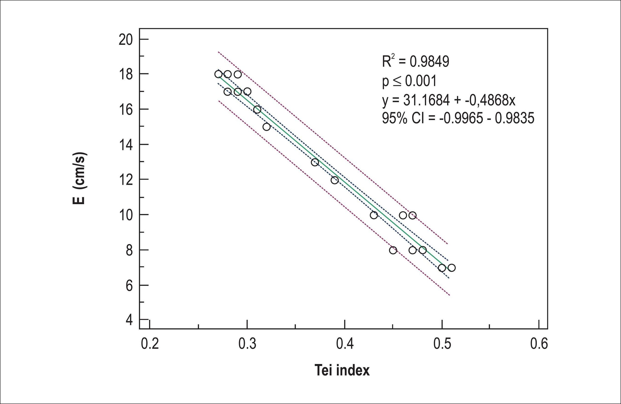
Compared to RV septum, transmitral E-wave was significantly affected in RV apical pacing (95.38 ± 9.19 vs 83 ± 18.75, p = 0.038). Likewise, parameters at the lateral annular tissue Doppler imaging (TDI) were significantly affected in children paced at the RV apex. The E´ wave correlated inversely with TDI lateral myocardial performance index (Tei index) (R2= 0.9849, p ≤ 0.001). RV apex pacing (Odds ratio, 0.648; confidence interval, 0.067-0.652; p = 0.003) and TDI lateral Tei index (Odds ratio, 31.21; confidence interval, 54.6-177.4; p = 0.025) predicted significantly decreased LV diastolic function.

Of the two sites studied, RV septum prevents pacing-induced reduction of LV diastolic function.

  • Child
  • Heart Defects, Congenital
  • Pacemaker, Artificial
  • Ventricular Function, Left
  • Ventricular Function, Right
Read Full Article
Creative Commons Attribution (CC BY) Creative Commons license
01/08/2015 DOI: 10.5935/abc.20150077 Views 1
Left ventricular synchrony and function in pediatric patients with definitive pacemakers
ABC
Artigo Original

Left ventricular synchrony and function in pediatric patients with definitive pacemakers

Michel Cabrera Ortega et al. Adel Eladio Gonzales Morejón Giselle Serrano Ricardo

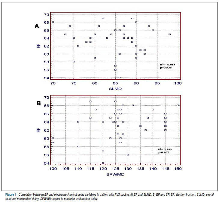
BACKGROUND: Chronic right ventricular pacing (RVP) induces a dyssynchronous contraction pattern,producing interventricular and intraventricular asynchrony. Many studies have shown the relationship of RVP with impaired left ventricular (LV) form and function. OBJECTIVE: The aim of this study was to evaluate LV synchrony and function in pediatric patients receiving RVP in comparison with those receiving LV pacing (LVP). METHODS:LV systolic and diastolic function and synchrony were evaluated in 80 pediatric patients with either nonsurgical or postsurgical complete atrioventricular block, with pacing from either the RV endocardium (n = 40) or the LV epicardium (n = 40). Echocardiographic data obtained before pacemaker implantation, immediately after it, and at the end of a mean follow-up of 6.8 years were analyzed. RESULTS: LV diastolic function did not change in any patient during follow-up. LV systolic function was preserved in patients with LVP. However, in children with RVP the shortening fraction and ejection fraction decreased from medians of 41% ± 2.6% and 70% ± 6.9% before implantation to 32% ± 4.2% and 64% ± 2.5% (p < 0.0001 and p < 0.0001), respectively, at final follow-up. Interventricular mechanical delay was significantly larger with RVP (66 ± 13 ms) than with LVP (20 ± 8 ms). Similarly, the following parameters were significantly different in the two groups: LV mechanical delay (RVP: 69 ± 6 ms, LVP: 30 ± 11 ms, p < 0.0001); septal to lateral wall motion delay (RVP: 75 ± 19 ms, LVP: 42 ± 10 ms, p < 0.0001); and, septal to posterior wall motion delay (RVP: 127 ± 33 ms, LVP: 58 ± 17 ms, p < 0.0001). CONCLUSION: Compared with RV endocardium, LV epicardium is an optimal site for pacing to preserve cardiac synchrony and function.

  • Artificial
  • Cardiac Pacing, Artificial
  • Child
  • Myocardial Contraction
  • Pacemaker
  • Ventricular Function, Left
Read Full Article
Creative Commons Attribution (CC BY) Creative Commons license
24/09/2013 DOI: 10.5935/abc.20130189
3D Echo pilot study of geometric Left Ventricular changes after acute myocardial infarction
ABC
Artigo Original

3D Echo pilot study of geometric Left Ventricular changes after acute myocardial infarction

Marcelo Luiz Campos Vieira et al. Wercules Antonio Oliveira Adriana Cordovil Ana Clara Tude Rodrigues … Cláudia Gianini Mônaco Tânia Afonso Edgar Bezerra Lira Filho Marco Perin Cláudio Henrique Fischer Samira Saady Morhy

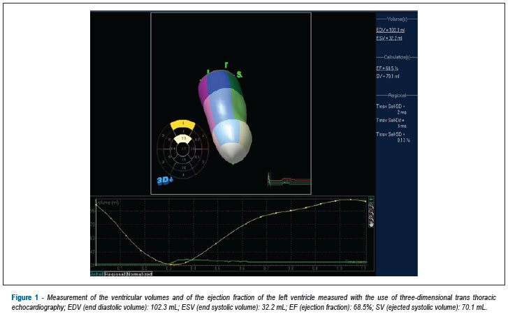
BACKGROUND: Left ventricular remodeling (LVR) after AMI characterizes a factor of poor prognosis. There is little information in the literature on the LVR analyzed with three-dimensional echocardiography (3D ECHO). OBJECTIVE: To analyze, with 3D ECHO, the geometric and volumetric modifications of the left ventricle (VE) six months after AMI in patients subjected to percutaneous primary treatment. METHODS: Prospective study with 3D ECHO of 21 subjects (16 men, 56 ± 12 years-old), affected by AMI with ST segment elevation. The morphological and functional analysis (LV) with 3D ECHO (volumes, LVEF, 3D sphericity index) was carried out up to seven days and six months after the AMI. The LVR was considered for increase > 15% of the end diastolic volume of the LV (LVEDV) six months after the AMI, compared to the LVEDV up to seven days from the event. RESULTS: Eight (38%) patients have presented LVR. Echocardiographic measurements (n = 21 patients): I- up to seven days after the AMI: 1- LVEDV: 92.3 ± 22.3 mL; 2- LVEF: 0.51 ± 0.01; 3- sphericity index: 0.38 ± 0.05; II- after six months: 1- LVEDV: 107.3 ± 26.8 mL; 2- LVEF: 0.59 ± 0.01; 3- sphericity index: 0.31 ± 0.05. Correlation coefficient (r) between the sphericity index up to seven days after the AMI and the LVEDV at six months (n = 8) after the AMI: r: 0.74, p = 0.0007; (r) between the sphericity index six months after the AMI and the LVEDV at six months after the AMI: r: 0.85, p < 0.0001. CONCLUSION: In this series, LVR has been observed in 38% of the patients six months after the AMI. The three-dimensional sphericity index has been associated to the occurrence of LVR.

  • Angioplasty
  • Balloon, Coronary
  • Echocardiography, Three-Dimensional
  • Myocardial Infarction
  • Stents
  • Ventricular Function, Left
Read Full Article
Creative Commons Attribution (CC BY) Creative Commons license
31/05/2013 DOI: 10.5935/abc.20130112
Metalloproteinases-2 and -9 predict left ventricular remodeling after myocardial infarction
ABC
Artigo Original

Metalloproteinases-2 and -9 predict left ventricular remodeling after myocardial infarction

Ana Lucia Cogni et al. Elaine Farah Marcos F. Minicucci Paula S. Azevedo … Katashi Okoshi Beatriz B. Matsubara Silméia Zanati Rodrigo Haggeman Sergio A. R. Paiva Leonardo A. M. Zornoff

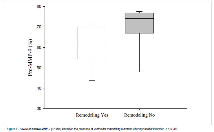
BACKGROUND: The role of serum metalloproteinases (MMP) after myocardial infarction (MI) is unknown. OBJECTIVE: The aim of this study was to evaluate the role of serum MMP-2 and -9 as predictors of ventricular remodeling six months after anterior MI. METHODS: We prospectively enrolled patients after their first anterior MI. MMP activity was assayed 12 to 72 hours after the MI. An echocardiogram was performed during the hospitalization and six months later. RESULTS: We included 29 patients; 62% exhibited ventricular remodeling. The patients who exhibited remodeling had higher infarct size based on creatine phosphokinase (CPK) peak values (p = 0.037), higher prevalence of in-hospital congestive heart failure (p = 0.004), and decreased ejection fraction (EF) (p = 0.007). The patients with ventricular remodeling had significantly lower serum levels of inactive MMP-9 (p = 0.007) and significantly higher levels of the active form of MMP-2 (p = 0.011). In a multivariate logistic regression model, adjusted by age, CPK peak, EF and prevalence of heart failure, MMP-2 and -9 serum levels remained associated with remodeling (p = 0.033 and 0.044, respectively). CONCLUSIONS: Higher serum levels of inactive MMP-9 were associated with the preservation of left ventricular volumes, and higher serum levels of the active form of MMP-2 were a predictor of remodeling 6 months after MI.

  • Matrix Metalloproteinase 2
  • Matrix Metalloproteinase 9
  • Myocardial Infarction
  • Ventricular Function, Left
Read Full Article
Creative Commons Attribution (CC BY) Creative Commons license
22/03/2013 DOI: 10.5935/abc.20130049
ABC
Artigo Original

Quantification of left ventricular dilatation in myocardial perfusion scintigraphy

Mauren B. Azambuja Gonzalez et al. Roberto Alves Azambuja Luiz Carlos Bodanese

BACKGROUND: The rate of transient dilatation can be determined by exercise testing or pharmacological stress test. It is unknown whether the type of stress has an impact on average transient dilatation index values. OBJECTIVE: To compare average transient dilation index values in 99mTc-sestamibi scintigraphy in patients undergoing treadmill stress test, versus dipyridamole stress test. The secondary purpose was to evaluate the impact on the average index value by demographic characteristics, risk factors for coronary artery disease and severity of ischemia. METHODS: The cross-sectional study included 200 patients between 40 and 70 years old, with or without risk factors for ischemic heart disease, with or without a previous diagnosis of ischemic heart disease. The separation between groups was sequential. The software 4D-MSPECT calculated the transient dilatation index and provided a scoring system for perfusion analysis. RESULTS: The average transient dilation index value of the group undergoing exercise stress test was 1.06 (±0.23). For the group undergoing the dipyridamole stress test, it was 1.10 (±0.22); (p = 0.200). There was no association between the type of stress and the average transient dilatation index values. An association was found between the average index values and age only for those patients from the exercise test group (p = 0.009). CONCLUSION: The results of our study demonstrate that the transient dilation index does not differ when patients undergo exercise stress test on a treadmill or pharmacological stress by dipyridamole.

  • Dilatation
  • Dipyridamole
  • Exercise Test
  • Radionuclide Imaging
  • Ventricular Function, Left
Read Full Article
Creative Commons Attribution (CC BY) Creative Commons license
15/04/2011 DOI: 10.1590/S0066-782X2011005000042
ABC
Artigo Original

Predictors of global left ventricular function in metabolic syndrome

Branislava Aleksa Ivanovic et al. Marijana Vaso Tadic Dragan Vojislav Simic

BACKGROUND: The metabolic syndrome (MS) represents a cluster of cardiovascular risk factors that act synergistically. OBJECTIVE: The aim of this study was to determine which parameters were independently associated with the global left ventricular (LV) function in subjects with MS estimated with the Tei index. METHODS: The study included 234 subjects with MS and 96 controls adjusted by age. MS was defined by the presence of three or more of ATP- NCEP III criteria. All subjects underwent laboratory blood tests and two-dimensional, pulsed and tissue Doppler echocardiography. Appropriate tissue Doppler time intervals for the estimation of the Tei index were also assessed. RESULTS: The Tei index was increased in subjects with MS (0.35 ± 0.05 vs 0.49 ± 0.10, p < 0.001). Multiple regression analysis of the clinical parameters showed that systolic blood pressure (β= 0.289, p < 0.001), fasting glucose (β= 0.205, p = 0.009), LV mass index (β= 0.301, p < 0.001), E/e'septal (β= 0.267, p < 0.001), and e'septal (β= -0.176, p = 0.011) were independently associated with the global left ventricular function estimated by Tei index. CONCLUSION: MS has a significant impact on LV global function. Systolic blood pressure, fasting glucose, LV mass index, E/e'septal, and e'septal were independently associated with the LV global function.

  • Metabolic Syndrome
  • Myocardial Ischemia
  • Risk Factors
  • Ventricular Function, Left
Read Full Article
Creative Commons Attribution (CC BY) Creative Commons license
01/04/2011 DOI: 10.1590/S0066-782X2011005000039
Alternative endocardial sites for artificial cardiac stimulation
ABC
Review Article

Alternative endocardial sites for artificial cardiac stimulation

Otaviano da Silva Júnior et al. Celso Salgado de Melo Marcelo Marra Dalmo Correia

The conventional right ventricular stimulation can be associated with deleterious effects on cardiac function. The need for a more physiological artificial cardiac stimulation is undoubtedly one of the most important points in the area of cardiac electrotherapy. The programming algorithms for the maintenance of adequate atrioventricular conduction, the stimulation of alternative endocardial sites and the cardiac resynchronization therapy are used with the objective of attaining these goals. The stimulation of the bundle of His and the septal stimulation have been studied as alternative endocardial sites for the positioning of the electrode on the right ventricle. The septal stimulation represents a simple and practical alternative, with no additional costs involved and with potential benefits in decreasing the deleterious effects of the right ventricular stimulation. However, this alternative site involves a heterogeneous group of patients and presents conflicting results regarding its long-term clinical benefit. This article reviews the scientific evidence on the alternative sites for right ventricular stimulation, with emphasis on the safety of the procedure, the measurement of the electrophysiological parameters, assessment of the left ventricular function and the clinical follow-up of patients.

  • Pacemaker, Artificial
  • septal stimulation
  • Ventricular Function, Left
Read Full Article
Creative Commons Attribution (CC BY) Creative Commons license
04/02/2011 DOI: 10.1590/S0066-782X2011000100013 Views 1
Treatment of heart failure with normal ejection fraction
ABC
Revisão

Treatment of heart failure with normal ejection fraction

Evandro Tinoco Mesquita et al. Antonio José Lagoeiro Jorge

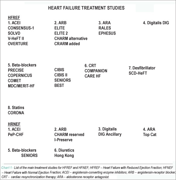
Different from heart failure with reduced ejection fraction (HFREF), for which large studies have demonstrated the effectiveness of drug treatment to improve morbidity and mortality, no treatment study has shown definitive results in patients with heart failure with normal ejection fraction (HFNEF). HFNEF is more prevalent in women and the elderly and it is associated with multiple comorbidities. Although the optimum treatment has yet to be defined, the control of arterial hypertension and decreased fluid overload are important measures to control the syndrome. In Brazil, the recommendations for the treatment of HF are based on the Review of the II Directives of the Brazilian Society of Cardiology for the diagnosis and treatment of Heart Failure – 2002. However, none of the recommendations is specific for patients with HFNEF, as they are based on general measures proposed by specialists. This review aims at demonstrating the scientific evidence from recent clinical trials in HFNEF and future perspectives in terms of new medications.

  • Heart Failure
  • Hypertrophy Left Ventricular
  • Ventricular Function, Left
Read Full Article
Creative Commons Attribution (CC BY) Creative Commons license
28/04/2010 DOI: 10.1590/S0066-782X2010000300021
ABC
Artigo Original

Systolic function of patients with myocardial infarction undergoing autologous bone marrow transplantation

Fernanda Belloni dos Santos Nogueira et al. Suzana Alves Silva Andrea Ferreira Haddad Cintia M. Peixoto … Rodrigo Moreira de Carvalho Fabio Antonio A. Tuche Vinício Elia Soares André Luiz Silveira Sousa Arnaldo Rabischoffsky Claudio Tinoco Mesquita Radovan Borojevic Hans Fernando Rocha Dohmann

BACKGROUND: Several studies have been published on the effect of bone-marrow stem cells on the left ventricle when acting on post- acute myocardial infarction remodeling. However, the results have been controversial. OBJECTIVE: To carry out an echocardiographic analysis of the systolic function of patients with acute myocardial infarction after autologous mononuclear bone marrow cell transplantation (AMBMCT) as performed via the intracoronary and intravenous routes. METHODS: This is an open-label, prospective, randomized study. Inclusion criteria: patients admitted for ST-elevation acute myocardial infarction (MI) who had undergone mechanical or chemical reperfusion within 24 hours of the onset of symptoms and whose echocardiogram showed decreased segmental wall motion and fixed perfusion defect related to the culprit artery. Autologous bone marrow was aspirated from the posterior iliac crest under sedation and analgesia of the patients randomly assigned for the treatment group. After laboratory manipulation, intracoronary or intravenous injection of 100 x 106 mononuclear cells was performed. Echocardiography (Vivid 7) was used to assess ventricular function before and three and six months after cell infusion. RESULTS: A total of 30 patients were included, 14 in the arterial group (AG), 10 in the venous group (VG), and six in the control group (CG). No statistical difference was found between the groups for the echocardiographic parameters studied. CONCLUSION: Autologous mononuclear bone marrow cell transplantation did not improve the echocardiographic parameters of systolic function.

  • bone marrow
  • Myocardial Infarction
  • transplantation, autologous
  • Ventricular Function, Left
Read Full Article
Creative Commons Attribution (CC BY) Creative Commons license
24/11/2009 DOI: 10.1590/S0066-782X2009001000010 Views 1

Could not load more articles. Please try again.

How to cite this article

EndNote RIS

Electronic Document Format (Vancouver)

Petronilho A, Gois MO, Sakaguchi C, Frade MCM, Roscani MG, Catai AM. Effects of Physical Exercise on Left Ventricular Function in Type 2 Diabetes Mellitus: A Systematic Review. International Journal of Cardiovascular Sciences 2023;36:e20220020.

Electronic Document Format (ABNT)

Petronilho, Ariane; Gois, Mariana de Oliveira; Sakaguchi, Camila; Frade, Maria Cecília Moraes; Roscani, Meliza Goi; Catai, Aparecida Maria. Effects of Physical Exercise on Left Ventricular Function in Type 2 Diabetes Mellitus: A Systematic Review. International Journal of Cardiovascular Sciences, v. 36, e20220020, Mar. 2023.

Electronic Document Format (APA)

Petronilho, A., Gois, M. O., Sakaguchi, C., Frade, M. C. M., Roscani, M. G., & Catai, A. M. (2023). Effects of Physical Exercise on Left Ventricular Function in Type 2 Diabetes Mellitus: A Systematic Review. International Journal of Cardiovascular Sciences, 36, e20220020.

Electronic Document Format (ISO)

Petronilho, Ariane and Gois, Mariana de Oliveira and Sakaguchi, Camila and Frade, Maria Cecília Moraes and Roscani, Meliza Goi and Catai, Aparecida Maria. Effects of Physical Exercise on Left Ventricular Function in Type 2 Diabetes Mellitus: A Systematic Review. International Journal of Cardiovascular Sciences [online]. 2023, vol. 36, [cited 2026-10-14], e20220020. Available from: <https://journals.cardiol.br/en/article/effects-of-physical-exercise-on-left-ventricular-function-in-type-2-diabetes-mellitus-a-systematic-review/>. ISSN 2359-4802

EndNote RIS

Electronic Document Format (Vancouver)

Barberato SH, Bruneto EG, Reis GS, Oliveira PRF, Possamai AF, Silvestre O, Silva MMF, et al. Abnormal Echocardiographic Findings in Hospitalized Patients with Covid-19: A Systematic Review and Meta-analysis. Arquivos Brasileiros de Cardiologia 2022;119(2):267-79.

Electronic Document Format (ABNT)

Barberato, Silvio Henrique; Bruneto, Eduardo G.; Reis, Gabriel S.; Oliveira, Paula Rauen Franco de; Possamai, Alexandre F.; Silvestre, Odilson; Silva, Miguel M. Fernandes. Abnormal Echocardiographic Findings in Hospitalized Patients with Covid-19: A Systematic Review and Meta-analysis. Arquivos Brasileiros de Cardiologia, v. 119, n. 2, p. 267-279, Jul. 2022.

Electronic Document Format (APA)

Barberato, S. H., Bruneto, E. G., Reis, G. S., Oliveira, P. R. F. , Possamai, A. F., Silvestre, O., & Silva, M. M. F. (2022). Abnormal Echocardiographic Findings in Hospitalized Patients with Covid-19: A Systematic Review and Meta-analysis. Arquivos Brasileiros de Cardiologia, 119(2), 267-279.

Electronic Document Format (ISO)

Barberato, Silvio Henrique and Bruneto, Eduardo G. and Reis, Gabriel S. and Oliveira, Paula Rauen Franco de and Possamai, Alexandre F. and Silvestre, Odilson and Silva, Miguel M. Fernandes. Abnormal Echocardiographic Findings in Hospitalized Patients with Covid-19: A Systematic Review and Meta-analysis. Arquivos Brasileiros de Cardiologia [online]. 2022, vol. 119, n. 2, [cited 2026-10-14], pp. 267-279. Available from: <https://journals.cardiol.br/en/article/abnormal-echocardiographic-findings-in-hospitalized-patients-with-covid-19-a-systematic-review-and-meta-analysis/>. ISSN 0066-782X

EndNote RIS

Electronic Document Format (Vancouver)

Tavares BG, Aguiar MO, Tsutsui J, Oliveira M, Soeiro AM, Nicolau J, Ribeiro H, et al. Sonothrombolysis Promotes Improvement in Left Ventricular Wall Motion and Perfusion Scores after Acute Myocardial Infarction. Arquivos Brasileiros de Cardiologia 2022;118(4):756-65.

Electronic Document Format (ABNT)

Tavares, Bruno G.; Aguiar, Miguel Osman; Tsutsui, Jeane; Oliveira, Mucio; Soeiro, Alexandre de Matos; Nicolau, José; Ribeiro, Henrique; PoChiang, Hsu; Sbano, João; Rochitte, Carlos Eduardo; Lopes, Bernardo; Ramirez, José; Kalil Filho, Roberto; Mathias, Wilson. Sonothrombolysis Promotes Improvement in Left Ventricular Wall Motion and Perfusion Scores after Acute Myocardial Infarction. Arquivos Brasileiros de Cardiologia, v. 118, n. 4, p. 756-765, Apr. 2022.

Electronic Document Format (APA)

Tavares, B. G., Aguiar, M. O., Tsutsui, J., Oliveira, M., Soeiro, A. M., Nicolau, J., Ribeiro, H., PoChiang, H., Sbano, J., Rochitte, C. E., Lopes, B., Ramirez, J., Kalil Filho, R., & Mathias, W. (2022). Sonothrombolysis Promotes Improvement in Left Ventricular Wall Motion and Perfusion Scores after Acute Myocardial Infarction. Arquivos Brasileiros de Cardiologia, 118(4), 756-765.

Electronic Document Format (ISO)

Tavares, Bruno G. and Aguiar, Miguel Osman and Tsutsui, Jeane and Oliveira, Mucio and Soeiro, Alexandre de Matos and Nicolau, José and Ribeiro, Henrique and PoChiang, Hsu and Sbano, João and Rochitte, Carlos Eduardo and Lopes, Bernardo and Ramirez, José and Kalil Filho, Roberto and Mathias, Wilson. Sonothrombolysis Promotes Improvement in Left Ventricular Wall Motion and Perfusion Scores after Acute Myocardial Infarction. Arquivos Brasileiros de Cardiologia [online]. 2022, vol. 118, n. 4, [cited 2026-10-14], pp. 756-765. Available from: <https://journals.cardiol.br/en/article/sonothrombolysis-promotes-improvement-in-left-ventricular-wall-motion-and-perfusion-scores-after-acute-myocardial-infarction/>. ISSN 0066-782X

EndNote RIS

Electronic Document Format (Vancouver)

Santos CCL, Oliveira RP, Sena J, Oliveira AD, Ferreira MG, Santos Filho A, Guissoni H, et al. Factors That Impact the Decision to Perform Left Ventriculography in Coronary Artery Disease. Arquivos Brasileiros de Cardiologia 2022;118(3):607-13.

Electronic Document Format (ABNT)

Santos, Claudia de Castro Lima; Oliveira, Ricardo Peixoto; Sena, Joberto; Oliveira, Adriano Dourado; Ferreira, Marcelo Gottschald; Santos Filho, Ademar; Guissoni, Heitor; Brito, José Carlos; Feitosa, Gilson Soares; Feitosa-Filho, Gilson Soares. Factors That Impact the Decision to Perform Left Ventriculography in Coronary Artery Disease. Arquivos Brasileiros de Cardiologia, v. 118, n. 3, p. 607-613, Mar. 2022.

Electronic Document Format (APA)

Santos, C. C. L., Oliveira, R. P., Sena, J., Oliveira, A. D., Ferreira, M. G., Santos Filho, A., Guissoni, H., Brito, J. C., Feitosa, G. S., & Feitosa-Filho, G. S. (2022). Factors That Impact the Decision to Perform Left Ventriculography in Coronary Artery Disease. Arquivos Brasileiros de Cardiologia, 118(3), 607-613.

Electronic Document Format (ISO)

Santos, Claudia de Castro Lima and Oliveira, Ricardo Peixoto and Sena, Joberto and Oliveira, Adriano Dourado and Ferreira, Marcelo Gottschald and Santos Filho, Ademar and Guissoni, Heitor and Brito, José Carlos and Feitosa, Gilson Soares and Feitosa-Filho, Gilson Soares. Factors That Impact the Decision to Perform Left Ventriculography in Coronary Artery Disease. Arquivos Brasileiros de Cardiologia [online]. 2022, vol. 118, n. 3, [cited 2026-10-14], pp. 607-613. Available from: <https://journals.cardiol.br/en/article/factors-that-impact-the-decision-to-perform-left-ventriculography-in-coronary-artery-disease/>. ISSN 0066-782X

EndNote RIS

Electronic Document Format (Vancouver)

Borsoi R, Silva MMF, Barberato SH. Semiautomatic Quantification of Left Ventricular Ejection Fraction and Volumes by Two-Dimensional Echocardiography: Comparison with Automatic Three-Dimensional Echocardiography. Arquivos Brasileiros de Cardiologia Imagem cardiovascular 2021;33(4):1-6.

Electronic Document Format (ABNT)

Borsoi, Rafael; Silva, Miguel Morita Fernandes da; Barberato, Silvio Henrique. Semiautomatic Quantification of Left Ventricular Ejection Fraction and Volumes by Two-Dimensional Echocardiography: Comparison with Automatic Three-Dimensional Echocardiography. Arquivos Brasileiros de Cardiologia Imagem cardiovascular, v. 33, n. 4, p. 1-6, Dec. 2021.

Electronic Document Format (APA)

Borsoi, R., Silva, M. M. F. , & Barberato, S. H. (2021). Semiautomatic Quantification of Left Ventricular Ejection Fraction and Volumes by Two-Dimensional Echocardiography: Comparison with Automatic Three-Dimensional Echocardiography. Arquivos Brasileiros de Cardiologia Imagem cardiovascular, 33(4), 1-6.

Electronic Document Format (ISO)

Borsoi, Rafael and Silva, Miguel Morita Fernandes da and Barberato, Silvio Henrique. Semiautomatic Quantification of Left Ventricular Ejection Fraction and Volumes by Two-Dimensional Echocardiography: Comparison with Automatic Three-Dimensional Echocardiography. Arquivos Brasileiros de Cardiologia Imagem cardiovascular [online]. 2021, vol. 33, n. 4, [cited 2026-10-14], pp. 1-6. Available from: <https://journals.cardiol.br/en/article/semiautomatic-quantification-of-left-ventricular-ejection-fraction-and-volumes-by-two-dimensional-echocardiography-comparison-with-automatic-three-dimensional-echocardiography/>. ISSN 2318-8219

EndNote RIS

Electronic Document Format (Vancouver)

Kosger P, Akin T, Kiztanir H, Ucar B. Arterial Stiffness and Left Ventricular Myocardial Function in Children with a Well-Functioning Bicuspid Aortic Valve. Arquivos Brasileiros de Cardiologia 2021;117(6):1126-33.

Electronic Document Format (ABNT)

Kosger, Pelin; Akin, Tugcem; Kiztanir, Hikmet; Ucar, Birsen. Arterial Stiffness and Left Ventricular Myocardial Function in Children with a Well-Functioning Bicuspid Aortic Valve. Arquivos Brasileiros de Cardiologia, v. 117, n. 6, p. 1126-1133, Sep. 2021.

Electronic Document Format (APA)

Kosger, P., Akin, T., Kiztanir, H., & Ucar, B. (2021). Arterial Stiffness and Left Ventricular Myocardial Function in Children with a Well-Functioning Bicuspid Aortic Valve. Arquivos Brasileiros de Cardiologia, 117(6), 1126-1133.

Electronic Document Format (ISO)

Kosger, Pelin and Akin, Tugcem and Kiztanir, Hikmet and Ucar, Birsen. Arterial Stiffness and Left Ventricular Myocardial Function in Children with a Well-Functioning Bicuspid Aortic Valve. Arquivos Brasileiros de Cardiologia [online]. 2021, vol. 117, n. 6, [cited 2026-10-14], pp. 1126-1133. Available from: <https://journals.cardiol.br/en/article/arterial-stiffness-and-left-ventricular-myocardial-function-in-children-with-a-well-functioning-bicuspid-aortic-valve/>. ISSN 0066-782X

EndNote RIS

Electronic Document Format (Vancouver)

Costa JPS, Ritt LEF, Fernandes RM, Campos FAD, Borges Q, Darzé ES. Takotsubo Syndrome in the Context of Transmural Acute Myocardial Infarction: Prevalence and How to Differentiate?. International Journal of Cardiovascular Sciences 2020;33(1):94-7.

Electronic Document Format (ABNT)

Costa, João Paulo Soares; Ritt, Luiz Eduardo Fonteles; Fernandes, Rafael Modesto; Campos, Felipe Alvarenga Duarte; Borges, Queila; Darzé, Eduardo Sahade. Takotsubo Syndrome in the Context of Transmural Acute Myocardial Infarction: Prevalence and How to Differentiate?. International Journal of Cardiovascular Sciences, v. 33, n. 1, p. 94-97, Feb. 2020.

Electronic Document Format (APA)

Costa, J. P. S., Ritt, L. E. F., Fernandes, R. M., Campos, F. A. D., Borges, Q., & Darzé, E. S. (2020). Takotsubo Syndrome in the Context of Transmural Acute Myocardial Infarction: Prevalence and How to Differentiate?. International Journal of Cardiovascular Sciences, 33(1), 94-97.

Electronic Document Format (ISO)

Costa, João Paulo Soares and Ritt, Luiz Eduardo Fonteles and Fernandes, Rafael Modesto and Campos, Felipe Alvarenga Duarte and Borges, Queila and Darzé, Eduardo Sahade. Takotsubo Syndrome in the Context of Transmural Acute Myocardial Infarction: Prevalence and How to Differentiate?. International Journal of Cardiovascular Sciences [online]. 2020, vol. 33, n. 1, [cited 2026-10-14], pp. 94-97. Available from: <https://journals.cardiol.br/en/article/takotsubo-syndrome-in-the-context-of-transmural-acute-myocardial-infarction-prevalence-and-how-to-differentiate/>. ISSN 2359-4802

EndNote RIS

Electronic Document Format (Vancouver)

Correia ETO, Barbetta LMS. Left Ventricle Mass Index, a Confounding Variable of Global Longitudinal Strain to be Noticed. Arquivos Brasileiros de Cardiologia 2020;114(1):136.

Electronic Document Format (ABNT)

Correia, Eduardo Thadeu de Oliveira; Barbetta, Letícia Mara dos Santos. Left Ventricle Mass Index, a Confounding Variable of Global Longitudinal Strain to be Noticed. Arquivos Brasileiros de Cardiologia, v. 114, n. 1, p. 136-136, Feb. 2020.

Electronic Document Format (APA)

Correia, E. T. O., & Barbetta, L. M. S. (2020). Left Ventricle Mass Index, a Confounding Variable of Global Longitudinal Strain to be Noticed. Arquivos Brasileiros de Cardiologia, 114(1), 136-136.

Electronic Document Format (ISO)

Correia, Eduardo Thadeu de Oliveira and Barbetta, Letícia Mara dos Santos. Left Ventricle Mass Index, a Confounding Variable of Global Longitudinal Strain to be Noticed. Arquivos Brasileiros de Cardiologia [online]. 2020, vol. 114, n. 1, [cited 2026-10-14], pp. 136-136. Available from: <https://journals.cardiol.br/en/article/left-ventricle-mass-index-a-confounding-variable-of-global-longitudinal-strain-to-be-noticed/>. ISSN 0066-782X

EndNote RIS

Electronic Document Format (Vancouver)

Póvoa R. Aortic Arch Calcification on routine Chest Radiography is Strongly and Independently Associated with Non-Dipper Blood Pressure Pattern. Arquivos Brasileiros de Cardiologia 2020;114(1):118-9.

Electronic Document Format (ABNT)

Póvoa, Rui. Aortic Arch Calcification on routine Chest Radiography is Strongly and Independently Associated with Non-Dipper Blood Pressure Pattern. Arquivos Brasileiros de Cardiologia, v. 114, n. 1, p. 118-119, Feb. 2020.

Electronic Document Format (APA)

Póvoa, R. (2020). Aortic Arch Calcification on routine Chest Radiography is Strongly and Independently Associated with Non-Dipper Blood Pressure Pattern. Arquivos Brasileiros de Cardiologia, 114(1), 118-119.

Electronic Document Format (ISO)

Póvoa, Rui. Aortic Arch Calcification on routine Chest Radiography is Strongly and Independently Associated with Non-Dipper Blood Pressure Pattern. Arquivos Brasileiros de Cardiologia [online]. 2020, vol. 114, n. 1, [cited 2026-10-14], pp. 118-119. Available from: <https://journals.cardiol.br/en/article/aortic-arch-calcification-on-routine-chest-radiography-is-strongly-and-independently-associated-with-non-dipper-blood-pressure-pattern-2/>. ISSN 0066-782X

EndNote RIS

Electronic Document Format (Vancouver)

Mancuso FJN. Real-Time Three-Dimensional Echocardiography and Myocardial Strain: Ready for Use in Clinical Practice. Arquivos Brasileiros de Cardiologia 2019;113(5):946-7.

Electronic Document Format (ABNT)

Mancuso, Frederico Jose Neves. Real-Time Three-Dimensional Echocardiography and Myocardial Strain: Ready for Use in Clinical Practice. Arquivos Brasileiros de Cardiologia, v. 113, n. 5, p. 946-947, Dec. 2019.

Electronic Document Format (APA)

Mancuso, F. J. N. (2019). Real-Time Three-Dimensional Echocardiography and Myocardial Strain: Ready for Use in Clinical Practice. Arquivos Brasileiros de Cardiologia, 113(5), 946-947.

Electronic Document Format (ISO)

Mancuso, Frederico Jose Neves. Real-Time Three-Dimensional Echocardiography and Myocardial Strain: Ready for Use in Clinical Practice. Arquivos Brasileiros de Cardiologia [online]. 2019, vol. 113, n. 5, [cited 2026-10-14], pp. 946-947. Available from: <https://journals.cardiol.br/en/article/real-time-three-dimensional-echocardiography-and-myocardial-strain-ready-for-use-in-clinical-practice/>. ISSN 0066-782X

EndNote RIS

Electronic Document Format (Vancouver)

Gil TCP, Castier MB, Gondar AFP, Sales AF, Santos MO, Lima FCS, Mourilhe-Rocha R, et al. Strain Analysis of Left Ventricular Function in the Association of Hypertrophic Cardiomyopathy and Systemic Arterial Hypertension. Arquivos Brasileiros de Cardiologia 2019;113(4):677-84.

Electronic Document Format (ABNT)

Gil, Thereza Cristina Pereira; Castier, Marcia Bueno; Gondar, Alyne Freitas Pereira; Sales, Ana Ferreira; Santos, Marceli de Oliveira; Lima, Fernanda Cristina da Silva de; Mourilhe-Rocha, Ricardo. Strain Analysis of Left Ventricular Function in the Association of Hypertrophic Cardiomyopathy and Systemic Arterial Hypertension. Arquivos Brasileiros de Cardiologia, v. 113, n. 4, p. 677-684, Sep. 2019.

Electronic Document Format (APA)

Gil, T. C. P., Castier, M. B., Gondar, A. F. P., Sales, A. F., Santos, M. O., Lima, F. C. S. , & Mourilhe-Rocha, R. (2019). Strain Analysis of Left Ventricular Function in the Association of Hypertrophic Cardiomyopathy and Systemic Arterial Hypertension. Arquivos Brasileiros de Cardiologia, 113(4), 677-684.

Electronic Document Format (ISO)

Gil, Thereza Cristina Pereira and Castier, Marcia Bueno and Gondar, Alyne Freitas Pereira and Sales, Ana Ferreira and Santos, Marceli de Oliveira and Lima, Fernanda Cristina da Silva de and Mourilhe-Rocha, Ricardo. Strain Analysis of Left Ventricular Function in the Association of Hypertrophic Cardiomyopathy and Systemic Arterial Hypertension. Arquivos Brasileiros de Cardiologia [online]. 2019, vol. 113, n. 4, [cited 2026-10-14], pp. 677-684. Available from: <https://journals.cardiol.br/en/article/strain-analysis-of-left-ventricular-function-in-the-association-of-hypertrophic-cardiomyopathy-and-systemic-arterial-hypertension/>. ISSN 0066-782X

EndNote RIS

Electronic Document Format (Vancouver)

Kalkan AK, Cakmak HA, Erturk M, Kalkan KE, Uzun F, Tasbulak O, Diker VO, et al. Adropin and Irisin in Patients with Cardiac Cachexia. Arquivos Brasileiros de Cardiologia 2018;111(1):39-47.

Electronic Document Format (ABNT)

Kalkan, Ali Kemal; Cakmak, Huseyin Altug; Erturk, Mehmet; Kalkan, Kübra Erol; Uzun, Fatih; Tasbulak, Omer; Diker, Vesile Ornek; Aydin, Suleyman; Celik, Ahmet. Adropin and Irisin in Patients with Cardiac Cachexia. Arquivos Brasileiros de Cardiologia, v. 111, n. 1, p. 39-47, Jul. 2018.

Electronic Document Format (APA)

Kalkan, A. K., Cakmak, H. A., Erturk, M., Kalkan, K. E., Uzun, F., Tasbulak, O., Diker, V. O., Aydin, S., & Celik, A. (2018). Adropin and Irisin in Patients with Cardiac Cachexia. Arquivos Brasileiros de Cardiologia, 111(1), 39-47.

Electronic Document Format (ISO)

Kalkan, Ali Kemal and Cakmak, Huseyin Altug and Erturk, Mehmet and Kalkan, Kübra Erol and Uzun, Fatih and Tasbulak, Omer and Diker, Vesile Ornek and Aydin, Suleyman and Celik, Ahmet. Adropin and Irisin in Patients with Cardiac Cachexia. Arquivos Brasileiros de Cardiologia [online]. 2018, vol. 111, n. 1, [cited 2026-10-14], pp. 39-47. Available from: <https://journals.cardiol.br/en/article/adropin-and-irisin-in-patients-with-cardiac-cachexia-2/>. ISSN 0066-782X

EndNote RIS

Electronic Document Format (Vancouver)

Winter SCN, Macedo RM, Francisco JC, Santos PC, Lopes APS, Meira LF, Carvalho KAT, et al. Impact of a High-Intensity Training on Ventricular Function in Rats After Acute Myocardial Infarction. Arquivos Brasileiros de Cardiologia 2018;110(4):373-80.

Electronic Document Format (ABNT)

Winter, Simone de Campos Neitzke; Macedo, Rafael Michel de; Francisco, Júlio Cesar; Santos, Paula Costa; Lopes, Ana Paula Sarraff; Meira, Leanderson Franco de; Carvalho, Katherine A. Teixeira de; Faria Neto, José Rocha; Macedo, Ana Carolina Brandt de; Guarita-Souza, Luiz César. Impact of a High-Intensity Training on Ventricular Function in Rats After Acute Myocardial Infarction. Arquivos Brasileiros de Cardiologia, v. 110, n. 4, p. 373-380, Mar. 2018.

Electronic Document Format (APA)

Winter, S. C. N., Macedo, R. M. , Francisco, J. C., Santos, P. C., Lopes, A. P. S., Meira, L. F. , Carvalho, K. A. T. , Faria Neto, J. R., Macedo, A. C. B. , & Guarita-Souza, L. C. (2018). Impact of a High-Intensity Training on Ventricular Function in Rats After Acute Myocardial Infarction. Arquivos Brasileiros de Cardiologia, 110(4), 373-380.

Electronic Document Format (ISO)

Winter, Simone de Campos Neitzke and Macedo, Rafael Michel de and Francisco, Júlio Cesar and Santos, Paula Costa and Lopes, Ana Paula Sarraff and Meira, Leanderson Franco de and Carvalho, Katherine A. Teixeira de and Faria Neto, José Rocha and Macedo, Ana Carolina Brandt de and Guarita-Souza, Luiz César. Impact of a High-Intensity Training on Ventricular Function in Rats After Acute Myocardial Infarction. Arquivos Brasileiros de Cardiologia [online]. 2018, vol. 110, n. 4, [cited 2026-10-14], pp. 373-380. Available from: <https://journals.cardiol.br/en/article/impact-of-a-high-intensity-training-on-ventricular-function-in-rats-after-acute-myocardial-infarction/>. ISSN 0066-782X

EndNote RIS

Electronic Document Format (Vancouver)

Almeida Junior GLG, Clausell N, Garcia MI, Esporcatte R, Rangel FOD, Rocha RM, Beck-da-Silva L, et al. Natriuretic Peptide and Clinical Evaluation in the Diagnosis of Heart Failure Hemodynamic Profile: Comparison with Tissue Doppler Echocardiography. Arquivos Brasileiros de Cardiologia 2018;110(3):270-7.

Electronic Document Format (ABNT)

Almeida Junior, Gustavo Luiz Gouvêa de; Clausell, Nadine; Garcia, Marcelo Iorio; Esporcatte, Roberto; Rangel, Fernando Oswaldo Dias; Rocha, Ricardo Mourilhe; Beck-da-Silva, Luis; Silva, Fabricio Braga da; Gorgulho, Paula de Castro Carvalho; Xavier, Sergio Salles. Natriuretic Peptide and Clinical Evaluation in the Diagnosis of Heart Failure Hemodynamic Profile: Comparison with Tissue Doppler Echocardiography. Arquivos Brasileiros de Cardiologia, v. 110, n. 3, p. 270-277, Feb. 2018.

Electronic Document Format (APA)

Almeida Junior, G. L. G. , Clausell, N., Garcia, M. I., Esporcatte, R., Rangel, F. O. D., Rocha, R. M., Beck-da-Silva, L., Silva, F. B. , Gorgulho, P. C. C., & Xavier, S. S. (2018). Natriuretic Peptide and Clinical Evaluation in the Diagnosis of Heart Failure Hemodynamic Profile: Comparison with Tissue Doppler Echocardiography. Arquivos Brasileiros de Cardiologia, 110(3), 270-277.

Electronic Document Format (ISO)

Almeida Junior, Gustavo Luiz Gouvêa de and Clausell, Nadine and Garcia, Marcelo Iorio and Esporcatte, Roberto and Rangel, Fernando Oswaldo Dias and Rocha, Ricardo Mourilhe and Beck-da-Silva, Luis and Silva, Fabricio Braga da and Gorgulho, Paula de Castro Carvalho and Xavier, Sergio Salles. Natriuretic Peptide and Clinical Evaluation in the Diagnosis of Heart Failure Hemodynamic Profile: Comparison with Tissue Doppler Echocardiography. Arquivos Brasileiros de Cardiologia [online]. 2018, vol. 110, n. 3, [cited 2026-10-14], pp. 270-277. Available from: <https://journals.cardiol.br/en/article/natriuretic-peptide-and-clinical-evaluation-in-the-diagnosis-of-heart-failure-hemodynamic-profile-comparison-with-tissue-doppler-echocardiography/>. ISSN 0066-782X

EndNote RIS

Electronic Document Format (Vancouver)

Oliveira LFL, O’Connell JL, Carvalho EEV, Pulici CC, Romano MMD, Maciel BC, Simões MV, et al. Comparison between Radionuclide Ventriculography and Echocardiography for Quantification of Left Ventricular Systolic Function in Rats Exposed to Doxorubicin. Arquivos Brasileiros de Cardiologia 2017;108(1):12-20.

Electronic Document Format (ABNT)

Oliveira, Luciano Fonseca Lemos de; O’Connell, João Lucas; Carvalho, Eduardo Elias Vieira de; Pulici, Érica Carolina Campos; Romano, Minna Moreira Dias; Maciel, Benedito Carlos; Simões, Marcus Vinicius. Comparison between Radionuclide Ventriculography and Echocardiography for Quantification of Left Ventricular Systolic Function in Rats Exposed to Doxorubicin. Arquivos Brasileiros de Cardiologia, v. 108, n. 1, p. 12-20, Dec. 2016.

Electronic Document Format (APA)

Oliveira, L. F. L. , O’Connell, J. L., Carvalho, E. E. V. , Pulici, C. C., Romano, M. M. D., Maciel, B. C., & Simões, M. V. (2017). Comparison between Radionuclide Ventriculography and Echocardiography for Quantification of Left Ventricular Systolic Function in Rats Exposed to Doxorubicin. Arquivos Brasileiros de Cardiologia, 108(1), 12-20.

Electronic Document Format (ISO)

Oliveira, Luciano Fonseca Lemos de and O’Connell, João Lucas and Carvalho, Eduardo Elias Vieira de and Pulici, Érica Carolina Campos and Romano, Minna Moreira Dias and Maciel, Benedito Carlos and Simões, Marcus Vinicius. Comparison between Radionuclide Ventriculography and Echocardiography for Quantification of Left Ventricular Systolic Function in Rats Exposed to Doxorubicin. Arquivos Brasileiros de Cardiologia [online]. 2017, vol. 108, n. 1, [cited 2026-10-14], pp. 12-20. Available from: <https://journals.cardiol.br/en/article/comparison-between-radionuclide-ventriculography-and-echocardiography-for-quantification-of-left-ventricular-systolic-function-in-rats-exposed-to-doxorubicin/>. ISSN 0066-782X

EndNote RIS

Electronic Document Format (Vancouver)

Ortega MC, Morejon AEG, Serrano GR, Ramos DBB. Diastolic Function in Paced Children with Cardiac Defects: Septum vs Apex. Arquivos Brasileiros de Cardiologia 2015;105(2):184-7.

Electronic Document Format (ABNT)

Ortega, Michel Cabrera; Morejon, Adel Eladio Gonzalez; Serrano, Giselle Ricardo; Ramos, Dunia Barbara Benitez. Diastolic Function in Paced Children with Cardiac Defects: Septum vs Apex. Arquivos Brasileiros de Cardiologia, v. 105, n. 2, p. 184-187, Jul. 2015.

Electronic Document Format (APA)

Ortega, M. C., Morejon, A. E. G., Serrano, G. R., & Ramos, D. B. B. (2015). Diastolic Function in Paced Children with Cardiac Defects: Septum vs Apex. Arquivos Brasileiros de Cardiologia, 105(2), 184-187.

Electronic Document Format (ISO)

Ortega, Michel Cabrera and Morejon, Adel Eladio Gonzalez and Serrano, Giselle Ricardo and Ramos, Dunia Barbara Benitez. Diastolic Function in Paced Children with Cardiac Defects: Septum vs Apex. Arquivos Brasileiros de Cardiologia [online]. 2015, vol. 105, n. 2, [cited 2026-10-14], pp. 184-187. Available from: <https://journals.cardiol.br/en/article/diastolic-function-in-paced-children-with-cardiac-defects-septum-vs-apex/>. ISSN 0066-782X

EndNote RIS

Electronic Document Format (Vancouver)

Ortega MC, Morejón AEG, Ricardo GS. Left ventricular synchrony and function in pediatric patients with definitive pacemakers. Arquivos Brasileiros de Cardiologia 2013;101(5):410-7.

Electronic Document Format (ABNT)

Ortega, Michel Cabrera; Morejón, Adel Eladio Gonzales; Ricardo, Giselle Serrano. Left ventricular synchrony and function in pediatric patients with definitive pacemakers. Arquivos Brasileiros de Cardiologia, v. 101, n. 5, p. 410-417, Sep. 2013.

Electronic Document Format (APA)

Ortega, M. C., Morejón, A. E. G., & Ricardo, G. S. (2013). Left ventricular synchrony and function in pediatric patients with definitive pacemakers. Arquivos Brasileiros de Cardiologia, 101(5), 410-417.

Electronic Document Format (ISO)

Ortega, Michel Cabrera and Morejón, Adel Eladio Gonzales and Ricardo, Giselle Serrano. Left ventricular synchrony and function in pediatric patients with definitive pacemakers. Arquivos Brasileiros de Cardiologia [online]. 2013, vol. 101, n. 5, [cited 2026-10-14], pp. 410-417. Available from: <https://journals.cardiol.br/en/article/left-ventricular-synchrony-and-function-in-pediatric-patients-with-definitive-pacemakers/>. ISSN 0066-782X

EndNote RIS

Electronic Document Format (Vancouver)

Vieira MLC, Oliveira WA, Cordovil A, Rodrigues ACT, Mônaco CG, Afonso T, Lira Filho EB, et al. 3D Echo pilot study of geometric Left Ventricular changes after acute myocardial infarction. Arquivos Brasileiros de Cardiologia 2013;101(1):43-51.

Electronic Document Format (ABNT)

Vieira, Marcelo Luiz Campos; Oliveira, Wercules Antonio; Cordovil, Adriana; Rodrigues, Ana Clara Tude; Mônaco, Cláudia Gianini; Afonso, Tânia; Lira Filho, Edgar Bezerra; Perin, Marco; Fischer, Cláudio Henrique; Morhy, Samira Saady. 3D Echo pilot study of geometric Left Ventricular changes after acute myocardial infarction. Arquivos Brasileiros de Cardiologia, v. 101, n. 1, p. 43-51, May. 2013.

Electronic Document Format (APA)

Vieira, M. L. C., Oliveira, W. A., Cordovil, A., Rodrigues, A. C. T., Mônaco, C. G., Afonso, T., Lira Filho, E. B., Perin, M., Fischer, C. H., & Morhy, S. S. (2013). 3D Echo pilot study of geometric Left Ventricular changes after acute myocardial infarction. Arquivos Brasileiros de Cardiologia, 101(1), 43-51.

Electronic Document Format (ISO)

Vieira, Marcelo Luiz Campos and Oliveira, Wercules Antonio and Cordovil, Adriana and Rodrigues, Ana Clara Tude and Mônaco, Cláudia Gianini and Afonso, Tânia and Lira Filho, Edgar Bezerra and Perin, Marco and Fischer, Cláudio Henrique and Morhy, Samira Saady. 3D Echo pilot study of geometric Left Ventricular changes after acute myocardial infarction. Arquivos Brasileiros de Cardiologia [online]. 2013, vol. 101, n. 1, [cited 2026-10-14], pp. 43-51. Available from: <https://journals.cardiol.br/en/article/3d-echo-pilot-study-of-geometric-left-ventricular-changes-after-acute-myocardial-infarction/>. ISSN 0066-782X

EndNote RIS

Electronic Document Format (Vancouver)

Cogni AL, Farah E, Minicucci MF, Azevedo PS, Okoshi K, Matsubara BB, Zanati S, et al. Metalloproteinases-2 and -9 predict left ventricular remodeling after myocardial infarction. Arquivos Brasileiros de Cardiologia 2013;100(4):315-21.

Electronic Document Format (ABNT)

Cogni, Ana Lucia; Farah, Elaine; Minicucci, Marcos F.; Azevedo, Paula S.; Okoshi, Katashi; Matsubara, Beatriz B.; Zanati, Silméia; Haggeman, Rodrigo; Paiva, Sergio A. R.; Zornoff, Leonardo A. M.. Metalloproteinases-2 and -9 predict left ventricular remodeling after myocardial infarction. Arquivos Brasileiros de Cardiologia, v. 100, n. 4, p. 315-321, Mar. 2013.

Electronic Document Format (APA)

Cogni, A. L., Farah, E., Minicucci, M. F., Azevedo, P. S., Okoshi, K., Matsubara, B. B., Zanati, S., Haggeman, R., Paiva, S. A. R., & Zornoff, L. A. M. (2013). Metalloproteinases-2 and -9 predict left ventricular remodeling after myocardial infarction. Arquivos Brasileiros de Cardiologia, 100(4), 315-321.

Electronic Document Format (ISO)

Cogni, Ana Lucia and Farah, Elaine and Minicucci, Marcos F. and Azevedo, Paula S. and Okoshi, Katashi and Matsubara, Beatriz B. and Zanati, Silméia and Haggeman, Rodrigo and Paiva, Sergio A. R. and Zornoff, Leonardo A. M.. Metalloproteinases-2 and -9 predict left ventricular remodeling after myocardial infarction. Arquivos Brasileiros de Cardiologia [online]. 2013, vol. 100, n. 4, [cited 2026-10-14], pp. 315-321. Available from: <https://journals.cardiol.br/en/article/metalloproteinases-2-and-9-predict-left-ventricular-remodeling-after-myocardial-infarction/>. ISSN 0066-782X

EndNote RIS

Electronic Document Format (Vancouver)

Gonzalez MBA, Azambuja RA, Bodanese LC. Quantification of left ventricular dilatation in myocardial perfusion scintigraphy. Arquivos Brasileiros de Cardiologia 2011;96(5):363-9.

Electronic Document Format (ABNT)

Gonzalez, Mauren B. Azambuja; Azambuja, Roberto Alves; Bodanese, Luiz Carlos. Quantification of left ventricular dilatation in myocardial perfusion scintigraphy. Arquivos Brasileiros de Cardiologia, v. 96, n. 5, p. 363-369, Apr. 2011.

Electronic Document Format (APA)

Gonzalez, M. B. A., Azambuja, R. A., & Bodanese, L. C. (2011). Quantification of left ventricular dilatation in myocardial perfusion scintigraphy. Arquivos Brasileiros de Cardiologia, 96(5), 363-369.

Electronic Document Format (ISO)

Gonzalez, Mauren B. Azambuja and Azambuja, Roberto Alves and Bodanese, Luiz Carlos. Quantification of left ventricular dilatation in myocardial perfusion scintigraphy. Arquivos Brasileiros de Cardiologia [online]. 2011, vol. 96, n. 5, [cited 2026-10-14], pp. 363-369. Available from: <https://journals.cardiol.br/en/article/quantification-of-left-ventricular-dilatation-in-myocardial-perfusion-scintigraphy/>. ISSN 0066-782X

EndNote RIS

Electronic Document Format (Vancouver)

Ivanovic BA, Tadic MV, Simic DV. Predictors of global left ventricular function in metabolic syndrome. Arquivos Brasileiros de Cardiologia 2011;96(5):377-85.

Electronic Document Format (ABNT)

Ivanovic, Branislava Aleksa; Tadic, Marijana Vaso; Simic, Dragan Vojislav. Predictors of global left ventricular function in metabolic syndrome. Arquivos Brasileiros de Cardiologia, v. 96, n. 5, p. 377-385, Mar. 2011.

Electronic Document Format (APA)

Ivanovic, B. A., Tadic, M. V., & Simic, D. V. (2011). Predictors of global left ventricular function in metabolic syndrome. Arquivos Brasileiros de Cardiologia, 96(5), 377-385.

Electronic Document Format (ISO)

Ivanovic, Branislava Aleksa and Tadic, Marijana Vaso and Simic, Dragan Vojislav. Predictors of global left ventricular function in metabolic syndrome. Arquivos Brasileiros de Cardiologia [online]. 2011, vol. 96, n. 5, [cited 2026-10-14], pp. 377-385. Available from: <https://journals.cardiol.br/en/article/predictors-of-global-left-ventricular-function-in-metabolic-syndrome/>. ISSN 0066-782X

EndNote RIS

Electronic Document Format (Vancouver)

Silva Júnior O, Melo CS, Marra M, Correia D. Alternative endocardial sites for artificial cardiac stimulation. Arquivos Brasileiros de Cardiologia 2011;96(1):76-85.

Electronic Document Format (ABNT)

Silva Júnior, Otaviano da; Melo, Celso Salgado de; Marra, Marcelo; Correia, Dalmo. Alternative endocardial sites for artificial cardiac stimulation. Arquivos Brasileiros de Cardiologia, v. 96, n. 1, p. 76-85, Feb. 2011.

Electronic Document Format (APA)

Silva Júnior, O. , Melo, C. S. , Marra, M., & Correia, D. (2011). Alternative endocardial sites for artificial cardiac stimulation. Arquivos Brasileiros de Cardiologia, 96(1), 76-85.

Electronic Document Format (ISO)

Silva Júnior, Otaviano da and Melo, Celso Salgado de and Marra, Marcelo and Correia, Dalmo. Alternative endocardial sites for artificial cardiac stimulation. Arquivos Brasileiros de Cardiologia [online]. 2011, vol. 96, n. 1, [cited 2026-10-14], pp. 76-85. Available from: <https://journals.cardiol.br/en/article/alternative-endocardial-sites-for-artificial-cardiac-stimulation/>. ISSN 0066-782X

EndNote RIS

Electronic Document Format (Vancouver)

Mesquita ET, Jorge AJL. Treatment of heart failure with normal ejection fraction. Arquivos Brasileiros de Cardiologia 2010;94(3):414-26.

Electronic Document Format (ABNT)

Mesquita, Evandro Tinoco; Jorge, Antonio José Lagoeiro. Treatment of heart failure with normal ejection fraction. Arquivos Brasileiros de Cardiologia, v. 94, n. 3, p. 414-426, Apr. 2010.

Electronic Document Format (APA)

Mesquita, E. T., & Jorge, A. J. L. (2010). Treatment of heart failure with normal ejection fraction. Arquivos Brasileiros de Cardiologia, 94(3), 414-426.

Electronic Document Format (ISO)

Mesquita, Evandro Tinoco and Jorge, Antonio José Lagoeiro. Treatment of heart failure with normal ejection fraction. Arquivos Brasileiros de Cardiologia [online]. 2010, vol. 94, n. 3, [cited 2026-10-14], pp. 414-426. Available from: <https://journals.cardiol.br/en/article/treatment-of-heart-failure-with-normal-ejection-fraction/>. ISSN 0066-782X

EndNote RIS

Electronic Document Format (Vancouver)

Nogueira FBS, Silva SA, Haddad AF, Peixoto CM, Carvalho RM, Tuche FAA, Soares VE, et al. Systolic function of patients with myocardial infarction undergoing autologous bone marrow transplantation. Arquivos Brasileiros de Cardiologia 2009;93(4):374-9.

Electronic Document Format (ABNT)

Nogueira, Fernanda Belloni dos Santos; Silva, Suzana Alves; Haddad, Andrea Ferreira; Peixoto, Cintia M.; Carvalho, Rodrigo Moreira de; Tuche, Fabio Antonio A.; Soares, Vinício Elia; Sousa, André Luiz Silveira; Rabischoffsky, Arnaldo; Mesquita, Claudio Tinoco; Borojevic, Radovan; Dohmann, Hans Fernando Rocha. Systolic function of patients with myocardial infarction undergoing autologous bone marrow transplantation. Arquivos Brasileiros de Cardiologia, v. 93, n. 4, p. 374-379, Nov. 2009.

Electronic Document Format (APA)

Nogueira, F. B. S., Silva, S. A., Haddad, A. F., Peixoto, C. M., Carvalho, R. M. , Tuche, F. A. A., Soares, V. E., Sousa, A. L. S., Rabischoffsky, A., Mesquita, C. T., Borojevic, R., & Dohmann, H. F. R. (2009). Systolic function of patients with myocardial infarction undergoing autologous bone marrow transplantation. Arquivos Brasileiros de Cardiologia, 93(4), 374-379.

Electronic Document Format (ISO)

Nogueira, Fernanda Belloni dos Santos and Silva, Suzana Alves and Haddad, Andrea Ferreira and Peixoto, Cintia M. and Carvalho, Rodrigo Moreira de and Tuche, Fabio Antonio A. and Soares, Vinício Elia and Sousa, André Luiz Silveira and Rabischoffsky, Arnaldo and Mesquita, Claudio Tinoco and Borojevic, Radovan and Dohmann, Hans Fernando Rocha. Systolic function of patients with myocardial infarction undergoing autologous bone marrow transplantation. Arquivos Brasileiros de Cardiologia [online]. 2009, vol. 93, n. 4, [cited 2026-10-14], pp. 374-379. Available from: <https://journals.cardiol.br/en/article/systolic-function-of-patients-with-myocardial-infarction-undergoing-autologous-bone-marrow-transplantation/>. ISSN 0066-782X

  • About ABC Family
  • Internal Regulation
  • Editorial Board
  • Brazilian Society of Cardiology (SBC)

Issues

SBC Guidelines

  • Videos
  • Podcasts
  • News