ABC Family Arquivos Brasileiros de Cardiologia International Journal of Cardiovascular Sciences ABC Heart Failure & Cardiomyopathy ABC Imagem Cardiovascular
Português English
ABC Family
Português English
  • About
    • About ABC Family
    • Internal Regulation
    • Editorial Board
    • Brazilian Society of Cardiology (SBC)
  • Issues
  • SBC Guidelines
  • Content
    • Videos
    • Podcasts
    • News
  • About
    • About ABC Family
    • Internal Regulation
    • Editorial Board
    • Brazilian Society of Cardiology (SBC)
  • Issues
  • SBC Guidelines
  • Content
    • Videos
    • Podcasts
    • News

ABC Family

  • ABC Family
  • Arquivos Brasileiros de Cardiologia
  • International Journal of Cardiovascular Sciences
  • ABC Heart Failure & Cardiomyopathy
  • ABC Imagem Cardiovascular
Search in

No content found.

Journals
Ordering
Article types
Others

  1. Home
  2. Keyword: Radionuclide Imaging
Figure 1
IJCS
Case Report

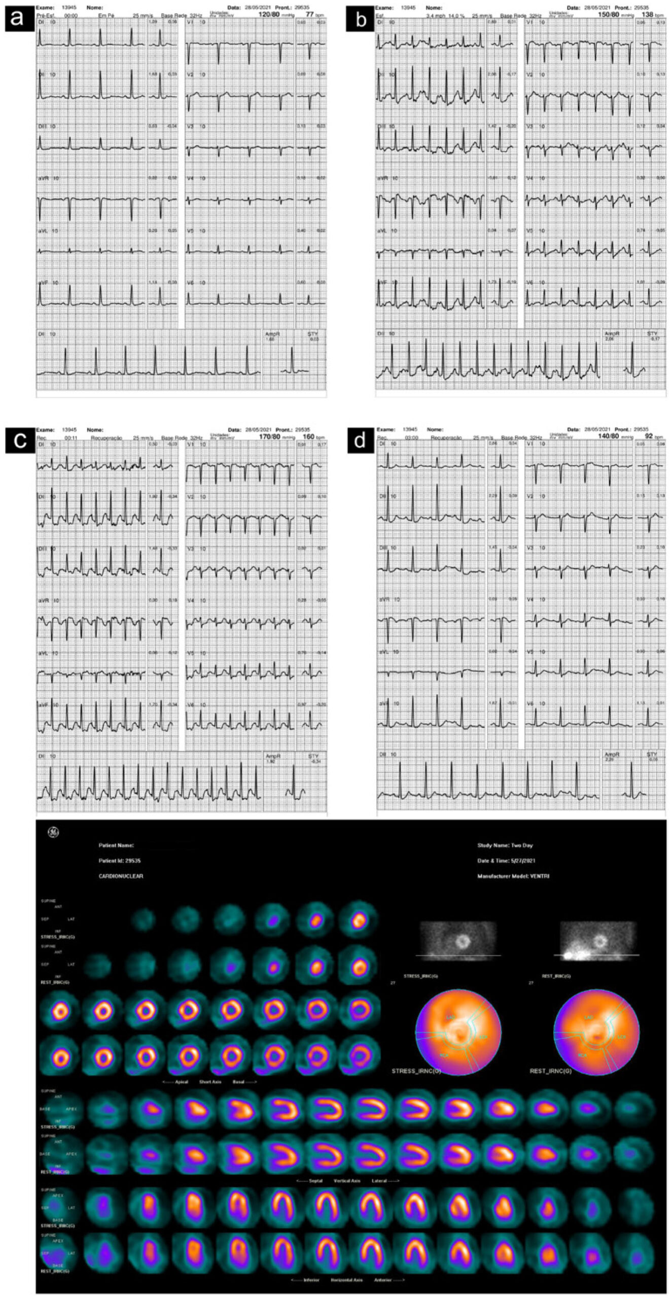
ECG's AVR ST elevation Analysis by Perfusion Scintilography in the Context of Myocardial Ischemia

Miriana Basso Gomes et al. Carolina Feijó Bombana Felipe C. Fuchs

Introduction

The presence of ST-segment elevation in two or more contiguous leads indicates a current subepicardial lesion that often requires early intervention, either by angioplasty or thrombolytic therapy., ST elevation, only in aVR, may also indicate the presence of an acute infarction. This finding was first made by Wellens et al. However, this is not always true, and physicians should be aware of the details surrounding changes in Right Vector amplification (aVR).

The medical literature has a wide range of articles reporting on the association between ST elevation in aVR during exercise testing and left main coronary artery injury.– However, this concept began at a time when fewer imaging tests and catheterizations were performed. Altered stress tests are a common indication of myocardial scintigraphy, which is why the Nuclear Medicine laboratory is the perfect place to perform this type of analysis. There is now evidence that aVR elevation in this context may not indicate coronary lesions and may be present, for example, in patients with aortic stenosis, hypertrophy and hypoxia..– Since these are stress tests, it is important to analyze the heart rate in which the aVR elevation occurs as well as the pattern of the other leads.

[…]

  • Electrocardiography
  • Exercise Test
  • Myocardial Ischemia
  • Radionuclide Imaging
Read Full Article
Creative Commons Attribution (CC BY) Creative Commons license
11/04/2025 DOI: 10.36660/ijcs.20230080 Views 1
Prevalence of Cardiac Amyloidosis in Elderly Patients With Aortic Stenosis
IJCS
Original Article

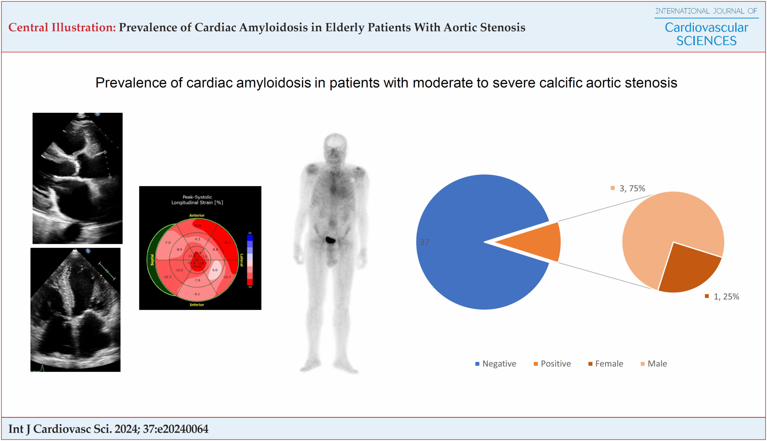
Prevalence of Cardiac Amyloidosis in Elderly Patients With Aortic Stenosis

Ng Kin Key et al. Adelina Sanches de Melo Joberto Pinheiro Sena Adriano Dias Dourado … Julia Micheli Perez Jose Carlos Raimundo Brito Gilson Feitosa Gilson Soares Feitosa-Filho

Abstract

Background:

Aortic Stenosis (AS) is one of the most common valvular heart diseases in adults, especially the elderly. The persistence of heart failure symptoms and worse outcomes despite valve replacement treatment may be associated with the coexistence of Cardiac Amyloidosis (CA).

Objective:

To determine the prevalence of CA in patients with calcific AS.

Methods:

This was a cross-sectional study. Elderly patients with moderate to severe AS underwent cardiac scintigraphy with bone radiotracer for diagnosis of CA. Patients diagnosed with Transthyretin (TTR) amyloidosis by scintigraphy underwent genetic studies. Groups with altered and unaltered scintigraphy results for continuous variables were compared using an independent t-test for normally distributed variables and a Mann-Whitney test for those with asymmetric distribution. Statistical significance was set at p-value < 0.05

Results:

Forty-one patients with AS underwent pyrophosphate scintigraphy. The mean age of the cases was 79 ± 6 years, 23 (56%) were female, 23 (56%) had classic severe AS pattern, and 8 (19.5%) had low-flow low-gradient AS with reduced Ejection Fraction (EF). Carpal tunnel syndrome was present in 2 (4%) patients, 8 (19%) had polyneuropathy. A RAISE score ≥ 2 was found in 4 patients (9%). Four patients were diagnosed with TTR amyloidosis, one of whom had a Val122I genetic mutation. Three (75%) of the patients diagnosed with AE/AI were male. There were no statistically significant differences between the isolated AS and AS/CA groups.

Conclusion:

The prevalence of CA in patients with moderate to severe AS of probable calcified etiology in our sample was 10%.

  • Amyloidosis
  • Aortic Valve Stenosis
  • Prealbumin
  • Radionuclide Imaging
Read Full Article
Creative Commons Attribution (CC BY) Creative Commons license
22/11/2024 DOI: 10.36660/ijcs.20240064 Views 3
Pyrophosphate Scintigraphy: Use in the Diagnosis and Monitoring of Cardiac Amyloidosis
IJCS
Review Article

Pyrophosphate Scintigraphy: Use in the Diagnosis and Monitoring of Cardiac Amyloidosis

Claudio Tinoco Mesquita et al. Davi Shunji Yahiro José Felipe Ramos dos Santos Luís Eduardo Machado … Jose Gregorio Valero Rodriguez Giovane Leal de Azevedo Junior Marcus Simões

Abstract

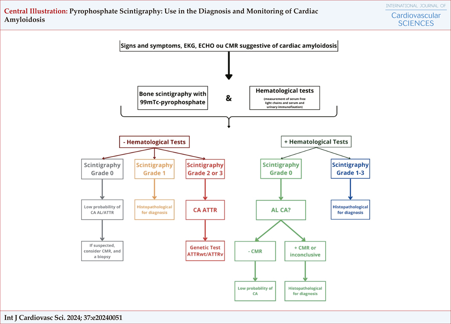
Cardiac amyloidosis (CA) is a progressive condition marked by the accumulation of amyloid fibrils in cardiac tissue, resulting in cardiac dysfunction and, ultimately, heart failure. Pyrophosphate scintigraphy has emerged as a promising tool for the early detection and monitoring of CA, providing valuable insights into the disease’s extent and severity. This review examines the current use of pyrophosphate scintigraphy in distinguishing between different types of amyloidosis, assessing risk levels, and monitoring patients with CA. It underscores the clinical significance and prospects of this imaging technique.

  • Amyloidosis
  • Diagnosis
  • Nuclear Medicine
  • Radionuclide Imaging
Read Full Article
Creative Commons Attribution (CC BY) Creative Commons license
19/08/2024 DOI: 10.36660/ijcs.20240051 Views 1
Figure 1
IJCS
Review Article

Multimodality Imaging of Chagas Heart Disease: Review and Case Presentation

Amalia Peix et al. Raul Araujo Veronica Gomez Aylen Perez Barreda … Maurizio Dondi Anita Brink Erika Minoshima Diana Paez

Abstract

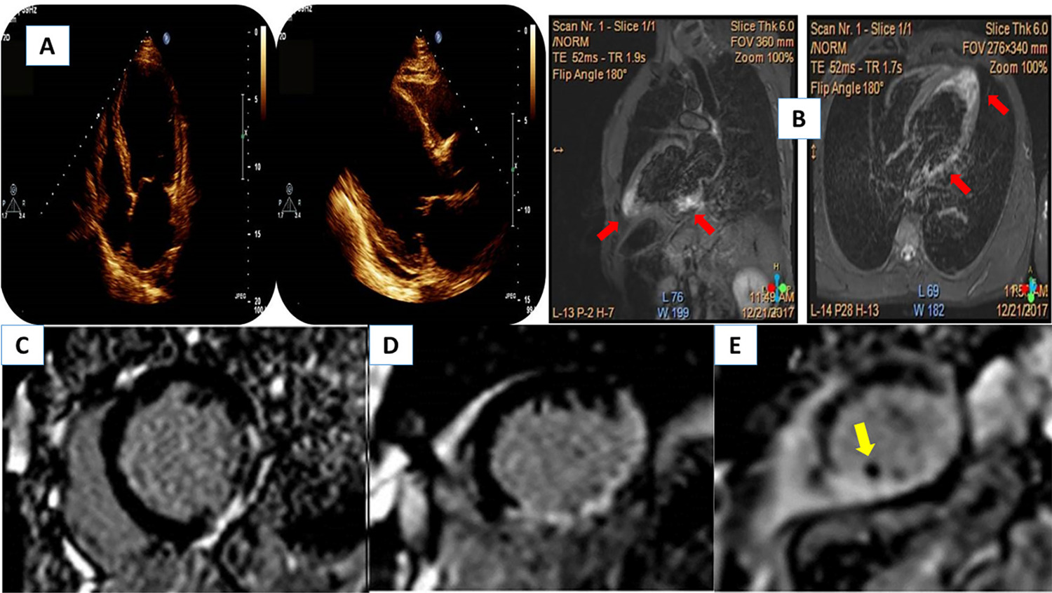
Chagas disease (CD) is a neglected tropical disease (NTD) caused by Trypanosoma cruzi, a hemoflagellate protozoa. CD is diagnosed by epidemiologic history and two positive serological tests. The infection has two main clinical phases: acute, early after acquiring the infection, and chronic. The electrocardiogram (ECG) is the first tool to diagnose chronic Chagas heart disease after CD serological diagnosis. In addition, the different cardiac imaging modalities, including chest X-ray, echocardiography, single photon emission computed tomography (SPECT), computed tomography (CT), and magnetic resonance, play a crucial role in detecting the cardiac involvement, establishing the prognosis, stratifying patient risk, and addressing the management of CD patients. In this review of the current literature, we summarize the epidemiology, pathophysiology, and clinical presentation of CD. We also discuss the indications, advantages, and limitations of the different cardiac imaging modalities. We also present some case examples, where cardiac imaging proved helpful in cardiac CD (CCD). The early mortality and substantial disability caused by CD represents a health, social and economic burden, particularly in the Latin America. To harness the full potential of medical imaging for the effective management of patients with CD, a comprehensive approach is essential. This involves a concerted effort to invest in the education of medical providers and to facilitate widespread access to diagnostic tools. Accessible and timely diagnosis is crucial for initiating prompt and targeted interventions. Finally, continuous investment in research is vital to advance our understanding of CD and develop innovative solutions for improved patient care.

  • Cardiac Imaging Techniques
  • Chagas disease
  • Radionuclide Imaging
Read Full Article
Creative Commons Attribution (CC BY) Creative Commons license
19/08/2024 DOI: 10.36660/ijcs.20240114 Views 1
Figure 1
ABC
Original Article

Scintigraphic and Echocardiographic Study of Patients with Pathogenic or Probably Pathogenic Variants of the TTR Gene without Overt Cardiac Involvement

Tonnison de Oliveira Silva et al. Eduardo Sahade Darzé Marcela Machado Costa Luiz José Junior … Antonio A. B. Ximenes Fábio Fernandes Mário de Seixas Rocha Marcia M. Noya-Rabelo Luiz Eduardo Fonteles Ritt

Abstract

Background:

Transthyretin amyloidosis (ATTR) is an infiltrative disease caused by abnormal protein deposition mainly in the heart and peripheral nervous system. When it affects the heart, the disease presents as restrictive cardiomyopathy; when it affects the peripheral and autonomic nervous system, it manifests as polyneuropathy, and is called familial amyloid polyneuropathy (FAP). There are two ATTR subtypes: wild-type ATTR, where there is no mutation, and mutant ATTR (ATTRm), which is characterized by a mutation in the gene encoding the transthyretin protein (TTR). In both subtypes, cardiac involvement is the major marker of poor prognosis.

Objectives:

To assess the prevalence of subclinical cardiac involvement in a sample of patients with TTR gene mutation by using pyrophosphate scintigraphy and strain echocardiography; to compare scintigraphy and strain findings; to evaluate the association between neurological manifestations (FAP) and subclinical cardiac involvement; and to analyze whether there is an association between any specific mutation and cardiac involvement.

Methods:

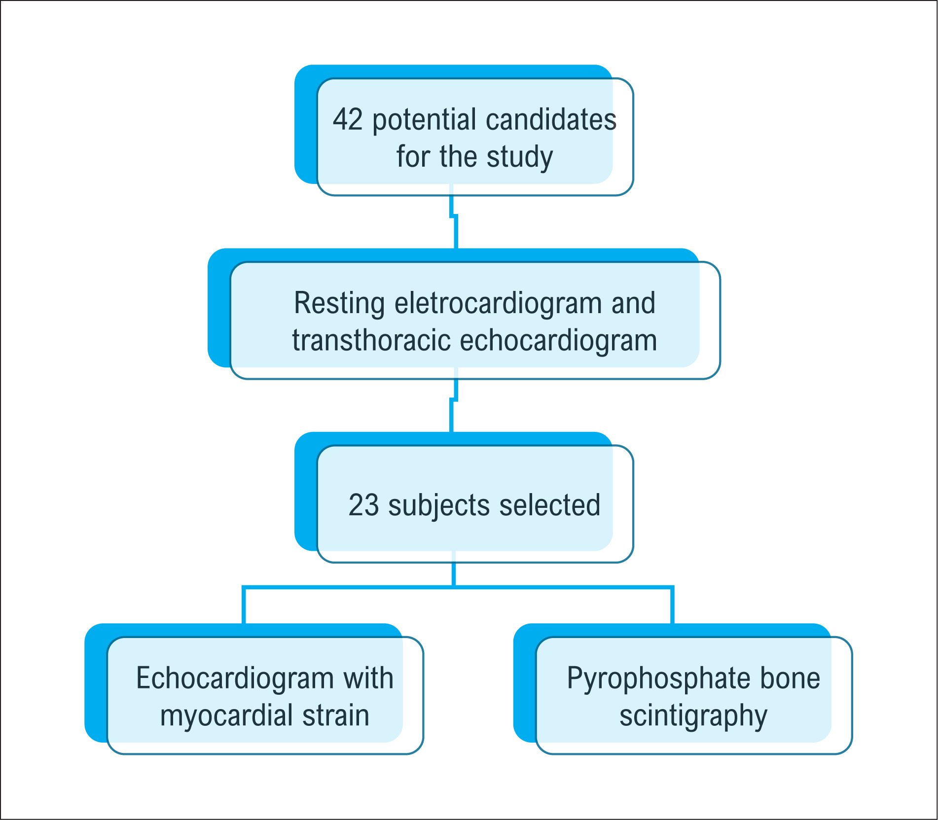
This is a cross-sectional study with carriers of the TTR gene mutation, without cardiovascular symptoms or changes in electrocardiographic or conventional echocardiographic parameters. All patients underwent pyrophosphate scintigraphy and strain echocardiography. Subclinical cardiac involvement was defined as a Perugini score ≥ 2, heart-to-contralateral lung (H/CL) ratio ≥ 1.5 at 1 h, H/CL ≥1.3 at 3 h, or global longitudinal strain (GLS) ≤ −17%. Descriptive and analytical analyses were performed and Fisher’s exact test and Mann–Whitney test were applied. A value of p < 0.05 was considered significant.

Results:

The 23 patients evaluated had a median age of 51 years (IQR 37–57 years), 15 (65.2%) were female, 12 (52.2%) were Pardo, nine (39.1%) had systemic arterial hypertension, and nine (39.1%) had a previous diagnosis of FAP. Of the nine patients with FAP, 8 (34.8%) were on tafamidis. The associated mutations were Val142IIe, Val50Met, and IIe127Val. The median GLS in the sample was −19% (−16% to −20%). Of the 23 patients, nine (39.1%; 95% CI = 29–49%) met criteria for cardiac involvement, six (26%) by the GLS-based criteria only. There was no association between having FAP and being an asymptomatic carrier, as assessed by strain echocardiography and pyrophosphate scintigraphy (p = 0.19). The prevalence of systemic arterial hypertension, diabetes mellitus, dyslipidemia, smoking, and reduced GLS did not differ between groups. Septal e’ wave velocity was the only variable that significantly differed between individuals with and without reduced GLS, with an area under the ROC curve of 0.80 (95% CI = 0.61–0.98, p = 0.027). The best diagnostic accuracy was achieved with a septal e’ velocity ≤ 8.5 cm/s. There was no association between mutation type and preclinical cardiac involvement, nor between tafamidis use and lower degree of cardiac involvement (37.5% versus 40.0%, p = 0.90).

Conclusion:

Subclinical cardiac involvement was common in a sample of TTR mutation carriers without cardiac involvement. Reduced left ventricular GLS was the most frequent finding. There was no association between the presence of amyloid polyneuropathy and subclinical cardiac involvement. Type of mutation was not associated with early cardiac involvement. In this sample, the use of tafamidis 20 mg/day was not associated with a lower prevalence of subclinical cardiac involvement.

  • Amiloidose
  • Echocardiography
  • Familial Amyloid Neuropathies
  • Radionuclide Imaging
Read Full Article
Creative Commons Attribution (CC BY) Creative Commons license
20/05/2024 DOI: 10.36660/abc.20230216i
Incremental Determinants of The Combined Supine-Prone Protocol for Myocardial Perfusion Scintigraphy
ABCIC
Original Article

Incremental Determinants of The Combined Supine-Prone Protocol for Myocardial Perfusion Scintigraphy

Tiago Oliveira Costa et al. Mário de Seixas Rocha Gilson Feitosa Soares Filho Raphael Abegão de Camargo Cristiano Ricardo Bastos de Macedo

Abstract

Background:

Myocardial perfusion scintigraphy is an important non-invasive method for diagnosing coronary artery disease, but it is not free from artifacts. Performing the combined supine-prone protocol can bring benefits to images with artifacts from the standard protocol in the supine position. The lack of unanimity in the implementation of the combined protocol may be related to the lack of objective data for the prior selection of patients.

Objective:

To evaluate which anthropometric profiles may be associated with a greater benefit in excluding artifacts for the optimized performance of the combined protocol.

Methods:

A cross-sectional and analytical study was carried out with 370 patients at the Nuclear Medicine Group Clinic between April and August 2022. The estimated level of statistical significance was 5%. Statistical analysis using binary logistic regression was used to evaluate the association between anthropometric data and the change in the initial test result after carrying out the combined protocol.

Results:

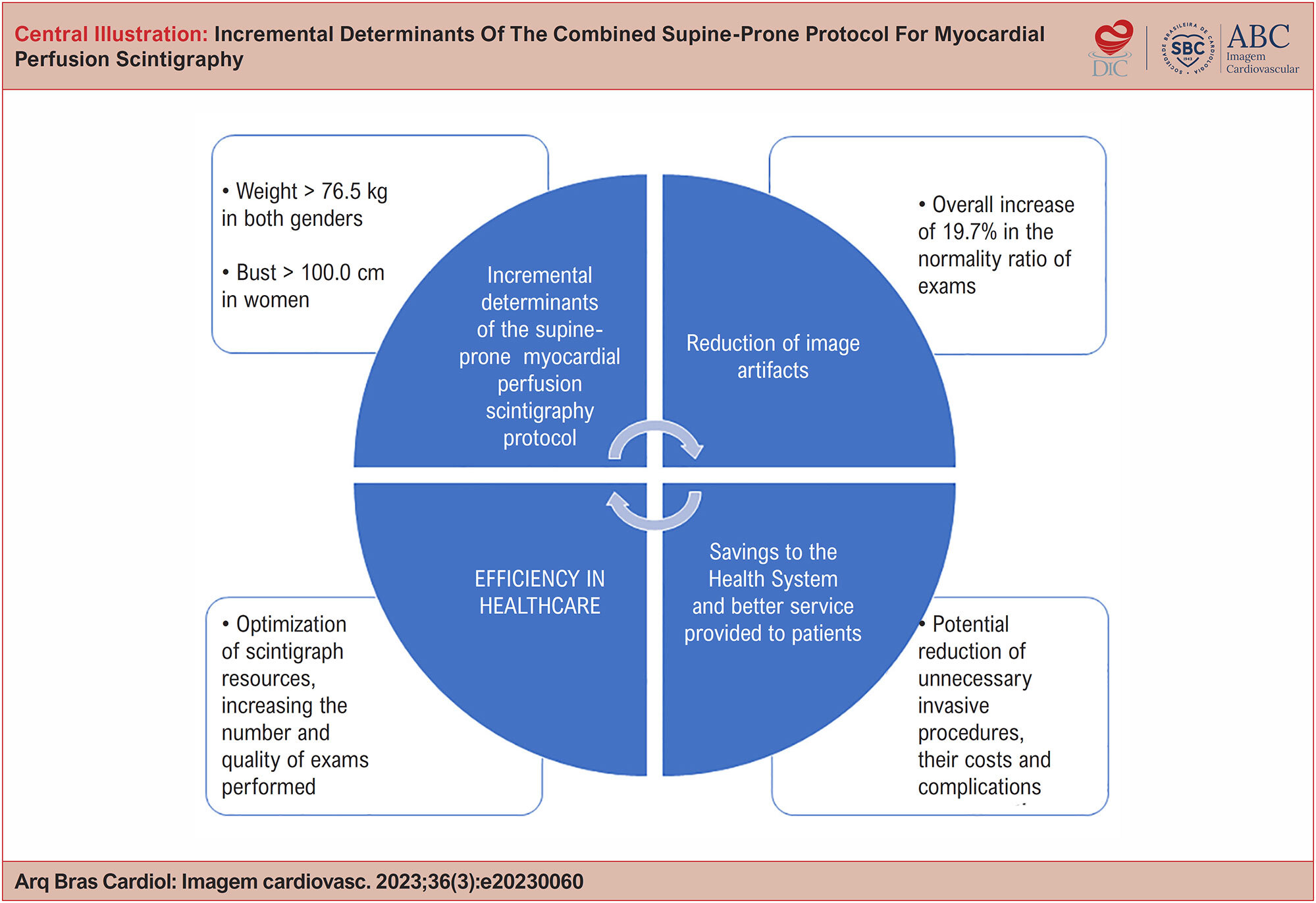
The combined protocol promoted a 19.7% increase in the normality ratio of the standard protocol. The variables weight, for both genders [OR = 1.02 (95% CI, 1.0002 – 1.04; p = 0.047)], and bust in women [OR = 1.06 (95% CI, 1. 01 – 1.11; p = 0.014)] represented incremental determinants with statistical significance. Weight greater than 76.5kg for both genders (S: 58.5%; E: 61.5%) and bust greater than 100.0 cm in women (S: 73.9%; E: 53.4%) were the best cutoff points in the receiver operating characteristic (ROC) curve.

Conclusion:

The optimized implementation of the combined protocol using the described weight and bust measurement criteria can promote greater efficiency in image acquisition.

  • Anthropometry
  • Coronary Disease
  • Myocardial Perfusion Imaging
  • Prone Position
  • Radionuclide Imaging
Read Full Article
Creative Commons Attribution (CC BY) Creative Commons license
02/10/2023 DOI: 10.36660/abcimg.20230060i Views 1
Figure 1
ABC
Editorial

Precision Medicine: Can 18F-FDG PET Detect Cardiotoxicity Phenotypes?

Claudio Tinoco Mesquita et al. Maria Fernanda Rezende

The publication of the article by Dourado et al. in the Arquivos Brasileiros de Cardiologia should be carefully considered by cardiologists engaged in Precision Medicine. In this study, the authors investigated the intensity of 2-deoxy-2[18F] fluoro-D-glucose (18F-FDG) myocardial uptake in 70 patients with lymphoma by positron emission tomography associated with computed tomography (PET/CT) scans before, during and after chemotherapy. They observed a progressive increase in glucose metabolism in the left ventricle from baseline PET/CT to interim PET/CT, and from interim PET/CT to post-therapy PET/CT. More than half of patients showed an increase of ≥ 30% in cardiac 18F-FDG uptake measured by left ventricular SUV max. The authors inferred that PET/CT is a reliable method to assess the intensity of 18F-FDG uptake in patients with lymphoma during and after chemotherapy. More importantly, the authors could identify a group of patients in which the metabolic effects of chemotherapy on the left ventricle were the greatest. These findings can contribute to a strategy for the early identification of patients who are more sensitive to cardiotoxicity of chemotherapeutic agents and for the definition of individualized preventive measures of irreversible myocardial damage.

Precision Medicine is commonly defined as an approach for disease treatment and prevention that takes into account individual variability and disease manifestation for each person. For this purpose, it is fundamental to clarify specific mechanisms of disease and key points for the implementation of effective interventions. This process is known as deep phenotyping, where endotypes, i.e., phenotypes underlying the common phenotype are identified, allowing a more effective guidance of therapeutic approaches.

[…]

  • Positron-Emission Tomography
  • Radionuclide Imaging
  • Toxicity
Read Full Article
Creative Commons Attribution (CC BY) Creative Commons license
11/07/2022 DOI: 10.36660/abc.20220393
Figure 1
ABC
Brief Communication

Risk Stratification and Cardiac Sympathetic Activity Assessment Using Myocardial [123I] MIBG Imaging in Renal Denervation

Joana Delgado-Silva et al. Ana Paula Moreira Gracinda Costa Lino Gonçalves

Abstract

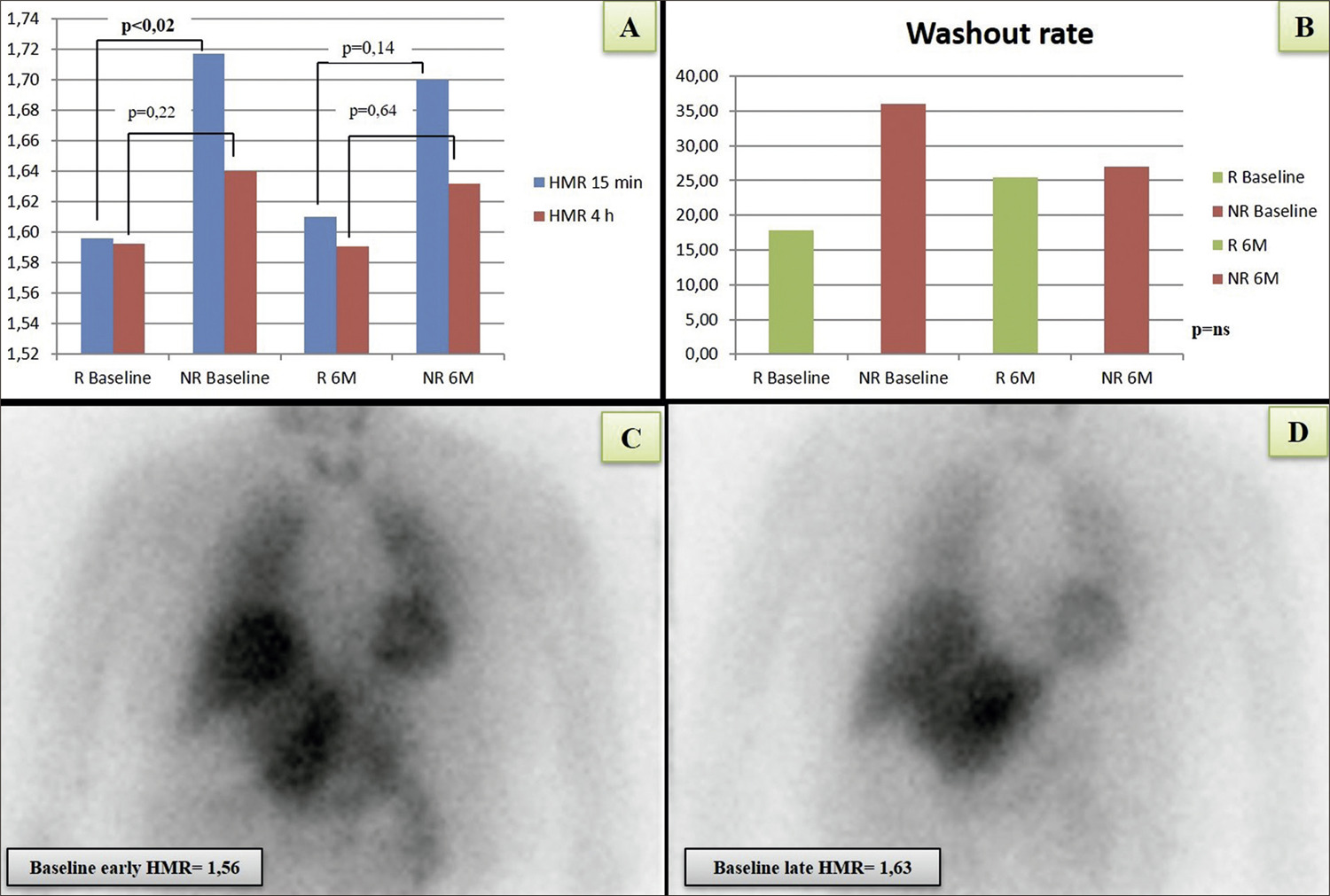
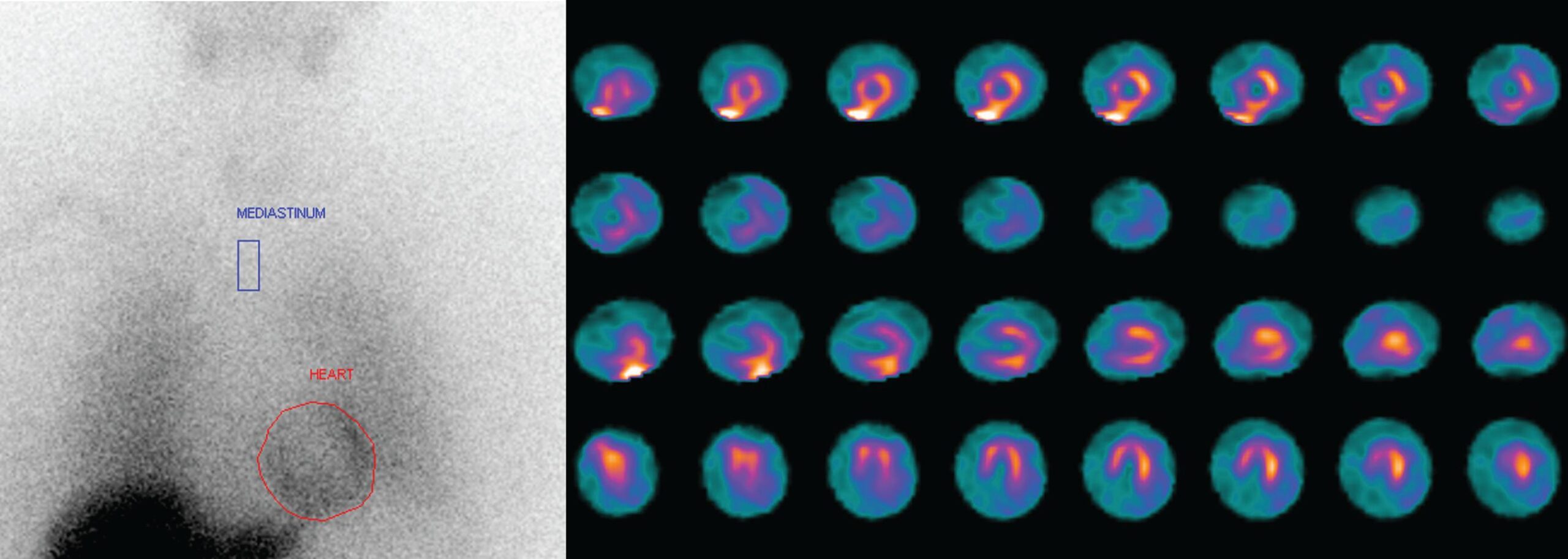
Hyperactivation of the sympathetic nervous system plays a central role in the pathophysiology of hypertension. The aim of this study was to assess cardiac sympathetic activity and investigate the role of myocardial123I-labelled meta-iodo benzyl guanidine ([123I] MIBG) scintigraphy in cardiovascular risk stratification of patients with resistant hypertension treated with renal denervation (RDN). Eighteen patients were included in this prospective study (mean age 56 ± 10 years old, 27.8% females). Transthoracic echocardiogram, general blood analysis and myocardial ([123I] MIBG scintigraphy were performed before and six-months after RDN. A patient was considered a responder (R) if a drop ≥ 5mmHg on mean systolic ambulatory blood pressure (BP) monitoring was observed at the six-month follow-up. 66.7% of patients were R (drop in systolic BP of 20.6 ± 14.5mmHg, vs minus 8 ± 11.6mmHg in non-responders (NR), p=0.001). Early heart-mediastinum ratio (HMR) was significantly lower at baseline in the R group (1.6 ± 0.1 vs 1.72 ± 0.1, p<0.02) but similar at six months. Considering both instants in time, the R group had lower early HMR values than the NR group (p<0.05). Both the late HMR and the washout rate were identical and no significant correlation between response to RDN or any MIBG imaging index was found. Renal denervation effectively lowered blood pressure in the majority of patients but [123I] MIBG was not useful in predicting the response. However, there was evidence of sympathetic overdrive and, both early and late HMR were overall reduced, probably putting this population at a higher risk of adverse events.

  • 3-iodobenzylguanidine
  • Kidney/denervation
  • Myocardial/diagnostic imaging
  • Radionuclide Imaging
  • Resistant Hypertension
  • Sympathetic Nervous System
Read Full Article
Creative Commons Attribution (CC BY) Creative Commons license
07/03/2022 DOI: 10.36660/abc.20201253 Views 2
ABCIC
Case Report

Myocardial Viability in a Patient with Systemic Lupus Erythematosus

Elisa Carla Hilgemberg et al. Ursula Cristina Barbosa de Almeida Daco Roessler Larissa Ticiane Machado dos Santos Alair Sarmet Damas Claúdio Tinoco Mesquita

[…]

  • Atrioventricular block
  • Diagnostic Imaging
  • Lupus Erithematosus Systemic
  • Myocardial Infarction
  • Radionuclide Imaging
  • Women
Read Full Article
Creative Commons Attribution (CC BY) Creative Commons license
20/01/2022 DOI: 10.5935/2318-8219.20180033
ABCIC
Review Article

What is the Value of Perfusion Scintigraphy and Cardiac PET in Hypertrophic Cardiomyopathy?

Elry Medeiros Vieira Segundo Neto et al. Rafael Willain Lopes Simone Cristina Soares Brandão

Abstract

Hypertrophic cardiomyopathy is the most common hereditary heart disease and affects about 1:500 individuals in the general population. Diagnosis is not always simple due to phenotypic variation and concomitance with other pathologies. It is initially based on electrocardiographic and echocardiographic criteria and on the absence of other diseases occurring with ventricular hypertrophy. Having myofibrillar derangement and fibrosis as a cellular base resulting in hemodynamic abnormalities, hypertrophic cardiomyopathy may reveal myocardial ischemia (not related to atherosclerosis) and sudden death. Therefore, evaluation of functional repercussion with myocardial perfusion scintigraphy using the Single Photon Emission Computed Tomography (SPECT) has gained space, since 25% of patients with hypertrophic cardiomyopathy have fixed or ischemic perfusion defects. In this context, some perfusion disorders are not necessarily associated with the type of hypertrophic cardiomyopathy, but are able to predict morbidity and mortality in these individuals. Another recent scintigraphy technique is the positron emission tomography (PET), which stands out in the evaluation of microcirculation, coronary flow reserve and myocardial metabolism. In patients with hypertrophic cardiomyopathy, studies have shown unfavorable results when myocardial blood flow and coronary flow reserve are lower. Metabolic myocardial evaluation using PET seems to be useful in the pathophysiological understanding of this disease and in the prognostic evaluation of alcohol ablation, a procedure performed in severe obstructive forms. This review addresses the role of nuclear cardiology using SPECT and myocardial PET in the diagnostic, prognostic and therapeutic evaluation of hypertrophic cardiomyopathy.

  • Cardiomyopathy, Hypertrophic, Familial
  • Echocardiography
  • Myocardial Ischemia
  • Myocardial Perfusion Imaging/methods
  • Positron-Emission Tomography/heart
  • Radionuclide Imaging
Read Full Article
Creative Commons Attribution (CC BY) Creative Commons license
20/01/2022 DOI: 10.5935/2318-8219.20190007
ABCIC
Case Report

Acute myocardial infarction after myocardial scintigraphy with dipyridamole

Rafael Alessandro Ferreira Gomes et al. Isabelle Conceição Albuquerque Machado Moreira Otávio Guilherme de Moraes Cardoso Alexandre Gomes de Souza Melo Aluísio Roberto Andrade Macêdo Junior

[…]

  • Dipyridamole
  • Myocardial Infarction
  • Radionuclide Imaging
Read Full Article
Creative Commons Attribution (CC BY) Creative Commons license
14/01/2022 DOI: 10.5935/2318-8219.20200012
Figure 1
ABC
Case Report

Large Bilateral Coronary Artery Fistula: 10-year Follow-up in Clinical Treatment

Rodrigo Melo Kulchetscki et al. Luka David Lechinewski Luciana Oliveira Cascaes Dourado Whady Armindo Hueb Luiz Antonio Machado César

Case Report

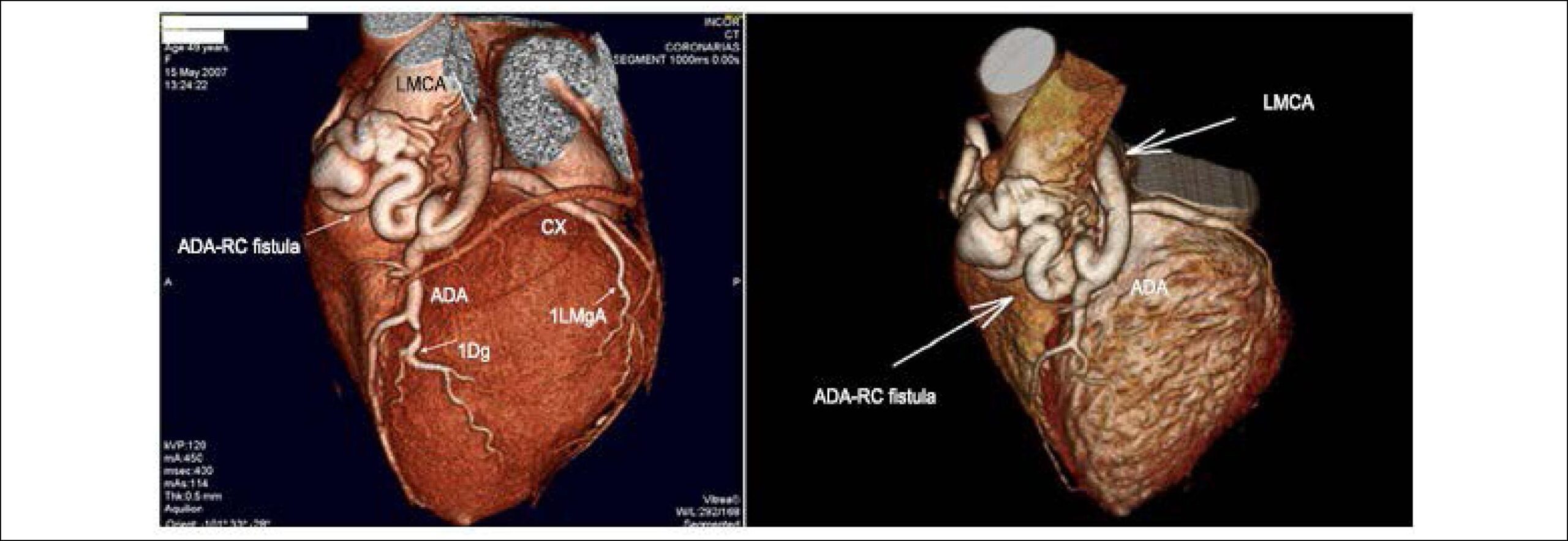
A 59-year-old asymptomatic female patient, with a diagnosis of a large bilateral coronary-pulmonary artery fistula made in 2007 was investigated after a cardiac murmur was identified on a routine examination. At the time, conservative treatment was chosen. Cardiac auscultation showed a more audible systolic-diastolic murmur in the upper left sternal border, with a more audible component in systole. There were no other findings in the cardiological physical examination or even the overall segmental examination. The patient had no comorbidities at the time, except for a prior history of smoking (10-pack-years). During the evolution, at the annual outpatient follow-up, she had diagnoses of dyslipidemia, glucose intolerance and depression. At the last consultation, in 2017, the patient was asymptomatic. She used atenolol 25 mg/ day, metformin 850 mg/day, atorvastatin 20 mg/day and sertraline 50 mg/day.

The examinations performed after 10 years of follow-up were compared with those at the time of diagnosis. The current echocardiogram showed right coronary (RC) with 4 mm of diameter at the origin and 7 mm in the middle third; the left main coronary artery (LMCA) with 8 mm. The patient had a fistulous trajectory with tortuous flow communicating both coronaries with the pulmonary trunk, without the presence of pulmonary hyperflow. Additionally, the evolution of mitral regurgitation showed to be of an important degree. shows the echocardiographic parameters during follow-up.

[…]

  • Arterio-Arterial Fistula/diagnosis
  • Coronary Angiography
  • Coronary Vessel Anomalies
  • Diagnostic Imaging
  • Mitral Valve Insufficiency
  • Myocardial Ischemia
  • Radionuclide Imaging
Read Full Article
Creative Commons Attribution (CC BY) Creative Commons license
01/02/2019 DOI: 10.5935/abc.20180267
Figure 1
ABC
Original Article

Relationship of Electromechanical Dyssynchrony in Patients Submitted to CRT With LV Lead Implantation Guided by Gated Myocardial Perfusion Spect

Erivelton Alessandro do Nascimento et al. Christiane Cigagna Wiefels Reis Fernanda Baptista Ribeiro Christiane Rodrigues Alves … Eduardo Nani Silva Mario Luiz Ribeiro Claudio Tinoco Mesquita

Abstract

Background:

Heart failure (HF) affects more than 5 million individuals in the United States, with more than 1 million hospital admissions per year. Cardiac resynchronization therapy (CRT) can benefit patients with advanced HF and prolonged QRS. A significant percentage of patients, however, does not respond to CRT. Electrical dyssynchrony isolated might not be a good predictor of response, and the last left ventricular (LV) segment to contract can influence the response.

Objectives:

To assess electromechanical dyssynchrony in CRT with LV lead implantation guided by GATED SPECT.

Methods:

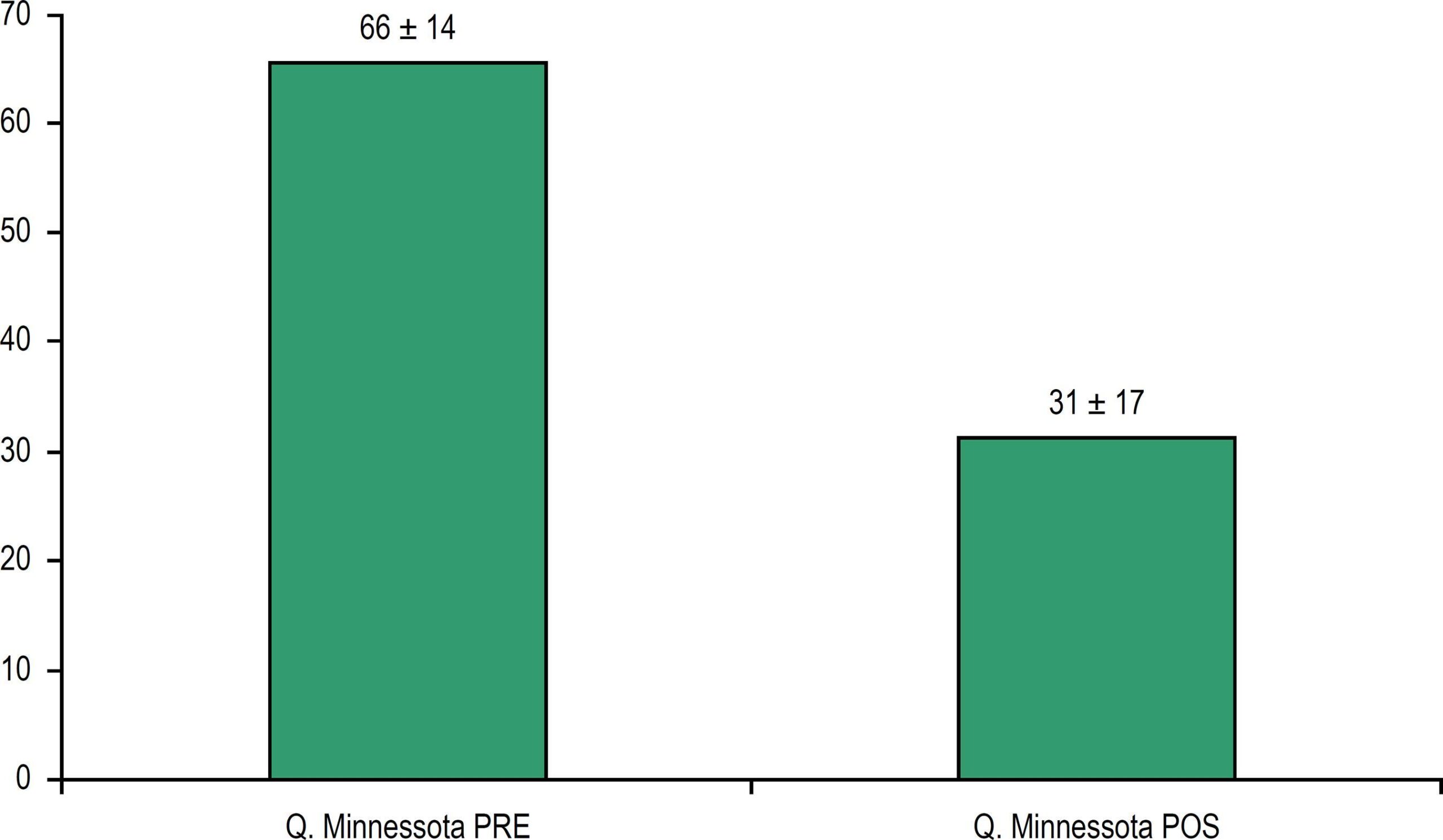
This study included 15 patients with functional class II-IV HF and clinically optimized, ejection fraction of 35%, sinus rhythm, left bundle-branch block, and QRS ≥ 120 ms. The patients underwent electrocardiography, answered the Minnesota Living with Heart Failure Questionnaire (MLHFQ), and underwent gated myocardial perfusion SPECT up to 4 weeks before CRT, being reassessed 6 months later. The primary analysis aimed at determining the proportion of patients with a reduction in QRS duration and favorable response to CRT, depending on concordance of the LV lead position, using chi-square test. The pre- and post-CRT variables were analyzed by use of Student t test, adopting the significance level of 5%.

Results:

We implanted 15 CRT devices, and 2 patients died during follow-up. The durations of the QRS (212 ms vs 136 ms) and the PR interval (179 ms vs 126 ms) were significantly reduced (p < 0.001). In 54% of the patients, the lead position was concordant with the maximal delay site. In the responder group, the lateral position was prevalent. The MLHFQ showed a significant improvement in quality of life (p < 0.0002).

Conclusion:

CRT determines improvement in the quality of life and in electrical synchronism. Electromechanical synchronism relates to response to CRT. Positioning the LV lead in the maximal delay site has limitations.

  • Cardiac Resynchronization Therapy
  • Eletrodes,Implanted
  • Heart Failure
  • Radionuclide Imaging
  • Stroke Volume
Read Full Article
Creative Commons Attribution (CC BY) Creative Commons license
23/08/2018 DOI: 10.5935/abc.20180159 Views 2
Figure 1
IJCS
Review Article

The use of Cardiac 123I-mIBG Scintigraphy in Clinical Practice: The Necessity to Standardize!

Euclides Timóteo da Rocha et al. Wilson Eduardo Furlan Matos Alves Derk O. Verschure Hein J Verberne

Abstract

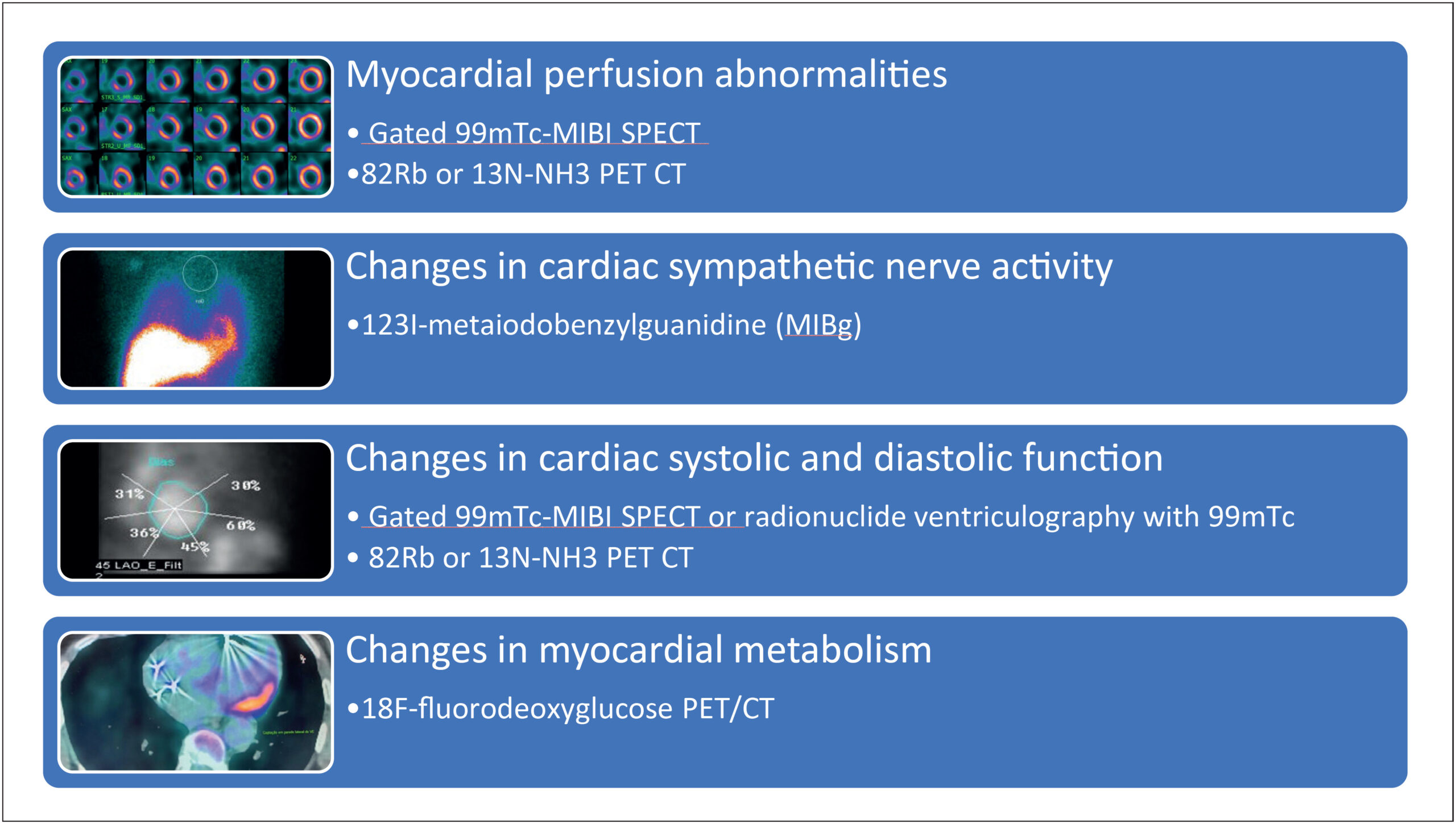
Cardiac adrenergic imaging has a great potential in a wide variety of clinical applications. Cardiac 123I-meta-iodobenzylguanidine (123I-mIBG) scintigraphy has a key role to assess chronic heart failure (CHF) by risk stratifying patients for cardiac events. The mIBG is a norepinephrine (NE) analogue that can evaluate cardiac sympathetic activity by assessing the down expression of β-adrenergic receptor (β-AR) in CHF. Furthermore, 123I-mIBG scintigraphy in combination with other parameters of left ventricular function can be used to identify the best responder for implantable cardiac devices, as well as to assess oncological cardiotoxicity. Despite its usefulness, 123I-mIBG scintigraphy is not widely performed because of the lack of standardization between different institutions. Thus, standardization and validation may contribute to its acceptance in clinical setting.

  • 3-iodobenzylguanidine
  • Heart Failure
  • Myocardium/diagnostic imaging
  • Radionuclide Imaging
  • Sympathetic Nervous System
Read Full Article
Creative Commons Attribution (CC BY) Creative Commons license
21/09/2017 DOI: 10.5935/2359-4802.20170070 Views 1
Figure 1
ABC
Case Report

Myocardial Bridge and Angiotomography of the Coronary Arteries: Perfusion under Pharmacological Stress

Wilter dos Santos Ker et al. Daniel Gama Neves Alair Sarmet A. A. Damas Cláudio Tinoco Mesquita Marcelo Souto Nacif

Introduction

The myocardial bridge is one of the most prevalent congenital anomalies that involve the coronary circulation, and its incidence in the general population is high, affecting from 23 to 55% in necropsy studies.The impairment of the anterior descending artery is more frequent, on its proximal 2/3.In most patients, the myocardial bridges do not cause symptoms, because in order to have ischemia, there must be an imbalance between supply and consumption of oxygen. The superficial bridges, with small or slender muscle band, are the most common ones, and they may account for 75% of the cases, with average length of 1.5 cm and usually without causing symptoms. In approximately 24% of the cases, we observed deep myocardial bridges, with thicker muscle band.,

Atherosclerosis is the most common cause of ischemic heart disease. However, other causes for ischemia are frequent, and among them, we highlight the myocardial bridge, which may provoke typical or atypicalchest angina,acute myocardial infarction and sudden death.–

[…]

  • Computed Tomography
  • Coronary Artery Disease
  • Myocardial Bridge
  • Myocardial Ischemia
  • Perfusion
  • Radionuclide Imaging
Read Full Article
Creative Commons Attribution (CC BY) Creative Commons license
01/06/2017 DOI: 10.5935/abc.20170021
ABC
Letter to the Editor

The Tip of The Iceberg: Non-Calcified Coronary Plague and Epicardial Adipose Tissue

Levent Cerit

To the Editor,

I have read with great interest the article entitled “Relationship between Calcium Score and Myocardial Scintigraphy in the Diagnosis of Coronary Disease” by Siqueira et al., recently published in Arquivos Brasileiros de Cardiologia 2016; 107:367-74. The investigators reported the possibility of removing extensive coronary artery disease (CAD) by means of a zero calcium score, or by indicating the presence of an extensive disease when it is severely increased, which justifies the use of this method in the initial or joint evaluation in asymptomatic patients with suspected CAD and in cardiovascular risk stratification. The evaluation of symptomatic low-risk patients, despite suggestive evidence, should be re-evaluated in upcoming guidelines.

[…]

  • Adipose Tissue / pathology
  • Biomarkers / analysis
  • Calcium Signaling
  • Coronary Artery Disease
  • Radionuclide Imaging
  • Tomography, Emission Computed
Read Full Article
Creative Commons Attribution (CC BY) Creative Commons license
01/04/2017 DOI: 10.5935/abc.20170046
Figure 1
ABC
Case Report

Case Report: Multivessel Coronary Disease Assessment with SPECT 99mTc-Sestamibi and Rubidium-82 PET/CT

Bruno Gomes Padilha et al. Daniela Sabino Maria Clementina Giorgi José Soares Jr. … Marisa Izaki José Claudio Meneghetti

Introduction

Coronary angiography (CAG) is the standard diagnostic method for detection of coronary artery disease (CAD). However, it is often necessary to evaluate the expression of a coronary obstruction in relation to myocardial perfusion, before defining the best patient management.

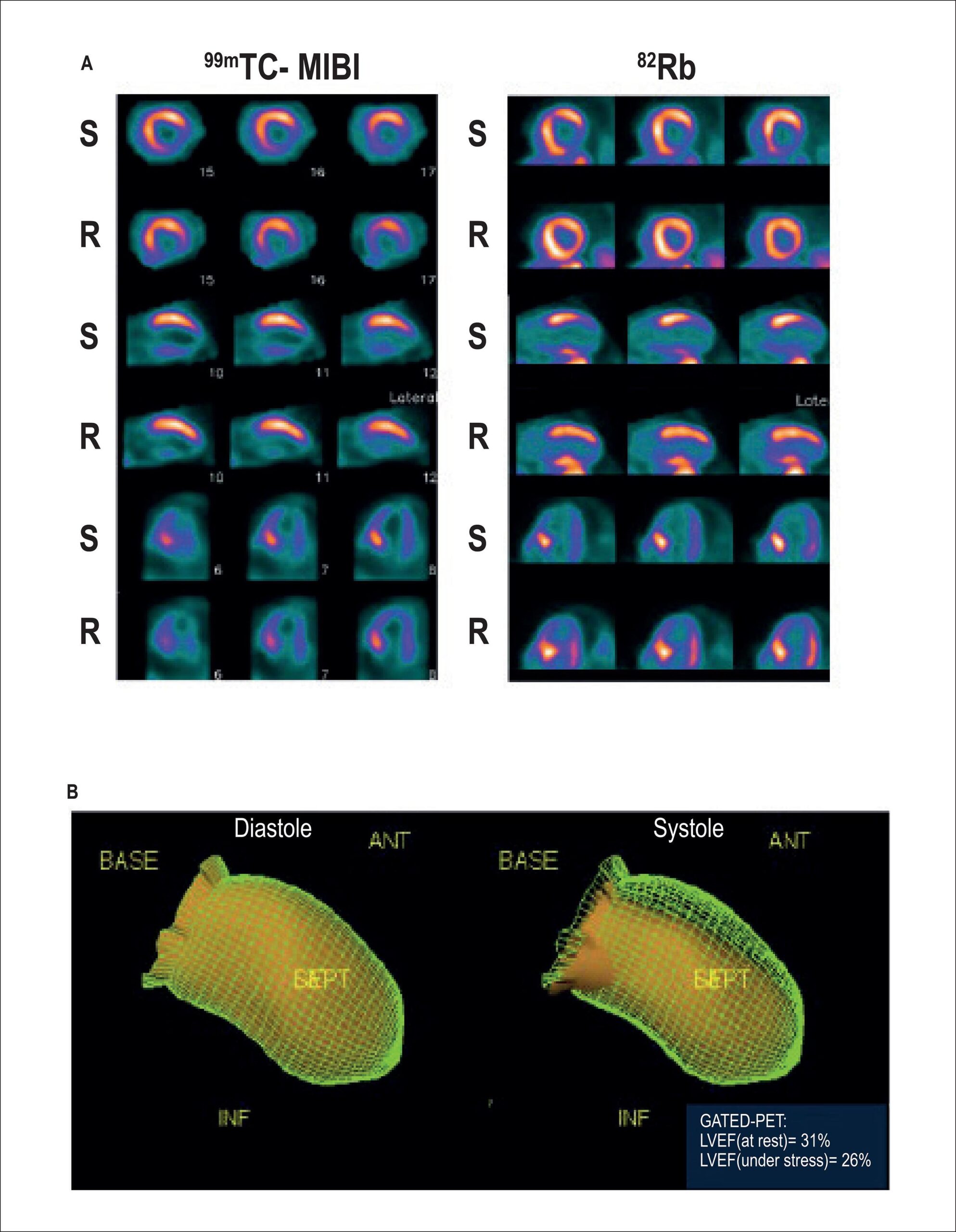
Myocardial perfusion scintigraphy with technetium-99m-Sestamibi (99mTc-sestamibi) allows early detection and evaluation of disease extension and cardiovascular risk in patients with suspected or established CAD, helping in decision-making regarding the start and type of therapy to be implemented. This method has been widely used, but shows difficulties in some situations such as balanced multivessel disease, in which the proportional flow distribution in the myocardial regions can hinder ischemia detection. In such cases, additional assessment data, such as evaluation of contractility, decrease in left ventricular ejection fraction (LVEF) under stress, electrocardiographic alterations or symptoms during stress, dilation of the left ventricle (LV) cavity under stress can provide evidence of ischemia, indicating further diagnostic investigation.

[…]

  • Cardiac Catheterization
  • Coronary Artery Disease
  • Myocardial Perfusion Imaging
  • Radionuclide Imaging
  • Rubidium-82
  • Technetium Tc 99m Sestamibi
Read Full Article
Creative Commons Attribution (CC BY) Creative Commons license
01/01/2017 DOI: 10.5935/abc.20160198
Myocardial perfusion imaging and cardiac involvement in the indeterminate phase of chagas disease
ABC
Artigo Original

Myocardial perfusion imaging and cardiac involvement in the indeterminate phase of chagas disease

Amalia Peix et al. Raymid García Jesús Sánchez Lázaro O. Cabrera … Kenia Padrón Oscar Vedia Hugo V. Choque Jorge Fraga Juan Bandera Alberto Hernández-Cañero

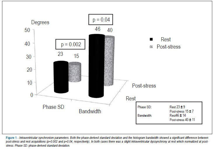
BACKGROUND: Myocardial perfusion imaging (MPI) has been used in the assessment of chagasic heart disease. OBJECTIVE: To investigate the value of gated-single photon emission computed tomography (gated-SPECT) MPI to detect early cardiac involvement in chagasic patients in the indeterminate phase, who present segmental motion abnormalities detected by tissue Doppler imaging (TDI)-derived strain. METHODS: Forty individuals (mean age: 25±2 years, 50% males) from an endemic area of Chagas disease and with positive serologic diagnosis, were included. All underwent gated-SPECT two-day (stress-rest) MPI and echocardiography. RESULTS: Thirty individuals (75%) showed a normal scan. In three cases (8%) the MPI was slightly abnormal, and in seven it was equivocal. In all cases with reversible defects, the affected segments were coincident with those with motion abnormalities. A post-stress left ventricular ejection fraction (LVEF) reduction > 5% (ΔLVEF < -5%) was found in 11 out of 40 individuals (28%). Both the phase-derived standard deviation and the histogram bandwidth showed a significant difference between post-stress and rest. In both cases there was a slight dyssynchrony at rest which normalized at post-stress. CONCLUSIONS: A stress-rest gated-SPECT is a valid approach to detect early myocardial alterations, as well as intraventricular dyssynchrony in the indeterminate phase of Chagas disease in patients with segmental motion abnormalities previously detected by TDI-derived strain.

  • Chagas disease
  • Myocardial Reperfusion
  • Radionuclide Imaging
  • Stroke Volume
Read Full Article
Creative Commons Attribution (CC BY) Creative Commons license
11/03/2013 DOI: 10.5935/abc.20130023
Diagnostic value of myocardial radionuclide imaging in patients with multivessel coronary disease
ABC
Artigo Original

Diagnostic value of myocardial radionuclide imaging in patients with multivessel coronary disease

Maria Eduarda Menezes de Siqueira et al. Erly Medeiros Vieira Segundo Neto Juliana Fernandes Kelendjian Paola Emanuela Pogio Smanio

BACKGROUND: Myocardial perfusion radionuclide imaging (MPRI) is a noninvasive method extremely useful for evaluating ischemia in patients with coronary disease. However, the idea that this method would not be effective for patients with multivessel coronary disease is still entertained. OBJECTIVE: Assess the value of MPRI with gated-SPECT for tracing pharmacological stress-induced ischemia in patients with multivessel obstructive coronary disease. METHODS: We evaluated 68 patients with multivessel obstructive coronary disease applying coronary angiography for performing MPRI under pharmacologic stress with dipyridamole. The tests were analyzed by two nuclear medicine physicians without prior knowledge of the result of the coronary angiography. RESULTS: Gated-SPECT revealed that, out of the patients, 64 (92.8%) showed images of perfusion abnormalities and 4 (7.2%) showed normal perfusion, given that three of the latter showed functional changes. CONCLUSION: MPRI has shown high value for identifying patients with multivessel coronary disease, since most patients had perfusion abnormalities indicative of ischemia.

  • Coronary Artery Disease
  • Dipyridamole
  • Myocardial Reperfusion
  • Radionuclide Imaging
Read Full Article
Creative Commons Attribution (CC BY) Creative Commons license
29/07/2011 DOI: 10.1590/S0066-782X2011005000082
ABC
Artigo Original

Quantification of left ventricular dilatation in myocardial perfusion scintigraphy

Mauren B. Azambuja Gonzalez et al. Roberto Alves Azambuja Luiz Carlos Bodanese

BACKGROUND: The rate of transient dilatation can be determined by exercise testing or pharmacological stress test. It is unknown whether the type of stress has an impact on average transient dilatation index values. OBJECTIVE: To compare average transient dilation index values in 99mTc-sestamibi scintigraphy in patients undergoing treadmill stress test, versus dipyridamole stress test. The secondary purpose was to evaluate the impact on the average index value by demographic characteristics, risk factors for coronary artery disease and severity of ischemia. METHODS: The cross-sectional study included 200 patients between 40 and 70 years old, with or without risk factors for ischemic heart disease, with or without a previous diagnosis of ischemic heart disease. The separation between groups was sequential. The software 4D-MSPECT calculated the transient dilatation index and provided a scoring system for perfusion analysis. RESULTS: The average transient dilation index value of the group undergoing exercise stress test was 1.06 (±0.23). For the group undergoing the dipyridamole stress test, it was 1.10 (±0.22); (p = 0.200). There was no association between the type of stress and the average transient dilatation index values. An association was found between the average index values and age only for those patients from the exercise test group (p = 0.009). CONCLUSION: The results of our study demonstrate that the transient dilation index does not differ when patients undergo exercise stress test on a treadmill or pharmacological stress by dipyridamole.

  • Dilatation
  • Dipyridamole
  • Exercise Test
  • Radionuclide Imaging
  • Ventricular Function, Left
Read Full Article
Creative Commons Attribution (CC BY) Creative Commons license
15/04/2011 DOI: 10.1590/S0066-782X2011005000042
Assessment of myocardial perfusion and function in victims of scorpion envenomation using Gated-SPECT
ABC
Artigo Original

Assessment of myocardial perfusion and function in victims of scorpion envenomation using Gated-SPECT

Alexandre Baldini de Figueiredo et al. Palmira Cupo Antônio O Pintya Fábio Caligaris … José A Marin-Neto Sylvia E Hering Marcus Vinicius Simões

BACKGROUND: Cardiogenic shock and acute pulmonary edema are the major causes of death of patients with scorpion envenomation, whose pathophysiological mechanism remains controversial. OBJECTIVE: To investigate the correlation between myocardial perfusion abnormalities and left ventricular contractile function in victims of scorpion envenomation. METHODS: Fifteen patients underwent ECG-gated myocardial perfusion scintigraphy (gated SPECT) within 72 hours of, and 15 days after scorpion envenomation. Images were analyzed by means of a semi-quantitative visual perfusion score (0 = normal, 4 = absent) and motion score (0 = normal, 4 = akinesia), using the 17-segment model. Summed perfusion (SPS) and summed motion (SMS) scores were calculated for each patient. Ejection fraction (LVEF) was calculated by a commercially available software. RESULTS: At baseline, 12 out of the 15 patients presented abnormal myocardial perfusion and contractility. Mean values of SPS, SMS and LVEF were 12.5 ± 7.3, 17.0 ± 12.8, and 44.6 ± 16.0%, respectively. A positive correlation between SPS and SMS (r = 0.68; p = 0.005) and negative correlation between SPS and LVEF (r = -0.75; p = 0.0021) were found. The follow-up studies showed recovery of global contractility (LVEF of 68.9 ± 9.5, p = 0.0002), segmental contractility (SMS of 2.6 ± 3.1, p = 0.0009) and perfusion (SPS of 3.7 ± 3.3, p = 0.0003). Improvement of LVEF correlated positively with improvement of SPS (r = 0.72; p = 0.0035). CONCLUSIONS: Myocardial perfusion abnormalities are common in scorpion envenomation and correlate topographically with the contractile dysfunction. Recovery of contractility correlates with reversibility of perfusion defects. These findings suggest the participation of myocardial perfusion abnormalities in the pathophysiology of this form of acute ventricular failure. (Arq Bras Cardiol 2010;94(4): 418-425)

  • Perfusion
  • Pulmonary Edema
  • Radionuclide Imaging
  • Scorpions
  • Ventricular Dysfunction
Read Full Article
Creative Commons Attribution (CC BY) Creative Commons license
22/09/2010 DOI: 10.1590/S0066-782X2010000400003
Effects of short-term carvedilol on the cardiac sympathetic activity assessed by 123I-MIBG scintigraphy
ABC
Artigo Original

Effects of short-term carvedilol on the cardiac sympathetic activity assessed by 123I-MIBG scintigraphy

Sandra Marina Ribeiro de Miranda et al. Evandro Tinoco Mesquita Hans Fernando da Rocha Dohmann Jader Cunha Azevedo … Gustavo Borges Barbirato Fabiano de Lima Freire Mário Luiz Ribeiro Antonio Cláudio Lucas da Nóbrega Alexandro Coimbra Cláudio Tinoco Mesquita

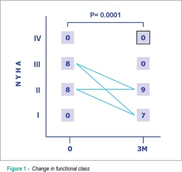
BACKGROUND: Autonomic alterations in heart failure are associated with an increase in morbimortality. Several noninvasive methods have been employed to evaluate the sympathetic function, including the Meta-Iodobenzylguanidine (123I-MIBG) scintigraphy imaging of the heart. OBJECTIVE: to evaluate the cardiac sympathetic activity through 123I-MIBG scintigraphy, before and after three months of carvedilol therapy in patients with heart failure and left ventricular ejection fraction (LVEF) < 45%. PATIENTS AND METHODS: Sixteen patients, aged 56.3 ± 12.6 years (11 males), with a mean LVEF of 28% ± 8% and no previous use of beta-blockers were recruited for the study. Images of the heart innervation were acquired with 123I-MIBG, and the serum levels of catecholamines (epinephrine, dopamine and norepinephrine) were measured; the radioisotope ventriculography (RIV) was performed before and after a three-month therapy with carvedilol. RESULTS: Patients' functional class showed improvement: before the treatment, 50% of the patients were FC II and 50% were FC III. After 3 months, 7 patients were FC I (43.8%) and 9 were FC II (56.2%), (p = 0.0001). The mean LVEF assessed by RIV increased from 29% to 33% (p = 0.017). There was no significant variation in cardiac adrenergic activity assessed by 123I-MIBG (early and late resting images and washout rate). No significant variation was observed regarding the measurement of catecholamines. CONCLUSION: The short-term treatment with carvedilol promoted the clinical and LVEF improvement. However, this was not associated to an improvement in the cardiac adrenergic activity, assessed by 123I-MIBG scintigraphy, as well as the measurement of circulating catecholamines.

  • 3-iodobenzylguanidine
  • Heart Failure /therapy
  • Radionuclide Imaging
Read Full Article
Creative Commons Attribution (CC BY) Creative Commons license
28/04/2010 DOI: 10.1590/S0066-782X2010000300008
Radionuclide imaging of myocardium with mental stress in the investigation of thoracic pain
ABC
Case Report

Radionuclide imaging of myocardium with mental stress in the investigation of thoracic pain

Lívia Maria Lima da Silva et al. Gustavo Borges Barbirato Cantídio Drumond Neto Antonio Claudio Lucas da Nóbrega … Alexandro Coimbra Claudio Tinoco Mesquita

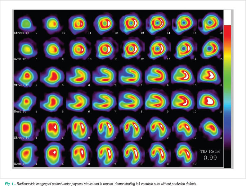
Thoracic pain is a common symptom in emergency services, and stress radionuclide imaging represents one of the phases of risk stratification in these individuals. However, a group of patients with negative functional exams after physical or pharmacological stress develops myocardial ischemia during this psychological stress. Alterations in vascular tonus as a response to endogenous mechanisms are the physiopathologic basis for such alterations. We report a case that illustrates how mental stress radionuclide imaging has the potential to be used in the assessment of myocardial ischemia non-detected by conventional methods in patients with suspicion of ischemic thoracic pain.

  • Myocardial Ischemia
  • Psychological Stress
  • Radionuclide Imaging
Read Full Article
Creative Commons Attribution (CC BY) Creative Commons license
24/11/2009 DOI: 10.1590/S0066-782X2009001000024 Views 1
Ventricular function following coronary artery bypass grafting: comparison between Gated SPECT and cardiac magnetic resonance imaging
ABC
Artigo Original

Ventricular function following coronary artery bypass grafting: comparison between Gated SPECT and cardiac magnetic resonance imaging

Cláudio Tinoco Mesquita et al. Maria Carolina Pinheiro Pessoa Paulo Pontes Vasconcelos Amarino Carvalho Oliveira Júnior … Hans Fernando Rocha Dohmann Adair Gomes dos Reis Lea Mirian Barbosa da Fonseca

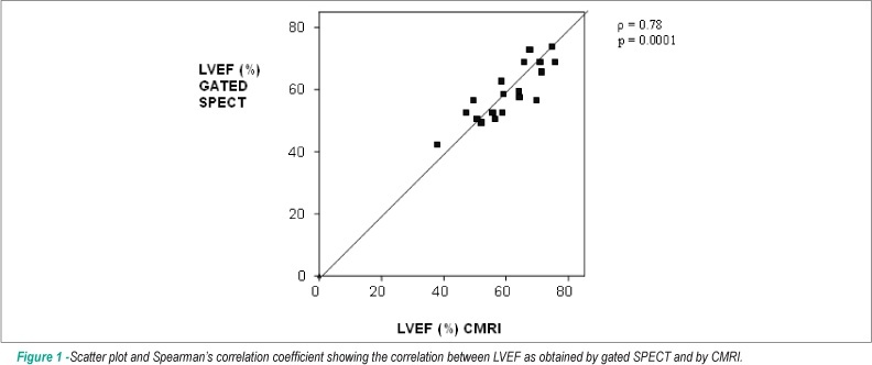
BACKGROUND: The assessment of left ventricular function may be impaired by the abnormal interventricular septal motion frequently found after coronary artery bypass grafting (CABG). Studies on the validation of gated SPECT as a tool for the assessment of left ventricular function in this patient group are scarce. OBJECTIVE: We investigated the agreement and correlation between left ventricular ejection fraction (LVEF), end-diastolic volume (EDV), and end-systolic volume (ESV) as obtained using electrocardiogram-gated myocardial perfusion scintigraphy (gated SPECT) and cardiac magnetic resonance imaging in 20 patients undergoing coronary artery bypass grafting. METHODS: Correlation was measured using Spearman’s correlation coefficient (ρ). Agreement was assessed using Bland-Altman analysis. RESULTS: A good correlation was found between gated SPECT and cardiac magnetic resonance imaging in patients after CABG with regard to left ventricular ejection fraction (ρ = 0.85; p =0.0001), moderate correlation for end-diastolic volume (ρ = 0.51; p = 0.02), and non-significant correlation for end-diastolic volume (ρ = 0.13; p = 0.5). Agreement ranges for LVEF, ESV and EDV were: -20% to 12%; -38 to 54 ml and; -96 to 100 ml, respectively. CONCLUSION: A reliable correlation was found for left ventricular ejection fraction as obtained by gated SPECT and magnetic resonance imaging in patients undergoing CABG. For ventricular volumes, however, the correlation is not adequate.

  • Magnetic Resonance Imaging
  • Myocardial Reperfusion
  • Myocardial Revascularization
  • Radionuclide Imaging
  • Ventricular Function, Left
Read Full Article
Creative Commons Attribution (CC BY) Creative Commons license
20/07/2009 DOI: 10.1590/S0066-782X2009000500004

Could not load more articles. Please try again.

How to cite this article

EndNote RIS

Electronic Document Format (Vancouver)

Gomes MB, Bombana CF, Fuchs FC. ECG’s AVR ST elevation Analysis by Perfusion Scintilography in the Context of Myocardial Ischemia. International Journal of Cardiovascular Sciences 2025;38:e20230080.

Electronic Document Format (ABNT)

Gomes, Miriana Basso; Bombana, Carolina Feijó; Fuchs, Felipe C.. ECG’s AVR ST elevation Analysis by Perfusion Scintilography in the Context of Myocardial Ischemia. International Journal of Cardiovascular Sciences, v. 38, e20230080, Apr. 2025.

Electronic Document Format (APA)

Gomes, M. B., Bombana, C. F., & Fuchs, F. C. (2025). ECG’s AVR ST elevation Analysis by Perfusion Scintilography in the Context of Myocardial Ischemia. International Journal of Cardiovascular Sciences, 38, e20230080.

Electronic Document Format (ISO)

Gomes, Miriana Basso and Bombana, Carolina Feijó and Fuchs, Felipe C.. ECG’s AVR ST elevation Analysis by Perfusion Scintilography in the Context of Myocardial Ischemia. International Journal of Cardiovascular Sciences [online]. 2025, vol. 38, [cited 2026-10-14], e20230080. Available from: <https://journals.cardiol.br/en/article/ecgs-avr-st-elevation-analysis-by-perfusion-scintilography-in-the-context-of-myocardial-ischemia/>. ISSN 2359-4802

EndNote RIS

Electronic Document Format (Vancouver)

Key NK, Melo AS, Sena JP, Dourado AD, Perez JM, Brito JCR, Feitosa G, et al. Prevalence of Cardiac Amyloidosis in Elderly Patients With Aortic Stenosis. International Journal of Cardiovascular Sciences 2024;37:e20240064.

Electronic Document Format (ABNT)

Key, Ng Kin; Melo, Adelina Sanches de; Sena, Joberto Pinheiro; Dourado, Adriano Dias; Perez, Julia Micheli; Brito, Jose Carlos Raimundo; Feitosa, Gilson; Feitosa-Filho, Gilson Soares. Prevalence of Cardiac Amyloidosis in Elderly Patients With Aortic Stenosis. International Journal of Cardiovascular Sciences, v. 37, e20240064, Nov. 2024.

Electronic Document Format (APA)

Key, N. K., Melo, A. S. , Sena, J. P., Dourado, A. D., Perez, J. M., Brito, J. C. R., Feitosa, G., & Feitosa-Filho, G. S. (2024). Prevalence of Cardiac Amyloidosis in Elderly Patients With Aortic Stenosis. International Journal of Cardiovascular Sciences, 37, e20240064.

Electronic Document Format (ISO)

Key, Ng Kin and Melo, Adelina Sanches de and Sena, Joberto Pinheiro and Dourado, Adriano Dias and Perez, Julia Micheli and Brito, Jose Carlos Raimundo and Feitosa, Gilson and Feitosa-Filho, Gilson Soares. Prevalence of Cardiac Amyloidosis in Elderly Patients With Aortic Stenosis. International Journal of Cardiovascular Sciences [online]. 2024, vol. 37, [cited 2026-10-14], e20240064. Available from: <https://journals.cardiol.br/en/article/prevalence-of-cardiac-amyloidosis-in-elderly-patients-with-aortic-stenosis/>. ISSN 2359-4802

EndNote RIS

Electronic Document Format (Vancouver)

Mesquita CT, Yahiro DS, Santos JFR, Machado LE, Rodriguez JGV, Azevedo Junior GL, Simões M, et al. Pyrophosphate Scintigraphy: Use in the Diagnosis and Monitoring of Cardiac Amyloidosis. International Journal of Cardiovascular Sciences 2024;37:e20240051.

Electronic Document Format (ABNT)

Mesquita, Claudio Tinoco; Yahiro, Davi Shunji; Santos, José Felipe Ramos dos; Machado, Luís Eduardo; Rodriguez, Jose Gregorio Valero; Azevedo Junior, Giovane Leal de; Simões, Marcus. Pyrophosphate Scintigraphy: Use in the Diagnosis and Monitoring of Cardiac Amyloidosis. International Journal of Cardiovascular Sciences, v. 37, e20240051, Aug. 2024.

Electronic Document Format (APA)

Mesquita, C. T., Yahiro, D. S., Santos, J. F. R. , Machado, L. E., Rodriguez, J. G. V., Azevedo Junior, G. L. , & Simões, M. (2024). Pyrophosphate Scintigraphy: Use in the Diagnosis and Monitoring of Cardiac Amyloidosis. International Journal of Cardiovascular Sciences, 37, e20240051.

Electronic Document Format (ISO)

Mesquita, Claudio Tinoco and Yahiro, Davi Shunji and Santos, José Felipe Ramos dos and Machado, Luís Eduardo and Rodriguez, Jose Gregorio Valero and Azevedo Junior, Giovane Leal de and Simões, Marcus. Pyrophosphate Scintigraphy: Use in the Diagnosis and Monitoring of Cardiac Amyloidosis. International Journal of Cardiovascular Sciences [online]. 2024, vol. 37, [cited 2026-10-14], e20240051. Available from: <https://journals.cardiol.br/en/article/pyrophosphate-scintigraphy-use-in-the-diagnosis-and-monitoring-of-cardiac-amyloidosis/>. ISSN 2359-4802

EndNote RIS

Electronic Document Format (Vancouver)

Peix A, Araujo R, Gomez V, Barreda AP, Dondi M, Brink A, Minoshima E, et al. Multimodality Imaging of Chagas Heart Disease: Review and Case Presentation. International Journal of Cardiovascular Sciences 2024;37:e20240114.

Electronic Document Format (ABNT)

Peix, Amalia; Araujo, Raul; Gomez, Veronica; Barreda, Aylen Perez; Dondi, Maurizio; Brink, Anita; Minoshima, Erika; Paez, Diana. Multimodality Imaging of Chagas Heart Disease: Review and Case Presentation. International Journal of Cardiovascular Sciences, v. 37, e20240114, Aug. 2024.

Electronic Document Format (APA)

Peix, A., Araujo, R., Gomez, V., Barreda, A. P., Dondi, M., Brink, A., Minoshima, E., & Paez, D. (2024). Multimodality Imaging of Chagas Heart Disease: Review and Case Presentation. International Journal of Cardiovascular Sciences, 37, e20240114.

Electronic Document Format (ISO)

Peix, Amalia and Araujo, Raul and Gomez, Veronica and Barreda, Aylen Perez and Dondi, Maurizio and Brink, Anita and Minoshima, Erika and Paez, Diana. Multimodality Imaging of Chagas Heart Disease: Review and Case Presentation. International Journal of Cardiovascular Sciences [online]. 2024, vol. 37, [cited 2026-10-14], e20240114. Available from: <https://journals.cardiol.br/en/article/multimodality-imaging-of-chagas-heart-disease-review-and-case-presentation/>. ISSN 2359-4802

EndNote RIS

Electronic Document Format (Vancouver)

Silva TO, Darzé ES, Costa MM, José Junior L, Ximenes AAB, Fernandes F, Rocha MS, et al. Scintigraphic and Echocardiographic Study of Patients with Pathogenic or Probably Pathogenic Variants of the TTR Gene without Overt Cardiac Involvement. Arquivos Brasileiros de Cardiologia 2024;121(4):e20230216.

Electronic Document Format (ABNT)

Silva, Tonnison de Oliveira; Darzé, Eduardo Sahade; Costa, Marcela Machado; José Junior, Luiz; Ximenes, Antonio A. B.; Fernandes, Fábio; Rocha, Mário de Seixas; Noya-Rabelo, Marcia M.; Ritt, Luiz Eduardo Fonteles. Scintigraphic and Echocardiographic Study of Patients with Pathogenic or Probably Pathogenic Variants of the TTR Gene without Overt Cardiac Involvement. Arquivos Brasileiros de Cardiologia, v. 121, n. 4, e20230216, May. 2024.

Electronic Document Format (APA)

Silva, T. O., Darzé, E. S., Costa, M. M., José Junior, L., Ximenes, A. A. B., Fernandes, F., Rocha, M. S., Noya-Rabelo, M. M., & Ritt, L. E. F. (2024). Scintigraphic and Echocardiographic Study of Patients with Pathogenic or Probably Pathogenic Variants of the TTR Gene without Overt Cardiac Involvement. Arquivos Brasileiros de Cardiologia, 121(4), e20230216.

Electronic Document Format (ISO)

Silva, Tonnison de Oliveira and Darzé, Eduardo Sahade and Costa, Marcela Machado and José Junior, Luiz and Ximenes, Antonio A. B. and Fernandes, Fábio and Rocha, Mário de Seixas and Noya-Rabelo, Marcia M. and Ritt, Luiz Eduardo Fonteles. Scintigraphic and Echocardiographic Study of Patients with Pathogenic or Probably Pathogenic Variants of the TTR Gene without Overt Cardiac Involvement. Arquivos Brasileiros de Cardiologia [online]. 2024, vol. 121, n. 4, [cited 2026-10-14], e20230216. Available from: <https://journals.cardiol.br/en/article/scintigraphic-and-echocardiographic-study-of-patients-with-pathogenic-or-probably-pathogenic-variants-of-the-ttr-gene-without-overt-cardiac-involvement/>. ISSN 0066-782X

EndNote RIS

Electronic Document Format (Vancouver)

Costa TO, Rocha MS, Soares Filho GF, Camargo RA, Macedo CRB. Incremental Determinants of The Combined Supine-Prone Protocol for Myocardial Perfusion Scintigraphy. ABC Imagem Cardiovascular 2023;36(3):e20230060.

Electronic Document Format (ABNT)

Costa, Tiago Oliveira; Rocha, Mário de Seixas; Soares Filho, Gilson Feitosa; Camargo, Raphael Abegão de; Macedo, Cristiano Ricardo Bastos de. Incremental Determinants of The Combined Supine-Prone Protocol for Myocardial Perfusion Scintigraphy. ABC Imagem Cardiovascular, v. 36, n. 3, e20230060, Oct. 2023.

Electronic Document Format (APA)

Costa, T. O., Rocha, M. S., Soares Filho, G. F., Camargo, R. A. , & Macedo, C. R. B. (2023). Incremental Determinants of The Combined Supine-Prone Protocol for Myocardial Perfusion Scintigraphy. ABC Imagem Cardiovascular, 36(3), e20230060.

Electronic Document Format (ISO)

Costa, Tiago Oliveira and Rocha, Mário de Seixas and Soares Filho, Gilson Feitosa and Camargo, Raphael Abegão de and Macedo, Cristiano Ricardo Bastos de. Incremental Determinants of The Combined Supine-Prone Protocol for Myocardial Perfusion Scintigraphy. ABC Imagem Cardiovascular [online]. 2023, vol. 36, n. 3, [cited 2026-10-14], e20230060. Available from: <https://journals.cardiol.br/en/article/incremental-determinants-of-the-combined-supine-prone-protocol-for-myocardial-perfusion-scintigraphy/>. ISSN 2318-8219

EndNote RIS

Electronic Document Format (Vancouver)

Mesquita CT, Rezende MF. Precision Medicine: Can 18F-FDG PET Detect Cardiotoxicity Phenotypes?. Arquivos Brasileiros de Cardiologia 2022;119(1):109-10.

Electronic Document Format (ABNT)

Mesquita, Claudio Tinoco; Rezende, Maria Fernanda. Precision Medicine: Can 18F-FDG PET Detect Cardiotoxicity Phenotypes?. Arquivos Brasileiros de Cardiologia, v. 119, n. 1, p. 109-110, Jul. 2022.

Electronic Document Format (APA)

Mesquita, C. T., & Rezende, M. F. (2022). Precision Medicine: Can 18F-FDG PET Detect Cardiotoxicity Phenotypes?. Arquivos Brasileiros de Cardiologia, 119(1), 109-110.

Electronic Document Format (ISO)

Mesquita, Claudio Tinoco and Rezende, Maria Fernanda. Precision Medicine: Can 18F-FDG PET Detect Cardiotoxicity Phenotypes?. Arquivos Brasileiros de Cardiologia [online]. 2022, vol. 119, n. 1, [cited 2026-10-14], pp. 109-110. Available from: <https://journals.cardiol.br/en/article/precision-medicine-can-18f-fdg-pet-detect-cardiotoxicity-phenotypes/>. ISSN 0066-782X

EndNote RIS

Electronic Document Format (Vancouver)

Delgado-Silva J, Moreira AP, Costa G, Gonçalves L. Risk Stratification and Cardiac Sympathetic Activity Assessment Using Myocardial [123I] MIBG Imaging in Renal Denervation. Arquivos Brasileiros de Cardiologia 2022;118(2):519-24.

Electronic Document Format (ABNT)

Delgado-Silva, Joana; Moreira, Ana Paula; Costa, Gracinda; Gonçalves, Lino. Risk Stratification and Cardiac Sympathetic Activity Assessment Using Myocardial [123I] MIBG Imaging in Renal Denervation. Arquivos Brasileiros de Cardiologia, v. 118, n. 2, p. 519-524, Mar. 2022.

Electronic Document Format (APA)

Delgado-Silva, J., Moreira, A. P., Costa, G., & Gonçalves, L. (2022). Risk Stratification and Cardiac Sympathetic Activity Assessment Using Myocardial [123I] MIBG Imaging in Renal Denervation. Arquivos Brasileiros de Cardiologia, 118(2), 519-524.

Electronic Document Format (ISO)

Delgado-Silva, Joana and Moreira, Ana Paula and Costa, Gracinda and Gonçalves, Lino. Risk Stratification and Cardiac Sympathetic Activity Assessment Using Myocardial [123I] MIBG Imaging in Renal Denervation. Arquivos Brasileiros de Cardiologia [online]. 2022, vol. 118, n. 2, [cited 2026-10-14], pp. 519-524. Available from: <https://journals.cardiol.br/en/article/risk-stratification-and-cardiac-sympathetic-activity-assessment-using-myocardial-123i-mibg-imaging-in-renal-denervation/>. ISSN 0066-782X

EndNote RIS

Electronic Document Format (Vancouver)

Hilgemberg EC, Roessler UCBAD, Santos LTM, Damas AS, Mesquita CT. Myocardial Viability in a Patient with Systemic Lupus Erythematosus. Arquivos Brasileiros de Cardiologia Imagem cardiovascular 2022;31(3):215-7.

Electronic Document Format (ABNT)

Hilgemberg, Elisa Carla; Roessler, Ursula Cristina Barbosa de Almeida Daco; Santos, Larissa Ticiane Machado dos; Damas, Alair Sarmet; Mesquita, Claúdio Tinoco. Myocardial Viability in a Patient with Systemic Lupus Erythematosus. Arquivos Brasileiros de Cardiologia Imagem cardiovascular, v. 31, n. 3, p. 215-217, Jan. 2022.

Electronic Document Format (APA)

Hilgemberg, E. C., Roessler, U. C. B. A. D., Santos, L. T. M. , Damas, A. S., & Mesquita, C. T. (2022). Myocardial Viability in a Patient with Systemic Lupus Erythematosus. Arquivos Brasileiros de Cardiologia Imagem cardiovascular, 31(3), 215-217.

Electronic Document Format (ISO)

Hilgemberg, Elisa Carla and Roessler, Ursula Cristina Barbosa de Almeida Daco and Santos, Larissa Ticiane Machado dos and Damas, Alair Sarmet and Mesquita, Claúdio Tinoco. Myocardial Viability in a Patient with Systemic Lupus Erythematosus. Arquivos Brasileiros de Cardiologia Imagem cardiovascular [online]. 2022, vol. 31, n. 3, [cited 2026-10-14], pp. 215-217. Available from: <https://journals.cardiol.br/en/article/myocardial-viability-in-a-patient-with-systemic-lupus-erythematosus/>. ISSN 2318-8219

EndNote RIS

Electronic Document Format (Vancouver)

Segundo Neto EMV, Lopes RW, Brandão SCS. What is the Value of Perfusion Scintigraphy and Cardiac PET in Hypertrophic Cardiomyopathy?. Arquivos Brasileiros de Cardiologia Imagem cardiovascular 2022;32(1):19-28.

Electronic Document Format (ABNT)

Segundo Neto, Elry Medeiros Vieira; Lopes, Rafael Willain; Brandão, Simone Cristina Soares. What is the Value of Perfusion Scintigraphy and Cardiac PET in Hypertrophic Cardiomyopathy?. Arquivos Brasileiros de Cardiologia Imagem cardiovascular, v. 32, n. 1, p. 19-28, Jan. 2022.

Electronic Document Format (APA)

Segundo Neto, E. M. V., Lopes, R. W., & Brandão, S. C. S. (2022). What is the Value of Perfusion Scintigraphy and Cardiac PET in Hypertrophic Cardiomyopathy?. Arquivos Brasileiros de Cardiologia Imagem cardiovascular, 32(1), 19-28.

Electronic Document Format (ISO)

Segundo Neto, Elry Medeiros Vieira and Lopes, Rafael Willain and Brandão, Simone Cristina Soares. What is the Value of Perfusion Scintigraphy and Cardiac PET in Hypertrophic Cardiomyopathy?. Arquivos Brasileiros de Cardiologia Imagem cardiovascular [online]. 2022, vol. 32, n. 1, [cited 2026-10-14], pp. 19-28. Available from: <https://journals.cardiol.br/en/article/what-is-the-value-of-perfusion-scintigraphy-and-cardiac-pet-in-hypertrophic-cardiomyopathy/>. ISSN 2318-8219

EndNote RIS

Electronic Document Format (Vancouver)

Gomes RAF, Moreira ICAM, Cardoso OGM, Melo AGS, Macêdo Junior ARA. Acute myocardial infarction after myocardial scintigraphy with dipyridamole. Arquivos Brasileiros de Cardiologia Imagem cardiovascular 2022;33(1):1-3.

Electronic Document Format (ABNT)

Gomes, Rafael Alessandro Ferreira; Moreira, Isabelle Conceição Albuquerque Machado; Cardoso, Otávio Guilherme de Moraes; Melo, Alexandre Gomes de Souza; Macêdo Junior, Aluísio Roberto Andrade. Acute myocardial infarction after myocardial scintigraphy with dipyridamole. Arquivos Brasileiros de Cardiologia Imagem cardiovascular, v. 33, n. 1, p. 1-3, Jan. 2022.

Electronic Document Format (APA)

Gomes, R. A. F., Moreira, I. C. A. M., Cardoso, O. G. M., Melo, A. G. S., & Macêdo Junior, A. R. A. (2022). Acute myocardial infarction after myocardial scintigraphy with dipyridamole. Arquivos Brasileiros de Cardiologia Imagem cardiovascular, 33(1), 1-3.

Electronic Document Format (ISO)

Gomes, Rafael Alessandro Ferreira and Moreira, Isabelle Conceição Albuquerque Machado and Cardoso, Otávio Guilherme de Moraes and Melo, Alexandre Gomes de Souza and Macêdo Junior, Aluísio Roberto Andrade. Acute myocardial infarction after myocardial scintigraphy with dipyridamole. Arquivos Brasileiros de Cardiologia Imagem cardiovascular [online]. 2022, vol. 33, n. 1, [cited 2026-10-14], pp. 1-3. Available from: <https://journals.cardiol.br/en/article/acute-myocardial-infarction-after-myocardial-scintigraphy-with-dipyridamole/>. ISSN 2318-8219

EndNote RIS

Electronic Document Format (Vancouver)

Kulchetscki RM, Lechinewski LD, Dourado LOC, Hueb WA, César LAM. Large Bilateral Coronary Artery Fistula: 10-year Follow-up in Clinical Treatment. Arquivos Brasileiros de Cardiologia 2019;112(2):211-3.

Electronic Document Format (ABNT)

Kulchetscki, Rodrigo Melo; Lechinewski, Luka David; Dourado, Luciana Oliveira Cascaes; Hueb, Whady Armindo; César, Luiz Antonio Machado. Large Bilateral Coronary Artery Fistula: 10-year Follow-up in Clinical Treatment. Arquivos Brasileiros de Cardiologia, v. 112, n. 2, p. 211-213, Jan. 2019.

Electronic Document Format (APA)

Kulchetscki, R. M., Lechinewski, L. D., Dourado, L. O. C., Hueb, W. A., & César, L. A. M. (2019). Large Bilateral Coronary Artery Fistula: 10-year Follow-up in Clinical Treatment. Arquivos Brasileiros de Cardiologia, 112(2), 211-213.

Electronic Document Format (ISO)

Kulchetscki, Rodrigo Melo and Lechinewski, Luka David and Dourado, Luciana Oliveira Cascaes and Hueb, Whady Armindo and César, Luiz Antonio Machado. Large Bilateral Coronary Artery Fistula: 10-year Follow-up in Clinical Treatment. Arquivos Brasileiros de Cardiologia [online]. 2019, vol. 112, n. 2, [cited 2026-10-14], pp. 211-213. Available from: <https://journals.cardiol.br/en/article/large-bilateral-coronary-artery-fistula-10-year-follow-up-in-clinical-treatment/>. ISSN 0066-782X

EndNote RIS

Electronic Document Format (Vancouver)

Nascimento EA, Reis CCW, Ribeiro FB, Alves CR, Silva EN, Ribeiro ML, Mesquita CT, et al. Relationship of Electromechanical Dyssynchrony in Patients Submitted to CRT With LV Lead Implantation Guided by Gated Myocardial Perfusion Spect. Arquivos Brasileiros de Cardiologia 2018;111(4):607-15.

Electronic Document Format (ABNT)

Nascimento, Erivelton Alessandro do; Reis, Christiane Cigagna Wiefels; Ribeiro, Fernanda Baptista; Alves, Christiane Rodrigues; Silva, Eduardo Nani; Ribeiro, Mario Luiz; Mesquita, Claudio Tinoco. Relationship of Electromechanical Dyssynchrony in Patients Submitted to CRT With LV Lead Implantation Guided by Gated Myocardial Perfusion Spect. Arquivos Brasileiros de Cardiologia, v. 111, n. 4, p. 607-615, Aug. 2018.

Electronic Document Format (APA)

Nascimento, E. A. , Reis, C. C. W., Ribeiro, F. B., Alves, C. R., Silva, E. N., Ribeiro, M. L., & Mesquita, C. T. (2018). Relationship of Electromechanical Dyssynchrony in Patients Submitted to CRT With LV Lead Implantation Guided by Gated Myocardial Perfusion Spect. Arquivos Brasileiros de Cardiologia, 111(4), 607-615.

Electronic Document Format (ISO)

Nascimento, Erivelton Alessandro do and Reis, Christiane Cigagna Wiefels and Ribeiro, Fernanda Baptista and Alves, Christiane Rodrigues and Silva, Eduardo Nani and Ribeiro, Mario Luiz and Mesquita, Claudio Tinoco. Relationship of Electromechanical Dyssynchrony in Patients Submitted to CRT With LV Lead Implantation Guided by Gated Myocardial Perfusion Spect. Arquivos Brasileiros de Cardiologia [online]. 2018, vol. 111, n. 4, [cited 2026-10-14], pp. 607-615. Available from: <https://journals.cardiol.br/en/article/relationship-of-electromechanical-dyssynchrony-in-patients-submitted-to-crt-with-lv-lead-implantation-guided-by-gated-myocardial-perfusion-spect/>. ISSN 0066-782X

EndNote RIS

Electronic Document Format (Vancouver)

Rocha ET, Alves WEFM, Verschure DO, Verberne HJ. The use of Cardiac 123I-mIBG Scintigraphy in Clinical Practice: The Necessity to Standardize!. International Journal of Cardiovascular Sciences 2017;30(6):533-41.

Electronic Document Format (ABNT)

Rocha, Euclides Timóteo da; Alves, Wilson Eduardo Furlan Matos; Verschure, Derk O.; Verberne, Hein J. The use of Cardiac 123I-mIBG Scintigraphy in Clinical Practice: The Necessity to Standardize!. International Journal of Cardiovascular Sciences, v. 30, n. 6, p. 533-541, Sep. 2017.

Electronic Document Format (APA)

Rocha, E. T. , Alves, W. E. F. M., Verschure, D. O., & Verberne, H. J. (2017). The use of Cardiac 123I-mIBG Scintigraphy in Clinical Practice: The Necessity to Standardize!. International Journal of Cardiovascular Sciences, 30(6), 533-541.

Electronic Document Format (ISO)

Rocha, Euclides Timóteo da and Alves, Wilson Eduardo Furlan Matos and Verschure, Derk O. and Verberne, Hein J. The use of Cardiac 123I-mIBG Scintigraphy in Clinical Practice: The Necessity to Standardize!. International Journal of Cardiovascular Sciences [online]. 2017, vol. 30, n. 6, [cited 2026-10-14], pp. 533-541. Available from: <https://journals.cardiol.br/en/article/the-use-of-cardiac-123i-mibg-scintigraphy-in-clinical-practice-the-necessity-to-standardize/>. ISSN 2359-4802

EndNote RIS

Electronic Document Format (Vancouver)

Ker WS, Neves DG, Damas ASAA, Mesquita CT, Nacif MS. Myocardial Bridge and Angiotomography of the Coronary Arteries: Perfusion under Pharmacological Stress. Arquivos Brasileiros de Cardiologia 2017;108(6):572-5.

Electronic Document Format (ABNT)

Ker, Wilter dos Santos; Neves, Daniel Gama; Damas, Alair Sarmet A. A.; Mesquita, Cláudio Tinoco; Nacif, Marcelo Souto. Myocardial Bridge and Angiotomography of the Coronary Arteries: Perfusion under Pharmacological Stress. Arquivos Brasileiros de Cardiologia, v. 108, n. 6, p. 572-575, May. 2017.

Electronic Document Format (APA)

Ker, W. S., Neves, D. G., Damas, A. S. A. A., Mesquita, C. T., & Nacif, M. S. (2017). Myocardial Bridge and Angiotomography of the Coronary Arteries: Perfusion under Pharmacological Stress. Arquivos Brasileiros de Cardiologia, 108(6), 572-575.

Electronic Document Format (ISO)

Ker, Wilter dos Santos and Neves, Daniel Gama and Damas, Alair Sarmet A. A. and Mesquita, Cláudio Tinoco and Nacif, Marcelo Souto. Myocardial Bridge and Angiotomography of the Coronary Arteries: Perfusion under Pharmacological Stress. Arquivos Brasileiros de Cardiologia [online]. 2017, vol. 108, n. 6, [cited 2026-10-14], pp. 572-575. Available from: <https://journals.cardiol.br/en/article/myocardial-bridge-and-angiotomography-of-the-coronary-arteries-perfusion-under-pharmacological-stress/>. ISSN 0066-782X

EndNote RIS

Electronic Document Format (Vancouver)

Cerit L. The Tip of The Iceberg: Non-Calcified Coronary Plague and Epicardial Adipose Tissue. Arquivos Brasileiros de Cardiologia 2017;108(4):383-5.

Electronic Document Format (ABNT)

Cerit, Levent. The Tip of The Iceberg: Non-Calcified Coronary Plague and Epicardial Adipose Tissue. Arquivos Brasileiros de Cardiologia, v. 108, n. 4, p. 383-385, Mar. 2017.

Electronic Document Format (APA)

Cerit, L. (2017). The Tip of The Iceberg: Non-Calcified Coronary Plague and Epicardial Adipose Tissue. Arquivos Brasileiros de Cardiologia, 108(4), 383-385.

Electronic Document Format (ISO)

Cerit, Levent. The Tip of The Iceberg: Non-Calcified Coronary Plague and Epicardial Adipose Tissue. Arquivos Brasileiros de Cardiologia [online]. 2017, vol. 108, n. 4, [cited 2026-10-14], pp. 383-385. Available from: <https://journals.cardiol.br/en/article/the-tip-of-the-iceberg-non-calcified-coronary-plague-and-epicardial-adipose-tissue/>. ISSN 0066-782X

EndNote RIS

Electronic Document Format (Vancouver)

Padilha BG, Sabino D, Giorgi MC, Soares Jr. J, Izaki M, Meneghetti JC. Case Report: Multivessel Coronary Disease Assessment with SPECT 99mTc-Sestamibi and Rubidium-82 PET/CT. Arquivos Brasileiros de Cardiologia 2017;108(1):87-90.

Electronic Document Format (ABNT)

Padilha, Bruno Gomes; Sabino, Daniela; Giorgi, Maria Clementina; Soares Jr., José; Izaki, Marisa; Meneghetti, José Claudio. Case Report: Multivessel Coronary Disease Assessment with SPECT 99mTc-Sestamibi and Rubidium-82 PET/CT. Arquivos Brasileiros de Cardiologia, v. 108, n. 1, p. 87-90, Dec. 2016.

Electronic Document Format (APA)

Padilha, B. G., Sabino, D., Giorgi, M. C., Soares Jr., J., Izaki, M., & Meneghetti, J. C. (2017). Case Report: Multivessel Coronary Disease Assessment with SPECT 99mTc-Sestamibi and Rubidium-82 PET/CT. Arquivos Brasileiros de Cardiologia, 108(1), 87-90.

Electronic Document Format (ISO)

Padilha, Bruno Gomes and Sabino, Daniela and Giorgi, Maria Clementina and Soares Jr., José and Izaki, Marisa and Meneghetti, José Claudio. Case Report: Multivessel Coronary Disease Assessment with SPECT 99mTc-Sestamibi and Rubidium-82 PET/CT. Arquivos Brasileiros de Cardiologia [online]. 2017, vol. 108, n. 1, [cited 2026-10-14], pp. 87-90. Available from: <https://journals.cardiol.br/en/article/case-report-multivessel-coronary-disease-assessment-with-spect-99mtc-sestamibi-and-rubidium-82-pet-ct/>. ISSN 0066-782X

EndNote RIS

Electronic Document Format (Vancouver)

Peix A, García R, Sánchez J, Cabrera LO, Padrón K, Vedia O, Choque HV, et al. Myocardial perfusion imaging and cardiac involvement in the indeterminate phase of chagas disease. Arquivos Brasileiros de Cardiologia 2013;100(2):114-7.

Electronic Document Format (ABNT)

Peix, Amalia; García, Raymid; Sánchez, Jesús; Cabrera, Lázaro O.; Padrón, Kenia; Vedia, Oscar; Choque, Hugo V.; Fraga, Jorge; Bandera, Juan; Hernández-Cañero, Alberto. Myocardial perfusion imaging and cardiac involvement in the indeterminate phase of chagas disease. Arquivos Brasileiros de Cardiologia, v. 100, n. 2, p. 114-117, Mar. 2013.

Electronic Document Format (APA)

Peix, A., García, R., Sánchez, J., Cabrera, L. O., Padrón, K., Vedia, O., Choque, H. V., Fraga, J., Bandera, J., & Hernández-Cañero, A. (2013). Myocardial perfusion imaging and cardiac involvement in the indeterminate phase of chagas disease. Arquivos Brasileiros de Cardiologia, 100(2), 114-117.

Electronic Document Format (ISO)

Peix, Amalia and García, Raymid and Sánchez, Jesús and Cabrera, Lázaro O. and Padrón, Kenia and Vedia, Oscar and Choque, Hugo V. and Fraga, Jorge and Bandera, Juan and Hernández-Cañero, Alberto. Myocardial perfusion imaging and cardiac involvement in the indeterminate phase of chagas disease. Arquivos Brasileiros de Cardiologia [online]. 2013, vol. 100, n. 2, [cited 2026-10-14], pp. 114-117. Available from: <https://journals.cardiol.br/en/article/myocardial-perfusion-imaging-and-cardiac-involvement-in-the-indeterminate-phase-of-chagas-disease/>. ISSN 0066-782X

EndNote RIS

Electronic Document Format (Vancouver)

Siqueira MEM, Segundo Neto EMV, Kelendjian JF, Smanio PEP. Diagnostic value of myocardial radionuclide imaging in patients with multivessel coronary disease. Arquivos Brasileiros de Cardiologia 2011;97(3):194-8.

Electronic Document Format (ABNT)

Siqueira, Maria Eduarda Menezes de; Segundo Neto, Erly Medeiros Vieira; Kelendjian, Juliana Fernandes; Smanio, Paola Emanuela Pogio. Diagnostic value of myocardial radionuclide imaging in patients with multivessel coronary disease. Arquivos Brasileiros de Cardiologia, v. 97, n. 3, p. 194-198, Jul. 2011.

Electronic Document Format (APA)

Siqueira, M. E. M. , Segundo Neto, E. M. V., Kelendjian, J. F., & Smanio, P. E. P. (2011). Diagnostic value of myocardial radionuclide imaging in patients with multivessel coronary disease. Arquivos Brasileiros de Cardiologia, 97(3), 194-198.

Electronic Document Format (ISO)

Siqueira, Maria Eduarda Menezes de and Segundo Neto, Erly Medeiros Vieira and Kelendjian, Juliana Fernandes and Smanio, Paola Emanuela Pogio. Diagnostic value of myocardial radionuclide imaging in patients with multivessel coronary disease. Arquivos Brasileiros de Cardiologia [online]. 2011, vol. 97, n. 3, [cited 2026-10-14], pp. 194-198. Available from: <https://journals.cardiol.br/en/article/diagnostic-value-of-myocardial-radionuclide-imaging-in-patients-with-multivessel-coronary-disease/>. ISSN 0066-782X

EndNote RIS

Electronic Document Format (Vancouver)

Gonzalez MBA, Azambuja RA, Bodanese LC. Quantification of left ventricular dilatation in myocardial perfusion scintigraphy. Arquivos Brasileiros de Cardiologia 2011;96(5):363-9.

Electronic Document Format (ABNT)

Gonzalez, Mauren B. Azambuja; Azambuja, Roberto Alves; Bodanese, Luiz Carlos. Quantification of left ventricular dilatation in myocardial perfusion scintigraphy. Arquivos Brasileiros de Cardiologia, v. 96, n. 5, p. 363-369, Apr. 2011.

Electronic Document Format (APA)

Gonzalez, M. B. A., Azambuja, R. A., & Bodanese, L. C. (2011). Quantification of left ventricular dilatation in myocardial perfusion scintigraphy. Arquivos Brasileiros de Cardiologia, 96(5), 363-369.

Electronic Document Format (ISO)

Gonzalez, Mauren B. Azambuja and Azambuja, Roberto Alves and Bodanese, Luiz Carlos. Quantification of left ventricular dilatation in myocardial perfusion scintigraphy. Arquivos Brasileiros de Cardiologia [online]. 2011, vol. 96, n. 5, [cited 2026-10-14], pp. 363-369. Available from: <https://journals.cardiol.br/en/article/quantification-of-left-ventricular-dilatation-in-myocardial-perfusion-scintigraphy/>. ISSN 0066-782X

EndNote RIS

Electronic Document Format (Vancouver)

Figueiredo AB, Cupo P, Pintya AO, Caligaris F, Marin-Neto JA, Hering SE, Simões MV, et al. Assessment of myocardial perfusion and function in victims of scorpion envenomation using Gated-SPECT. Arquivos Brasileiros de Cardiologia 2010;94(4):444-51.

Electronic Document Format (ABNT)

Figueiredo, Alexandre Baldini de; Cupo, Palmira; Pintya, Antônio O; Caligaris, Fábio; Marin-Neto, José A; Hering, Sylvia E; Simões, Marcus Vinicius. Assessment of myocardial perfusion and function in victims of scorpion envenomation using Gated-SPECT. Arquivos Brasileiros de Cardiologia, v. 94, n. 4, p. 444-451, Sep. 2010.

Electronic Document Format (APA)

Figueiredo, A. B. , Cupo, P., Pintya, A. O., Caligaris, F., Marin-Neto, J. A., Hering, S. E., & Simões, M. V. (2010). Assessment of myocardial perfusion and function in victims of scorpion envenomation using Gated-SPECT. Arquivos Brasileiros de Cardiologia, 94(4), 444-451.

Electronic Document Format (ISO)

Figueiredo, Alexandre Baldini de and Cupo, Palmira and Pintya, Antônio O and Caligaris, Fábio and Marin-Neto, José A and Hering, Sylvia E and Simões, Marcus Vinicius. Assessment of myocardial perfusion and function in victims of scorpion envenomation using Gated-SPECT. Arquivos Brasileiros de Cardiologia [online]. 2010, vol. 94, n. 4, [cited 2026-10-14], pp. 444-451. Available from: <https://journals.cardiol.br/en/article/assessment-of-myocardial-perfusion-and-function-in-victims-of-scorpion-envenomation-using-gated-spect/>. ISSN 0066-782X

EndNote RIS

Electronic Document Format (Vancouver)

Miranda SMR, Mesquita ET, Dohmann HFR, Azevedo JC, Barbirato GB, Freire FL, Ribeiro ML, et al. Effects of short-term carvedilol on the cardiac sympathetic activity assessed by 123I-MIBG scintigraphy. Arquivos Brasileiros de Cardiologia 2010;94(3):328-32.

Electronic Document Format (ABNT)

Miranda, Sandra Marina Ribeiro de; Mesquita, Evandro Tinoco; Dohmann, Hans Fernando da Rocha; Azevedo, Jader Cunha; Barbirato, Gustavo Borges; Freire, Fabiano de Lima; Ribeiro, Mário Luiz; Nóbrega, Antonio Cláudio Lucas da; Coimbra, Alexandro; Mesquita, Cláudio Tinoco. Effects of short-term carvedilol on the cardiac sympathetic activity assessed by 123I-MIBG scintigraphy. Arquivos Brasileiros de Cardiologia, v. 94, n. 3, p. 328-332, Apr. 2010.

Electronic Document Format (APA)

Miranda, S. M. R. , Mesquita, E. T., Dohmann, H. F. R., Azevedo, J. C., Barbirato, G. B., Freire, F. L., Ribeiro, M. L., Nóbrega, A. C. L. , Coimbra, A., & Mesquita, C. T. (2010). Effects of short-term carvedilol on the cardiac sympathetic activity assessed by 123I-MIBG scintigraphy. Arquivos Brasileiros de Cardiologia, 94(3), 328-332.

Electronic Document Format (ISO)

Miranda, Sandra Marina Ribeiro de and Mesquita, Evandro Tinoco and Dohmann, Hans Fernando da Rocha and Azevedo, Jader Cunha and Barbirato, Gustavo Borges and Freire, Fabiano de Lima and Ribeiro, Mário Luiz and Nóbrega, Antonio Cláudio Lucas da and Coimbra, Alexandro and Mesquita, Cláudio Tinoco. Effects of short-term carvedilol on the cardiac sympathetic activity assessed by 123I-MIBG scintigraphy. Arquivos Brasileiros de Cardiologia [online]. 2010, vol. 94, n. 3, [cited 2026-10-14], pp. 328-332. Available from: <https://journals.cardiol.br/en/article/effects-of-short-term-carvedilol-on-the-cardiac-sympathetic-activity-assessed-by-123i-mibg-scintigraphy/>. ISSN 0066-782X

EndNote RIS

Electronic Document Format (Vancouver)

Silva LML, Barbirato GB, Drumond Neto C, Nóbrega ACL, Coimbra A, Mesquita CT. Radionuclide imaging of myocardium with mental stress in the investigation of thoracic pain. Arquivos Brasileiros de Cardiologia 2009;93(4):e63-6.

Electronic Document Format (ABNT)

Silva, Lívia Maria Lima da; Barbirato, Gustavo Borges; Drumond Neto, Cantídio; Nóbrega, Antonio Claudio Lucas da; Coimbra, Alexandro; Mesquita, Claudio Tinoco. Radionuclide imaging of myocardium with mental stress in the investigation of thoracic pain. Arquivos Brasileiros de Cardiologia, v. 93, n. 4, p. e63-e66, Nov. 2009.

Electronic Document Format (APA)

Silva, L. M. L. , Barbirato, G. B., Drumond Neto, C., Nóbrega, A. C. L. , Coimbra, A., & Mesquita, C. T. (2009). Radionuclide imaging of myocardium with mental stress in the investigation of thoracic pain. Arquivos Brasileiros de Cardiologia, 93(4), e63-e66.

Electronic Document Format (ISO)

Silva, Lívia Maria Lima da and Barbirato, Gustavo Borges and Drumond Neto, Cantídio and Nóbrega, Antonio Claudio Lucas da and Coimbra, Alexandro and Mesquita, Claudio Tinoco. Radionuclide imaging of myocardium with mental stress in the investigation of thoracic pain. Arquivos Brasileiros de Cardiologia [online]. 2009, vol. 93, n. 4, [cited 2026-10-14], pp. e63-e66. Available from: <https://journals.cardiol.br/en/article/radionuclide-imaging-of-myocardium-with-mental-stress-in-the-investigation-of-thoracic-pain/>. ISSN 0066-782X

EndNote RIS

Electronic Document Format (Vancouver)

Mesquita CT, Pessoa MCP, Vasconcelos PP, Oliveira Júnior AC, Dohmann HFR, Reis AG, Fonseca LMB, et al. Ventricular function following coronary artery bypass grafting: comparison between Gated SPECT and cardiac magnetic resonance imaging. Arquivos Brasileiros de Cardiologia 2009;92(5):357-63.

Electronic Document Format (ABNT)

Mesquita, Cláudio Tinoco; Pessoa, Maria Carolina Pinheiro; Vasconcelos, Paulo Pontes; Oliveira Júnior, Amarino Carvalho; Dohmann, Hans Fernando Rocha; Reis, Adair Gomes dos; Fonseca, Lea Mirian Barbosa da. Ventricular function following coronary artery bypass grafting: comparison between Gated SPECT and cardiac magnetic resonance imaging. Arquivos Brasileiros de Cardiologia, v. 92, n. 5, p. 357-363, Jul. 2009.

Electronic Document Format (APA)

Mesquita, C. T., Pessoa, M. C. P., Vasconcelos, P. P., Oliveira Júnior, A. C., Dohmann, H. F. R., Reis, A. G. , & Fonseca, L. M. B. (2009). Ventricular function following coronary artery bypass grafting: comparison between Gated SPECT and cardiac magnetic resonance imaging. Arquivos Brasileiros de Cardiologia, 92(5), 357-363.

Electronic Document Format (ISO)

Mesquita, Cláudio Tinoco and Pessoa, Maria Carolina Pinheiro and Vasconcelos, Paulo Pontes and Oliveira Júnior, Amarino Carvalho and Dohmann, Hans Fernando Rocha and Reis, Adair Gomes dos and Fonseca, Lea Mirian Barbosa da. Ventricular function following coronary artery bypass grafting: comparison between Gated SPECT and cardiac magnetic resonance imaging. Arquivos Brasileiros de Cardiologia [online]. 2009, vol. 92, n. 5, [cited 2026-10-14], pp. 357-363. Available from: <https://journals.cardiol.br/en/article/ventricular-function-following-coronary-artery-bypass-grafting-comparison-between-gated-spect-and-cardiac-magnetic-resonance-imaging/>. ISSN 0066-782X

  • About ABC Family
  • Internal Regulation
  • Editorial Board
  • Brazilian Society of Cardiology (SBC)

Issues

SBC Guidelines

  • Videos
  • Podcasts
  • News