ABC Family Arquivos Brasileiros de Cardiologia International Journal of Cardiovascular Sciences ABC Heart Failure & Cardiomyopathy ABC Imagem Cardiovascular
Português English
ABC Family
Português English
  • About
    • About ABC Family
    • Internal Regulation
    • Editorial Board
    • Brazilian Society of Cardiology (SBC)
  • Issues
  • SBC Guidelines
  • Content
    • Videos
    • Podcasts
    • News
  • About
    • About ABC Family
    • Internal Regulation
    • Editorial Board
    • Brazilian Society of Cardiology (SBC)
  • Issues
  • SBC Guidelines
  • Content
    • Videos
    • Podcasts
    • News

ABC Family

  • ABC Family
  • Arquivos Brasileiros de Cardiologia
  • International Journal of Cardiovascular Sciences
  • ABC Heart Failure & Cardiomyopathy
  • ABC Imagem Cardiovascular
Search in

No content found.

Journals
Ordering
Article types
Others

  1. Home
  2. Keyword: Echocardiography, Three-Dimensional
ABCIC
Review Article

Use of Three-Dimensional Echocardiography in the Analysis of Ventricular Function in Chagas Disease

Antonio Carlos Leite de Barros Filho et al. Minna Moreira Dias Romano

Abstract

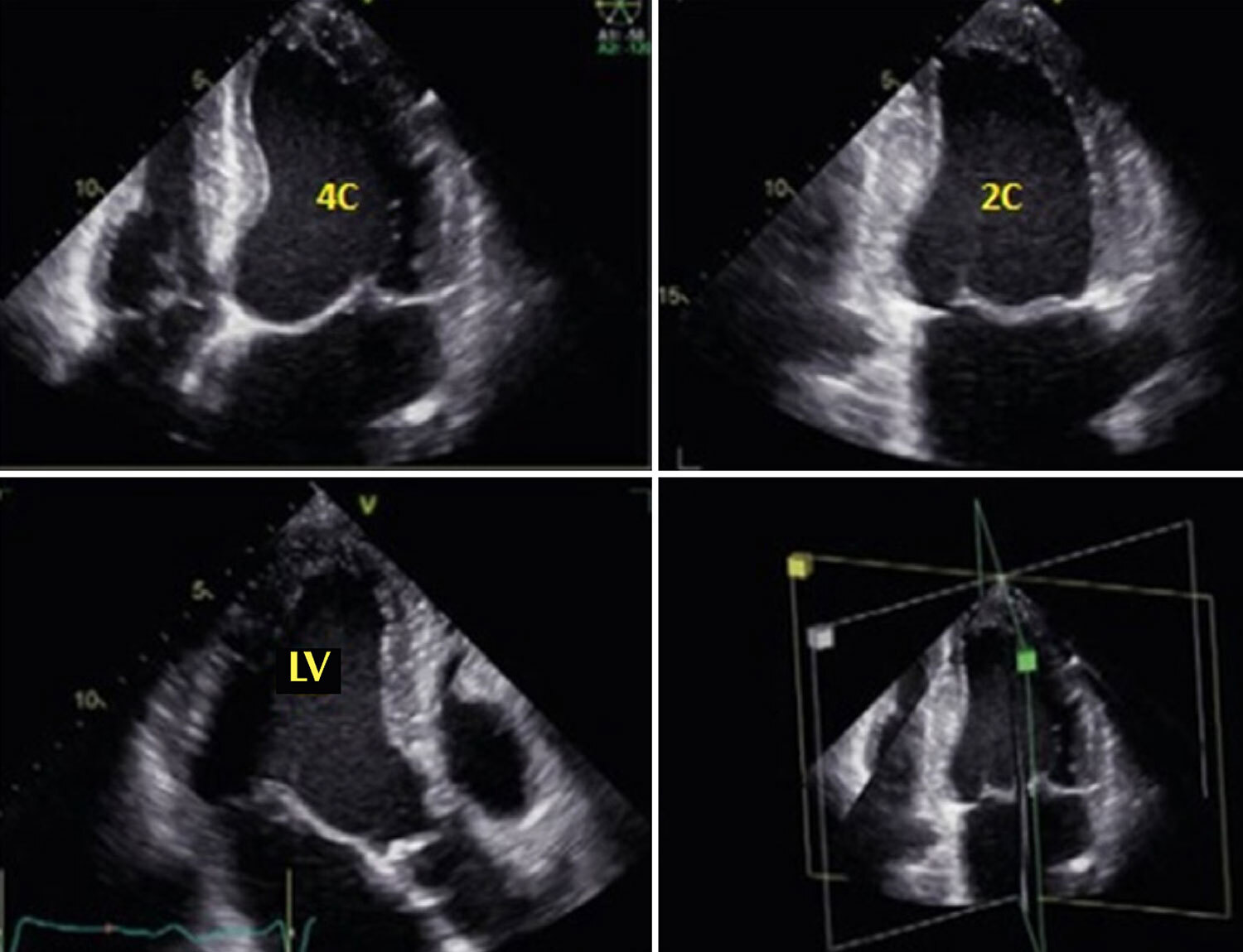
Chagas disease is recognized as a global public health issue, capable of causing cardiac, digestive, and neurological manifestations. The echocardiogram is a fundamental complementary exam in assessing patients with cardiac involvement, evaluating various parameters of cardiac structure and function. This review aims to discuss the use of three-dimensional echocardiography in analyzing ventricular function in patients with Chagas disease.

  • Chagas Cardiomyopathy
  • Echocardiography, Three-Dimensional
  • Ventricular Function
Read Full Article
Creative Commons Attribution (CC BY) Creative Commons license
18/12/2023 DOI: 10.36660/abcimg.20230093i Views 1
Figure 1
ABCIC
Case Report

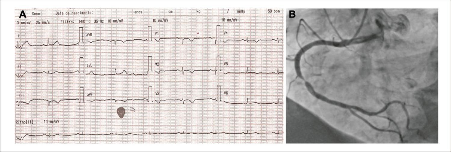
Incidental Finding of Arrhythmogenic Right Ventricular Cardiomyopathy in a 72-Year-Old Man Admitted With Acute Coronary Syndrome

Rafael Modesto Fernandes et al. Luciana Cunha Weber Vitor Queiroz de Castro Souza Mariana Baptista Guedes … Diogo Freitas Cardoso de Azevedo Marcia Maria Nova Rabelo

Introduction

Arrhythmogenic right ventricular cardiomyopathy (ARVC) – also known as arrhythmogenic right ventricular (RV) dysplasia – is a genetic disorder characterized by progressive loss of RV cardiomyocytes, which are replaced with fibrofatty tissue. This replacement may delay intraventricular conduction and contribute to ventricular arrhythmias through a fibrosis-related macro-reentry mechanism. Thus, ARVC is one of the main causes of arrhythmic cardiac arrest in young people and athletes.

Although initially designated as dysplasia, ARVC is not a congenital defect in the myocardium development. It is caused by mutations in the genes that encode desmosomal proteins, which are responsible for cell-to-cell adhesion. This discovery led to the disease being recognized as cardiomyopathy and included in the classification of cardiomyopathies by the American Heart Association. ,

[…]

  • Arrhythmogenic right ventricular dysplasia;
  • Echocardiography, Three-Dimensional
  • Global Longitudinal Strain
Read Full Article
Creative Commons Attribution (CC BY) Creative Commons license
06/09/2023 DOI: 10.36660/abcimg.20230046i
Figure 1
ABCIC
Case Report

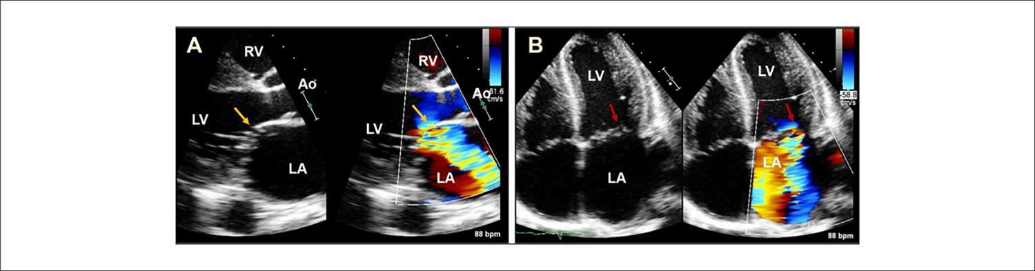
Dual Mechanism of Mitral Valve Injury: Additional Value of Three-Dimensional Transesophageal Echocardiography

Larissa Neto Espíndola et al. Angele Azevedo Alves Mattoso Geórgia dos Santos Couto Pompílio Sampaio Britto

Abstract

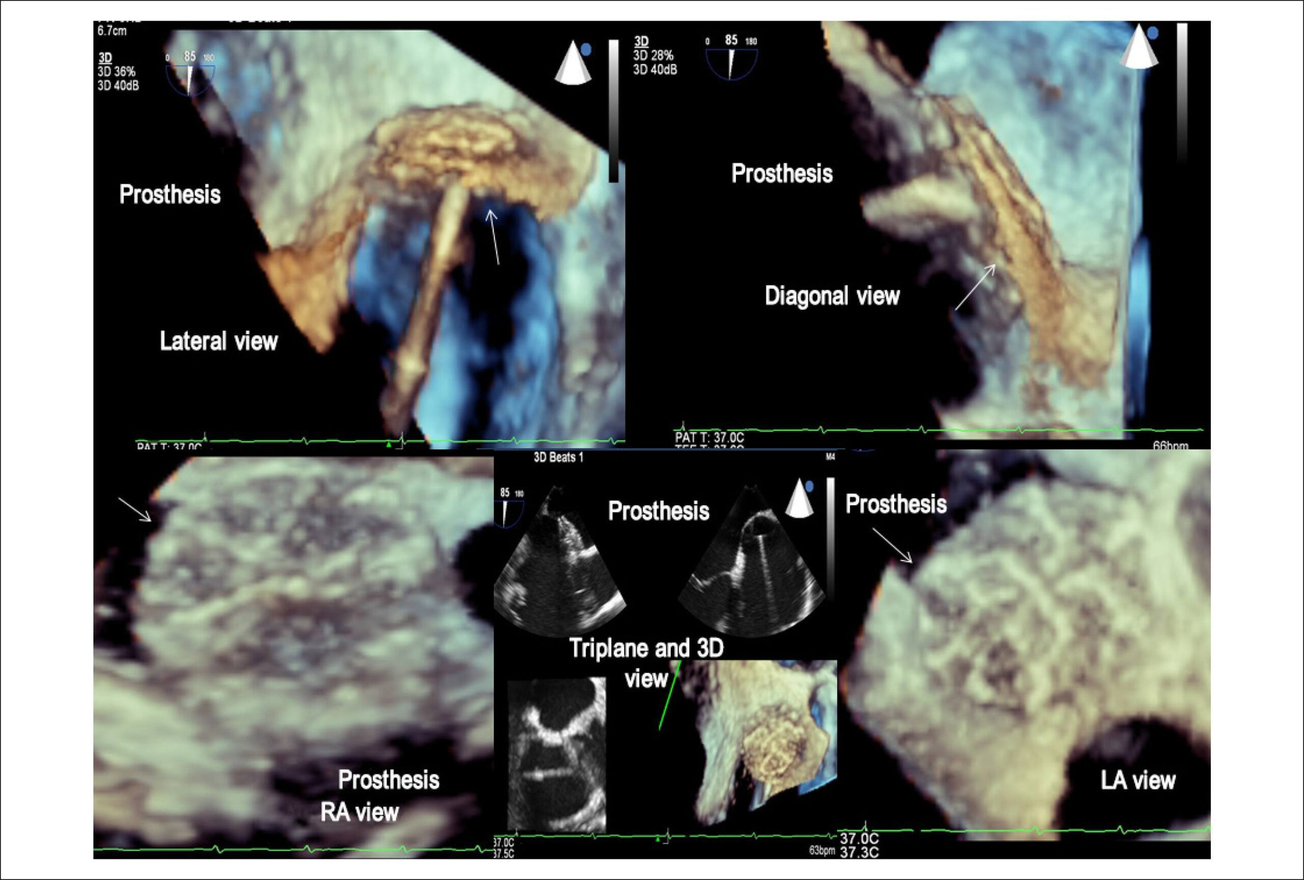
Infective endocarditis (IE) is a life-threatening condition and during its course several complications may occur, including embolic events, perivalvular extension and valvular destruction. Echocardiography is fundamental across all stages of the disease, from the diagnosis until the follow-up after completion of the definitive therapy. Three-dimensional transesophageal echocardiography (TEE) can demonstrate a better anatomical definition and it is especially useful in the assessment of perivalvular extension of the infection, prosthetic valve dehiscence and valve perforation. In this article we report a case of double damage in the same mitral valve: leaflet perforation and rupture of chords in a patient with IE.

  • Echocardiography, Three-Dimensional
  • Endocarditis
  • Mitral Valve
Read Full Article
Creative Commons Attribution (CC BY) Creative Commons license
30/06/2023 DOI: 10.36660/abcimg.2023322i Views 1
Figure 1
ABCIC
Review Article

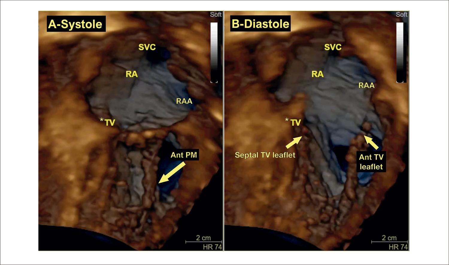
My Approach to Three-Dimensional Echocardiography for Pathophysiological Classification of Tricuspid Regurgitation

Alex dos Santos Felix et al. Monica Luiza de Alcantara Konstantinos Papadopoulos

Introduction

Although it was known as the “forgotten valve” in the past, there has been increasing interest in studying the tricuspid valve (TV) during the last two decades. Tricuspid regurgitation (TR) has been identified as a prognostic marker not only when associated with other cardiac diseases, like heart failure, mitral regurgitation, or aortic stenosis, – but also as an isolated entity. Understanding the pathophysiological mechanisms of this disease is of paramount importance, since new options of transcatheter devices and techniques are emerging, offering invasive treatment for high-risk patients unsuitable for surgical intervention.

The TV is difficult to image in transesophageal echocardiography (TEE) for the following reasons: 1) the leaflets are much thinner compared to mitral leaflets, with greater anatomic variability; 2) it is an anterior structure, far away from the esophagus, with acoustic shadowing from the fibrous heart skeleton; and 3) it cannot be aligned to the esophageal probe in order to acquire en face views and requires use of lateral resolution. These restrictions may limit the ability of traditional TEE to evaluate the TV, making it necessary to complement the evaluation with special windows from the lower esophagus and transgastric views. Different from other cardiac valves, images of the TV obtained from transthoracic echocardiography (TTE) usually have better resolution than images obtained by TEE. Even though all protocols start with two-dimensional (2D) assessment of the TV, three-dimensional echocardiography (3DE) plays an important role in the evaluation of TV diseases, due not only to its ability to precisely depict the anatomy of the valve and the subvalvular apparatus, but also to its accuracy in quantitation of right ventricular (RV) and right atrial volumes and function and functional analysis of valvular dysfunction, especially for grading TR and evaluating dynamics of the tricuspid annulus (TA) through dedicated software. All of this information is of unparalleled importance for patient management and pre-procedural planning in surgical and transcatheter approaches.

[…]

  • Echocardiography, Three-Dimensional
  • Tricuspid Valve
  • Tricuspid Valve Insufficiency
Read Full Article
Creative Commons Attribution (CC BY) Creative Commons license
30/06/2023 DOI: 10.36660/abcimg.20230055i Views 3
Figure 1
ABCIC
Case Report

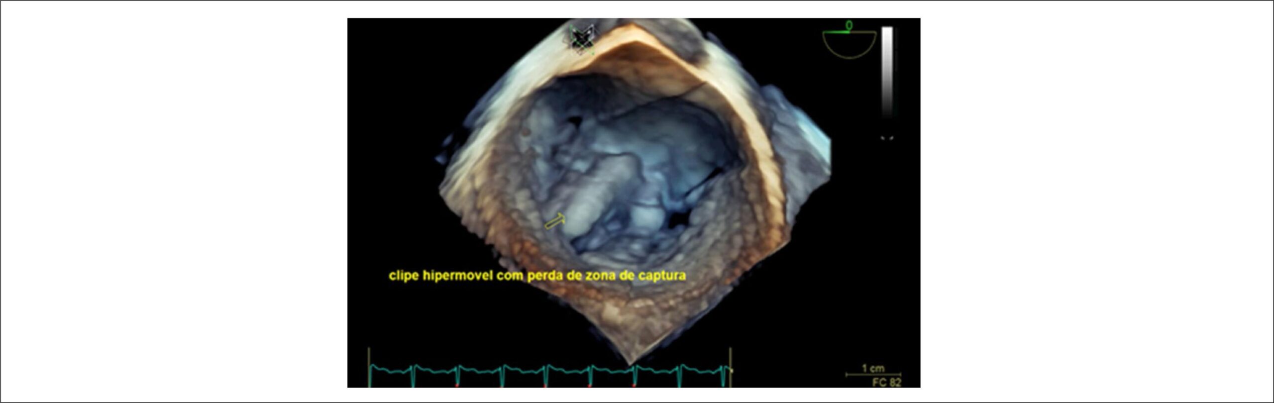
Real-time Three-Dimensional Echocardiogram in the Identification of Late Complications in the Follow-Up After Mitraclip® Implantation

Alexandre Costa et al. Bruna de Mattos Ivo Junqueira Carolina Thé Macedo Marcus Vinicius Silva Freire de Carvalho … Cristiano Guedes Bezerra Rodrigo Morel Vieira de Melo

Introduction

Conventional surgery is the treatment of choice for severe mitral valve regurgitation. However, it is not feasible for patients at high surgical risk, who have high morbidity and mortality rates despite clinical treatment. Percutaneous valve treatment (MitraClip) has been evolving as a therapeutic perspective for this population, with reduced heart failure (HF) hospitalization and all-cause mortality rates.,,

Three-dimensional transesophageal echocardiography (3DTEE) has an additive role during clinical follow-up, especially in cases of suspected late complications with high detail of valve anatomy and clip positioning with cardiac structures. Additionally, due to the greater anatomical accuracy of this test, complications resulting from the procedure can be safely detected in real time.

[…]

  • Echocardiography, Three-Dimensional
  • Mitral Valve Insufficiency
  • Postoperative Complications, Cardiovascular Surgical Procedures
Read Full Article
Creative Commons Attribution (CC BY) Creative Commons license
01/12/2022 DOI: 10.47593/2675-312X/20223504eabc298i Views 1
Figure 1
ABCIC
Review Article

Stress Echocardiography Using Real-Time 3D Imaging

José Roberto Matos-Souza et al. Guilherme De Rossi

Abstract

The need to examine the heart using a three-dimensional (3D) tool is not new. This complex and dynamic organ has always required 3D and real-time understanding. Without this feature, the examiner has to transform two-dimensional images to understand its volume, which requires complex knowledge and approximation interactions. Echocardiography was invented three decades ago, and its improvements resulted in commercial products at the beginning of the century. Some studies demonstrate 3D equivalence with gains in handling the necessary time. We use triplane modalities in our routine, with time gain and less stress on the examiner’s upper limb. Thus, 3D examinations can answer more complex questions and provide a more geometric approach to contraction, with thickening being analyzed in the background.

  • Echocardiography, Stress
  • Echocardiography, Three-Dimensional
  • Imaging, Three-Dimensional
Read Full Article
Creative Commons Attribution (CC BY) Creative Commons license
07/07/2022 DOI: 10.47593/2675-312X/20223503erer_07
ABCIC
Case Report

Carcinoid Syndrome with Involvement of Tricuspid and Pulmonary Valves: Findings on Three-Dimensional Echocardiography

Eduardo Menti et al. Renata Patrícia Moreira Camargo Marcia Castilhos Puchalski Felipe Gropelli Crochemore Fabio Moreira Cañelas

[…]

  • Carcinoid Heart Disease
  • Carcinoid Tumor
  • Echocardiography, Three-Dimensional
  • Pulmonary Valve/dysfunction
  • Tricuspid Valve/dysfunction
Read Full Article
Creative Commons Attribution (CC BY) Creative Commons license
04/02/2022 DOI: 10.5935/2318-8219.20170022 Views 1
ABCIC
Editorial

Embolic Complications in Infective Endocarditis: How Can We Predict Using a Risk Evaluator Score (SORTIE or ABCDE)

Cesar Augusto da Silva Nascimento

[…]

  • Echocardiography, Three-Dimensional
  • Echocardiography, Transesophageal
  • Echocardiography, Transthoracic
  • Endocarditis
Read Full Article
Creative Commons Attribution (CC BY) Creative Commons license
14/01/2022 DOI: 10.5935/2318-8219.20190044
ABCIC
Original Article

Semiautomatic Quantification of Left Ventricular Ejection Fraction and Volumes by Two-Dimensional Echocardiography: Comparison with Automatic Three-Dimensional Echocardiography

Rafael Borsoi et al. Miguel Morita Fernandes da Silva Silvio Henrique Barberato

Abstract

Background

Left ventricular ejection fraction is one of the most used echocardiographic parameters in clinical practice. Its estimation by two-dimensional manual method (Simpson method) has limited reproducibility and accuracy, and semi-automatic methods have been proposed. It becomes necessary to compare the semi-automatic two-dimensional method with more accurate methods of assessing left ventricular ejection fraction, such as measurement by automatic three-dimensional echocardiography.

Objective

To compare the left ventricular ejection fraction, and left ventricular end-diastolic and end-systolic volumes estimates by the semi-automatic two-dimensional method with those obtained using the automatic three-dimensional method.

Method

Observational cross-sectional study, including patients in sinus rhythm, left ventricular ejection fraction > 50% and without significant structural heart disease, submitted to transthoracic echocardiography. Student’s t test, Pearson’s coefficient and Bland-Altman analysis were used in the statistical analysis.

Results

Forty patients were included, 53% women, 35% with arterial hypertension, 25% with dyslipidemia, 10% diabetic, 10% smokers and 13% with previous angioplasty. The mean values of left ventricular ejection fraction by three-dimensional and two-dimensional were 62.1 ± 5.8% and 61.7 ± 5.9% (p = 0.50), respectively. There was a strong correlation between left ventricular ejection fraction determined by two-dimensional and three-dimensional (r = 0.75; p <0.001), as well as with left ventricular end-diastolic (r = 0.75; p <0.001) and end-systolic (r = 0.76; p < 0.001). There was good agreement between two-dimensional and three-dimensional left ventricular ejection fraction (mean difference: -0.39; 95% confidence interval -1.7-0.9).

Conclusion

Left ventricular ejection fraction estimated by the two-dimensional semi-automatic method showed good agreement with the automatic three-dimensional method. The findings suggest the two-dimensional semi-automatic method as a reliable alternative for assessing volumes and left ventricular ejection fraction.

  • Echocardiography, Doppler
  • Echocardiography, Three-Dimensional
  • Stroke Volume
  • Ventricular Function, Left
Read Full Article
Creative Commons Attribution (CC BY) Creative Commons license
17/12/2021 DOI: 10.47593/2675-312X/20203304eabc98
ABC
Editorial

Assessment of Aortic Valve Regurgitation by Echocardiography: Basic and New Concepts

Said Alsidawi

Conclusion

Introducing new concepts or techniques that can help grading AR severity is a valuable resource. AR VTI is a promising concept that is physiologically sound and appears reproducible. Larger clinical trials will be needed to further assess its role and, more importantly, its prognostic value and correlation with clinical outcomes.

It is imperative, however, to keep in mind that it is very unlikely that we will find a single echocardiographic marker that will be a gold standard when assessing AR severity. The echocardiographist has to keep an open mind and integrate all available data to come to a final conclusion. This includes the following:

[…]

  • Aortic Regurgitation
  • Aortic Valve Insufficiency
  • Blood Fow Velocity
  • Diagnostic Imaging
  • Echocardiography
  • Echocardiography, Doppler
  • Echocardiography, Three-Dimensional
Read Full Article
Creative Commons Attribution (CC BY) Creative Commons license
28/08/2020 DOI: 10.36660/abc.20200559
ABC
Editorial

Real-Time Three-Dimensional Echocardiography and Myocardial Strain: Ready for Use in Clinical Practice

Frederico Jose Neves Mancuso

The ability of echocardiography to quantify volumes and to evaluate the contractile function of cardiac chambers has evolved greatly in recent years, particularly due to the development of real-time three-dimensional echocardiography (echo3D) and the possibility myocardial deformation (Strain) analysis by the speckle tracking technique.–

Unlike two-dimensional echocardiography, where geometric inferences and mathematical calculations are required, the echo3D allows direct measurement of ventricular and atrial volumes, from which function data such as left and right ventricular ejection fraction, and left atrial function, including total emptying fraction and active emptying fraction parameters.–

[…]

  • Echocardiography, Three-Dimensional
  • Reference Values
  • Speckle Tracking
  • Strain
  • Ventricular Function, Left
Read Full Article
Creative Commons Attribution (CC BY) Creative Commons license
02/12/2019 DOI: 10.5935/abc.20190179
Figure 1
ABC
Original Article

3-Dimensional Echocardiography and 2-D Strain Analysis of Left Ventricular, Left Atrial and Right Ventricular Function in Healthy Brazilian Volunteers

Roberto M. Saraiva et al. Eliza Maria B. Scolin Nicole P. Pacheco Maria Eduarda Bouret … Mauro Felippe Felix Mediano Marcelo T. Holanda Andréa R. da Costa

Abstract

Background:

New echocardiographic techniques are used in the diagnosis and prognosis of many heart diseases. However, reference values in different populations are still needed for several of these new indexes. We studied these new echocardiographic parameters in a group of Brazilians with no known cardiovascular disease.

Objective:

To study values for new echocardiographic indexes in Brazilians without known cardiovascular disease and their correlation with age.

Methods:

Cross-sectional study that included healthy individuals who underwent three-dimensional echocardiography (3DE) and two-dimensional speckle tracking echocardiography (STE) strain (e) analysis. Left atrial (LA) and left ventricular (LV) function were analyzed by 3DE and STE, and right ventricular (RV) function by STE. P values < 0.05 were considered significant.

Results:

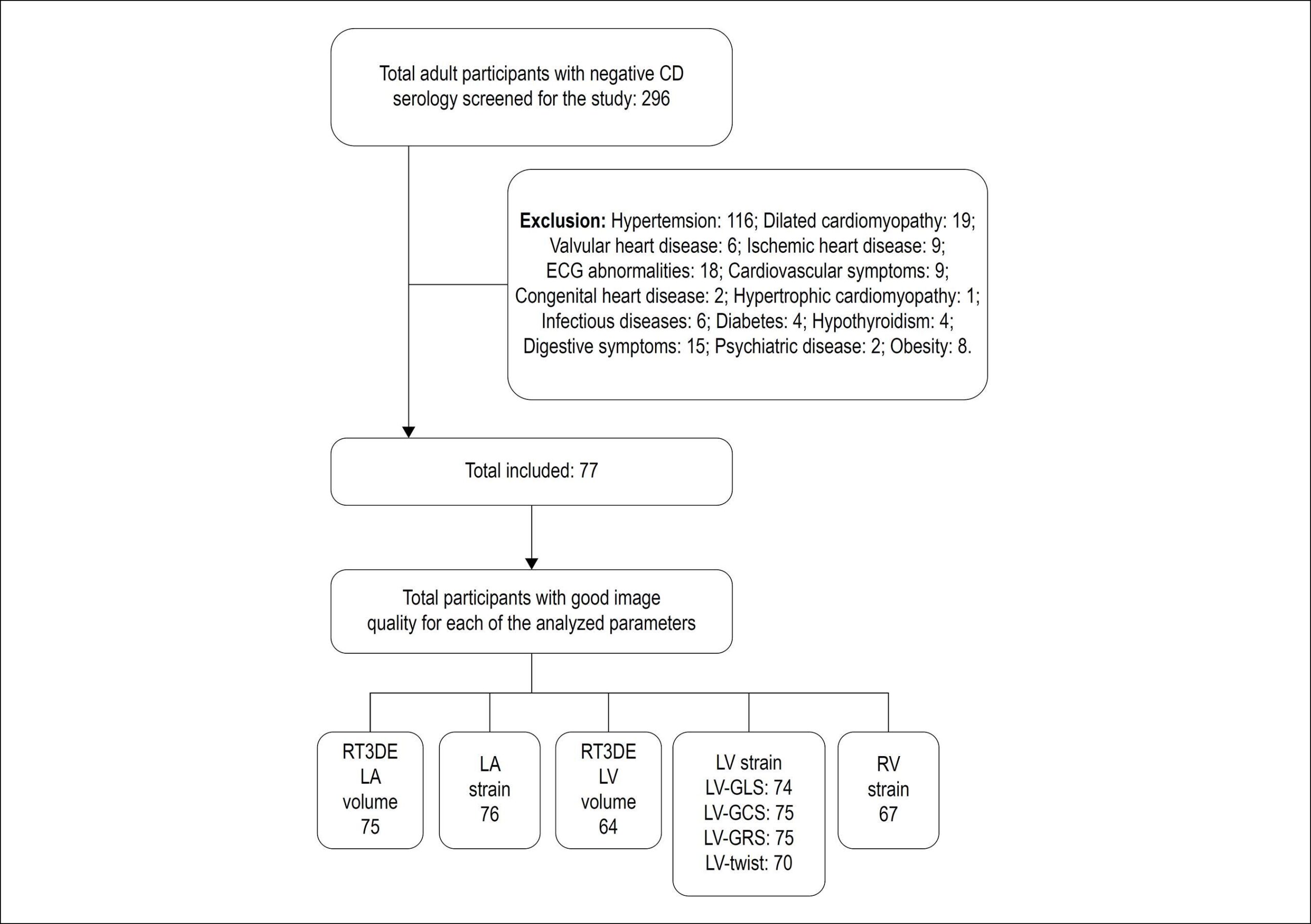
Seventy-seven subjects (46.7% men; 40.4 ± 10.4 years) were included. Maximum, minimum and pre-atrial contraction (pre-A) LA volumes (ml/m2) were 21.2 ± 5.5, 7.8 ± 2.5, and 11.0 ± 3.1, respectively. Peak positive global LA e (LAScd), peak negative global LA e and total global LA e (LASr) were 17.4 ± 5.2%, -13.2 ± 2.0% and 30.5 ± 5.9%, respectively. LV end-diastolic and end-systolic volumes (ml/m2) measured 57 ± 12 and 24 ± 6, and 3D LV ejection fraction measured 58 ± 6%. Global LV longitudinal, circumferential and radial e were -19 ± 2%, -19 ± 3%, and 46 ± 12%, respectively. LV torsion measured 1.6 ± 0.70 /cm. Global longitudinal RV e (RV-GLS) and RV free wall strain were -22 ± 3% and -24 ± 5%. Minimum LA and pre-A volumes, LV apical rotation, torsion and RV-GLS increased with age, while total and passive LA emptying fractions, LAScd, LASr, LV end-diastolic and end-systolic volumes decreased with age.

Conclusion:

Values for new echocardiographic indexes in Brazilians without known cardiovascular disease and their correlation with age are presented.

  • Cardiovascular Diseases
  • Echocardiography, Three-Dimensional
  • Left Ventricular Function
  • Reference Values
  • Right Ventricular Function
  • Speckle Tracking
  • Strain
Read Full Article
Creative Commons Attribution (CC BY) Creative Commons license
29/08/2019 DOI: 10.5935/abc.20190155
ABC
Editorial

Three-dimensional Speckle Tracking Echocardiography in Amyloidosis: A New Assessment Method for a Rare Disease

Brivaldo Markman Filho

Immunoglobulin deposition in the myocardium characterizes involvement of cardiac amyloidosis (CA). Fibrillary infiltration, which may happen in every heart cavity, leads to the restrictive cardiomyopathy phenotype, with complex pathophysiological mechanisms, which will result in the syndromic diagnosis of congestive heart failure. Diastolic dysfunction is dominant in most cases, and it may or may not, follow diverse levels of systolic dysfunction in the most advanced phases of the disease. Atrial remodeling by amyloid infiltration may contribute to cardiac output decrease by means of insufficient or nonexistent telediastolic atrial contraction. The onset of atrial electrical instability, ending in atrial fibrillation, highlights symptomatic worsening and these patients’ reserved prognosis.

Historically, the right cavities of the heart have been neglected in echocardiographic assessments. The complex morphology of the right ventricle (RV) has possibly contributed for the lack of reproducible data on echocardiographic cutting plans, diversely from the left ventricle (LV). The development of three-dimensional echocardiography (3DE) has allowed for a more accurate calculation of right ventricle volume and function in the diverse pathologies involving that chamber.

[…]

  • Cardiomyopathy, Restrictive/psysiopathology
  • Echocardiography, Three-Dimensional
  • Heart Failure
  • Immunoglobulin Light-Chain Amyloidosis
Read Full Article
Creative Commons Attribution (CC BY) Creative Commons license
01/09/2018 DOI: 10.5935/abc.20180182
Figure 1
ABC
Image

Challenging Evaluation of Aortic Regurgitation: More Than a Quadricuspid Valve

Gonçalo Pestana et al. Carla Sousa Teresa Pinho Sara Maia M. Júlia Maciel

A 61-year-old female patient with chronic obstructive pulmonary disease, and no other comorbidities, was referred for cardiological assessment due to aggravated exertional dyspnea (New York Heart Association – NYHA class III) and atypical chest pain. Physical examination disclosed only a diastolic murmur in the second intercostal space in the right sternal border.

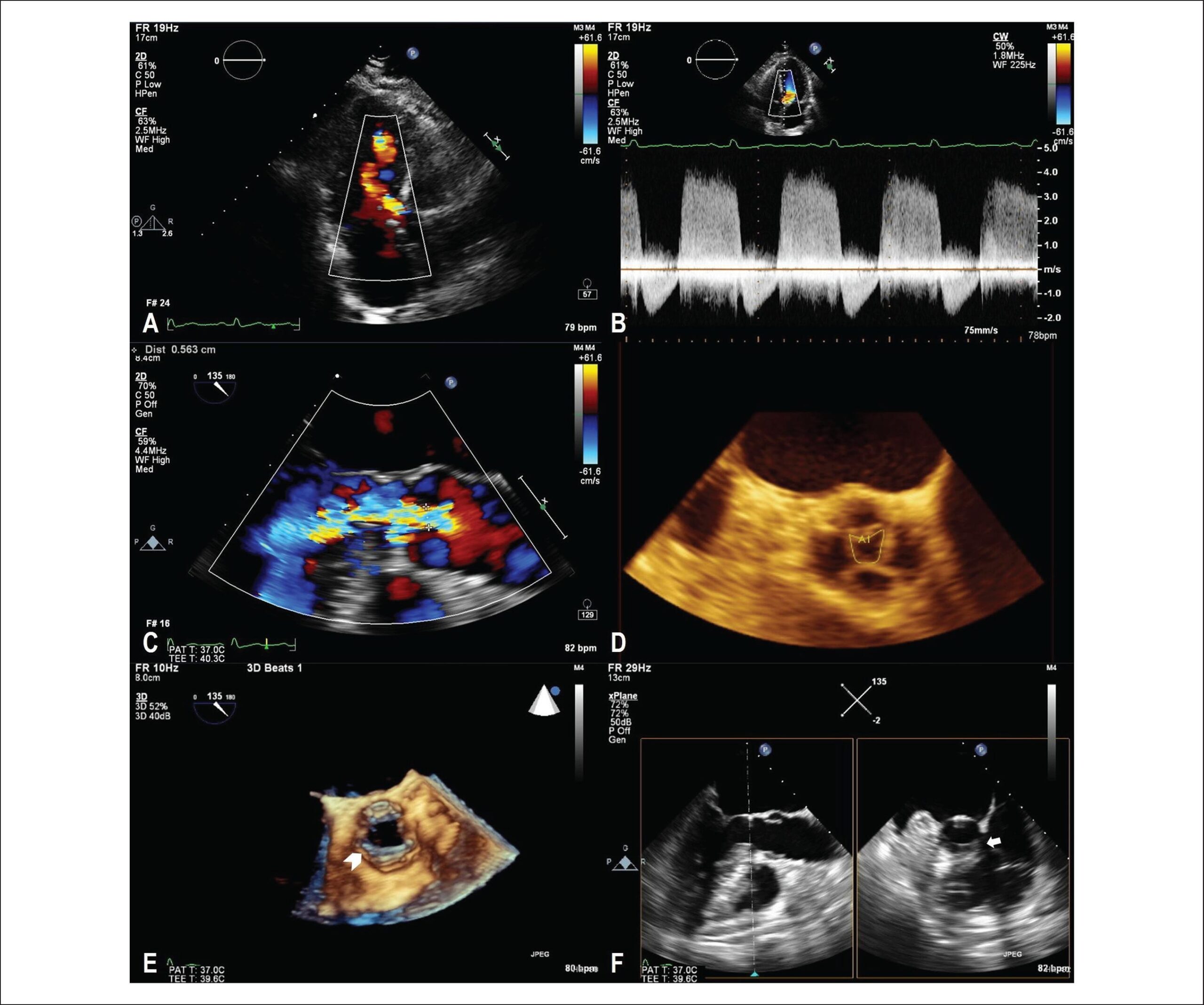
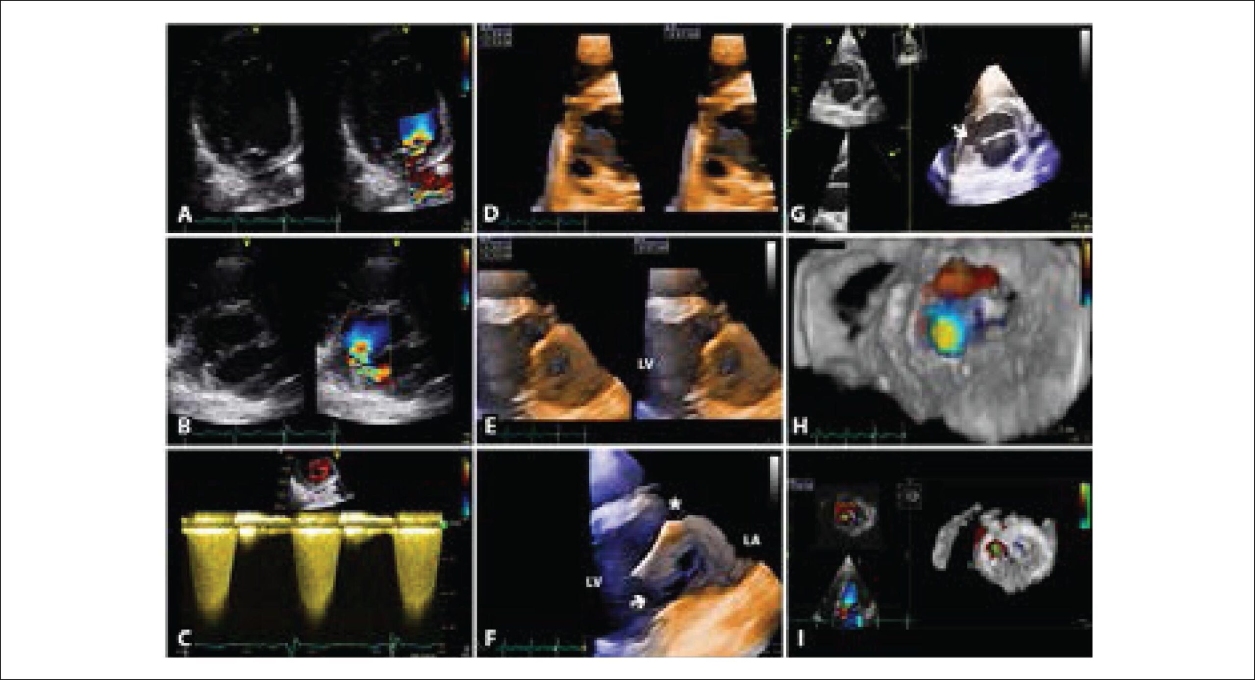
The transthoracic echocardiogram (TTE) showed aortic regurgitation (), with non-dilated cardiac chambers and preserved biventricular function. The acoustic window limited the evaluation of the valvular lesion severity and its importance in the context of the complaints, although the continuous Doppler spectrum suggested significant regurgitation (). The evaluation was further impaired by a systolic flow acceleration in the Left Ventricular Outflow Tract (LVOT), with no significant gradient and an undetermined cause. The aortic root had normal size, but it was not possible to carry out an adequate morphological and functional characterization of the valve.

[…]

  • Aortic Valve Insufficiency
  • Echocardiography, Three-Dimensional
  • Echocardiography, Transesophageal
  • Pulmonary Disease, Chronic Obstructive
Read Full Article
Creative Commons Attribution (CC BY) Creative Commons license
01/07/2018 DOI: 10.5935/abc.20180106
ABC
Letter to the Editor

Challenges For Percutaneous Left Atrial Appendage Closure: Imaging And Residual Flow

Tarik Yildirim et al. Ibrahim Altun Mustafa Ozcan Soylu

We have read with great interest the paper entitled ‘First results of the Brazilian Registry of Percutaneous Left Atrial Appendage Closure’ by Guérios et al. is a very important study. We have some suggestions about this trial.

Firstly, two-dimensional transeosophageal echocardiography (2D-TEE) can evaluate the morphology of left atrial appendage in multiple views. But real-time three-dimensional transeosophageal echocardiography (3D-TEE) provides more detailed images of the left atrial appendage (LAA) anatomy than 2D-TEE. A competent physician can accurately evaluate the LAA depth and landing zone. Moreover LAA closure depends on an accurate determination of anatomical structure. Therefore the 3D-TEE may show advantantages in relation to 2D-TEE in transcathater LAA closure.

[…]

  • Atrial Appendage
  • Echocardiography, Three-Dimensional
  • Medical Records
  • Septal Occluder Devices
Read Full Article
Creative Commons Attribution (CC BY) Creative Commons license
01/07/2018 DOI: 10.5935/abc.20180135
Figure 1
ABC
Original Article

Three-Dimensional Volumetric Assessment of Diastolic Function by Cardiac Magnetic Resonance Imaging: The Multi-Ethnic Study of Atherosclerosis (MESA)

Marcelo S Nacif et al. Andre L. C. Almeida Alistair A Young Brett R Cowan … Anderson C Armstrong Eunice Yang Christopher T Sibley W. Gregory Hundley Songtao Liu Joao AC Lima David A Bluemke

Abstract

Background:

Cardiac Magnetic Resonance is in need of a simple and robust method for diastolic function assessment that can be done with routine protocol sequences.

Objective:

To develop and validate a three-dimensional (3D) model-based volumetric assessment of diastolic function using cardiac magnetic resonance (CMR) imaging and compare the results obtained with the model with those obtained by echocardiography.

Methods:

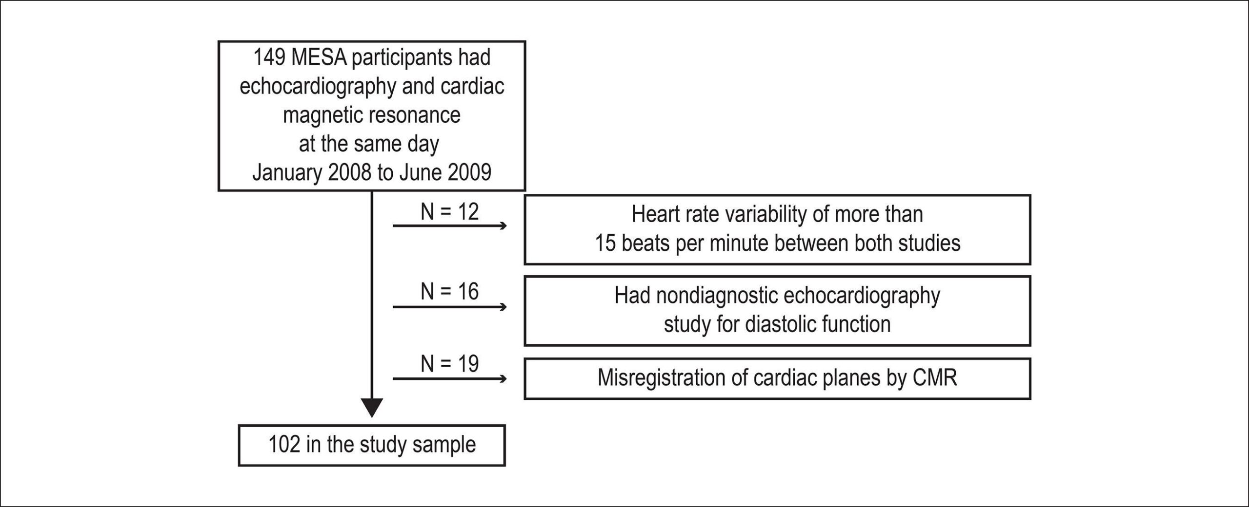
The study participants provided written informed consent and were included if having undergone both echocardiography and cine steady-state free precession (SSFP) CMR on the same day. Guide points at the septal and lateral mitral annulus were used to define the early longitudinal relaxation rate (E’), while a time-volume curve from the 3D model was used to assess diastolic filling parameters. We determined the correlation between 3D CMR and echocardiography and the accuracy of CMR in classifying the diastolic function grade.

Results:

The study included 102 subjects. The E/A ratio by CMR was positively associated with the E/A ratio by echocardiography (r = 0.71, p < 0.0001). The early diastolic relaxation velocity by tissue Doppler and longitudinal relaxation rate for the lateral mitral annulus displacement were positively associated (p = 0.007), as were the ratio between Doppler E/e' and CMR E/E' (p = 0.01). CMR-determined normalized peak E (NE) and deceleration time (DT) were able to predict diastolic dysfunction (areas under the curve [AUCs] = 0.70 and 0.72, respectively). In addition, the lateral E/E' ratio showed good utility in identifying diastolic dysfunction (AUC = 0.80). Overall, echocardiography and CMR interobserver and intraobserver agreements were excellent (intraclass correlation coefficient range 0.72 – 0.97).

Conclusion:

3D modeling of standard cine CMR images was able to identify study subjects with reduced diastolic function and showed good reproducibility, suggesting a potential for a routine diastolic function assessment by CMR.

  • Echocardiography, Three-Dimensional
  • Evaluation
  • Imaging, Three-Dimensional
  • Magnetic Resonance
  • Ventricular Function
Read Full Article
Creative Commons Attribution (CC BY) Creative Commons license
25/05/2017 DOI: 10.5935/abc.20170063 Views 1
Figure 1
ABC
Image

Three-dimensional Echocardiography for Percutaneous Occlusion of Patent Foramen Ovale

Robinson Poffo et al. Paola Keese Montanhesi Renato Bastos Pope Alisson Parrilha Toschi Marcelo Campos Vieira

Female patient, 31 years old, with a history of ischemic stroke 7 years before, was referred for treatment of Patent Foramen Ovale (PFO). She only had a complaint of migraine with aura, with no focal neurological abnormalities.

On July 2, 2015, she was submitted to an uneventful percutaneous PFO occlusion using a Cardia IntraseptTM prosthesis (CARDIA, Inc., Burnsville, MN, USA). Intraoperative three-dimensional transesophageal echocardiography confirmed correct prosthesis positioning and absence of residual shunt through the bubble test. Current guidelines of the two most important international societies of echocardiography, the European Association of Cardiovascular Imaging and the American Society of Echocardiography, determine that the three-dimensional echocardiography is the echocardiographic modality of choice for monitoring, following and guiding percutaneous procedures performed in hemodynamics and hybrid rooms, as in this case.

[…]

  • Echocardiography, Three-Dimensional
  • Foramen Ovale, Patent / surgery
  • Minimally Invasive Surgical Procedures
Read Full Article
Creative Commons Attribution (CC BY) Creative Commons license
01/03/2016 DOI: 10.5935/abc.20160036
Figure 1
ABC
Image

Multimodality Images of a Mixed Atrial Septal Defect

Zafer Işılak et al. Uğur Küçük Omer Uz Murat Yalçın Veysel Temizkan

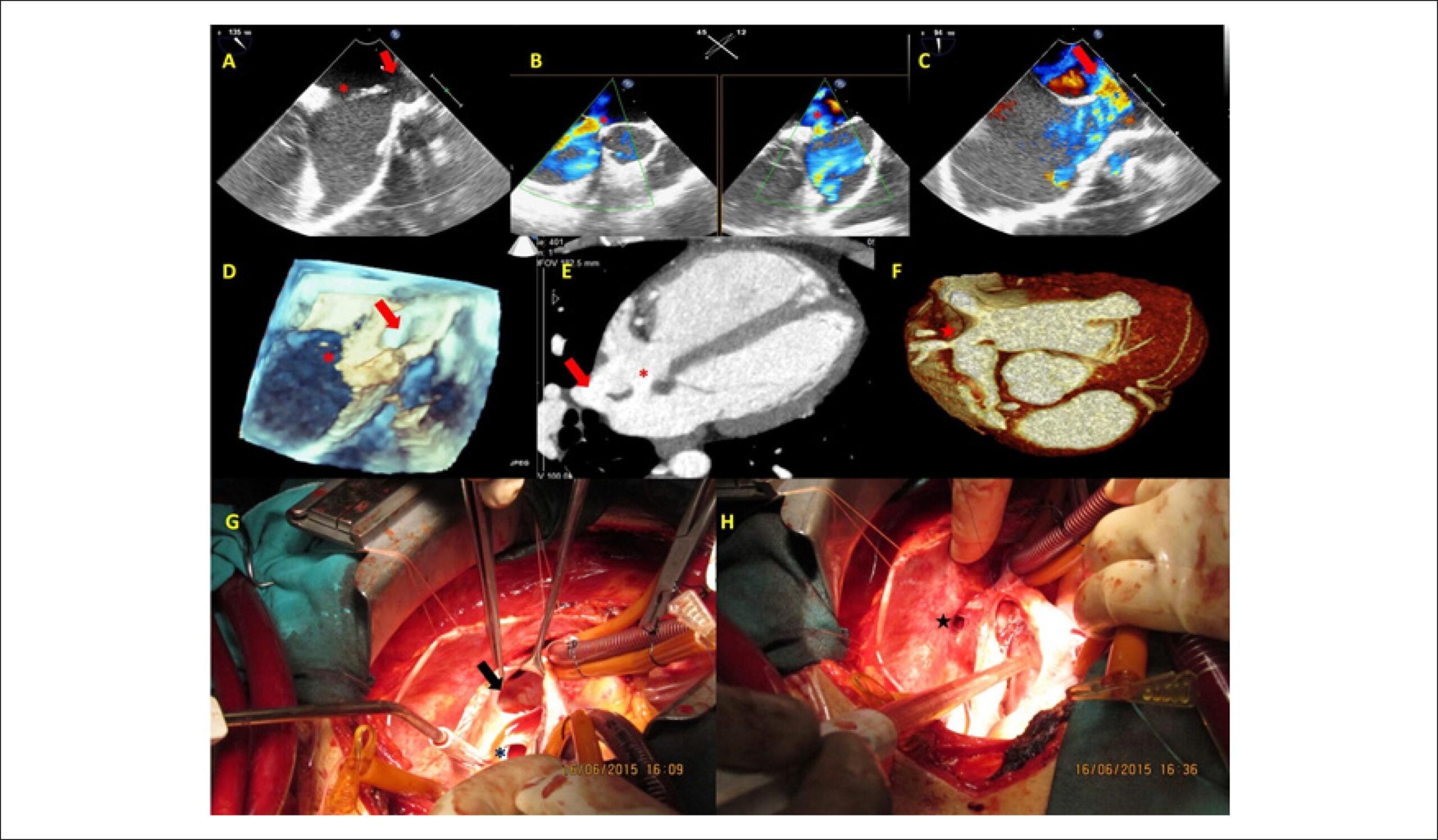
A 20-year-old male patient with dyspnea complaint was referred to our hospital. On physical examination there was grade 2/6 systolic murmur, which is best heard at the second left intercostal space and fixed split S2. ECG revealed sinus rhythm with complete right bundle brunch block. Transthoracic echocardiography demonstrated a secundum atrial septal defect (ASD) and Qp/Qs ratio was 1.6. The right ventricle was severely dilated, inconsistent with the size of the defect. Two-dimensional and color Doppler transesophageal echocardiographic examination confirmed the secundum type ASD (asterisk) and revealed an additional sinus venosus type ASD between the right atrium and the superior vena cava (SVC)(, , ; ). Three-dimensional transesophageal echocardiography verified both septal defects (). The patient underwent cardiac tomography for further anatomical outlining the. and clearly show the secundum ASD (asterisk), sinus venosus ASD (arrow) and anomalous drainage of the right superior pulmonary vein to the SVC (star). The patient underwent surgery. and show intraoperative images of the defects.

[…]

  • Atrial Septum / physiopathology
  • Echocardiography, Three-Dimensional
  • Heart Septal Defects, Atrial / physiopathology
Read Full Article
Creative Commons Attribution (CC BY) Creative Commons license
01/02/2016 DOI: 10.5935/abc.20160016
Figure 1
ABC
Original Article

Left Atrial Volume Determinants in Patients with Non-Ischemic Dilated Cardiomyopathy

Frederico José Neves Mancuso et al. Valdir Ambrósio Moisés Dirceu Rodrigues Almeida Dalva Poyares … Luciana Julio Storti Wércules Antonio Oliveira Flavio Souza Brito Angelo Amato Vincenzo de Paola Antonio Carlos Camargo Carvalho Orlando Campos

Background:

Left atrial volume (LAV) is a predictor of prognosis in patients with heart failure.

Objective:

We aimed to evaluate the determinants of LAV in patients with dilated cardiomyopathy (DCM).

Methods:

Ninety patients with DCM and left ventricular (LV) ejection fraction ≤ 0.50 were included. LAV was measured with real-time three-dimensional echocardiography (eco3D). The variables evaluated were heart rate, systolic blood pressure, LV end-diastolic volume and end-systolic volume and ejection fraction (eco3D), mitral inflow E wave, tissue Doppler e´ wave, E/e´ ratio, intraventricular dyssynchrony, 3D dyssynchrony index and mitral regurgitation vena contracta. Pearson´s coefficient was used to identify the correlation of the LAV with the assessed variables. A multiple linear regression model was developed that included LAV as the dependent variable and the variables correlated with it as the predictive variables.

Results:

Mean age was 52 ± 11 years-old, LV ejection fraction: 31.5 ± 8.0% (16-50%) and LAV: 39.2±15.7 ml/m2. The variables that correlated with the LAV were LV end-diastolic volume (r = 0.38; p < 0.01), LV end-systolic volume (r = 0.43; p < 0.001), LV ejection fraction (r = -0.36; p < 0.01), E wave (r = 0.50; p < 0.01), E/e´ ratio (r = 0.51; p < 0.01) and mitral regurgitation (r = 0.53; p < 0.01). A multivariate analysis identified the E/e´ ratio (p = 0.02) and mitral regurgitation (p = 0.02) as the only independent variables associated with LAV increase.

Conclusion:

The LAV is independently determined by LV filling pressures (E/e´ ratio) and mitral regurgitation in DCM.

  • Cardiomyopathy, Dilated
  • Chagas Cardiomyopathy
  • Echocardiography, Three-Dimensional
  • Heart Atria
  • Organ Size
Read Full Article
Creative Commons Attribution (CC BY) Creative Commons license
08/05/2015 DOI: 10.5935/abc.20150042 Views 1
Figure 1
ABC
Case Report

Isolated Anterior Mitral Valve Leaflet Cleft: 3D Transthoracic Echocardiography-Guided Surgical Strategy

Marcelo Haertel Miglioranza et al. Denisa Muraru Sorina Mihaila José Carlos de Araujo Haertel … Sabino Iliceto Luigi Paolo Badano

Introduction

Isolated cleft of the anterior mitral leaflet (not associated with atrioventricular septal defect) is a rare cause of congenital mitral regurgitation. When feasible, mitral valve repair (direct suturing of the cleft with or without prosthetic annular ring insertion) is preferable to valve replacement. We report a clinical case in which we describe the usefulness of three-dimensional (3D) transthoracic echocardiography (TTE) for diagnosis and morphological assessment of the defect to assist in planning the surgical procedure.

[…]

  • Echocardiography
  • Echocardiography, Three-Dimensional
  • Heart Defects, Congenital
  • Mitral Valve Insufficiency
Read Full Article
Creative Commons Attribution (CC BY) Creative Commons license
01/05/2015 DOI: 10.5935/abc.20140191
3D Echo pilot study of geometric Left Ventricular changes after acute myocardial infarction
ABC
Artigo Original

3D Echo pilot study of geometric Left Ventricular changes after acute myocardial infarction

Marcelo Luiz Campos Vieira et al. Wercules Antonio Oliveira Adriana Cordovil Ana Clara Tude Rodrigues … Cláudia Gianini Mônaco Tânia Afonso Edgar Bezerra Lira Filho Marco Perin Cláudio Henrique Fischer Samira Saady Morhy

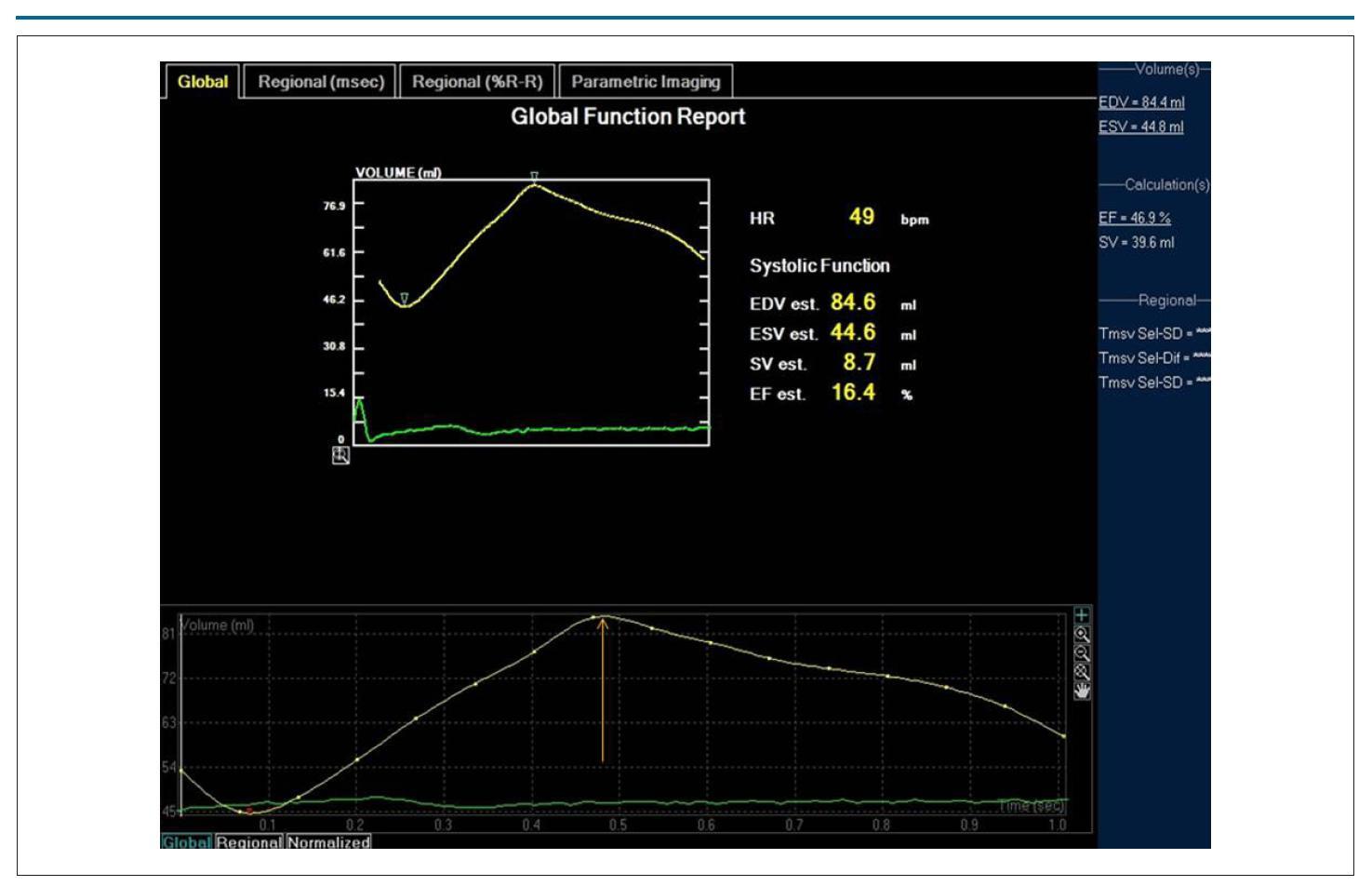
BACKGROUND: Left ventricular remodeling (LVR) after AMI characterizes a factor of poor prognosis. There is little information in the literature on the LVR analyzed with three-dimensional echocardiography (3D ECHO). OBJECTIVE: To analyze, with 3D ECHO, the geometric and volumetric modifications of the left ventricle (VE) six months after AMI in patients subjected to percutaneous primary treatment. METHODS: Prospective study with 3D ECHO of 21 subjects (16 men, 56 ± 12 years-old), affected by AMI with ST segment elevation. The morphological and functional analysis (LV) with 3D ECHO (volumes, LVEF, 3D sphericity index) was carried out up to seven days and six months after the AMI. The LVR was considered for increase > 15% of the end diastolic volume of the LV (LVEDV) six months after the AMI, compared to the LVEDV up to seven days from the event. RESULTS: Eight (38%) patients have presented LVR. Echocardiographic measurements (n = 21 patients): I- up to seven days after the AMI: 1- LVEDV: 92.3 ± 22.3 mL; 2- LVEF: 0.51 ± 0.01; 3- sphericity index: 0.38 ± 0.05; II- after six months: 1- LVEDV: 107.3 ± 26.8 mL; 2- LVEF: 0.59 ± 0.01; 3- sphericity index: 0.31 ± 0.05. Correlation coefficient (r) between the sphericity index up to seven days after the AMI and the LVEDV at six months (n = 8) after the AMI: r: 0.74, p = 0.0007; (r) between the sphericity index six months after the AMI and the LVEDV at six months after the AMI: r: 0.85, p < 0.0001. CONCLUSION: In this series, LVR has been observed in 38% of the patients six months after the AMI. The three-dimensional sphericity index has been associated to the occurrence of LVR.

  • Angioplasty
  • Balloon, Coronary
  • Echocardiography, Three-Dimensional
  • Myocardial Infarction
  • Stents
  • Ventricular Function, Left
Read Full Article
Creative Commons Attribution (CC BY) Creative Commons license
31/05/2013 DOI: 10.5935/abc.20130112
"Hyper-response" evaluated by 3D echocardiography after cardiac resynchronization therapy
ABC
Case Report

"Hyper-response" evaluated by 3D echocardiography after cardiac resynchronization therapy

Viviane Tiemi Hotta et al. Marcelo Luiz Campos Vieira Daniela do Carmo Rassi Silvana Angelina D'orio Nishioka … Martino Martinelli Filho Wilson Mathias Jr

Cardiac resynchronization therapy consists of a promising treatment for patients with severe heart failure, but about 30% of patients do not exhibit clinical improvement with this procedure. However, approximately 10% of patients undergoing this therapy may have hyperresponsiveness, and three-dimensional echocardiography can provide an interesting option for the selection and evaluation of such patients.

  • Cardiomyopathy, Dilated
  • Echocardiography, Three-Dimensional
  • Heart Failure
  • left ventricular reverse remodeling
Read Full Article
Creative Commons Attribution (CC BY) Creative Commons license
03/10/2011 DOI: 10.1590/S0066-782X2011000600019 Views 1
Comparison between 2D and 3D echocardiography in the evaluation of reverse remodeling after CRT
ABC
Artigo Original

Comparison between 2D and 3D echocardiography in the evaluation of reverse remodeling after CRT

Viviane Tiemi Hotta et al. Martino Martinelli Filho Charles Mady Wilson Mathias Jr. Marcelo L.C. Vieira

BACKGROUND: Echocardiography is a useful method for screening and assessing response to cardiac resynchronization therapy (CRT). 3D echocardiography has already established its role in the evaluation of ventricular volumes and ejection fraction (LVEF) with excellent correlation of results when compared with MRI. OBJECTIVE: To compare the evaluation of ventricular volumes (LVDV, LVSV), LVEF, and LV mass before and after CRT by 2D echocardiography and three-dimensional echocardiography. METHODS: We evaluated 24 patients with HF FCIIIouIV (NYHA), sinus rhythm QRS > 150 ms, during an optimized therapy for HF undergoing CRT. We conducted electrocardiogram (ECG), clinical evaluation, 2D and 3D echocardiography before, three and six months after CRT. The comparison between the techniques was performed using Pearson’s correlation (r). Results: At baseline, the correlation between methods was 0.96 for evaluation of LVDV, 0.95 for evaluation of LVSV, 0.87 for LVEF and 0.72 for LV mass. After three months of CRT, the correlation between methods for analysis of LVDV was 0.96, 0.95 for LVSV, 0.95 for LVEF, and 0.77 for LV mass. After six months of CRT, the correlation between 2D and 3D echocardiography for analysis of LVDV was 0.98, 0.91 for LVSV, 0.96 for LVEF, and 0.85 for LV mass. CONCLUSION: This study reported was a reduction of LVDV, LVSV, besides improvement in LVEF after CRT. There was an excellent correlation between the 2D and 3D echocardiography for evaluation of ventricular volumes and LVEF, and a good correlation between methods for evaluation of left ventricular mass before and after CRT.

  • Echocardiography
  • Echocardiography, Three-Dimensional
  • Stroke Volume
  • Ventricular Dysfunction, Left
Read Full Article
Creative Commons Attribution (CC BY) Creative Commons license
17/06/2011 DOI: 10.1590/S0066-782X2011005000073 Views 1

How to cite this article

EndNote RIS

Electronic Document Format (Vancouver)

Barros Filho ACL, Romano MMD. Use of Three-Dimensional Echocardiography in the Analysis of Ventricular Function in Chagas Disease. ABC Imagem Cardiovascular 2023;36(4):e20230093.

Electronic Document Format (ABNT)

Barros Filho, Antonio Carlos Leite de; Romano, Minna Moreira Dias. Use of Three-Dimensional Echocardiography in the Analysis of Ventricular Function in Chagas Disease. ABC Imagem Cardiovascular, v. 36, n. 4, e20230093, Dec. 2023.

Electronic Document Format (APA)

Barros Filho, A. C. L. , & Romano, M. M. D. (2023). Use of Three-Dimensional Echocardiography in the Analysis of Ventricular Function in Chagas Disease. ABC Imagem Cardiovascular, 36(4), e20230093.

Electronic Document Format (ISO)

Barros Filho, Antonio Carlos Leite de and Romano, Minna Moreira Dias. Use of Three-Dimensional Echocardiography in the Analysis of Ventricular Function in Chagas Disease. ABC Imagem Cardiovascular [online]. 2023, vol. 36, n. 4, [cited 2026-10-14], e20230093. Available from: <https://journals.cardiol.br/en/article/use-of-three-dimensional-echocardiography-in-the-analysis-of-ventricular-function-in-chagas-disease/>. ISSN 2318-8219

EndNote RIS

Electronic Document Format (Vancouver)

Fernandes RM, Weber LC, Souza VQC, Guedes MB, Azevedo DFC, Rabelo MMN. Incidental Finding of Arrhythmogenic Right Ventricular Cardiomyopathy in a 72-Year-Old Man Admitted With Acute Coronary Syndrome. ABC Imagem Cardiovascular 2023;36(3):e20230046.

Electronic Document Format (ABNT)

Fernandes, Rafael Modesto; Weber, Luciana Cunha; Souza, Vitor Queiroz de Castro; Guedes, Mariana Baptista; Azevedo, Diogo Freitas Cardoso de; Rabelo, Marcia Maria Nova. Incidental Finding of Arrhythmogenic Right Ventricular Cardiomyopathy in a 72-Year-Old Man Admitted With Acute Coronary Syndrome. ABC Imagem Cardiovascular, v. 36, n. 3, e20230046, Sep. 2023.

Electronic Document Format (APA)

Fernandes, R. M., Weber, L. C., Souza, V. Q. C., Guedes, M. B., Azevedo, D. F. C. , & Rabelo, M. M. N. (2023). Incidental Finding of Arrhythmogenic Right Ventricular Cardiomyopathy in a 72-Year-Old Man Admitted With Acute Coronary Syndrome. ABC Imagem Cardiovascular, 36(3), e20230046.

Electronic Document Format (ISO)

Fernandes, Rafael Modesto and Weber, Luciana Cunha and Souza, Vitor Queiroz de Castro and Guedes, Mariana Baptista and Azevedo, Diogo Freitas Cardoso de and Rabelo, Marcia Maria Nova. Incidental Finding of Arrhythmogenic Right Ventricular Cardiomyopathy in a 72-Year-Old Man Admitted With Acute Coronary Syndrome. ABC Imagem Cardiovascular [online]. 2023, vol. 36, n. 3, [cited 2026-10-14], e20230046. Available from: <https://journals.cardiol.br/en/article/incidental-finding-of-arrhythmogenic-right-ventricular-cardiomyopathy-in-a-72-year-old-man-admitted-with-acute-coronary-syndrome/>. ISSN 2318-8219

EndNote RIS

Electronic Document Format (Vancouver)

Espíndola LN, Mattoso AAA, Couto GS, Britto PS. Dual Mechanism of Mitral Valve Injury: Additional Value of Three-Dimensional Transesophageal Echocardiography. ABC Imagem Cardiovascular 2023;36(2):e322.

Electronic Document Format (ABNT)

Espíndola, Larissa Neto; Mattoso, Angele Azevedo Alves; Couto, Geórgia dos Santos; Britto, Pompílio Sampaio. Dual Mechanism of Mitral Valve Injury: Additional Value of Three-Dimensional Transesophageal Echocardiography. ABC Imagem Cardiovascular, v. 36, n. 2, e322, Jun. 2023.

Electronic Document Format (APA)

Espíndola, L. N., Mattoso, A. A. A., Couto, G. S., & Britto, P. S. (2023). Dual Mechanism of Mitral Valve Injury: Additional Value of Three-Dimensional Transesophageal Echocardiography. ABC Imagem Cardiovascular, 36(2), e322.

Electronic Document Format (ISO)

Espíndola, Larissa Neto and Mattoso, Angele Azevedo Alves and Couto, Geórgia dos Santos and Britto, Pompílio Sampaio. Dual Mechanism of Mitral Valve Injury: Additional Value of Three-Dimensional Transesophageal Echocardiography. ABC Imagem Cardiovascular [online]. 2023, vol. 36, n. 2, [cited 2026-10-14], e322. Available from: <https://journals.cardiol.br/en/article/dual-mechanism-of-mitral-valve-injury-additional-value-of-three-dimensional-transesophageal-echocardiography/>. ISSN 2318-8219

EndNote RIS

Electronic Document Format (Vancouver)

Felix AS, Alcantara ML, Papadopoulos K. My Approach to Three-Dimensional Echocardiography for Pathophysiological Classification of Tricuspid Regurgitation. ABC Imagem Cardiovascular 2023;36(2):e20230055.

Electronic Document Format (ABNT)

Felix, Alex dos Santos; Alcantara, Monica Luiza de; Papadopoulos, Konstantinos. My Approach to Three-Dimensional Echocardiography for Pathophysiological Classification of Tricuspid Regurgitation. ABC Imagem Cardiovascular, v. 36, n. 2, e20230055, Jun. 2023.

Electronic Document Format (APA)

Felix, A. S., Alcantara, M. L. , & Papadopoulos, K. (2023). My Approach to Three-Dimensional Echocardiography for Pathophysiological Classification of Tricuspid Regurgitation. ABC Imagem Cardiovascular, 36(2), e20230055.

Electronic Document Format (ISO)

Felix, Alex dos Santos and Alcantara, Monica Luiza de and Papadopoulos, Konstantinos. My Approach to Three-Dimensional Echocardiography for Pathophysiological Classification of Tricuspid Regurgitation. ABC Imagem Cardiovascular [online]. 2023, vol. 36, n. 2, [cited 2026-10-14], e20230055. Available from: <https://journals.cardiol.br/en/article/my-approach-to-three-dimensional-echocardiography-for-pathophysiological-classification-of-tricuspid-regurgitation/>. ISSN 2318-8219

EndNote RIS

Electronic Document Format (Vancouver)

Costa A, Junqueira BMI, Macedo CT, Carvalho MVSF, Bezerra CG, Melo RMV. Real-time Three-Dimensional Echocardiogram in the Identification of Late Complications in the Follow-Up After Mitraclip® Implantation. ABC Imagem Cardiovascular 2022;35(4):eabc298.

Electronic Document Format (ABNT)

Costa, Alexandre; Junqueira, Bruna de Mattos Ivo; Macedo, Carolina Thé; Carvalho, Marcus Vinicius Silva Freire de; Bezerra, Cristiano Guedes; Melo, Rodrigo Morel Vieira de. Real-time Three-Dimensional Echocardiogram in the Identification of Late Complications in the Follow-Up After Mitraclip® Implantation. ABC Imagem Cardiovascular, v. 35, n. 4, eabc298, Nov. 2022.

Electronic Document Format (APA)

Costa, A., Junqueira, B. M. I., Macedo, C. T., Carvalho, M. V. S. F. , Bezerra, C. G., & Melo, R. M. V. (2022). Real-time Three-Dimensional Echocardiogram in the Identification of Late Complications in the Follow-Up After Mitraclip® Implantation. ABC Imagem Cardiovascular, 35(4), eabc298.

Electronic Document Format (ISO)

Costa, Alexandre and Junqueira, Bruna de Mattos Ivo and Macedo, Carolina Thé and Carvalho, Marcus Vinicius Silva Freire de and Bezerra, Cristiano Guedes and Melo, Rodrigo Morel Vieira de. Real-time Three-Dimensional Echocardiogram in the Identification of Late Complications in the Follow-Up After Mitraclip® Implantation. ABC Imagem Cardiovascular [online]. 2022, vol. 35, n. 4, [cited 2026-10-14], eabc298. Available from: <https://journals.cardiol.br/article/real-time-three-dimensional-echocardiogram-in-the-identification-of-late-complications-in-the-follow-up-after-mitraclip-implantation/>. ISSN 2318-8219

EndNote RIS

Electronic Document Format (Vancouver)

Matos-Souza JR, Rossi GD. Stress Echocardiography Using Real-Time 3D Imaging. ABC Imagem Cardiovascular 2022;35(3):erer_07.

Electronic Document Format (ABNT)

Matos-Souza, José Roberto; Rossi, Guilherme De. Stress Echocardiography Using Real-Time 3D Imaging. ABC Imagem Cardiovascular, v. 35, n. 3, erer_07, Jul. 2022.

Electronic Document Format (APA)

Matos-Souza, J. R., & Rossi, G. D. (2022). Stress Echocardiography Using Real-Time 3D Imaging. ABC Imagem Cardiovascular, 35(3), erer_07.

Electronic Document Format (ISO)

Matos-Souza, José Roberto and Rossi, Guilherme De. Stress Echocardiography Using Real-Time 3D Imaging. ABC Imagem Cardiovascular [online]. 2022, vol. 35, n. 3, [cited 2026-10-14], erer_07. Available from: <https://journals.cardiol.br/en/article/stress-echocardiography-using-real-time-3d-imaging/>. ISSN 2318-8219

EndNote RIS

Electronic Document Format (Vancouver)

Menti E, Camargo RPM, Puchalski MC, Crochemore FG, Cañelas FM. Carcinoid Syndrome with Involvement of Tricuspid and Pulmonary Valves: Findings on Three-Dimensional Echocardiography. Arquivos Brasileiros de Cardiologia Imagem cardiovascular 2022;30(3):101-5.

Electronic Document Format (ABNT)

Menti, Eduardo; Camargo, Renata Patrícia Moreira; Puchalski, Marcia Castilhos; Crochemore, Felipe Gropelli; Cañelas, Fabio Moreira. Carcinoid Syndrome with Involvement of Tricuspid and Pulmonary Valves: Findings on Three-Dimensional Echocardiography. Arquivos Brasileiros de Cardiologia Imagem cardiovascular, v. 30, n. 3, p. 101-105, Feb. 2022.

Electronic Document Format (APA)

Menti, E., Camargo, R. P. M., Puchalski, M. C., Crochemore, F. G., & Cañelas, F. M. (2022). Carcinoid Syndrome with Involvement of Tricuspid and Pulmonary Valves: Findings on Three-Dimensional Echocardiography. Arquivos Brasileiros de Cardiologia Imagem cardiovascular, 30(3), 101-105.

Electronic Document Format (ISO)

Menti, Eduardo and Camargo, Renata Patrícia Moreira and Puchalski, Marcia Castilhos and Crochemore, Felipe Gropelli and Cañelas, Fabio Moreira. Carcinoid Syndrome with Involvement of Tricuspid and Pulmonary Valves: Findings on Three-Dimensional Echocardiography. Arquivos Brasileiros de Cardiologia Imagem cardiovascular [online]. 2022, vol. 30, n. 3, [cited 2026-10-14], pp. 101-105. Available from: <https://journals.cardiol.br/en/article/carcinoid-syndrome-with-involvement-of-tricuspid-and-pulmonary-valves-findings-on-three-dimensional-echocardiography/>. ISSN 2318-8219

EndNote RIS

Electronic Document Format (Vancouver)

Nascimento CAS. Embolic Complications in Infective Endocarditis: How Can We Predict Using a Risk Evaluator Score (SORTIE or ABCDE). Arquivos Brasileiros de Cardiologia Imagem cardiovascular 2022;32(4):251-3.

Electronic Document Format (ABNT)

Nascimento, Cesar Augusto da Silva. Embolic Complications in Infective Endocarditis: How Can We Predict Using a Risk Evaluator Score (SORTIE or ABCDE). Arquivos Brasileiros de Cardiologia Imagem cardiovascular, v. 32, n. 4, p. 251-253, Jan. 2022.

Electronic Document Format (APA)

Nascimento, C. A. S. (2022). Embolic Complications in Infective Endocarditis: How Can We Predict Using a Risk Evaluator Score (SORTIE or ABCDE). Arquivos Brasileiros de Cardiologia Imagem cardiovascular, 32(4), 251-253.

Electronic Document Format (ISO)

Nascimento, Cesar Augusto da Silva. Embolic Complications in Infective Endocarditis: How Can We Predict Using a Risk Evaluator Score (SORTIE or ABCDE). Arquivos Brasileiros de Cardiologia Imagem cardiovascular [online]. 2022, vol. 32, n. 4, [cited 2026-10-14], pp. 251-253. Available from: <https://journals.cardiol.br/en/article/embolic-complications-in-infective-endocarditis-how-can-we-predict-using-a-risk-evaluator-score-sortie-or-abcde/>. ISSN 2318-8219

EndNote RIS

Electronic Document Format (Vancouver)

Borsoi R, Silva MMF, Barberato SH. Semiautomatic Quantification of Left Ventricular Ejection Fraction and Volumes by Two-Dimensional Echocardiography: Comparison with Automatic Three-Dimensional Echocardiography. Arquivos Brasileiros de Cardiologia Imagem cardiovascular 2021;33(4):1-6.

Electronic Document Format (ABNT)

Borsoi, Rafael; Silva, Miguel Morita Fernandes da; Barberato, Silvio Henrique. Semiautomatic Quantification of Left Ventricular Ejection Fraction and Volumes by Two-Dimensional Echocardiography: Comparison with Automatic Three-Dimensional Echocardiography. Arquivos Brasileiros de Cardiologia Imagem cardiovascular, v. 33, n. 4, p. 1-6, Dec. 2021.

Electronic Document Format (APA)

Borsoi, R., Silva, M. M. F. , & Barberato, S. H. (2021). Semiautomatic Quantification of Left Ventricular Ejection Fraction and Volumes by Two-Dimensional Echocardiography: Comparison with Automatic Three-Dimensional Echocardiography. Arquivos Brasileiros de Cardiologia Imagem cardiovascular, 33(4), 1-6.

Electronic Document Format (ISO)

Borsoi, Rafael and Silva, Miguel Morita Fernandes da and Barberato, Silvio Henrique. Semiautomatic Quantification of Left Ventricular Ejection Fraction and Volumes by Two-Dimensional Echocardiography: Comparison with Automatic Three-Dimensional Echocardiography. Arquivos Brasileiros de Cardiologia Imagem cardiovascular [online]. 2021, vol. 33, n. 4, [cited 2026-10-14], pp. 1-6. Available from: <https://journals.cardiol.br/en/article/semiautomatic-quantification-of-left-ventricular-ejection-fraction-and-volumes-by-two-dimensional-echocardiography-comparison-with-automatic-three-dimensional-echocardiography/>. ISSN 2318-8219

EndNote RIS

Electronic Document Format (Vancouver)

Alsidawi S. Assessment of Aortic Valve Regurgitation by Echocardiography: Basic and New Concepts. Arquivos Brasileiros de Cardiologia 2020;115(2):261-2.

Electronic Document Format (ABNT)

Alsidawi, Said. Assessment of Aortic Valve Regurgitation by Echocardiography: Basic and New Concepts. Arquivos Brasileiros de Cardiologia, v. 115, n. 2, p. 261-262, Aug. 2020.

Electronic Document Format (APA)

Alsidawi, S. (2020). Assessment of Aortic Valve Regurgitation by Echocardiography: Basic and New Concepts. Arquivos Brasileiros de Cardiologia, 115(2), 261-262.

Electronic Document Format (ISO)

Alsidawi, Said. Assessment of Aortic Valve Regurgitation by Echocardiography: Basic and New Concepts. Arquivos Brasileiros de Cardiologia [online]. 2020, vol. 115, n. 2, [cited 2026-10-14], pp. 261-262. Available from: <https://journals.cardiol.br/en/article/assessment-of-aortic-valve-regurgitation-by-echocardiography-basic-and-new-concepts/>. ISSN 0066-782X

EndNote RIS

Electronic Document Format (Vancouver)

Mancuso FJN. Real-Time Three-Dimensional Echocardiography and Myocardial Strain: Ready for Use in Clinical Practice. Arquivos Brasileiros de Cardiologia 2019;113(5):946-7.

Electronic Document Format (ABNT)

Mancuso, Frederico Jose Neves. Real-Time Three-Dimensional Echocardiography and Myocardial Strain: Ready for Use in Clinical Practice. Arquivos Brasileiros de Cardiologia, v. 113, n. 5, p. 946-947, Dec. 2019.

Electronic Document Format (APA)

Mancuso, F. J. N. (2019). Real-Time Three-Dimensional Echocardiography and Myocardial Strain: Ready for Use in Clinical Practice. Arquivos Brasileiros de Cardiologia, 113(5), 946-947.

Electronic Document Format (ISO)

Mancuso, Frederico Jose Neves. Real-Time Three-Dimensional Echocardiography and Myocardial Strain: Ready for Use in Clinical Practice. Arquivos Brasileiros de Cardiologia [online]. 2019, vol. 113, n. 5, [cited 2026-10-14], pp. 946-947. Available from: <https://journals.cardiol.br/en/article/real-time-three-dimensional-echocardiography-and-myocardial-strain-ready-for-use-in-clinical-practice/>. ISSN 0066-782X

EndNote RIS

Electronic Document Format (Vancouver)

Saraiva RM, Scolin EMB, Pacheco NP, Bouret ME, Mediano MFF, Holanda MT, Costa AR, et al. 3-Dimensional Echocardiography and 2-D Strain Analysis of Left Ventricular, Left Atrial and Right Ventricular Function in Healthy Brazilian Volunteers. Arquivos Brasileiros de Cardiologia 2019;113(5):935-4.

Electronic Document Format (ABNT)

Saraiva, Roberto M.; Scolin, Eliza Maria B.; Pacheco, Nicole P.; Bouret, Maria Eduarda; Mediano, Mauro Felippe Felix; Holanda, Marcelo T.; Costa, Andréa R. da. 3-Dimensional Echocardiography and 2-D Strain Analysis of Left Ventricular, Left Atrial and Right Ventricular Function in Healthy Brazilian Volunteers. Arquivos Brasileiros de Cardiologia, v. 113, n. 5, p. 935-945, Aug. 2019.

Electronic Document Format (APA)

Saraiva, R. M., Scolin, E. M. B., Pacheco, N. P., Bouret, M. E., Mediano, M. F. F., Holanda, M. T., & Costa, A. R. (2019). 3-Dimensional Echocardiography and 2-D Strain Analysis of Left Ventricular, Left Atrial and Right Ventricular Function in Healthy Brazilian Volunteers. Arquivos Brasileiros de Cardiologia, 113(5), 935-945.

Electronic Document Format (ISO)

Saraiva, Roberto M. and Scolin, Eliza Maria B. and Pacheco, Nicole P. and Bouret, Maria Eduarda and Mediano, Mauro Felippe Felix and Holanda, Marcelo T. and Costa, Andréa R. da. 3-Dimensional Echocardiography and 2-D Strain Analysis of Left Ventricular, Left Atrial and Right Ventricular Function in Healthy Brazilian Volunteers. Arquivos Brasileiros de Cardiologia [online]. 2019, vol. 113, n. 5, [cited 2026-10-14], pp. 935-945. Available from: <https://journals.cardiol.br/en/article/3-dimensional-echocardiography-and-2-d-strain-analysis-of-left-ventricular-left-atrial-and-right-ventricular-function-in-healthy-brazilian-volunteers/>. ISSN 0066-782X

EndNote RIS

Electronic Document Format (Vancouver)

Markman Filho B. Three-dimensional Speckle Tracking Echocardiography in Amyloidosis: A New Assessment Method for a Rare Disease. Arquivos Brasileiros de Cardiologia 2018;111(3):392-3.

Electronic Document Format (ABNT)

Markman Filho, Brivaldo. Three-dimensional Speckle Tracking Echocardiography in Amyloidosis: A New Assessment Method for a Rare Disease. Arquivos Brasileiros de Cardiologia, v. 111, n. 3, p. 392-393, Aug. 2018.

Electronic Document Format (APA)

Markman Filho, B. (2018). Three-dimensional Speckle Tracking Echocardiography in Amyloidosis: A New Assessment Method for a Rare Disease. Arquivos Brasileiros de Cardiologia, 111(3), 392-393.

Electronic Document Format (ISO)

Markman Filho, Brivaldo. Three-dimensional Speckle Tracking Echocardiography in Amyloidosis: A New Assessment Method for a Rare Disease. Arquivos Brasileiros de Cardiologia [online]. 2018, vol. 111, n. 3, [cited 2026-10-14], pp. 392-393. Available from: <https://journals.cardiol.br/en/article/three-dimensional-speckle-tracking-echocardiography-in-amyloidosis-a-new-assessment-method-for-a-rare-disease/>. ISSN 0066-782X

EndNote RIS

Electronic Document Format (Vancouver)

Pestana G, Sousa C, Pinho T, Maia S, Maciel MJ. Challenging Evaluation of Aortic Regurgitation: More Than a Quadricuspid Valve. Arquivos Brasileiros de Cardiologia 2018;111(1):115-6.

Electronic Document Format (ABNT)

Pestana, Gonçalo; Sousa, Carla; Pinho, Teresa; Maia, Sara; Maciel, M. Júlia. Challenging Evaluation of Aortic Regurgitation: More Than a Quadricuspid Valve. Arquivos Brasileiros de Cardiologia, v. 111, n. 1, p. 115-116, Jun. 2018.

Electronic Document Format (APA)

Pestana, G., Sousa, C., Pinho, T., Maia, S., & Maciel, M. J. (2018). Challenging Evaluation of Aortic Regurgitation: More Than a Quadricuspid Valve. Arquivos Brasileiros de Cardiologia, 111(1), 115-116.

Electronic Document Format (ISO)

Pestana, Gonçalo and Sousa, Carla and Pinho, Teresa and Maia, Sara and Maciel, M. Júlia. Challenging Evaluation of Aortic Regurgitation: More Than a Quadricuspid Valve. Arquivos Brasileiros de Cardiologia [online]. 2018, vol. 111, n. 1, [cited 2026-10-14], pp. 115-116. Available from: <https://journals.cardiol.br/en/article/challenging-evaluation-of-aortic-regurgitation-more-than-a-quadricuspid-valve/>. ISSN 0066-782X

EndNote RIS

Electronic Document Format (Vancouver)

Yildirim T, Altun I, Soylu MO. Challenges For Percutaneous Left Atrial Appendage Closure: Imaging And Residual Flow. Arquivos Brasileiros de Cardiologia 2018;111(1):117.

Electronic Document Format (ABNT)

Yildirim, Tarik; Altun, Ibrahim; Soylu, Mustafa Ozcan. Challenges For Percutaneous Left Atrial Appendage Closure: Imaging And Residual Flow. Arquivos Brasileiros de Cardiologia, v. 111, n. 1, p. 117-117, Jun. 2018.

Electronic Document Format (APA)

Yildirim, T., Altun, I., & Soylu, M. O. (2018). Challenges For Percutaneous Left Atrial Appendage Closure: Imaging And Residual Flow. Arquivos Brasileiros de Cardiologia, 111(1), 117-117.

Electronic Document Format (ISO)

Yildirim, Tarik and Altun, Ibrahim and Soylu, Mustafa Ozcan. Challenges For Percutaneous Left Atrial Appendage Closure: Imaging And Residual Flow. Arquivos Brasileiros de Cardiologia [online]. 2018, vol. 111, n. 1, [cited 2026-10-14], pp. 117-117. Available from: <https://journals.cardiol.br/en/article/challenges-for-percutaneous-left-atrial-appendage-closure-imaging-and-residual-flow/>. ISSN 0066-782X

EndNote RIS

Electronic Document Format (Vancouver)

Nacif MS, Almeida ALC, Young AA, Cowan BR, Armstrong AC, Yang E, Sibley CT, et al. Three-Dimensional Volumetric Assessment of Diastolic Function by Cardiac Magnetic Resonance Imaging: The Multi-Ethnic Study of Atherosclerosis (MESA). Arquivos Brasileiros de Cardiologia 2017;108(6):552-63.

Electronic Document Format (ABNT)

Nacif, Marcelo S; Almeida, Andre L. C.; Young, Alistair A; Cowan, Brett R; Armstrong, Anderson C; Yang, Eunice; Sibley, Christopher T; Hundley, W. Gregory; Liu, Songtao; Lima, Joao AC; Bluemke, David A. Three-Dimensional Volumetric Assessment of Diastolic Function by Cardiac Magnetic Resonance Imaging: The Multi-Ethnic Study of Atherosclerosis (MESA). Arquivos Brasileiros de Cardiologia, v. 108, n. 6, p. 552-563, May. 2017.

Electronic Document Format (APA)

Nacif, M. S., Almeida, A. L. C., Young, A. A., Cowan, B. R., Armstrong, A. C., Yang, E., Sibley, C. T., Hundley, W. G., Liu, S., Lima, J. A., & Bluemke, D. A. (2017). Three-Dimensional Volumetric Assessment of Diastolic Function by Cardiac Magnetic Resonance Imaging: The Multi-Ethnic Study of Atherosclerosis (MESA). Arquivos Brasileiros de Cardiologia, 108(6), 552-563.

Electronic Document Format (ISO)

Nacif, Marcelo S and Almeida, Andre L. C. and Young, Alistair A and Cowan, Brett R and Armstrong, Anderson C and Yang, Eunice and Sibley, Christopher T and Hundley, W. Gregory and Liu, Songtao and Lima, Joao AC and Bluemke, David A. Three-Dimensional Volumetric Assessment of Diastolic Function by Cardiac Magnetic Resonance Imaging: The Multi-Ethnic Study of Atherosclerosis (MESA). Arquivos Brasileiros de Cardiologia [online]. 2017, vol. 108, n. 6, [cited 2026-10-14], pp. 552-563. Available from: <https://journals.cardiol.br/en/article/three-dimensional-volumetric-assessment-of-diastolic-function-by-cardiac-magnetic-resonance-imaging-the-multi-ethnic-study-of-atherosclerosis-mesa/>. ISSN 0066-782X

EndNote RIS

Electronic Document Format (Vancouver)

Poffo R, Montanhesi PK, Pope RB, Toschi AP, Vieira MC. Three-dimensional Echocardiography for Percutaneous Occlusion of Patent Foramen Ovale. Arquivos Brasileiros de Cardiologia 2016;106(3):263-4.

Electronic Document Format (ABNT)

Poffo, Robinson; Montanhesi, Paola Keese; Pope, Renato Bastos; Toschi, Alisson Parrilha; Vieira, Marcelo Campos. Three-dimensional Echocardiography for Percutaneous Occlusion of Patent Foramen Ovale. Arquivos Brasileiros de Cardiologia, v. 106, n. 3, p. 263-264, Feb. 2016.

Electronic Document Format (APA)

Poffo, R., Montanhesi, P. K., Pope, R. B., Toschi, A. P., & Vieira, M. C. (2016). Three-dimensional Echocardiography for Percutaneous Occlusion of Patent Foramen Ovale. Arquivos Brasileiros de Cardiologia, 106(3), 263-264.

Electronic Document Format (ISO)

Poffo, Robinson and Montanhesi, Paola Keese and Pope, Renato Bastos and Toschi, Alisson Parrilha and Vieira, Marcelo Campos. Three-dimensional Echocardiography for Percutaneous Occlusion of Patent Foramen Ovale. Arquivos Brasileiros de Cardiologia [online]. 2016, vol. 106, n. 3, [cited 2026-10-14], pp. 263-264. Available from: <https://journals.cardiol.br/en/article/three-dimensional-echocardiography-for-percutaneous-occlusion-of-patent-foramen-ovale/>. ISSN 0066-782X

EndNote RIS

Electronic Document Format (Vancouver)

Işılak Z, Küçük U, Uz O, Yalçın M, Temizkan V. Multimodality Images of a Mixed Atrial Septal Defect. Arquivos Brasileiros de Cardiologia 2016;106(2):160-1.

Electronic Document Format (ABNT)

Işılak, Zafer; Küçük, Uğur; Uz, Omer; Yalçın, Murat; Temizkan, Veysel. Multimodality Images of a Mixed Atrial Septal Defect. Arquivos Brasileiros de Cardiologia, v. 106, n. 2, p. 160-161, Jan. 2016.

Electronic Document Format (APA)

Işılak, Z., Küçük, U., Uz, O., Yalçın, M., & Temizkan, V. (2016). Multimodality Images of a Mixed Atrial Septal Defect. Arquivos Brasileiros de Cardiologia, 106(2), 160-161.

Electronic Document Format (ISO)

Işılak, Zafer and Küçük, Uğur and Uz, Omer and Yalçın, Murat and Temizkan, Veysel. Multimodality Images of a Mixed Atrial Septal Defect. Arquivos Brasileiros de Cardiologia [online]. 2016, vol. 106, n. 2, [cited 2026-10-14], pp. 160-161. Available from: <https://journals.cardiol.br/en/article/multimodality-images-of-a-mixed-atrial-septal-defect/>. ISSN 0066-782X

EndNote RIS

Electronic Document Format (Vancouver)

Mancuso FJN, Moisés VA, Almeida DR, Poyares D, Storti LJ, Oliveira WA, Brito FS, et al. Left Atrial Volume Determinants in Patients with Non-Ischemic Dilated Cardiomyopathy. Arquivos Brasileiros de Cardiologia 2015;105(1):65-70.

Electronic Document Format (ABNT)

Mancuso, Frederico José Neves; Moisés, Valdir Ambrósio; Almeida, Dirceu Rodrigues; Poyares, Dalva; Storti, Luciana Julio; Oliveira, Wércules Antonio; Brito, Flavio Souza; Paola, Angelo Amato Vincenzo de; Carvalho, Antonio Carlos Camargo; Campos, Orlando. Left Atrial Volume Determinants in Patients with Non-Ischemic Dilated Cardiomyopathy. Arquivos Brasileiros de Cardiologia, v. 105, n. 1, p. 65-70, May. 2015.

Electronic Document Format (APA)

Mancuso, F. J. N., Moisés, V. A., Almeida, D. R., Poyares, D., Storti, L. J., Oliveira, W. A., Brito, F. S., Paola, A. A. V. , Carvalho, A. C. C., & Campos, O. (2015). Left Atrial Volume Determinants in Patients with Non-Ischemic Dilated Cardiomyopathy. Arquivos Brasileiros de Cardiologia, 105(1), 65-70.

Electronic Document Format (ISO)

Mancuso, Frederico José Neves and Moisés, Valdir Ambrósio and Almeida, Dirceu Rodrigues and Poyares, Dalva and Storti, Luciana Julio and Oliveira, Wércules Antonio and Brito, Flavio Souza and Paola, Angelo Amato Vincenzo de and Carvalho, Antonio Carlos Camargo and Campos, Orlando. Left Atrial Volume Determinants in Patients with Non-Ischemic Dilated Cardiomyopathy. Arquivos Brasileiros de Cardiologia [online]. 2015, vol. 105, n. 1, [cited 2026-10-14], pp. 65-70. Available from: <https://journals.cardiol.br/en/article/left-atrial-volume-determinants-in-patients-with-non-ischemic-dilated-cardiomyopathy/>. ISSN 0066-782X

EndNote RIS

Electronic Document Format (Vancouver)

Miglioranza MH, Muraru D, Mihaila S, Haertel JCA, Iliceto S, Badano LP. Isolated Anterior Mitral Valve Leaflet Cleft: 3D Transthoracic Echocardiography-Guided Surgical Strategy. Arquivos Brasileiros de Cardiologia 2015;104(5):e49-52.

Electronic Document Format (ABNT)

Miglioranza, Marcelo Haertel; Muraru, Denisa; Mihaila, Sorina; Haertel, José Carlos de Araujo; Iliceto, Sabino; Badano, Luigi Paolo. Isolated Anterior Mitral Valve Leaflet Cleft: 3D Transthoracic Echocardiography-Guided Surgical Strategy. Arquivos Brasileiros de Cardiologia, v. 104, n. 5, p. e49-e52, Apr. 2015.

Electronic Document Format (APA)

Miglioranza, M. H., Muraru, D., Mihaila, S., Haertel, J. C. A., Iliceto, S., & Badano, L. P. (2015). Isolated Anterior Mitral Valve Leaflet Cleft: 3D Transthoracic Echocardiography-Guided Surgical Strategy. Arquivos Brasileiros de Cardiologia, 104(5), e49-e52.

Electronic Document Format (ISO)

Miglioranza, Marcelo Haertel and Muraru, Denisa and Mihaila, Sorina and Haertel, José Carlos de Araujo and Iliceto, Sabino and Badano, Luigi Paolo. Isolated Anterior Mitral Valve Leaflet Cleft: 3D Transthoracic Echocardiography-Guided Surgical Strategy. Arquivos Brasileiros de Cardiologia [online]. 2015, vol. 104, n. 5, [cited 2026-10-14], pp. e49-e52. Available from: <https://journals.cardiol.br/en/article/isolated-anterior-mitral-valve-leaflet-cleft-3d-transthoracic-echocardiography-guided-surgical-strategy/>. ISSN 0066-782X

EndNote RIS

Electronic Document Format (Vancouver)

Vieira MLC, Oliveira WA, Cordovil A, Rodrigues ACT, Mônaco CG, Afonso T, Lira Filho EB, et al. 3D Echo pilot study of geometric Left Ventricular changes after acute myocardial infarction. Arquivos Brasileiros de Cardiologia 2013;101(1):43-51.

Electronic Document Format (ABNT)

Vieira, Marcelo Luiz Campos; Oliveira, Wercules Antonio; Cordovil, Adriana; Rodrigues, Ana Clara Tude; Mônaco, Cláudia Gianini; Afonso, Tânia; Lira Filho, Edgar Bezerra; Perin, Marco; Fischer, Cláudio Henrique; Morhy, Samira Saady. 3D Echo pilot study of geometric Left Ventricular changes after acute myocardial infarction. Arquivos Brasileiros de Cardiologia, v. 101, n. 1, p. 43-51, May. 2013.

Electronic Document Format (APA)

Vieira, M. L. C., Oliveira, W. A., Cordovil, A., Rodrigues, A. C. T., Mônaco, C. G., Afonso, T., Lira Filho, E. B., Perin, M., Fischer, C. H., & Morhy, S. S. (2013). 3D Echo pilot study of geometric Left Ventricular changes after acute myocardial infarction. Arquivos Brasileiros de Cardiologia, 101(1), 43-51.

Electronic Document Format (ISO)

Vieira, Marcelo Luiz Campos and Oliveira, Wercules Antonio and Cordovil, Adriana and Rodrigues, Ana Clara Tude and Mônaco, Cláudia Gianini and Afonso, Tânia and Lira Filho, Edgar Bezerra and Perin, Marco and Fischer, Cláudio Henrique and Morhy, Samira Saady. 3D Echo pilot study of geometric Left Ventricular changes after acute myocardial infarction. Arquivos Brasileiros de Cardiologia [online]. 2013, vol. 101, n. 1, [cited 2026-10-14], pp. 43-51. Available from: <https://journals.cardiol.br/en/article/3d-echo-pilot-study-of-geometric-left-ventricular-changes-after-acute-myocardial-infarction/>. ISSN 0066-782X

EndNote RIS

Electronic Document Format (Vancouver)

Hotta VT, Vieira MLC, Rassi DC, Nishioka SAD, Martinelli Filho M, Mathias Jr W. “Hyper-response” evaluated by 3D echocardiography after cardiac resynchronization therapy. Arquivos Brasileiros de Cardiologia 2011;96(6):e119-22.

Electronic Document Format (ABNT)

Hotta, Viviane Tiemi; Vieira, Marcelo Luiz Campos; Rassi, Daniela do Carmo; Nishioka, Silvana Angelina D’orio; Martinelli Filho, Martino; Mathias Jr, Wilson. “Hyper-response” evaluated by 3D echocardiography after cardiac resynchronization therapy. Arquivos Brasileiros de Cardiologia, v. 96, n. 6, p. e119-e122, Oct. 2011.

Electronic Document Format (APA)

Hotta, V. T., Vieira, M. L. C., Rassi, D. C., Nishioka, S. A. D., Martinelli Filho, M., & Mathias Jr, W. (2011). “Hyper-response” evaluated by 3D echocardiography after cardiac resynchronization therapy. Arquivos Brasileiros de Cardiologia, 96(6), e119-e122.

Electronic Document Format (ISO)

Hotta, Viviane Tiemi and Vieira, Marcelo Luiz Campos and Rassi, Daniela do Carmo and Nishioka, Silvana Angelina D’orio and Martinelli Filho, Martino and Mathias Jr, Wilson. “Hyper-response” evaluated by 3D echocardiography after cardiac resynchronization therapy. Arquivos Brasileiros de Cardiologia [online]. 2011, vol. 96, n. 6, [cited 2026-10-14], pp. e119-e122. Available from: <https://journals.cardiol.br/en/article/hyper-response-evaluated-by-3d-echocardiography-after-cardiac-resynchronization-therapy/>. ISSN 0066-782X

EndNote RIS

Electronic Document Format (Vancouver)

Hotta VT, Martinelli Filho M, Mady C, Mathias Jr. W, Vieira ML. Comparison between 2D and 3D echocardiography in the evaluation of reverse remodeling after CRT. Arquivos Brasileiros de Cardiologia 2011;97(2):111-2.

Electronic Document Format (ABNT)

Hotta, Viviane Tiemi; Martinelli Filho, Martino; Mady, Charles; Mathias Jr., Wilson; Vieira, Marcelo L.C.. Comparison between 2D and 3D echocardiography in the evaluation of reverse remodeling after CRT. Arquivos Brasileiros de Cardiologia, v. 97, n. 2, p. 111-121, Jun. 2011.

Electronic Document Format (APA)

Hotta, V. T., Martinelli Filho, M., Mady, C., Mathias Jr., W., & Vieira, M. L. (2011). Comparison between 2D and 3D echocardiography in the evaluation of reverse remodeling after CRT. Arquivos Brasileiros de Cardiologia, 97(2), 111-121.

Electronic Document Format (ISO)

Hotta, Viviane Tiemi and Martinelli Filho, Martino and Mady, Charles and Mathias Jr., Wilson and Vieira, Marcelo L.C.. Comparison between 2D and 3D echocardiography in the evaluation of reverse remodeling after CRT. Arquivos Brasileiros de Cardiologia [online]. 2011, vol. 97, n. 2, [cited 2026-10-14], pp. 111-121. Available from: <https://journals.cardiol.br/en/article/comparison-between-2d-and-3d-echocardiography-in-the-evaluation-of-reverse-remodeling-after-crt/>. ISSN 0066-782X

  • About ABC Family
  • Internal Regulation
  • Editorial Board
  • Brazilian Society of Cardiology (SBC)

Issues

SBC Guidelines

  • Videos
  • Podcasts
  • News