ABC Family Arquivos Brasileiros de Cardiologia International Journal of Cardiovascular Sciences ABC Heart Failure & Cardiomyopathy ABC Imagem Cardiovascular
Português English
ABC Family
Português English
  • About
    • About ABC Family
    • Internal Regulation
    • Editorial Board
    • Brazilian Society of Cardiology (SBC)
  • Issues
  • SBC Guidelines
  • Content
    • Videos
    • Podcasts
    • News
  • About
    • About ABC Family
    • Internal Regulation
    • Editorial Board
    • Brazilian Society of Cardiology (SBC)
  • Issues
  • SBC Guidelines
  • Content
    • Videos
    • Podcasts
    • News

ABC Family

  • ABC Family
  • Arquivos Brasileiros de Cardiologia
  • International Journal of Cardiovascular Sciences
  • ABC Heart Failure & Cardiomyopathy
  • ABC Imagem Cardiovascular
Search in

No content found.

Journals
Ordering
Article types
Others

  1. Home
  2. Keyword: Hypertrophy Left Ventricular
ABC
Editorial

Zebras Exist, Too!

Murillo de Oliveira Antunes

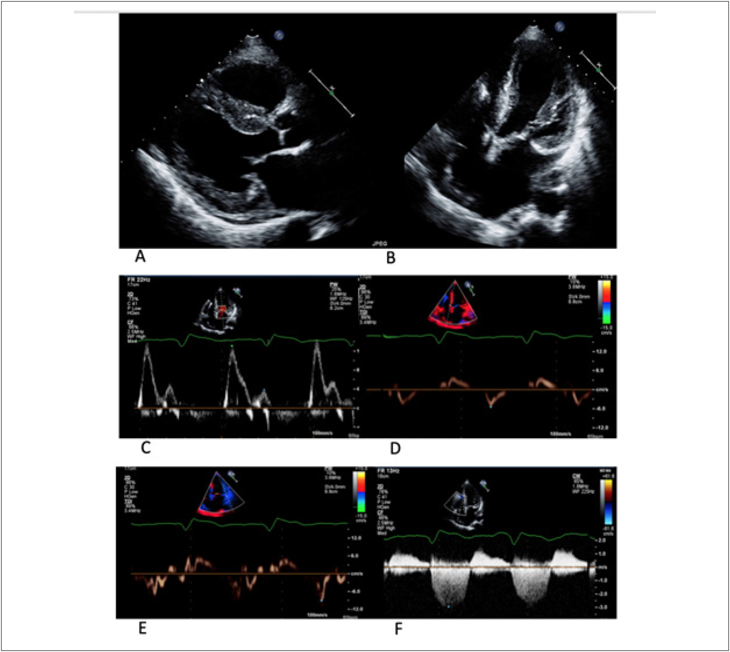
Fabry disease (FD) is a rare X-linked hereditary lysosomal storage disease caused by deficient activity of the enzyme alpha-galactosidase A that leads to the accumulation of globotriasylceramide (Gb3) in affected tissues, including the heart, brain, and kidneys. In FD, cardiac damage begins early and progresses subclinically before the appearance of symptoms and generally manifests as left ventricular hypertrophy simulating hypertrophic cardiomyopathy (HCM). Recent studies have demonstrated a prevalence of FD of up to 5% in patients whose initial diagnosis was HCM. After the introduction of enzyme replacement therapy, FD deserves special attention in the differential diagnosis in patients with unexplained ventricular hypertrophy and HCM, as it is a heart disease that requires specific treatment. However, early diagnosis and treatment are essential to prevent the progression of the disease by reducing cardiovascular event rates.–

With the aim of investigating the clinical and exam differences between HCM and DF, the study of Akhan et al. retrospectively evaluated 60 patients with HCM and 40 with FD. In this population, with a similar average age, the authors showed, in general, that the characteristics of the two cardiac conditions share more similarities than differences, making diagnosis a challenge in clinical practice.

[…]

  • Cardiomyopathy, Hypertrophic/diagnostic, imaging
  • Enzyme Replacement Therapy
  • Fabry disease / complications
  • Hypertrophy Left Ventricular
  • infant, newborn, diseases
  • Rare Disease
Read Full Article
Creative Commons Attribution (CC BY) Creative Commons license
08/03/2024 DOI: 10.36660/abc.20230834
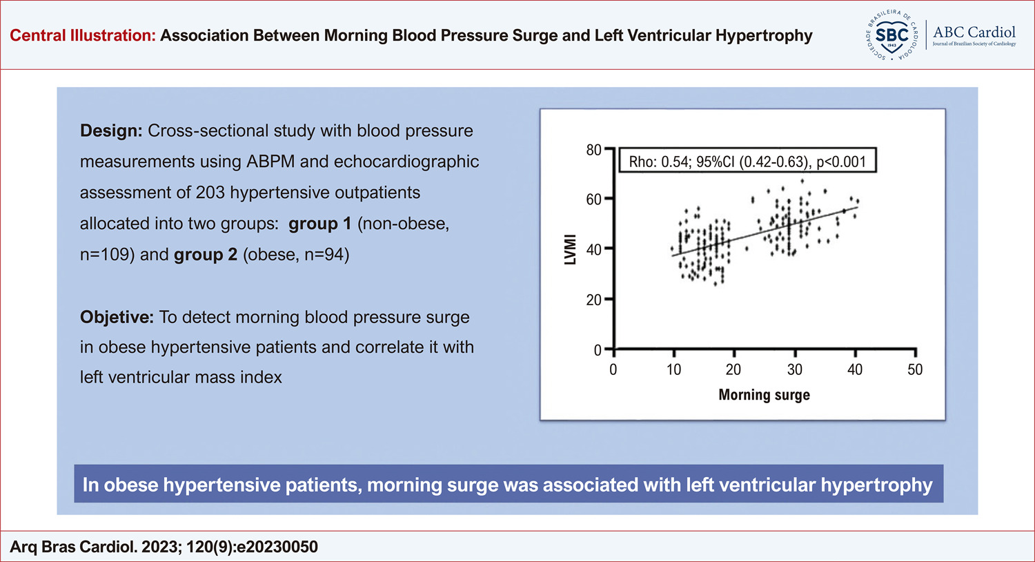
Association between Morning Surge and Left Ventricular Hypertrophy in Obese Hypertensive Patients
ABC
Original Article

Association between Morning Surge and Left Ventricular Hypertrophy in Obese Hypertensive Patients

Natascha Gonçalves Francisco Palmeira et al. Henrique Tria Bianco Maria Teresa Nogueira Bombig Fernando Focaccia Povoa … Francisco A. H. Fonseca Maria Cristina Izar José Marcos Thalenberg Braulio Luna Filho Fabiane Marui Simone Matheus Fischer Celso Amodeo Dilma do Socorro Moraes de Souza Rui Povoa

Abstract

Background

Weight gain can trigger mechanisms that increase blood pressure. Nevertheless, obesity causes structural changes in the myocardium, including increased ventricular mass, atrial dilatation, and diastolic and systolic dysfunction. Additionally, blood pressure variations, like morning surge (MS) in obese hypertensive patients may have clinical relevance in cardiovascular events. Although morning blood pressure surge is a physiological phenomenon, excess MS can be considered an independent risk factor for cardiovascular events.

Objective

To evaluate MS values and their association with left ventricular hypertrophy (LVH) and nocturnal dipping (ND) in obese and non-obese hypertensive patients.

Methods

A cross-sectional study that evaluated BP measurements by ambulatory blood pressure monitoring (ABPM) and the presence of LVH by echocardiography in 203 hypertensive outpatients, divided into two groups: 109 non-obese and 94 obese hypertensives patients. The significance level was set at 0.05 in two-tailed tests.

Results

A MS above 20 mmHg by ABPM was detected in 59.2% of patients in the non-obese group and 40.6% in the obese group. LVH was found in 18.1% and 39.3% of patients in the non-obese and obese groups, respectively, p16 mmHg was associated with LVH, [prevalence ratio: 2.80; 95%CI (1.12–6.98), p=0.03]. For the non-obese group, the cut-off point of MS for this association was >22 mmHg.

Conclusion

High MS was positively associated with LVH, with a particular behavior in the hypertensive obese group.

  • Blood Pressure Monitoring, Ambulatory
  • Hypertrophy Left Ventricular
  • Obesity
Read Full Article
Creative Commons Attribution (CC BY) Creative Commons license
09/10/2023 DOI: 10.36660/abc.20230050
Figure 1
ABCIC
Case Report

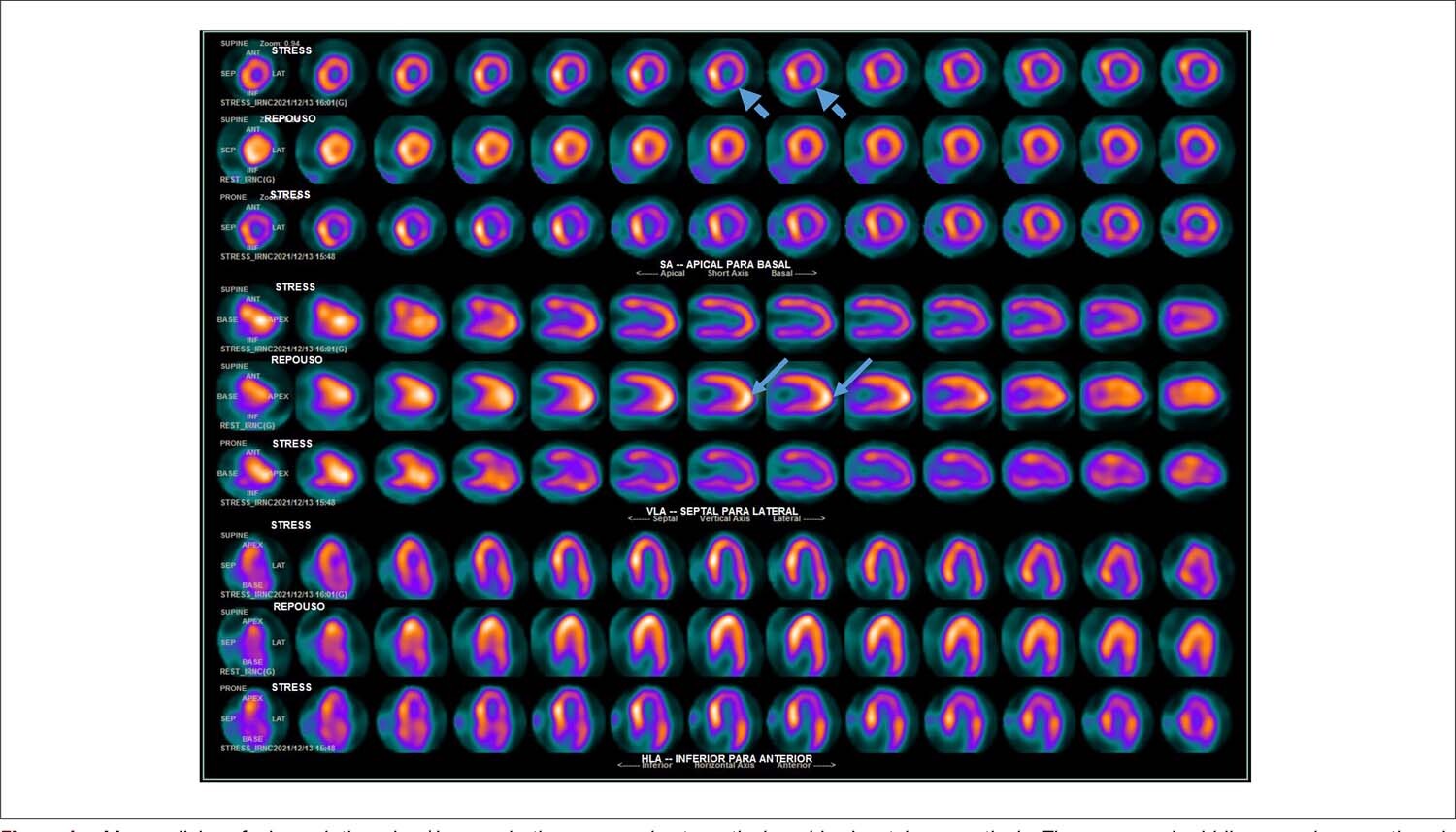
Evaluation of Coronary Flow Reserve by Myocardial Scintigraphy in Left Ventricular Hypertrophy

Renata Christian Martins Felix et al. Felipe Hermely Villela Pedras Odorico de Souza Lima Neto Leonardo Medeiros Vitório

Introduction

The microvascular integrity of the myocardial bed can be assessed by coronary flow reserve (CFR), which is a calculation of the ratio between the absolute coronary blood flow under vasodilator stress and under resting conditions. This measurement is capable of estimating vasomotor dysfunction, and it reflects the hemodynamic effect of focal, diffuse, or small-vessel coronary disease.

Currently, it is possible to measure CFR invasively using the techniques of thermodilution or intracoronary Doppler and through non-invasive methods, such as echocardiography, magnetic resonance imaging, computed tomography, or nuclear medicine.

[…]

  • Hypertrophy Left Ventricular
  • Microvascular Angina
  • Myocardial Perfusion Imaging
  • Regional Blood Flow
Read Full Article
Creative Commons Attribution (CC BY) Creative Commons license
26/09/2023 DOI: 10.36660/abcimg.20230053i Views 2
ABC
Editorial

PRKAG2 Cardiomyopathy

Eduardo Back Sternick

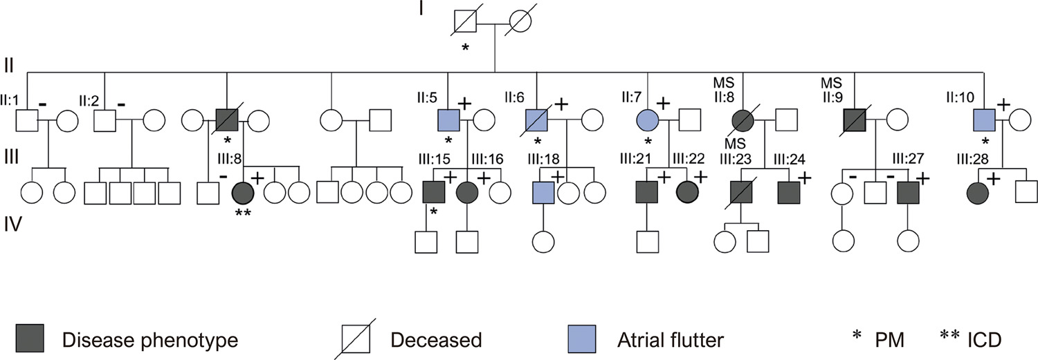
Magalhães et al. assessed atrial flutter’s clinical, electrocardiographic, and electrophysiologic characteristics in patients with and without PRKAG2 cardiomyopathy. Although genetic sequencing was not carried out in their control patients, the absence of clinical features of this pathology is acceptable evidence of a non-carrier state. In humans, mutations in the PRKAG2 gene result in a highly penetrant phenotype. It is dominated by the cardiac features of left ventricular hypertrophy, ventricular pre-excitation, atrial tachyarrhythmia, cardiac conduction disease, and myocardial glycogen storage. It would be very unlikely to find a patient with a positive genotype for PRKAG2 in the setting of a negative phenotype.

Our group pioneered the study of PRKAG2 cardiomyopathy in Brazil. The first patient was assessed in 1994, a 36-years old male, with mild hypertension, with recurrent episodes of common atrial flutter. The electrocardiogram showed ventricular pre-excitation, enlarged P waves, a significant increase in the QRS voltage, and sinus bradycardia. Of 8 brothers, six had very similar electrocardiographic findings. Their mother had had a pacemaker implanted at the age of 42. The echocardiogram showed a non-obstructive form of asymmetric left ventricular hypertrophy and mild left atrial enlargement. There were sufficient elements to allow a presumptive diagnosis of a phenocopy of hypertrophic cardiomyopathy! In 1994, however, we were not able to make the diagnosis. The mutations of the PRKAG2 gene had yet to be reported in 1994, but it would be 7 years later. Then, in 2004, we undertook genetic sequencing of the patients. We presented our experience during the NASPE Congress of 2005. Our research received the first prize in clinical arrhythmia during the Brazilian Congress of Cardiac Arrhythmias of 2004.

[…]

  • Arrhythmias Cardiac
  • Atrial flutter
  • Atrioventricular block
  • Cardiomyopathy, Hypertrophic
  • Glycogen Storage Disease
  • Hypertrophy Left Ventricular
Read Full Article
Creative Commons Attribution (CC BY) Creative Commons license
25/11/2022 DOI: 10.36660/abc.20220694 Views 1
Figure 1
ABC
Original Article

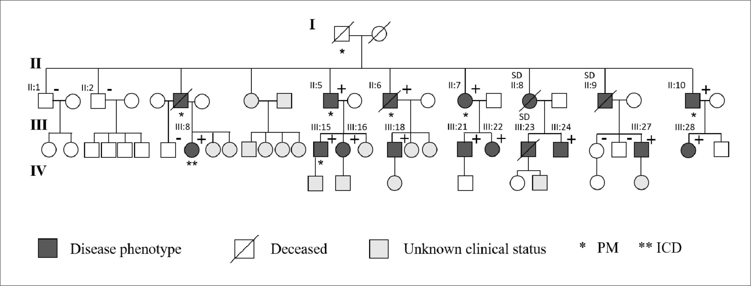
Atrial Flutter in PRKAG2 Syndrome: Clinical and Electrophysiological Characteristics

Eduardo Faria Soares de Magalhães et al. Luiz Pereira de Magalhães Jussara de Oliveira Pinheiro Alex Teixeira Guabiru Roque Aras

Abstract

Background

PRKAG2 syndrome is a rare autosomal dominant disease, a phenocopy of hypertrophic cardiomyopathy characterized by intracellular glycogen accumulation. Clinical manifestations include ventricular preexcitation, cardiac conduction disorder, ventricular hypertrophy, and atrial arrhythmias.

Objective

To compare the clinical and electrophysiological characteristics observed in patients with atrial flutter, with and without PRKAG2 syndrome.

Methods

An observational study comparing patients with atrial flutter: group A consisted of five patients with PRKAG2 syndrome from a family, and group B consisted of 25 patients without phenotype of PRKAG2 syndrome. The level of significance was 5%.

Results

All patients in group A had ventricular preexcitation and right branch block, and four had pacemakers (80%). Patients in group A were younger (39±5.4 vs 58.6±17.6 years, p=0.021), had greater interventricular septum (median=18 vs 10 mm; p<0.001) and posterior wall thickness (median=14 vs 10 mm; p=0.001). In group A, four patients were submitted to an electrophysiological study, showing a fasciculoventricular pathway, and atrial flutter ablation was performed in tree. All patients in group B were submitted to ablation of atrial flutter, with no evidence of accessory pathway. Group B had a higher prevalence of hypertension, diabetes mellitus, coronary artery disease and sleep apnea, with no statistically significant difference.

Conclusion

Patients with PRKAG2 syndrome presented atrial flutter at an earlier age and had fewer comorbidities when compared to patients with atrial flutter without mutation phenotype. The occurrence of atrial flutter in young individuals, especially in the presence of ventricular preexcitation and familial ventricular hypertrophy, should raise the suspicion of PRKAG2 syndrome.

  • Atrial flutter
  • Atrioventricular block
  • Cardiac Arrhythmias
  • Cardiomyopathy, Hypertrophic
  • Glycogen Storage Disease
  • Hypertrophy Left Ventricular
Read Full Article
Creative Commons Attribution (CC BY) Creative Commons license
06/09/2022 DOI: 10.36660/abc.20210792 Views 2
ABC
Editorial

Can Simple Echocardiographic Parameters Replace The ASCVD Probabilistic Model Calculation?

Tonnison de Oliveira Silva et al. Luiz Eduardo Fonteles Ritt

The study by Fernandes et al. used data from the ELSA-BRASIL Cohort. , It showed that in asymptomatic patients with no history of cardiovascular disease, echocardiographic measurements that are part of the daily routine of any echocardiography service are independently associated with the ASCVD predictor model. A total of 2,973 Brazilian participants without cardiovascular disease were evaluated between 2008 and 2010. The ASCVD score calculation used data produced in 2008-2010 and 2012-2014; echocardiography was performed exclusively at the initial moment (period 1). After multivariate logistic regression analysis, the echocardiographic parameters with statistical significance (controlled for body mass index, hypertriglyceridemia, physical activity, educational level and excessive alcohol consumption) were diastolic dysfunction, left ventricular hypertrophy, and left atrial volume indexed by body surface area. Left ventricular diastolic dysfunction was the strongest predictor of association with a high risk of cardiovascular events (ASCVD > 7.5%).

Diastolic dysfunction is a marker of cardiovascular events, including total mortality and hospitalizations for HF. In the ischemic cascade, diastolic dysfunction, symptomatic or not, is one of the earliest manifestations. It is also useful in identifying those patients with stage B heart failure. Echocardiography is the non-invasive, available and low-cost tool most used to evaluate this alteration. The major obstacle is that ventricular diastole mechanisms are complex, multifactorial, age-related, and subject to both acute and chronic hemodynamic and coronary flow changes. –

[…]

  • Cardiovascular Diseases
  • Diastolic Dysfunction
  • Echocardiography/methods
  • Hospitalization
  • Hypertrophy Left Ventricular
  • Mortality
  • risk stratification
Read Full Article
Creative Commons Attribution (CC BY) Creative Commons license
13/05/2022 DOI: 10.36660/abc.20220270 Views 1
Figure 1
ABC
Research Letter

Long-Term Cardiac Complications of PRKAG2 Syndrome

Luiz Pereira de Magalhães et al. Eduardo Faria Soares de Magalhães Jussara de Oliveira Pinheiro Alex Teixeira Guabiru … Francisco José Farias Borges dos Reis Roque Aras

Introduction

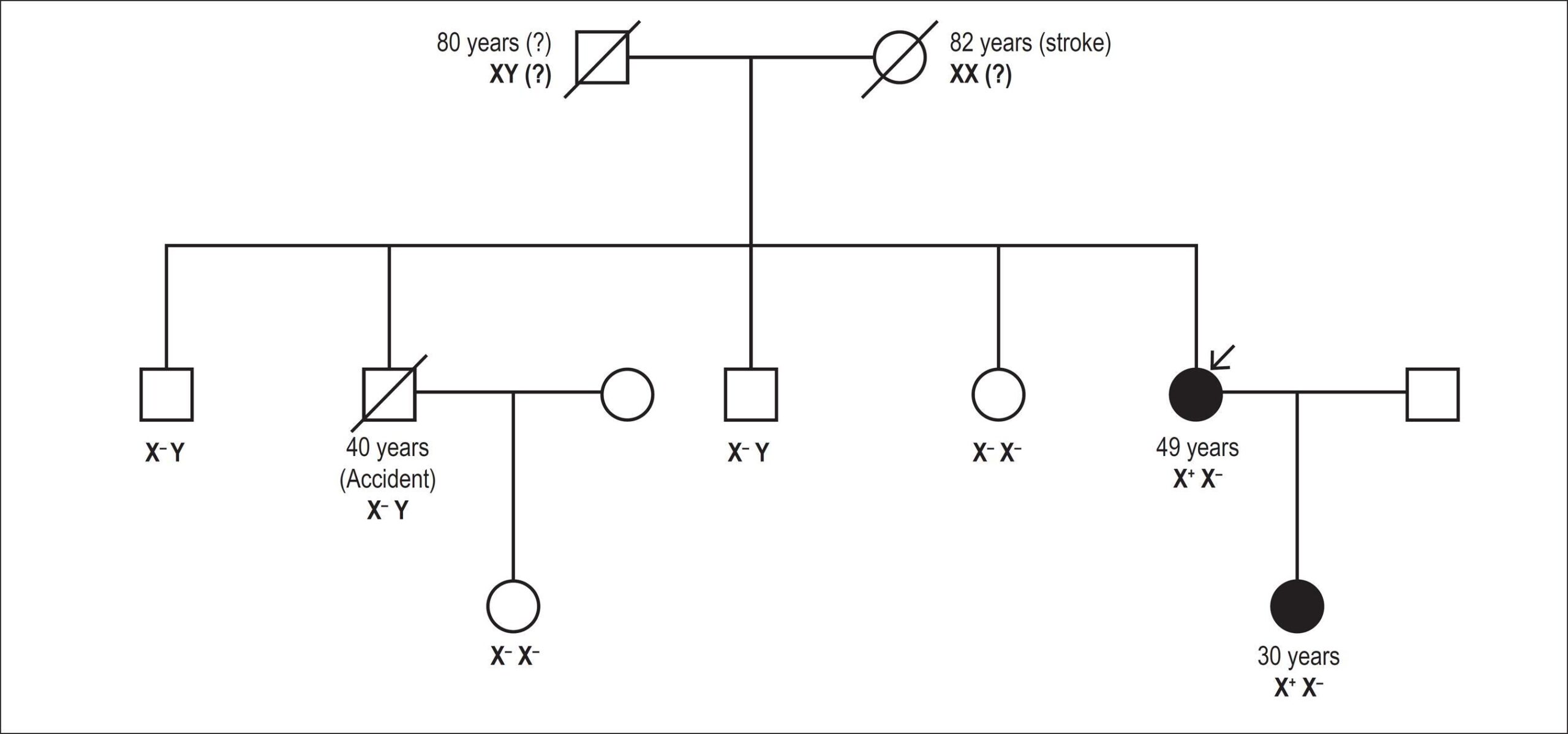
The PRKAG2 syndrome is a rare autosomal dominant inherited disease caused by mutations in the gene encoding the γ regulatory subunit of AMP-activated protein kinase., It has been associated with abnormal glycogen accumulation in cardiomyocytes, predisposing to ventricular hypertrophy, arrhythmias, and sudden death. Gollob et al. were the first to describe the mutation in 2001, calling attention to the need for differential diagnosis with hypertrophic cardiomyopathy., Although the prevalence of PRKAG2 syndrome is not established, the number of reported cases may be increasing due to wider availability of genotyping. Despite that, possible prognostic factors have not yet been described in the literature. Given the possible severity of clinical presentations and the scarcity of data regarding the natural history, our objectives were to evaluate the clinical course of patients with PRKAG2 syndrome over time and analyze the incidence of long-term cardiac complications.

[…]

  • Atrial flutter
  • Atrioventricular block
  • Hypertrophy Left Ventricular
  • Pacemaker, Artificial
Read Full Article
Creative Commons Attribution (CC BY) Creative Commons license
21/02/2022 DOI: 10.36660/abc.20210062 Views 2
Figure 1
ABC
Research Letter

Left Ventricular Hypertrophy: One Phenotype, Two Hypotheses, Three Lessons

Patrícia Rodrigues et al. Ana Rita Soares Ricardo Taipa Sofia Ferreira Hipólito Reis

Past medical history

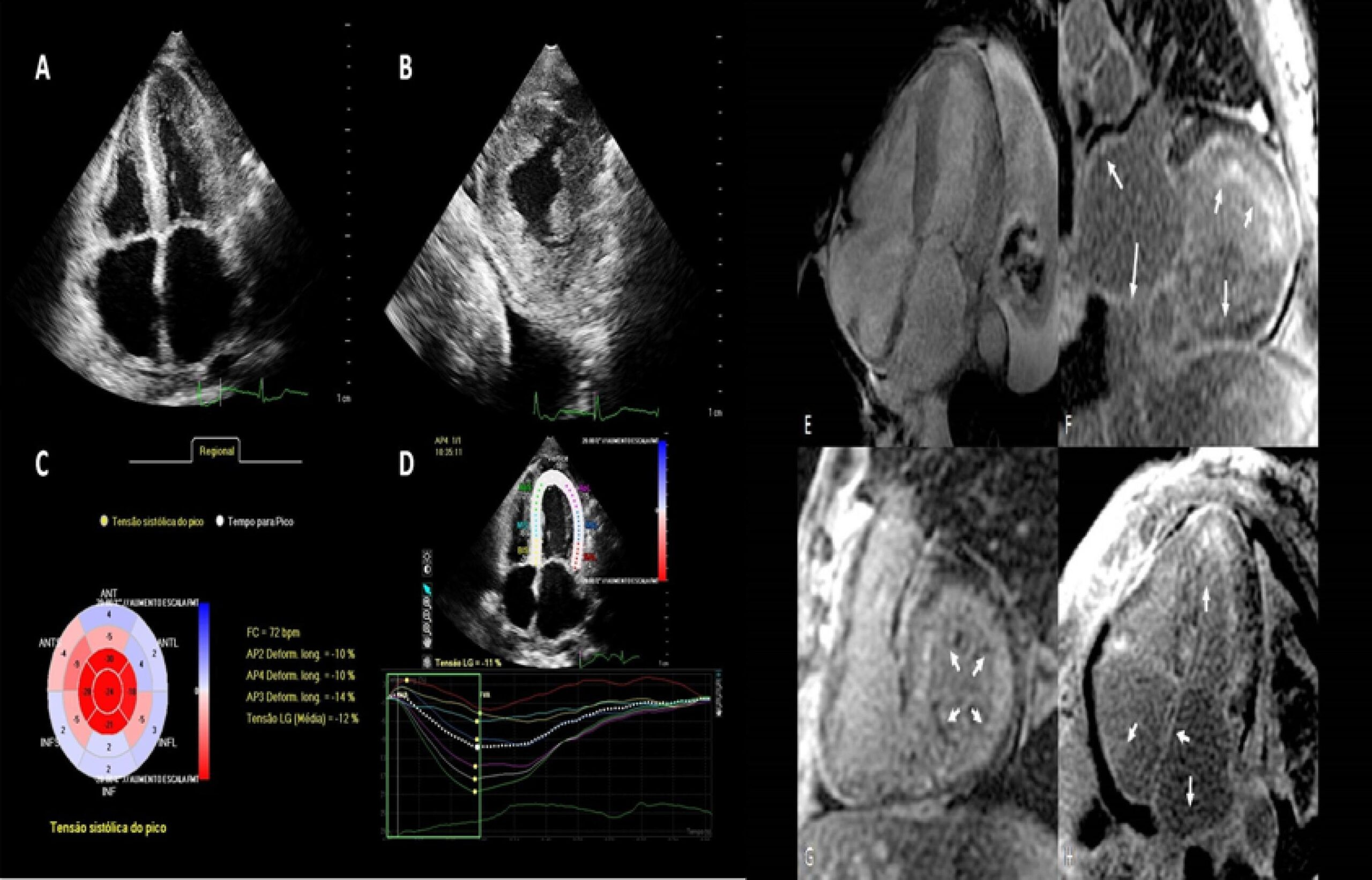
A 58-year-old woman with familial amyloid polyneuropathy caused by the Val30Met (p.Val50Met) mutation in the transthyretin (TTR) gene started showing neuropathic symptoms by the age of 37 and a salivary gland biopsy confirmed TTR amyloid deposition. She was submitted to liver transplantation 7 years after symptom onset and was under immunosuppressive drugs, with stable neurological changes since then. She also had chronic kidney failure (stage 3b) and a pacemaker implanted due to sick sinus syndrome.

[…]

  • Amyloid Neuropathies, Familial/diagnostic imaging
  • Amyloidosis
  • Cardiomyopathy, Hypertrophic
  • Heart Failure
  • Hypertrophy Left Ventricular
Read Full Article
Creative Commons Attribution (CC BY) Creative Commons license
22/11/2021 DOI: 10.36660/abc.20210103 Views 1
Figure 1
ABC
Original Article

Performance of the Electrocardiogram in the Diagnosis of Left Ventricular Hypertrophy in Older and Very Older Hypertensive Patients

Fernando Focaccia Povoa et al. Braulio Luna Filho Henrique Tria Bianco Celso Amodeo … Rui Povoa Maria Teresa Nogueira Bombig Roberto Dischinger Miranda Simone Matheus Fischer Maria Cristina Oliveira Izar Francisco A. H. Fonseca

Abstract

Background:

Left ventricular hypertrophy (LVH) is an important cardiovascular risk factor, regardless of arterial hypertension. Despite the evolution of imaging tests, the electrocardiogram (ECG) is still the most used in the initial evaluation, however, with low sensitivity.

Objective:

To evaluate the performance of the main electrocardiographic criteria for LVH in elderly and very elderly hypertensive individuals.

Methods:

In a cohort of hypertensive patients, ECGs and doppler echocardiographies (ECHO) were performed and separated into three age groups: <60 years, Group I; 60-79 years Group II; and ≥80 years, Group III. The most used electrocardiographic criteria were applied for the diagnosis of LVH: Perugia; Pegaro-Lo Presti; Gubner-Ungerleider; Narita; (Rm+Sm) x duration; Cornell voltage; Cornell voltage duration; Sokolow-Lyon voltage; R of aVL ≥11 mm; RaVL duration. In evaluating the performance of these criteria, in addition to sensitivity (Sen) and specificity (Esp), the “Diagnostic Odds Ratios” (DOR) were analyzed. We considered p-value <0.05 for the analyses, with two-tailed tests.

Results:

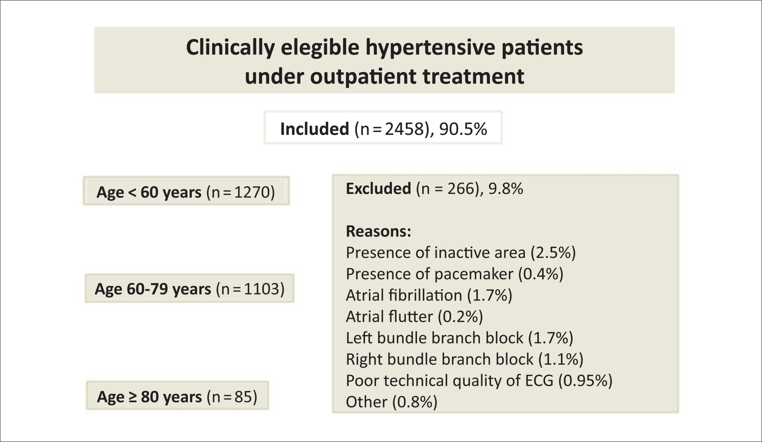
In 2,458 patients, LVH was present by ECHO in 781 (31.7%). In Groups I and II, the best performances were for the criteria of Narita, Perugia, (Rm+Sm) x duration, with no statistical differences between them. In Group III (very elderly) the Perugia criteria and (Rm+Sm) x duration had the best performances: Perugia [44,7/89.3; (Sen/Esp)] and (Rm+Sm) duration [39.4%/91.3%; (Sen/Esp), p<0.05)], with the best PAIN results:6.8. This suggests that in this very elderly population, these criteria have greater discriminatory power to separate patients with LVH.

Conclusion:

In very elderly hypertensive patients, the Perugia electrocardiographic criteria and (Rm+Sm) x duration showed the best diagnostic performance for LVH.

  • Aged
  • Electrocardiography/methods
  • Heart Failure
  • Hypertension
  • Hypertrophy Left Ventricular
  • Myocardial Infarction
  • Stroke
Read Full Article
Creative Commons Attribution (CC BY) Creative Commons license
06/08/2021 DOI: 10.36660/abc.20200600 Views 1
Figure 1
IJCS
Review Article

Transcatheter Interatrial Shunts for the Treatment of Heart Failure with Preserved Ejection Fraction

Anju Bhardwaj et al. Vishal Y. Parikh Ajith Nair

Abstract

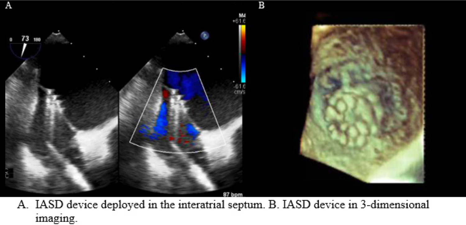
Heart failure with preserved ejection fraction (HFpEF) is a clinical syndrome, which accounts for about 50% of patients with heart failure (HF). The morbidity and mortality associated with HFpEF is similar to HFrEF. Clinical trials to date have failed to show a benefit of medical therapy for HFpEF, which may due to lack of uniform phenotypes and heterogeneous population. In addition, medical therapy proven for HFrEF may not address the pathophysiologic basis for HFpEF. Left atrial remodeling and dysfunction is central to HFpEF and accounts for secondary pulmonary hypertension and pulmonary vascular congestion that frequently occurs with exertion. Interatrial shunts represent a novel treatment modality for HFpEF. These shunts allow for left atrial decongestion and a reduction in pulmonary venous hypertension during exercise leading to improvements in hemodynamics, functional status and quality of life. Trials to date have demonstrated safety and short-term efficacy of these devices for HFpEF. The long-term benefits are currently being evaluated in ongoing trials. If effective, the use of interatrial shunts may be a new therapeutic paradigm for the treatment of HFpEF.

  • Heart Atria
  • Heart Failure
  • Hypertrophy Left Ventricular
  • Stroke Volume
  • transcatheter
Read Full Article
Creative Commons Attribution (CC BY) Creative Commons license
01/02/2021 DOI: 10.36660/ijcs.20200236 Views 2
ABC
Editorial

Which Risk Score Best Assesses Clinical Objectives in Patients with Hypertrophic Cardiomyopathy?

Eduardo Alberto de Castro Roque

Hypertrophic cardiomyopathy (HCM) is a genetic disorder characterized by commonly asymmetric unexplained left ventricular hypertrophy, with greater thickening of the basal interventricular septum. Left ventricular outflow tract obstruction is present at rest in around one third of patients and can be caused in another third. The histological features of HCM include hypertrophy and myocyte disorder, in addition to interstitial fibrosis. Hypertrophy is also often associated with left ventricular diastolic dysfunction.

The first case of HCM was described by Henri Liouville in 1869 in the Gazette Medecine Paris. In 1907, Dr. A. Schmincke, a German pathologist, described two hearts with left ventricular hypertrophy; both were seen in women in their fifth decade of life. Levy and von Glahn, in 1944, from the University of Colombia, in New York, published a series of cases that resembled HCM. In 1949, William Evans, a cardiologist from London, described the familial occurrence of cardiac hypertrophy in a series of patients similar to those described in the article by Levy and von Glahn. Dr. Eugene Braunwald and Dr. Andrew Glenn Morrow published a series of studies where they detailed the clinical and hemodynamic aspects of this disease, allowing the establishment of therapeutic objectives. –

[…]

  • Cardiomyopathy, Hypertrophic, Familial
  • Defibrillators, Implantable
  • Discrete Subaortic Stenosis
  • Hypertrophy Left Ventricular
  • Safety Management
Read Full Article
Creative Commons Attribution (CC BY) Creative Commons license
28/08/2020 DOI: 10.36660/abc.20200543 Views 1
Figure 1
IJCS
Case Report

Mitral Valve Replacement with Regent Aortic Valve in Severe Mitral Stenosis

Negin Yavari et al. Mina Ghorbanpour Landy Negar Omidi Mahmoud Shirzad Seyed Hossein Ahmadi Tafti

Introduction

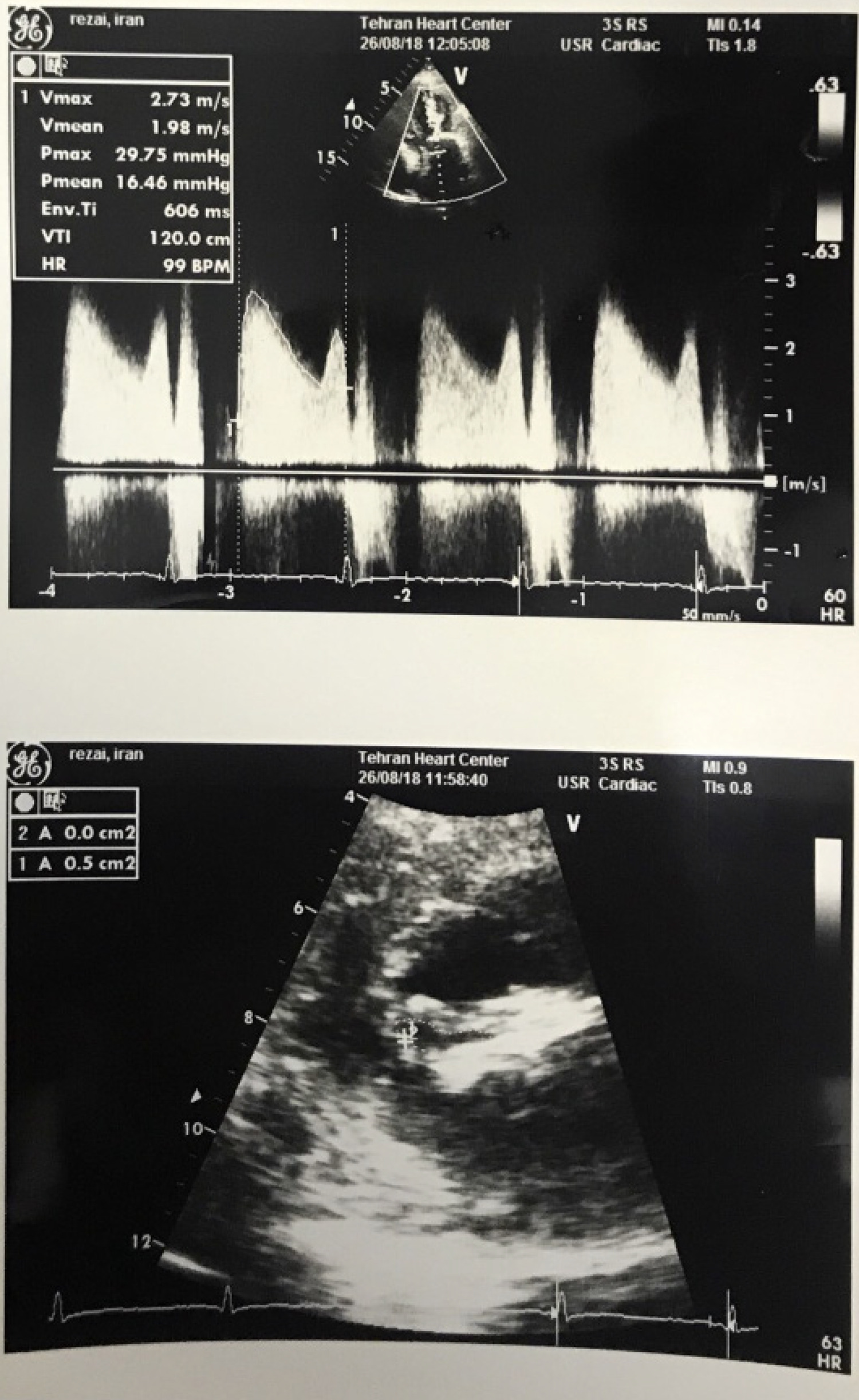
Mitral valve replacement (MVR) in the context of calcification of the mitral valve annulus (<25 millimeters diameter) is challenging, with an increased risk of postoperative complications. Mitral annular calcification is more common in elderly patients due to secondary aging and tissue degeneration. It can also be found in younger patients with connective tissue disorders and inflammatory conditions including the Marfan’s syndrome, rheumatoid arthritis, and rheumatic fever. Calcification of the mitral annulus has been associated with arrhythmias, heart block, stenosis, valve insufficiency, bacterial endocarditis, and arterial embolization. Although the condition is not common, calcified mitral annulus may been found in patients requiring surgery for mitral valve dysfunction. However, replacement of a calcified valve is challenging especially because of the difficulty in placing the sutures through the calcified annulus, increasing the risk of leakage, poor positioning and dehiscence. , For this reason, some studies have suggested the implantation of an aortic mechanical valve (St. Jude Medical prosthesis) in mitral position due to its smaller surface area. Advantages of the St. Jude Medical valve prosthesis over other mechanical prostheses include a reduced sewing cuff size and a reduced frame diameter to allow a greater orifice area for a given annular size. There is limited number of studies on this kind of replacement surgeries, and here we report a case of successful placement of a St. Jude Medical aortic valve prosthesis in a calcified mitral annulus without postoperative complication.

[…]

  • Calcinosis/complications
  • Heart Valve Prosthesis
  • Hypertrophy Left Ventricular
  • Mitral Valve/surgery
  • Rheumatic Heart Diseases/complications
  • Stenosil mitral valve/surgery
Read Full Article
Creative Commons Attribution (CC BY) Creative Commons license
15/07/2020 DOI: 10.36660/ijcs.20190198 Views 1
ABC
Editorial

Do We Need to Know the Left Ventricular Geometry Patterns of the Brazilian Population?

Roberto M. Saraiva

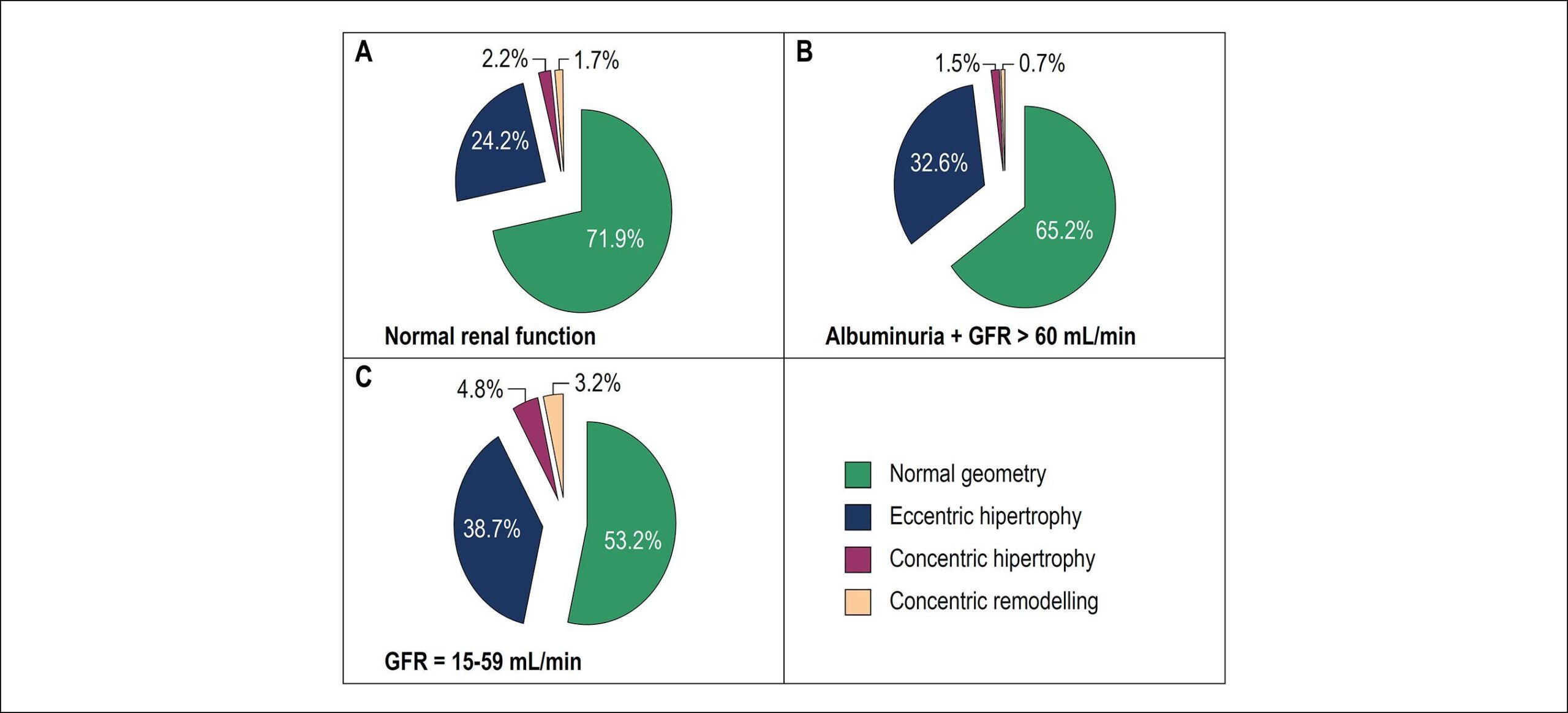
In this issue of the Arquivos Brasileiros de Cardiologia, Almeida et al. describe the left ventricular (LV) remodeling patterns frequencies found in a Brazilian population followed at primary healthcare clinics in Niterói city, state of Rio de Janeiro. The authors found that a LV abnormal geometry was present in up to 33% of 636 studied individuals (mean age 59.5±10.3 years old; 62% women). Eccentric LV hypertrophy (LVH) was the most common abnormal LV geometry pattern (29%), followed by concentric LVH and concentric remodeling (2% each).

LV remodeling is no longer considered solely an adaptative mechanism but a response to several different stimuli that lead to gene activation, cellular hypertrophy, apoptosis, fibrosis, and, finally, LV remodeling with different degrees of LV function compromise and increase in cardiovascular risk. In fact, the relation between LVH diagnosed by electrocardiogram and mortality has been long recognized. LV mass is considered an independent risk factor for heart failure (HF),, stroke, sudden cardiac death, supraventricular and ventricular tachycardia, and all-cause and cardiovascular mortality. Therefore, hypertension (HTN) is considered stage A HF and LVH is considered stage B HF on the American College of Cardiology/American Heart Association guidelines on HF management. Surprisingly, it was not up to the study of Almeida et al. that LV geometry patterns were studied in Brazilian population. We need to know exactly what are the frequency and value of LV geometry patterns in the Brazilian population and not only apply knowledge obtained with other populations.

[…]

  • Echocardiography/métodos
  • Heart Failure
  • Heart Ventricles/cirurgia
  • Hypertension
  • Hypertrophy Left Ventricular
  • Ventricular Remodeling
Read Full Article
Creative Commons Attribution (CC BY) Creative Commons license
10/02/2020 DOI: 10.36660/abc.20190838 Views 3
Figure 1
ABC
Original Article

Left Ventricular Remodeling Patterns in Primary Healthcare

Roberto de Castro Meirelles de Almeida et al. Antonio José Lagoeiro Jorge Maria Luiza Garcia Rosa Adson Renato Leite … Dayse Mary S. Correia Evandro Tinoco Mesquita Sergio Chermont Jocemir Ronaldo Lugon Wolney de Andrade Martins

Abstract

Background:

Left ventricular remodeling (LVR) is related to both non-fatal and fatal outcomes.

Objective:

To describe the geometric patterns of the LV and their associations.

Methods:

A total of 636 individuals between the ages of 45 and 99 years in Rio de Janeiro, Brazil, were submitted to clinical evaluation, laboratory tests, electrocardiogram, and tissue Doppler echocardiography (TDE). The difference between categories was tested with Kruskall-Wallis with post hoc tests, once all variables studied are non-normally distributed and Pearson’s Qui-square (categorical variables). Gross and adjusted ORs were estimated by logistic regression. The level of significance was 5% for all tests. Subjects had LVR characterized as: normal geometry (NG), concentric remodeling (CR), concentric hypertrophy (CH), and eccentric hypertrophy (EH).

Results:

The prevalence of altered patterns was 33%. Subjects presented NG (n = 423; 67%); EH (n = 186; 29%); CH (n = 14; 2%); and CR (n = 13; 2%). The variables of gender, age, level of education and albumin/creatinine ratio (A/C), showed a relationship with the chance of EH even after adjustment.

Conclusion:

Approximately one third of the studied individuals had LVR and were at risk for developing heart failure. Altered A/C in urine was associated with EH, indicating an early relationship between cardiac and renal dysfunction.

  • Cardiovascular Diseases/physiopathology
  • Comorbidity
  • Heart Failure
  • Hypertrophy Left Ventricular
  • Renal Insufficiency
  • Risk Factors/complications
  • Ventricular Remodeling
Read Full Article
Creative Commons Attribution (CC BY) Creative Commons license
10/02/2020 DOI: 10.36660/abc.20180258 Views 1
ABC
Letter to the Editor

Left Ventricle Mass Index, a Confounding Variable of Global Longitudinal Strain to be Noticed

Eduardo Thadeu de Oliveira Correia et al. Letícia Mara dos Santos Barbetta

Dear Editor,

We read with great interest the article entitled as “Strain Analysis of Left Ventricular Function in the Association of Hypertrophic Cardiomyopathy and Systemic Arterial Hypertension”. In this paper, the authors evaluated the global longitudinal strain (GLS) of the left ventricle (LV) in two distinct groups: patients with hypertrophic cardiomyopathy (HCM) and patients with HCM and systemic arterial hypertension (SAH) and demonstrated that GLS was lower in the second group. This important finding may indicate greater impairment of LV function in patients with SAH and HCM.

[…]

  • Cardiomyopathy, Hypertrophic
  • Heart Failure
  • Hypertension
  • Hypertrophy Left Ventricular
  • Strain
  • Ventricular Function, Left
Read Full Article
Creative Commons Attribution (CC BY) Creative Commons license
10/02/2020 DOI: 10.36660/abc.20190708 Views 1
Figure 1
IJCS
Original Article

Coarctation of The Aorta: A Case-Series from a Tertiary Care Hospital

Joaquim Barreto et al. Juliana Roda Carlos Wustemberg Germano Ana Paula Damiano Thiago Quinaglia

Abstract

Background:

Coarctation of the aorta is a congenital segmental narrowing of the aortic arch with severe hemodynamic repercussions and increased cardiovascular mortality. Early surgical correction and life-time echocardiographic follow-up must be performed to improve prognosis. However, this goal has been challenged by high rates of underdiagnosis, which delay surgical correction, and by recoarctation in up to one third of operated patients.

Objectives:

The objectives of this study were: (i) to register the frequency of common clinical signs at diagnosis of coarctation of the aorta; (ii) to describe the course of echocardiographic parameters before and during the follow-up of coartectomized subjects; (iii) to analyze the clinical prognosis of patients according to baseline characteristics, occurrence of recoarctation and associated malformations.

Methods:

Case-series of 72 patients coarctectomized between June 1996 and November 2016 in a tertiary care hospital. Clinical, echocardiographic and surgical variables were considered. All patients were submitted to coarctectomy by posterolateral thoracotomy and end-to-end anastomosis. Data were classified as parametric or non-parametric by Kolmogorov-Smirnov test. Parametric data were expressed as mean and standard deviation, and non-parametric data as median and interquartile range. Continuous variables were analyzed using paired t-tests, and categorical variables were compared by chi-square test. For all analysis, a p-value of less than 0.05 was considered statistically significant. Statistical analysis was performed using SPSS, version 20.0 (IBM, Chicago, IL, USA).

Results:

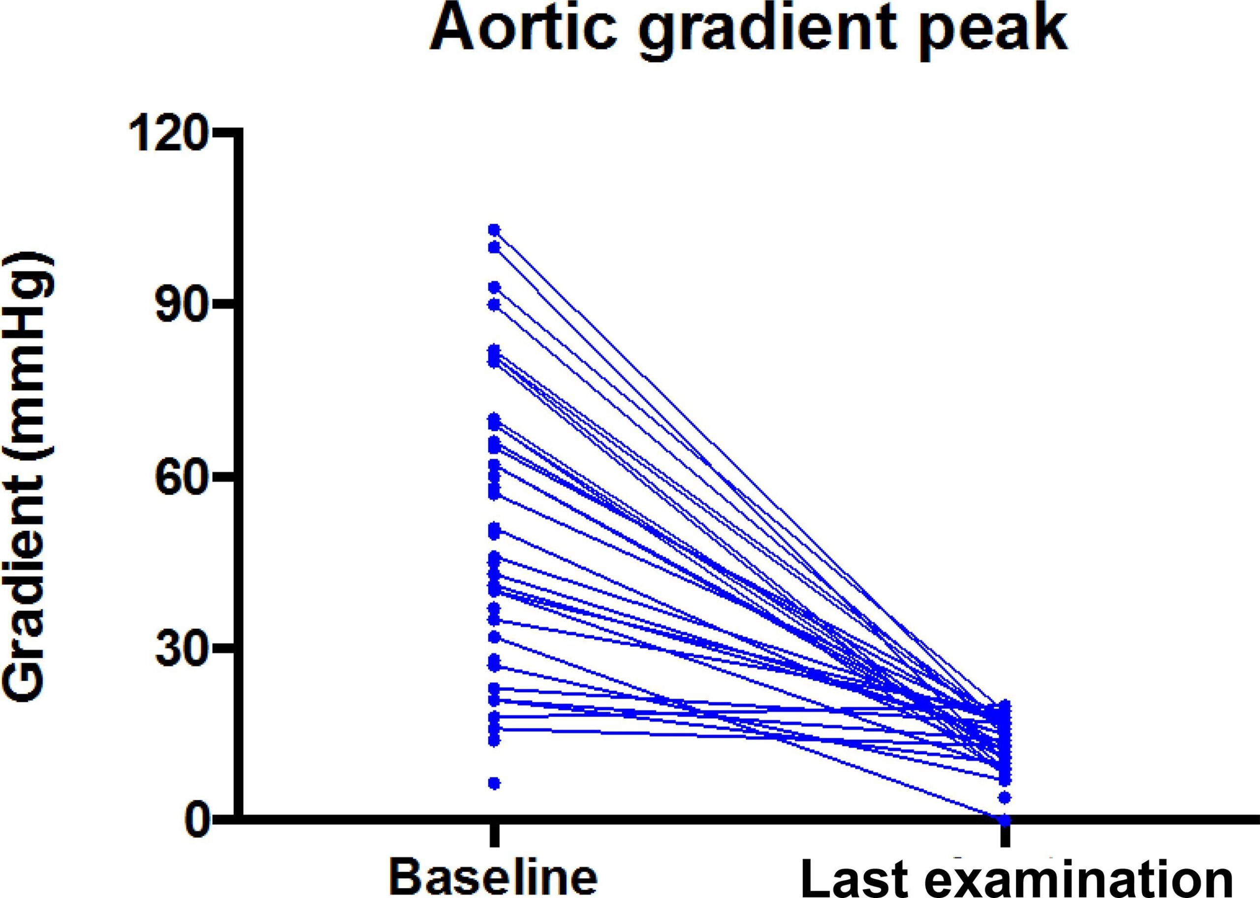
The mean follow-up time was 5.8 years (range: 0-20 years). At diagnosis, most patients had heart murmur (88%), non-palpable pulse in the lower limbs (50%), left ventricular hypertrophy (78%), and bicuspid aortic valve (33%), with a mean aortic peak gradient of 55 mmHg. After surgical correction, those without recoarctation were less symptomatic (60 vs 4.5%; p < 0.001), had lower aortic peak gradient (54 ± 3.8 vs 13 ± 0.8; p = 0.01) and left ventricle mass (95 ± 9.2 vs. 63 ± 11; p = 0.01), and the most common complications were late hypertension (39.2%), and recoarctation (27.6%). Recoarcted patients did not show improvement of neither clinical nor echocardiographic variables. Age at repair and bicuspid aortic valve groups had comparable results with controls. Surgical procedure was safe; mean time of hospitalization was 10 days and mean surgery time 2.3 hours.

Conclusions:

Coarctectomy improves cardiac symptoms and left ventricular hypertrophy, with a slight effect on the incidence of hypertension. Recoarctation occurs in one-third of patients and draws attention for the need of lifelong surveillance by echocardiography.

  • Aortic Coactation/surgery
  • Echocardiography/methods
  • Heart Defects, Congenital
  • Hypertension
  • Hypertrophy Left Ventricular
Read Full Article
Creative Commons Attribution (CC BY) Creative Commons license
08/08/2019 DOI: 10.5935/2359-4802.20190067 Views 1
Figure 1
ABC
Original Article

GLA Gene Mutation in Hypertrophic Cardiomyopathy with a New Variant Description: Is it Fabry's Disease?

Ândrea Virgínia Chaves-Markman et al. Manuel Markman Eveline Barros Calado Ricardo Flores Pires … Marcelo Antônio Oliveira Santos-Veloso Catarina Maria Fonseca Pereira Andréa Bezerra de Melo da Silveira Lordsleem Sandro Gonçalves de Lima Brivaldo Markman Filho Dinaldo Cavalcanti de Oliveira

Abstract

Background:

Fabry disease (FD) is an X-linked lysosomal storage disorder caused by mutations in the alpha galactosidase A gene (GLA) that lead to the enzymatic deficiency of alpha galactosidase (α-Gal A), resulting in the accumulation of globotriaosylceramide (Gb3) and globotriaosylsphingosine (lyso-Gb3), causing multiple organ dysfunctions.

Objective:

To perform GLA gene screening in a group of patients with echocardiographic diagnosis of hypertrophic cardiomyopathy (HCM).

Methods:

a cross-sectional study was conducted with HCM patients from a university hospital. Patients with coronary artery disease and valvulopathies were excluded. Mutation analysis of the GLA gene was performed. In male subjects, the analysis was performed after evidence of low α-Gal A activity.

Results:

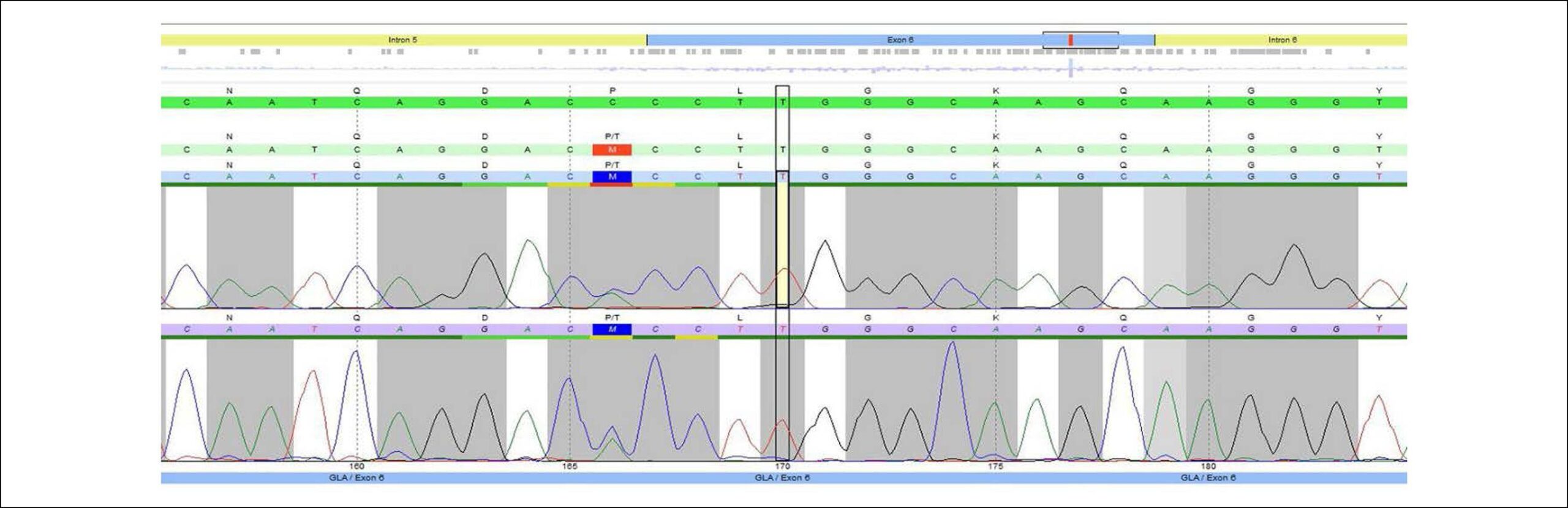
60 patients with echocardiographic diagnosis of HCM were included. Age ranged from 12 to 85 years and 60% were women. Mean myocardial fibrosis percentage on MRI was 10.7 ± 13.1% and mean ventricular thickness was18.7 ± 6.7 mm. Four patients had the following GLA gene mutations: c.967C>A (p.Pro323Thr), not yet described in the literature; c.937G>T (p.Asp313Tyr); and c.352C>T (p.Arg118Cys). All patients had normal levels of lyso-Gb3 and non-ischemic myocardial fibrosis on magnetic resonance imaging; one patient had proteinuria and one patient had ventricular tachycardia.

Conclusion:

in this study, the frequency of mutation in the GLA gene in patients with HCM was 6.7%. A novel mutation in exon 6 of the GLA gene, c.967C>A (p.Pro323Thr), was identified. Patients with HCM may have GLA mutations and FD should be ruled out. Plasma (lyso-Gb3) levels do not seem to be sufficient to attain a diagnosis and organ biopsy should be considered.

  • Cardiomyopathy, Hypertrophic
  • Fabry Disease/genetic
  • Glycosphingolipids
  • Hypertrophy Left Ventricular
Read Full Article
Creative Commons Attribution (CC BY) Creative Commons license
10/07/2019 DOI: 10.5935/abc.20190112 Views 2
Figure 1
IJCS
Review Article

Impact of Obesity and Bariatric Surgery in the Outcome of Patients with Heart Failure

Ricardo Mourilhe-Rocha et al. Nathália Felix Araujo Salvino

Abstract

Currently, the association between obesity and heart failure (HF) is increasingly known. Patients with advanced obesity who suffer from HF without an identifiable cause can be diagnosed as having obesity-associated cardiomyopathy. Although data suggest that obesity may reduce mortality in HF, weight loss, especially in the presence of morbid obesity, reduces symptoms and improves the quality of life of those patients.

Bariatric surgery is the major treatment available for sustained weight loss in morbid obesity. Observational studies have demonstrated an improvement in ventricular structure and function of morbidly obese patients with HF who underwent that procedure.

Thus, despite the risks, bariatric surgery should be considered for patients with HF, because of its potential for reducing associated comorbidities and improving quality of life and functional capacity, in addition to making eligible for heart transplantation those excluded due to high body mass index.

  • bariatric surgery
  • Comorbidity
  • Heart Failure
  • Hypertrophy Left Ventricular
  • Obesity
  • Quality of life
Read Full Article
Creative Commons Attribution (CC BY) Creative Commons license
18/02/2019 DOI: 10.5935/2359-4802.20190001 Views 2
ABC
Letter to the Editor

QTc and QTcd Measurements and Their Relationships with Left Ventricular Hypertrophy in Hemodialysis Patients

Sora Yasri et al. Viroj Wiwanitkit

Editor,

We read the publication on “QTc and QTcd Measurements and Their Relationships with Left Ventricular Hypertrophy in Hemodialysis Patients”, which is very interesting. Alonso et al. concluded that “We found that QTc interval, in contrast to QTcd, is a reproducible and reliable measure and had a weak but positive correlation with LVMi in HD patients.” This report used an unmatched control group; hence, the selection bias can be expected. In fact, the hypertrophy might be expected in a hemodialysis patient who might have underlying metabolic syndrome and vascular disease. To test the reproducibility, the repeated analysis is needed and there is a need to assess the within-run and between-run precision. In the present report, one cannot conclude that the test has a good reproducibility.

[…]

  • Electrocardiography
  • Hypertrophy Left Ventricular
  • renal dialysis
Read Full Article
Creative Commons Attribution (CC BY) Creative Commons license
01/06/2018 DOI: 10.5935/abc.20180096 Views 1
Figure 1
IJCS
Case Report

Cardiac Amyloidosis with Heart Failure and Middle Range Ejection Fraction

Antonio Jose Lagoeiro Jorge et al. Diane Xavier de Ávila Enoï Guedes Vilar Mario Luiz Ribeiro … Karima Elias Hallack Bruno Ana Carolina Pires

Introduction

Cardiac amyloidosis (CA) is a disease with a difficult diagnosis, limited management and a reserved prognosis., A high level of suspicion is necessary for its identification. There are some clinical clues, such as elderly individuals with unexplained left ventricular hypertrophy (LVH), heart failure with preserved ejection fraction (HFpEF) and restrictive pattern, dissociation between LVH on echocardiography and low voltage on the electrocardiogram, among others.,

CA can occur with several hemodynamic forms and patterns of remodeling, according to the disease evolution stage. It may occur as the restrictive form, with left ventricular ejection fraction > 50%; and dilated form, with reduced ejection fraction., Recently, the European Society of Cardiology has established a new classification with the creation of “Heart Failure with Mid-Range Ejection Fraction” (HFmrEF). We report a case of CA with HFmrEF.

[…]

  • Aged
  • Amyloidosis
  • Heart Failure/physiopathology
  • Hypertrophy Left Ventricular
  • Stroke Volume
Read Full Article
Creative Commons Attribution (CC BY) Creative Commons license
01/01/2018 DOI: 10.5935/2359-4802.20180039 Views 1
Figure 1
ABC
Original Article

Pericardial Parietal Mesothelial Cells: Source of the Angiotensin-Converting-Enzyme of the Bovine Pericardial Fluid

Ilsione Ribeiro de Sousa Filho et al. Isabela Cabral Cavicchioli Pereira Lourimar José de Morais Livia das Graças Vieito Lombardi Teodoro … Maria Laura Pinto Rodrigues Roseli Aparecida da Silva Gomes

Abstract

Background:

Angiotensin II (Ang II), the primary effector hormone of the renin-angiotensin system (RAS), acts systemically or locally, being produced by the action of angiotensin-converting-enzyme (ACE) on angiotensin I. Although several tissue RASs, such as cardiac RAS, have been described, little is known about the presence of an RAS in the pericardial fluid and its possible sources. Locally produced Ang II has paracrine and autocrine effects, inducing left ventricular hypertrophy, fibrosis, heart failure and cardiac dysfunction. Because of the difficulties inherent in human pericardial fluid collection, appropriate experimental models are useful to obtain data regarding the characteristics of the pericardial fluid and surrounding tissues.

Objectives:

To evidence the presence of constituents of the Ang II production paths in bovine pericardial fluid and parietal pericardium.

Methods:

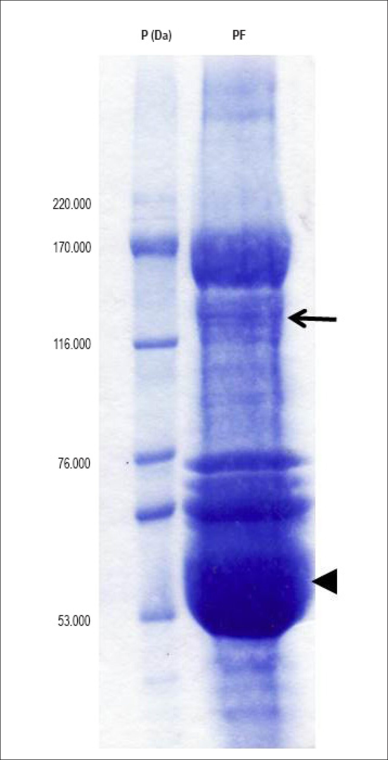
Albumin-free crude extracts of bovine pericardial fluid, immunoprecipitated with anti-ACE antibody, were submitted to electrophoresis (SDS-PAGE) and gels stained with coomassie blue. Duplicates of gels were probed with anti-ACE antibody. In the pericardial membranes, ACE was detected by use of immunofluorescence.

Results:

Immunodetection on nitrocellulose membranes showed a 146-KDa ACE isoform in the bovine pericardial fluid. On the pericardial membrane sections, ACE was immunolocalized in the mesothelial layer.

Conclusions:

The ACE isoform in the bovine pericardial fluid and parietal pericardium should account at least partially for the production of Ang II in the pericardial space, and should be considered when assessing the cardiac RAS.

  • Cattle
  • Hypertrophy Left Ventricular
  • Peptidyl-Dipeptidase A
  • Pericardial Fluid
  • Renin-Angiotensin System
Read Full Article
Creative Commons Attribution (CC BY) Creative Commons license
01/11/2017 DOI: 10.5935/abc.20170155 Views 1
Figure 1
ABC
Original Article

Reproducibility and Reliability Of QTc and QTcd Measurements and Their Relationships with Left Ventricular Hypertrophy in Hemodialysis Patients

Maria Angélica Gonçalves Alonso et al. Valentine de Almeida Costa de Castro Lima Maria Angela Magalhães de Queiroz Carreira Jocemir Ronaldo Lugon

Abstract

Background:

Left ventricular hypertrophy (LVH) is very common in hemodialysis patients and an independent risk factor for mortality in this population. The myocardial remodeling underlying the LVH can affect ventricular repolarization causing abnormalities in QT interval.

Objective:

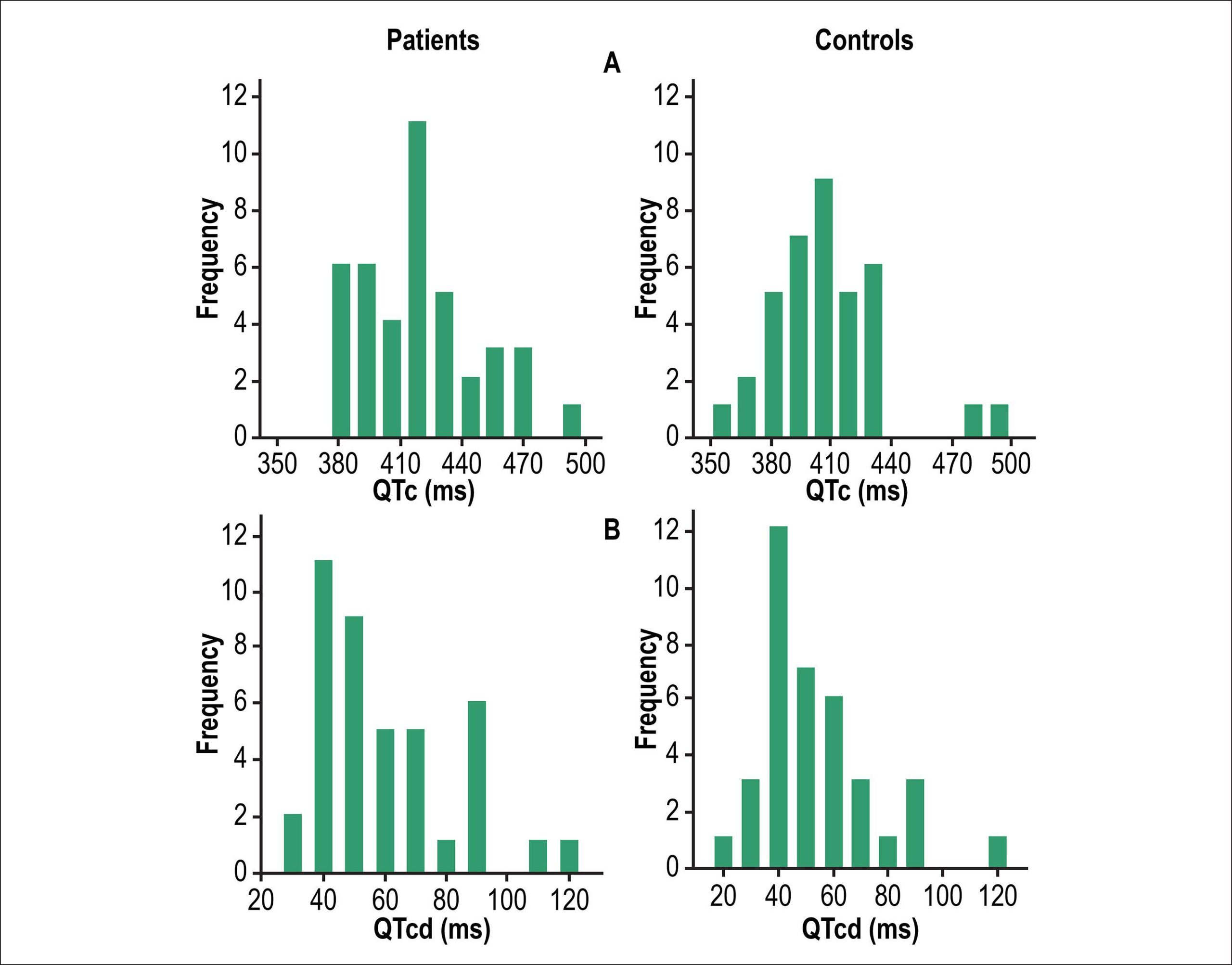
to evaluate the reproducibility and reliability of measurements of corrected QT interval (QTc) and its dispersion (QTcd) and correlate these parameters with LVH in hemodialysis patients.

Methods:

Case-control study involving hemodialysis patients and a control group. Clinical examination, blood sampling, transthoracic echocardiogram, and electrocardiogram were performed. Intra- and interobserver correlation and concordance tests were performed by Pearson´s correlation, Cohen’s Kappa coefficient and Bland Altman diagram. Linear regression was used to analyze association of QTc or QTcd with HVE.

Results:

Forty-one HD patients and 37 controls concluded the study. Hemodialysis patients tended to have higher values of QTc, QTcd and left ventricular mass index (LVMi) than controls but statistical significance was not found. Correlation and concordance tests depicted better results for QTc than for QTcd. In HD patients, a poor but significant correlation was found between QTc and LVMi (R2 = 0.12; p = 0.03). No correlation was found between values of QTcd and LVMi (R2= 0.00; p=0.940). For the control group, the correspondent values were R2= 0.00; p = 0.67 and R2= 0.00; p = 0.94, respectively.

Conclusion:

We found that QTc interval, in contrast to QTcd, is a reproducible and reliable measure and had a weak but positive correlation with LVMi in HD patients.

  • Cardiomyopathy, Hypertrophic
  • Coronary Artery Disease
  • Electrocardiography
  • Hypertrophy Left Ventricular
  • renal dialysis
Read Full Article
Creative Commons Attribution (CC BY) Creative Commons license
07/08/2017 DOI: 10.5935/abc.20170112 Views 1
ABC
Original Article

Association between Clinical and Doppler Echocardiographic Parameters with Sudden Death in Hemodialysis Patients

Silvio Henrique Barberato et al. Sérgio Gardano Elias Bucharles Marcia Ferreira Alves Barberato Roberto Pecoits-Filho

Abstract

Background:

Sudden cardiac death (SCD) is the leading cause of death in maintenance hemodialysis (HD) patients, but there is little information about underlying risk factors.

Objectives:

Evaluate the association between clinical and echocardiographic variables with SCD on HD patients.

Methods:

Retrospective nested case-control study on chronic HD patients who were prospectively followed. The primary endpoint was SCD. Variables were compared by Student t test, Mann-Whitney or Chi-Square, and independent predictors of SCD were evidenced by multivariate logistic regression.

Results:

We followed 153 patients (50 ± 15 years, 58% men) for 23 ± 14 months and observed 35 deaths, 17 of which were SCD events. When compared to the control group (matched for gender, age, and body mass index) there were no differences regarding time on dialysis, traditional biochemical parameters, blood pressure, smoking, use of cardiovascular protective drugs, ejection fraction, left ventricular dimensions, and diastolic function indices. On the other hand, in the SCD group, we found a higher prevalence of previous heart failure, acute myocardial infarction and diabetes, greater left ventricular mass index, greater left atrial size and lower global myocardial performance. After multivariate logistic regression analysis, diabetes (OR = 2.6; CI = 1.3-7.5; p = 0.023) and left ventricular mass index ≥ 101 g/m2.7 (OR = 1.04; CI = 1.01-1.08; p = 0.028) showed independent association with SCD events.

Conclusions:

HD patients with diabetes mellitus and left ventricular hypertrophy appear to have the highest risk of SCD. Preventive and therapeutic strategies should be encouraged in addressing these risk factors to minimize the occurrence of SCD in HD patients.

  • Death, Sudden, Cardiac
  • Echocardiography, Doppler
  • Hypertrophy Left Ventricular
  • renal dialysis
  • Risk Factors
Read Full Article
Creative Commons Attribution (CC BY) Creative Commons license
11/07/2016 DOI: 10.5935/abc.20160098 Views 1
Figure 1
ABC
Original Article

Screening for Fabry Disease in Left Ventricular Hypertrophy: Documentation of a Novel Mutation

Ana Baptista et al. Pedro Magalhães Sílvia Leão Sofia Carvalho … Pedro Mateus Ilídio Moreira

Abstract

Background:

Fabry disease is a lysosomal storage disease caused by enzyme α-galactosidase A deficiency as a result of mutations in the GLA gene. Cardiac involvement is characterized by progressive left ventricular hypertrophy.

Objective:

To estimate the prevalence of Fabry disease in a population with left ventricular hypertrophy.

Methods:

The patients were assessed for the presence of left ventricular hypertrophy defined as a left ventricular mass index ≥ 96 g/m2 for women or ≥ 116 g/m2 for men. Severe aortic stenosis and arterial hypertension with mild left ventricular hypertrophy were exclusion criteria. All patients included were assessed for enzyme α-galactosidase A activity using dry spot testing. Genetic study was performed whenever the enzyme activity was decreased.

Results:

A total of 47 patients with a mean left ventricular mass index of 141.1 g/m2 (± 28.5; 99.2 to 228.5 g/m2] were included. Most of the patients were females (51.1%). Nine (19.1%) showed decreased α-galactosidase A activity, but only one positive genetic test − [GLA] c.785G>T; p.W262L (exon 5), a mutation not previously described in the literature. This clinical investigation was able to establish the association between the mutation and the clinical presentation.

Conclusion:

In a population of patients with left ventricular hypertrophy, we documented a Fabry disease prevalence of 2.1%. This novel case was defined in the sequence of a mutation of unknown meaning in the GLA gene with further pathogenicity study. Thus, this study permitted the definition of a novel causal mutation for Fabry disease – [GLA] c.785G>T; p.W262L (exon 5).

  • Alpha-Galactosidase / genetics
  • Fabry disease / complications
  • Hypertrophy Left Ventricular
Read Full Article
Creative Commons Attribution (CC BY) Creative Commons license
07/08/2015 DOI: 10.5935/abc.20150090 Views 1

Could not load more articles. Please try again.

How to cite this article

EndNote RIS

Electronic Document Format (Vancouver)

Antunes MO. Zebras Exist, Too!. Arquivos Brasileiros de Cardiologia 2024;121(1):e20230834.

Electronic Document Format (ABNT)

Antunes, Murillo de Oliveira. Zebras Exist, Too!. Arquivos Brasileiros de Cardiologia, v. 121, n. 1, e20230834, Mar. 2024.

Electronic Document Format (APA)

Antunes, M. O. (2024). Zebras Exist, Too!. Arquivos Brasileiros de Cardiologia, 121(1), e20230834.

Electronic Document Format (ISO)

Antunes, Murillo de Oliveira. Zebras Exist, Too!. Arquivos Brasileiros de Cardiologia [online]. 2024, vol. 121, n. 1, [cited 2026-10-14], e20230834. Available from: <https://journals.cardiol.br/en/article/zebras-exist-too/>. ISSN 0066-782X

EndNote RIS

Electronic Document Format (Vancouver)

Palmeira NGF, Bianco HT, Bombig MTN, Povoa FF, Fonseca FAH, Izar MC, Thalenberg JM, et al. Association between Morning Surge and Left Ventricular Hypertrophy in Obese Hypertensive Patients. Arquivos Brasileiros de Cardiologia 2023;120(9):e20230050.

Electronic Document Format (ABNT)

Palmeira, Natascha Gonçalves Francisco; Bianco, Henrique Tria; Bombig, Maria Teresa Nogueira; Povoa, Fernando Focaccia; Fonseca, Francisco A. H.; Izar, Maria Cristina; Thalenberg, José Marcos; Luna Filho, Braulio; Marui, Fabiane; Fischer, Simone Matheus; Amodeo, Celso; Souza, Dilma do Socorro Moraes de; Povoa, Rui. Association between Morning Surge and Left Ventricular Hypertrophy in Obese Hypertensive Patients. Arquivos Brasileiros de Cardiologia, v. 120, n. 9, e20230050, Oct. 2023.

Electronic Document Format (APA)

Palmeira, N. G. F., Bianco, H. T., Bombig, M. T. N., Povoa, F. F., Fonseca, F. A. H., Izar, M. C., Thalenberg, J. M., Luna Filho, B., Marui, F., Fischer, S. M., Amodeo, C., Souza, D. S. M. , & Povoa, R. (2023). Association between Morning Surge and Left Ventricular Hypertrophy in Obese Hypertensive Patients. Arquivos Brasileiros de Cardiologia, 120(9), e20230050.

Electronic Document Format (ISO)

Palmeira, Natascha Gonçalves Francisco and Bianco, Henrique Tria and Bombig, Maria Teresa Nogueira and Povoa, Fernando Focaccia and Fonseca, Francisco A. H. and Izar, Maria Cristina and Thalenberg, José Marcos and Luna Filho, Braulio and Marui, Fabiane and Fischer, Simone Matheus and Amodeo, Celso and Souza, Dilma do Socorro Moraes de and Povoa, Rui. Association between Morning Surge and Left Ventricular Hypertrophy in Obese Hypertensive Patients. Arquivos Brasileiros de Cardiologia [online]. 2023, vol. 120, n. 9, [cited 2026-10-14], e20230050. Available from: <https://journals.cardiol.br/en/article/association-between-morning-surge-and-left-ventricular-hypertrophy-in-obese-hypertensive-patients/>. ISSN 0066-782X

EndNote RIS

Electronic Document Format (Vancouver)

Felix RCM, Pedras FHV, Lima Neto OS, Vitório LM. Evaluation of Coronary Flow Reserve by Myocardial Scintigraphy in Left Ventricular Hypertrophy. ABC Imagem Cardiovascular 2023;36(3):e20230053.

Electronic Document Format (ABNT)

Felix, Renata Christian Martins; Pedras, Felipe Hermely Villela; Lima Neto, Odorico de Souza; Vitório, Leonardo Medeiros. Evaluation of Coronary Flow Reserve by Myocardial Scintigraphy in Left Ventricular Hypertrophy. ABC Imagem Cardiovascular, v. 36, n. 3, e20230053, Sep. 2023.

Electronic Document Format (APA)

Felix, R. C. M., Pedras, F. H. V., Lima Neto, O. S., & Vitório, L. M. (2023). Evaluation of Coronary Flow Reserve by Myocardial Scintigraphy in Left Ventricular Hypertrophy. ABC Imagem Cardiovascular, 36(3), e20230053.

Electronic Document Format (ISO)

Felix, Renata Christian Martins and Pedras, Felipe Hermely Villela and Lima Neto, Odorico de Souza and Vitório, Leonardo Medeiros. Evaluation of Coronary Flow Reserve by Myocardial Scintigraphy in Left Ventricular Hypertrophy. ABC Imagem Cardiovascular [online]. 2023, vol. 36, n. 3, [cited 2026-10-14], e20230053. Available from: <https://journals.cardiol.br/en/article/evaluation-of-coronary-flow-reserve-by-myocardial-scintigraphy-in-left-ventricular-hypertrophy/>. ISSN 2318-8219

EndNote RIS

Electronic Document Format (Vancouver)

Sternick EB. PRKAG2 Cardiomyopathy. Arquivos Brasileiros de Cardiologia 2022;119(5):689-90.

Electronic Document Format (ABNT)

Sternick, Eduardo Back. PRKAG2 Cardiomyopathy. Arquivos Brasileiros de Cardiologia, v. 119, n. 5, p. 689-690, Nov. 2022.

Electronic Document Format (APA)

Sternick, E. B. (2022). PRKAG2 Cardiomyopathy. Arquivos Brasileiros de Cardiologia, 119(5), 689-690.

Electronic Document Format (ISO)

Sternick, Eduardo Back. PRKAG2 Cardiomyopathy. Arquivos Brasileiros de Cardiologia [online]. 2022, vol. 119, n. 5, [cited 2026-10-14], pp. 689-690. Available from: <https://journals.cardiol.br/en/article/prkag2-cardiomyopathy/>. ISSN 0066-782X

EndNote RIS

Electronic Document Format (Vancouver)

Magalhães EFS, Magalhães LP, Pinheiro JO, Guabiru AT, Aras R. Atrial Flutter in PRKAG2 Syndrome: Clinical and Electrophysiological Characteristics. Arquivos Brasileiros de Cardiologia 2022;119(5):681-8.

Electronic Document Format (ABNT)

Magalhães, Eduardo Faria Soares de; Magalhães, Luiz Pereira de; Pinheiro, Jussara de Oliveira; Guabiru, Alex Teixeira; Aras, Roque. Atrial Flutter in PRKAG2 Syndrome: Clinical and Electrophysiological Characteristics. Arquivos Brasileiros de Cardiologia, v. 119, n. 5, p. 681-688, Sep. 2022.

Electronic Document Format (APA)

Magalhães, E. F. S. , Magalhães, L. P. , Pinheiro, J. O., Guabiru, A. T., & Aras, R. (2022). Atrial Flutter in PRKAG2 Syndrome: Clinical and Electrophysiological Characteristics. Arquivos Brasileiros de Cardiologia, 119(5), 681-688.

Electronic Document Format (ISO)

Magalhães, Eduardo Faria Soares de and Magalhães, Luiz Pereira de and Pinheiro, Jussara de Oliveira and Guabiru, Alex Teixeira and Aras, Roque. Atrial Flutter in PRKAG2 Syndrome: Clinical and Electrophysiological Characteristics. Arquivos Brasileiros de Cardiologia [online]. 2022, vol. 119, n. 5, [cited 2026-10-14], pp. 681-688. Available from: <https://journals.cardiol.br/en/article/atrial-flutter-in-prkag2-syndrome-clinical-and-electrophysiological-characteristics/>. ISSN 0066-782X

EndNote RIS

Electronic Document Format (Vancouver)

Silva TO, Ritt LEF. Can Simple Echocardiographic Parameters Replace The ASCVD Probabilistic Model Calculation?. Arquivos Brasileiros de Cardiologia 2022;118(5):925-6.

Electronic Document Format (ABNT)

Silva, Tonnison de Oliveira; Ritt, Luiz Eduardo Fonteles. Can Simple Echocardiographic Parameters Replace The ASCVD Probabilistic Model Calculation?. Arquivos Brasileiros de Cardiologia, v. 118, n. 5, p. 925-926, May. 2022.

Electronic Document Format (APA)

Silva, T. O., & Ritt, L. E. F. (2022). Can Simple Echocardiographic Parameters Replace The ASCVD Probabilistic Model Calculation?. Arquivos Brasileiros de Cardiologia, 118(5), 925-926.

Electronic Document Format (ISO)

Silva, Tonnison de Oliveira and Ritt, Luiz Eduardo Fonteles. Can Simple Echocardiographic Parameters Replace The ASCVD Probabilistic Model Calculation?. Arquivos Brasileiros de Cardiologia [online]. 2022, vol. 118, n. 5, [cited 2026-10-14], pp. 925-926. Available from: <https://journals.cardiol.br/en/article/can-simple-echocardiographic-parameters-replace-the-ascvd-probabilistic-model-calculation/>. ISSN 0066-782X

EndNote RIS

Electronic Document Format (Vancouver)

Magalhães LP, Magalhães EFS, Pinheiro JO, Guabiru AT, Reis FJFB, Aras R. Long-Term Cardiac Complications of PRKAG2 Syndrome. Arquivos Brasileiros de Cardiologia 2022;118(1):106-9.

Electronic Document Format (ABNT)

Magalhães, Luiz Pereira de; Magalhães, Eduardo Faria Soares de; Pinheiro, Jussara de Oliveira; Guabiru, Alex Teixeira; Reis, Francisco José Farias Borges dos; Aras, Roque. Long-Term Cardiac Complications of PRKAG2 Syndrome. Arquivos Brasileiros de Cardiologia, v. 118, n. 1, p. 106-109, Feb. 2022.

Electronic Document Format (APA)

Magalhães, L. P. , Magalhães, E. F. S. , Pinheiro, J. O., Guabiru, A. T., Reis, F. J. F. B. , & Aras, R. (2022). Long-Term Cardiac Complications of PRKAG2 Syndrome. Arquivos Brasileiros de Cardiologia, 118(1), 106-109.

Electronic Document Format (ISO)

Magalhães, Luiz Pereira de and Magalhães, Eduardo Faria Soares de and Pinheiro, Jussara de Oliveira and Guabiru, Alex Teixeira and Reis, Francisco José Farias Borges dos and Aras, Roque. Long-Term Cardiac Complications of PRKAG2 Syndrome. Arquivos Brasileiros de Cardiologia [online]. 2022, vol. 118, n. 1, [cited 2026-10-14], pp. 106-109. Available from: <https://journals.cardiol.br/en/article/long-term-cardiac-complications-of-prkag2-syndrome/>. ISSN 0066-782X

EndNote RIS

Electronic Document Format (Vancouver)

Rodrigues P, Soares AR, Taipa R, Ferreira S, Reis H. Left Ventricular Hypertrophy: One Phenotype, Two Hypotheses, Three Lessons. Arquivos Brasileiros de Cardiologia 2021;117(5):1056-9.

Electronic Document Format (ABNT)

Rodrigues, Patrícia; Soares, Ana Rita; Taipa, Ricardo; Ferreira, Sofia; Reis, Hipólito. Left Ventricular Hypertrophy: One Phenotype, Two Hypotheses, Three Lessons. Arquivos Brasileiros de Cardiologia, v. 117, n. 5, p. 1056-1059, Nov. 2021.

Electronic Document Format (APA)

Rodrigues, P., Soares, A. R., Taipa, R., Ferreira, S., & Reis, H. (2021). Left Ventricular Hypertrophy: One Phenotype, Two Hypotheses, Three Lessons. Arquivos Brasileiros de Cardiologia, 117(5), 1056-1059.

Electronic Document Format (ISO)

Rodrigues, Patrícia and Soares, Ana Rita and Taipa, Ricardo and Ferreira, Sofia and Reis, Hipólito. Left Ventricular Hypertrophy: One Phenotype, Two Hypotheses, Three Lessons. Arquivos Brasileiros de Cardiologia [online]. 2021, vol. 117, n. 5, [cited 2026-10-14], pp. 1056-1059. Available from: <https://journals.cardiol.br/en/article/left-ventricular-hypertrophy-one-phenotype-two-hypotheses-three-lessons/>. ISSN 0066-782X

EndNote RIS

Electronic Document Format (Vancouver)

Povoa FF, Luna Filho B, Bianco HT, Amodeo C, Povoa R, Bombig MTN, Miranda RD, et al. Performance of the Electrocardiogram in the Diagnosis of Left Ventricular Hypertrophy in Older and Very Older Hypertensive Patients. Arquivos Brasileiros de Cardiologia 2021;117(5):924-31.

Electronic Document Format (ABNT)

Povoa, Fernando Focaccia; Luna Filho, Braulio; Bianco, Henrique Tria; Amodeo, Celso; Povoa, Rui; Bombig, Maria Teresa Nogueira; Miranda, Roberto Dischinger; Fischer, Simone Matheus; Izar, Maria Cristina Oliveira; Fonseca, Francisco A. H.. Performance of the Electrocardiogram in the Diagnosis of Left Ventricular Hypertrophy in Older and Very Older Hypertensive Patients. Arquivos Brasileiros de Cardiologia, v. 117, n. 5, p. 924-931, Aug. 2021.

Electronic Document Format (APA)

Povoa, F. F., Luna Filho, B., Bianco, H. T., Amodeo, C., Povoa, R., Bombig, M. T. N., Miranda, R. D., Fischer, S. M., Izar, M. C. O., & Fonseca, F. A. H. (2021). Performance of the Electrocardiogram in the Diagnosis of Left Ventricular Hypertrophy in Older and Very Older Hypertensive Patients. Arquivos Brasileiros de Cardiologia, 117(5), 924-931.

Electronic Document Format (ISO)

Povoa, Fernando Focaccia and Luna Filho, Braulio and Bianco, Henrique Tria and Amodeo, Celso and Povoa, Rui and Bombig, Maria Teresa Nogueira and Miranda, Roberto Dischinger and Fischer, Simone Matheus and Izar, Maria Cristina Oliveira and Fonseca, Francisco A. H.. Performance of the Electrocardiogram in the Diagnosis of Left Ventricular Hypertrophy in Older and Very Older Hypertensive Patients. Arquivos Brasileiros de Cardiologia [online]. 2021, vol. 117, n. 5, [cited 2026-10-14], pp. 924-931. Available from: <https://journals.cardiol.br/en/article/performance-of-the-electrocardiogram-in-the-diagnosis-of-left-ventricular-hypertrophy-in-older-and-very-older-hypertensive-patients/>. ISSN 0066-782X

EndNote RIS

Electronic Document Format (Vancouver)

Bhardwaj A, Parikh VY, Nair A. Transcatheter Interatrial Shunts for the Treatment of Heart Failure with Preserved Ejection Fraction. International Journal of Cardiovascular Sciences 2021;34(1):81-8.

Electronic Document Format (ABNT)

Bhardwaj, Anju; Parikh, Vishal Y.; Nair, Ajith. Transcatheter Interatrial Shunts for the Treatment of Heart Failure with Preserved Ejection Fraction. International Journal of Cardiovascular Sciences, v. 34, n. 1, p. 81-88, Jan. 2021.

Electronic Document Format (APA)

Bhardwaj, A., Parikh, V. Y., & Nair, A. (2021). Transcatheter Interatrial Shunts for the Treatment of Heart Failure with Preserved Ejection Fraction. International Journal of Cardiovascular Sciences, 34(1), 81-88.

Electronic Document Format (ISO)

Bhardwaj, Anju and Parikh, Vishal Y. and Nair, Ajith. Transcatheter Interatrial Shunts for the Treatment of Heart Failure with Preserved Ejection Fraction. International Journal of Cardiovascular Sciences [online]. 2021, vol. 34, n. 1, [cited 2026-10-14], pp. 81-88. Available from: <https://journals.cardiol.br/en/article/transcatheter-interatrial-shunts-for-the-treatment-of-heart-failure-with-preserved-ejection-fraction/>. ISSN 2359-4802

EndNote RIS

Electronic Document Format (Vancouver)

Roque EAC. Which Risk Score Best Assesses Clinical Objectives in Patients with Hypertrophic Cardiomyopathy?. Arquivos Brasileiros de Cardiologia 2020;115(2):205-6.

Electronic Document Format (ABNT)

Roque, Eduardo Alberto de Castro. Which Risk Score Best Assesses Clinical Objectives in Patients with Hypertrophic Cardiomyopathy?. Arquivos Brasileiros de Cardiologia, v. 115, n. 2, p. 205-206, Aug. 2020.

Electronic Document Format (APA)

Roque, E. A. C. (2020). Which Risk Score Best Assesses Clinical Objectives in Patients with Hypertrophic Cardiomyopathy?. Arquivos Brasileiros de Cardiologia, 115(2), 205-206.

Electronic Document Format (ISO)

Roque, Eduardo Alberto de Castro. Which Risk Score Best Assesses Clinical Objectives in Patients with Hypertrophic Cardiomyopathy?. Arquivos Brasileiros de Cardiologia [online]. 2020, vol. 115, n. 2, [cited 2026-10-14], pp. 205-206. Available from: <https://journals.cardiol.br/en/article/which-risk-score-best-assesses-clinical-objectives-in-patients-with-hypertrophic-cardiomyopathy/>. ISSN 0066-782X

EndNote RIS

Electronic Document Format (Vancouver)

Yavari N, Landy MG, Omidi N, Shirzad M, Tafti SHA. Mitral Valve Replacement with Regent Aortic Valve in Severe Mitral Stenosis. International Journal of Cardiovascular Sciences 2020;34(1):103-6.

Electronic Document Format (ABNT)

Yavari, Negin; Landy, Mina Ghorbanpour; Omidi, Negar; Shirzad, Mahmoud; Tafti, Seyed Hossein Ahmadi. Mitral Valve Replacement with Regent Aortic Valve in Severe Mitral Stenosis. International Journal of Cardiovascular Sciences, v. 34, n. 1, p. 103-106, Jul. 2020.

Electronic Document Format (APA)

Yavari, N., Landy, M. G., Omidi, N., Shirzad, M., & Tafti, S. H. A. (2020). Mitral Valve Replacement with Regent Aortic Valve in Severe Mitral Stenosis. International Journal of Cardiovascular Sciences, 34(1), 103-106.

Electronic Document Format (ISO)

Yavari, Negin and Landy, Mina Ghorbanpour and Omidi, Negar and Shirzad, Mahmoud and Tafti, Seyed Hossein Ahmadi. Mitral Valve Replacement with Regent Aortic Valve in Severe Mitral Stenosis. International Journal of Cardiovascular Sciences [online]. 2020, vol. 34, n. 1, [cited 2026-10-14], pp. 103-106. Available from: <https://journals.cardiol.br/en/article/mitral-valve-replacement-with-regent-aortic-valve-in-severe-mitral-stenosis/>. ISSN 2359-4802

EndNote RIS

Electronic Document Format (Vancouver)

Saraiva RM. Do We Need to Know the Left Ventricular Geometry Patterns of the Brazilian Population?. Arquivos Brasileiros de Cardiologia 2020;114(1):66-7.

Electronic Document Format (ABNT)

Saraiva, Roberto M.. Do We Need to Know the Left Ventricular Geometry Patterns of the Brazilian Population?. Arquivos Brasileiros de Cardiologia, v. 114, n. 1, p. 66-67, Feb. 2020.

Electronic Document Format (APA)

Saraiva, R. M. (2020). Do We Need to Know the Left Ventricular Geometry Patterns of the Brazilian Population?. Arquivos Brasileiros de Cardiologia, 114(1), 66-67.

Electronic Document Format (ISO)

Saraiva, Roberto M.. Do We Need to Know the Left Ventricular Geometry Patterns of the Brazilian Population?. Arquivos Brasileiros de Cardiologia [online]. 2020, vol. 114, n. 1, [cited 2026-10-14], pp. 66-67. Available from: <https://journals.cardiol.br/en/article/do-we-need-to-know-the-left-ventricular-geometry-patterns-of-the-brazilian-population/>. ISSN 0066-782X

EndNote RIS

Electronic Document Format (Vancouver)

Almeida RCM, Jorge AJL, Rosa MLG, Leite AR, Correia DMS, Mesquita ET, Chermont S, et al. Left Ventricular Remodeling Patterns in Primary Healthcare. Arquivos Brasileiros de Cardiologia 2020;114(1):59-65.

Electronic Document Format (ABNT)

Almeida, Roberto de Castro Meirelles de; Jorge, Antonio José Lagoeiro; Rosa, Maria Luiza Garcia; Leite, Adson Renato; Correia, Dayse Mary S.; Mesquita, Evandro Tinoco; Chermont, Sergio; Lugon, Jocemir Ronaldo; Martins, Wolney de Andrade. Left Ventricular Remodeling Patterns in Primary Healthcare. Arquivos Brasileiros de Cardiologia, v. 114, n. 1, p. 59-65, Feb. 2020.

Electronic Document Format (APA)

Almeida, R. C. M. , Jorge, A. J. L., Rosa, M. L. G., Leite, A. R., Correia, D. M. S., Mesquita, E. T., Chermont, S., Lugon, J. R., & Martins, W. A. (2020). Left Ventricular Remodeling Patterns in Primary Healthcare. Arquivos Brasileiros de Cardiologia, 114(1), 59-65.

Electronic Document Format (ISO)

Almeida, Roberto de Castro Meirelles de and Jorge, Antonio José Lagoeiro and Rosa, Maria Luiza Garcia and Leite, Adson Renato and Correia, Dayse Mary S. and Mesquita, Evandro Tinoco and Chermont, Sergio and Lugon, Jocemir Ronaldo and Martins, Wolney de Andrade. Left Ventricular Remodeling Patterns in Primary Healthcare. Arquivos Brasileiros de Cardiologia [online]. 2020, vol. 114, n. 1, [cited 2026-10-14], pp. 59-65. Available from: <https://journals.cardiol.br/en/article/left-ventricular-remodeling-patterns-in-primary-healthcare/>. ISSN 0066-782X

EndNote RIS

Electronic Document Format (Vancouver)

Correia ETO, Barbetta LMS. Left Ventricle Mass Index, a Confounding Variable of Global Longitudinal Strain to be Noticed. Arquivos Brasileiros de Cardiologia 2020;114(1):136.

Electronic Document Format (ABNT)

Correia, Eduardo Thadeu de Oliveira; Barbetta, Letícia Mara dos Santos. Left Ventricle Mass Index, a Confounding Variable of Global Longitudinal Strain to be Noticed. Arquivos Brasileiros de Cardiologia, v. 114, n. 1, p. 136-136, Feb. 2020.

Electronic Document Format (APA)

Correia, E. T. O., & Barbetta, L. M. S. (2020). Left Ventricle Mass Index, a Confounding Variable of Global Longitudinal Strain to be Noticed. Arquivos Brasileiros de Cardiologia, 114(1), 136-136.

Electronic Document Format (ISO)

Correia, Eduardo Thadeu de Oliveira and Barbetta, Letícia Mara dos Santos. Left Ventricle Mass Index, a Confounding Variable of Global Longitudinal Strain to be Noticed. Arquivos Brasileiros de Cardiologia [online]. 2020, vol. 114, n. 1, [cited 2026-10-14], pp. 136-136. Available from: <https://journals.cardiol.br/en/article/left-ventricle-mass-index-a-confounding-variable-of-global-longitudinal-strain-to-be-noticed/>. ISSN 0066-782X

EndNote RIS

Electronic Document Format (Vancouver)

Barreto J, Roda J, Germano CW, Damiano AP, Quinaglia T. Coarctation of The Aorta: A Case-Series from a Tertiary Care Hospital. International Journal of Cardiovascular Sciences 2019;33(1):3-11.

Electronic Document Format (ABNT)

Barreto, Joaquim; Roda, Juliana; Germano, Carlos Wustemberg; Damiano, Ana Paula; Quinaglia, Thiago. Coarctation of The Aorta: A Case-Series from a Tertiary Care Hospital. International Journal of Cardiovascular Sciences, v. 33, n. 1, p. 3-11, Aug. 2019.

Electronic Document Format (APA)

Barreto, J., Roda, J., Germano, C. W., Damiano, A. P., & Quinaglia, T. (2019). Coarctation of The Aorta: A Case-Series from a Tertiary Care Hospital. International Journal of Cardiovascular Sciences, 33(1), 3-11.

Electronic Document Format (ISO)

Barreto, Joaquim and Roda, Juliana and Germano, Carlos Wustemberg and Damiano, Ana Paula and Quinaglia, Thiago. Coarctation of The Aorta: A Case-Series from a Tertiary Care Hospital. International Journal of Cardiovascular Sciences [online]. 2019, vol. 33, n. 1, [cited 2026-10-14], pp. 3-11. Available from: <https://journals.cardiol.br/en/article/coarctation-of-the-aorta-a-case-series-from-a-tertiary-care-hospital/>. ISSN 2359-4802

EndNote RIS

Electronic Document Format (Vancouver)

Chaves-Markman V, Markman M, Calado EB, Pires RF, Santos-Veloso MAO, Pereira CMF, Lordsleem ABMS, et al. GLA Gene Mutation in Hypertrophic Cardiomyopathy with a New Variant Description: Is it Fabry’s Disease?. Arquivos Brasileiros de Cardiologia 2019;113(1):77-84.

Electronic Document Format (ABNT)

Chaves-Markman, Ândrea Virgínia; Markman, Manuel; Calado, Eveline Barros; Pires, Ricardo Flores; Santos-Veloso, Marcelo Antônio Oliveira; Pereira, Catarina Maria Fonseca; Lordsleem, Andréa Bezerra de Melo da Silveira; Lima, Sandro Gonçalves de; Markman Filho, Brivaldo; Oliveira, Dinaldo Cavalcanti de. GLA Gene Mutation in Hypertrophic Cardiomyopathy with a New Variant Description: Is it Fabry’s Disease?. Arquivos Brasileiros de Cardiologia, v. 113, n. 1, p. 77-84, Jul. 2019.

Electronic Document Format (APA)

Chaves-Markman, V., Markman, M., Calado, E. B., Pires, R. F., Santos-Veloso, M. A. O., Pereira, C. M. F., Lordsleem, A. B. M. S., Lima, S. G. , Markman Filho, B., & Oliveira, D. C. (2019). GLA Gene Mutation in Hypertrophic Cardiomyopathy with a New Variant Description: Is it Fabry’s Disease?. Arquivos Brasileiros de Cardiologia, 113(1), 77-84.

Electronic Document Format (ISO)

Chaves-Markman, Ândrea Virgínia and Markman, Manuel and Calado, Eveline Barros and Pires, Ricardo Flores and Santos-Veloso, Marcelo Antônio Oliveira and Pereira, Catarina Maria Fonseca and Lordsleem, Andréa Bezerra de Melo da Silveira and Lima, Sandro Gonçalves de and Markman Filho, Brivaldo and Oliveira, Dinaldo Cavalcanti de. GLA Gene Mutation in Hypertrophic Cardiomyopathy with a New Variant Description: Is it Fabry’s Disease?. Arquivos Brasileiros de Cardiologia [online]. 2019, vol. 113, n. 1, [cited 2026-10-14], pp. 77-84. Available from: <https://journals.cardiol.br/en/article/gla-gene-mutation-in-hypertrophic-cardiomyopathy-with-a-new-variant-description-is-it-fabrys-disease/>. ISSN 0066-782X

EndNote RIS

Electronic Document Format (Vancouver)

Mourilhe-Rocha R, Salvino NFA. Impact of Obesity and Bariatric Surgery in the Outcome of Patients with Heart Failure. International Journal of Cardiovascular Sciences 2019;32(5):527-35.

Electronic Document Format (ABNT)

Mourilhe-Rocha, Ricardo; Salvino, Nathália Felix Araujo. Impact of Obesity and Bariatric Surgery in the Outcome of Patients with Heart Failure. International Journal of Cardiovascular Sciences, v. 32, n. 5, p. 527-535, Feb. 2019.

Electronic Document Format (APA)

Mourilhe-Rocha, R., & Salvino, N. F. A. (2019). Impact of Obesity and Bariatric Surgery in the Outcome of Patients with Heart Failure. International Journal of Cardiovascular Sciences, 32(5), 527-535.

Electronic Document Format (ISO)

Mourilhe-Rocha, Ricardo and Salvino, Nathália Felix Araujo. Impact of Obesity and Bariatric Surgery in the Outcome of Patients with Heart Failure. International Journal of Cardiovascular Sciences [online]. 2019, vol. 32, n. 5, [cited 2026-10-14], pp. 527-535. Available from: <https://journals.cardiol.br/en/article/impact-of-obesity-and-bariatric-surgery-in-the-outcome-of-patients-with-heart-failure/>. ISSN 2359-4802

EndNote RIS

Electronic Document Format (Vancouver)

Yasri S, Wiwanitkit V. QTc and QTcd Measurements and Their Relationships with Left Ventricular Hypertrophy in Hemodialysis Patients. Arquivos Brasileiros de Cardiologia 2018;110(6):602.

Electronic Document Format (ABNT)

Yasri, Sora; Wiwanitkit, Viroj. QTc and QTcd Measurements and Their Relationships with Left Ventricular Hypertrophy in Hemodialysis Patients. Arquivos Brasileiros de Cardiologia, v. 110, n. 6, p. 602-602, May. 2018.

Electronic Document Format (APA)

Yasri, S., & Wiwanitkit, V. (2018). QTc and QTcd Measurements and Their Relationships with Left Ventricular Hypertrophy in Hemodialysis Patients. Arquivos Brasileiros de Cardiologia, 110(6), 602-602.

Electronic Document Format (ISO)

Yasri, Sora and Wiwanitkit, Viroj. QTc and QTcd Measurements and Their Relationships with Left Ventricular Hypertrophy in Hemodialysis Patients. Arquivos Brasileiros de Cardiologia [online]. 2018, vol. 110, n. 6, [cited 2026-10-14], pp. 602-602. Available from: <https://journals.cardiol.br/en/article/qtc-and-qtcd-measurements-and-their-relationships-with-left-ventricular-hypertrophy-in-hemodialysis-patients/>. ISSN 0066-782X

EndNote RIS

Electronic Document Format (Vancouver)

Jorge AJL, Ávila DX, Vilar EG, Ribeiro ML, Bruno KEH, Pires AC. Cardiac Amyloidosis with Heart Failure and Middle Range Ejection Fraction. International Journal of Cardiovascular Sciences 2018;31(4):457-60.

Electronic Document Format (ABNT)

Jorge, Antonio Jose Lagoeiro; Ávila, Diane Xavier de; Vilar, Enoï Guedes; Ribeiro, Mario Luiz; Bruno, Karima Elias Hallack; Pires, Ana Carolina. Cardiac Amyloidosis with Heart Failure and Middle Range Ejection Fraction. International Journal of Cardiovascular Sciences, v. 31, n. 4, p. 457-460, Dec. 2017.

Electronic Document Format (APA)

Jorge, A. J. L., Ávila, D. X. , Vilar, E. G., Ribeiro, M. L., Bruno, K. E. H., & Pires, A. C. (2018). Cardiac Amyloidosis with Heart Failure and Middle Range Ejection Fraction. International Journal of Cardiovascular Sciences, 31(4), 457-460.

Electronic Document Format (ISO)

Jorge, Antonio Jose Lagoeiro and Ávila, Diane Xavier de and Vilar, Enoï Guedes and Ribeiro, Mario Luiz and Bruno, Karima Elias Hallack and Pires, Ana Carolina. Cardiac Amyloidosis with Heart Failure and Middle Range Ejection Fraction. International Journal of Cardiovascular Sciences [online]. 2018, vol. 31, n. 4, [cited 2026-10-14], pp. 457-460. Available from: <https://journals.cardiol.br/en/article/cardiac-amyloidosis-with-heart-failure-and-middle-range-ejection-fraction/>. ISSN 2359-4802

EndNote RIS

Electronic Document Format (Vancouver)

Sousa Filho IR, Pereira ICC, Morais LJ, Teodoro LGVL, Rodrigues MLP, Gomes RAS. Pericardial Parietal Mesothelial Cells: Source of the Angiotensin-Converting-Enzyme of the Bovine Pericardial Fluid. Arquivos Brasileiros de Cardiologia 2017;109(5):425-31.

Electronic Document Format (ABNT)

Sousa Filho, Ilsione Ribeiro de; Pereira, Isabela Cabral Cavicchioli; Morais, Lourimar José de; Teodoro, Livia das Graças Vieito Lombardi; Rodrigues, Maria Laura Pinto; Gomes, Roseli Aparecida da Silva. Pericardial Parietal Mesothelial Cells: Source of the Angiotensin-Converting-Enzyme of the Bovine Pericardial Fluid. Arquivos Brasileiros de Cardiologia, v. 109, n. 5, p. 425-431, Oct. 2017.

Electronic Document Format (APA)

Sousa Filho, I. R. , Pereira, I. C. C., Morais, L. J. , Teodoro, L. G. V. L., Rodrigues, M. L. P., & Gomes, R. A. S. (2017). Pericardial Parietal Mesothelial Cells: Source of the Angiotensin-Converting-Enzyme of the Bovine Pericardial Fluid. Arquivos Brasileiros de Cardiologia, 109(5), 425-431.

Electronic Document Format (ISO)

Sousa Filho, Ilsione Ribeiro de and Pereira, Isabela Cabral Cavicchioli and Morais, Lourimar José de and Teodoro, Livia das Graças Vieito Lombardi and Rodrigues, Maria Laura Pinto and Gomes, Roseli Aparecida da Silva. Pericardial Parietal Mesothelial Cells: Source of the Angiotensin-Converting-Enzyme of the Bovine Pericardial Fluid. Arquivos Brasileiros de Cardiologia [online]. 2017, vol. 109, n. 5, [cited 2026-10-14], pp. 425-431. Available from: <https://journals.cardiol.br/en/article/pericardial-parietal-mesothelial-cells-source-of-the-angiotensin-converting-enzyme-of-the-bovine-pericardial-fluid/>. ISSN 0066-782X

EndNote RIS

Electronic Document Format (Vancouver)

Alonso MAG, Lima VACC, Carreira MAMQ, Lugon JR. Reproducibility and Reliability Of QTc and QTcd Measurements and Their Relationships with Left Ventricular Hypertrophy in Hemodialysis Patients. Arquivos Brasileiros de Cardiologia 2017;109(3):222-30.

Electronic Document Format (ABNT)

Alonso, Maria Angélica Gonçalves; Lima, Valentine de Almeida Costa de Castro; Carreira, Maria Angela Magalhães de Queiroz; Lugon, Jocemir Ronaldo. Reproducibility and Reliability Of QTc and QTcd Measurements and Their Relationships with Left Ventricular Hypertrophy in Hemodialysis Patients. Arquivos Brasileiros de Cardiologia, v. 109, n. 3, p. 222-230, Aug. 2017.

Electronic Document Format (APA)

Alonso, M. A. G., Lima, V. A. C. C., Carreira, M. A. M. Q., & Lugon, J. R. (2017). Reproducibility and Reliability Of QTc and QTcd Measurements and Their Relationships with Left Ventricular Hypertrophy in Hemodialysis Patients. Arquivos Brasileiros de Cardiologia, 109(3), 222-230.

Electronic Document Format (ISO)

Alonso, Maria Angélica Gonçalves and Lima, Valentine de Almeida Costa de Castro and Carreira, Maria Angela Magalhães de Queiroz and Lugon, Jocemir Ronaldo. Reproducibility and Reliability Of QTc and QTcd Measurements and Their Relationships with Left Ventricular Hypertrophy in Hemodialysis Patients. Arquivos Brasileiros de Cardiologia [online]. 2017, vol. 109, n. 3, [cited 2026-10-14], pp. 222-230. Available from: <https://journals.cardiol.br/en/article/reproducibility-and-reliability-of-qtc-and-qtcd-measurements-and-their-relationships-with-left-ventricular-hypertrophy-in-hemodialysis-patients/>. ISSN 0066-782X

EndNote RIS

Electronic Document Format (Vancouver)

Barberato SH, Bucharles SGE, Barberato MFA, Pecoits-Filho R. Association between Clinical and Doppler Echocardiographic Parameters with Sudden Death in Hemodialysis Patients. Arquivos Brasileiros de Cardiologia 2016;107(2):124-30.

Electronic Document Format (ABNT)

Barberato, Silvio Henrique; Bucharles, Sérgio Gardano Elias; Barberato, Marcia Ferreira Alves; Pecoits-Filho, Roberto. Association between Clinical and Doppler Echocardiographic Parameters with Sudden Death in Hemodialysis Patients. Arquivos Brasileiros de Cardiologia, v. 107, n. 2, p. 124-130, Jul. 2016.

Electronic Document Format (APA)

Barberato, S. H., Bucharles, S. G. E., Barberato, M. F. A., & Pecoits-Filho, R. (2016). Association between Clinical and Doppler Echocardiographic Parameters with Sudden Death in Hemodialysis Patients. Arquivos Brasileiros de Cardiologia, 107(2), 124-130.

Electronic Document Format (ISO)

Barberato, Silvio Henrique and Bucharles, Sérgio Gardano Elias and Barberato, Marcia Ferreira Alves and Pecoits-Filho, Roberto. Association between Clinical and Doppler Echocardiographic Parameters with Sudden Death in Hemodialysis Patients. Arquivos Brasileiros de Cardiologia [online]. 2016, vol. 107, n. 2, [cited 2026-10-14], pp. 124-130. Available from: <https://journals.cardiol.br/en/article/association-between-clinical-and-doppler-echocardiographic-parameters-with-sudden-death-in-hemodialysis-patients/>. ISSN 0066-782X

EndNote RIS

Electronic Document Format (Vancouver)

Baptista A, Magalhães P, Leão S, Carvalho S, Mateus P, Moreira I. Screening for Fabry Disease in Left Ventricular Hypertrophy: Documentation of a Novel Mutation. Arquivos Brasileiros de Cardiologia 2015;105(2):139-44.

Electronic Document Format (ABNT)

Baptista, Ana; Magalhães, Pedro; Leão, Sílvia; Carvalho, Sofia; Mateus, Pedro; Moreira, Ilídio. Screening for Fabry Disease in Left Ventricular Hypertrophy: Documentation of a Novel Mutation. Arquivos Brasileiros de Cardiologia, v. 105, n. 2, p. 139-144, Aug. 2015.

Electronic Document Format (APA)

Baptista, A., Magalhães, P., Leão, S., Carvalho, S., Mateus, P., & Moreira, I. (2015). Screening for Fabry Disease in Left Ventricular Hypertrophy: Documentation of a Novel Mutation. Arquivos Brasileiros de Cardiologia, 105(2), 139-144.

Electronic Document Format (ISO)

Baptista, Ana and Magalhães, Pedro and Leão, Sílvia and Carvalho, Sofia and Mateus, Pedro and Moreira, Ilídio. Screening for Fabry Disease in Left Ventricular Hypertrophy: Documentation of a Novel Mutation. Arquivos Brasileiros de Cardiologia [online]. 2015, vol. 105, n. 2, [cited 2026-10-14], pp. 139-144. Available from: <https://journals.cardiol.br/en/article/screening-for-fabry-disease-in-left-ventricular-hypertrophy-documentation-of-a-novel-mutation/>. ISSN 0066-782X

  • About ABC Family
  • Internal Regulation
  • Editorial Board
  • Brazilian Society of Cardiology (SBC)

Issues

SBC Guidelines

  • Videos
  • Podcasts
  • News