ABC Family Arquivos Brasileiros de Cardiologia International Journal of Cardiovascular Sciences ABC Heart Failure & Cardiomyopathy ABC Imagem Cardiovascular
Português English
ABC Family
Português English
  • About
    • About ABC Family
    • Internal Regulation
    • Editorial Board
    • Brazilian Society of Cardiology (SBC)
  • Issues
  • SBC Guidelines
  • Content
    • Videos
    • Podcasts
    • News
  • About
    • About ABC Family
    • Internal Regulation
    • Editorial Board
    • Brazilian Society of Cardiology (SBC)
  • Issues
  • SBC Guidelines
  • Content
    • Videos
    • Podcasts
    • News

ABC Family

  • ABC Family
  • Arquivos Brasileiros de Cardiologia
  • International Journal of Cardiovascular Sciences
  • ABC Heart Failure & Cardiomyopathy
  • ABC Imagem Cardiovascular
Search in

No content found.

Journals
Ordering
Article types
Others

  1. Home
  2. Keyword: Reference Values
ABC
Editorial

Reference Values in Cardiovascular Magnetic Resonance Imaging: Between Local Validation and Global Harmonization

Miguel Correia

Cardiovascular magnetic resonance imaging (CMR) has become established in recent decades as the gold standard method for cardiac morphofunctional assessment, allowing for the precise quantification of ventricular volumes, ejection fraction, and myocardial mass, as well as the tissue characterization of multiple heart diseases.– The high reproducibility of its measurements is one of its main advantages over other imaging modalities, making CMR an essential tool in both clinical practice and cardiovascular research.,

However, the proper interpretation of parameters obtained by CMR depends on the existence of robust reference values applicable to the studied population. In this context, the article “Normal Reference Values for Cardiovascular Magnetic Resonance: An Analytical–Comparative Review with Emphasis on the Brazilian Population”, published in this issue of the Arquivos Brasileiros de Cardiologia, addresses a question that is particularly relevant to Brazilian cardiology: the applicability of international normative values to the national reality.

[…]

  • Magnetic Resonance Imaging
  • Reference Standards
  • Reference Values
Read Full Article
Creative Commons Attribution (CC BY) Creative Commons license
10/08/2026 DOI: 10.36660/abc.2026458i Views 2
Normal Reference Values for Cardiovascular Magnetic Resonance: An Analytical–Comparative Review with Emphasis on the Brazilian Population
ABC
Original Article

Normal Reference Values for Cardiovascular Magnetic Resonance: An Analytical–Comparative Review with Emphasis on the Brazilian Population

Jorge A. Torreão et al. Isabela Bispo Santos da Silva Costa Antonildes N. Assunção-Jr Ilan Gottlieb … Leonardo Sara da Silva Tiago Senra Tiago Augusto Magalhães Marly Uellendahl Hugo Bizetto Zampa Gabriela Liberato Walther Yoshiharu Ishikawa Henrique Trad Marcelo Hadlich Otavio Coelho-Filho Roberto Sasdelli Neto Juliano Lara Fernandes Luiz Francisco Rodrigues de Ávila Ibraim Masciarelli F. Pinto Paulo R. Schvartzman Carlos E. Rochitte

ABSTRACT

Background:

Cardiovascular magnetic resonance (CMR) is the reference standard for morphofunctional cardiac assessment. In Brazil, the application of international normal reference values faces challenges related to population admixture, technical heterogeneity among centers, and the absence of national standardization, all of which may affect diagnostic accuracy.

Objectives:

To compare normal reference values for morphofunctional parameters obtained by CMR between a multicenter international review and a Brazilian publication, discussing concordance, methodological differences, and implications for Brazilian clinical practice.

Methods:

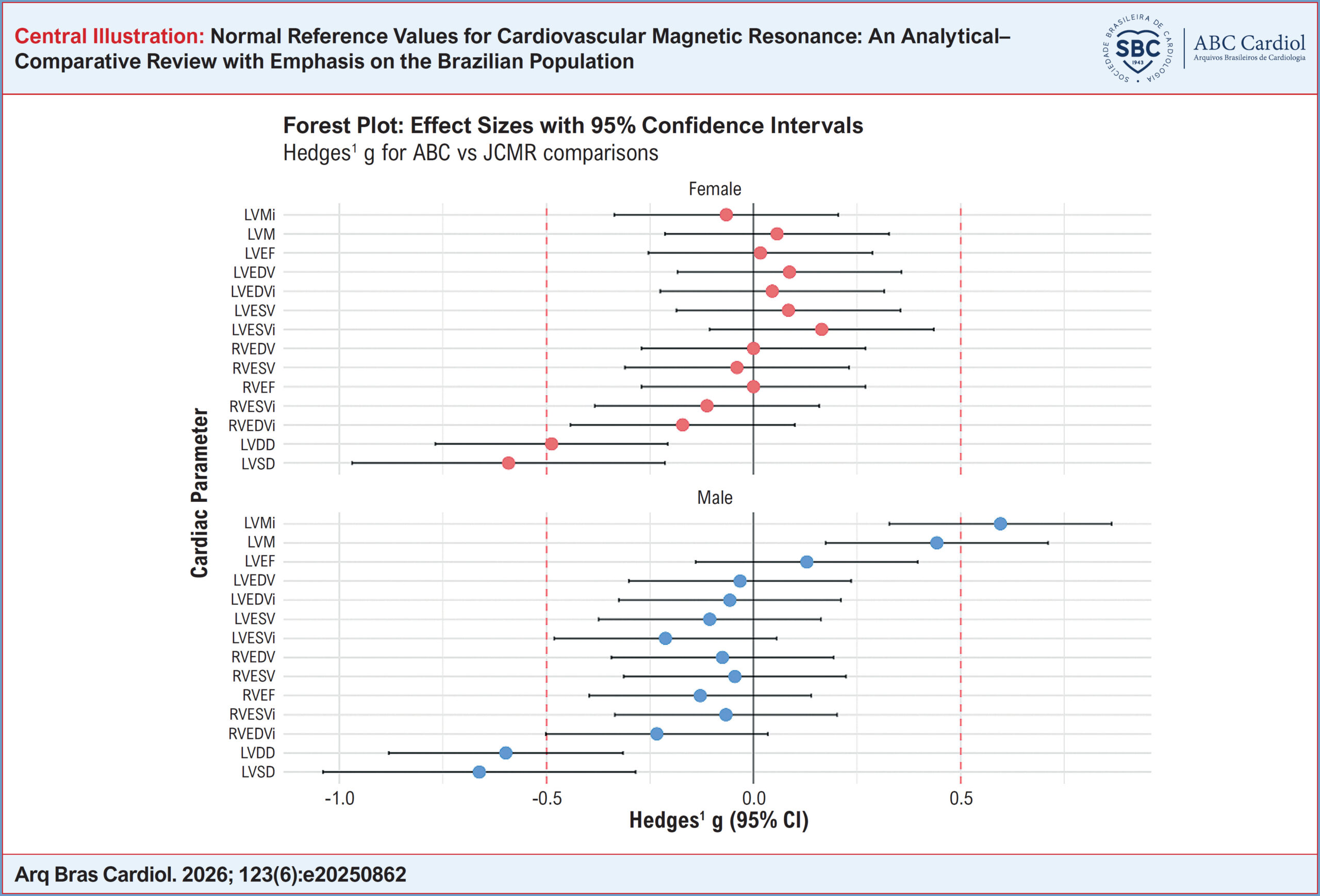
An analytical–comparative review was performed using summarized data from an international reference-value review and a Brazilian study. Means and standard deviations of key ventricular variables were compared, with assessment of 95% CI overlap, Welch’s t-test, and effect size (Hedges’ g). Standard-deviation–based cut-off points were proposed for grading abnormality.

Results:

Overall concordance between means was observed for most variables. Statistically significant differences occurred in a few parameters, mainly ventricular diameters and left ventricular mass, with small to moderate effect sizes. Discrepancies were largely attributed to methodological differences in measurement and indexation.

Conclusions:

International normal reference values may be used in Brazilian clinical practice, provided they are adjusted for body surface area and interpreted in light of local particularities. Standardization of protocols and national multicenter studies are needed to strengthen regional validation. Keywords: Normal Values, Cardiac Magnetic Resonance, Reference Interval.

  • Cardiovascular disease
  • Magnetic Resonance Imaging
  • Reference Values
Read Full Article
Creative Commons Attribution (CC BY) Creative Commons license
03/08/2026 DOI: 10.36660/abc.20250862i Views 1
Figure 1
ABC
Editorial

Echocardiographic Reference Values: Why Brazil Needs to Speak With its Own Voice

Imara Correia de Queiroz Barbosa et al. Israel Nilton de Almeida Feitosa

Echocardiographic measurements are cornerstones of diagnosis, risk stratification, and therapeutic decision-making across a wide range of cardiovascular conditions. Despite this, clinical practice in Brazil still relies largely on reference ranges derived from European and North American populations. In a country of continental dimensions, marked by substantial ethnic, anthropometric, and socioeconomic heterogeneity, such extrapolation raises legitimate concerns regarding diagnostic accuracy and equity in cardiovascular assessment.

In this issue of the Arquivos Brasileiros de Cardiologia, Rodrigues et al. present a timely and methodologically sound study establishing contemporary reference values for echocardiographic parameters in healthy Brazilian adults. The inclusion of 496 volunteers recruited from 20 echocardiography laboratories across the five major geographic regions of the country provides unprecedented national representativeness and addresses a longstanding gap in Brazilian cardiology.

[…]

  • Echocardiography
  • Global Longitudinal Strain
  • Reference Values
Read Full Article
Creative Commons Attribution (CC BY) Creative Commons license
22/05/2026 DOI: 10.36660/abc.20260155i Views 2
Reference Echocardiographic Values for Cardiac Chambers in Brazil: A Multiregional, Multi-Racial Study
ABC
Original Article

Reference Echocardiographic Values for Cardiac Chambers in Brazil: A Multiregional, Multi-Racial Study

Ana Clara Tude Rodrigues et al. Roberto Magalhães Saraiva Viviane T. Hotta Gabriel Doreto Rodrigues … Fabiana Camelier de Assis Cardoso Anderson da Costa Armstrong Cláudia Gianini Monaco Adelino Parro Junior José Luis de Castro e Silva Pretto João Marcos Bemfica Barbosa Ferreira Renata Santos Lopes Teixeira de Lima Marcelo Salame Salustiano Pereira de Araujo Carlos Eduardo Suaide Silva Cíntia Galhardo Tressino Fabio Canellas Moreira Ana Cristina Camarozano Minna Moreira Dias Romano Andre Luiz Cerqueira Almeida Claudio Henrique Fischer Alex dos Santos Felix Clarissa Borguezan-Daros Diogo Silva Piardi Diego Cunha Marcelo Dantas Tavares de Melo Rafael Modesto Silvio Henrique Barberato Wercules Antonio Oliveira Marcelo Luiz Campos Vieira Samira Saady Morhy

Abstract

Background

Reference values for cardiac chamber quantification are essential for clinical decision-making. Brazil is a continental country with marked racial diversity and substantial regional socioeconomic and anthropometric variability, which may not be adequately represented in international echocardiographic standards.

Objective

To establish reference echocardiographic measurements for healthy Brazilian adults and to investigate geographic and sex-related differences across Brazil’s major regions.

Methods

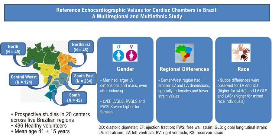
In this prospective, multicenter study, transthoracic echocardiograms were performed in 496 healthy volunteers (mean age 41 ± 15 years; 55% women) from five Brazilian regions. Left ventricular (LV) and left atrial (LA) dimensions, volumes, mass, ejection fraction, and global longitudinal strain (GLS) were analyzed in accordance with current guidelines. Comparisons were conducted by sex (Student’s t test or Mann-Whitney U test) and by region (analysis of variance).

Results

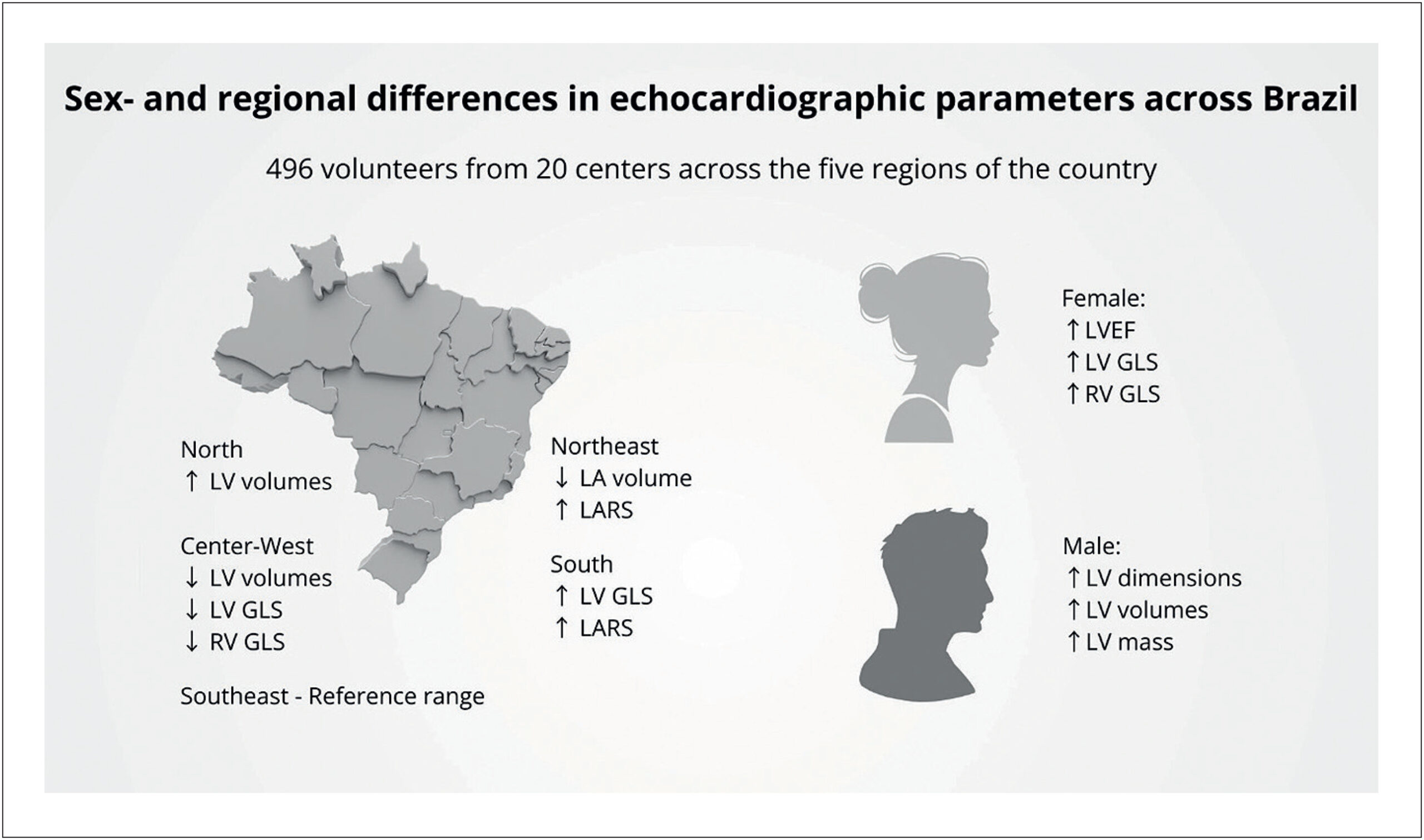
Cardiac chamber dimensions and LV mass were greater in men, even after indexing for body surface area. LA volumes were also higher in men, although these differences were attenuated after indexing. Absolute LVGLS and right ventricle (RV) GLS (RVGLS) values were higher in women, whereas LA strain reservoir (LASr) was similar between sexes. Regionally, the Center-West exhibited significantly smaller LV volumes, a difference primarily driven by women, along with lower LVGLS and RVGLS (p < 0.001). In contrast, LA volumes were smaller in the Northeast region. No significant associations were observed between self-reported race and echocardiographic parameters, except for RVGLS and LASr, which were lower in White participants.

Conclusion

This study establishes echocardiographic reference values in a nationally representative Brazilian cohort, confirms expected sex-related differences, and identifies previously unrecognized regional variations in cardiac chamber dimensions. These findings underscore the need for population-specific standards to ensure accurate cardiac quantification.

  • Echocardiography
  • Heart
  • Reference Values
Read Full Article
Creative Commons Attribution (CC BY) Creative Commons license
23/03/2026 DOI: 10.36660/abc.20250628i Views 1
ABC
Original Article

Handgrip Strength in Heart Failure: Developing a Reference Equation

Suena Medeiros Parahiba et al. Édina Caroline Ternus Ribeiro Ingrid da Silveira Knobloch Débowra Dapper … Ingrid Dalira Schweigert Perry Nadine Oliveira Clausell Vivian Luft Gabriela Corrêa Souza Eneida Rejane Rabelo-Silva

Abstract

Background:

Handgrip strength (HGS) is a key indicator of overall muscle strength and functional capacity in patients with heart failure (HF). However, no reference equations specific to this population have been previously published.

Objectives:

This study aimed to develop and validate a reference equation for predicting HGS in patients with HF.

Methods:

A cross-sectional study was conducted on patients with stable HF, aged 18–79 years, diagnosed for at least three months. Maximum HGS value was obtained from three consecutive measurements. Clinical data and anthropometric assessments were collected. The sample was randomly divided into two-thirds (n=174) for derivation and one third (n=100) for validation. A multivariate regression model was applied to develop the predictive equation, including variables with a p-value < 0.25 as determined by the Wald test.

Results:

Derivation and validation samples showed no significant differences at baseline. Patients were predominantly male, older adults, and white. The derived equation was: Predicted HGS = −39.732 + (10.771 * gender [female = 0; male = 1]) – (0.158 * age [years]) + (35.096 * height [m]) + (0.448 * calf circumference [cm]) – (4.224 * the New York Heart Association class [I /II = 0; III/IV = 1]). When applied to the validation sample, the equation underestimated actual HGS by 0.68 ± 8.93 Kg.

Conclusion:

Age, sex, height, calf circumference, and NYHA class were key determinants of HGS in HF patients. The derived equation showed good predictive accuracy and may serve as a useful reference for interpreting grip strength in this population.

  • Hand Strength
  • Heart Failure
  • Reference Values
Read Full Article
Creative Commons Attribution (CC BY) Creative Commons license
01/12/2025 DOI: 10.36660/abc.20240777i Views 1
ABC
Editorial

A Step Forward in the Assessment of Handgrip Strength in Heart Failure

Evandro José Cesarino et al. Marildes Luiza de Castro Regina Célia Garcia de Andrade Carolina Baraldi Araujo Restini

Significance of Handgrip Strength in HF

HGS is a simple, non-invasive measure of muscle strength, correlating with physical condition and considered a prognostic marker in HF., A systematic review of 7,350 HF patients revealed that a 1 kg reduction in HGS increases mortality risk by 8% (RR 1.08, 95% CI 1.05–1.11). Unlike complex measures, such as cardiopulmonary exercise testing, HGS is low-cost, accessible, and feasible in all clinical settings, making it a straightforward tool to assess frailty and guide rehabilitation., However, existing HGS reference equations are mainly derived from healthy populations, failing to account for HF-specific factors, such as reduced cardiac output (CO) and muscle atrophy, leading to inaccurate conclusions. The manuscript’s development of an HF-specific HGS equation represents a timely advance.

[…]

  • Diastolic Heart Failure
  • Hand Strength
  • Heart Failure
  • Reference Values
  • Systolic Heart Failure
Read Full Article
Creative Commons Attribution (CC BY) Creative Commons license
14/11/2025 DOI: 10.36660/abc.20250579i Views 1
ABC
Research Letter

What are the Optimal Reference Values for Home Blood Pressure Monitoring?

Audes D. M. Feitosa et al. Marco A. Mota-Gomes Fernando Nobre Decio Mion Jr. … Annelise M.G. Paiva Fábio Argenta Weimar K.S. Barroso Roberto D. Miranda Eduardo C. D. Barbosa Andréa A. Brandão Thiago S.V. Jardim Paulo C.B.V. Jardim Wilson Nadruz

Introduction

The diagnosis of hypertension usually relies on office blood pressure (BP) measures. However, this approach might underestimate or overestimate the true prevalence of hypertension due to the presence of alternative BP phenotypes, such as masked hypertension and white-coat hypertension. In this regard, current hypertension guidelines have recommended the evaluation of out-of-office BP by ambulatory BP monitoring (ABPM) or home blood pressure monitoring (HBPM), when these techniques are available, to confirm the diagnosis and to provide a more adequate management of hypertension.–

HBPM reference values used to define hypertension have been more consistently suggested since the end of 1990s. In 1998, results of a meta-analysis including data from 17 studies and 5,422 untreated participants suggested that elevated HBPM values should be ≥ 137/89 mmHg or ≥ 135/86 mmHg based on analysis of means + 2 standard deviations or ninety-fifth percentiles, respectively. In 1999, the analysis of the ninety-fifth percentiles of HBPM values from 2,401 normotensive individuals at the office estimated that elevated HBPM values were ≥ 137/85 mmHg. Furthermore, the evaluation of 1,913 individuals (69% not treated with antihypertensive medications) from the Ohasama Study showed that HBPM values ≥ 137/84 mmHg were associated with greater risk of death after 5 years of follow-up. Subsequent hypertension guidelines rounded the reference values suggested by these latter studies and recommended that abnormal HBPM measures should be considered when ≥ 135/85 mmHg., HBPM thresholds of 135/85 mmHg were then incorporated into clinical practice, and they have been used to define abnormal HBPM values by hypertension guidelines from various societies,, including the Seventh Brazilian Hypertension Guidelines published in 2016 and the Sixth Brazilian Guidelines of ABPM and Fourth Brazilian Guidelines of HBPM published in 2018.

[…]

  • Blood Pressure
  • Blood Pressure Self-Monitoring
  • Hypertension
  • Reference Standards
  • Reference Values
Read Full Article
Creative Commons Attribution (CC BY) Creative Commons license
23/04/2021 DOI: 10.36660/abc.20201109 Views 1
ABC
Editorial

Real-Time Three-Dimensional Echocardiography and Myocardial Strain: Ready for Use in Clinical Practice

Frederico Jose Neves Mancuso

The ability of echocardiography to quantify volumes and to evaluate the contractile function of cardiac chambers has evolved greatly in recent years, particularly due to the development of real-time three-dimensional echocardiography (echo3D) and the possibility myocardial deformation (Strain) analysis by the speckle tracking technique.–

Unlike two-dimensional echocardiography, where geometric inferences and mathematical calculations are required, the echo3D allows direct measurement of ventricular and atrial volumes, from which function data such as left and right ventricular ejection fraction, and left atrial function, including total emptying fraction and active emptying fraction parameters.–

[…]

  • Echocardiography, Three-Dimensional
  • Reference Values
  • Speckle Tracking
  • Strain
  • Ventricular Function, Left
Read Full Article
Creative Commons Attribution (CC BY) Creative Commons license
02/12/2019 DOI: 10.5935/abc.20190179
Figure 1
ABC
Original Article

3-Dimensional Echocardiography and 2-D Strain Analysis of Left Ventricular, Left Atrial and Right Ventricular Function in Healthy Brazilian Volunteers

Roberto M. Saraiva et al. Eliza Maria B. Scolin Nicole P. Pacheco Maria Eduarda Bouret … Mauro Felippe Felix Mediano Marcelo T. Holanda Andréa R. da Costa

Abstract

Background:

New echocardiographic techniques are used in the diagnosis and prognosis of many heart diseases. However, reference values in different populations are still needed for several of these new indexes. We studied these new echocardiographic parameters in a group of Brazilians with no known cardiovascular disease.

Objective:

To study values for new echocardiographic indexes in Brazilians without known cardiovascular disease and their correlation with age.

Methods:

Cross-sectional study that included healthy individuals who underwent three-dimensional echocardiography (3DE) and two-dimensional speckle tracking echocardiography (STE) strain (e) analysis. Left atrial (LA) and left ventricular (LV) function were analyzed by 3DE and STE, and right ventricular (RV) function by STE. P values < 0.05 were considered significant.

Results:

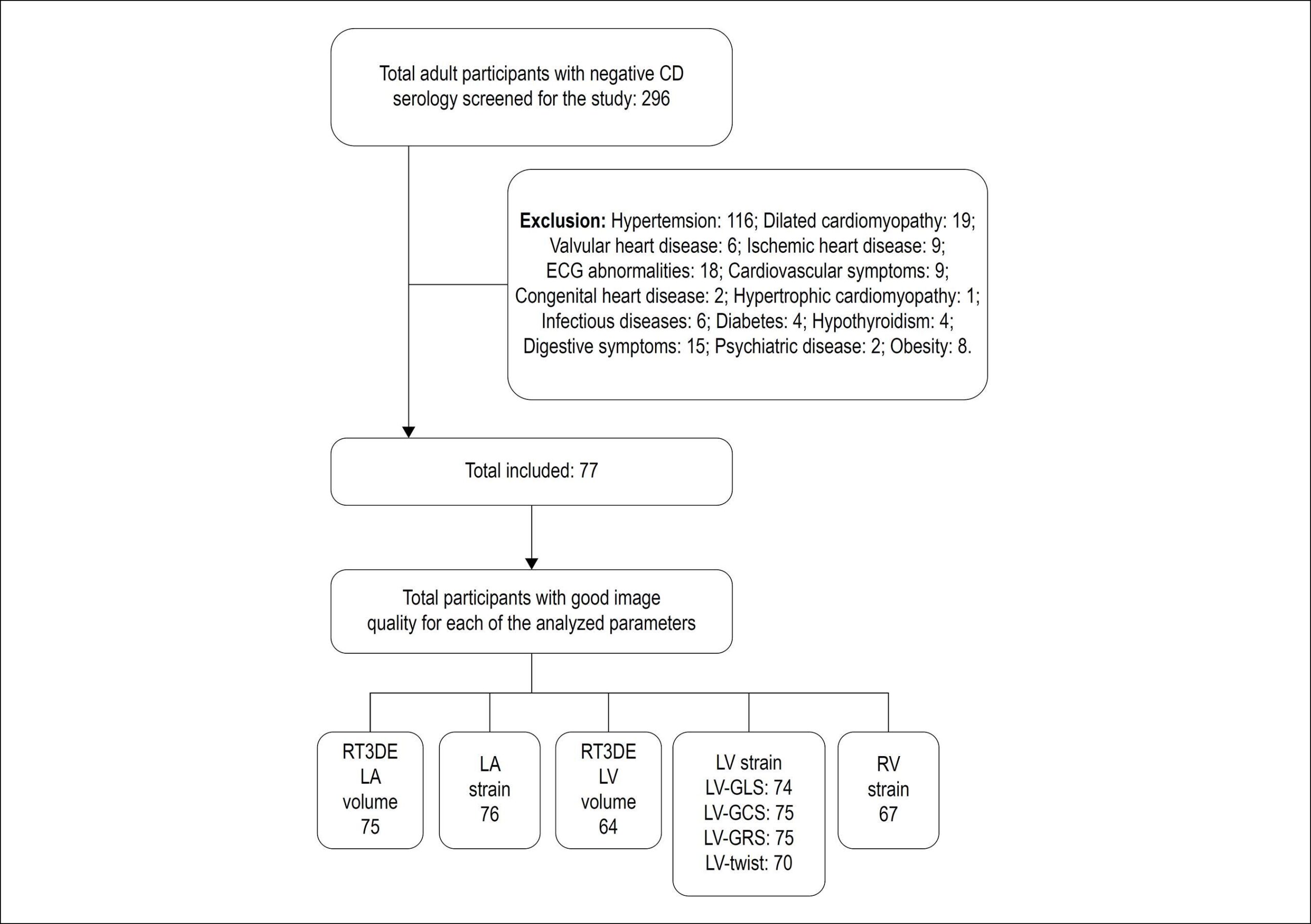
Seventy-seven subjects (46.7% men; 40.4 ± 10.4 years) were included. Maximum, minimum and pre-atrial contraction (pre-A) LA volumes (ml/m2) were 21.2 ± 5.5, 7.8 ± 2.5, and 11.0 ± 3.1, respectively. Peak positive global LA e (LAScd), peak negative global LA e and total global LA e (LASr) were 17.4 ± 5.2%, -13.2 ± 2.0% and 30.5 ± 5.9%, respectively. LV end-diastolic and end-systolic volumes (ml/m2) measured 57 ± 12 and 24 ± 6, and 3D LV ejection fraction measured 58 ± 6%. Global LV longitudinal, circumferential and radial e were -19 ± 2%, -19 ± 3%, and 46 ± 12%, respectively. LV torsion measured 1.6 ± 0.70 /cm. Global longitudinal RV e (RV-GLS) and RV free wall strain were -22 ± 3% and -24 ± 5%. Minimum LA and pre-A volumes, LV apical rotation, torsion and RV-GLS increased with age, while total and passive LA emptying fractions, LAScd, LASr, LV end-diastolic and end-systolic volumes decreased with age.

Conclusion:

Values for new echocardiographic indexes in Brazilians without known cardiovascular disease and their correlation with age are presented.

  • Cardiovascular Diseases
  • Echocardiography, Three-Dimensional
  • Left Ventricular Function
  • Reference Values
  • Right Ventricular Function
  • Speckle Tracking
  • Strain
Read Full Article
Creative Commons Attribution (CC BY) Creative Commons license
29/08/2019 DOI: 10.5935/abc.20190155
ABC
Special Article

Positioning about the Flexibility of Fasting for Lipid Profiling

Marileia Scartezini et al. Carlos Eduardo dos Santos Ferreira Maria Cristina Oliveira Izar Marcello Bertoluci … Sergio Vencio Gustavo Aguiar Campana Nairo Massakazu Sumita Luiz Fernando Barcelos André A. Faludi Raul D. Santos Marcus Vinícius Bolívar Malachias Jerolino Lopes Aquino César Alex de Oliveira Galoro Cleide Sabino Maria Helane Costa Gurgel Luiz Alberto Andreotti Turatti Alexandre Hohl Tania Leme da Rocha Martinez

Justifications

The review of the need of fasting for lipid profile analysis (total cholesterol, LDL-C, HDL-C, non-HDL-C, and triglycerides [TG]) is based on the following grounds:

[…]

  • Fasting / metabolism
  • Lipid profile
  • Postprandial
  • Reference Values
  • Risk category
Read Full Article
Creative Commons Attribution (CC BY) Creative Commons license
01/03/2017 DOI: 10.5935/abc.20170039 Views 1
ABC
Letter to the Editor

Measurement of Epicardial Fat Thickness by Echocardiography Presents Challenges

Mustafa Gulgun et al. Fatih Alparslan Genç

To the Editor,

It was with great interest that we read the paper by Kaplan et al. entitled ”Evaluation of Electrocardiographic T-peak to T-end Interval in Subjects with Increased Epicardial Fat Tissue Thickness” published on Arquivos Brasileiros de Cardiologia in December 2015. They aimed to investigate the relationship between epicardial adipose tissue (EFT) and ventricular repolarization. They demonstrated some ventricular repolarization abnormalities on electrocardiography in patients with higher EFT thickness. We would like to thank Kaplan et al. for their great effort in this study, and share some of our thoughts.

[…]

  • Adipose Tissue
  • Arrhythmias Cardiac
  • Echocardiography
  • Electrocardiography
  • Magnetic Resonance Imaging / trends
  • Pericardium
  • Reference Values
Read Full Article
Creative Commons Attribution (CC BY) Creative Commons license
01/11/2016 DOI: 10.5935/abc.20160167
Figure 1
ABC
Original Article

Evaluation of Electrocardiographic T-peak to T-end Interval in Subjects with Increased Epicardial Fat Tissue Thickness

Ozgur Kaplan et al. Ertugrul Kurtoglu Gokay Nar Erdogan Yasar … Gokhan Gozubuyuk Cem Dogan Ahmet Ugur Boz Sıho Hidayet Hasan Pekdemir

Abstract

Background:

The association between periatrial adiposity and atrial arrhythmias has been shown in previous studies. However, there are not enough available data on the association between epicardial fat tissue (EFT) thickness and parameters of ventricular repolarization. Thus, we aimed to evaluate the association of EFT thickness with indices of ventricular repolarization by using T-peak to T-end (Tp-e) interval and Tp-e/QT ratio.

Methods:

The present study included 50 patients whose EFT thickness ≥ 9 mm (group 1) and 40 control subjects with EFT thickness < 9 mm (group 2). Transthoracic echocardiographic examination was performed in all participants. QT parameters, Tp-e intervals and Tp-e/QT ratio were measured from the 12-lead electrocardiogram.

Results:

QTd (41.1 ± 2.5 vs 38.6 ± 3.2, p < 0.001) and corrected QTd (46.7 ± 4.7 vs 43.7 ± 4, p = 0.002) were significantly higher in group 1 when compared to group 2. The Tp-e interval (76.5 ± 6.3, 70.3 ± 6.8, p < 0.001), cTp-e interval (83.1 ± 4.3 vs. 76±4.9, p < 0.001), Tp-e/QT (0.20 ± 0.02 vs. 0.2 ± 0.02, p < 0.001) and Tp-e/QTc ratios (0.2 ± 0.01 vs. 0.18 ± 0.01, p < 0.001) were increased in group 1 in comparison to group 2. Significant positive correlations were found between EFT thickness and Tp-e interval (r = 0.548, p < 0.001), cTp-e interval (r = 0.259, p = 0.01), and Tp-e/QT (r = 0.662, p < 0.001) and Tp-e/QTc ratios (r = 0.560, p < 0.001).

Conclusion:

The present study shows that Tp-e and cTp-e interval, Tp-e/QT and Tp-e/QTc ratios were increased in subjects with increased EFT, which may suggest an increased risk of ventricular arrhythmia.

  • Adipose Tissue
  • Arrhythmias
  • cardiac
  • Electracardiography
  • Pericardium
  • Reference Values
Read Full Article
Creative Commons Attribution (CC BY) Creative Commons license
02/10/2015 DOI: 10.5935/abc.20150124
Morphological and functional measurements of the heart obtained by magnetic resonance imaging in Brazilians
ABC
Artigo Original

Morphological and functional measurements of the heart obtained by magnetic resonance imaging in Brazilians

Robson Macedo et al. Juliano Lara Fernandes Solange Souza Andrade Carlos Eduardo Rochitte … Kênio Costa Lima Álvaro Campos Cavalcanti Maciel Fernanda Cunha Maciel Geraldo Souza Pinho Alves Otávio Rizzi Coelho Rosiane Viana Zuza Diniz

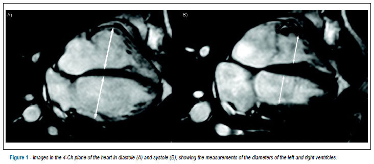
BACKGROUND: Still today, measurements used as a reference in the cardiac magnetic resonance imaging have been obtained mainly from studies carried out in North-American and European populations. OBJECTIVE: To obtain measurements of the diastolic diameter, systolic diameter, end diastolic volume, end systolic volume, ejection fraction, and myocardial mass of the left and right ventricles in Brazilians. METHODS: 54 men and 53 women, with mean age of 43.4 ± 13.1 years, asymptomatic, with no cardiomyopathies, have been subjected to the cardiac magnetic resonance imaging, using a balanced steady state free precession technique. RESULTS: The averages and the standard deviations of the parameters for the left ventricle have been: diastolic diameter = 4.8 ± 0.5 cm; systolic diameter = 3.0 ± 0.6 cm; end diastolic volume = 128.4 ± 29.6 mL; end systolic volume = 45.2 ± 16.6 mL; ejection fraction = 65.5 ± 6.3%; mass = 95.2 ± 30.8 g. For the right ventricle, they have been: diastolic diameter = 3.9 ± 1.3 cm; systolic diameter = 2.5 ± 0.5 cm; end diastolic volume = 126.5 ± 30.7 mL; end systolic volume = 53.6 ± 18.4 mL; ejection fraction = 58.3 ± 8.0%, and mass = 26.1 ± 6.1 g. The masses and the volumes were significantly greater in the men, except for the end systolic volume of the left ventricle. The ejection fraction of the right ventricle has been significantly greater in the women. There has been a significant and inverted correlation of the systolic volume of the right volume with the progression of the age. CONCLUSION: This study has described, for the first time, cardiac measurements obtained through the cardiac magnetic resonance imaging in Brazilians, asymptomatic, with no cardiomyopathies, showing differences in accordance with gender and age.

  • Genetics, Population
  • Heart Ventricle
  • Magnetic Resonance Imaging
  • Reference Values
  • Ventricular Function
Read Full Article
Creative Commons Attribution (CC BY) Creative Commons license
11/06/2013 DOI: 10.5935/abc.20130113 Views 1
Reference curve of the fetal ventricular septum area by the STIC method: preliminary study
ABC
Artigo Original

Reference curve of the fetal ventricular septum area by the STIC method: preliminary study

Liliam Cristine Rolo et al. Luciano Marcondes Machado Nardozza Edward Araujo Júnior Christiane Simioni … Marina Maccagnano Zamith Antonio Fernandes Moron

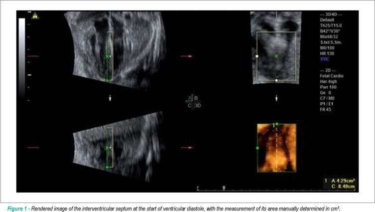
BACKGROUND: Early detection of septal changes such as septal hypertrophy commonly present in fetuses of diabetic mothers would help reduce the high rates of infant mortality. OBJECTIVE: Determine reference ranges for the fetal ventricular septal area through three-dimensional ultrasound (US3D) using the STIC method (Spatio-Temporal Image Correlation). METHODS: We conducted a cross-sectional study with 69 pregnant women between the 18th and 33rd weeks of pregnancy. We used as a reference the four-chamber plane with the ROI (Region of Interest) positioned from the ventricles; the septum area were manually marked. To assess the correlation of the interventricular septum area with gestational age (GA), we constructed scatter plots and calculated Pearson’s correlation coefficient (r), and the adjustment was performed by the coefficient of determination (R²). We calculated averages, medians, standard deviations (sd), as well as maximum and minimum values. To calculate the intraobserver reproducibility, we used the intraclass correlation coefficient (ICC). The interventricular septum thickness was measured and it was correlated with gestational age and the septal area rendered in 52 patients using the ICC. RESULTS: The interventricular septum area was highly correlated with gestational age (r = 0.81), and the average increased from 0.47 cm² in the 18th week to 2.42 cm² in the 33rd of gestation. The intraobserver reproducibility was excellent with ICC = 0.994. No significant correlation was observed between the interventricular septum measurement and the GA (R² = 0.200), as well as there was no correlation with the septal area rendered with ICC = 0.150. CONCLUSION: Reference intervals for the interventricular septum area between the 18th and the 33rd pregnancy week were determined to be highly reproducible.

  • fetal heart
  • Imaging, Three-Dimensional
  • Pregnancy
  • Reference Values
  • ventricular septum
Read Full Article
Creative Commons Attribution (CC BY) Creative Commons license
01/04/2011 DOI: 10.1590/S0066-782X2011005000036 Views 1
ABC
Atualização

Reference equations for the 6-minute walk test in healthy individuals

Victor Zuniga Dourado

El test de caminata de 6 minutos (TC6) ha sido ampliamente utilizado en el ambiente clínico. Algunas ecuaciones de referencia para la previsión de la distancia recorrida en el test (DTC6) están disponibles en la literatura. Esta revisión tuvo como objetivo discutir críticamente los hallazgos de la literatura, publicados en portugués y en inglés (LILACS, SCIELO, MEDLINE y PUBMED), que evaluaron los valores normales y elaboraron ecuaciones de referencia para la previsión de la DTC6 en individuos sanos, comparándolos a los resultados recientemente obtenidos en individuos brasileños. Edad, género, peso, estatura e índice de masa corporal fueron los atributos demográficos y antropométricos más frecuentemente correlacionados con la DTC6. Las ecuaciones resultantes de esos atributos fueron capaces de explicar entre 25 y 66% de la variabilidad total de la DTC6. Lamentablemente, las ecuaciones extranjeras no son adecuadas para la población brasileña. Aun cuando el test es realizado bajo estandarización rigurosa, la diferencia de performance en el TC6 entre extranjeros y brasileños permanece, indicando la necesidad de los valores de referencias específicos para cada población y/o etnia. En ese sentido, las ecuaciones desarrolladas recientemente en el Brasil son, probablemente, las más apropiadas para interpretar la performance de caminata de nuestros compatriotas con enfermedades crónicas que afectan su capacidad para realizar ejercicios. Estudios futuros con muestras substancialmente mayores (p.e. multicéntricos) y con técnica de muestreo randomizada son necesarios para que los valores de referencia de la DTC6 sean más representativos.

  • Exercise
  • Reference Values
  • walking
Read Full Article
Creative Commons Attribution (CC BY) Creative Commons license
25/02/2011 DOI: 10.1590/S0066-782X2011005000024 Views 2
Reference values for cardiopulmonary exercise testing for sedentary and active men and women
ABC
Artigo Original

Reference values for cardiopulmonary exercise testing for sedentary and active men and women

Artur Haddad Herdy et al. Dorian Uhlendorf

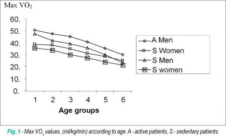
BACKGROUND: The reference values for cardiopulmonary exercise testing (CPET) available in Brazil were derived from a cycle ergometer in a sedentary and relatively small population. OBJECTIVE: Provide reference values for CPET in Brazilians of both sexes, either sedentary or active. METHODS: From 2006 to 2008, 3,992 CEPT of healthy individuals were selected from our laboratory. Athletes, smokers, patients with any known pathology, users of continuous medication and obese patients were excluded. Peak VO2 was considered max VO2. We also analyzed the anaerobic threshold VO2, maximum ventilation and oxygen pulse according to sex, age, sedentary and active patients. Age groups were divided as follows: G1 (15-24), G2 (25-34), G3 (35-44), G4 (45-54), G5 (55-64) and G6 (65-74). RESULTS: According to age groups, the mean values of VO2 in ml/kg/min with their standard deviations were: Active man: G1-50.6 ± 7.3, G2-47, 4 ± 7.4, G3-45, 4 ± 6.8, G4-40.5 ± 6.5; G5-35.3 ± 6.2; G6-30.0 ± 6.1. Active woman: G1-38.9 ± 5.7; G2-38.1 ± 6.6; G3-34.9 ± 5.9; G4-31.1 ± 5.4; G5-28.6 ± 6.1; G6-25.1 ± 4.4. Sedentary man: G1-47.4 ± 7.9; G2-41.9 ± 7.2; G3-39.0 ± 6.8; G4-35.6 ± 7.7; G5-30.0 ± 6.3; G6-23.1 ± 6.3. Sedentary woman: G1-35.6 ± 5.7; G2-34.0 ± 4.8; G3-30.0 ± 5.4; G4-27.2 ± 5.0; G5-23.9 ± 4.2; G6-21.2 ± 3.4. CONCLUSION: This article provides reference values of max VO2, among other parameters, in the Cardiopulmonary Exercise Testing performed on the treadmill in individuals of both sexes, either active and sedentary.

  • Exercise Test
  • Pulmonary Ventilation
  • Reference Values
  • Sedentary Lifestyle
Read Full Article
Creative Commons Attribution (CC BY) Creative Commons license
26/11/2010 DOI: 10.1590/S0066-782X2010005000155 Views 3
ABC
Artigo Original

Echocardiographic reference values in a sample of asymptomatic adult Brazilian population

Lílian Cláudia Souza Ângelo et al. Marcelo Luiz Campos Vieira Sérgio Lamêgo Rodrigues Renato Lírio Morelato … Alexandre C. Pereira José Geraldo Mill José Eduardo Krieger

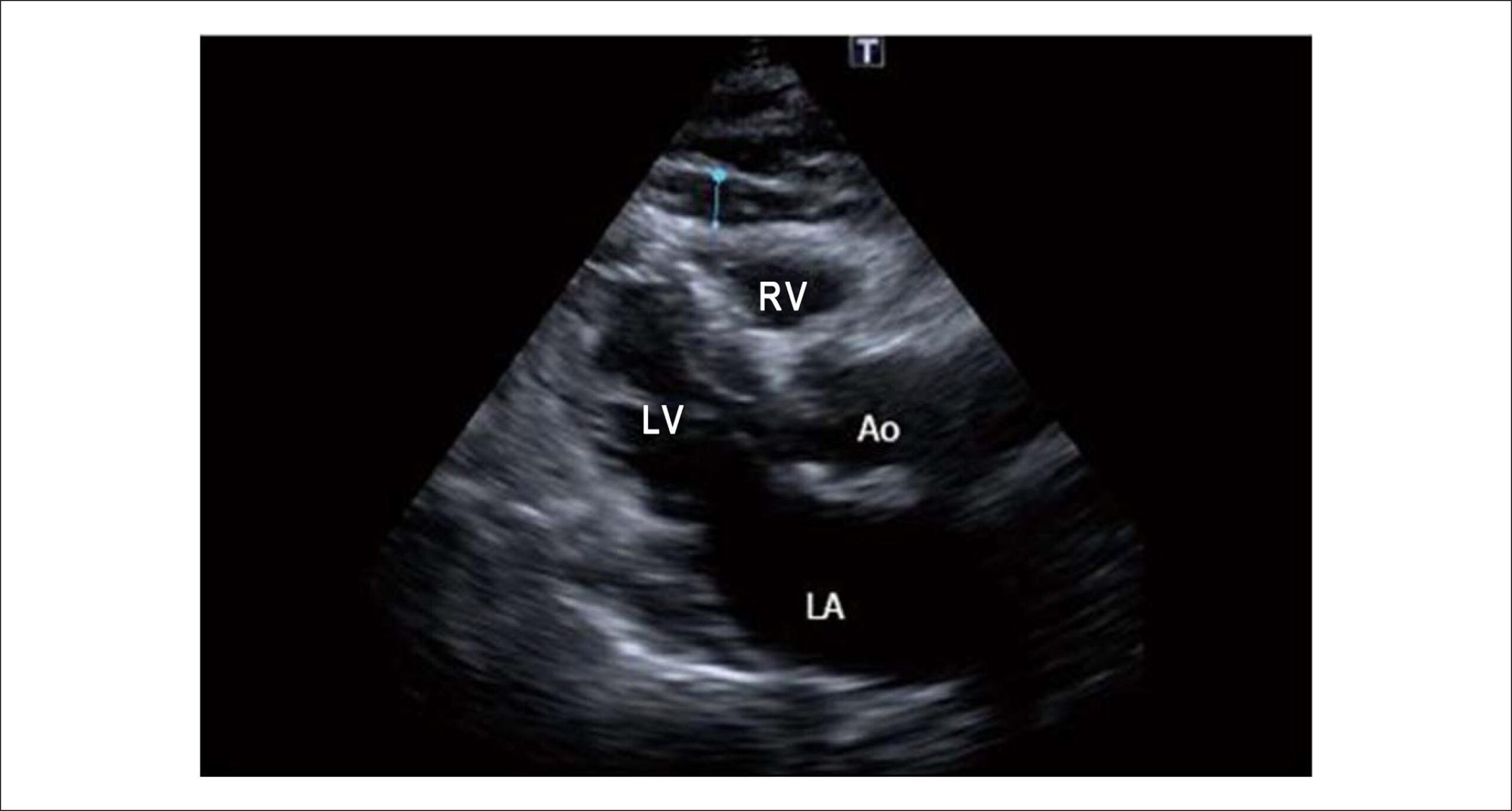
OBJECTIVE: To determine echocardiographic reference values for cardiac chambers, left ventricular mass, and left ventricular (LV) mass indexes in an asymptomatic adult population sample. METHODS: This was an observational study based on a randomly selected population sample from the city of Vitória, Brazil. Two hundred and ninety-five volunteers (61.7% women) with no past history of cardiovascular disease underwent transthoracic echocardiography. The following M-mode echocardiographic parameters were measured: ventricular diameters, interventricular septal thickness, LV posterior wall thickness, LV mass, left ventricular mass indexes, plus aortic and left atrial diameters. Values were expressed as mean and standard deviation and percentiles, with a 95% confidence interval. RESULTS: Echocardiographic values were slightly influenced by gender and age. Overall, cardiac measurements were higher in the male gender. LV posterior wall thickness, mass indexes corrected for height and diastolic diameter were influenced by age. The 95% percentiles of interventricular septum and LV posterior wall were 9.9 mm and 9.6 mm for men, respectively, and 9.3 mm for septum and posterior wall for women. CONCLUSION: The 95% percentile values of interventricular septum and posterior wall and, therefore, of both absolute and indexed left ventricular mass found in our study conducted in the Vitória population are lower than those reported in previous studies. In this framework, our results will be useful as a reference, since they are consistent with the new limits suggested in the literature for the echocardiographic diagnosis of left ventricular hypertrophy.

  • Echocardiography
  • population
  • random and systematic sampling
  • Reference Values
Read Full Article
Creative Commons Attribution (CC BY) Creative Commons license
25/09/2007 DOI: 10.1590/S0066-782X2007001500007
Percentile curves of normal values of echocardiographic measurements in normal children from the central-southern region of the State of São Paulo, Brazil
ABC
Artigo Original

Percentile curves of normal values of echocardiographic measurements in normal children from the central-southern region of the State of São Paulo, Brazil

Rossano César Bonatto et al. José Roberto Fioretto Katashi Okoshi Beatriz Bojikian Matsubara … Carlos Roberto Padovani Thiago Colletti Remond Manfrin Michelle de Farias Gobbi Rodolfo Silva De Martino Edson Antônio Bregagnollo

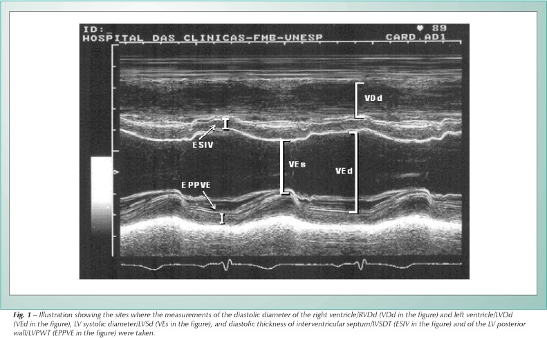
OBJECTIVE: To asses the values of echocardiographic measurements in normal children without cardiopathy and to build percentile curves, relating them to the body surface (BS, m²). METHODS: We analyzed M-mode echocardiographic measurements for children between one and 144 months of age. We assessed right ventricular diastolic diameter (RVDd, mm) and left ventricular diastolic diameter (LVDd, mm), LV systolic diameter (LVSd, mm), right ventricular outflow tract diameter (RVOT, mm), aortic diameter (AoD, mm) and left atrial diameter (LAD, mm); left ventricular ejection fraction (LVEF, %); percentage of variation of left ventricular diameter (deltaLV, %); interventricular septum diastolic thickness (IVSDT, mm) and LV posterior wall diastolic thickness (PWDT, mm); left ventricular mass (LVM, g) and LV mass index (LVMI, g/m²). RESULTS: At the end of the study, 595 children (326 male) were assessed. The values of echocardiographic measurements showed a good correlation with BS and allowed the constructing of percentile curves (3%, 25%, 50%, 75% and 97%). Statistically significant differences between the genders were evidenced for the following variables: LVSd, LVDd, RVOT, AoD, LVM and LVMI, and the highest values were observed in male children. CONCLUSION: The percentile curves of the values obtained can be used as a reference to assess children with suspicion of cardiopathy or to follow-up on those with diagnosed cardiopathy or under treatment with potentially cardiotoxic drugs.

  • body surface area
  • Child
  • Echocardiography
  • Reference Values
Read Full Article
Creative Commons Attribution (CC BY) Creative Commons license
18/01/2007 DOI: 10.1590/S0066-782X2006001900006 Views 2

How to cite this article

EndNote RIS

Electronic Document Format (Vancouver)

Correia M. Reference Values in Cardiovascular Magnetic Resonance Imaging: Between Local Validation and Global Harmonization. Arquivos Brasileiros de Cardiologia 2026;123(6):e20260458.

Electronic Document Format (ABNT)

Correia, Miguel. Reference Values in Cardiovascular Magnetic Resonance Imaging: Between Local Validation and Global Harmonization. Arquivos Brasileiros de Cardiologia, v. 123, n. 6, e20260458, Aug. 2026.

Electronic Document Format (APA)

Correia, M. (2026). Reference Values in Cardiovascular Magnetic Resonance Imaging: Between Local Validation and Global Harmonization. Arquivos Brasileiros de Cardiologia, 123(6), e20260458.

Electronic Document Format (ISO)

Correia, Miguel. Reference Values in Cardiovascular Magnetic Resonance Imaging: Between Local Validation and Global Harmonization. Arquivos Brasileiros de Cardiologia [online]. 2026, vol. 123, n. 6, [cited 2026-10-14], e20260458. Available from: <https://journals.cardiol.br/en/article/reference-values-in-cardiovascular-magnetic-resonance-imaging-between-local-validation-and-global-harmonization/>. ISSN 0066-782X

EndNote RIS

Electronic Document Format (Vancouver)

Torreão JA, Costa IBSS, Assunção-Jr AN, Gottlieb I, Silva LS, Senra T, Magalhães TA, et al. Normal Reference Values for Cardiovascular Magnetic Resonance: An Analytical–Comparative Review with Emphasis on the Brazilian Population. Arquivos Brasileiros de Cardiologia 2026;123(6):e20250862.

Electronic Document Format (ABNT)

Torreão, Jorge A.; Costa, Isabela Bispo Santos da Silva; Assunção-Jr, Antonildes N.; Gottlieb, Ilan; Silva, Leonardo Sara da; Senra, Tiago; Magalhães, Tiago Augusto; Uellendahl, Marly; Zampa, Hugo Bizetto; Liberato, Gabriela; Ishikawa, Walther Yoshiharu; Trad, Henrique; Hadlich, Marcelo; Coelho-Filho, Otavio; Sasdelli Neto, Roberto; Fernandes, Juliano Lara; Ávila, Luiz Francisco Rodrigues de; Pinto, Ibraim Masciarelli F.; Schvartzman, Paulo R.; Rochitte, Carlos E.. Normal Reference Values for Cardiovascular Magnetic Resonance: An Analytical–Comparative Review with Emphasis on the Brazilian Population. Arquivos Brasileiros de Cardiologia, v. 123, n. 6, e20250862, Aug. 2026.

Electronic Document Format (APA)

Torreão, J. A., Costa, I. B. S. S., Assunção-Jr, A. N., Gottlieb, I., Silva, L. S. , Senra, T., Magalhães, T. A., Uellendahl, M., Zampa, H. B., Liberato, G., Ishikawa, W. Y., Trad, H., Hadlich, M., Coelho-Filho, O., Sasdelli Neto, R., Fernandes, J. L., Ávila, L. F. R. , Pinto, I. M. F., Schvartzman, P. R., & Rochitte, C. E. (2026). Normal Reference Values for Cardiovascular Magnetic Resonance: An Analytical–Comparative Review with Emphasis on the Brazilian Population. Arquivos Brasileiros de Cardiologia, 123(6), e20250862.

Electronic Document Format (ISO)

Torreão, Jorge A. and Costa, Isabela Bispo Santos da Silva and Assunção-Jr, Antonildes N. and Gottlieb, Ilan and Silva, Leonardo Sara da and Senra, Tiago and Magalhães, Tiago Augusto and Uellendahl, Marly and Zampa, Hugo Bizetto and Liberato, Gabriela and Ishikawa, Walther Yoshiharu and Trad, Henrique and Hadlich, Marcelo and Coelho-Filho, Otavio and Sasdelli Neto, Roberto and Fernandes, Juliano Lara and Ávila, Luiz Francisco Rodrigues de and Pinto, Ibraim Masciarelli F. and Schvartzman, Paulo R. and Rochitte, Carlos E.. Normal Reference Values for Cardiovascular Magnetic Resonance: An Analytical–Comparative Review with Emphasis on the Brazilian Population. Arquivos Brasileiros de Cardiologia [online]. 2026, vol. 123, n. 6, [cited 2026-10-14], e20250862. Available from: <https://journals.cardiol.br/en/article/normal-reference-values-for-cardiovascular-magnetic-resonance-an-analytical-comparative-review-with-emphasis-on-the-brazilian-population/>. ISSN 0066-782X

EndNote RIS

Electronic Document Format (Vancouver)

Barbosa ICQ, Feitosa INA. Echocardiographic Reference Values: Why Brazil Needs to Speak With its Own Voice. Arquivos Brasileiros de Cardiologia 2026;123(2):e20260155.

Electronic Document Format (ABNT)

Barbosa, Imara Correia de Queiroz; Feitosa, Israel Nilton de Almeida. Echocardiographic Reference Values: Why Brazil Needs to Speak With its Own Voice. Arquivos Brasileiros de Cardiologia, v. 123, n. 2, e20260155, May. 2026.

Electronic Document Format (APA)

Barbosa, I. C. Q., & Feitosa, I. N. A. (2026). Echocardiographic Reference Values: Why Brazil Needs to Speak With its Own Voice. Arquivos Brasileiros de Cardiologia, 123(2), e20260155.

Electronic Document Format (ISO)

Barbosa, Imara Correia de Queiroz and Feitosa, Israel Nilton de Almeida. Echocardiographic Reference Values: Why Brazil Needs to Speak With its Own Voice. Arquivos Brasileiros de Cardiologia [online]. 2026, vol. 123, n. 2, [cited 2026-10-14], e20260155. Available from: <https://journals.cardiol.br/en/article/echocardiographic-reference-values-why-brazil-needs-to-speak-with-its-own-voice/>. ISSN 0066-782X

EndNote RIS

Electronic Document Format (Vancouver)

Rodrigues ACT, Saraiva RM, Hotta VT, Rodrigues GD, Cardoso FCA, Armstrong AC, Monaco CG, et al. Reference Echocardiographic Values for Cardiac Chambers in Brazil: A Multiregional, Multi-Racial Study. Arquivos Brasileiros de Cardiologia 2026;123(2):e20250628.

Electronic Document Format (ABNT)

Rodrigues, Ana Clara Tude; Saraiva, Roberto Magalhães; Hotta, Viviane T.; Rodrigues, Gabriel Doreto; Cardoso, Fabiana Camelier de Assis; Armstrong, Anderson da Costa; Monaco, Cláudia Gianini; Parro Junior, Adelino; Pretto, José Luis de Castro e Silva; Ferreira, João Marcos Bemfica Barbosa; Lima, Renata Santos Lopes Teixeira de; Salame, Marcelo; Araujo, Salustiano Pereira de; Silva, Carlos Eduardo Suaide; Tressino, Cíntia Galhardo; Moreira, Fabio Canellas; Camarozano, Ana Cristina; Romano, Minna Moreira Dias; Almeida, Andre Luiz Cerqueira; Fischer, Claudio Henrique; Felix, Alex dos Santos; Borguezan-Daros, Clarissa; Piardi, Diogo Silva; Cunha, Diego; Melo, Marcelo Dantas Tavares de; Modesto, Rafael; Barberato, Silvio Henrique; Oliveira, Wercules Antonio; Vieira, Marcelo Luiz Campos; Morhy, Samira Saady. Reference Echocardiographic Values for Cardiac Chambers in Brazil: A Multiregional, Multi-Racial Study. Arquivos Brasileiros de Cardiologia, v. 123, n. 2, e20250628, Mar. 2026.

Electronic Document Format (APA)

Rodrigues, A. C. T., Saraiva, R. M., Hotta, V. T., Rodrigues, G. D., Cardoso, F. C. A., Armstrong, A. C., Monaco, C. G., Parro Junior, A., Pretto, J. L. C. S., Ferreira, J. M. B. B., Lima, R. S. L. T. , Salame, M., Araujo, S. P. , Silva, C. E. S., Tressino, C. G., Moreira, F. C., Camarozano, A. C., Romano, M. M. D., Almeida, A. L. C., Fischer, C. H., Felix, A. S., Borguezan-Daros, C., Piardi, D. S., Cunha, D., Melo, M. D. T. , Modesto, R., Barberato, S. H., Oliveira, W. A., Vieira, M. L. C., & Morhy, S. S. (2026). Reference Echocardiographic Values for Cardiac Chambers in Brazil: A Multiregional, Multi-Racial Study. Arquivos Brasileiros de Cardiologia, 123(2), e20250628.

Electronic Document Format (ISO)

Rodrigues, Ana Clara Tude and Saraiva, Roberto Magalhães and Hotta, Viviane T. and Rodrigues, Gabriel Doreto and Cardoso, Fabiana Camelier de Assis and Armstrong, Anderson da Costa and Monaco, Cláudia Gianini and Parro Junior, Adelino and Pretto, José Luis de Castro e Silva and Ferreira, João Marcos Bemfica Barbosa and Lima, Renata Santos Lopes Teixeira de and Salame, Marcelo and Araujo, Salustiano Pereira de and Silva, Carlos Eduardo Suaide and Tressino, Cíntia Galhardo and Moreira, Fabio Canellas and Camarozano, Ana Cristina and Romano, Minna Moreira Dias and Almeida, Andre Luiz Cerqueira and Fischer, Claudio Henrique and Felix, Alex dos Santos and Borguezan-Daros, Clarissa and Piardi, Diogo Silva and Cunha, Diego and Melo, Marcelo Dantas Tavares de and Modesto, Rafael and Barberato, Silvio Henrique and Oliveira, Wercules Antonio and Vieira, Marcelo Luiz Campos and Morhy, Samira Saady. Reference Echocardiographic Values for Cardiac Chambers in Brazil: A Multiregional, Multi-Racial Study. Arquivos Brasileiros de Cardiologia [online]. 2026, vol. 123, n. 2, [cited 2026-10-14], e20250628. Available from: <https://journals.cardiol.br/en/article/reference-echocardiographic-values-for-cardiac-chambers-in-brazil-a-multiregional-multi-racial-study/>. ISSN 0066-782X

EndNote RIS

Electronic Document Format (Vancouver)

Parahiba SM, Ribeiro CT, Knobloch IS, Dapper D, Perry IDS, Clausell NO, Luft V, et al. Handgrip Strength in Heart Failure: Developing a Reference Equation. Arquivos Brasileiros de Cardiologia 2025;122(9):e20240777.

Electronic Document Format (ABNT)

Parahiba, Suena Medeiros; Ribeiro, Édina Caroline Ternus; Knobloch, Ingrid da Silveira; Dapper, Débowra; Perry, Ingrid Dalira Schweigert; Clausell, Nadine Oliveira; Luft, Vivian; Souza, Gabriela Corrêa; Rabelo-Silva, Eneida Rejane. Handgrip Strength in Heart Failure: Developing a Reference Equation. Arquivos Brasileiros de Cardiologia, v. 122, n. 9, e20240777, Nov. 2025.

Electronic Document Format (APA)

Parahiba, S. M., Ribeiro, C. T., Knobloch, I. S., Dapper, D., Perry, I. D. S., Clausell, N. O., Luft, V., Souza, G. C., & Rabelo-Silva, E. R. (2025). Handgrip Strength in Heart Failure: Developing a Reference Equation. Arquivos Brasileiros de Cardiologia, 122(9), e20240777.

Electronic Document Format (ISO)

Parahiba, Suena Medeiros and Ribeiro, Édina Caroline Ternus and Knobloch, Ingrid da Silveira and Dapper, Débowra and Perry, Ingrid Dalira Schweigert and Clausell, Nadine Oliveira and Luft, Vivian and Souza, Gabriela Corrêa and Rabelo-Silva, Eneida Rejane. Handgrip Strength in Heart Failure: Developing a Reference Equation. Arquivos Brasileiros de Cardiologia [online]. 2025, vol. 122, n. 9, [cited 2026-10-14], e20240777. Available from: <https://journals.cardiol.br/en/article/handgrip-strength-in-heart-failure-developing-a-reference-equation/>. ISSN 0066-782X

EndNote RIS

Electronic Document Format (Vancouver)

Cesarino EJ, Castro ML, Andrade RCG, Restini CBA. A Step Forward in the Assessment of Handgrip Strength in Heart Failure. Arquivos Brasileiros de Cardiologia 2025;122(9):e20250579.

Electronic Document Format (ABNT)

Cesarino, Evandro José; Castro, Marildes Luiza de; Andrade, Regina Célia Garcia de; Restini, Carolina Baraldi Araujo. A Step Forward in the Assessment of Handgrip Strength in Heart Failure. Arquivos Brasileiros de Cardiologia, v. 122, n. 9, e20250579, Nov. 2025.

Electronic Document Format (APA)

Cesarino, E. J., Castro, M. L. , Andrade, R. C. G. , & Restini, C. B. A. (2025). A Step Forward in the Assessment of Handgrip Strength in Heart Failure. Arquivos Brasileiros de Cardiologia, 122(9), e20250579.

Electronic Document Format (ISO)

Cesarino, Evandro José and Castro, Marildes Luiza de and Andrade, Regina Célia Garcia de and Restini, Carolina Baraldi Araujo. A Step Forward in the Assessment of Handgrip Strength in Heart Failure. Arquivos Brasileiros de Cardiologia [online]. 2025, vol. 122, n. 9, [cited 2026-10-14], e20250579. Available from: <https://journals.cardiol.br/en/article/a-step-forward-in-the-assessment-of-handgrip-strength-in-heart-failure/>. ISSN 0066-782X

EndNote RIS

Electronic Document Format (Vancouver)

Feitosa ADM, Mota-Gomes MA, Nobre F, Mion Jr. D, Paiva AM, Argenta F, Barroso WK, et al. What are the Optimal Reference Values for Home Blood Pressure Monitoring?. Arquivos Brasileiros de Cardiologia 2021;116(3):501-3.

Electronic Document Format (ABNT)

Feitosa, Audes D. M.; Mota-Gomes, Marco A.; Nobre, Fernando; Mion Jr., Decio; Paiva, Annelise M.G.; Argenta, Fábio; Barroso, Weimar K.S.; Miranda, Roberto D.; Barbosa, Eduardo C. D.; Brandão, Andréa A.; Jardim, Thiago S.V.; Jardim, Paulo C.B.V.; Nadruz, Wilson. What are the Optimal Reference Values for Home Blood Pressure Monitoring?. Arquivos Brasileiros de Cardiologia, v. 116, n. 3, p. 501-503, Apr. 2021.

Electronic Document Format (APA)

Feitosa, A. D. M., Mota-Gomes, M. A., Nobre, F., Mion Jr., D., Paiva, A. M., Argenta, F., Barroso, W. K., Miranda, R. D., Barbosa, E. C. D., Brandão, A. A., Jardim, T. S., Jardim, P. C., & Nadruz, W. (2021). What are the Optimal Reference Values for Home Blood Pressure Monitoring?. Arquivos Brasileiros de Cardiologia, 116(3), 501-503.

Electronic Document Format (ISO)

Feitosa, Audes D. M. and Mota-Gomes, Marco A. and Nobre, Fernando and Mion Jr., Decio and Paiva, Annelise M.G. and Argenta, Fábio and Barroso, Weimar K.S. and Miranda, Roberto D. and Barbosa, Eduardo C. D. and Brandão, Andréa A. and Jardim, Thiago S.V. and Jardim, Paulo C.B.V. and Nadruz, Wilson. What are the Optimal Reference Values for Home Blood Pressure Monitoring?. Arquivos Brasileiros de Cardiologia [online]. 2021, vol. 116, n. 3, [cited 2026-10-14], pp. 501-503. Available from: <https://journals.cardiol.br/article/what-are-the-optimal-reference-values-for-home-blood-pressure-monitoring/>. ISSN 0066-782X

EndNote RIS

Electronic Document Format (Vancouver)

Mancuso FJN. Real-Time Three-Dimensional Echocardiography and Myocardial Strain: Ready for Use in Clinical Practice. Arquivos Brasileiros de Cardiologia 2019;113(5):946-7.

Electronic Document Format (ABNT)

Mancuso, Frederico Jose Neves. Real-Time Three-Dimensional Echocardiography and Myocardial Strain: Ready for Use in Clinical Practice. Arquivos Brasileiros de Cardiologia, v. 113, n. 5, p. 946-947, Dec. 2019.

Electronic Document Format (APA)

Mancuso, F. J. N. (2019). Real-Time Three-Dimensional Echocardiography and Myocardial Strain: Ready for Use in Clinical Practice. Arquivos Brasileiros de Cardiologia, 113(5), 946-947.

Electronic Document Format (ISO)

Mancuso, Frederico Jose Neves. Real-Time Three-Dimensional Echocardiography and Myocardial Strain: Ready for Use in Clinical Practice. Arquivos Brasileiros de Cardiologia [online]. 2019, vol. 113, n. 5, [cited 2026-10-14], pp. 946-947. Available from: <https://journals.cardiol.br/en/article/real-time-three-dimensional-echocardiography-and-myocardial-strain-ready-for-use-in-clinical-practice/>. ISSN 0066-782X

EndNote RIS

Electronic Document Format (Vancouver)

Saraiva RM, Scolin EMB, Pacheco NP, Bouret ME, Mediano MFF, Holanda MT, Costa AR, et al. 3-Dimensional Echocardiography and 2-D Strain Analysis of Left Ventricular, Left Atrial and Right Ventricular Function in Healthy Brazilian Volunteers. Arquivos Brasileiros de Cardiologia 2019;113(5):935-4.

Electronic Document Format (ABNT)

Saraiva, Roberto M.; Scolin, Eliza Maria B.; Pacheco, Nicole P.; Bouret, Maria Eduarda; Mediano, Mauro Felippe Felix; Holanda, Marcelo T.; Costa, Andréa R. da. 3-Dimensional Echocardiography and 2-D Strain Analysis of Left Ventricular, Left Atrial and Right Ventricular Function in Healthy Brazilian Volunteers. Arquivos Brasileiros de Cardiologia, v. 113, n. 5, p. 935-945, Aug. 2019.

Electronic Document Format (APA)

Saraiva, R. M., Scolin, E. M. B., Pacheco, N. P., Bouret, M. E., Mediano, M. F. F., Holanda, M. T., & Costa, A. R. (2019). 3-Dimensional Echocardiography and 2-D Strain Analysis of Left Ventricular, Left Atrial and Right Ventricular Function in Healthy Brazilian Volunteers. Arquivos Brasileiros de Cardiologia, 113(5), 935-945.

Electronic Document Format (ISO)

Saraiva, Roberto M. and Scolin, Eliza Maria B. and Pacheco, Nicole P. and Bouret, Maria Eduarda and Mediano, Mauro Felippe Felix and Holanda, Marcelo T. and Costa, Andréa R. da. 3-Dimensional Echocardiography and 2-D Strain Analysis of Left Ventricular, Left Atrial and Right Ventricular Function in Healthy Brazilian Volunteers. Arquivos Brasileiros de Cardiologia [online]. 2019, vol. 113, n. 5, [cited 2026-10-14], pp. 935-945. Available from: <https://journals.cardiol.br/en/article/3-dimensional-echocardiography-and-2-d-strain-analysis-of-left-ventricular-left-atrial-and-right-ventricular-function-in-healthy-brazilian-volunteers/>. ISSN 0066-782X

EndNote RIS

Electronic Document Format (Vancouver)

Scartezini M, Ferreira CES, Izar MCO, Bertoluci M, Vencio S, Campana GA, Sumita NM, et al. Positioning about the Flexibility of Fasting for Lipid Profiling. Arquivos Brasileiros de Cardiologia 2017;108(3):195-7.

Electronic Document Format (ABNT)

Scartezini, Marileia; Ferreira, Carlos Eduardo dos Santos; Izar, Maria Cristina Oliveira; Bertoluci, Marcello; Vencio, Sergio; Campana, Gustavo Aguiar; Sumita, Nairo Massakazu; Barcelos, Luiz Fernando; Faludi, André A.; Santos, Raul D.; Malachias, Marcus Vinícius Bolívar; Aquino, Jerolino Lopes; Galoro, César Alex de Oliveira; Sabino, Cleide; Gurgel, Maria Helane Costa; Turatti, Luiz Alberto Andreotti; Hohl, Alexandre; Martinez, Tania Leme da Rocha. Positioning about the Flexibility of Fasting for Lipid Profiling. Arquivos Brasileiros de Cardiologia, v. 108, n. 3, p. 195-197, Feb. 2017.

Electronic Document Format (APA)

Scartezini, M., Ferreira, C. E. S., Izar, M. C. O., Bertoluci, M., Vencio, S., Campana, G. A., Sumita, N. M., Barcelos, L. F., Faludi, A. A., Santos, R. D., Malachias, M. V. B., Aquino, J. L., Galoro, C. A. O., Sabino, C., Gurgel, M. H. C., Turatti, L. A. A., Hohl, A., & Martinez, T. L. R. (2017). Positioning about the Flexibility of Fasting for Lipid Profiling. Arquivos Brasileiros de Cardiologia, 108(3), 195-197.

Electronic Document Format (ISO)

Scartezini, Marileia and Ferreira, Carlos Eduardo dos Santos and Izar, Maria Cristina Oliveira and Bertoluci, Marcello and Vencio, Sergio and Campana, Gustavo Aguiar and Sumita, Nairo Massakazu and Barcelos, Luiz Fernando and Faludi, André A. and Santos, Raul D. and Malachias, Marcus Vinícius Bolívar and Aquino, Jerolino Lopes and Galoro, César Alex de Oliveira and Sabino, Cleide and Gurgel, Maria Helane Costa and Turatti, Luiz Alberto Andreotti and Hohl, Alexandre and Martinez, Tania Leme da Rocha. Positioning about the Flexibility of Fasting for Lipid Profiling. Arquivos Brasileiros de Cardiologia [online]. 2017, vol. 108, n. 3, [cited 2026-10-14], pp. 195-197. Available from: <https://journals.cardiol.br/en/article/positioning-about-the-flexibility-of-fasting-for-lipid-profiling/>. ISSN 0066-782X

EndNote RIS

Electronic Document Format (Vancouver)

Gulgun M, Genç FA. Measurement of Epicardial Fat Thickness by Echocardiography Presents Challenges. Arquivos Brasileiros de Cardiologia 2016;107(5):497-8.

Electronic Document Format (ABNT)

Gulgun, Mustafa; Genç, Fatih Alparslan. Measurement of Epicardial Fat Thickness by Echocardiography Presents Challenges. Arquivos Brasileiros de Cardiologia, v. 107, n. 5, p. 497-498, Oct. 2016.

Electronic Document Format (APA)

Gulgun, M., & Genç, F. A. (2016). Measurement of Epicardial Fat Thickness by Echocardiography Presents Challenges. Arquivos Brasileiros de Cardiologia, 107(5), 497-498.

Electronic Document Format (ISO)

Gulgun, Mustafa and Genç, Fatih Alparslan. Measurement of Epicardial Fat Thickness by Echocardiography Presents Challenges. Arquivos Brasileiros de Cardiologia [online]. 2016, vol. 107, n. 5, [cited 2026-10-14], pp. 497-498. Available from: <https://journals.cardiol.br/en/article/measurement-of-epicardial-fat-thickness-by-echocardiography-presents-challenges/>. ISSN 0066-782X

EndNote RIS

Electronic Document Format (Vancouver)

Kaplan O, Kurtoglu E, Nar G, Yasar E, Gozubuyuk G, Dogan C, Boz AU, et al. Evaluation of Electrocardiographic T-peak to T-end Interval in Subjects with Increased Epicardial Fat Tissue Thickness. Arquivos Brasileiros de Cardiologia 2015;105(6):566-72.

Electronic Document Format (ABNT)

Kaplan, Ozgur; Kurtoglu, Ertugrul; Nar, Gokay; Yasar, Erdogan; Gozubuyuk, Gokhan; Dogan, Cem; Boz, Ahmet Ugur; Hidayet, Sıho; Pekdemir, Hasan. Evaluation of Electrocardiographic T-peak to T-end Interval in Subjects with Increased Epicardial Fat Tissue Thickness. Arquivos Brasileiros de Cardiologia, v. 105, n. 6, p. 566-572, Oct. 2015.

Electronic Document Format (APA)

Kaplan, O., Kurtoglu, E., Nar, G., Yasar, E., Gozubuyuk, G., Dogan, C., Boz, A. U., Hidayet, S., & Pekdemir, H. (2015). Evaluation of Electrocardiographic T-peak to T-end Interval in Subjects with Increased Epicardial Fat Tissue Thickness. Arquivos Brasileiros de Cardiologia, 105(6), 566-572.

Electronic Document Format (ISO)

Kaplan, Ozgur and Kurtoglu, Ertugrul and Nar, Gokay and Yasar, Erdogan and Gozubuyuk, Gokhan and Dogan, Cem and Boz, Ahmet Ugur and Hidayet, Sıho and Pekdemir, Hasan. Evaluation of Electrocardiographic T-peak to T-end Interval in Subjects with Increased Epicardial Fat Tissue Thickness. Arquivos Brasileiros de Cardiologia [online]. 2015, vol. 105, n. 6, [cited 2026-10-14], pp. 566-572. Available from: <https://journals.cardiol.br/en/article/evaluation-of-electrocardiographic-t-peak-to-t-end-interval-in-subjects-with-increased-epicardial-fat-tissue-thickness/>. ISSN 0066-782X

EndNote RIS

Electronic Document Format (Vancouver)

Macedo R, Fernandes JL, Andrade SS, Rochitte CE, Lima KC, Maciel CC, Maciel FC, et al. Morphological and functional measurements of the heart obtained by magnetic resonance imaging in Brazilians. Arquivos Brasileiros de Cardiologia 2013;101(1):68-77.

Electronic Document Format (ABNT)

Macedo, Robson; Fernandes, Juliano Lara; Andrade, Solange Souza; Rochitte, Carlos Eduardo; Lima, Kênio Costa; Maciel, Álvaro Campos Cavalcanti; Maciel, Fernanda Cunha; Alves, Geraldo Souza Pinho; Coelho, Otávio Rizzi; Diniz, Rosiane Viana Zuza. Morphological and functional measurements of the heart obtained by magnetic resonance imaging in Brazilians. Arquivos Brasileiros de Cardiologia, v. 101, n. 1, p. 68-77, Jun. 2013.

Electronic Document Format (APA)

Macedo, R., Fernandes, J. L., Andrade, S. S., Rochitte, C. E., Lima, K. C., Maciel, C. C., Maciel, F. C., Alves, G. S. P., Coelho, O. R., & Diniz, R. V. Z. (2013). Morphological and functional measurements of the heart obtained by magnetic resonance imaging in Brazilians. Arquivos Brasileiros de Cardiologia, 101(1), 68-77.

Electronic Document Format (ISO)

Macedo, Robson and Fernandes, Juliano Lara and Andrade, Solange Souza and Rochitte, Carlos Eduardo and Lima, Kênio Costa and Maciel, Álvaro Campos Cavalcanti and Maciel, Fernanda Cunha and Alves, Geraldo Souza Pinho and Coelho, Otávio Rizzi and Diniz, Rosiane Viana Zuza. Morphological and functional measurements of the heart obtained by magnetic resonance imaging in Brazilians. Arquivos Brasileiros de Cardiologia [online]. 2013, vol. 101, n. 1, [cited 2026-10-14], pp. 68-77. Available from: <https://journals.cardiol.br/en/article/morphological-and-functional-measurements-of-the-heart-obtained-by-magnetic-resonance-imaging-in-brazilians/>. ISSN 0066-782X

EndNote RIS

Electronic Document Format (Vancouver)

Rolo LC, Nardozza LMM, Araujo Júnior E, Simioni C, Zamith MM, Moron AF. Reference curve of the fetal ventricular septum area by the STIC method: preliminary study. Arquivos Brasileiros de Cardiologia 2011;96(5):386-92.

Electronic Document Format (ABNT)

Rolo, Liliam Cristine; Nardozza, Luciano Marcondes Machado; Araujo Júnior, Edward; Simioni, Christiane; Zamith, Marina Maccagnano; Moron, Antonio Fernandes. Reference curve of the fetal ventricular septum area by the STIC method: preliminary study. Arquivos Brasileiros de Cardiologia, v. 96, n. 5, p. 386-392, Mar. 2011.

Electronic Document Format (APA)

Rolo, L. C., Nardozza, L. M. M., Araujo Júnior, E., Simioni, C., Zamith, M. M., & Moron, A. F. (2011). Reference curve of the fetal ventricular septum area by the STIC method: preliminary study. Arquivos Brasileiros de Cardiologia, 96(5), 386-392.

Electronic Document Format (ISO)

Rolo, Liliam Cristine and Nardozza, Luciano Marcondes Machado and Araujo Júnior, Edward and Simioni, Christiane and Zamith, Marina Maccagnano and Moron, Antonio Fernandes. Reference curve of the fetal ventricular septum area by the STIC method: preliminary study. Arquivos Brasileiros de Cardiologia [online]. 2011, vol. 96, n. 5, [cited 2026-10-14], pp. 386-392. Available from: <https://journals.cardiol.br/en/article/reference-curve-of-the-fetal-ventricular-septum-area-by-the-stic-method-preliminary-study/>. ISSN 0066-782X

EndNote RIS

Electronic Document Format (Vancouver)

Dourado VZ. Reference equations for the 6-minute walk test in healthy individuals. Arquivos Brasileiros de Cardiologia 2011;96(6):e128-3.

Electronic Document Format (ABNT)

Dourado, Victor Zuniga. Reference equations for the 6-minute walk test in healthy individuals. Arquivos Brasileiros de Cardiologia, v. 96, n. 6, p. e128-e138, Feb. 2011.

Electronic Document Format (APA)

Dourado, V. Z. (2011). Reference equations for the 6-minute walk test in healthy individuals. Arquivos Brasileiros de Cardiologia, 96(6), e128-e138.

Electronic Document Format (ISO)

Dourado, Victor Zuniga. Reference equations for the 6-minute walk test in healthy individuals. Arquivos Brasileiros de Cardiologia [online]. 2011, vol. 96, n. 6, [cited 2026-10-14], pp. e128-e138. Available from: <https://journals.cardiol.br/en/article/reference-equations-for-the-6-minute-walk-test-in-healthy-individuals/>. ISSN 0066-782X

EndNote RIS

Electronic Document Format (Vancouver)

Herdy AH, Uhlendorf D. Reference values for cardiopulmonary exercise testing for sedentary and active men and women. Arquivos Brasileiros de Cardiologia 2010;96(1):54-9.

Electronic Document Format (ABNT)

Herdy, Artur Haddad; Uhlendorf, Dorian. Reference values for cardiopulmonary exercise testing for sedentary and active men and women. Arquivos Brasileiros de Cardiologia, v. 96, n. 1, p. 54-59, Nov. 2010.

Electronic Document Format (APA)

Herdy, A. H., & Uhlendorf, D. (2010). Reference values for cardiopulmonary exercise testing for sedentary and active men and women. Arquivos Brasileiros de Cardiologia, 96(1), 54-59.

Electronic Document Format (ISO)

Herdy, Artur Haddad and Uhlendorf, Dorian. Reference values for cardiopulmonary exercise testing for sedentary and active men and women. Arquivos Brasileiros de Cardiologia [online]. 2010, vol. 96, n. 1, [cited 2026-10-14], pp. 54-59. Available from: <https://journals.cardiol.br/en/article/reference-values-for-cardiopulmonary-exercise-testing-for-sedentary-and-active-men-and-women/>. ISSN 0066-782X

EndNote RIS

Electronic Document Format (Vancouver)

Ângelo LCS, Vieira MLC, Rodrigues SL, Morelato RL, Pereira AC, Mill JG, Krieger JE, et al. Echocardiographic reference values in a sample of asymptomatic adult Brazilian population. Arquivos Brasileiros de Cardiologia 2007;89(3):184-90.

Electronic Document Format (ABNT)

Ângelo, Lílian Cláudia Souza; Vieira, Marcelo Luiz Campos; Rodrigues, Sérgio Lamêgo; Morelato, Renato Lírio; Pereira, Alexandre C.; Mill, José Geraldo; Krieger, José Eduardo. Echocardiographic reference values in a sample of asymptomatic adult Brazilian population. Arquivos Brasileiros de Cardiologia, v. 89, n. 3, p. 184-190, Sep. 2007.

Electronic Document Format (APA)

Ângelo, L. C. S., Vieira, M. L. C., Rodrigues, S. L., Morelato, R. L., Pereira, A. C., Mill, J. G., & Krieger, J. E. (2007). Echocardiographic reference values in a sample of asymptomatic adult Brazilian population. Arquivos Brasileiros de Cardiologia, 89(3), 184-190.

Electronic Document Format (ISO)

Ângelo, Lílian Cláudia Souza and Vieira, Marcelo Luiz Campos and Rodrigues, Sérgio Lamêgo and Morelato, Renato Lírio and Pereira, Alexandre C. and Mill, José Geraldo and Krieger, José Eduardo. Echocardiographic reference values in a sample of asymptomatic adult Brazilian population. Arquivos Brasileiros de Cardiologia [online]. 2007, vol. 89, n. 3, [cited 2026-10-14], pp. 184-190. Available from: <https://journals.cardiol.br/en/article/echocardiographic-reference-values-in-a-sample-of-asymptomatic-adult-brazilian-population/>. ISSN 0066-782X

EndNote RIS

Electronic Document Format (Vancouver)

Bonatto RC, Fioretto JR, Okoshi K, Matsubara BB, Padovani CR, Manfrin TCR, Gobbi MF, et al. Percentile curves of normal values of echocardiographic measurements in normal children from the central-southern region of the State of São Paulo, Brazil. Arquivos Brasileiros de Cardiologia 2007;87(6):711-2.

Electronic Document Format (ABNT)

Bonatto, Rossano César; Fioretto, José Roberto; Okoshi, Katashi; Matsubara, Beatriz Bojikian; Padovani, Carlos Roberto; Manfrin, Thiago Colletti Remond; Gobbi, Michelle de Farias; Martino, Rodolfo Silva De; Bregagnollo, Edson Antônio. Percentile curves of normal values of echocardiographic measurements in normal children from the central-southern region of the State of São Paulo, Brazil. Arquivos Brasileiros de Cardiologia, v. 87, n. 6, p. 711-721, Jan. 2007.

Electronic Document Format (APA)

Bonatto, R. C., Fioretto, J. R., Okoshi, K., Matsubara, B. B., Padovani, C. R., Manfrin, T. C. R., Gobbi, M. F., Martino, R. S. D., & Bregagnollo, E. A. (2007). Percentile curves of normal values of echocardiographic measurements in normal children from the central-southern region of the State of São Paulo, Brazil. Arquivos Brasileiros de Cardiologia, 87(6), 711-721.

Electronic Document Format (ISO)

Bonatto, Rossano César and Fioretto, José Roberto and Okoshi, Katashi and Matsubara, Beatriz Bojikian and Padovani, Carlos Roberto and Manfrin, Thiago Colletti Remond and Gobbi, Michelle de Farias and Martino, Rodolfo Silva De and Bregagnollo, Edson Antônio. Percentile curves of normal values of echocardiographic measurements in normal children from the central-southern region of the State of São Paulo, Brazil. Arquivos Brasileiros de Cardiologia [online]. 2007, vol. 87, n. 6, [cited 2026-10-14], pp. 711-721. Available from: <https://journals.cardiol.br/en/article/percentile-curves-of-normal-values-of-echocardiographic-measurements-in-normal-children-from-the-central-southern-region-of-the-state-of-sao-paulo-brazil/>. ISSN 0066-782X

  • About ABC Family
  • Internal Regulation
  • Editorial Board
  • Brazilian Society of Cardiology (SBC)

Issues

SBC Guidelines

  • Videos
  • Podcasts
  • News