ABC Family Arquivos Brasileiros de Cardiologia International Journal of Cardiovascular Sciences ABC Heart Failure & Cardiomyopathy ABC Imagem Cardiovascular
Português English
ABC Family
Português English
  • About
    • About ABC Family
    • Internal Regulation
    • Editorial Board
    • Brazilian Society of Cardiology (SBC)
  • Issues
  • SBC Guidelines
  • Content
    • Videos
    • Podcasts
    • News
  • About
    • About ABC Family
    • Internal Regulation
    • Editorial Board
    • Brazilian Society of Cardiology (SBC)
  • Issues
  • SBC Guidelines
  • Content
    • Videos
    • Podcasts
    • News

ABC Family

  • ABC Family
  • Arquivos Brasileiros de Cardiologia
  • International Journal of Cardiovascular Sciences
  • ABC Heart Failure & Cardiomyopathy
  • ABC Imagem Cardiovascular
Search in

No content found.

Journals
Ordering
Article types
Others

  1. Home
  2. Keyword: Echocardiography, Three Dimensional / methods
Figure 1
ABC
Image

Transesophageal Two- and Three-Dimensional Echocardiographic Assessment of Spontaneous Left Atrial Dissection

Javier Ivan Armenta-Moreno et al. Joaquin Berarducci Abel Mauricio Garcia-Cardenas José Carlos Armendariz-Ferrari … Jorge Luis Bermudez-Gonzalez Juan Ignacio Straface Jose Antonio Luna-Alvarez-Amezquita Nilda Espinola-Zavaleta

A 41-year-old woman was admitted to the emergency room with an onset of acute dyspnea, jugular ingurgitation, and with the first heart sound diminished, followed by a grade III/IV holosystolic murmur that was better heard at the apex and edema of the lower limbs.

The vital signs were heart rate 91 bpm, respiratory rate 21 rpm, blood pressure 110/60 mmHg, and oxygen saturation 91%. The chest x-ray showed cardiomegaly with a cardiothoracic index of 0.62 and pulmonary venocapillary hypertension.

[…]

  • Atrial Function, Left/physiology
  • Diagnostic Imaging/methods
  • Echocardiography, Three Dimensional / methods
  • Heart Failure/physiopathology
  • Shock, Cardiogenic
Read Full Article
Creative Commons Attribution (CC BY) Creative Commons license
05/09/2022 DOI: 10.36660/abc.20210740
Figure 1
ABC
Image

“Lightning The Heart” with 3D Echo: Transillumination of a Prosthetic Mitral Valve Dehiscence

Mariana Ribeiro Silva et al. Ana Isabel Azevedo Francisco Sampaio José Ribeiro Ricardo Fontes-Carvalho

Case 1

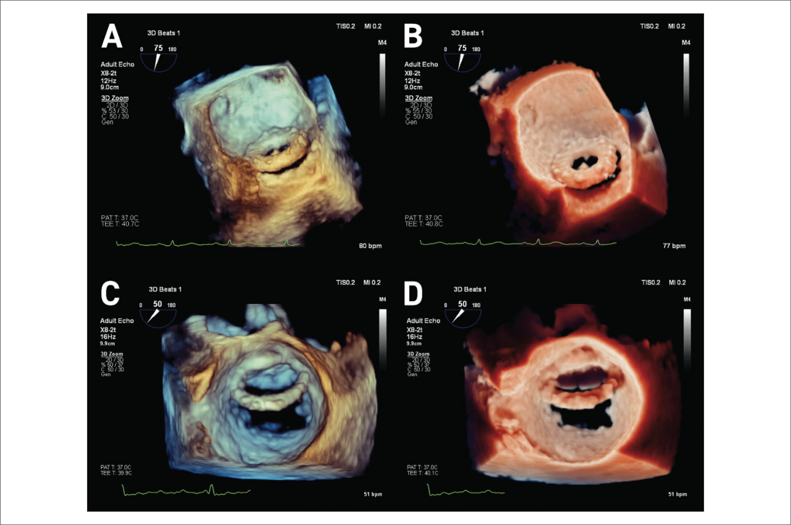
A 55-year-old man underwent mitral valve replacement with a caged-ball prosthesis (3M Starr-Edwards) at the age of 29. He was admitted with acute heart failure, functional New York Heart Association (NYHA) class III. Transthoracic echocardiography (TTE) showed a moderate-severe mitral periprosthetic leak; 3D-transesophageal echocardiography (TOE) showed prosthesis dehiscence, with a severe periprosthetic leak (). TI improved depth perception and accurate definition of the degree of prosthetic dehiscence, demonstrating a disinsertion of the mitral prosthesis, involving more than 50% of mitral circumference (; ).

[…]

  • Cardiovascular Diseases/diagnostic, imaging
  • Cardiovascular Diseases/physiopathology
  • Echocardiography, Three Dimensional / methods
  • Image Interpretation, Computer-Assisted
  • Mitral Valve/diagnosis
  • observer variation
  • Transillumination
Read Full Article
Creative Commons Attribution (CC BY) Creative Commons license
05/08/2022 DOI: 10.36660/abc.20210655
ABC
Editorial

Is it Possible to Non-Invasively Study the Hemodynamic Adaptations of Chagas Cardiomyopathy by the Volume-Time Curve Using 3D Echocardiography?

José Luiz Barros Pena

Three-dimensional echocardiography (3DE) represents a great innovation in cardiovascular ultrasound. Increased computer processing power and advances in the development of transducers have allowed acquiring of cardiac structures from any spatial point of view, without assumptions about their shape. Recent studies have demonstrated that when cardiac chamber sizes are quantified using 3DE, their volumes are similar to those obtained using cardiac magnetic resonance imaging compared to two-dimensional echocardiography (2DE)., The usefulness of 3DE has particularly been demonstrated primarily in realistic anatomical heart valve images and in guiding and monitoring cardiac procedures.

3DE allows volume calculation of the left ventricle (LV) throughout the cardiac cycle, making it possible to construct a volume-time curve. This method is more accurate than 2DE because the left ventricular volume is constructed by analyzing hundreds of points at the edge of the endocardium. No specific plane or geometric model is necessary to describe the complex LV structure. In this paper, Pinto et al. tested the hypothesis of studying the hemodynamic adaptations of non-invasive Chagas cardiomyopathy using the volume-time curve generated by 3DE. They generated a polynomial adjusted to the LV volume curve using specific software. Their objective was to present a cross-sectional study evaluating LV function, comparing volume curves in 20 patients with Chagas cardiomyopathy (CC) and 15 gender- and age-matched healthy controls.

[…]

  • Blood Pressure
  • Chagas Caddiomyopathy
  • Echocardiography, Three Dimensional / methods
  • Heart Failure
  • Stroke Volume
Read Full Article
Creative Commons Attribution (CC BY) Creative Commons license
10/06/2022 DOI: 10.36660/abc.20220284 Views 1
ABCIC
Original Article

Evaluation of Strain Parameters by Three Dimensional Speckle Tracking Echocardiography in Competitive Athletes

Edgar Daminello et al. Ana Clara Tude Rodrigues Adriana Cordovil Wércules Antônio Oliveira … Edgar Bezerra Lira Filho Rafael Bonafim Piveta Marcelo Luiz Campos Vieira Cláudio Henrique Fischer Samira S. Morhy

Summary

Introduction

Echocardiography is fundamental in the distinction between physiological adaptations promoted by physical activity and pathological abnormalities. Three-dimensional speckle tracking echocardiography (3D-STE) could prove accurate in detecting subclinical abnormalities in cardiac function.

Objectives

To determine the effect of exercise on the parameters of myocardial strain in athletes through 3D STE.

Method

Elite boxers underwent conventional three-dimensional echocardiography (3D-echo) and 3D-STE to analyze left ventricular (LV) volumes, left ventricular mass indexed to body surface area (LVMIBSA), ejection fraction (EF), longitudinal global strain (LGS), circumferential global strain (CGS), radial global strain (RGS), twist, torsion and tracking area. These data were compared with measurements performed on untrained control individuals.

Results

The analyses included 16 athletes and 14 controls with similar age (23 ± 4 vs. 21 ± 4 years; p = NS) and gender (14 vs. 12 males). LVEF was normal and similar in the 2 groups. LVMIBSA was higher in the athletes (83 ± 21 vs. 65 ± 15 g/m2, p < 0.05), as well as RGS (24.7 ± 5.2 vs. 16.3 ± 7.2; p = 0.007). There was no significant difference for the other parameters, such as CGS (-26 ± 2 vs. -28 ± 6), LGS (-16 ± 2 vs. -17 ± 3), twist (3.1 ± 1.3 vs. 3.7 ± 1.9), torsion (2.0 ± 0.8 vs. 1.4 ± 0.4) and tracking area (37 ± 4 vs. 41 ± 6).

Conclusion

Athletes and untrained individuals have comparable myocardial strain parameters on 3D-STE. However, an increase in RGS was observed only in the athletes. 3D-STE could help in the early detection of subclinical cardiac issues in athletes. (Arq Bras Cardiol: Imagem cardiovasc. 2017;30(3):92-97)
  • Athletes
  • Echocardiography, Three Dimensional / methods
  • Heart/diagnostic imaging
  • Parameters/analysis
  • Speckle Tracking
Read Full Article
Creative Commons Attribution (CC BY) Creative Commons license
04/02/2022 DOI: 10.5935/2318-8219.20170026 Views 1
Figure 1
ABC
Case Report

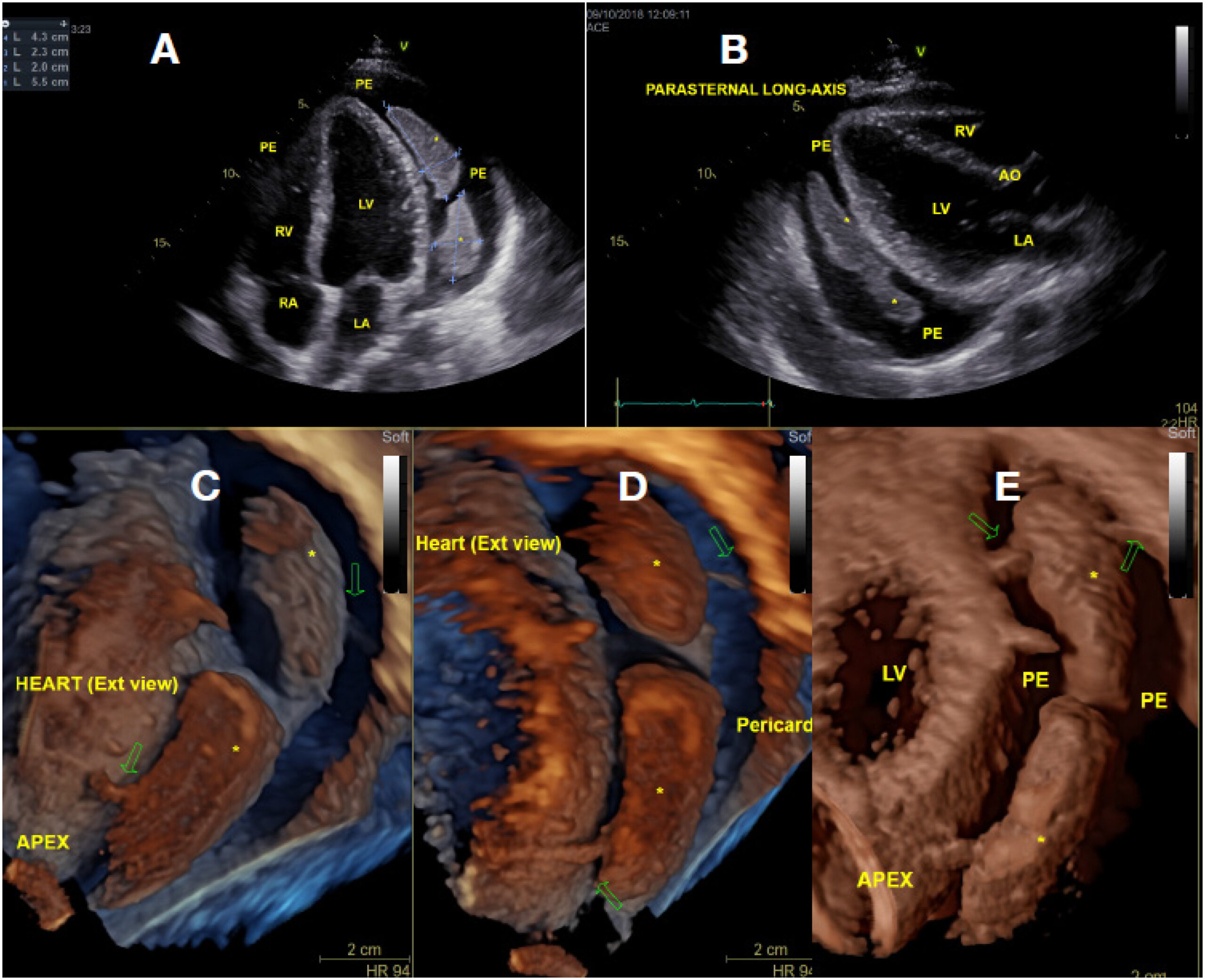
Pericardial Masses: A Rare Presentation of Tuberculous Pericarditis Documented by 3D Echocardiography

Alex dos Santos Felix et al. Viviane Belidio Pinheiro da Fonseca Rodrigo Coelho Segalote Larissa Franco de Andrade … Deborah Louize da Rocha Vianna Palmieri Ana Paula dos Reis Velloso Siciliano

Learning Points

[…]

  • Cardiac Tamponade/surgery
  • Diagnosis
  • Echocardiography, Three Dimensional / methods
  • Heart Neoplasms/surgery
  • Imaging/methods
  • Pericarditis, Tuberculous/physiopathology
Read Full Article
Creative Commons Attribution (CC BY) Creative Commons license
05/02/2021 DOI: 10.36660/abc.20190876
Figure 1
ABC
Case Report

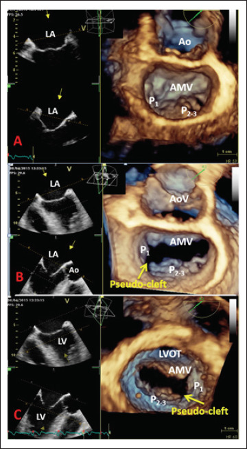
Three-dimensional Echocardiography Reveals the True Enemy in a Young Male with ST-Elevation Myocardial Infarction and Severe Mitral Regurgitation: Posterior Mitral Valve “Pseudo-Cleft” and Prolapse

Sorina Mihaila et al. Andreea Elena Velcea Luigi Paolo Badano Vinereanu Dragos Denisa Muraru

Introduction

Three-dimensional echocardiography (3DE) plays an increasingly important role in the diagnosis of valvular heart disease, in the assessment of valvular morphology in an anatomical manner, and in establishing valve repairability, beyond the limitations of conventional two-dimensional echocardiography (2DE).

We report the case of a young patient presenting with acute anterior ST-segment elevation myocardial infarction and severe mitral regurgitation (MR) after successful primary percutaneous coronary intervention (PCI) of the left anterior descending artery, whose three-dimensional transesophageal echocardiography (3D TEE) revealed an unexpected cause of the MR, namely, complex mitral valve (MV) pathology consisting of prolapse of the P2-3 scallops, flail chordae, and pseudo-cleft of the posterior leaflet separating the P1 from the P2 segment.

[…]

  • Diagnosis Imaging
  • Echocardiography, Three Dimensional / methods
  • Mitral Valve / abnormalities
  • Myocardial Infarction
  • Young Adult
Read Full Article
Creative Commons Attribution (CC BY) Creative Commons license
05/02/2021 DOI: 10.36660/abc.20190485
Figure 1
ABC
Original Article

Right Atrial Deformation Analysis in Cardiac Amyloidosis – Results from the Three-Dimensional Speckle-Tracking Echocardiographic MAGYAR-Path Study

Attila Nemes et al. Dóra Földeák Péter Domsik Anita Kalapos … Árpád Kormányos Zita Borbényi Tamás Forster

Abstract

Background:

Light-chain (AL) cardiac amyloidosis (CA) is characterized by fibril deposits, which are composed of monoclonal immunoglobulin light chains. The right ventricle is mostly involved in AL-CA and impairment of its function is a predictor of worse prognosis.

Objectives:

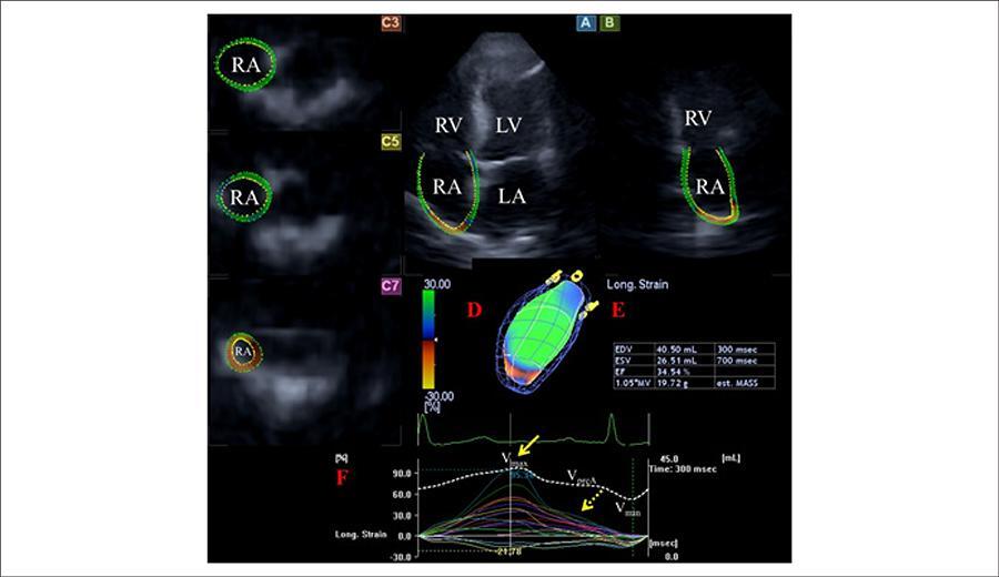
To characterize the volumetric and functional properties of the right atrium (RA) in AL-CA by three-dimensional speckle-tracking echocardiography (3DSTE).

Methods:

A total of 16 patients (mean age: 64.5 ± 10.1 years, 11 males) with AL-CA were examined. Their results were compared to that of 15 age- and gender-matched healthy controls (mean age: 58.9 ± 6.9 years, 8 males). All cases have undergone complete two-dimensional Doppler and 3DSTE. A two-tailed p value of less than 0.05 was considered statistically significant.

Results:

Significant differences could be demonstrated in RA volumes respecting cardiac cycle. Total (19.2 ± 9.3% vs. 27.9 ± 10.7%, p = 0.02) and active atrial emptying fractions (12.1 ± 8.1 vs. 18.6 ± 9.8%, p = 0.05) were significantly decreased in AL-CA patients. Peak global (16.7 ± 10.3% vs. 31.2 ± 19.4%, p = 0.01) and mean segmental (24.3 ± 11.1% vs. 38.6 ± 17.6%, p =0.01) RA area strains, together with some circumferential, longitudinal and segmental area strain parameters, proved to be reduced in patients with AL-CA. Global longitudinal (4.0 ± 5.2% vs. 8.2 ± 5.5%, p = 0.02) and area (7.8 ± 8.1% vs. 15.9 ± 10.3%, p = 0.03) strains at atrial contraction and some circumferential and area strain parameters at atrial contraction were reduced in AL-CA patients.

Conclusion:

Significantly increased RA volumes and deteriorated RA functions could be demonstrated in AL-CA.

  • Amyloidosis
  • Echocardiography, Three Dimensional / methods
  • Humans
  • Speckle Tracking
  • Ventricular Dysfunction Right
Read Full Article
Creative Commons Attribution (CC BY) Creative Commons license
20/08/2018 DOI: 10.5935/abc.20180150 Views 1
ABC
Editorial

New Method Improves the Assessment of Aortic Regurgitation Grade during TAVR by Aortography

Henrique B. Ribeiro

Transcatheter aortic valve replacement (TAVR) is a rapidly expanding alternative to surgical aortic valve replacement for patients deemed inoperable or at high or intermediate operative risk. Yet, residual aortic regurgitation (AR) secondary to paravalvular leaks (PVL) remains a procedural limitation. Although residual AR after TAVR is frequent, affecting up to approximately 70% of the treated patients,– it is moderate to severe in approximately 12% of these and steadily below 5% with current-generation devices, which come with specific sealing features. Noteworthy, moderate/severe AR has a detrimental clinical impact after TAVR, with a 3-fold increase in 30-day mortality and a 2.3-fold increase in 1-year mortality. Thus, its accurate assessment and quantification with a multimodality approach is key for appropriate utilization of additional procedures to reduce PVL, such as balloon post-dilatation (BPD), valve-in-valve, or leak closure.,

While Doppler echocardiography has been the most common method to assess AR following TAVR, its accurate quantification is challenging since AR jets are often multiple and eccentric.,– Therefore, other methods for proper AR assessment have been evaluated in recent years, such as 3D echocardiography, hemodynamic AR index, aortography and even cardiovascular magnetic resonance, each one with its specific advantages and disadvantages.,,

[…]

  • Aortic Valve Insufficiency/diagnostic imaging
  • Aortic Valve Insufficiency/mortality
  • Aortography/methods
  • Echocardiography, Three Dimensional / methods
  • Transcatheter Aortic Valve Replacement/methods
Read Full Article
Creative Commons Attribution (CC BY) Creative Commons license
01/08/2018 DOI: 10.5935/abc.20180158
Figure 1
ABC
Original Article

Left Atrial Deformation Analysis in Patients with Corrected Tetralogy of Fallot by 3D Speckle-Tracking Echocardiography (from the MAGYAR-Path Study)

Kálmán Havasi et al. Péter Domsik Anita Kalapos Jackie S. McGhie … Jolien W. Roos-Hesselink Tamás Forster Attila Nemes

Abstract

Background:

Three-dimensional (3D) echocardiography coupled with speckle-tracking echocardiographic (STE) capability is a novel methodology which has been demontrated to be useful for the assessment of left atrial (LA) volumes and functional properties. There is increased scientific interest on myocardial deformation analysis in adult patients with corrected tetralogy of Fallot (cTOF).

Objectives:

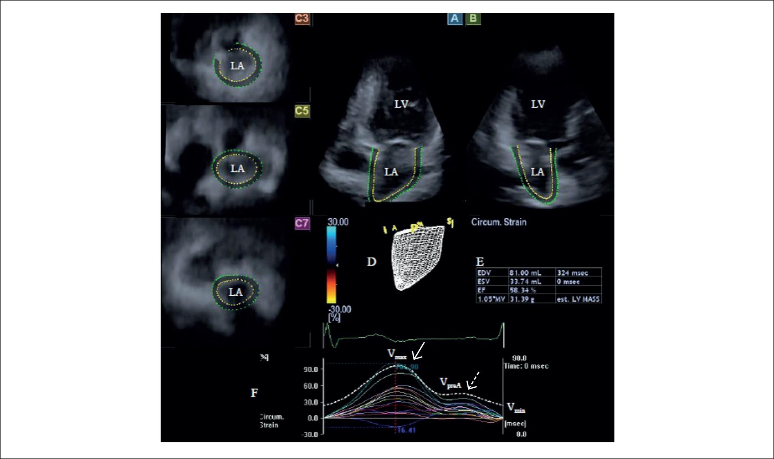
To compare LA volumes, volume-based functional properties and strain parameters between cTOF patients and age- and gender-matched healthy controls.

Methods:

The study population consisted of 19 consecutive adult patients with cTOF in sinus rhythm nursing at the University of Szeged, Hungary (mean age: 37.9 ± 11.3 years, 8 men, who had repair at the age of 4.1 ± 2.5 years). They all had undergone standard transthoracic two-dimensional Doppler echocardiographic study extended with 3DSTE. Their results were compared to 23 age- and gender-matched healthy controls (mean age: 39.2 ± 10.6 years, 14 men).

Results:

Increased LA volumes and reduced LA emptying fractions respecting cardiac cycle could be demonstrated in cTOF patients compared to controls. LA stroke volumes featuring all LA functions showed no differences between the 2 groups examined. LA global and mean segmental uni- and multidirectional peak strains featuring LA reservoir function were found to be diminished in adult patients with cTOF as compared to controls. Similarly to peak strains reduced global and mean segmental LA strains at atrial contraction characterizing atrial booster pump function could be demonstrated in cTOF patients as compared to controls.

Conclusions:

Significant deterioration of all LA functions could be demonstrated in adult patients with cTOF late after repair.

  • Echocardiography, Three Dimensional / methods
  • Heart Atria/abnormalities
  • Heart Defects, Congenital
  • Tetralogy of Fallot
Read Full Article
Creative Commons Attribution (CC BY) Creative Commons license
13/02/2017 DOI: 10.5935/abc.20170004 Views 1
Figure 1
ABC
Image

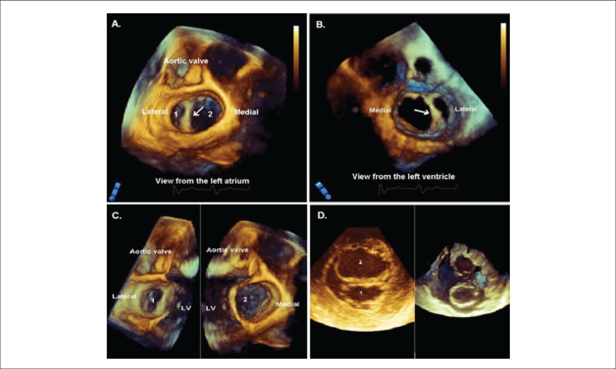
Isolated Double Orifice Mitral Valve – a Rare Finding in an Elderly Man

Andrea O Ciobanu et al. Sean Bennett Renato D Lopes Dragos Vinereanu

W

e

[…]

  • Aged
  • Echocardiography, Three Dimensional / methods
  • Heart Defects, Congenital
  • Mitral Valve / abnormalities
Read Full Article
Creative Commons Attribution (CC BY) Creative Commons license
01/05/2015 DOI: 10.5935/abc.20150043

How to cite this article

EndNote RIS

Electronic Document Format (Vancouver)

Armenta-Moreno JI, Berarducci J, Garcia-Cardenas AM, Armendariz-Ferrari JC, Bermudez-Gonzalez JL, Straface JI, Luna-Alvarez-Amezquita JA, et al. Transesophageal Two- and Three-Dimensional Echocardiographic Assessment of Spontaneous Left Atrial Dissection. Arquivos Brasileiros de Cardiologia 2022;119(3):499-501.

Electronic Document Format (ABNT)

Armenta-Moreno, Javier Ivan; Berarducci, Joaquin; Garcia-Cardenas, Abel Mauricio; Armendariz-Ferrari, José Carlos; Bermudez-Gonzalez, Jorge Luis; Straface, Juan Ignacio; Luna-Alvarez-Amezquita, Jose Antonio; Espinola-Zavaleta, Nilda. Transesophageal Two- and Three-Dimensional Echocardiographic Assessment of Spontaneous Left Atrial Dissection. Arquivos Brasileiros de Cardiologia, v. 119, n. 3, p. 499-501, Sep. 2022.

Electronic Document Format (APA)

Armenta-Moreno, J. I., Berarducci, J., Garcia-Cardenas, A. M., Armendariz-Ferrari, J. C., Bermudez-Gonzalez, J. L., Straface, J. I., Luna-Alvarez-Amezquita, J. A., & Espinola-Zavaleta, N. (2022). Transesophageal Two- and Three-Dimensional Echocardiographic Assessment of Spontaneous Left Atrial Dissection. Arquivos Brasileiros de Cardiologia, 119(3), 499-501.

Electronic Document Format (ISO)

Armenta-Moreno, Javier Ivan and Berarducci, Joaquin and Garcia-Cardenas, Abel Mauricio and Armendariz-Ferrari, José Carlos and Bermudez-Gonzalez, Jorge Luis and Straface, Juan Ignacio and Luna-Alvarez-Amezquita, Jose Antonio and Espinola-Zavaleta, Nilda. Transesophageal Two- and Three-Dimensional Echocardiographic Assessment of Spontaneous Left Atrial Dissection. Arquivos Brasileiros de Cardiologia [online]. 2022, vol. 119, n. 3, [cited 2026-10-14], pp. 499-501. Available from: <https://journals.cardiol.br/en/article/transesophageal-two-and-three-dimensional-echocardiographic-assessment-of-spontaneous-left-atrial-dissection/>. ISSN 0066-782X

EndNote RIS

Electronic Document Format (Vancouver)

Silva MR, Azevedo AI, Sampaio F, Ribeiro J, Fontes-Carvalho R. “Lightning The Heart” with 3D Echo: Transillumination of a Prosthetic Mitral Valve Dehiscence. Arquivos Brasileiros de Cardiologia 2022;119(2):358-60.

Electronic Document Format (ABNT)

Silva, Mariana Ribeiro; Azevedo, Ana Isabel; Sampaio, Francisco; Ribeiro, José; Fontes-Carvalho, Ricardo. “Lightning The Heart” with 3D Echo: Transillumination of a Prosthetic Mitral Valve Dehiscence. Arquivos Brasileiros de Cardiologia, v. 119, n. 2, p. 358-360, Aug. 2022.

Electronic Document Format (APA)

Silva, M. R., Azevedo, A. I., Sampaio, F., Ribeiro, J., & Fontes-Carvalho, R. (2022). “Lightning The Heart” with 3D Echo: Transillumination of a Prosthetic Mitral Valve Dehiscence. Arquivos Brasileiros de Cardiologia, 119(2), 358-360.

Electronic Document Format (ISO)

Silva, Mariana Ribeiro and Azevedo, Ana Isabel and Sampaio, Francisco and Ribeiro, José and Fontes-Carvalho, Ricardo. “Lightning The Heart” with 3D Echo: Transillumination of a Prosthetic Mitral Valve Dehiscence. Arquivos Brasileiros de Cardiologia [online]. 2022, vol. 119, n. 2, [cited 2026-10-14], pp. 358-360. Available from: <https://journals.cardiol.br/en/article/lightning-the-heart-with-3d-echo-transillumination-of-a-prosthetic-mitral-valve-dehiscence/>. ISSN 0066-782X

EndNote RIS

Electronic Document Format (Vancouver)

Pena JLB. Is it Possible to Non-Invasively Study the Hemodynamic Adaptations of Chagas Cardiomyopathy by the Volume-Time Curve Using 3D Echocardiography?. Arquivos Brasileiros de Cardiologia 2022;118(6):1106-7.

Electronic Document Format (ABNT)

Pena, José Luiz Barros. Is it Possible to Non-Invasively Study the Hemodynamic Adaptations of Chagas Cardiomyopathy by the Volume-Time Curve Using 3D Echocardiography?. Arquivos Brasileiros de Cardiologia, v. 118, n. 6, p. 1106-1107, Jun. 2022.

Electronic Document Format (APA)

Pena, J. L. B. (2022). Is it Possible to Non-Invasively Study the Hemodynamic Adaptations of Chagas Cardiomyopathy by the Volume-Time Curve Using 3D Echocardiography?. Arquivos Brasileiros de Cardiologia, 118(6), 1106-1107.

Electronic Document Format (ISO)

Pena, José Luiz Barros. Is it Possible to Non-Invasively Study the Hemodynamic Adaptations of Chagas Cardiomyopathy by the Volume-Time Curve Using 3D Echocardiography?. Arquivos Brasileiros de Cardiologia [online]. 2022, vol. 118, n. 6, [cited 2026-10-14], pp. 1106-1107. Available from: <https://journals.cardiol.br/en/article/is-it-possible-to-non-invasively-study-the-hemodynamic-adaptations-of-chagas-cardiomyopathy-by-the-volume-time-curve-using-3d-echocardiography/>. ISSN 0066-782X

EndNote RIS

Electronic Document Format (Vancouver)

Daminello E, Rodrigues ACT, Cordovil A, Oliveira WA, Lira Filho EB, Piveta RB, Vieira MLC, et al. Evaluation of Strain Parameters by Three Dimensional Speckle Tracking Echocardiography in Competitive Athletes. Arquivos Brasileiros de Cardiologia Imagem cardiovascular 2022;30(3):92-7.

Electronic Document Format (ABNT)

Daminello, Edgar; Rodrigues, Ana Clara Tude; Cordovil, Adriana; Oliveira, Wércules Antônio; Lira Filho, Edgar Bezerra; Piveta, Rafael Bonafim; Vieira, Marcelo Luiz Campos; Fischer, Cláudio Henrique; Morhy, Samira S.. Evaluation of Strain Parameters by Three Dimensional Speckle Tracking Echocardiography in Competitive Athletes. Arquivos Brasileiros de Cardiologia Imagem cardiovascular, v. 30, n. 3, p. 92-97, Feb. 2022.

Electronic Document Format (APA)

Daminello, E., Rodrigues, A. C. T., Cordovil, A., Oliveira, W. A., Lira Filho, E. B., Piveta, R. B., Vieira, M. L. C., Fischer, C. H., & Morhy, S. S. (2022). Evaluation of Strain Parameters by Three Dimensional Speckle Tracking Echocardiography in Competitive Athletes. Arquivos Brasileiros de Cardiologia Imagem cardiovascular, 30(3), 92-97.

Electronic Document Format (ISO)

Daminello, Edgar and Rodrigues, Ana Clara Tude and Cordovil, Adriana and Oliveira, Wércules Antônio and Lira Filho, Edgar Bezerra and Piveta, Rafael Bonafim and Vieira, Marcelo Luiz Campos and Fischer, Cláudio Henrique and Morhy, Samira S.. Evaluation of Strain Parameters by Three Dimensional Speckle Tracking Echocardiography in Competitive Athletes. Arquivos Brasileiros de Cardiologia Imagem cardiovascular [online]. 2022, vol. 30, n. 3, [cited 2026-10-14], pp. 92-97. Available from: <https://journals.cardiol.br/en/article/evaluation-of-strain-parameters-by-three-dimensional-speckle-tracking-echocardiography-in-competitive-athletes/>. ISSN 2318-8219

EndNote RIS

Electronic Document Format (Vancouver)

Felix AS, Fonseca VBP, Segalote RC, Andrade LF, Palmieri DLRV, Siciliano APRV. Pericardial Masses: A Rare Presentation of Tuberculous Pericarditis Documented by 3D Echocardiography. Arquivos Brasileiros de Cardiologia 2021;116(2 suppl 1):12-6.

Electronic Document Format (ABNT)

Felix, Alex dos Santos; Fonseca, Viviane Belidio Pinheiro da; Segalote, Rodrigo Coelho; Andrade, Larissa Franco de; Palmieri, Deborah Louize da Rocha Vianna; Siciliano, Ana Paula dos Reis Velloso. Pericardial Masses: A Rare Presentation of Tuberculous Pericarditis Documented by 3D Echocardiography. Arquivos Brasileiros de Cardiologia, v. 116, n. 2 suppl 1, p. 12-16, Feb. 2021.

Electronic Document Format (APA)

Felix, A. S., Fonseca, V. B. P. , Segalote, R. C., Andrade, L. F. , Palmieri, D. L. R. V., & Siciliano, A. P. R. V. (2021). Pericardial Masses: A Rare Presentation of Tuberculous Pericarditis Documented by 3D Echocardiography. Arquivos Brasileiros de Cardiologia, 116(2 suppl 1), 12-16.

Electronic Document Format (ISO)

Felix, Alex dos Santos and Fonseca, Viviane Belidio Pinheiro da and Segalote, Rodrigo Coelho and Andrade, Larissa Franco de and Palmieri, Deborah Louize da Rocha Vianna and Siciliano, Ana Paula dos Reis Velloso. Pericardial Masses: A Rare Presentation of Tuberculous Pericarditis Documented by 3D Echocardiography. Arquivos Brasileiros de Cardiologia [online]. 2021, vol. 116, n. 2 suppl 1, [cited 2026-10-14], pp. 12-16. Available from: <https://journals.cardiol.br/en/article/pericardial-masses-a-rare-presentation-of-tuberculous-pericarditis-documented-by-3d-echocardiography/>. ISSN 0066-782X

EndNote RIS

Electronic Document Format (Vancouver)

Mihaila S, Velcea AE, Badano LP, Dragos V, Muraru D. Three-dimensional Echocardiography Reveals the True Enemy in a Young Male with ST-Elevation Myocardial Infarction and Severe Mitral Regurgitation: Posterior Mitral Valve “Pseudo-Cleft” and Prolapse. Arquivos Brasileiros de Cardiologia 2021;116(2 suppl 1):36-8.

Electronic Document Format (ABNT)

Mihaila, Sorina; Velcea, Andreea Elena; Badano, Luigi Paolo; Dragos, Vinereanu; Muraru, Denisa. Three-dimensional Echocardiography Reveals the True Enemy in a Young Male with ST-Elevation Myocardial Infarction and Severe Mitral Regurgitation: Posterior Mitral Valve “Pseudo-Cleft” and Prolapse. Arquivos Brasileiros de Cardiologia, v. 116, n. 2 suppl 1, p. 36-38, Feb. 2021.

Electronic Document Format (APA)

Mihaila, S., Velcea, A. E., Badano, L. P., Dragos, V., & Muraru, D. (2021). Three-dimensional Echocardiography Reveals the True Enemy in a Young Male with ST-Elevation Myocardial Infarction and Severe Mitral Regurgitation: Posterior Mitral Valve “Pseudo-Cleft” and Prolapse. Arquivos Brasileiros de Cardiologia, 116(2 suppl 1), 36-38.

Electronic Document Format (ISO)

Mihaila, Sorina and Velcea, Andreea Elena and Badano, Luigi Paolo and Dragos, Vinereanu and Muraru, Denisa. Three-dimensional Echocardiography Reveals the True Enemy in a Young Male with ST-Elevation Myocardial Infarction and Severe Mitral Regurgitation: Posterior Mitral Valve “Pseudo-Cleft” and Prolapse. Arquivos Brasileiros de Cardiologia [online]. 2021, vol. 116, n. 2 suppl 1, [cited 2026-10-14], pp. 36-38. Available from: <https://journals.cardiol.br/en/article/three-dimensional-echocardiography-reveals-the-true-enemy-in-a-young-male-with-st-elevation-myocardial-infarction-and-severe-mitral-regurgitation-posterior-mitral-valve-pseudo-cleft/>. ISSN 0066-782X

EndNote RIS

Electronic Document Format (Vancouver)

Nemes A, Földeák D, Domsik P, Kalapos A, Kormányos , Borbényi Z, Forster T, et al. Right Atrial Deformation Analysis in Cardiac Amyloidosis – Results from the Three-Dimensional Speckle-Tracking Echocardiographic MAGYAR-Path Study. Arquivos Brasileiros de Cardiologia 2018;111(3):384-91.

Electronic Document Format (ABNT)

Nemes, Attila; Földeák, Dóra; Domsik, Péter; Kalapos, Anita; Kormányos, Árpád; Borbényi, Zita; Forster, Tamás. Right Atrial Deformation Analysis in Cardiac Amyloidosis – Results from the Three-Dimensional Speckle-Tracking Echocardiographic MAGYAR-Path Study. Arquivos Brasileiros de Cardiologia, v. 111, n. 3, p. 384-391, Aug. 2018.

Electronic Document Format (APA)

Nemes, A., Földeák, D., Domsik, P., Kalapos, A., Kormányos, , Borbényi, Z., & Forster, T. (2018). Right Atrial Deformation Analysis in Cardiac Amyloidosis – Results from the Three-Dimensional Speckle-Tracking Echocardiographic MAGYAR-Path Study. Arquivos Brasileiros de Cardiologia, 111(3), 384-391.

Electronic Document Format (ISO)

Nemes, Attila and Földeák, Dóra and Domsik, Péter and Kalapos, Anita and Kormányos, Árpád and Borbényi, Zita and Forster, Tamás. Right Atrial Deformation Analysis in Cardiac Amyloidosis – Results from the Three-Dimensional Speckle-Tracking Echocardiographic MAGYAR-Path Study. Arquivos Brasileiros de Cardiologia [online]. 2018, vol. 111, n. 3, [cited 2026-10-14], pp. 384-391. Available from: <https://journals.cardiol.br/en/article/right-atrial-deformation-analysis-in-cardiac-amyloidosis-results-from-the-three-dimensional-speckle-tracking-echocardiographic-magyar-path-study/>. ISSN 0066-782X

EndNote RIS

Electronic Document Format (Vancouver)

Ribeiro HB. New Method Improves the Assessment of Aortic Regurgitation Grade during TAVR by Aortography. Arquivos Brasileiros de Cardiologia 2018;111(2):203-4.

Electronic Document Format (ABNT)

Ribeiro, Henrique B.. New Method Improves the Assessment of Aortic Regurgitation Grade during TAVR by Aortography. Arquivos Brasileiros de Cardiologia, v. 111, n. 2, p. 203-204, Jul. 2018.

Electronic Document Format (APA)

Ribeiro, H. B. (2018). New Method Improves the Assessment of Aortic Regurgitation Grade during TAVR by Aortography. Arquivos Brasileiros de Cardiologia, 111(2), 203-204.

Electronic Document Format (ISO)

Ribeiro, Henrique B.. New Method Improves the Assessment of Aortic Regurgitation Grade during TAVR by Aortography. Arquivos Brasileiros de Cardiologia [online]. 2018, vol. 111, n. 2, [cited 2026-10-14], pp. 203-204. Available from: <https://journals.cardiol.br/en/article/new-method-improves-the-assessment-of-aortic-regurgitation-grade-during-tavr-by-aortography/>. ISSN 0066-782X

EndNote RIS

Electronic Document Format (Vancouver)

Havasi K, Domsik P, Kalapos A, McGhie JS, Roos-Hesselink JW, Forster T, Nemes A, et al. Left Atrial Deformation Analysis in Patients with Corrected Tetralogy of Fallot by 3D Speckle-Tracking Echocardiography (from the MAGYAR-Path Study). Arquivos Brasileiros de Cardiologia 2017;108(2):129-34.

Electronic Document Format (ABNT)

Havasi, Kálmán; Domsik, Péter; Kalapos, Anita; McGhie, Jackie S.; Roos-Hesselink, Jolien W.; Forster, Tamás; Nemes, Attila. Left Atrial Deformation Analysis in Patients with Corrected Tetralogy of Fallot by 3D Speckle-Tracking Echocardiography (from the MAGYAR-Path Study). Arquivos Brasileiros de Cardiologia, v. 108, n. 2, p. 129-134, Feb. 2017.

Electronic Document Format (APA)

Havasi, K., Domsik, P., Kalapos, A., McGhie, J. S., Roos-Hesselink, J. W., Forster, T., & Nemes, A. (2017). Left Atrial Deformation Analysis in Patients with Corrected Tetralogy of Fallot by 3D Speckle-Tracking Echocardiography (from the MAGYAR-Path Study). Arquivos Brasileiros de Cardiologia, 108(2), 129-134.

Electronic Document Format (ISO)

Havasi, Kálmán and Domsik, Péter and Kalapos, Anita and McGhie, Jackie S. and Roos-Hesselink, Jolien W. and Forster, Tamás and Nemes, Attila. Left Atrial Deformation Analysis in Patients with Corrected Tetralogy of Fallot by 3D Speckle-Tracking Echocardiography (from the MAGYAR-Path Study). Arquivos Brasileiros de Cardiologia [online]. 2017, vol. 108, n. 2, [cited 2026-10-14], pp. 129-134. Available from: <https://journals.cardiol.br/en/article/left-atrial-deformation-analysis-in-patients-with-corrected-tetralogy-of-fallot-by-3d-speckle-tracking-echocardiography-from-the-magyar-path-study/>. ISSN 0066-782X

EndNote RIS

Electronic Document Format (Vancouver)

Ciobanu AO, Bennett S, Lopes RD, Vinereanu D. Isolated Double Orifice Mitral Valve – a Rare Finding in an Elderly Man. Arquivos Brasileiros de Cardiologia 2015;104(5):e53-4.

Electronic Document Format (ABNT)

Ciobanu, Andrea O; Bennett, Sean; Lopes, Renato D; Vinereanu, Dragos. Isolated Double Orifice Mitral Valve – a Rare Finding in an Elderly Man. Arquivos Brasileiros de Cardiologia, v. 104, n. 5, p. e53-e54, Apr. 2015.

Electronic Document Format (APA)

Ciobanu, A. O., Bennett, S., Lopes, R. D., & Vinereanu, D. (2015). Isolated Double Orifice Mitral Valve – a Rare Finding in an Elderly Man. Arquivos Brasileiros de Cardiologia, 104(5), e53-e54.

Electronic Document Format (ISO)

Ciobanu, Andrea O and Bennett, Sean and Lopes, Renato D and Vinereanu, Dragos. Isolated Double Orifice Mitral Valve – a Rare Finding in an Elderly Man. Arquivos Brasileiros de Cardiologia [online]. 2015, vol. 104, n. 5, [cited 2026-10-14], pp. e53-e54. Available from: <https://journals.cardiol.br/en/article/isolated-double-orifice-mitral-valve-a-rare-finding-in-an-elderly-man/>. ISSN 0066-782X

  • About ABC Family
  • Internal Regulation
  • Editorial Board
  • Brazilian Society of Cardiology (SBC)

Issues

SBC Guidelines

  • Videos
  • Podcasts
  • News