ABC Family Arquivos Brasileiros de Cardiologia International Journal of Cardiovascular Sciences ABC Heart Failure & Cardiomyopathy ABC Imagem Cardiovascular
Português English
ABC Family
Português English
  • About
    • About ABC Family
    • Internal Regulation
    • Editorial Board
    • Brazilian Society of Cardiology (SBC)
  • Issues
  • SBC Guidelines
  • Content
    • Videos
    • Podcasts
    • News
  • About
    • About ABC Family
    • Internal Regulation
    • Editorial Board
    • Brazilian Society of Cardiology (SBC)
  • Issues
  • SBC Guidelines
  • Content
    • Videos
    • Podcasts
    • News

ABC Family

  • ABC Family
  • Arquivos Brasileiros de Cardiologia
  • International Journal of Cardiovascular Sciences
  • ABC Heart Failure & Cardiomyopathy
  • ABC Imagem Cardiovascular
Search in

No content found.

Journals
Ordering
Article types
Others

  1. Home
  2. Keyword: Tomography
ABCIC
Editorial

Coronary CT Angiography in 2025: Long-Term Evidence, Artificial Intelligence, and the Photon-Counting Era

Tiago A. Magalhães

Coronary CT Angiography (CCTA) has become established as a first-line test in the evaluation of chest pain, supported by robust evidence of prognostic impact, advances in Artificial Intelligence (AI), and technological evolution with photon-counting CT. Publications in 2025 reposition CCTA as a central platform for risk stratification and therapeutic guidance in coronary artery disease.

The 10-year follow-up of the SCOT-HEART trial represents a major milestone in this process. Among 4,146 patients with stable chest pain randomized to standard care with or without CCTA, there was a sustained reduction in death from coronary heart disease or nonfatal myocardial infarction in the CCTA-guided group (HR 0.79; 95% CI 0.63–0.99). Although revascularization rates did not differ between groups, the use of preventive therapies was more intensive and consistently maintained over time in the CCTA arm. These findings demonstrate that the direct identification of atherosclerosis leads to more aggressive preventive interventions, with durable effects on clinical outcomes, establishing CCTA as a disease-modifying tool in chronic coronary syndromes.

[…]

  • Coronary Angiography
  • Coronary Artery Disease
  • Tomography
Read Full Article
Creative Commons Attribution (CC BY) Creative Commons license
04/03/2026 DOI: 10.36660/abcimg.20250106i Views 8
Central Illustration
ABCIC
Review Article

My Approach To CAD-RADS 2.0 in the Assessment of Coronary Artery Disease by CT Angiogram

Bruno Maeda Fuzissima et al. Roberto Vitor Almeida Torres Renata Muller Couto Lucas de Pádua Gomes de Farias … Bernardo Salgado Pinto Oliveira José de Arimatéia Batista Araújo Filho

Abstract

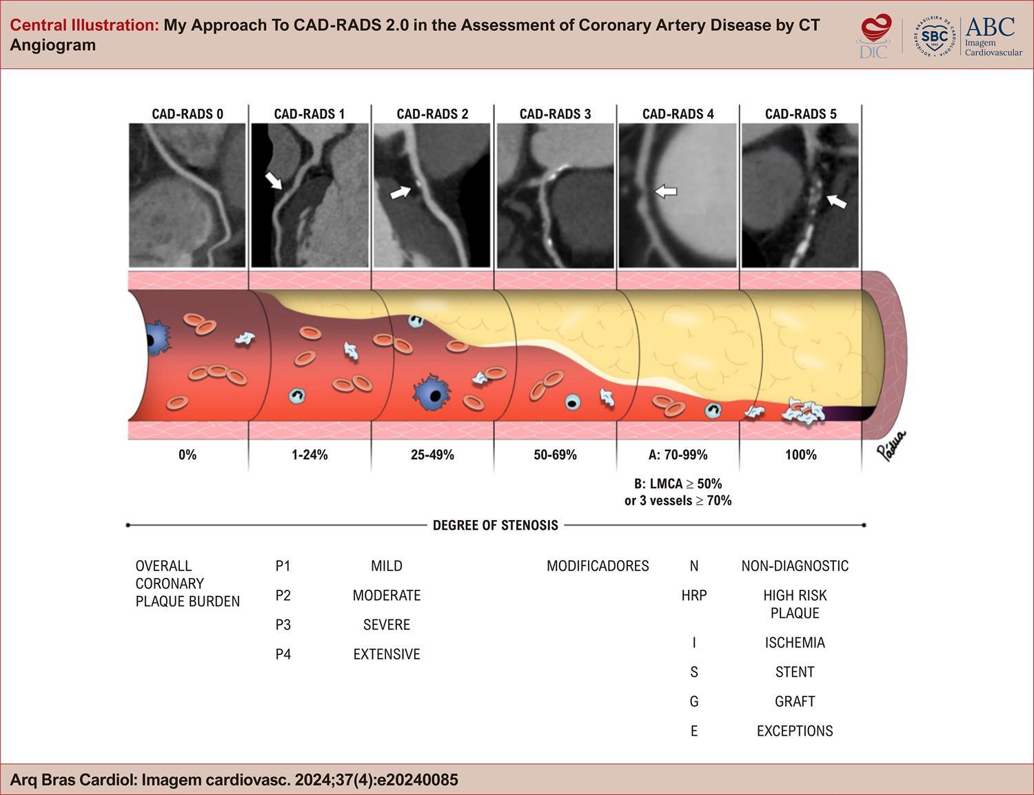
Coronary artery disease (CAD) is a common cause of chest pain, frequently seen in emergency departments and during elective consultations, and remains the leading cause of death worldwide. Coronary computed tomography angiography (CCTA) has become an established diagnostic tool for evaluating stable angina and acute chest pain in patients with low or intermediate risk. In this setting, CAD-RADS (Coronary Artery Disease-Reporting and Data System) was introduced as a CCTA reporting model, first published in 2016 and updated in 2022, with the aim of standardizing the description of imaging findings, improving communication between imaging specialists and clinicians, and providing guidance on patient management. This article reviews the CAD-RADS classification categories, which are based on the degree of stenosis, atherosclerotic plaque burden, and other modifying findings related to coronary abnormalities while also addressing certain exceptional situations. Through clinical cases, examples of structured reports, and recommendations for clinical management in different scenarios, we examine the objectives, benefits, limitations, and future potential of this classification, which remains underutilized in clinical practice.

  • Chest Pain
  • Coronary Artery Disease
  • Tomography
Read Full Article
Creative Commons Attribution (CC BY) Creative Commons license
21/11/2024 DOI: 10.36660/abcimg.20240085i
ABCIC
Review Article

The Indiscriminate Use Of Androgenic Anabolic Steroids: The Contribution of Cardiovascular Imaging

Irving Gabriel Araöşjo Bispo et al. Isabella Montanher Zago

Abstract

Anabolic androgenic steroids (AAS) are natural or synthetic compounds similar to the male hormone testosterone. The literature also describes cases of AAS abuse, such as in bodybuilding, cosmetics, recreational use, and aesthetic enhancement. It was discovered that AAS alone directly induce myocardial injury, with the main pathological finding in autopsied hearts being left ventricle (LV) hypertrophy in frequent association with myocyte hypertrophy, increased collagen deposition in the matrix, increased activity of the cardiac angiotensin, and myocardial fibrosis.

Patients who used illicit AAS present decreased LV systolic function. In athletes who self-administer AAS, LV hypertrophy and elevated sympathetic modulation were observed, as well as elevated blood pressure (BP). High doses of AAS can cause the user to have ventricular arrhythmias and sudden death. Furthermore, AAS users demonstrated greater coronary artery plaque volume than non-users. The objective of this review is to revisit the main effects of the use of AAS on cardiac changes through cardiovascular imaging as well as to establish a difference in relation to athletic heart syndrome.

  • Doppler Echocardiography
  • Global Longitudinal Strain
  • Steroids
  • Tomography
Read Full Article
Creative Commons Attribution (CC BY) Creative Commons license
21/03/2024 DOI: 10.36660/abcimg.20230113i
ABC
Editorial

Pericoronary Fat Attenuation on Computed Tomography Unveils a Guilty Factor of Coronary Artery Disease Associated with Anabolic–Androgenic Steroids

Jennifer Mancio

The illicit use of anabolic–androgenic steroids (AAS) is a growing public health problem that affects mainly nonathletes, bodybuilders, and young adults, including women. It is difficult to determine how many people use anabolic steroids for non-medical reasons. In a general population meta-analysis, 6.4% of males and 1.6% of females appealed to have used AAS in their lives. In Brazil, where aesthetic appearance, body cult, and beauty play a special social and economic role, the prevalence of AAS use can vary between 2.1% and 31.6%, according to the region. AAS are synthetic substances related to the male sex hormones, particularly testosterone. In normal doses and over a short time, AAS can improve muscle strength and increase lean body mass, but at higher doses (often 100 times above the clinical dose), AAS are associated with a 4.6-fold higher mortality rate compared to the general population. Several cases of acute cardiovascular events, including myocardial infarction and stroke (some of them fatal), have been described in the literature., The main pathophysiological mechanism behind this association in young adults is the promotion of an adverse lipidic and metabolic profile characterized by elevated LDL- and decreased HDL-cholesterol and insulin resistance, which, ultimately, can lead to atherosclerotic plaque build-up. Importantly, thrombosis without underlying atherosclerosis or vasospasm is also highly possible in AAS users, given its hypercoagulability state and increased blood viscosity because of thromboxane A2, and fibrinogen synthesis, inhibition of prostacyclin production, and increase erythropoiesis.

Diagnosing coronary artery disease (CAD) in AAS users (and abusers) may be challenging given its very low risk based on the classical risk scores. Computed tomography coronary angiography (CTCA) is the current first-line imaging method for the investigation of patients with suspicion of CAD. Despite its excellent spatial resolution, visual CTCA assessment has limitations in detecting coronary artery wall disease at early stages, when coronary plaques are not yet visible to the human eye. Researchers from the University of Oxford showed that more than half of the events happen in patients with non-obstructive plaques., Over the past 5 years, since the publication of the landmark CRISP-CT study, Oikonomou et al. have comprehensively demonstrated that coronary inflammation – the driver of CAD formation and instability – may be present despite the absence of visible plaques; they discovered that signals released from inflamed vessels into the surrounding perivascular adipose tissue block the differentiation of perivascular pre-adipocytes into mature, lipid-laden adipocytes. Thus, patients with CAD accumulate perivascular adipose tissue with smaller adipocyte size and lower lipid content near the vascular wall and, consequently, less negative fat attenuation values compared with non-diseased vessels. These changes in the perivascular fat composition can be tri-dimensionally captured by the Fat Attenuation Index (FAI) – a CTCA-derived biomarker developed to detect early CAD., Briefly, FAI is the mean perivascular fat signal around the vessel that can be obtained in any routinely acquired CTCA. This biomarker of coronary inflammation was developed and validated in large cohorts of European and American patients and further integrated (along with the cardiovascular risk factors) into a prognostic model to obtain the absolute individual risk of events. FAI calculation implies vessel wall contouring in contrasted coronary arteries, which is a time-consuming and operator-dependent task. Currently, an AI-tool is available to automatically segment the perivascular fat and provide each subject risk score for use in clinical practice.

[…]

  • Coronary Artery Disese
  • Plaque Atherosclerotic/pathology
  • Risk Factors
  • Testosterone Congeners/toxicity
  • Tomography
  • X-Ray Computed/methods
Read Full Article
Creative Commons Attribution (CC BY) Creative Commons license
11/03/2024 DOI: 10.36660/abc.20230843 Views 1
Figure 1
ABCIC
Original Article

Diagnostic Accuracy of the Coronary Calcium Score Acquired with Reduced Radiation Dose and Iterative Reconstruction

Daniela Rodrigues Carnaval et al. Adriano Liebl Miguel Morita Fernandes Silva Bruno Marques Sartori … Caio Nogara de Menezes Couto Gabriel Bergonse Garaluz da Silva Ítalo Gabriel Beltrame Vazzoler Vinícius Maksoud Medeiros Rodrigo Julio Cerci

Abstract

Background:

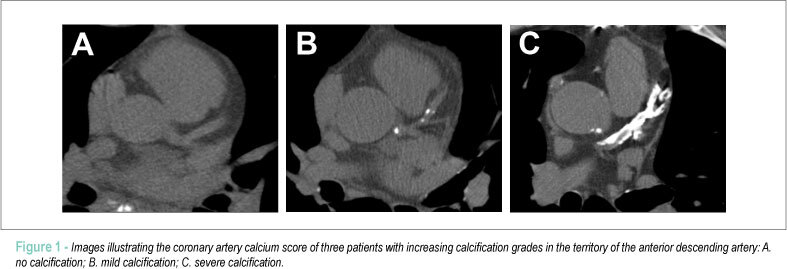
The calcium score is an imaging test used to evaluate cardiovascular risk by detecting coronary artery calcification (CAC). However, exposure to ionizing radiation during computed tomography (CT) has been a concern.

Objective:

The objective of this study was to evaluate radiation dose and the diagnostic quality of the calcium score using reduced tube voltage and iterative image reconstruction.

Methods:

This was a cross-sectional, observational study with patients over 18 years of age. Patients with previous coronary artery disease and patients who declined to participate were excluded. We acquired calcium score twice (120 kV and 100 kV) with different iterative reconstruction techniques. Two professionals interpreted the exams.

Results:

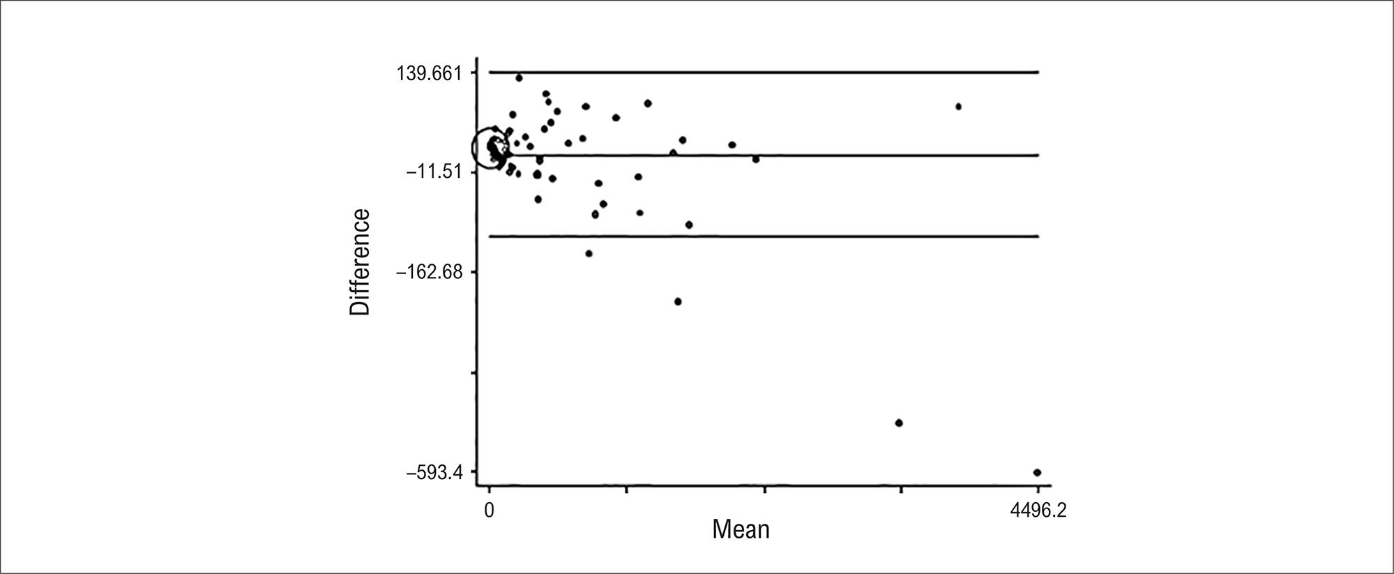
We evaluated 153 patients, and the reduction in radiation dose between acquisitions with 120 kV and 100 kV was 43%. The agreement of the degree of CAC between the different acquisitions was high, ranging from 93.9% to 96.2%. In Bland-Altman analysis, we observed a slight overestimation of the results in the acquisition with 100 kV compared to the acquisition with 120 kV.

Conclusion:

Calcium score acquired at 100 kV using Iterative Model Reconstruction (IMR) iterative reconstruction resulted in a significant reduction in radiation dose. Furthermore, agreement with acquisition at 120 kV was high, indicating that this approach may be a viable alternative to decrease radiation exposure during calcium score testing. However, it is important to underscore the slight overestimation of the results in the acquisition with 100 kV, which must be considered in the clinical interpretation.

  • Coronary Artery Disease
  • Radiation Dosage
  • Tomography
  • Vascular Calcification
Read Full Article
Creative Commons Attribution (CC BY) Creative Commons license
02/10/2023 DOI: 10.36660/abcimg.20230069i Views 1
Figure 1
ABC
Original Article

Predicting Significant Coronary Obstruction in a Population with Suspected Coronary Disease and Absence of Coronary Calcium: CORE-64 / CORE320 Studies

Anderson C. Armstrong et al. Rodrigo Cerci Matthew B. Matheson Tiago Magalhães … Satoru Kishi Jeff Brinker Melvin E. Clouse Carlos E. Rochitte Christopher Cox João A. C. Lima Armin Arbab-Zadeh

Abstract

Background

Coronary artery calcium (CAC) scanning can be performed using non-contrast computed tomography to predict cardiovascular events, but has less value for risk stratification in symptomatic patients.

Objective

To identify and validate predictors of significant coronary obstruction (SCO) in symptomatic patients without coronary artery calcification.

Methods

A total of 4,258 participants were screened from the CORE64 and CORE320 studies that enrolled patients referred for invasive angiography, and from the Quanta Registry that included patients referred for coronary computed tomography angiography (CTA). Logistic regression models evaluated associations between cardiovascular risk factors, CAC, and SCO. An algorithm to assess the risk of SCO was proposed for patients without CAC. Significance level of 5% was used in the analyses.

Results

Of the 509 participants of the CORE study, 117 (23%) had zero coronary calcium score; 13 (11%) patients without CAC had SCO. Zero calcium score was related to younger age, female gender, lower body mass index, no diabetes, and no dyslipidemia. Being a current smoker increased ~3.5 fold the probability of SCO and other CV risk factors were not significantly associated. Considering the clinical findings, an algorithm to further stratify zero calcium score patients was proposed and had a limited performance in the validation cohort (AUC 58; 95%CI 43, 72).

Conclusion

A lower cardiovascular risk profile is associated with zero calcium score in a setting of high-risk patients. Smoking is the strongest predictor of SCO in patients without CAC.

  • Chest Pain
  • Coronary Angiography
  • Coronary Artery Disease / complications
  • Diagnostic Imaging/methods
  • Indice Calcio
  • Tomography
  • X-Ray Computed/methods
Read Full Article
Creative Commons Attribution (CC BY) Creative Commons license
20/03/2023 DOI: 10.36660/abc.20220183
Figure 1
ABC
Image

Bulky Mitral Annulus Caseous Calcification in an Atypical Location

Joana Laranjeira Correia et al. Miguel Correia

Abstract

Mitral annular calcification (MAC) is a commonly observed chronic and degenerative fibrotic process of the base of the mitral valve, usually deemed as an incidental finding. Although initially, MAC was thought to be a consequence of an age-related degenerative process, recent findings suggest other independent contributive mechanisms, such as atherosclerosis and abnormal calcium-phosphorus metabolism.

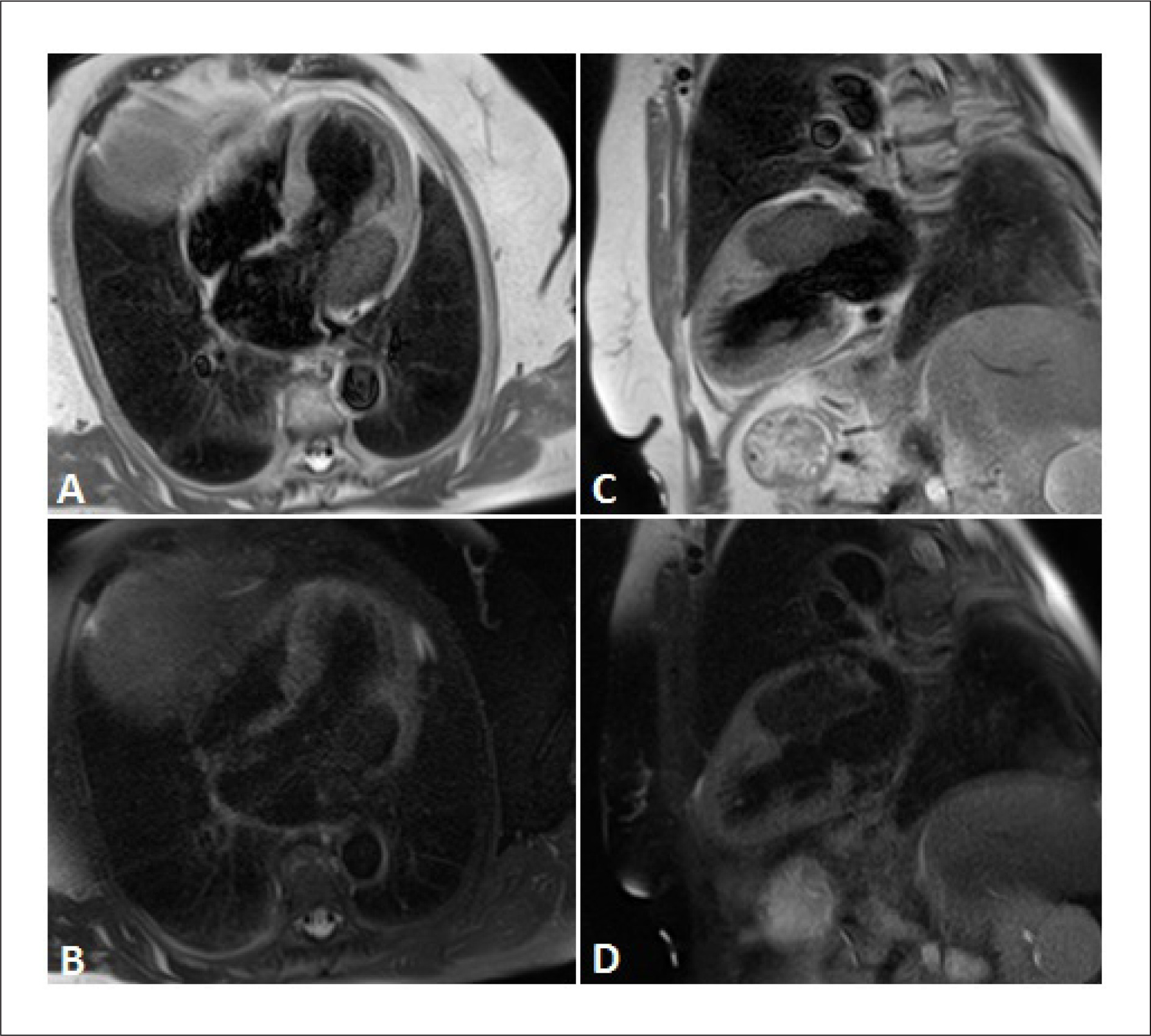
Caseous calcification of the mitral annulus (cMAC) is a rarely described variant of MAC, characterized by an ovoid, focal mass with internal caseous fluid-like calcifications and debris.

Differentiating a cMAC from other cardiac masses attached to the mitral annulus may be challenging. A single imaging modality, such as transthoracic echocardiography, may not be sufficient for a clear diagnosis. Therefore, a multimodal imaging approach is necessary, including cardiac tomography computerized imaging and cardiac magnetic resonance (CMR).

MAC and cMAC typically affect the posterior mitral annulus, with very few cases in the literature describing the involvement of the anterior annulus. We present a rare case of an anterior mitral annulus caseous calcification found in a CMR performed to evaluate a left atrial mass identified on a transthoracic echocardiogram.

  • Cardiac Imaging Techniques
  • Stroke
  • Tomography
  • X-Ray Computed
Read Full Article
Creative Commons Attribution (CC BY) Creative Commons license
19/12/2022 DOI: 10.36660/abc.20210949 Views 1
Figure 1
IJCS
Image

A New Kid on the Block in POCUS: “LISA”

Alex dos Santos Felix et al. Marcelo Dantas Tavares de Melo Thaíssa Santos Monteiro Maria Carolina Terra Cola … Rafael Castro Nicolas Merke

A new kid on the Block in POCUS: “LISA”

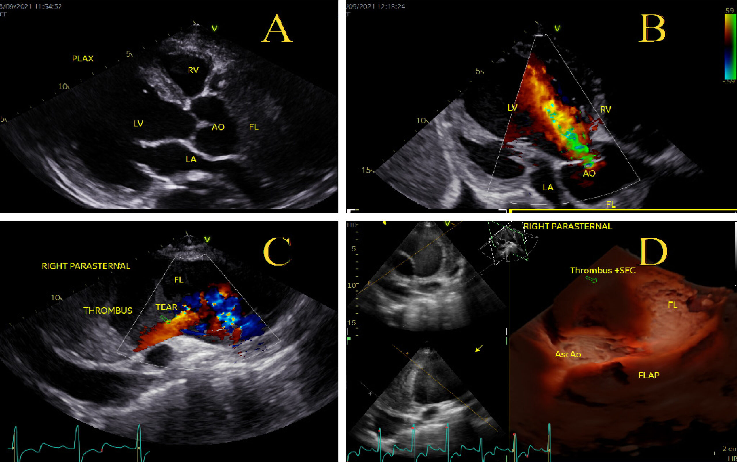
Transesophageal echocardiography (TTE) and computed tomography angiography (CTA) are methods of choice for definitive diagnosis and prognostic stratification of acute aortic syndromes (AAS). Point-of-care ultrasound (POCUS) has important applications in the initial workup of these patients in the emergency room (ER), ruling out other potential causes of chest pain and allowing a faster diagnosis, although conventional transthoracic echocardiographic acoustic windows do not allow for imaging of some segments of the thoracic aorta (TA), especially the descending TA (DTA).

A 49-year-old man, with a 20-day history of dorsal and abdominal pain, shortness of breath (NYHA-III) and peripheral edema was admitted to the hospital ER. He had uncontrolled systemic hypertension and a history of smoking, without a family history of aortic diseases or sudden death.

[…]

  • Aortic Diseases/diagnostic, imaging
  • Echocardiography
  • Takayasu Arteritis/complications
  • Tomography
  • Transesophageal/methods
  • X-Ray Computed/methods
Read Full Article
Creative Commons Attribution (CC BY) Creative Commons license
16/05/2022 DOI: 10.36660/ijcs.20210285
Figure 1
ABCIC
Case Report

Rare Incidental Finding on Coronary Artery CT Angiography: Left Main Coronary Artery Atresia

Maria Eduarda Menezes de Siqueira et al. Mayra Isabel Dias Ana Claudia Vincezi Raudan Uski Luciano Figueiredo de Aguiar Filho

Introduction

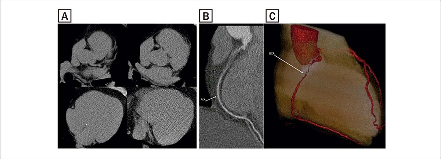
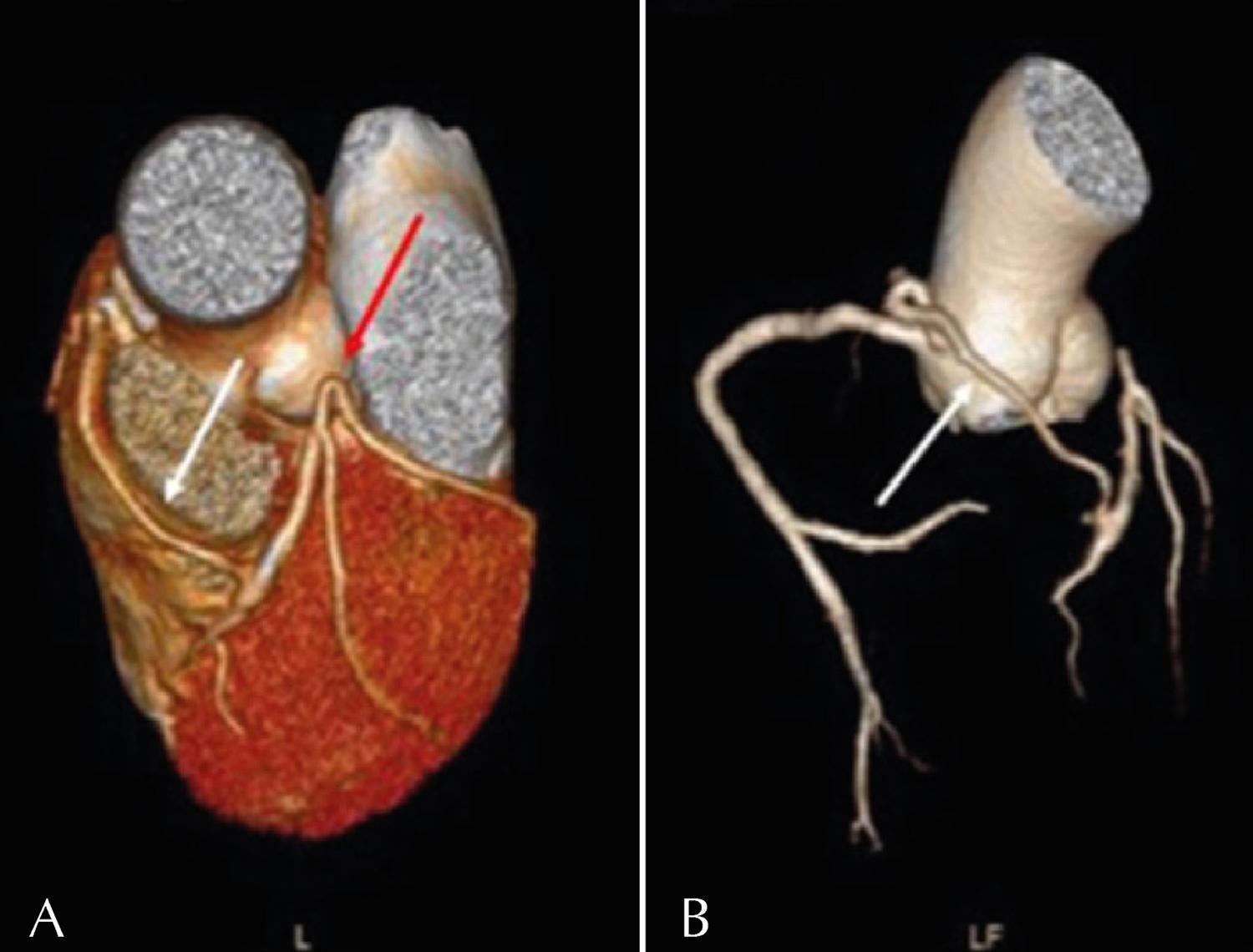
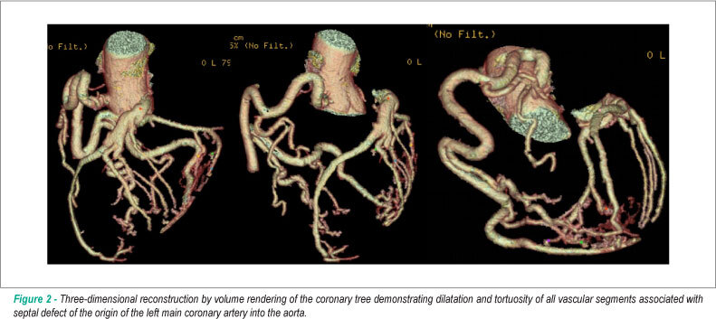
Congenital left main coronary artery atresia (CLMCAA) is a coronary anomaly in which the left coronary ostium or trunk is absent. The anterior descending (AD) and circumflex (CX) arteries are usually connected and proximally end blindly. Left chamber perfusion is provided by the right coronary (RC) collaterals. It is the rarest coronary anomaly, with fewer than 70 cases described in the literature. The clinical CLMCAA spectrum ranges from asymptomatic cases (due to an efficient collateral system) to symptoms such as angina, tachyarrhythmias, syncope, and sudden death., Surgical revascularization is recommended in symptomatic patients; however, preventive surgical treatment is controversial in asymptomatic patients without inducible myocardial ischemia.–

[…]

  • Abnormality, Heart
  • Anomalous Left Coronary Artery
  • Tomography
Read Full Article
Creative Commons Attribution (CC BY) Creative Commons license
25/03/2022 DOI: 10.47593/2675-312X/20213404eabc239
ABCIC
Image

A Deceiving Aorta

Helder Santos et al. Hugo Miranda Inês Almeida Mariana Santos … Samuel Almeida Lurdes Almeida

[…]

  • Aortic Aneurysm
  • Aortic Dissection
  • Tomography
  • Transthoracic Echocardiography
Read Full Article
Creative Commons Attribution (CC BY) Creative Commons license
14/01/2022 DOI: 10.5935/2318-8219.20190055
ABCIC
Editorial

What does the Cardiologist Expect from the Echocardiogram of a Transcatheter Aortic Valve Implant?

Gláucia Maria Moraes de Oliveira et al. Marcelo Antônio Cartaxo Queiroga Lopes

[…]

  • Aortic Valve Stenosis
  • Echocardiography
  • Magnetic Resonance
  • Tomography
Read Full Article
Creative Commons Attribution (CC BY) Creative Commons license
14/01/2022 DOI: 10.5935/2318-8219.20200024 Views 1
ABCIC
Case Report

Cardiac Metastasis in Patient with Rectal Carcinoid Tumor

Iuri Shen de Lacerda et al. Priscilla Ornellas Neves Lucianno Henrique Pereira Santos Joalbo Matos Andrade

[…]

  • Carcinoid Tumor
  • Heart
  • Magnetic Resonance Imaging
  • Neoplasm Metastasis
  • Tomography
Read Full Article
Creative Commons Attribution (CC BY) Creative Commons license
14/01/2022 DOI: 10.5935/2318-8219.20190052
ABCIC
Case Report

A Three-Leaflets Mitral Valve Associated to Three Papillary Muscles in a Patient with Hypertrophic Cardiomyopathy

Antonio Tito Paladino et al. Luiz Antônio Vasconcelos Leonardo Mello Guimarães de Toledo Natalia Massoni … Mariana Oliveira Rezende Natasha Soares Simões dos Santos David Costa de Souza Le Bihan Rodrigo Bellio de Mattos Barretto Jorge Eduardo Assef

Abstract

Mitral valve malformations are considered rare even in their association with other anomalies, except for mitral valve prolapse, which is found in approximately 10% of the population. Mitral valve abnormalities may affect any portion of the valve or subvalvular apparatus (annulus, cusp, tendinous cords or papillary muscles).1,2 Mitral valve anatomy and function can be assessed by echocardiography, resonance or cardiac tomography, and is essential for potential therapeutic programming.2

  • Echocardiography
  • Heart Defects, Congenital
  • Magnetic Resonance Imaging
  • Tomography
Read Full Article
Creative Commons Attribution (CC BY) Creative Commons license
14/01/2022 DOI: 10.5935/2318-8219.20200010 Views 1
ABCIC
Original Article

Fractional Flow Reserve by Tomography Diagnostic Performance in the Detection of Coronary Stenoses Hemodynamically Significant

Carlos Eduardo Elias dos Prazeres et al. Natália Boing Salvatti Hilka dos Santos Moraes de Carvalho Isaac Torquato Queiroz e Silva … Adriano Camargo de Castro Carneiro Tiago Augusto Magalhães Valéria de Melo Moreira Juliana Hiromi Silva Matsumoto Bello Carlos Eduardo Rochitte

Abstract

Background

Coronary computed tomography angiography is a noninvasive anatomic test for the diagnosis of coronary artery disease. The coronary fractional flow reserve is the gold standard method to determine if coronary stenosis is hemodynamically significant but requires an invasive procedure. Fractional flow reserve measurement by tomography is a new method to determine the hemodynamic significance of coronary luminal reduction.

Objective

To compare the accuracy of the invasive fractional flow reserve with the fractional flow reserve measurement by tomography.

Method

Patients submitted to invasive coronary computed tomography angiography and fractional flow reserve were included in the study. Flow-restricted stenosis was defined by fractional flow reserve measurement by tomography and fractional flow reserve ≤ 0.8, and anatomically significant coronary artery disease was defined as stenosis ≥ 50%. Diagnostic performance of fractional flow reserve measurement by tomography and stenosis by coronary computed tomography angiography was accessed with invasive fractional flow reserve as the gold standard.

Results

Among the patients, 33% had fractional flow reserve ≤0.8. There was no significant difference between the fractional flow reserve measurement by tomography and fractional flow reserve averages (0.87 versus 0.84; p = 0.4). The correlation between the values of the methods was r = 0.77, and concordance was moderate (k = 0.54). When fractional flow reserve ≤ 0.75 was analyzed, the concordance between the methods was absolute. The sensitivity, specificity, positive predictive value and negative predictive value of fractional flow reserve measurement by tomography were, respectively, 50%, 100%, 100% and 75%, for the threshold of 0.8. The mean difference between fractional flow reserve and fractional flow reserve measurement by tomography was -0.033 (95%CI -0.072-0.007), with concordance limits between -0.176 and -0.1111.

Conclusion

Fractional flow reserve measurement by tomography is a new method with high diagnostic performance in the detection of hemodynamically significant coronary stenoses and significantly elevates the specificity of the isolated coronary computed tomography angiography.

  • Coronary Disease
  • Diagnostic Imaging
  • Myocardial Ischemia
  • Tomography
Read Full Article
Creative Commons Attribution (CC BY) Creative Commons license
14/01/2022 DOI: 10.5935/2318-8219.20200037 Views 3
Figure 1
ABC
Case Report

Anomalous Origin of the Circumflex Coronary Artery from the Right Pulmonary Artery: Diagnosis Through Cardiac CT

Bebiana Faria et al. Lucy Calvo Sílvia Ribeiro Catarina Ruivo António Lourenço

Introduction

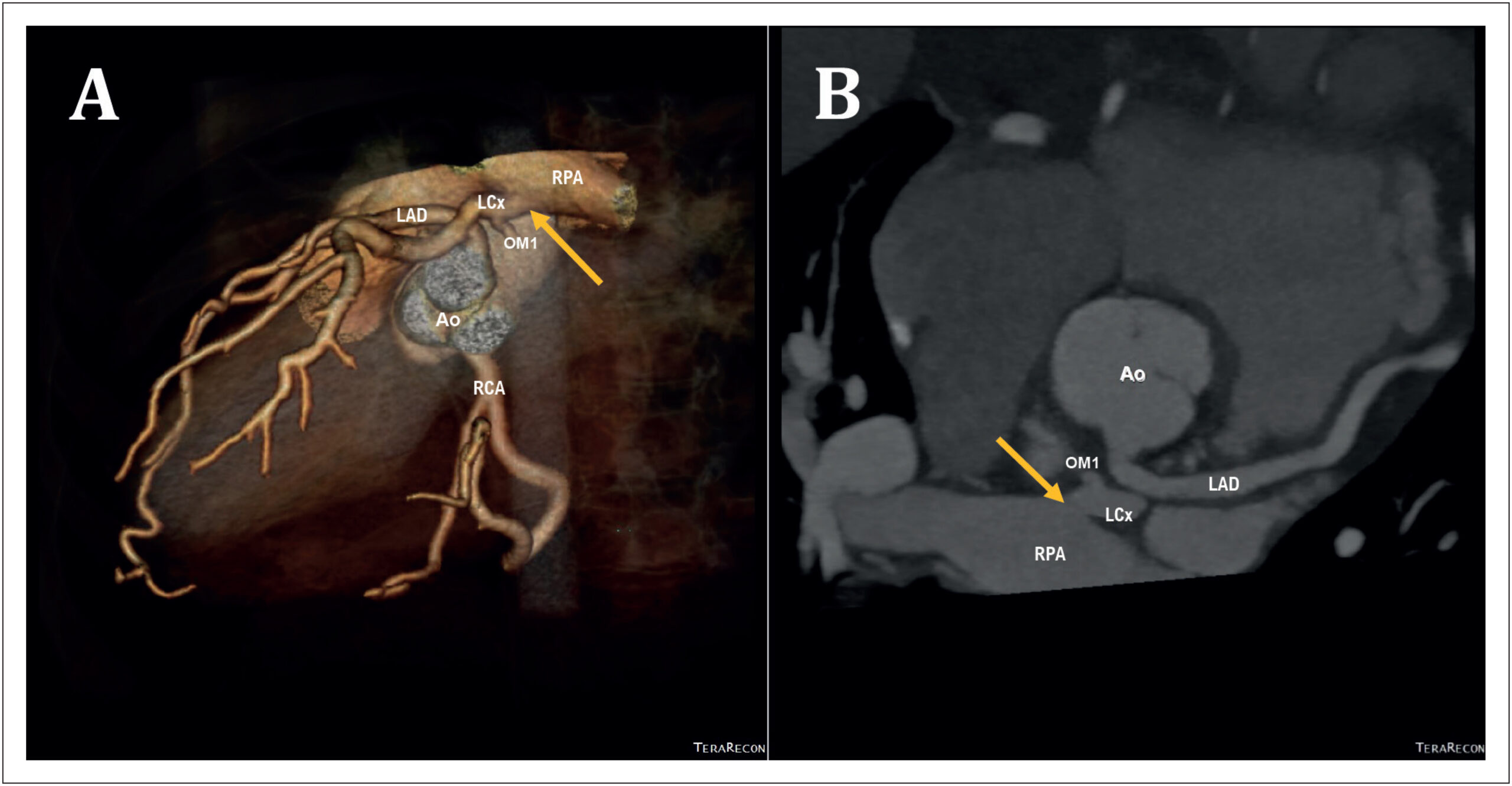
Anomalous origin of the left circumflex coronary artery (LCx) from the right pulmonary artery (PA) is an extremely rare coronary anomaly. Although the clinical course may be silent, the risk of sudden cardiac death is increased. The symptoms are related to collateralization and amount of myocardium that it supplies, and some patients need surgical treatment.,

[…]

  • Computed/methods
  • Congenital
  • Congenital Abnormalities
  • Coronary Artery Disease
  • Coronary Vessels
  • Heart Defects
  • Pulmonary Artery
  • Tomography
Read Full Article
Creative Commons Attribution (CC BY) Creative Commons license
19/07/2021 DOI: 10.36660/abc.20200060 Views 1
Figure 1
ABC
Case Report

Spider-Like Coronary Anatomy; the True Spider!

Levent Cerit et al. Hamza Duygu Kamil Gülşen Hatice Kemal Barcin Ozcem

Introduction

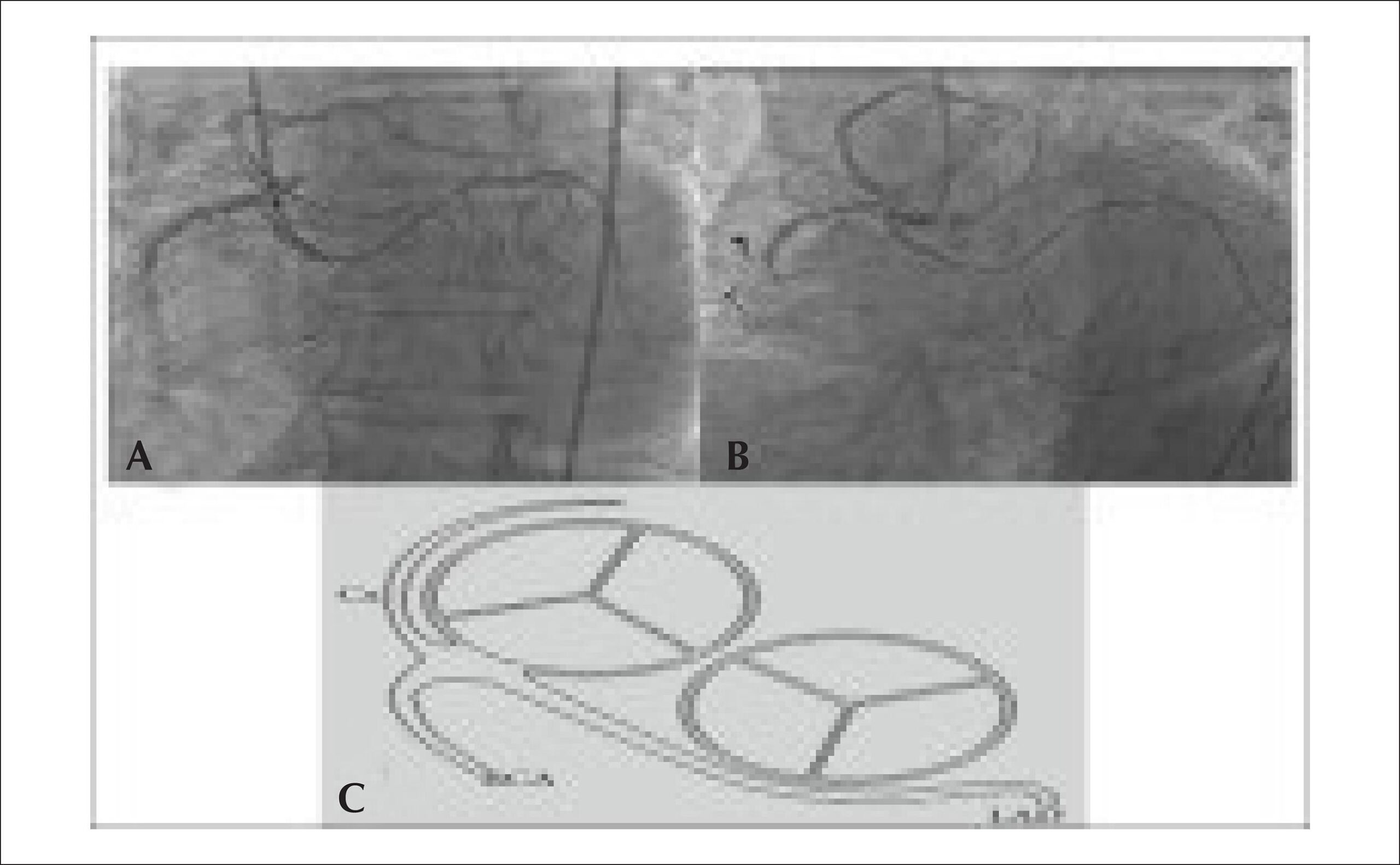
Coronary anomalies are uncommon, affecting approximately 0.3-5.6% of the general population according to the literature. However, some cases can result in severe life-threatening events, such as myocardial ischemia, arrhythmia, and acute myocardial infarction.,

[…]

  • Coronary Vessels / anatomy & histology
  • Coronary Vessels Anomalies
  • Tomography
  • X-Ray Computed
Read Full Article
Creative Commons Attribution (CC BY) Creative Commons license
01/10/2017 DOI: 10.5935/abc.20170101
Figure 1
ABC
Image

Patient with Atrial Myxoma and Signs of Obstruction of the Left Ventricular Outflow Tract

Antonio Fernando Diniz Freire et al. Alexandre Anderson de Sousa Soares Tatiana de Carvalho Andreucci Torres Leal Mucio Tavares de Oliveira Junior Alexandre de Matos Soeiro

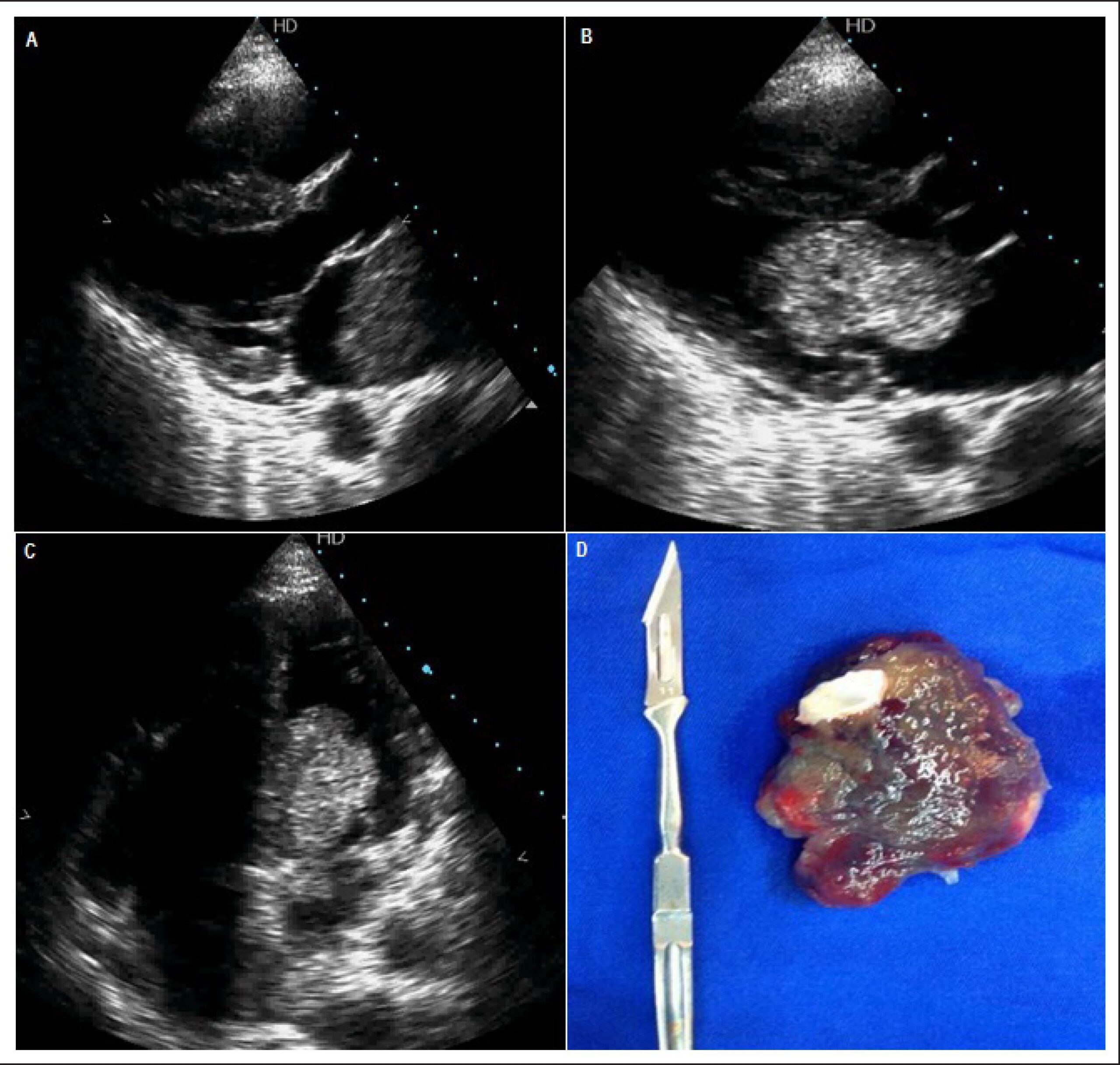
Female patient, 45 years-old, reported dyspnea on routine efforts with 6 months of evolution with dry cough. Previously healthy she refused to take regular medication. According to the physical examination, she had a heart rate of 105 bpm, blood pressure of 90x60mmHg, respiratory frequency 30 incursions per minute, arterial saturation of 88%, slow capillary filling time, rhythmic sounds, diastolic rumble murmur (+++/6) in mitral focus and bibasal pulmonary crepitations. Chest X-Ray showed a discreet widening of the mediastinum. Chest CT was performed to evaluate the mediastinum and pulmonary parenchyma and an intracardiac mass was found. It evolved with hypotension and worsening of pulmonary congestion.

Emergency echocardiogram showed a moving rounded hyperechoic image in the left atrium, measuring 66x36mm with a pedicle adhered to the membrane of the oval fossa dislocating to the left ventricle (LV) during systole causing hemodynamic repercussion (signs of LV outflow tract obstruction). The patient underwent emergency surgery. Retraction of the tumor revealed a cleft in the anterior cusp of the mitral valve and it was closed. In the immediate postoperative, the patient developed cardiogenic shock refractory to vasoactive drugs; an intra-aortic balloon was implanted, but the patient died 30 hours later. Anatomopathological confirmed the diagnosis of a myxoma: 7.5×4.6×3.4cm. In conclusion, the atrial myxoma with obstruction of the outflow tract of the LV is rare, its clinical manifestations can mislead the evaluator, clinical suspicion and the correct use of propaedeutic are essential for early diagnosis and successful clinical treatment.

[…]

  • Heart Atria/abnormalities
  • Hypotension
  • Myxoma/surgery
  • Tomography
Read Full Article
Creative Commons Attribution (CC BY) Creative Commons license
01/03/2017 DOI: 10.5935/abc.20170007
Figure 1
ABC
Review Article

Multimodality Imaging of Heart Valve Disease

Ronak Rajani et al. Rajdeep Khattar Amedeo Chiribiri Kelly Victor John Chambers

Unidentified heart valve disease is associated with a significant morbidity and mortality. It has therefore become important to accurately identify, assess and monitor patients with this condition in order that appropriate and timely intervention can occur. Although echocardiography has emerged as the predominant imaging modality for this purpose, recent advances in cardiac magnetic resonance and cardiac computed tomography indicate that they may have an important contribution to make. The current review describes the assessment of regurgitant and stenotic heart valves by multimodality imaging (echocardiography, cardiac computed tomography and cardiac magnetic resonance) and discusses their relative strengths and weaknesses.

  • Cardiac Imaging
  • Diagnostic Imaging
  • Heart Valve Diseases
  • Magnetic Resonance Imaging
  • Techniques / trends
  • Tomography
Read Full Article
Creative Commons Attribution (CC BY) Creative Commons license
09/05/2014 DOI: 10.5935/abc.20140057
Coronary computed tomography angiography in the assessment of acute chest pain in the emergency room
ABC
Review Article

Coronary computed tomography angiography in the assessment of acute chest pain in the emergency room

Carlos Eduardo Elias dos Prazeres et al. Roberto Caldeira Cury Adriano Camargo de Castro Carneiro Carlos Eduardo Rochitte

The coronary computed tomography angiography has recently emerged as an accurate diagnostic tool in the evaluation of coronary artery disease, providing diagnostic and prognostic data that correlate directly with the data provided by invasive coronary angiography. The association of recent technological developments has allowed improved temporal resolution and better spatial coverage of the cardiac volume with significant reduction in radiation dose, and with the crucial need for more effective protocols of risk stratification of patients with chest pain in the emergency room, recent evaluation of the computed tomography coronary angiography has been performed in the setting of acute chest pain, as about two thirds of invasive coronary angiographies show no significantly obstructive coronary artery disease. In daily practice, without the use of more efficient technologies, such as coronary angiography by computed tomography, safe and efficient stratification of patients with acute chest pain remains a challenge to the medical team in the emergency room. Recently, several studies, including three randomized trials, showed favorable results with the use of this technology in the emergency department for patients with low to intermediate likelihood of coronary artery disease. In this review, we show data resulting from coronary angiography by computed tomography in risk stratification of patients with chest pain in the emergency room, its diagnostic value, prognosis and cost-effectiveness and a critical analysis of recently published multicenter studies.

  • Chest Pain
  • Coronary Artery Disease
  • Emergency Service
  • hospital
  • Tomography
Read Full Article
Creative Commons Attribution (CC BY) Creative Commons license
22/10/2013 DOI: 10.5935/abc.20130208 Views 3
Coronary artery calcium score and coronary computed tomographic angiography for cardiovascular risk stratification
ABC
Review Article

Coronary artery calcium score and coronary computed tomographic angiography for cardiovascular risk stratification

Clerio F. Azevedo et al. Carlos E. Rochitte João A.C. Lima

Cardiovascular disease is the leading mortality cause worldwide. The capacity to identify among the asymptomatic individuals the subgroup at greater risk for developing cardiovascular events is fundamental in any strategy aimed at reducing the rate of cardiovascular events. The first step in cardiovascular risk stratification is the use of global risk scores, the Framingham risk score being the most frequently used. However, previous studies have shown that, although very useful, clinical scores, when used alone, have a limited capacity for stratifying cardiovascular risk in a significant part of the population. In that context, coronary artery calcium score (CACS) and coronary computed tomographic angiography might play an important role as complementary tools for risk stratification of asymptomatic patients. The CACS provides important prognostic information that is incremental to clinical scores based on traditional risk factors and other diagnostic modalities, such as C-reactive protein measurement. In addition, CACS has the potential to change and help the patients’ clinical management. On the other hand, coronary computed tomographic angiography provides a detailed assessment of the anatomy of the coronary arteries, allowing visualizing not only the lumen, but also the coronary arterial walls. Compared with conventional invasive coronary angiography, coronary computed tomographic angiography has excellent accuracy to identify and mainly exclude the presence of significant obstructive lesions. In addition, it proved to be able to provide incremental prognostic information to traditional risk factors and CACS.

  • Calcium
  • Coronary Angiography
  • Coronary Disease
  • Risk Assessment
  • Tomography
  • X-Ray Computed
Read Full Article
Creative Commons Attribution (CC BY) Creative Commons license
08/08/2012 DOI: 10.1590/S0066-782X2012000600012 Views 1
Diltiazem as an alternative to beta-blocker in coronary artery computed tomography angiography
ABC
Artigo Original

Diltiazem as an alternative to beta-blocker in coronary artery computed tomography angiography

Carlos Eduardo Rochitte et al. Guilherme Santana Antunes Azevedo Afonso Akio Shiozaki Clerio Francisco Azevedo Roberto Kalil Filho

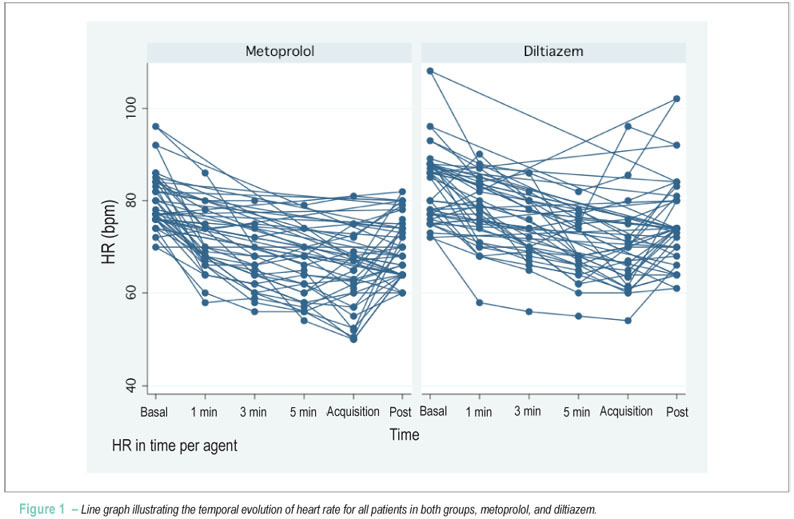
BACKGROUND: Reducing heart rate (HR) in CT angiography of the coronary arteries (CTACor) is critical to image quality. The effectiveness of calcium channel blockers as alternatives for patients with contraindications to beta-blockers has not been established. OBJECTIVES: To compare the efficacy in the reduction of HR and RR variability of metoprolol and diltiazem in CTACor. METHODS: Prospective, randomized, open study that included patients with clinical indication of CTACor in sinus rhythm with HR > 70 bpm and no use of agents that could interfere with HR. Fifty patients were randomized to the groups: metoprolol IV 5-15 mg or up to HR ≤ 60 bpm (M), and diltiazem IV 0.25 to 0.60 mg/kg or up to HR ≤ 60 bpm (D). Blood pressure (BP) and HR were measured at baseline, 1 minute, 3 minutes and 5 minutes after the agents, at the acquisition and after CTACor. RESULTS: HR reduction in absolute values was higher in group M than in group D (1, 3, 5 min, acquisition and post-test). The percentage reduction of HR was significantly higher in group M only 1 min and 3 min after the start of the agents. There was no difference in 5 min at acquisition and after examination. The percentage RR variability in group D was lower than that in group M during acquisition (RR variability/mean HR of acquisition). A single case of AVB, 2:1 Mobitz I occurred, which was spontaneously reverted (group D). CONCLUSION: We conclude that diltiazem is an effective and safe alternative to beta-blockers in the reduction of HR when performing computed tomography angiography of coronary arteries. (Arq Bras Cardiol. 2012; [online].ahead print, PP.0-0)

  • calcium channel blockers
  • Coronary Angiography
  • Coronary Vessels
  • Diltiazem
  • Tomography
Read Full Article
Creative Commons Attribution (CC BY) Creative Commons license
26/06/2012 DOI: 10.1590/S0066-782X2012005000059
Anomalous origin of Coronary Artery (ALCAPA) in 64-channel TC scanner
ABC
Case Report

Anomalous origin of Coronary Artery (ALCAPA) in 64-channel TC scanner

Marcelo Souto Nacif et al. José Hugo Mendes Luz Denise Madeira Moreira Carlos Eduardo Rochitte Amarino Carvalho de Oliveira Júnior

Multidetector computed tomography (MDCT) with 64 channels provides to clinical practice an excellent method to detect coronary artery anomalies. The diagnosis of coronary anomalies consisting of origin of left coronary artery in the pulmonary trunk in adults with no history of congenital disease has few reports in literature. We report a case in a 30-year old female patient complaining of fatigue on major efforts and positive scintigraphy for ischemia. The diagnosis was made through 64-channel MDCT and thus it appears that the method can be used as baseline.

  • Congenital
  • Coronary Disease
  • Heart Defects
  • Tomography
Read Full Article
Creative Commons Attribution (CC BY) Creative Commons license
12/07/2010 DOI: 10.1590/S0066-782X2010000600022 Views 2
Coronary calcium score as predictor of stenosis and events in pretransplant renal chronic failure
ABC
Artigo Original

Coronary calcium score as predictor of stenosis and events in pretransplant renal chronic failure

Miguel Abraão Rosário et al. José Jayme de Lima José R. Parga Luiz F. Ávila … Luis H. Gowdak Pedro A. Lemos Carlos E. Rochitte

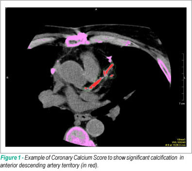
BACKGROUND: Coronary artery disease (CAD) is the major cause of death among chronic renal failure (CRF) patients. Traditional, non-invasive exams to detect CAD and to predict events have shown insufficient results in this group. CT Scan evaluation of Coronary Calcium Score (CCS) has proven to be of prognostic value for the population reporting no renal condition. OBJECTIVE: To investigate CCS accuracy in detecting obstructive CAD and in predicting cardiovascular events in candidates to renal transplant as compared to quantitative invasive coronary angiography (ICA). METHODS: Ninety-seven (97) CRF patients aged > 35 were evaluated. Obstructive CAD was considered as >50% or >70% stenosis on ICA. Descriptive data, concordance, diagnostic tests, Kaplan-Meier, and multivariate analysis were used. RESULTS: Agatston mean score was 580.6 ± 1,102.2. Minimum and maximum values were 0 and 7,994, with median at 176. Only 14 patients had zero calcium score. No differences were reported in regard to ethnicity. Highest regional calcium was associated to the highest probability of coronary stenosis in the same segment. Agatston calcium score showed high accuracy for the diagnosis of >50% and >70% stenosis, with area under ROC curve (AUC) of 0.75 and 0.70, respectively. At the threshold of 400, calcium score identified a subgroup with a higher rate of cardiovascular events at an average follow-up time of 29±11.0 months. CONCLUSION: CCS proved to have good diagnostic and prognostic performance for cardiovascular events evaluation in CRF patients.

  • calcio score
  • Chronic
  • Coronary Artery Disease
  • determination
  • emission-computed
  • Prognostic
  • Renal Insufficiency
  • Tomography
Read Full Article
Creative Commons Attribution (CC BY) Creative Commons license
05/05/2010 DOI: 10.1590/S0066-782X2010000200018 Views 2
Left ventricular ejection fraction and volumes as measured by 3d echocardiography and ultrafast computed tomography
ABC
Artigo Original

Left ventricular ejection fraction and volumes as measured by 3d echocardiography and ultrafast computed tomography

Marcelo Luiz Campos Vieira et al. César H. Nomura Bernardino Tranchesi Junior Wercules A. de Oliveira … Gustavo Naccarato Bruna S. Serpa Rodrigo B. D. Passos Marcelo B. G. Funari Cláudio H. Fischer Samira S. Morhy

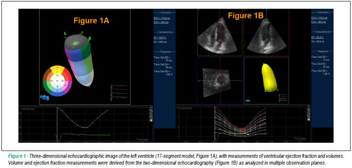
BACKGROUND: Real-time three-dimensional echocargiography (RT-3D-Echo) and ultrafast computed tomography (CT) are two novel methods for the analysis of LV ejection fraction and volumes. OBJECTIVE: To compare LVEF and volume measurements as obtained using RT-3D-Echo and ultrafast CT. METHODS: Thirty nine consecutive patients (27 men, mean age of 57±12 years) were studied using RT-3D-Echo and 64-slice ultrafast CT. LVEF and LV volumes were analyzed. Statistical analysis: coefficient of correlation (r: Pearson), Bland-Altman analysis, linear regression analysis, 95% CI, p<0.05. RESULTS: RT-3D-Echo measurements: LVEF ranged from 56.1 to 78.6 (65.5±5.58)%; end-diastolic volume ranged from 49.6 to 178.2 (87±27.8) ml; end-systolic volume ranged from 11.4 to 78 (33.1±13.6) ml. CT scan measurements: LVEF ranged from 53 to 86% (67.8±7.78); end-diastolic volume ranged from 51 to 186 (106.5±30.3) ml; end-systolic volume ranged from 7 to 72 (35.5±13.4)ml. Correlations between RT-3D-Echo and CT were: LVEF (r: 0.7888, p<0.0001, 95% CI 0.6301 to 0.8843); end-diastolic volume (r: 0.7695, p<0.0001, 95% CI 0.5995 to 0.8730); end-systolic volume (r: 0.8119, p<0.0001, 95% CI 0.6673 to 0.8975). CONCLUSION: Good correlation between LVEF and ventricular volume parameters as measured by RT-3D-Echo and 64-slice ultrafast CT was found in the present case series.

  • Echocardiography
  • emission-computed
  • Heart
  • Stroke Volume
  • Three-Dimensional
  • Tomography
Read Full Article
Creative Commons Attribution (CC BY) Creative Commons license
23/06/2009 DOI: 10.1590/S0066-782X2009000400008 Views 1

Could not load more articles. Please try again.

How to cite this article

EndNote RIS

Electronic Document Format (Vancouver)

Magalhães TA. Coronary CT Angiography in 2025: Long-Term Evidence, Artificial Intelligence, and the Photon-Counting Era. ABC Imagem Cardiovascular 2026;39(1):e20250106.

Electronic Document Format (ABNT)

Magalhães, Tiago A.. Coronary CT Angiography in 2025: Long-Term Evidence, Artificial Intelligence, and the Photon-Counting Era. ABC Imagem Cardiovascular, v. 39, n. 1, e20250106, Mar. 2026.

Electronic Document Format (APA)

Magalhães, T. A. (2026). Coronary CT Angiography in 2025: Long-Term Evidence, Artificial Intelligence, and the Photon-Counting Era. ABC Imagem Cardiovascular, 39(1), e20250106.

Electronic Document Format (ISO)

Magalhães, Tiago A.. Coronary CT Angiography in 2025: Long-Term Evidence, Artificial Intelligence, and the Photon-Counting Era. ABC Imagem Cardiovascular [online]. 2026, vol. 39, n. 1, [cited 2026-10-14], e20250106. Available from: <https://journals.cardiol.br/en/article/coronary-ct-angiography-in-2025-long-term-evidence-artificial-intelligence-and-the-photon-counting-era/>. ISSN 2318-8219

EndNote RIS

Electronic Document Format (Vancouver)

Fuzissima BM, Torres RVA, Couto RM, Farias LPG, Oliveira BSP, Araújo Filho JAB. My Approach To CAD-RADS 2.0 in the Assessment of Coronary Artery Disease by CT Angiogram. ABC Imagem Cardiovascular 2024;37(4):e20240085.

Electronic Document Format (ABNT)

Fuzissima, Bruno Maeda; Torres, Roberto Vitor Almeida; Couto, Renata Muller; Farias, Lucas de Pádua Gomes de; Oliveira, Bernardo Salgado Pinto; Araújo Filho, José de Arimatéia Batista. My Approach To CAD-RADS 2.0 in the Assessment of Coronary Artery Disease by CT Angiogram. ABC Imagem Cardiovascular, v. 37, n. 4, e20240085, Nov. 2024.

Electronic Document Format (APA)

Fuzissima, B. M., Torres, R. V. A., Couto, R. M., Farias, L. P. G. , Oliveira, B. S. P., & Araújo Filho, J. A. B. (2024). My Approach To CAD-RADS 2.0 in the Assessment of Coronary Artery Disease by CT Angiogram. ABC Imagem Cardiovascular, 37(4), e20240085.

Electronic Document Format (ISO)

Fuzissima, Bruno Maeda and Torres, Roberto Vitor Almeida and Couto, Renata Muller and Farias, Lucas de Pádua Gomes de and Oliveira, Bernardo Salgado Pinto and Araújo Filho, José de Arimatéia Batista. My Approach To CAD-RADS 2.0 in the Assessment of Coronary Artery Disease by CT Angiogram. ABC Imagem Cardiovascular [online]. 2024, vol. 37, n. 4, [cited 2026-10-14], e20240085. Available from: <https://journals.cardiol.br/en/article/my-approach-to-cad-rads-2-0-in-the-assessment-of-coronary-artery-disease-by-ct-angiogram/>. ISSN 2318-8219

EndNote RIS

Electronic Document Format (Vancouver)

Bispo IGA, Zago IM. The Indiscriminate Use Of Androgenic Anabolic Steroids: The Contribution of Cardiovascular Imaging. ABC Imagem Cardiovascular 2024;37(1):e20230113.

Electronic Document Format (ABNT)

Bispo, Irving Gabriel Araöşjo; Zago, Isabella Montanher. The Indiscriminate Use Of Androgenic Anabolic Steroids: The Contribution of Cardiovascular Imaging. ABC Imagem Cardiovascular, v. 37, n. 1, e20230113, Mar. 2024.

Electronic Document Format (APA)

Bispo, I. G. A., & Zago, I. M. (2024). The Indiscriminate Use Of Androgenic Anabolic Steroids: The Contribution of Cardiovascular Imaging. ABC Imagem Cardiovascular, 37(1), e20230113.

Electronic Document Format (ISO)

Bispo, Irving Gabriel Araöşjo and Zago, Isabella Montanher. The Indiscriminate Use Of Androgenic Anabolic Steroids: The Contribution of Cardiovascular Imaging. ABC Imagem Cardiovascular [online]. 2024, vol. 37, n. 1, [cited 2026-10-14], e20230113. Available from: <https://journals.cardiol.br/en/article/the-indiscriminate-use-of-androgenic-anabolic-steroids-the-contribution-of-cardiovascular-imaging/>. ISSN 2318-8219

EndNote RIS

Electronic Document Format (Vancouver)

Mancio J. Pericoronary Fat Attenuation on Computed Tomography Unveils a Guilty Factor of Coronary Artery Disease Associated with Anabolic–Androgenic Steroids. Arquivos Brasileiros de Cardiologia 2024;120(11):e20230843.

Electronic Document Format (ABNT)

Mancio, Jennifer. Pericoronary Fat Attenuation on Computed Tomography Unveils a Guilty Factor of Coronary Artery Disease Associated with Anabolic–Androgenic Steroids. Arquivos Brasileiros de Cardiologia, v. 120, n. 11, e20230843, Mar. 2024.

Electronic Document Format (APA)

Mancio, J. (2024). Pericoronary Fat Attenuation on Computed Tomography Unveils a Guilty Factor of Coronary Artery Disease Associated with Anabolic–Androgenic Steroids. Arquivos Brasileiros de Cardiologia, 120(11), e20230843.

Electronic Document Format (ISO)

Mancio, Jennifer. Pericoronary Fat Attenuation on Computed Tomography Unveils a Guilty Factor of Coronary Artery Disease Associated with Anabolic–Androgenic Steroids. Arquivos Brasileiros de Cardiologia [online]. 2024, vol. 120, n. 11, [cited 2026-10-14], e20230843. Available from: <https://journals.cardiol.br/en/article/pericoronary-fat-attenuation-on-computed-tomography-unveils-a-guilty-factor-of-coronary-artery-disease-associated-with-anabolic-androgenic-steroids/>. ISSN 0066-782X

EndNote RIS

Electronic Document Format (Vancouver)

Carnaval DR, Liebl A, Silva MMF, Sartori BM, Couto CNM, Silva GBG, Vazzoler GB, et al. Diagnostic Accuracy of the Coronary Calcium Score Acquired with Reduced Radiation Dose and Iterative Reconstruction. ABC Imagem Cardiovascular 2023;36(3):e20230069.

Electronic Document Format (ABNT)

Carnaval, Daniela Rodrigues; Liebl, Adriano; Silva, Miguel Morita Fernandes; Sartori, Bruno Marques; Couto, Caio Nogara de Menezes; Silva, Gabriel Bergonse Garaluz da; Vazzoler, Ítalo Gabriel Beltrame; Medeiros, Vinícius Maksoud; Cerci, Rodrigo Julio. Diagnostic Accuracy of the Coronary Calcium Score Acquired with Reduced Radiation Dose and Iterative Reconstruction. ABC Imagem Cardiovascular, v. 36, n. 3, e20230069, Oct. 2023.

Electronic Document Format (APA)

Carnaval, D. R., Liebl, A., Silva, M. M. F., Sartori, B. M., Couto, C. N. M., Silva, G. B. G. , Vazzoler, G. B., Medeiros, V. M., & Cerci, R. J. (2023). Diagnostic Accuracy of the Coronary Calcium Score Acquired with Reduced Radiation Dose and Iterative Reconstruction. ABC Imagem Cardiovascular, 36(3), e20230069.

Electronic Document Format (ISO)

Carnaval, Daniela Rodrigues and Liebl, Adriano and Silva, Miguel Morita Fernandes and Sartori, Bruno Marques and Couto, Caio Nogara de Menezes and Silva, Gabriel Bergonse Garaluz da and Vazzoler, Ítalo Gabriel Beltrame and Medeiros, Vinícius Maksoud and Cerci, Rodrigo Julio. Diagnostic Accuracy of the Coronary Calcium Score Acquired with Reduced Radiation Dose and Iterative Reconstruction. ABC Imagem Cardiovascular [online]. 2023, vol. 36, n. 3, [cited 2026-10-14], e20230069. Available from: <https://journals.cardiol.br/en/article/diagnostic-accuracy-of-the-coronary-calcium-score-acquired-with-reduced-radiation-dose-and-iterative-reconstruction/>. ISSN 2318-8219

EndNote RIS

Electronic Document Format (Vancouver)

Armstrong AC, Cerci R, Matheson MB, Magalhães T, Kishi S, Brinker J, Clouse ME, et al. Predicting Significant Coronary Obstruction in a Population with Suspected Coronary Disease and Absence of Coronary Calcium: CORE-64 / CORE320 Studies. Arquivos Brasileiros de Cardiologia 2023;120(3):e20220183.

Electronic Document Format (ABNT)

Armstrong, Anderson C.; Cerci, Rodrigo; Matheson, Matthew B.; Magalhães, Tiago; Kishi, Satoru; Brinker, Jeff; Clouse, Melvin E.; Rochitte, Carlos E.; Cox, Christopher; Lima, João A. C.; Arbab-Zadeh, Armin. Predicting Significant Coronary Obstruction in a Population with Suspected Coronary Disease and Absence of Coronary Calcium: CORE-64 / CORE320 Studies. Arquivos Brasileiros de Cardiologia, v. 120, n. 3, e20220183, Mar. 2023.

Electronic Document Format (APA)

Armstrong, A. C., Cerci, R., Matheson, M. B., Magalhães, T., Kishi, S., Brinker, J., Clouse, M. E., Rochitte, C. E., Cox, C., Lima, J. A. C., & Arbab-Zadeh, A. (2023). Predicting Significant Coronary Obstruction in a Population with Suspected Coronary Disease and Absence of Coronary Calcium: CORE-64 / CORE320 Studies. Arquivos Brasileiros de Cardiologia, 120(3), e20220183.

Electronic Document Format (ISO)

Armstrong, Anderson C. and Cerci, Rodrigo and Matheson, Matthew B. and Magalhães, Tiago and Kishi, Satoru and Brinker, Jeff and Clouse, Melvin E. and Rochitte, Carlos E. and Cox, Christopher and Lima, João A. C. and Arbab-Zadeh, Armin. Predicting Significant Coronary Obstruction in a Population with Suspected Coronary Disease and Absence of Coronary Calcium: CORE-64 / CORE320 Studies. Arquivos Brasileiros de Cardiologia [online]. 2023, vol. 120, n. 3, [cited 2026-10-14], e20220183. Available from: <https://journals.cardiol.br/en/article/predicting-significant-coronary-obstruction-in-a-population-with-suspected-coronary-disease-and-absence-of-coronary-calcium-core-64-core320-studies/>. ISSN 0066-782X

EndNote RIS

Electronic Document Format (Vancouver)

Correia JL, Correia M. Bulky Mitral Annulus Caseous Calcification in an Atypical Location. Arquivos Brasileiros de Cardiologia 2022;119(6):1002-5.

Electronic Document Format (ABNT)

Correia, Joana Laranjeira; Correia, Miguel. Bulky Mitral Annulus Caseous Calcification in an Atypical Location. Arquivos Brasileiros de Cardiologia, v. 119, n. 6, p. 1002-1005, Dec. 2022.

Electronic Document Format (APA)

Correia, J. L., & Correia, M. (2022). Bulky Mitral Annulus Caseous Calcification in an Atypical Location. Arquivos Brasileiros de Cardiologia, 119(6), 1002-1005.

Electronic Document Format (ISO)

Correia, Joana Laranjeira and Correia, Miguel. Bulky Mitral Annulus Caseous Calcification in an Atypical Location. Arquivos Brasileiros de Cardiologia [online]. 2022, vol. 119, n. 6, [cited 2026-10-14], pp. 1002-1005. Available from: <https://journals.cardiol.br/en/article/bulky-mitral-annulus-caseous-calcification-in-an-atypical-location/>. ISSN 0066-782X

EndNote RIS

Electronic Document Format (Vancouver)

Felix AS, Melo MDT, Monteiro TS, Cola MCT, Castro R, Merke N. A New Kid on the Block in POCUS: “LISA”. International Journal of Cardiovascular Sciences 2022;36:e20210285.

Electronic Document Format (ABNT)

Felix, Alex dos Santos; Melo, Marcelo Dantas Tavares de; Monteiro, Thaíssa Santos; Cola, Maria Carolina Terra; Castro, Rafael; Merke, Nicolas. A New Kid on the Block in POCUS: “LISA”. International Journal of Cardiovascular Sciences, v. 36, e20210285, May. 2022.

Electronic Document Format (APA)

Felix, A. S., Melo, M. D. T. , Monteiro, T. S., Cola, M. C. T., Castro, R., & Merke, N. (2022). A New Kid on the Block in POCUS: “LISA”. International Journal of Cardiovascular Sciences, 36, e20210285.

Electronic Document Format (ISO)

Felix, Alex dos Santos and Melo, Marcelo Dantas Tavares de and Monteiro, Thaíssa Santos and Cola, Maria Carolina Terra and Castro, Rafael and Merke, Nicolas. A New Kid on the Block in POCUS: “LISA”. International Journal of Cardiovascular Sciences [online]. 2022, vol. 36, [cited 2026-10-14], e20210285. Available from: <https://journals.cardiol.br/en/article/a-new-kid-on-the-block-in-pocus-lisa/>. ISSN 2359-4802

EndNote RIS

Electronic Document Format (Vancouver)

Siqueira MEM, Dias MI, Uski ACVR, Aguiar Filho LF. Rare Incidental Finding on Coronary Artery CT Angiography: Left Main Coronary Artery Atresia. Arquivos Brasileiros de Cardiologia Imagem cardiovascular 2022;34(4):eabc239.

Electronic Document Format (ABNT)

Siqueira, Maria Eduarda Menezes de; Dias, Mayra Isabel; Uski, Ana Claudia Vincezi Raudan; Aguiar Filho, Luciano Figueiredo de. Rare Incidental Finding on Coronary Artery CT Angiography: Left Main Coronary Artery Atresia. Arquivos Brasileiros de Cardiologia Imagem cardiovascular, v. 34, n. 4, eabc239, Mar. 2022.

Electronic Document Format (APA)

Siqueira, M. E. M. , Dias, M. I., Uski, A. C. V. R., & Aguiar Filho, L. F. (2022). Rare Incidental Finding on Coronary Artery CT Angiography: Left Main Coronary Artery Atresia. Arquivos Brasileiros de Cardiologia Imagem cardiovascular, 34(4), eabc239.

Electronic Document Format (ISO)

Siqueira, Maria Eduarda Menezes de and Dias, Mayra Isabel and Uski, Ana Claudia Vincezi Raudan and Aguiar Filho, Luciano Figueiredo de. Rare Incidental Finding on Coronary Artery CT Angiography: Left Main Coronary Artery Atresia. Arquivos Brasileiros de Cardiologia Imagem cardiovascular [online]. 2022, vol. 34, n. 4, [cited 2026-10-14], eabc239. Available from: <https://journals.cardiol.br/en/article/rare-incidental-finding-on-coronary-artery-ct-angiography-left-main-coronary-artery-atresia/>. ISSN 2318-8219

EndNote RIS

Electronic Document Format (Vancouver)

Santos H, Miranda H, Almeida I, Santos M, Almeida S, Almeida L. A Deceiving Aorta. Arquivos Brasileiros de Cardiologia Imagem cardiovascular 2022;32(4):345.

Electronic Document Format (ABNT)

Santos, Helder; Miranda, Hugo; Almeida, Inês; Santos, Mariana; Almeida, Samuel; Almeida, Lurdes. A Deceiving Aorta. Arquivos Brasileiros de Cardiologia Imagem cardiovascular, v. 32, n. 4, p. 345-345, Jan. 2022.

Electronic Document Format (APA)

Santos, H., Miranda, H., Almeida, I., Santos, M., Almeida, S., & Almeida, L. (2022). A Deceiving Aorta. Arquivos Brasileiros de Cardiologia Imagem cardiovascular, 32(4), 345-345.

Electronic Document Format (ISO)

Santos, Helder and Miranda, Hugo and Almeida, Inês and Santos, Mariana and Almeida, Samuel and Almeida, Lurdes. A Deceiving Aorta. Arquivos Brasileiros de Cardiologia Imagem cardiovascular [online]. 2022, vol. 32, n. 4, [cited 2026-10-14], pp. 345-345. Available from: <https://journals.cardiol.br/en/article/a-deceiving-aorta/>. ISSN 2318-8219

EndNote RIS

Electronic Document Format (Vancouver)

Oliveira GMM, Lopes MACQ. What does the Cardiologist Expect from the Echocardiogram of a Transcatheter Aortic Valve Implant?. Arquivos Brasileiros de Cardiologia Imagem cardiovascular 2022;33(2):1-3.

Electronic Document Format (ABNT)

Oliveira, Gláucia Maria Moraes de; Lopes, Marcelo Antônio Cartaxo Queiroga. What does the Cardiologist Expect from the Echocardiogram of a Transcatheter Aortic Valve Implant?. Arquivos Brasileiros de Cardiologia Imagem cardiovascular, v. 33, n. 2, p. 1-3, Jan. 2022.

Electronic Document Format (APA)

Oliveira, G. M. M. , & Lopes, M. A. C. Q. (2022). What does the Cardiologist Expect from the Echocardiogram of a Transcatheter Aortic Valve Implant?. Arquivos Brasileiros de Cardiologia Imagem cardiovascular, 33(2), 1-3.

Electronic Document Format (ISO)

Oliveira, Gláucia Maria Moraes de and Lopes, Marcelo Antônio Cartaxo Queiroga. What does the Cardiologist Expect from the Echocardiogram of a Transcatheter Aortic Valve Implant?. Arquivos Brasileiros de Cardiologia Imagem cardiovascular [online]. 2022, vol. 33, n. 2, [cited 2026-10-14], pp. 1-3. Available from: <https://journals.cardiol.br/en/article/what-does-the-cardiologist-expect-from-the-echocardiogram-of-a-transcatheter-aortic-valve-implant/>. ISSN 2318-8219

EndNote RIS

Electronic Document Format (Vancouver)

Lacerda IS, Neves PO, Santos LHP, Andrade JM. Cardiac Metastasis in Patient with Rectal Carcinoid Tumor. Arquivos Brasileiros de Cardiologia Imagem cardiovascular 2022;32(4):338-40.

Electronic Document Format (ABNT)

Lacerda, Iuri Shen de; Neves, Priscilla Ornellas; Santos, Lucianno Henrique Pereira; Andrade, Joalbo Matos. Cardiac Metastasis in Patient with Rectal Carcinoid Tumor. Arquivos Brasileiros de Cardiologia Imagem cardiovascular, v. 32, n. 4, p. 338-340, Jan. 2022.

Electronic Document Format (APA)

Lacerda, I. S. , Neves, P. O., Santos, L. H. P., & Andrade, J. M. (2022). Cardiac Metastasis in Patient with Rectal Carcinoid Tumor. Arquivos Brasileiros de Cardiologia Imagem cardiovascular, 32(4), 338-340.

Electronic Document Format (ISO)

Lacerda, Iuri Shen de and Neves, Priscilla Ornellas and Santos, Lucianno Henrique Pereira and Andrade, Joalbo Matos. Cardiac Metastasis in Patient with Rectal Carcinoid Tumor. Arquivos Brasileiros de Cardiologia Imagem cardiovascular [online]. 2022, vol. 32, n. 4, [cited 2026-10-14], pp. 338-340. Available from: <https://journals.cardiol.br/en/article/cardiac-metastasis-in-patient-with-rectal-carcinoid-tumor/>. ISSN 2318-8219

EndNote RIS

Electronic Document Format (Vancouver)

Paladino AT, Vasconcelos LA, Toledo LMG, Massoni N, Rezende MO, Santos NSS, Bihan DCSL, et al. A Three-Leaflets Mitral Valve Associated to Three Papillary Muscles in a Patient with Hypertrophic Cardiomyopathy. Arquivos Brasileiros de Cardiologia Imagem cardiovascular 2022;33(1):1-3.

Electronic Document Format (ABNT)

Paladino, Antonio Tito; Vasconcelos, Luiz Antônio; Toledo, Leonardo Mello Guimarães de; Massoni, Natalia; Rezende, Mariana Oliveira; Santos, Natasha Soares Simões dos; Bihan, David Costa de Souza Le; Barretto, Rodrigo Bellio de Mattos; Assef, Jorge Eduardo. A Three-Leaflets Mitral Valve Associated to Three Papillary Muscles in a Patient with Hypertrophic Cardiomyopathy. Arquivos Brasileiros de Cardiologia Imagem cardiovascular, v. 33, n. 1, p. 1-3, Jan. 2022.

Electronic Document Format (APA)

Paladino, A. T., Vasconcelos, L. A., Toledo, L. M. G. , Massoni, N., Rezende, M. O., Santos, N. S. S. , Bihan, D. C. S. L., Barretto, R. B. M., & Assef, J. E. (2022). A Three-Leaflets Mitral Valve Associated to Three Papillary Muscles in a Patient with Hypertrophic Cardiomyopathy. Arquivos Brasileiros de Cardiologia Imagem cardiovascular, 33(1), 1-3.

Electronic Document Format (ISO)

Paladino, Antonio Tito and Vasconcelos, Luiz Antônio and Toledo, Leonardo Mello Guimarães de and Massoni, Natalia and Rezende, Mariana Oliveira and Santos, Natasha Soares Simões dos and Bihan, David Costa de Souza Le and Barretto, Rodrigo Bellio de Mattos and Assef, Jorge Eduardo. A Three-Leaflets Mitral Valve Associated to Three Papillary Muscles in a Patient with Hypertrophic Cardiomyopathy. Arquivos Brasileiros de Cardiologia Imagem cardiovascular [online]. 2022, vol. 33, n. 1, [cited 2026-10-14], pp. 1-3. Available from: <https://journals.cardiol.br/en/article/a-three-leaflets-mitral-valve-associated-to-three-papillary-muscles-in-a-patient-with-hypertrophic-cardiomyopathy/>. ISSN 2318-8219

EndNote RIS

Electronic Document Format (Vancouver)

Prazeres CEE, Salvatti NB, Carvalho HSM, Silva ITQ, Carneiro ACC, Magalhães TA, Moreira VM, et al. Fractional Flow Reserve by Tomography Diagnostic Performance in the Detection of Coronary Stenoses Hemodynamically Significant. Arquivos Brasileiros de Cardiologia Imagem cardiovascular 2022;33(3):1-7.

Electronic Document Format (ABNT)

Prazeres, Carlos Eduardo Elias dos; Salvatti, Natália Boing; Carvalho, Hilka dos Santos Moraes de; Silva, Isaac Torquato Queiroz e; Carneiro, Adriano Camargo de Castro; Magalhães, Tiago Augusto; Moreira, Valéria de Melo; Bello, Juliana Hiromi Silva Matsumoto; Rochitte, Carlos Eduardo. Fractional Flow Reserve by Tomography Diagnostic Performance in the Detection of Coronary Stenoses Hemodynamically Significant. Arquivos Brasileiros de Cardiologia Imagem cardiovascular, v. 33, n. 3, p. 1-7, Jan. 2022.

Electronic Document Format (APA)

Prazeres, C. E. E. , Salvatti, N. B., Carvalho, H. S. M. , Silva, I. T. Q. , Carneiro, A. C. C., Magalhães, T. A., Moreira, V. M., Bello, J. H. S. M., & Rochitte, C. E. (2022). Fractional Flow Reserve by Tomography Diagnostic Performance in the Detection of Coronary Stenoses Hemodynamically Significant. Arquivos Brasileiros de Cardiologia Imagem cardiovascular, 33(3), 1-7.

Electronic Document Format (ISO)

Prazeres, Carlos Eduardo Elias dos and Salvatti, Natália Boing and Carvalho, Hilka dos Santos Moraes de and Silva, Isaac Torquato Queiroz e and Carneiro, Adriano Camargo de Castro and Magalhães, Tiago Augusto and Moreira, Valéria de Melo and Bello, Juliana Hiromi Silva Matsumoto and Rochitte, Carlos Eduardo. Fractional Flow Reserve by Tomography Diagnostic Performance in the Detection of Coronary Stenoses Hemodynamically Significant. Arquivos Brasileiros de Cardiologia Imagem cardiovascular [online]. 2022, vol. 33, n. 3, [cited 2026-10-14], pp. 1-7. Available from: <https://journals.cardiol.br/en/article/fractional-flow-reserve-by-tomography-diagnostic-performance-in-the-detection-of-coronary-stenoses-hemodynamically-significant/>. ISSN 2318-8219

EndNote RIS

Electronic Document Format (Vancouver)

Faria B, Calvo L, Ribeiro S, Ruivo C, Lourenço A. Anomalous Origin of the Circumflex Coronary Artery from the Right Pulmonary Artery: Diagnosis Through Cardiac CT. Arquivos Brasileiros de Cardiologia 2021;117(1 suppl 1):8-11.

Electronic Document Format (ABNT)

Faria, Bebiana; Calvo, Lucy; Ribeiro, Sílvia; Ruivo, Catarina; Lourenço, António. Anomalous Origin of the Circumflex Coronary Artery from the Right Pulmonary Artery: Diagnosis Through Cardiac CT. Arquivos Brasileiros de Cardiologia, v. 117, n. 1 suppl 1, p. 8-11, Jul. 2021.

Electronic Document Format (APA)

Faria, B., Calvo, L., Ribeiro, S., Ruivo, C., & Lourenço, A. (2021). Anomalous Origin of the Circumflex Coronary Artery from the Right Pulmonary Artery: Diagnosis Through Cardiac CT. Arquivos Brasileiros de Cardiologia, 117(1 suppl 1), 8-11.

Electronic Document Format (ISO)

Faria, Bebiana and Calvo, Lucy and Ribeiro, Sílvia and Ruivo, Catarina and Lourenço, António. Anomalous Origin of the Circumflex Coronary Artery from the Right Pulmonary Artery: Diagnosis Through Cardiac CT. Arquivos Brasileiros de Cardiologia [online]. 2021, vol. 117, n. 1 suppl 1, [cited 2026-10-14], pp. 8-11. Available from: <https://journals.cardiol.br/en/article/anomalous-origin-of-the-circumflex-coronary-artery-from-the-right-pulmonary-artery-diagnosis-through-cardiac-ct/>. ISSN 0066-782X

EndNote RIS

Electronic Document Format (Vancouver)

Cerit L, Duygu H, Gülşen K, Kemal H, Ozcem B. Spider-Like Coronary Anatomy; the True Spider!. Arquivos Brasileiros de Cardiologia 2017;109(4):376-7.

Electronic Document Format (ABNT)

Cerit, Levent; Duygu, Hamza; Gülşen, Kamil; Kemal, Hatice; Ozcem, Barcin. Spider-Like Coronary Anatomy; the True Spider!. Arquivos Brasileiros de Cardiologia, v. 109, n. 4, p. 376-377, Sep. 2017.

Electronic Document Format (APA)

Cerit, L., Duygu, H., Gülşen, K., Kemal, H., & Ozcem, B. (2017). Spider-Like Coronary Anatomy; the True Spider!. Arquivos Brasileiros de Cardiologia, 109(4), 376-377.

Electronic Document Format (ISO)

Cerit, Levent and Duygu, Hamza and Gülşen, Kamil and Kemal, Hatice and Ozcem, Barcin. Spider-Like Coronary Anatomy; the True Spider!. Arquivos Brasileiros de Cardiologia [online]. 2017, vol. 109, n. 4, [cited 2026-10-14], pp. 376-377. Available from: <https://journals.cardiol.br/en/article/spider-like-coronary-anatomy-the-true-spider/>. ISSN 0066-782X

EndNote RIS

Electronic Document Format (Vancouver)

Freire AFD, Soares AAS, Leal TCAT, Oliveira Junior MT, Soeiro AM. Patient with Atrial Myxoma and Signs of Obstruction of the Left Ventricular Outflow Tract. Arquivos Brasileiros de Cardiologia 2017;108(3):281-2.

Electronic Document Format (ABNT)

Freire, Antonio Fernando Diniz; Soares, Alexandre Anderson de Sousa; Leal, Tatiana de Carvalho Andreucci Torres; Oliveira Junior, Mucio Tavares de; Soeiro, Alexandre de Matos. Patient with Atrial Myxoma and Signs of Obstruction of the Left Ventricular Outflow Tract. Arquivos Brasileiros de Cardiologia, v. 108, n. 3, p. 281-282, Feb. 2017.

Electronic Document Format (APA)

Freire, A. F. D., Soares, A. A. S., Leal, T. C. A. T., Oliveira Junior, M. T. , & Soeiro, A. M. (2017). Patient with Atrial Myxoma and Signs of Obstruction of the Left Ventricular Outflow Tract. Arquivos Brasileiros de Cardiologia, 108(3), 281-282.

Electronic Document Format (ISO)

Freire, Antonio Fernando Diniz and Soares, Alexandre Anderson de Sousa and Leal, Tatiana de Carvalho Andreucci Torres and Oliveira Junior, Mucio Tavares de and Soeiro, Alexandre de Matos. Patient with Atrial Myxoma and Signs of Obstruction of the Left Ventricular Outflow Tract. Arquivos Brasileiros de Cardiologia [online]. 2017, vol. 108, n. 3, [cited 2026-10-14], pp. 281-282. Available from: <https://journals.cardiol.br/en/article/patient-with-atrial-myxoma-and-signs-of-obstruction-of-the-left-ventricular-outflow-tract/>. ISSN 0066-782X

EndNote RIS

Electronic Document Format (Vancouver)

Rajani R, Khattar R, Chiribiri A, Victor K, Chambers J. Multimodality Imaging of Heart Valve Disease. Arquivos Brasileiros de Cardiologia 2014;103(3):251-63.

Electronic Document Format (ABNT)

Rajani, Ronak; Khattar, Rajdeep; Chiribiri, Amedeo; Victor, Kelly; Chambers, John. Multimodality Imaging of Heart Valve Disease. Arquivos Brasileiros de Cardiologia, v. 103, n. 3, p. 251-263, May. 2014.

Electronic Document Format (APA)

Rajani, R., Khattar, R., Chiribiri, A., Victor, K., & Chambers, J. (2014). Multimodality Imaging of Heart Valve Disease. Arquivos Brasileiros de Cardiologia, 103(3), 251-263.

Electronic Document Format (ISO)

Rajani, Ronak and Khattar, Rajdeep and Chiribiri, Amedeo and Victor, Kelly and Chambers, John. Multimodality Imaging of Heart Valve Disease. Arquivos Brasileiros de Cardiologia [online]. 2014, vol. 103, n. 3, [cited 2026-10-14], pp. 251-263. Available from: <https://journals.cardiol.br/en/article/multimodality-imaging-of-heart-valve-disease/>. ISSN 0066-782X

EndNote RIS

Electronic Document Format (Vancouver)

Prazeres CEE, Cury RC, Carneiro ACC, Rochitte CE. Coronary computed tomography angiography in the assessment of acute chest pain in the emergency room. Arquivos Brasileiros de Cardiologia 2013;101(6):562-9.

Electronic Document Format (ABNT)

Prazeres, Carlos Eduardo Elias dos; Cury, Roberto Caldeira; Carneiro, Adriano Camargo de Castro; Rochitte, Carlos Eduardo. Coronary computed tomography angiography in the assessment of acute chest pain in the emergency room. Arquivos Brasileiros de Cardiologia, v. 101, n. 6, p. 562-569, Oct. 2013.

Electronic Document Format (APA)

Prazeres, C. E. E. , Cury, R. C., Carneiro, A. C. C., & Rochitte, C. E. (2013). Coronary computed tomography angiography in the assessment of acute chest pain in the emergency room. Arquivos Brasileiros de Cardiologia, 101(6), 562-569.

Electronic Document Format (ISO)

Prazeres, Carlos Eduardo Elias dos and Cury, Roberto Caldeira and Carneiro, Adriano Camargo de Castro and Rochitte, Carlos Eduardo. Coronary computed tomography angiography in the assessment of acute chest pain in the emergency room. Arquivos Brasileiros de Cardiologia [online]. 2013, vol. 101, n. 6, [cited 2026-10-14], pp. 562-569. Available from: <https://journals.cardiol.br/en/article/coronary-computed-tomography-angiography-in-the-assessment-of-acute-chest-pain-in-the-emergency-room/>. ISSN 0066-782X

EndNote RIS

Electronic Document Format (Vancouver)

Azevedo CF, Rochitte CE, Lima JA. Coronary artery calcium score and coronary computed tomographic angiography for cardiovascular risk stratification. Arquivos Brasileiros de Cardiologia 2012;98(6):559-68.

Electronic Document Format (ABNT)

Azevedo, Clerio F.; Rochitte, Carlos E.; Lima, João A.C.. Coronary artery calcium score and coronary computed tomographic angiography for cardiovascular risk stratification. Arquivos Brasileiros de Cardiologia, v. 98, n. 6, p. 559-568, Aug. 2012.

Electronic Document Format (APA)

Azevedo, C. F., Rochitte, C. E., & Lima, J. A. (2012). Coronary artery calcium score and coronary computed tomographic angiography for cardiovascular risk stratification. Arquivos Brasileiros de Cardiologia, 98(6), 559-568.

Electronic Document Format (ISO)

Azevedo, Clerio F. and Rochitte, Carlos E. and Lima, João A.C.. Coronary artery calcium score and coronary computed tomographic angiography for cardiovascular risk stratification. Arquivos Brasileiros de Cardiologia [online]. 2012, vol. 98, n. 6, [cited 2026-10-14], pp. 559-568. Available from: <https://journals.cardiol.br/en/article/coronary-artery-calcium-score-and-coronary-computed-tomographic-angiography-for-cardiovascular-risk-stratification/>. ISSN 0066-782X

EndNote RIS

Electronic Document Format (Vancouver)

Rochitte CE, Azevedo GSA, Shiozaki AA, Azevedo CF, Kalil Filho R. Diltiazem as an alternative to beta-blocker in coronary artery computed tomography angiography. Arquivos Brasileiros de Cardiologia 2012;99(2):706-13.

Electronic Document Format (ABNT)

Rochitte, Carlos Eduardo; Azevedo, Guilherme Santana Antunes; Shiozaki, Afonso Akio; Azevedo, Clerio Francisco; Kalil Filho, Roberto. Diltiazem as an alternative to beta-blocker in coronary artery computed tomography angiography. Arquivos Brasileiros de Cardiologia, v. 99, n. 2, p. 706-713, Jun. 2012.

Electronic Document Format (APA)

Rochitte, C. E., Azevedo, G. S. A., Shiozaki, A. A., Azevedo, C. F., & Kalil Filho, R. (2012). Diltiazem as an alternative to beta-blocker in coronary artery computed tomography angiography. Arquivos Brasileiros de Cardiologia, 99(2), 706-713.

Electronic Document Format (ISO)

Rochitte, Carlos Eduardo and Azevedo, Guilherme Santana Antunes and Shiozaki, Afonso Akio and Azevedo, Clerio Francisco and Kalil Filho, Roberto. Diltiazem as an alternative to beta-blocker in coronary artery computed tomography angiography. Arquivos Brasileiros de Cardiologia [online]. 2012, vol. 99, n. 2, [cited 2026-10-14], pp. 706-713. Available from: <https://journals.cardiol.br/en/article/diltiazem-as-an-alternative-to-beta-blocker-in-coronary-artery-computed-tomography-angiography/>. ISSN 0066-782X

EndNote RIS

Electronic Document Format (Vancouver)

Nacif MS, Luz JHM, Moreira DM, Rochitte CE, Oliveira Júnior AC. Anomalous origin of Coronary Artery (ALCAPA) in 64-channel TC scanner. Arquivos Brasileiros de Cardiologia 2010;94(6):143-6.

Electronic Document Format (ABNT)

Nacif, Marcelo Souto; Luz, José Hugo Mendes; Moreira, Denise Madeira; Rochitte, Carlos Eduardo; Oliveira Júnior, Amarino Carvalho de. Anomalous origin of Coronary Artery (ALCAPA) in 64-channel TC scanner. Arquivos Brasileiros de Cardiologia, v. 94, n. 6, p. 143-146, Jul. 2010.

Electronic Document Format (APA)

Nacif, M. S., Luz, J. H. M., Moreira, D. M., Rochitte, C. E., & Oliveira Júnior, A. C. (2010). Anomalous origin of Coronary Artery (ALCAPA) in 64-channel TC scanner. Arquivos Brasileiros de Cardiologia, 94(6), 143-146.

Electronic Document Format (ISO)

Nacif, Marcelo Souto and Luz, José Hugo Mendes and Moreira, Denise Madeira and Rochitte, Carlos Eduardo and Oliveira Júnior, Amarino Carvalho de. Anomalous origin of Coronary Artery (ALCAPA) in 64-channel TC scanner. Arquivos Brasileiros de Cardiologia [online]. 2010, vol. 94, n. 6, [cited 2026-10-14], pp. 143-146. Available from: <https://journals.cardiol.br/en/article/anomalous-origin-of-coronary-artery-alcapa-in-64-channel-tc-scanner/>. ISSN 0066-782X

EndNote RIS

Electronic Document Format (Vancouver)

Rosário MA, Lima JJ, Parga JR, Ávila LF, Gowdak LH, Lemos PA, Rochitte CE, et al. Coronary calcium score as predictor of stenosis and events in pretransplant renal chronic failure. Arquivos Brasileiros de Cardiologia 2010;94(2):252-60.

Electronic Document Format (ABNT)

Rosário, Miguel Abraão; Lima, José Jayme de; Parga, José R.; Ávila, Luiz F.; Gowdak, Luis H.; Lemos, Pedro A.; Rochitte, Carlos E.. Coronary calcium score as predictor of stenosis and events in pretransplant renal chronic failure. Arquivos Brasileiros de Cardiologia, v. 94, n. 2, p. 252-260, May. 2010.

Electronic Document Format (APA)

Rosário, M. A., Lima, J. J. , Parga, J. R., Ávila, L. F., Gowdak, L. H., Lemos, P. A., & Rochitte, C. E. (2010). Coronary calcium score as predictor of stenosis and events in pretransplant renal chronic failure. Arquivos Brasileiros de Cardiologia, 94(2), 252-260.

Electronic Document Format (ISO)

Rosário, Miguel Abraão and Lima, José Jayme de and Parga, José R. and Ávila, Luiz F. and Gowdak, Luis H. and Lemos, Pedro A. and Rochitte, Carlos E.. Coronary calcium score as predictor of stenosis and events in pretransplant renal chronic failure. Arquivos Brasileiros de Cardiologia [online]. 2010, vol. 94, n. 2, [cited 2026-10-14], pp. 252-260. Available from: <https://journals.cardiol.br/en/article/coronary-calcium-score-as-predictor-of-stenosis-and-events-in-pretransplant-renal-chronic-failure/>. ISSN 0066-782X

EndNote RIS

Electronic Document Format (Vancouver)

Vieira MLC, Nomura CH, Tranchesi Junior B, Oliveira WA, Naccarato G, Serpa BS, Passos RBD, et al. Left ventricular ejection fraction and volumes as measured by 3d echocardiography and ultrafast computed tomography. Arquivos Brasileiros de Cardiologia 2009;92(4):294-301.

Electronic Document Format (ABNT)

Vieira, Marcelo Luiz Campos; Nomura, César H.; Tranchesi Junior, Bernardino; Oliveira, Wercules A. de; Naccarato, Gustavo; Serpa, Bruna S.; Passos, Rodrigo B. D.; Funari, Marcelo B. G.; Fischer, Cláudio H.; Morhy, Samira S.. Left ventricular ejection fraction and volumes as measured by 3d echocardiography and ultrafast computed tomography. Arquivos Brasileiros de Cardiologia, v. 92, n. 4, p. 294-301, Jun. 2009.

Electronic Document Format (APA)

Vieira, M. L. C., Nomura, C. H., Tranchesi Junior, B., Oliveira, W. A. , Naccarato, G., Serpa, B. S., Passos, R. B. D., Funari, M. B. G., Fischer, C. H., & Morhy, S. S. (2009). Left ventricular ejection fraction and volumes as measured by 3d echocardiography and ultrafast computed tomography. Arquivos Brasileiros de Cardiologia, 92(4), 294-301.

Electronic Document Format (ISO)

Vieira, Marcelo Luiz Campos and Nomura, César H. and Tranchesi Junior, Bernardino and Oliveira, Wercules A. de and Naccarato, Gustavo and Serpa, Bruna S. and Passos, Rodrigo B. D. and Funari, Marcelo B. G. and Fischer, Cláudio H. and Morhy, Samira S.. Left ventricular ejection fraction and volumes as measured by 3d echocardiography and ultrafast computed tomography. Arquivos Brasileiros de Cardiologia [online]. 2009, vol. 92, n. 4, [cited 2026-10-14], pp. 294-301. Available from: <https://journals.cardiol.br/en/article/left-ventricular-ejection-fraction-and-volumes-as-measured-by-3d-echocardiography-and-ultrafast-computed-tomography/>. ISSN 0066-782X

  • About ABC Family
  • Internal Regulation
  • Editorial Board
  • Brazilian Society of Cardiology (SBC)

Issues

SBC Guidelines

  • Videos
  • Podcasts
  • News