ABC Family Arquivos Brasileiros de Cardiologia International Journal of Cardiovascular Sciences ABC Heart Failure & Cardiomyopathy ABC Imagem Cardiovascular
Português English
ABC Family
Português English
  • About
    • About ABC Family
    • Internal Regulation
    • Editorial Board
    • Brazilian Society of Cardiology (SBC)
  • Issues
  • SBC Guidelines
  • Content
    • Videos
    • Podcasts
    • News
  • About
    • About ABC Family
    • Internal Regulation
    • Editorial Board
    • Brazilian Society of Cardiology (SBC)
  • Issues
  • SBC Guidelines
  • Content
    • Videos
    • Podcasts
    • News

ABC Family

  • ABC Family
  • Arquivos Brasileiros de Cardiologia
  • International Journal of Cardiovascular Sciences
  • ABC Heart Failure & Cardiomyopathy
  • ABC Imagem Cardiovascular
Search in

No content found.

Journals
Ordering
Article types
Others

  1. Home
  2. Keyword: Ultrasonography/methods
ABC
Editorial

COVID-19 and Late Cardiovascular Manifestations – Building Up Evidence

Sofia Cabral

In early 2020, the world faced the outbreak of a new pandemic, Coronavirus Disease 2019 (COVID-19), caused by a novel coronavirus (SARS-CoV-2 virus). We soon discovered that while the respiratory tract was the prime target, many other organ systems might also be affected. COVID-19 cardiovascular (CV) manifestations are among the most common and feared and may manifest as myocardial injury, arrhythmias, acute coronary syndromes, heart failure, vascular dysfunction, and thromboembolic disease. Both pre-existing and comorbid cardiovascular disease and the concurrent presence of CV risk factors are harbingers of a poorer disease course and prognosis. Notably, acute CV involvement is a strong independent predictor of COVID-19 in-hospital mortality. The way in which the infection interacts with the CV system remains a subject of study, but apart from direct cell injury by the virus, the activation of inflammatory pathways is thought to play a key role. A comprehensive account of the pathophysiology of CV involvement in COVID-19 is beyond the scope of this commentary and has been extensively covered elsewhere.

While it is now clear that both cardiac and vascular dysregulations are to be expected during the active stage of the infection, concerns now focus on long-term residual CV abnormalities. Huang et al. reported that more than half of convalescent COVID-19 patients displayed signs of myocardial inflammation or fibrosis on cardiac magnetic resonance (CMR) imaging. Similarly, the meta-analysis by Kim et al. showed that nearly half of recovered COVID-19 patients exhibited one or more abnormal CMR results. Likewise, and somewhat unsurprisingly, the infection may also induce enduring vascular injury. In light of the tropism of SARS-CoV-2 for membrane-bound angiotensin-converting enzyme 2-expressing cells, it is no wonder that the vascular endothelium is a major player in the infection. Nonetheless, the effects of COVID-19 infection on vascular endothelium and the complex interplay between the two are still poorly understood. In essence, CV homeostasis is primarily dependent on the role of the endothelium. It regulates vascular tone, cell adhesion, thromboresistance, smooth muscle cell proliferation, and ultimately, vessel wall inflammation. Whenever the endothelium is activated, resulting in endothelial dysfunction, cell-signaling shifts from nitric oxide – mediated vasodilatory to vasoconstrictor redox processes. This can occur as a transient acute phenomenon, with little to no consequences, or as a sustained detrimental endothelium activation, eventually leading to a pro-atherogenic, pro-thrombotic milieu.

[…]
  • Covid-19/complications
  • Endothelium, Vascular
  • Flow Dilatation
  • Inflammation
  • Ultrasonography/methods
Read Full Article
Creative Commons Attribution (CC BY) Creative Commons license
05/08/2022 DOI: 10.36660/abc.20220435
ABC
Original Article

Evaluation of Endothelial Dysfunction in COVID-19 With Flow-Mediated Dilatation

Asli Kurtar Mansiroglu et al. Hande Seymen Isa Sincer Yilmaz Gunes

Abstract

Background:

Inflammation is known to play a crucial role in many diseases, including COVID-19.

Objective:

Using flow-mediated dilatation (FMD), we aimed to assess the effects of inflammation on endothelial function in COVID-19 patients.

Methods:

This study was conducted with a total of 161 subjects, of whom 80 were diagnosed with COVID-19 within the last six months (comprising 48 women and 32 men with a mean age of 32.10 ± 5.87 years) and 81 were healthy controls (comprising 45 women and 36 men with a mean age of 30.51 ± 7.33 years). We analyzed the findings of transthoracic echocardiography and FMD in all subjects. All results were considered statistically significant at the level of p < 0.05.

Results:

The echocardiography and FMD of the COVID-19 group were performed 35 days (range: 25-178) after diagnosis. There was no statistically significant difference in echocardiographic parameters. Differently, FMD (%) was significantly higher in the control group (9.52 ± 5.98 vs. 12.01 ± 6.18, p=0.01). In multivariate analysis with the forward stepwise model, FMD was significantly different in the control group compared to the COVID-19 group (1.086 (1.026 – 1.149), p=0.04). A Spearman’s correlation test indicated that FMD (r=0.27, p=0.006) had a weak positive correlation with the presence of COVID-19.

Conclusion:

Our findings point to COVID-19-induced endothelial dysfunction, as assessed by FMD, in the early recovery phase.

  • Covid-19/complications
  • Diagnostic Imaging/methods
  • Echocardiography/methods
  • Endothelial, Cells/infection
  • Endothelium Vascular/injuries
  • Flow Dilatation
  • Myalgia
  • Olfaction Disorders
  • Taste Disorders
  • Ultrasonography/methods
Read Full Article
Creative Commons Attribution (CC BY) Creative Commons license
03/06/2022 DOI: 10.36660/abc.20210561 Views 1
ABCIC
Case Report

Right Ventricular Endomyocardial Fibrosis: Case Report

Stella de Souza Carneiro et al. Patrick Ventorim Costa Aline Valério de Lima Fernando Luiz Torres Gomes … Ludmilla Ventura Lirio Danielle Cristina Filgueira Alves Batista

[…]

  • EchocardiographyDoppler/methods
  • Endomyocardial Fibrosis/complications
  • Endomyocardial Fibrosis/diagnostic, imaging
  • Eosinophilia
  • Heart Failure
  • Ultrasonography/methods
  • Ventricular Dysfunction, Right/complications
Read Full Article
Creative Commons Attribution (CC BY) Creative Commons license
20/01/2022 DOI: 10.5935/2318-8219.20190014 Views 2
Figure 1
ABC
Case Report

Antenatal Diagnosis of Double Aortic Arch

Natália Noronha et al. Angie Hobbs Patricia Caldas

Introduction

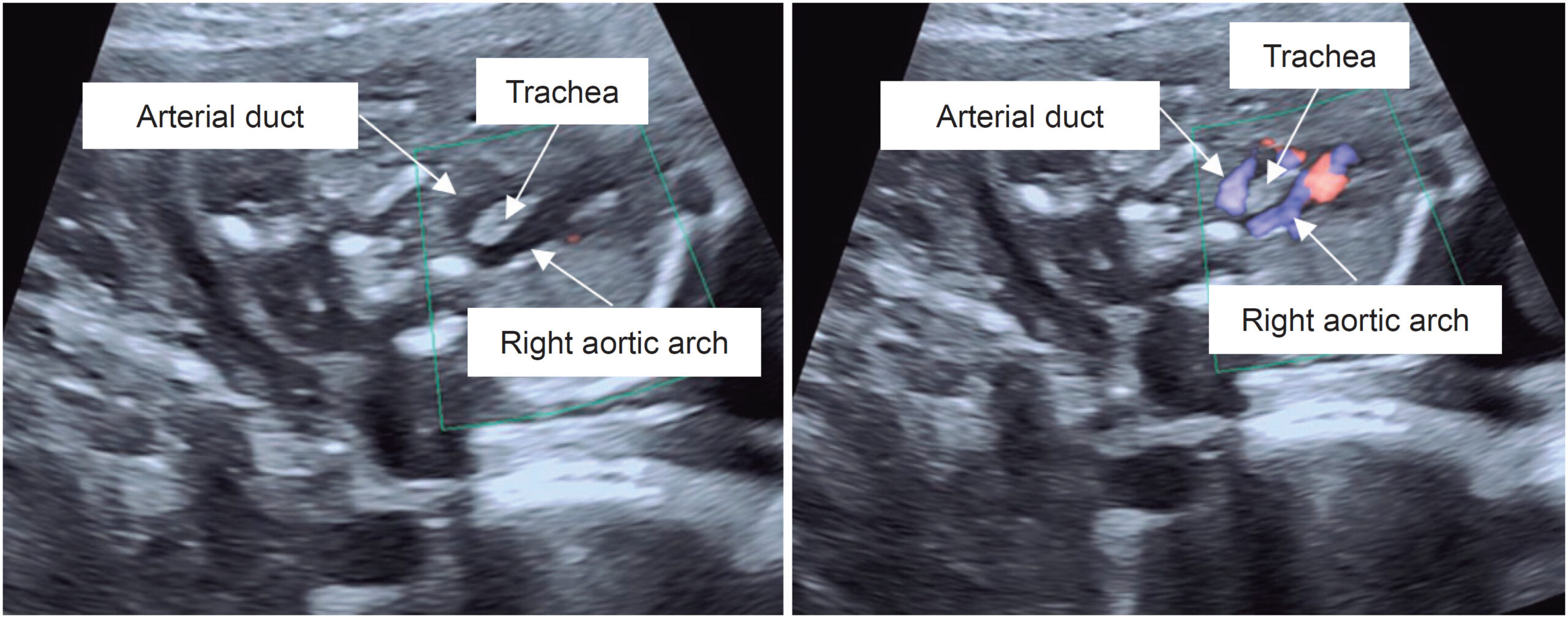
Congenital anomalies of the aortic arch affect 1-2% of the population and comprise a wide variety of abnormalities in the position and/or branching pattern of the aortic arch. Double aortic arch (DAA) represents 1-2% of all aortic arch anomalies and is characterized by persistence of both left and right embryonic aortic arches. It can be found either in isolation or, less frequently, in association with other cardiovascular or chromosomal abnormalities. Antenatal diagnosis of DAA may be challenging, as its distinction from other arch abnormalities like right aortic arch with left arterial duct or with an aberrant left subclavian artery is not always straightforward. The authors describe a case of antenatal diagnosis of DAA.

[…]

  • Aorta, Thoracic
  • Bronchomalacia/congenital
  • Chromosome Aberrations
  • Echocardiography/methods
  • Heart Defects Congenital/diagnosis
  • Heart Defects, Congenital/surgery
  • Ultrasonography/methods
  • Vocal Cord, Paralysis
Read Full Article
Creative Commons Attribution (CC BY) Creative Commons license
05/02/2021 DOI: 10.36660/abc.20190310
IJCS
Editorial

Lung Ultrasound in COVID 19 Outbreak: Can we Reduce the Burden from the Overloaded CT Departments?

Filipe Penna de Carvalho et al. Alex dos Santos Felix

Since the first descriptions on the use of lung ultrasound (LUS) for bedside evaluation of critically ill patients, important publications have underscored the utility of LUS for the detection of a wide array of pulmonary and pleural diseases, as well as for the differential diagnosis of acute respiratory failure, proving to be an important tool for a point-of-care based examination and an extended resource for examining critical patients, cited by many as the “new stethoscope”.,

The coronavirus disease-2019 (COVID-19) outbreak has emerged as a global health burden since late 2019, challenging healthcare systems around the world due to the constant need for intensive care support and isolation of patients, many of whom evolve with severe hemodynamic involvement, leading to high morbidity and mortality. This critical scenario highlights the importance of appropriateness and best appliance of all diagnostic and therapeutic resources. In this regard, the use of point-of-care ultrasound (POCUS) is proving to be a cost-effective tool for the management of these potentially critical patients, from the initial presentation (triage) to in-hospital management and post-discharge follow-up.

[…]

  • Computed Tomography
  • COVID-19/complications, Pandemics, Acute Failure Respiratory
  • Critic care
  • Morbidity
  • Mortality
  • Pleural Diseases/Diagnosis
  • Ultrasonography/methods
Read Full Article
Creative Commons Attribution (CC BY) Creative Commons license
25/11/2020 DOI: 10.36660/ijcs.20200283
Figure 1
IJCS
Original Article

Lung Ultrasound as a Triage Tool in an Emergency Setting during the Covid-19 Outbreak: comparison with CT Findings

Monica Luiza de Alcantara et al. Marcos Paulo Lacerda Bernardo Tatiana Bagrichevsky Autran Rodolfo de Paula Lustosa … Marcelo Tayah Lucia Antunes Chagas Dequitier Carvalho Machado

Abstract

Background

Lung ultrasound (LUS) can detect interstitial alveolar changes confined to the subpleural region, like those described in Covid-19.

Objetive

To evaluate how LUS findings correlate with chest computed tomography (CT) in patients admitted to the emergency department (ED) with suspicion of Covid-19.

Methods

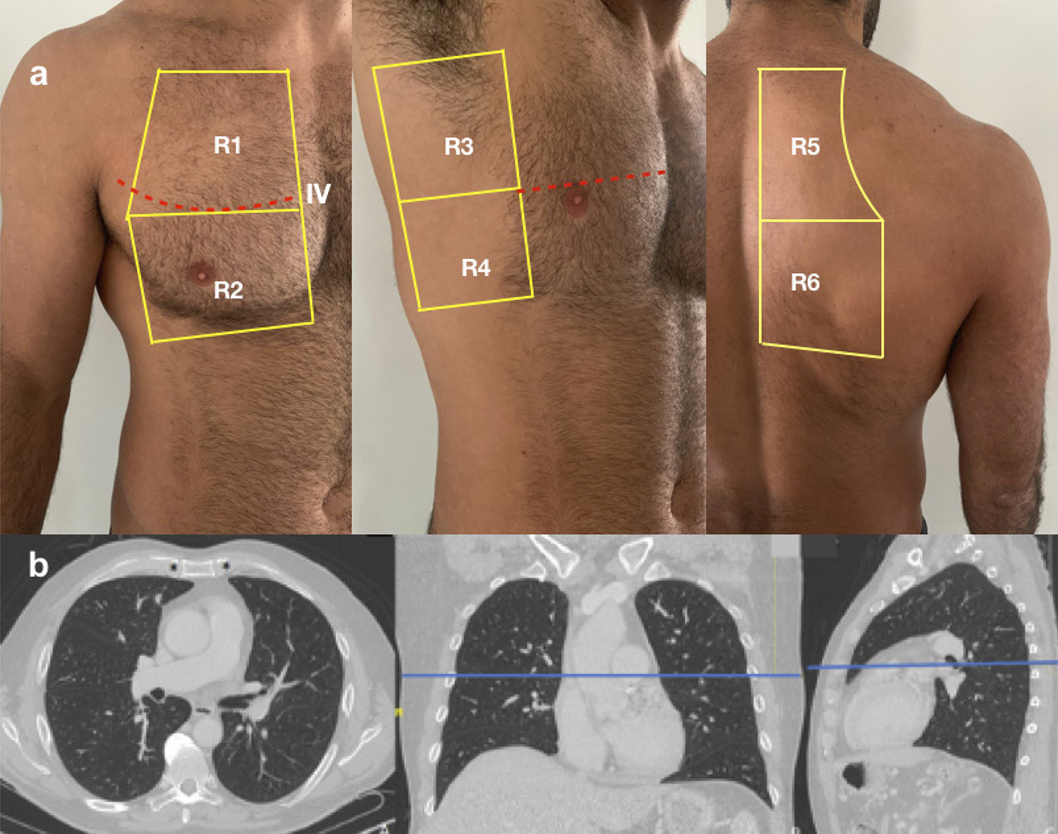
Cross-sectional study of 20 patients (median age 43 years; interquartile range, 37–63 years; 50% male). All patients underwent LUS and chest CT on the day of ED admission. Each hemithorax was divided into 6 segments with similar landmarks, and equivalent scores (sc) of lesion severity were defined for both methods. The number of affected segments on LUS (LUSseg) was divided into tertiles (0-1, 2-5, and ≥6), and compared with number of affected segments on CT (CTseg), LUSsc, CTsc, and percentage of affected lung parenchyma through visual analysis (CTvis). ANOVA or Kruskal–Wallis test for continuous variables, chi-square test for categorical variables, and receiver operating characteristic (ROC) curve analysis to define optimal cutoff points were performed. P<0.05 was considered statistically significant.

Results

Median LUSsc, CTsc, CTseg, and CTvis were significantly different between groups. A clear separation between groups was demonstrated; patients with 7 LUSsc and >3 LUSseg, respectively.

Conclusion

LUS findings correlate with chest CT, and can help identify patients with normal lung or minor pulmonary involvement secondary to Covid-19. Int J Cardiovasc Sci. 2020; [online].ahead print, PP.0-0

  • COVID-19
  • Lung
  • Pandemics
  • Pleura
  • Pulmonary Alveoli
  • Tomography, X Ray-Computed/methods
  • Triage
  • Ultrasonography/methods
Read Full Article
Creative Commons Attribution (CC BY) Creative Commons license
28/08/2020 DOI: 10.36660/ijcs.20200133 Views 1
Figure 1
ABC
Image

Prenatal Diagnosis of Intracardiac Tumors

Maria José Morais et al. Fátima Silva Mónica Melo Ana Carriço Francisco Valente

Clinical Case

Fetal cardiac tumors are a rare finding in prenatal ultrasonography with an incidence of 1-2/10000. Rhabdomyomas are the most common tumors in intrauterine life, accounting for 60-86% of primary fetal cardiac tumors.,

According to some case series and mostly case reports, the prevalence of tuberous sclerosis associated with fetal cardiac rhabdomyoma is 50-80%, resulting in a perinatal mortality rate of 0-100%.

[…]

  • Echocardiography/métodos
  • Heart Neoplasms/diagnosis
  • Prenatal Care
  • Rhabdomyoma
  • Tuberous Sclerosis
  • Ultrasonography/methods
Read Full Article
Creative Commons Attribution (CC BY) Creative Commons license
01/12/2016 DOI: 10.5935/abc.20160183 Views 1

How to cite this article

EndNote RIS

Electronic Document Format (Vancouver)

Cabral S. COVID-19 and Late Cardiovascular Manifestations – Building Up Evidence. Arquivos Brasileiros de Cardiologia 2022;119(2):326-7.

Electronic Document Format (ABNT)

Cabral, Sofia. COVID-19 and Late Cardiovascular Manifestations – Building Up Evidence. Arquivos Brasileiros de Cardiologia, v. 119, n. 2, p. 326-327, Aug. 2022.

Electronic Document Format (APA)

Cabral, S. (2022). COVID-19 and Late Cardiovascular Manifestations – Building Up Evidence. Arquivos Brasileiros de Cardiologia, 119(2), 326-327.

Electronic Document Format (ISO)

Cabral, Sofia. COVID-19 and Late Cardiovascular Manifestations – Building Up Evidence. Arquivos Brasileiros de Cardiologia [online]. 2022, vol. 119, n. 2, [cited 2026-10-14], pp. 326-327. Available from: <https://journals.cardiol.br/en/article/covid-19-and-late-cardiovascular-manifestations-building-up-evidence/>. ISSN 0066-782X

EndNote RIS

Electronic Document Format (Vancouver)

Mansiroglu AK, Seymen H, Sincer I, Gunes Y. Evaluation of Endothelial Dysfunction in COVID-19 With Flow-Mediated Dilatation. Arquivos Brasileiros de Cardiologia 2022;119(2):319-25.

Electronic Document Format (ABNT)

Mansiroglu, Asli Kurtar; Seymen, Hande; Sincer, Isa; Gunes, Yilmaz. Evaluation of Endothelial Dysfunction in COVID-19 With Flow-Mediated Dilatation. Arquivos Brasileiros de Cardiologia, v. 119, n. 2, p. 319-325, Jun. 2022.

Electronic Document Format (APA)

Mansiroglu, A. K., Seymen, H., Sincer, I., & Gunes, Y. (2022). Evaluation of Endothelial Dysfunction in COVID-19 With Flow-Mediated Dilatation. Arquivos Brasileiros de Cardiologia, 119(2), 319-325.

Electronic Document Format (ISO)

Mansiroglu, Asli Kurtar and Seymen, Hande and Sincer, Isa and Gunes, Yilmaz. Evaluation of Endothelial Dysfunction in COVID-19 With Flow-Mediated Dilatation. Arquivos Brasileiros de Cardiologia [online]. 2022, vol. 119, n. 2, [cited 2026-10-14], pp. 319-325. Available from: <https://journals.cardiol.br/en/article/evaluation-of-endothelial-dysfunction-in-covid-19-with-flow-mediated-dilatation/>. ISSN 0066-782X

EndNote RIS

Electronic Document Format (Vancouver)

Carneiro SS, Costa PV, Lima AV, Gomes FLT, Lirio LV, Batista DCFA. Right Ventricular Endomyocardial Fibrosis: Case Report. Arquivos Brasileiros de Cardiologia Imagem cardiovascular 2022;32(1):71-4.

Electronic Document Format (ABNT)

Carneiro, Stella de Souza; Costa, Patrick Ventorim; Lima, Aline Valério de; Gomes, Fernando Luiz Torres; Lirio, Ludmilla Ventura; Batista, Danielle Cristina Filgueira Alves. Right Ventricular Endomyocardial Fibrosis: Case Report. Arquivos Brasileiros de Cardiologia Imagem cardiovascular, v. 32, n. 1, p. 71-74, Jan. 2022.

Electronic Document Format (APA)

Carneiro, S. S., Costa, P. V., Lima, A. V. , Gomes, F. L. T., Lirio, L. V., & Batista, D. C. F. A. (2022). Right Ventricular Endomyocardial Fibrosis: Case Report. Arquivos Brasileiros de Cardiologia Imagem cardiovascular, 32(1), 71-74.

Electronic Document Format (ISO)

Carneiro, Stella de Souza and Costa, Patrick Ventorim and Lima, Aline Valério de and Gomes, Fernando Luiz Torres and Lirio, Ludmilla Ventura and Batista, Danielle Cristina Filgueira Alves. Right Ventricular Endomyocardial Fibrosis: Case Report. Arquivos Brasileiros de Cardiologia Imagem cardiovascular [online]. 2022, vol. 32, n. 1, [cited 2026-10-14], pp. 71-74. Available from: <https://journals.cardiol.br/en/article/right-ventricular-endomyocardial-fibrosis-case-report/>. ISSN 2318-8219

EndNote RIS

Electronic Document Format (Vancouver)

Noronha N, Hobbs A, Caldas P. Antenatal Diagnosis of Double Aortic Arch. Arquivos Brasileiros de Cardiologia 2021;116(2 suppl 1):4-7.

Electronic Document Format (ABNT)

Noronha, Natália; Hobbs, Angie; Caldas, Patricia. Antenatal Diagnosis of Double Aortic Arch. Arquivos Brasileiros de Cardiologia, v. 116, n. 2 suppl 1, p. 4-7, Feb. 2021.

Electronic Document Format (APA)

Noronha, N., Hobbs, A., & Caldas, P. (2021). Antenatal Diagnosis of Double Aortic Arch. Arquivos Brasileiros de Cardiologia, 116(2 suppl 1), 4-7.

Electronic Document Format (ISO)

Noronha, Natália and Hobbs, Angie and Caldas, Patricia. Antenatal Diagnosis of Double Aortic Arch. Arquivos Brasileiros de Cardiologia [online]. 2021, vol. 116, n. 2 suppl 1, [cited 2026-10-14], pp. 4-7. Available from: <https://journals.cardiol.br/en/article/antenatal-diagnosis-of-double-aortic-arch/>. ISSN 0066-782X

EndNote RIS

Electronic Document Format (Vancouver)

Carvalho FP, Felix AS. Lung Ultrasound in COVID 19 Outbreak: Can we Reduce the Burden from the Overloaded CT Departments?. International Journal of Cardiovascular Sciences 2020;33(6):604-5.

Electronic Document Format (ABNT)

Carvalho, Filipe Penna de; Felix, Alex dos Santos. Lung Ultrasound in COVID 19 Outbreak: Can we Reduce the Burden from the Overloaded CT Departments?. International Journal of Cardiovascular Sciences, v. 33, n. 6, p. 604-605, Nov. 2020.

Electronic Document Format (APA)

Carvalho, F. P. , & Felix, A. S. (2020). Lung Ultrasound in COVID 19 Outbreak: Can we Reduce the Burden from the Overloaded CT Departments?. International Journal of Cardiovascular Sciences, 33(6), 604-605.

Electronic Document Format (ISO)

Carvalho, Filipe Penna de and Felix, Alex dos Santos. Lung Ultrasound in COVID 19 Outbreak: Can we Reduce the Burden from the Overloaded CT Departments?. International Journal of Cardiovascular Sciences [online]. 2020, vol. 33, n. 6, [cited 2026-10-14], pp. 604-605. Available from: <https://journals.cardiol.br/en/article/lung-ultrasound-in-covid-19-outbreak-can-we-reduce-the-burden-from-the-overloaded-ct-departments/>. ISSN 2359-4802

EndNote RIS

Electronic Document Format (Vancouver)

Alcantara ML, Bernardo MPL, Autran TB, Lustosa RP, Tayah M, Chagas LA, Machado DC, et al. Lung Ultrasound as a Triage Tool in an Emergency Setting during the Covid-19 Outbreak: comparison with CT Findings. International Journal of Cardiovascular Sciences 2020;33(5):479-87.

Electronic Document Format (ABNT)

Alcantara, Monica Luiza de; Bernardo, Marcos Paulo Lacerda; Autran, Tatiana Bagrichevsky; Lustosa, Rodolfo de Paula; Tayah, Marcelo; Chagas, Lucia Antunes; Machado, Dequitier Carvalho. Lung Ultrasound as a Triage Tool in an Emergency Setting during the Covid-19 Outbreak: comparison with CT Findings. International Journal of Cardiovascular Sciences, v. 33, n. 5, p. 479-487, Aug. 2020.

Electronic Document Format (APA)

Alcantara, M. L. , Bernardo, M. P. L., Autran, T. B., Lustosa, R. P., Tayah, M., Chagas, L. A., & Machado, D. C. (2020). Lung Ultrasound as a Triage Tool in an Emergency Setting during the Covid-19 Outbreak: comparison with CT Findings. International Journal of Cardiovascular Sciences, 33(5), 479-487.

Electronic Document Format (ISO)

Alcantara, Monica Luiza de and Bernardo, Marcos Paulo Lacerda and Autran, Tatiana Bagrichevsky and Lustosa, Rodolfo de Paula and Tayah, Marcelo and Chagas, Lucia Antunes and Machado, Dequitier Carvalho. Lung Ultrasound as a Triage Tool in an Emergency Setting during the Covid-19 Outbreak: comparison with CT Findings. International Journal of Cardiovascular Sciences [online]. 2020, vol. 33, n. 5, [cited 2026-10-14], pp. 479-487. Available from: <https://journals.cardiol.br/en/article/lung-ultrasound-as-a-triage-tool-in-an-emergency-setting-during-the-covid-19-outbreak-comparison-with-ct-findings/>. ISSN 2359-4802

EndNote RIS

Electronic Document Format (Vancouver)

Morais MJ, Silva F, Melo M, Carriço A, Valente F. Prenatal Diagnosis of Intracardiac Tumors. Arquivos Brasileiros de Cardiologia 2016;107(6):605-6.

Electronic Document Format (ABNT)

Morais, Maria José; Silva, Fátima; Melo, Mónica; Carriço, Ana; Valente, Francisco. Prenatal Diagnosis of Intracardiac Tumors. Arquivos Brasileiros de Cardiologia, v. 107, n. 6, p. 605-606, Nov. 2016.

Electronic Document Format (APA)

Morais, M. J., Silva, F., Melo, M., Carriço, A., & Valente, F. (2016). Prenatal Diagnosis of Intracardiac Tumors. Arquivos Brasileiros de Cardiologia, 107(6), 605-606.

Electronic Document Format (ISO)

Morais, Maria José and Silva, Fátima and Melo, Mónica and Carriço, Ana and Valente, Francisco. Prenatal Diagnosis of Intracardiac Tumors. Arquivos Brasileiros de Cardiologia [online]. 2016, vol. 107, n. 6, [cited 2026-10-14], pp. 605-606. Available from: <https://journals.cardiol.br/en/article/prenatal-diagnosis-of-intracardiac-tumors/>. ISSN 0066-782X

  • About ABC Family
  • Internal Regulation
  • Editorial Board
  • Brazilian Society of Cardiology (SBC)

Issues

SBC Guidelines

  • Videos
  • Podcasts
  • News