ABC Family Arquivos Brasileiros de Cardiologia International Journal of Cardiovascular Sciences ABC Heart Failure & Cardiomyopathy ABC Imagem Cardiovascular
Português English
ABC Family
Português English
  • About
    • About ABC Family
    • Internal Regulation
    • Editorial Board
    • Brazilian Society of Cardiology (SBC)
  • Issues
  • SBC Guidelines
  • Content
    • Videos
    • Podcasts
    • News
  • About
    • About ABC Family
    • Internal Regulation
    • Editorial Board
    • Brazilian Society of Cardiology (SBC)
  • Issues
  • SBC Guidelines
  • Content
    • Videos
    • Podcasts
    • News

ABC Family

  • ABC Family
  • Arquivos Brasileiros de Cardiologia
  • International Journal of Cardiovascular Sciences
  • ABC Heart Failure & Cardiomyopathy
  • ABC Imagem Cardiovascular
Search in

No content found.

Journals
Ordering
Article types
Others

  1. Home
  2. Keyword: Mitral Valve Insufficiency
Figure 1
IJCS
Case Report

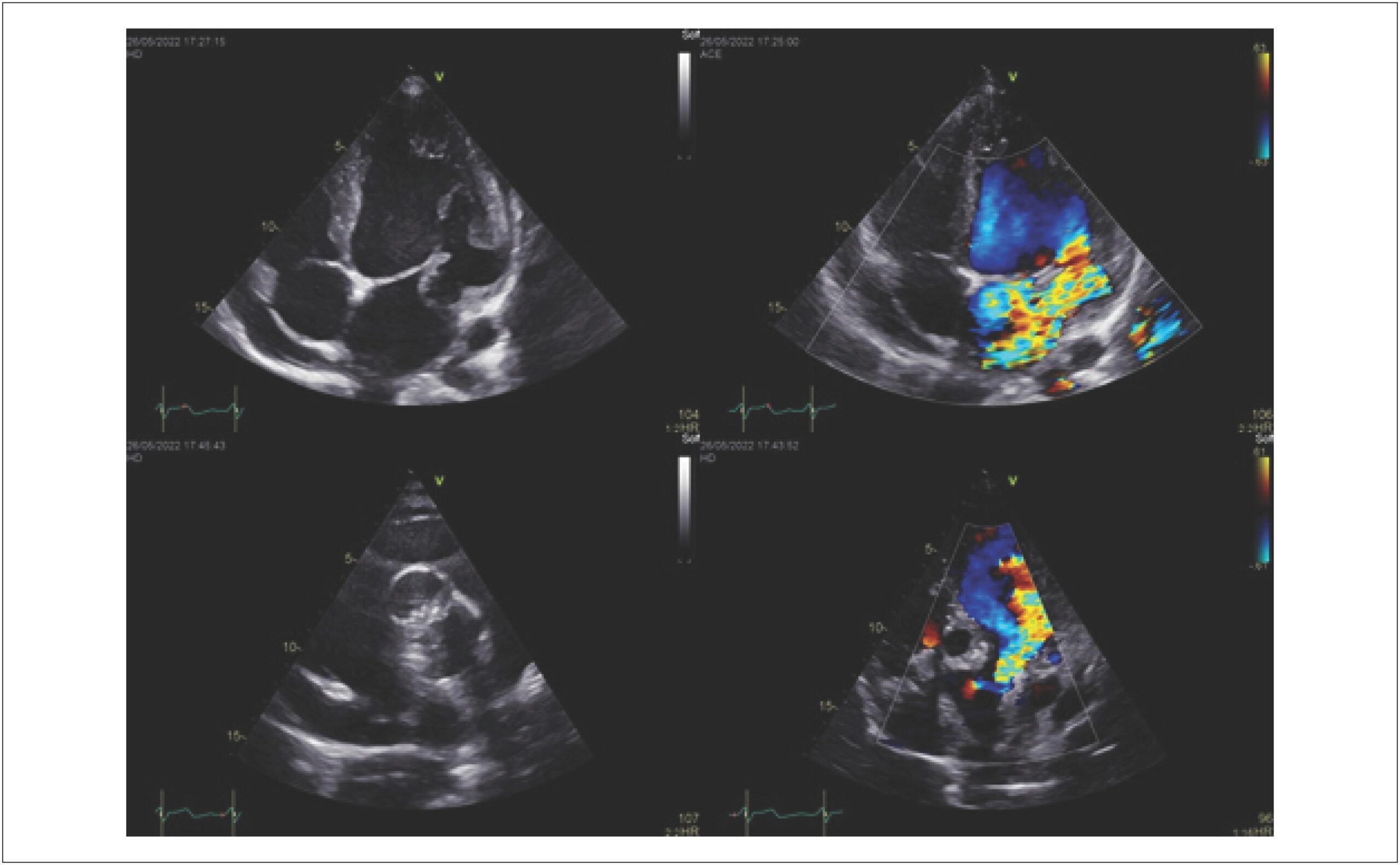
Double-Orifice Mitral Valve with Severe Regurgitation Associated with Bicuspid Aortic Valve: A Case Report

Patricia Delgado Cedeño et al. Estefania Carolina Arteaga Herrera Cesar João Tumbaco Mite Elsie Valdivieso Valenzuela … Juan Javier Zuñiga Bohorquez Guillermo Navas Moreira

Introduction

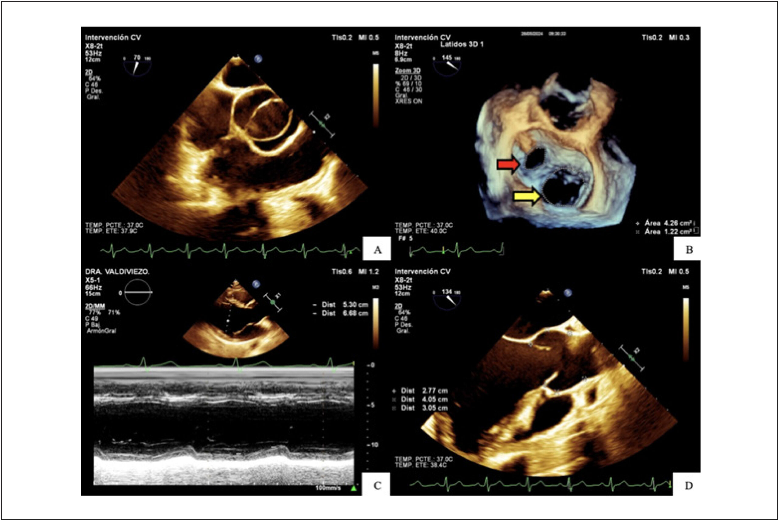
Double-orifice mitral valve (DOMV) was first described in 1876 by the British pathologist William Smith Greenfield. It is a very rare congenital or acquired cardiac anomaly with limited epidemiologic data and no clear sex predilection.

Congenital DOMV rarely occurs in isolation and is commonly associated with other cardiac anomalies, such as aortic coarctation, patent ductus arteriosus, atrioventricular septal defects, and bicuspid aortic valve (BAV), among others. Functionally, it may behave normally and remain asymptomatic; however, symptoms may arise from mitral valve dysfunction, such as stenosis or regurgitation.

[…]

  • Mitral Valve
  • Mitral Valve Annuloplasty
  • Mitral Valve Insufficiency
Read Full Article
Creative Commons Attribution (CC BY) Creative Commons license
11/09/2026 DOI: 10.36660/ijcs.20250214 Views 2
Figure 1
ABCIC
Case Report

Biventricular Noncompaction Cardiomyopathy Associated With Neuropathy in a Young Patient: A Case Report

Mônica de Oliveira Gonçalo et al. André Alexandre dos Santos Gomes Matheus Martins Monteiro Nicolas Babilônia Cavalcanti … Fernando Almeida Bezerra Moisés Abtibol Machado Dênison Clark Corrêa de Miranda Kátia do Nascimento Couceiro João Marcos Bemfica Barbosa Ferreira

Introduction

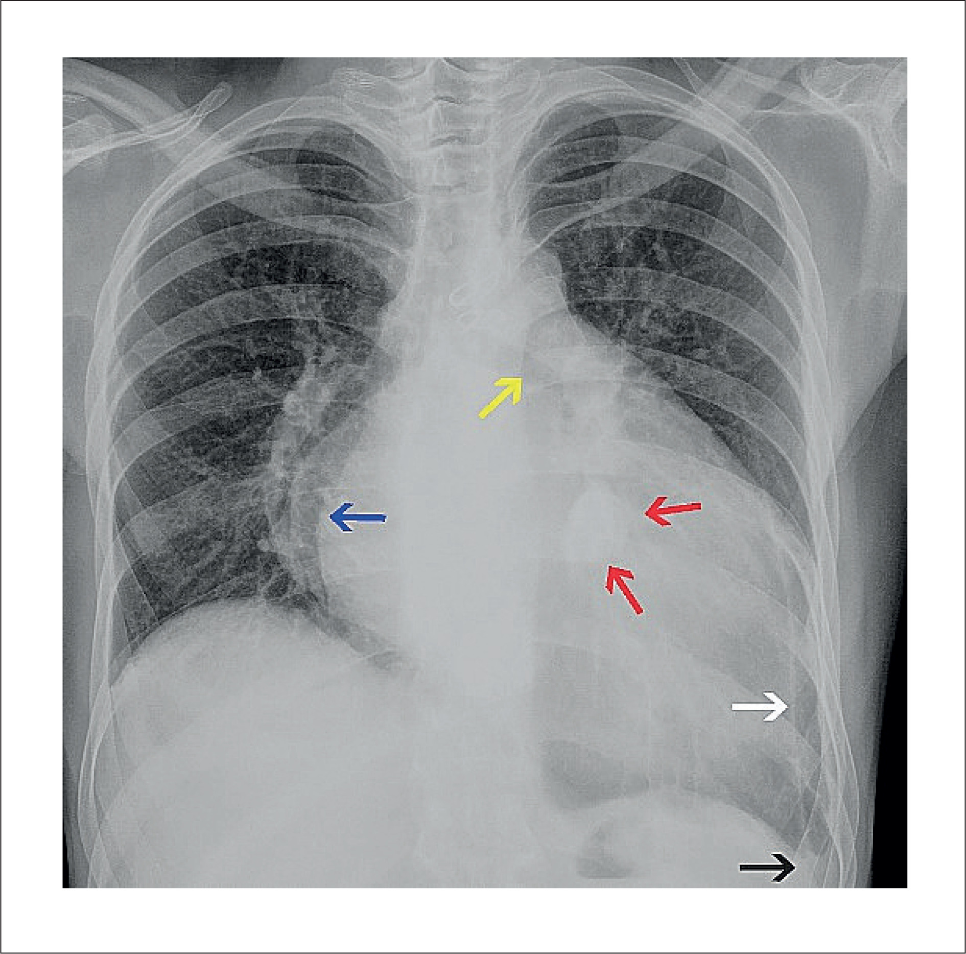
Ventricular hypertrabeculation, also known as ventricular noncompaction, is a myocardial phenotype characterized by excessive trabeculation and deep intertrabecular recesses caused by incomplete myocardial compaction during embryonic development. The earliest reported cases were associated with congenital heart defects, including ventricular outflow tract obstruction, complex cyanotic congenital heart disease, and coronary artery anomalies.,

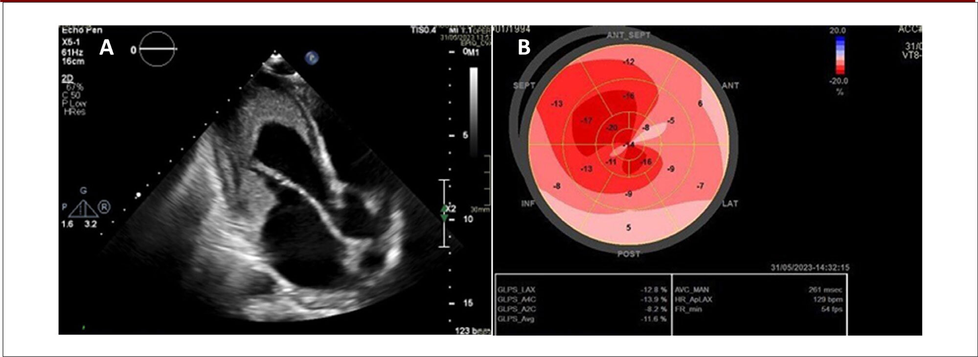
The clinical presentation is highly heterogeneous and may include heart failure (HF), arrhythmias, thromboembolic events, and sudden cardiac death. In addition to ventricular structural abnormalities, ventricular hypertrabeculation may be associated with cardiac conduction disorders, atrial arrhythmias, and functional valvular regurgitation (FVR) secondary to cardiac chamber dilatation, all of which contribute to greater clinical complexity and a poorer prognosis.

[…]

  • Case Reports
  • Left Ventricular Hypertrophy
  • Mitral Valve Insufficiency
Read Full Article
Creative Commons Attribution (CC BY) Creative Commons license
03/09/2026 DOI: 10.36660/abcimg.20260078i Views 1
My Approach to Assessing Mitral Regurgitation with Splay: What Does It Mean for Severity?
ABCIC
Review Article

My Approach to Assessing Mitral Regurgitation with Splay: What Does It Mean for Severity?

Gustavo Nishida et al. Natasha Soares Simões dos Santos Jorge Eduardo Assef Andrea de Andrade Vilela

Abstract

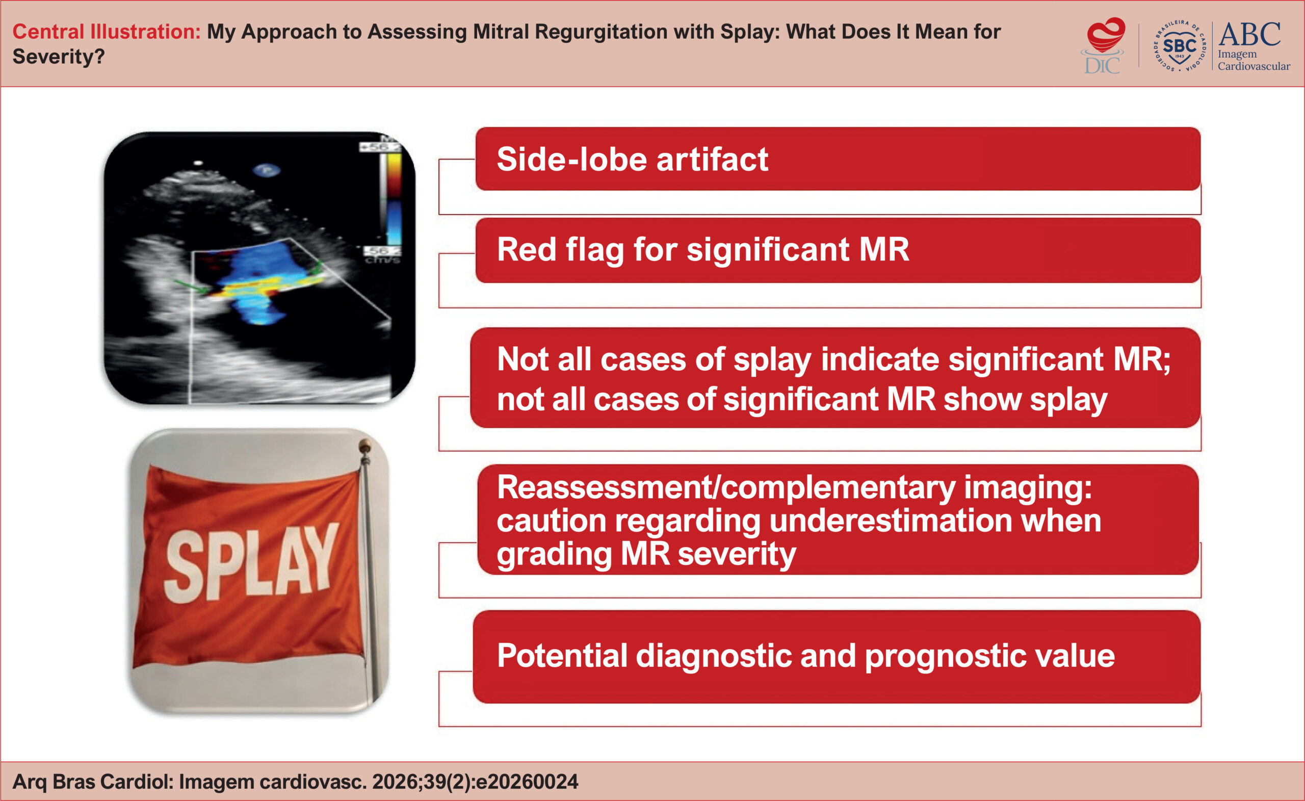
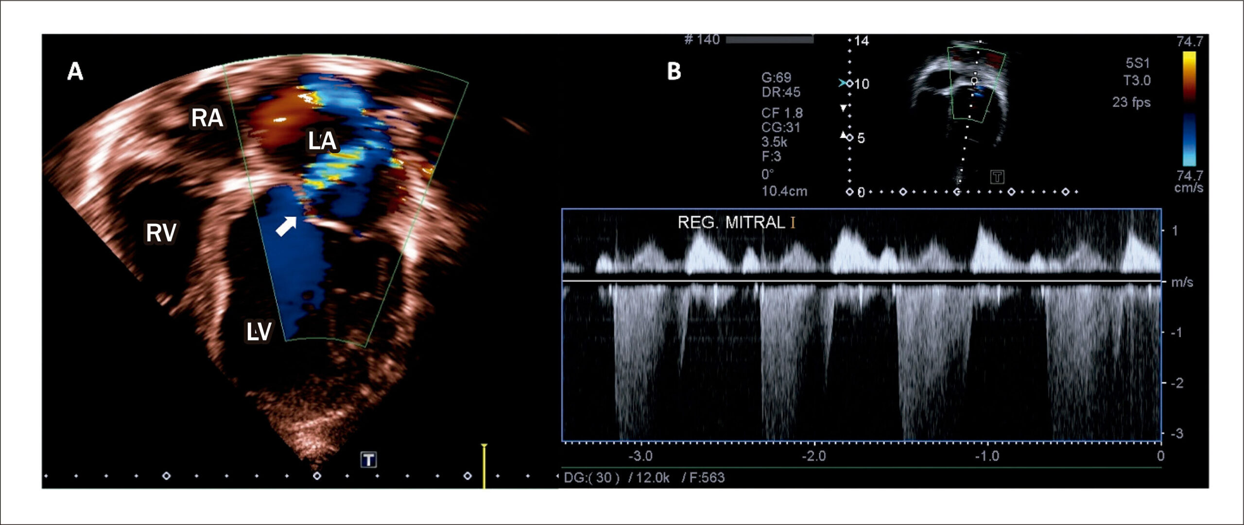
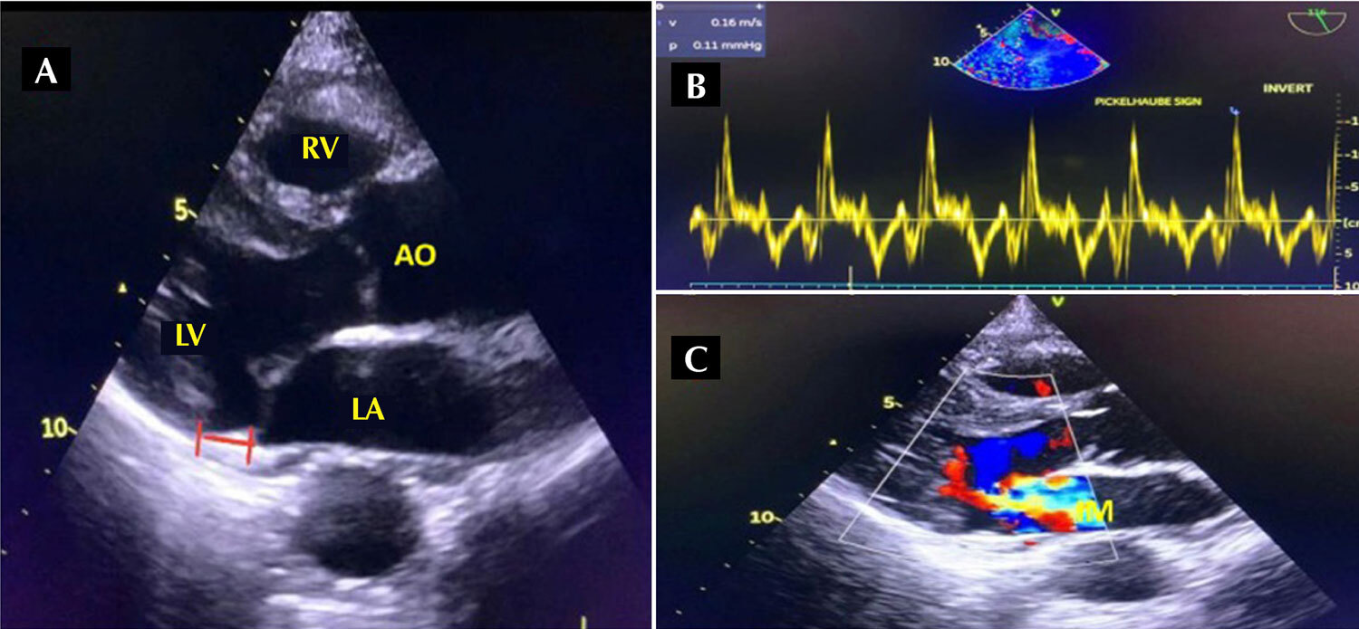
Echocardiographic assessment of mitral regurgitation (MR) is multiparametric and often challenging. The artifact known as splay has been described as an additional tool for estimating the severity of regurgitation. The term refers to a side-lobe artifact that forms a horizontal arc on color Doppler imaging. This same phenomenon can also be observed in other valvular heart diseases, such as aortic regurgitation. In 2020, Wiener et al. reported that splay was present in 81% of cases of significant MR, reaching 93% in eccentric jets, whereas the prevalence was only 16% in mild MR. Verbeke et al. associated the artifact with larger regurgitant volumes. A splay width greater than 29 mm was identified as an independent predictor of adverse cardiovascular outcomes. Although its presence alone does not denote severe MR, splay acts as a red flag, suggesting that regurgitation may be greater than it appears and indicating the need for careful reassessment of the transthoracic echocardiogram or, possibly, complementary transesophageal echocardiography. Further evidence and systematic evaluations are needed to investigate the best strategy for incorporating these data into the multiparametric approach to MR.

  • Echocardiography
  • Mitral Valve Insufficiency
  • Severity of Illness Index
Read Full Article
Creative Commons Attribution (CC BY) Creative Commons license
10/07/2026 DOI: 10.36660/abcimg.20260024i
The Causal Relationship between Blood Cell Traits and Valvular Heart Disease: A Two-Sample Mendelian Randomization Study
ABC
Original Article

The Causal Relationship between Blood Cell Traits and Valvular Heart Disease: A Two-Sample Mendelian Randomization Study

Xiaoli Wu et al. Yufeng Li Jinyu Tian

Abstract

Background

The pathogenesis of valvular heart disease (VHD) remains unclear, highlighting the need for further research on risk factors.

Objectives

To explore the potential causal links between blood cell traits and four types of VHD: rheumatic heart disease, mitral valve disease, aortic valve disease, and tricuspid valve disease.

Methods

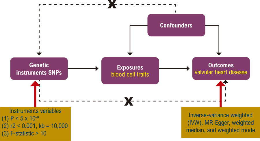
This study utilized summary statistics from large-scale GWAS and a two-sample Mendelian randomization approach, analyzing thirteen blood cell traits as exposures and four VHD phenotypes as outcomes. The primary MR method was inverse-variance weighted (IVW), supplemented by MR-Egger, weighted median, and weighted mode methods, with extensive sensitivity analyses to verify result reliability, where a p value < 0.05 was considered statistically significant.

Results

Among the thirteen blood cell traits examined, neutrophil count displayed a positive causal association with mitral valve disease (OR = 1.46, 95% CI: 1.16-1.85, p < 0.001) using the IVW method. This finding was further corroborated by the MR-Egger method (OR = 1.73, 95% CI: 1.09-2.75, p = 0.02), strengthening the evidence for this causal relationship. For this analysis, 253 robust genetic variants were used as instrumental variables. Additionally, mean corpuscular volume (MCV) showed a negative causal association with tricuspid valve disease (OR = 0.65, 95% CI: 0.44-0.96, p = 0.03) using the IVW method, based on 268 instrumental variables. Other blood cell traits did not show significant causal relationships with VHD. A comprehensive sensitivity analysis supported the robustness of our results.

Conclusions

The findings of this study suggest potential causal involvement of neutrophil count and MCV in the development of mitral valve disease and tricuspid valve disease, respectively.
  • Blood Cells
  • Heart Valve Diseases
  • Mendelian Randomization Analysis
  • Mitral Valve Insufficiency
Read Full Article
Creative Commons Attribution (CC BY) Creative Commons license
11/05/2026 DOI: 10.36660/abc.20250063i Views 2
Development and Validation of a Predictive Model for Atrial Functional Mitral Regurgitation
ABCIC
Original Article

Development and Validation of a Predictive Model for Atrial Functional Mitral Regurgitation

Alexandre Costa Souza et al. Bruna de Mattos Ivo Junqueira Stephanie de Azevedo Drubi Priscila Pinheiro … Laila Caroline Gomes Pedro Henrique Correia Filgueiras Ricardo André Sales Pereira Guedes Marco André Moraes Sales Yuri Xavier de Carvalho Carolina Thê Macêdo

Abstract

Background

Identifying atrial etiology in patients with mitral regurgitation remains challenging because the diagnosis is often established by exclusion. The use of a multivariable model may enhance diagnostic accuracy in the context of atrial functional mitral regurgitation.

Objective

To develop and validate a multivariable logistic regression model based on clinical and echocardiographic characteristics to predict atrial functional mitral regurgitation.

Methods

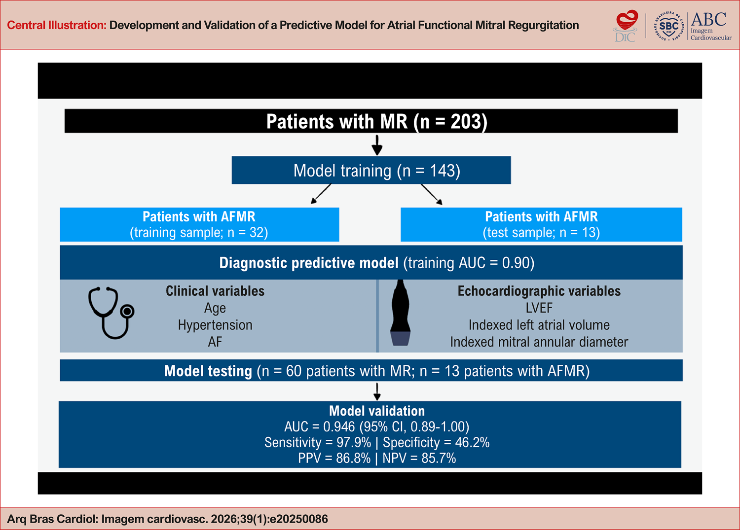
This cross-sectional study included patients with significant mitral regurgitation diagnosed by transesophageal echocardiography. The dataset was randomly divided into a training set (70%) and a validation set (30%). Statistical analyses were performed using a significance level of 5%.

Results

A total of 203 patients were included. The median age was 79 years in the atrial group and 72 years in the non-atrial group (p = 0.0022). Receiver operating characteristic curve analysis demonstrated good discriminative performance, with an area under the curve of 0.896 (95% CI, 0.845-0.947) in the training set. In the validation set, the model achieved an area under the curve of 0.946 (95% CI, 0.89-1.00), which indicates high predictive accuracy. Model calibration assessed by the Hosmer-Lemeshow test (chi-square test = 5.197; df = 8; p = 0.736) demonstrated good agreement between predicted and observed outcomes.

Conclusion

A multivariable model was derived and validated as a useful tool for predicting atrial etiology in patients with mitral regurgitation, potentially reducing diagnostic variability in clinical practice.

  • Echocardiography
  • Logistic Models
  • Mitral Valve Insufficiency
  • Statistical Models
Read Full Article
Creative Commons Attribution (CC BY) Creative Commons license
17/04/2026 DOI: 10.36660/abcimg.20250086i Views 3
ABCIC
Editorial

Functional Mitral Regurgitation of Atrial Origin: Search for Diagnostic Criteria

Minna Moreira Dias Romano

Identifying the various mechanisms of Mitral Regurgitation (MR) is essential for understanding the disease, prognosis, and planning structural therapeutic approaches. Recently, several studies on valvular heart disease have recognized functional MR. This condition is associated with the impact of cardiac geometry on mitral valve function, rather than primary valvular injury such as rheumatic, degenerative, infectious (endocarditis), or congenital diseases.

Furthermore, within the group of patients with functional MR, a subgroup has been identified that does not present Left Ventricular (LV) dilation or dysfunction. In this subgroup, the likely mechanism of MR is chronic enlargement of the Left Atrium (LA) and the mitral annulus, commonly associated with atrial fibrillation. Preliminary studies suggest associated left atrial dysfunction, which can be measured through tissue mechanics analysis. These factors likely contribute to the reduction of the coaptation surface of the mitral leaflet edges.

[…]

  • Atrial Fibrillation
  • Echocardiography
  • Mitral Valve Insufficiency
Read Full Article
Creative Commons Attribution (CC BY) Creative Commons license
10/04/2026 DOI: 10.36660/abcimg.20260008i Views 2
Figure 1
ABCIC
Case Report

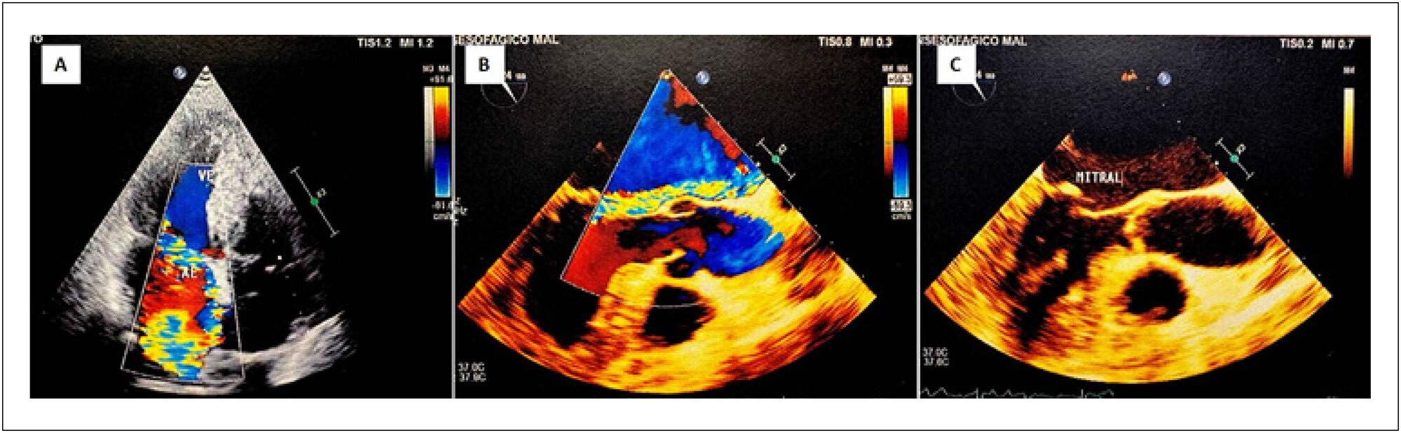
Important Mitral Regurgitation and Ventricular Dysfunction in Hypereosinophilic Syndrome: A Case Report

Marcel Pina Ciuffo et al. Lucas Mori Tais Araújo Ingrid Kowatsch Ana Clara Tude Rodrigues

Introduction

Hypereosinophilic syndrome (HES) represents a group of rare hematologic diseases characterized by an increased number of eosinophils in the peripheral blood. Once activated, these eosinophils can cause damage to host tissues and organs. Patients with HES generally present with dermatological, respiratory, and gastrointestinal signs and symptoms. Involvement of the cardiovascular system is uncommon and can lead to serious consequences such as heart failure, arrhythmias, and thromboembolic phenomena.

This article describes a case of eosinophilic endomyocardial disease, also known as Loeffler syndrome, a manifestation of HES characterized by eosinophil-mediated cardiac damage. Despite adequate treatment and control of the hematologic disease, the patient developed biventricular systolic dysfunction and important mitral regurgitation (MR).

[…]

  • Heart Failure
  • Hypereosinophilic Syndrome
  • Mitral Valve Insufficiency
Read Full Article
Creative Commons Attribution (CC BY) Creative Commons license
27/06/2025 DOI: 10.36660/abcimg.20250029i
Mitral Annulus Diameter's Relevance in the Diagnosis of Atrial Etiology in Mitral Regurgitation: A Comparative Analysis
ABCIC
Original Article

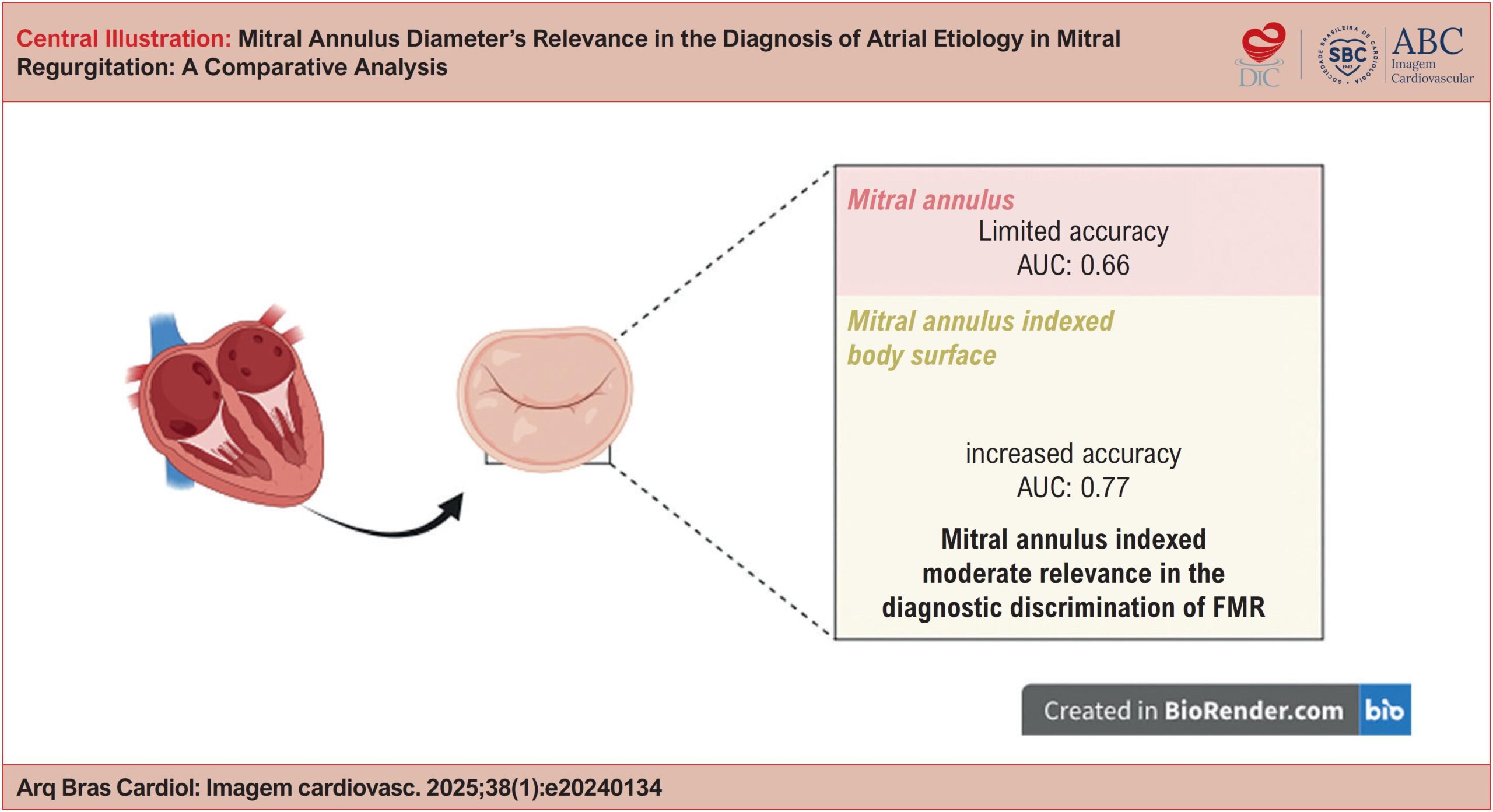
Mitral Annulus Diameter's Relevance in the Diagnosis of Atrial Etiology in Mitral Regurgitation: A Comparative Analysis

Alexandre Costa Souza et al. Bruna de Mattos Ivo Junqueira Stephanie de Azevedo Drubi Laila Caroline Gomes … Marcus Vinicius Freire Priscila Pinheiro Alessandra Ghattas Basile Marco André Moraes Sales Ricardo André Sales Pereira Guedes Carolina Thé Macêdo

Abstract

Background:

Atrial Functional Mitral Regurgitation (FMR) is defined by mitral annular dilation and functional alterations without structural impairment of the leaflets or subvalvular apparatus. It is associated with higher cardiovascular mortality, being often characterized as an exclusion diagnosis due to the absence of more precise diagnostic criteria. Therefore, it is crucial to identify markers that distinguish it from other Mitral Regurgitation (MR) types.

Objective:

To assess the accuracy of the intercommissural diameter of the mitral annulus and its value indexed to body surface area in differentiating atrial etiology in patients with MR.

Methods:

This is an observational cross-sectional study with 109 patients diagnosed with moderate or severe MR. Data were obtained between October 2022 and January 2024, from transesophageal echocardiograms performed at a referral hospital in the city of Salvador-BA.

Results:

The mean age was 69 ± 15, with 67 males and 28 cases of atrial etiology. The comparison between patients with MR of Atrial etiology versus Non-atrial etiology revealed significantly increased diameters of the intercommissural mitral annulus and its indexed value, with considerably larger diameters in the Atrial group (p = 0.009; 95% CI: 0.501 to 3.507). The receiver operating characteristic (ROC) curve analysis identified an optimal cut-off value of 20.8750, in which the sensitivity and specificity were 67.9%.

Conclusion:

Indexing the mitral annular diameter to the body surface area improves diagnostic accuracy in identifying the atrial etiology of MR compared to diameter alone, supporting its potential role in more comprehensive diagnostic algorithms.

  • Atrial Fibrillation
  • Heart Failure
  • Mitral Valve Insufficiency
Read Full Article
Creative Commons Attribution (CC BY) Creative Commons license
28/03/2025 DOI: 10.36660/abcimg.20240134i
Figure 1
ABCIC
Editorial

The Role of Objective Assessment of the Mitral Annulus

Minna Moreira Dias Romano

The mitral annulus is a complex and dynamic structure responsive to both structural and functional changes involving the mitral valve, as well as those affecting the geometry of the left ventricle (LV) and left atrium (LA). Its bean-shaped or “D”-shaped three-dimensional structure is similar to a horse’s saddle, in which the nadir (height) varies throughout the cardiac cycle. The malleability of the mitral annulus is important for valve competence. The posterior portion of the annulus, which is less fibrotic, moves during left ventricular systole, increasing the height of the saddle and reducing its circumferential area. In situations of left atrial dilation and dysfunction, mainly accompanied by atrial fibrillation (AF) and commonly associated with various causes of LV diastolic dysfunction, mitral annulus’ dynamics are often compromised. This results in reduced mobility of the posterior leaflet, annulus stiffness, and diminished oscillation of the saddle height, in addition to valve retraction (tethering), which contribute to mitral coaptation failure.

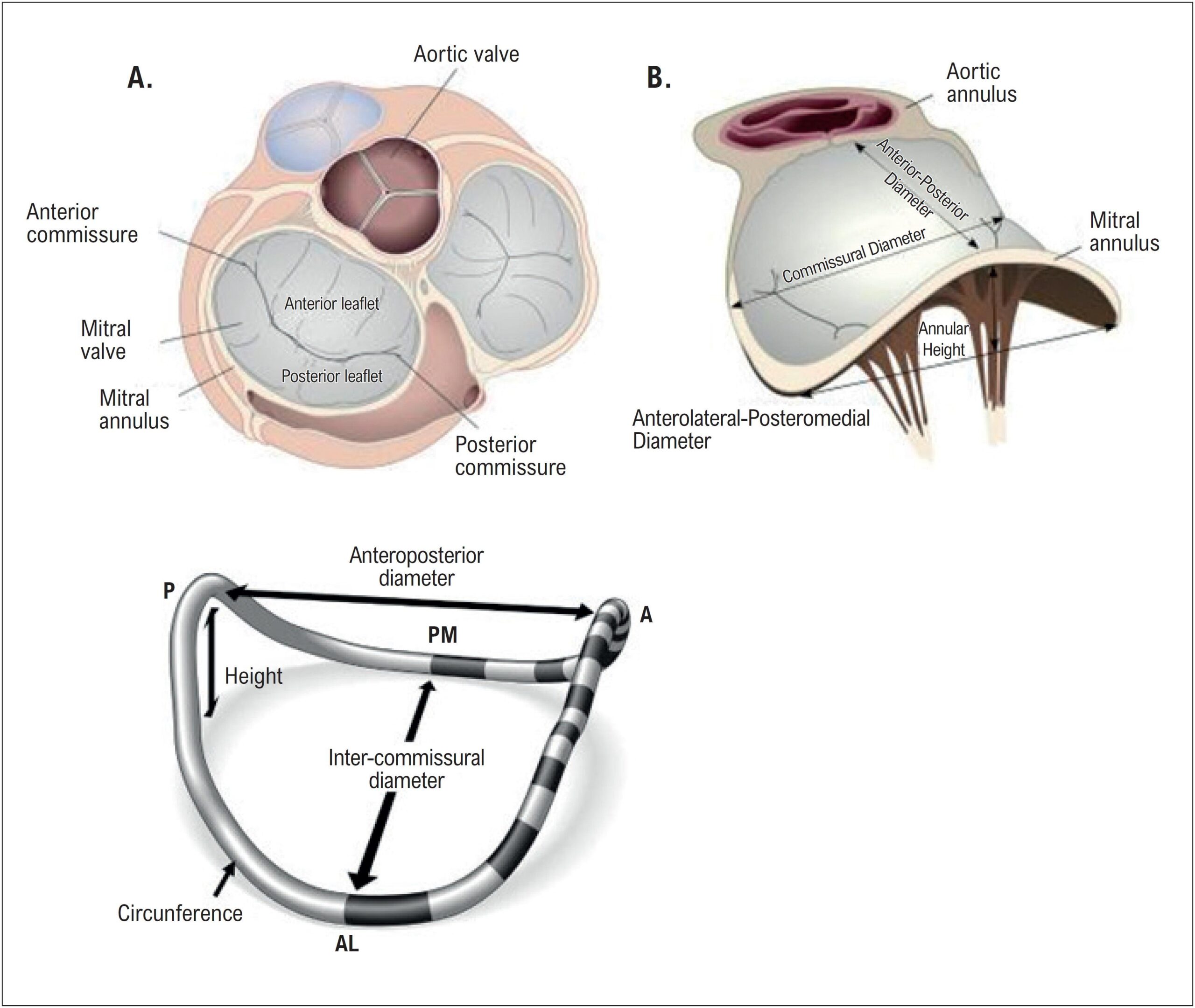
The mitral annulus can be studied using echocardiography, both two-dimensional and three-dimensional, with transesophageal imaging being the preferred approach. Three-dimensional imaging allows for various quantitative measurements facilitated by computational reconstruction software. These measurements can also be dynamic, capturing structural modifications throughout the cardiac cycle in both systole and diastole (). Key measurements include annular circumference and area, anteroposterior and inter-commissural diameters, and saddle height. The three-dimensional approach offers significant advantages, particularly when segmentation of valve anatomy is required, such as in cases of mitral valve prolapse that necessitate detailed anatomical analysis of valve bulges. Different etiologies of mitral regurgitation (MR) are accompanied by different degrees of mitral annulus deformation, with the most subtle ones observed in ischemic MR and the most significant in the most advanced spectra of mitral degenerative diseases, such as Barlow’s disease. For example, in rheumatic disease—still largely common in Brazil despite its low prevalence in more developed countries—mitral annulus changes are characterized by a “flattening of the saddle shape” and an increased area without a significant change in the perimeter.

[…]

  • Atrial Fibrillation
  • Echocardiography
  • Mitral Valve Insufficiency
Read Full Article
Creative Commons Attribution (CC BY) Creative Commons license
28/03/2025 DOI: 10.36660/abcimg.20250010i Views 1
Figure 1
ABC
Editorial

Mitral Regurgitation after Acute Myocardial Infarction: A Multi-Faceted Condition

Carlos Henrique Miranda

There are two leading causes of mitral regurgitation (MR) after acute myocardial infarction (AMI). First, the partial or complete papillary muscle rupture leads to MR; in this case, surgical repair is mandatory. Fortunately, the prevalence of this condition is low, around 0.05%. Second, the valve and the subvalvular apparatus are normal, and the MR results from leaflet tethering and regional left ventricular dilatation. The primary mechanism is the separation and excess angulation of the papillary muscle related to the regional wall motion in the myocardial infarction area. This second presentation is called the “functional” phenotype. This functional MR is a common echocardiographic finding after AMI. A recent investigation showed a mild MR in 76%, moderate MR in 21%, and severe MR in 3% of the patients after AMI. Vyas et al. found a significant MR (moderate + severe) prevalence of 7.21% after non-ST elevation myocardial infarction (NSTEMI). An important aspect to highlight is that most previous studies analyzed MR prevalence in all types of myocardial infarction together or ST-elevation AMI alone. The actual prevalence of MR in NSTEMI was unknown, and this study showed a considerable MR prevalence in this setting.

According to other previous studies,, this recent investigation showed that significant MR worst the patients’ prognosis, increasing the risk of death, pulmonary edema, heart failure, and cardiogenic shock. There are no doubts about the clinical impact of the MR after AMI. The more significant problem is that the mortality associated with this complication has not changed for decades, and there are still many doubts about the better management of these patients. Another feature deserves to be commented on: the clinical repercussions of severe MR are much more documented than those of moderate MR, and this classification is critical in making decisions about the treatment. In this recent investigation, the researchers analyzed the moderate and severe MR together, called significant MR; it was understandable because of the small sample size of severe MR; however, in the clinical setting, these two classifications have entirely different outcomes during the follow-up.

[…]

  • Mitral Valve Insufficiency
  • Myocardial Infarction
  • Myocardial Revascularization
Read Full Article
Creative Commons Attribution (CC BY) Creative Commons license
10/02/2025 DOI: 10.36660/abc.20240707i
NSTEMI and Ischemic Mitral Regurgitation: Incidence and Long-Term Clinical Outcomes with Respect to Management Strategy
ABC
Original Article

NSTEMI and Ischemic Mitral Regurgitation: Incidence and Long-Term Clinical Outcomes with Respect to Management Strategy

Pooja Vyas et al. Radhakishan Dake Kewal Kanabar Iva Patel … Ashish Mishra Vishal Sharma Tirth Nathwani Kunal Parwani Mital Rathod

Abstract

Background

The optimal treatment for ischemic mitral regurgitation (IMR) in patients of non-ST elevation myocardial infarction (NSTEMI) is a debated topic.

Objective

To evaluate the long term outcome on patients with NSTEMI and IMR, particularly emphasizing the comparison of treatments in those with moderate to severe MR.

Methods

We enrolled patients with NSTEMI and classified non/trivial to mild regurgitation as insignificant IMR and moderate to severe regurgitation as significant IMR. Furthermore, patients with substantial IMR were assessed for long-term clinical outcomes with respect to different management strategies. A test was considered statistically significant based on the probability value p<0.05.

Results

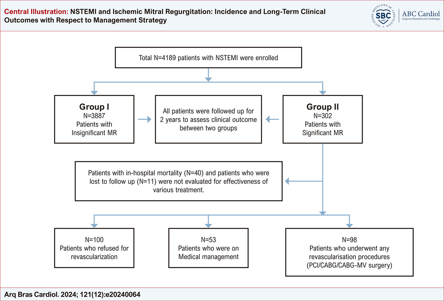
From a total of 4,189 patients of NSTEMI, significant IMR was found in 7.21% of patients. A significantly higher number of patients with death (1.21% vs. 13.24%, p<0.0001), cardiogenic shock (0.46% vs. 13.24%, p<0.0001) and heart failure (1.03% vs. 11.59%, p<0.0001) were found during hospitalization in patients with significant IMR. At a 2-year follow-up, a higher event rate was observed in the significant IMR group. Patients with significant IMR re-vascularized either by percutaneous coronary intervention (PCI), coronary artery bypass grafting (CABG), or CABG+ mitral valve (MV) surgery showed substantial improvement in MR grade ( 32.65% vs. 6% vs. 16.98%, p<0.0001) and LVEF (27.55% vs. 1% vs. 1.89%, p<0.0001) at 1 year follow up and significantly improved outcomes were identified compared to refused revascularization and medical management group with (-5.10% vs. 15% vs. 13.21%, p=0.04) mortality, (-33.67% vs. 61% vs. 73.58%, p<0.0001) readmission, and (-15.31% vs. 27% vs. 33.96%, p=0.01) heart failure at 2 years follow up.

Conclusion

Higher mortality and admission rates were observed in patients with significant IMR compared to those with in-significant IMR. Notably, significant IMR patients who underwent PCI, CABG, or CABG+MV surgery showed improved outcomes compared to non-revascularized counterparts.

  • Coronary Angioplasty Balloon
  • Mitral Valve Insufficiency
  • ST Elevation Myocardial Infarction
Read Full Article
Creative Commons Attribution (CC BY) Creative Commons license
13/12/2024 DOI: 10.36660/abc.20240064i Views 1
Figure 1
ABCIC
Review Article

My Approach to PISA EROA in Mitral Regurgitation: A Critical Review

Nicolas Merke

Abstract

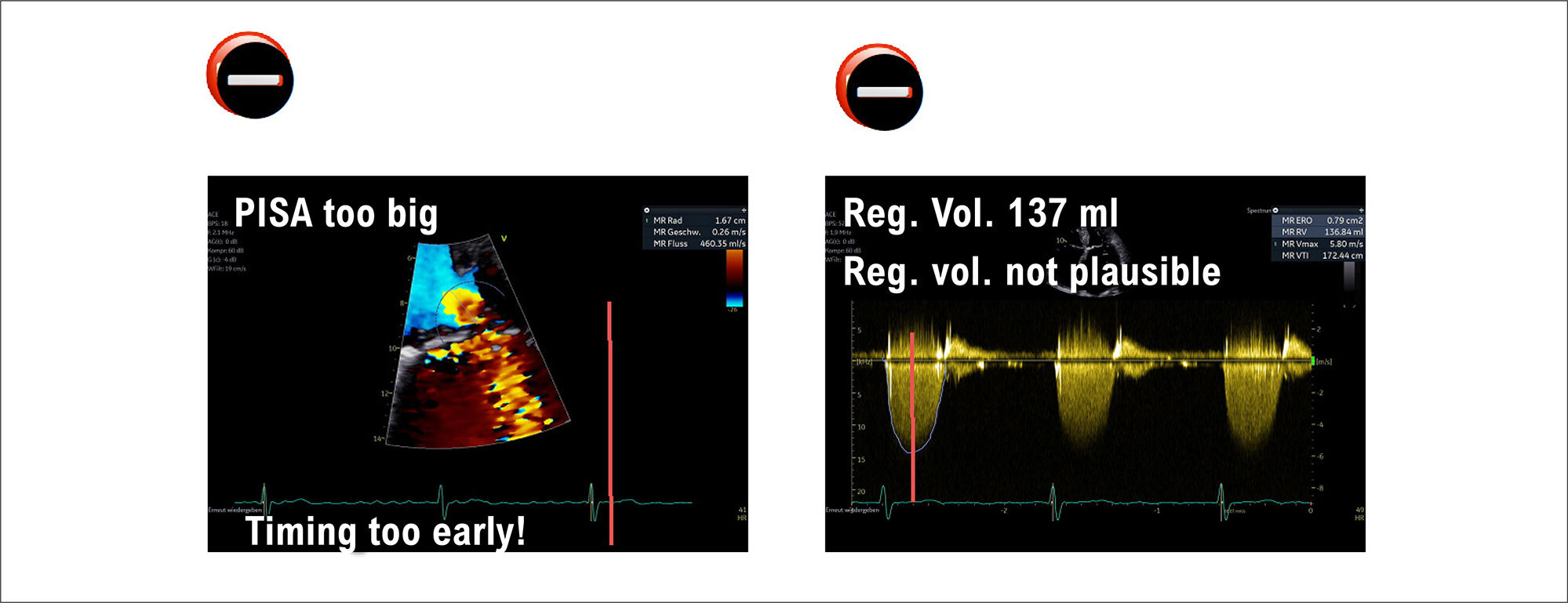
Mitral valve regurgitation is the most common valvular heart disease. In order to guide proper therapy, it is paramount to follow and use the guideline-recommended echocardiographic tools. The flow convergent method, which calculates the effective regurgitant orifice area and the regurgitant volume, plays an important role in the diagnostic workup and is widely used by echocardiographers and sonographers. Only by knowing the correct application and avoiding its pitfalls can we guarantee comprehensive and plausible values. This critical review will show in a step-by-step manner all critical moments by applying the Proximal Isovelocity Surface Area (PISA) and Effective Regurgitant Opening Area (EROA) method.

  • Heart Valve Diseases
  • Mitral Valve Insufficiency
  • Risk
Read Full Article
Creative Commons Attribution (CC BY) Creative Commons license
13/09/2024 DOI: 10.36660/abcimg.20240061i Views 1
Figure 1
ABCIC
Case Report

Idiopathic Mitral and Aortic Subvalvular Aneurysm in a Young Patient

João Pedro Laydner et al. Ana Paula Siciliano Rafael Castro da Silva Alex dos Santos Felix Maximiliano Freire Dutra

Abstract

Left ventricular aneurysms are primarily caused by coronary artery disease. Among them, there are also less common types, such as those located in the subvalvular regions of idiopathic origin. This article describes the case of a patient with a history of aortic subvalvular and Sinus of Valsalva aneurysm repairs who developed a new mitral subvalvular aneurysm.

  • Aortico-Ventricular Tunnel
  • Heart Aneurysm
  • Mitral Valve Insufficiency
Read Full Article
Creative Commons Attribution (CC BY) Creative Commons license
23/04/2024 DOI: 10.36660/abcimg.20240021i Views 2
Figure 1
ABCIC
Review Article

Atrial Functional Mitral Regurgitation

Alexsander da Silva Pretto

Abstract

Secondary mitral insufficiency (MI) in the setting of ventricular disease has always been a topic at conferences and has achieved extensive pathophysiological knowledge over the years. More recently, with the increasing incidence of atrial fibrillation (AF) and heart failure with preserved ejection fraction (HFpEF), a new phenotype that had been little discussed has come to light: atrial functional mitral regurgitation. In this entity, special attention should be addressed to the left atrium and the mitral annulus because, in early stages, they present normal left ventricular dimensions and function.

  • Atrial Fibrillation
  • Echocardiography
  • Heart Failure
  • Mitral Valve Insufficiency
Read Full Article
Creative Commons Attribution (CC BY) Creative Commons license
29/01/2024 DOI: 10.36660/abcimg.20230097i Views 1
Figure 1
ABCIC
Case Report

Takotsubo Syndrome After Percutaneous Mitral Valve Repair With Mitraclip®: A Case Report

Anna Luiza Souza et al. Maurício Lopes Prudente Débora Rodrigues Ana Cecília Campos Nogueira Giulliano Gardenghi

Introduction

Takotsubo syndrome (TTS), also known as stress cardiomyopathy or broken heart syndrome, is a condition whose pathophysiology is not well established, but which results from multiple factors that act synergistically in a setting of stress perception and cardiocirculatory response. It was first described in 1990, and the disproportionate and excessive increase in serum catecholamines is known to play a central role in its pathogenesis by favoring the occurrence of complications. Although rare, TTS deserves attention due to its inherent risk of decompensated heart function.,

[…]

  • Case Reports
  • Mitral Valve Insufficiency
  • Takotsubo Cardiomyopathy
Read Full Article
Creative Commons Attribution (CC BY) Creative Commons license
21/12/2023 DOI: 10.36660/abcimg.20230094i Views 1
ABCIC
Review Article

Using Three-Dimensional Printing in Surgical Mitral Valve Repairs

Rodrigo Bahiense Visconti et al. Luciano Herman Juaçaba Belem Antonio Carlos Dos Santos Nogueira Maria Claudia Lembo Cornélio Luis Henrique Weitzel

Abstract

Mitral regurgitation is an important cause of morbidity and mortality, and surgical mitral valve repair continues to be the treatment of choice when feasible. We use three-dimensional echocardiography associated with three-dimensional printing to create surgical models that allow trainees to practice or improve surgical techniques in order to improve the outcomes of surgical repairs, contributing to more advanced medical training.

  • Echocardiography
  • Mitral Valve Annuloplasty
  • Mitral Valve Insufficiency
  • Thoracic Surgery
  • Three-Dimensional Printing
Read Full Article
Creative Commons Attribution (CC BY) Creative Commons license
20/12/2023 DOI: 10.36660/abcimg.20230101i Views 1
Figure 1
ABCIC
Case Report

Importance of Echocardiography in the Differential Diagnosis of Rheumatic Mitral Regurgitation in Children and Adolescents

Camila Magalhães Silva et al. Maik Arantes Fátima Derlene da Rocha Araújo Adriana Furletti Machado Guimarães Zilda Maria Alves Meira

Introduction

Mitral regurgitation (MR) can be a result of abnormalities in different locations of the valve apparatus (cusps, annulus, tendinous cords and papillary muscles), being of acquired or congenital etiology. MR can be classified as primary, when resulting from a deformity in the valve structure, or secondary, when related to another heart or systemic disease. In our field, the main cause of MR in students and adolescents is the acquired one. Rheumatic fever (RF) is among the causes, as well as bacterial endocarditis, which should be considered in the differential diagnosis., However, congenital anomalies, such as mitral valve (MV) prolapse, mitral cleft (MC), parachute MV and, rarely, isolated cleft MV (ICMV) should be ruled out., The etiological diagnosis of MR is very important to determine the proper treatment, once it can be clinical or surgical, with definitive correction of the lesion.

We present the report of a student whose clinical and echocardiographic scenario was first diagnosed as acute RF, showing the importance of echocardiography in the posterior differential diagnosis of MR.

[…]

  • Diagnosis, Differential
  • Echocardiography
  • Mitral Valve Insufficiency
  • Pediatrics
  • Rheumatic Fever
Read Full Article
Creative Commons Attribution (CC BY) Creative Commons license
24/03/2023 DOI: 10.36660/abcimg.2023360i Views 1
Figure 1
ABC
Image

Tricky Diagnosis: An Aberrant Mitral Valve Chord

Marina Santos et al. Mariana Paiva Joana Ferreira Sara Guerreiro

Case

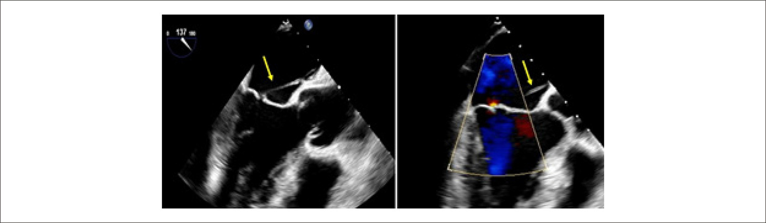
We present the case of a 52-year-old man with hepatitis C and a history of intravenous drug abuse, admitted to the infectology department with spondylodiscitis and psoas muscle abscess. Staphylococcus aureus was isolated from blood, urine, and spinal fluid cultures, and the transthoracic echocardiogram was negative for vegetation, abscess, or fistula. The transesophageal echocardiogram (TEE) showed a filamentous and mobile structure attached to the atrial surface of the mitral valve (MV), and a diagnosis of infective endocarditis was presumed. Seven days later, the patient was referred to our tertiary institution for a detailed TEE. In 2D images, we found a thin, well-delineated strand connecting the interatrial septum (IAS) to the tip of the anterior mitral leaflet without significant regurgitation ( , supplementary video 1). 3D imaging confirmed the presence of an anomalous chord connecting the A2 scallop of MV to the middle of the interatrial septum ( , supplementary video 2). There was no evidence of infective endocarditis, and this strand was found to be compatible with an anomalous left atrial MV chord.

[…]

  • Diagnostic Imaging/methods
  • Endocarditis, bacterial
  • Heart Defects, Congenital Atrial Septum
  • Mitral Valve Insufficiency
  • Staphylococcus Aureus
Read Full Article
Creative Commons Attribution (CC BY) Creative Commons license
10/03/2023 DOI: 10.36660/abc.20220573
Figure 1
ABC
Research Letter

Caseous Calcification of the Mitral Annulus: A Post-Heart Transplant Diagnosis

Bruno Jordão Chaves et al. Matheus Bitencourt Duarte Luiz Guilherme Passaglia Claudio Gelape … Paulo Hernane Rabelo Azevedo Geraldo Brasileiro Filho

Introduction

Caseous calcified mitral annulus is a non-neoplastic cardiac lesion considered to be a variant of mitral annular calcification that should be suspected when heart masses are detected by echocardiogram, chest x-ray or other radiologic studies.– The majority of the patients is asymptomatic, but signs and symptoms of mitral regurgitation, systemic embolization and atrioventricular blocks have been described.³ The diagnosis is confirmed by anatomopathologic examination. The prognosis is good. Here, we present a caseous calcification of the mitral annulus case diagnosed after the pathologic analysis of explanted heart.

[…]

  • Calcinosis
  • Cardiomyopathies
  • Diagnostic Imaging/methods
  • Heart Neoplasms/surgery
  • Heart Transplantation
  • Mitral Valve Insufficiency
Read Full Article
Creative Commons Attribution (CC BY) Creative Commons license
19/12/2022 DOI: 10.36660/abc.20210906
Figure 1
ABCIC
Case Report

Real-time Three-Dimensional Echocardiogram in the Identification of Late Complications in the Follow-Up After Mitraclip® Implantation

Alexandre Costa et al. Bruna de Mattos Ivo Junqueira Carolina Thé Macedo Marcus Vinicius Silva Freire de Carvalho … Cristiano Guedes Bezerra Rodrigo Morel Vieira de Melo

Introduction

Conventional surgery is the treatment of choice for severe mitral valve regurgitation. However, it is not feasible for patients at high surgical risk, who have high morbidity and mortality rates despite clinical treatment. Percutaneous valve treatment (MitraClip) has been evolving as a therapeutic perspective for this population, with reduced heart failure (HF) hospitalization and all-cause mortality rates.,,

Three-dimensional transesophageal echocardiography (3DTEE) has an additive role during clinical follow-up, especially in cases of suspected late complications with high detail of valve anatomy and clip positioning with cardiac structures. Additionally, due to the greater anatomical accuracy of this test, complications resulting from the procedure can be safely detected in real time.

[…]

  • Echocardiography, Three-Dimensional
  • Mitral Valve Insufficiency
  • Postoperative Complications, Cardiovascular Surgical Procedures
Read Full Article
Creative Commons Attribution (CC BY) Creative Commons license
01/12/2022 DOI: 10.47593/2675-312X/20223504eabc298i Views 1
Figure 1
ABCIC
Review Article

Mitral Annulus Disjunction: Diagnostic Modalities, Clinical Implications, and Prognostic Progression

Alexandre Costa Souza et al. Marcus Vinicius Silva Freire de Carvalho Marco André Sales Suellen Lacerda Bezerra … Jorge Adinon Torreão Carolina Thé Macêdo

Abstract

Mitral annulus disjunction (MAD) is an abnormal insertion of the flexion line of the mitral annulus into the atrial wall. The annulus presents a separation (disjunction) between the posterior leaflet–atrial wall junction and the left ventricular myocardial crest.1 MAD was first described more than 30 years ago in an autopsy study and is reportedly related to mitral valve prolapse (MVP) in 92% of cases.2 Since then, several studies have been conducted, and reports on the prevalence of MAD in patients with MVP have varied. Ultimately, it may or may not be associated with mitral regurgitation. Transthoracic echocardiography is part of initial MVP assessment, allowing its diagnosis and the assessment of related complications. As new diagnostic methods emerged, cardiac magnetic resonance imaging and transesophageal echocardiography improved the assessment of this pathology in terms of its diagnosis, extension, and location. However, the phenotypic characteristics of MVP that are more closely associated with MAD remain uncertain mainly due to the limited number of patients in classic studies on the subject. Patients with MAD may develop symptoms related to ventricular arrhythmias, configuring the MAD arrhythmic syndrome, which may progress to sudden death. The literature presents conflicting prognostic data among several studies on the subject from clear diagnostic criteria and best imaging method to be used to treatment and prognosis. This review describes MAD characteristics associated (or not) with valve prolapse to improve the diagnosis and management of this important pathology.

  • Diagnosis
  • Mitral Valve Insufficiency
  • Mitral Valve Prolapse
Read Full Article
Creative Commons Attribution (CC BY) Creative Commons license
07/07/2022 DOI: 10.47593/2675-312X/20223503eabc300 Views 1
Figure 1
ABCIC
Original Article

Left Atrial Function Analysis in Patients with Functional Mitral Regurgitation Associated with Dilated Cardiomyopathy

Stephanie Macedo Andrade et al. Rodrigo Bellio de Mattos Barretto Francisco Thiago Tomaz de Sousa David Costa de Souza Le Bihan

Abstract

Background

Functional mitral regurgitation (FMR) is associated with dilated cardiomyopathy (DC), heart failure (HF) and worsening left atrial function (LAF). Patients with DC and FMR may present left atrial dysfunction resulting from both ventricular dysfunction and valve disease, but it is unknown whether the presence of valve disease will lead to greater LAF impairment.

Objective

This study aimed to evaluate the relationship between LAF parameters and FMR degree in patients with DC.

Methods

This cross-sectional observational study included 214 patients with DC, 46 without FMR (control group) and 168 with mild, moderate or severe FMR. An LAF analysis was performed by speckle tracking echocardiography (STE) and atrial volumetric variation.

Results

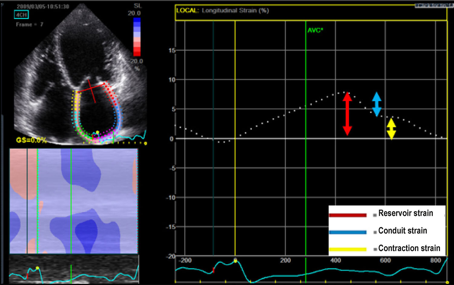
LAF analyzed by STE by means of reservoir strain, conduit strain and active contraction strain was reduced in the sample, with values of 14.3%, 8.49% and 5.92%, respectively. FMR degree was significantly associated with reservoir strain (0.27 ± 0.16 versus 0.15 ± 0.09; p < 0.001) and contraction strain (19.2 ± 7.3 versus 11.2 ± 2.7; p < 0.001). FMR was also associated with a reduced LAF assessed by volumetric analysis: total atrial emptying fraction of 0.51 ± 0.13 versus 0.34 ± 0.11 and active atrial emptying fraction of 0 .27 ± 0.16 versus 0.15 ± 0.09 (p < 0.001).

Conclusion

In a population with DC, FMR was associated with reduced LAF assessed by STE and atrial volume variation.

  • Atrial Function, Left
  • Cardiomyopathy, Dilated
  • Echocardiography
  • Mitral Valve Insufficiency
Read Full Article
Creative Commons Attribution (CC BY) Creative Commons license
30/06/2022 DOI: 10.47593/2675-312X/20223502eabc281 Views 1
ABCIC
Case Report

Rheumatic Mitral Valvopathy and Papillary Fibroelastoma An Unusual Association

Rafael Macedo Mustafé et al. Elinthon Tavares Veronesi Carlos Manuel Almeida Brandão Márcio Silva Miguel Lima

[…]

  • Echocardiography, Transesophageal
  • Mitral Valve Insufficiency
  • Mitral Valve Stenosis
  • Neoplasms
  • Rheumatic Fever
Read Full Article
Creative Commons Attribution (CC BY) Creative Commons license
04/02/2022 DOI: 10.5935/2318-8219.20180040 Views 1
ABCIC
Editorial

Choice of Imaging Methods in Hypertrophic Cardiomyopathy

Murillo de Oliveira Antunes et al. Edmundo Arteaga-Fernández Charles Mady

[…]

  • Cardiomyopathy,Hypertrophic
  • Diagnostic Imaging
  • Discrete Subaortic Stenosis
  • Echocardiography/methods
  • Electrocardiography/methods
  • Magnetic Resonance Spectroscopy/ methods
  • Mitral Valve Insufficiency
Read Full Article
Creative Commons Attribution (CC BY) Creative Commons license
20/01/2022 DOI: 10.5935/2318-8219.20190003 Views 1

Could not load more articles. Please try again.

How to cite this article

EndNote RIS

Electronic Document Format (Vancouver)

Cedeño PD, Herrera ECA, Mite CJT, Valenzuela EV, Bohorquez JJZ, Moreira GN. Double-Orifice Mitral Valve with Severe Regurgitation Associated with Bicuspid Aortic Valve: A Case Report. International Journal of Cardiovascular Sciences 2026;39:e20250214.

Electronic Document Format (ABNT)

Cedeño, Patricia Delgado; Herrera, Estefania Carolina Arteaga; Mite, Cesar João Tumbaco; Valenzuela, Elsie Valdivieso; Bohorquez, Juan Javier Zuñiga; Moreira, Guillermo Navas. Double-Orifice Mitral Valve with Severe Regurgitation Associated with Bicuspid Aortic Valve: A Case Report. International Journal of Cardiovascular Sciences, v. 39, e20250214, Sep. 2026.

Electronic Document Format (APA)

Cedeño, P. D., Herrera, E. C. A., Mite, C. J. T., Valenzuela, E. V., Bohorquez, J. J. Z., & Moreira, G. N. (2026). Double-Orifice Mitral Valve with Severe Regurgitation Associated with Bicuspid Aortic Valve: A Case Report. International Journal of Cardiovascular Sciences, 39, e20250214.

Electronic Document Format (ISO)

Cedeño, Patricia Delgado and Herrera, Estefania Carolina Arteaga and Mite, Cesar João Tumbaco and Valenzuela, Elsie Valdivieso and Bohorquez, Juan Javier Zuñiga and Moreira, Guillermo Navas. Double-Orifice Mitral Valve with Severe Regurgitation Associated with Bicuspid Aortic Valve: A Case Report. International Journal of Cardiovascular Sciences [online]. 2026, vol. 39, [cited 2026-10-14], e20250214. Available from: <https://journals.cardiol.br/en/article/double-orifice-mitral-valve-with-severe-regurgitation-associated-with-bicuspid-aortic-valve-a-case-report/>. ISSN 2359-4802

EndNote RIS

Electronic Document Format (Vancouver)

Gonçalo MO, Gomes AAS, Monteiro MM, Cavalcanti NB, Bezerra FA, Machado MA, Miranda DCC, et al. Biventricular Noncompaction Cardiomyopathy Associated With Neuropathy in a Young Patient: A Case Report. ABC Imagem Cardiovascular 2026;39(3):e20260078.

Electronic Document Format (ABNT)

Gonçalo, Mônica de Oliveira; Gomes, André Alexandre dos Santos; Monteiro, Matheus Martins; Cavalcanti, Nicolas Babilônia; Bezerra, Fernando Almeida; Machado, Moisés Abtibol; Miranda, Dênison Clark Corrêa de; Couceiro, Kátia do Nascimento; Ferreira, João Marcos Bemfica Barbosa. Biventricular Noncompaction Cardiomyopathy Associated With Neuropathy in a Young Patient: A Case Report. ABC Imagem Cardiovascular, v. 39, n. 3, e20260078, Sep. 2026.

Electronic Document Format (APA)

Gonçalo, M. O., Gomes, A. A. S., Monteiro, M. M., Cavalcanti, N. B., Bezerra, F. A., Machado, M. A., Miranda, D. C. C. , Couceiro, K. N., & Ferreira, J. M. B. B. (2026). Biventricular Noncompaction Cardiomyopathy Associated With Neuropathy in a Young Patient: A Case Report. ABC Imagem Cardiovascular, 39(3), e20260078.

Electronic Document Format (ISO)

Gonçalo, Mônica de Oliveira and Gomes, André Alexandre dos Santos and Monteiro, Matheus Martins and Cavalcanti, Nicolas Babilônia and Bezerra, Fernando Almeida and Machado, Moisés Abtibol and Miranda, Dênison Clark Corrêa de and Couceiro, Kátia do Nascimento and Ferreira, João Marcos Bemfica Barbosa. Biventricular Noncompaction Cardiomyopathy Associated With Neuropathy in a Young Patient: A Case Report. ABC Imagem Cardiovascular [online]. 2026, vol. 39, n. 3, [cited 2026-10-14], e20260078. Available from: <https://journals.cardiol.br/en/article/biventricular-noncompaction-cardiomyopathy-associated-with-neuropathy-in-a-young-patient-a-case-report/>. ISSN 2318-8219

EndNote RIS

Electronic Document Format (Vancouver)

Nishida G, Santos NSS, Assef JE, Vilela AA. My Approach to Assessing Mitral Regurgitation with Splay: What Does It Mean for Severity?. ABC Imagem Cardiovascular 2026;39(2):e20260024.

Electronic Document Format (ABNT)

Nishida, Gustavo; Santos, Natasha Soares Simões dos; Assef, Jorge Eduardo; Vilela, Andrea de Andrade. My Approach to Assessing Mitral Regurgitation with Splay: What Does It Mean for Severity?. ABC Imagem Cardiovascular, v. 39, n. 2, e20260024, Jul. 2026.

Electronic Document Format (APA)

Nishida, G., Santos, N. S. S. , Assef, J. E., & Vilela, A. A. (2026). My Approach to Assessing Mitral Regurgitation with Splay: What Does It Mean for Severity?. ABC Imagem Cardiovascular, 39(2), e20260024.

Electronic Document Format (ISO)

Nishida, Gustavo and Santos, Natasha Soares Simões dos and Assef, Jorge Eduardo and Vilela, Andrea de Andrade. My Approach to Assessing Mitral Regurgitation with Splay: What Does It Mean for Severity?. ABC Imagem Cardiovascular [online]. 2026, vol. 39, n. 2, [cited 2026-10-14], e20260024. Available from: <https://journals.cardiol.br/en/article/my-approach-to-assessing-mitral-regurgitation-with-splay-what-does-it-mean-for-severity/>. ISSN 2318-8219

EndNote RIS

Electronic Document Format (Vancouver)

Wu X, Li Y, Tian J. The Causal Relationship between Blood Cell Traits and Valvular Heart Disease: A Two-Sample Mendelian Randomization Study. Arquivos Brasileiros de Cardiologia 2026;123(3):e20250063.

Electronic Document Format (ABNT)

Wu, Xiaoli; Li, Yufeng; Tian, Jinyu. The Causal Relationship between Blood Cell Traits and Valvular Heart Disease: A Two-Sample Mendelian Randomization Study. Arquivos Brasileiros de Cardiologia, v. 123, n. 3, e20250063, May. 2026.

Electronic Document Format (APA)

Wu, X., Li, Y., & Tian, J. (2026). The Causal Relationship between Blood Cell Traits and Valvular Heart Disease: A Two-Sample Mendelian Randomization Study. Arquivos Brasileiros de Cardiologia, 123(3), e20250063.

Electronic Document Format (ISO)

Wu, Xiaoli and Li, Yufeng and Tian, Jinyu. The Causal Relationship between Blood Cell Traits and Valvular Heart Disease: A Two-Sample Mendelian Randomization Study. Arquivos Brasileiros de Cardiologia [online]. 2026, vol. 123, n. 3, [cited 2026-10-14], e20250063. Available from: <https://journals.cardiol.br/en/article/the-causal-relationship-between-blood-cell-traits-and-valvular-heart-disease-a-two-sample-mendelian-randomization-study/>. ISSN 0066-782X

EndNote RIS

Electronic Document Format (Vancouver)

Souza AC, Junqueira BMI, Drubi SA, Pinheiro P, Gomes LC, Filgueiras PHC, Guedes RASP, et al. Development and Validation of a Predictive Model for Atrial Functional Mitral Regurgitation. ABC Imagem Cardiovascular 2026;39(1):e20250086.

Electronic Document Format (ABNT)

Souza, Alexandre Costa; Junqueira, Bruna de Mattos Ivo; Drubi, Stephanie de Azevedo; Pinheiro, Priscila; Gomes, Laila Caroline; Filgueiras, Pedro Henrique Correia; Guedes, Ricardo André Sales Pereira; Sales, Marco André Moraes; Carvalho, Yuri Xavier de; Macêdo, Carolina Thê. Development and Validation of a Predictive Model for Atrial Functional Mitral Regurgitation. ABC Imagem Cardiovascular, v. 39, n. 1, e20250086, Apr. 2026.

Electronic Document Format (APA)

Souza, A. C., Junqueira, B. M. I., Drubi, S. A., Pinheiro, P., Gomes, L. C., Filgueiras, P. H. C., Guedes, R. A. S. P., Sales, M. A. M., Carvalho, Y. X. , & Macêdo, C. T. (2026). Development and Validation of a Predictive Model for Atrial Functional Mitral Regurgitation. ABC Imagem Cardiovascular, 39(1), e20250086.

Electronic Document Format (ISO)

Souza, Alexandre Costa and Junqueira, Bruna de Mattos Ivo and Drubi, Stephanie de Azevedo and Pinheiro, Priscila and Gomes, Laila Caroline and Filgueiras, Pedro Henrique Correia and Guedes, Ricardo André Sales Pereira and Sales, Marco André Moraes and Carvalho, Yuri Xavier de and Macêdo, Carolina Thê. Development and Validation of a Predictive Model for Atrial Functional Mitral Regurgitation. ABC Imagem Cardiovascular [online]. 2026, vol. 39, n. 1, [cited 2026-10-14], e20250086. Available from: <https://journals.cardiol.br/en/article/development-and-validation-of-a-predictive-model-for-atrial-functional-mitral-regurgitation/>. ISSN 2318-8219

EndNote RIS

Electronic Document Format (Vancouver)

Romano MMD. Functional Mitral Regurgitation of Atrial Origin: Search for Diagnostic Criteria. ABC Imagem Cardiovascular 2026;39(1):e20260008.

Electronic Document Format (ABNT)

Romano, Minna Moreira Dias. Functional Mitral Regurgitation of Atrial Origin: Search for Diagnostic Criteria. ABC Imagem Cardiovascular, v. 39, n. 1, e20260008, Apr. 2026.

Electronic Document Format (APA)

Romano, M. M. D. (2026). Functional Mitral Regurgitation of Atrial Origin: Search for Diagnostic Criteria. ABC Imagem Cardiovascular, 39(1), e20260008.

Electronic Document Format (ISO)

Romano, Minna Moreira Dias. Functional Mitral Regurgitation of Atrial Origin: Search for Diagnostic Criteria. ABC Imagem Cardiovascular [online]. 2026, vol. 39, n. 1, [cited 2026-10-14], e20260008. Available from: <https://journals.cardiol.br/en/article/functional-mitral-regurgitation-of-atrial-origin-search-for-diagnostic-criteria/>. ISSN 2318-8219

EndNote RIS

Electronic Document Format (Vancouver)

Ciuffo MP, Mori L, Araújo T, Kowatsch I, Rodrigues ACT. Important Mitral Regurgitation and Ventricular Dysfunction in Hypereosinophilic Syndrome: A Case Report. ABC Imagem Cardiovascular 2025;38(2):e20250029.

Electronic Document Format (ABNT)

Ciuffo, Marcel Pina; Mori, Lucas; Araújo, Tais; Kowatsch, Ingrid; Rodrigues, Ana Clara Tude. Important Mitral Regurgitation and Ventricular Dysfunction in Hypereosinophilic Syndrome: A Case Report. ABC Imagem Cardiovascular, v. 38, n. 2, e20250029, Jun. 2025.

Electronic Document Format (APA)

Ciuffo, M. P., Mori, L., Araújo, T., Kowatsch, I., & Rodrigues, A. C. T. (2025). Important Mitral Regurgitation and Ventricular Dysfunction in Hypereosinophilic Syndrome: A Case Report. ABC Imagem Cardiovascular, 38(2), e20250029.

Electronic Document Format (ISO)

Ciuffo, Marcel Pina and Mori, Lucas and Araújo, Tais and Kowatsch, Ingrid and Rodrigues, Ana Clara Tude. Important Mitral Regurgitation and Ventricular Dysfunction in Hypereosinophilic Syndrome: A Case Report. ABC Imagem Cardiovascular [online]. 2025, vol. 38, n. 2, [cited 2026-10-14], e20250029. Available from: <https://journals.cardiol.br/en/article/important-mitral-regurgitation-and-ventricular-dysfunction-in-hypereosinophilic-syndrome-a-case-report/>. ISSN 2318-8219

EndNote RIS

Electronic Document Format (Vancouver)

Souza AC, Junqueira BMI, Drubi SA, Gomes LC, Freire MV, Pinheiro P, Basile AG, et al. Mitral Annulus Diameter’s Relevance in the Diagnosis of Atrial Etiology in Mitral Regurgitation: A Comparative Analysis. ABC Imagem Cardiovascular 2025;38(1):e20240134.

Electronic Document Format (ABNT)

Souza, Alexandre Costa; Junqueira, Bruna de Mattos Ivo; Drubi, Stephanie de Azevedo; Gomes, Laila Caroline; Freire, Marcus Vinicius; Pinheiro, Priscila; Basile, Alessandra Ghattas; Sales, Marco André Moraes; Guedes, Ricardo André Sales Pereira; Macêdo, Carolina Thé. Mitral Annulus Diameter’s Relevance in the Diagnosis of Atrial Etiology in Mitral Regurgitation: A Comparative Analysis. ABC Imagem Cardiovascular, v. 38, n. 1, e20240134, Mar. 2025.

Electronic Document Format (APA)

Souza, A. C., Junqueira, B. M. I., Drubi, S. A., Gomes, L. C., Freire, M. V., Pinheiro, P., Basile, A. G., Sales, M. A. M., Guedes, R. A. S. P., & Macêdo, C. T. (2025). Mitral Annulus Diameter’s Relevance in the Diagnosis of Atrial Etiology in Mitral Regurgitation: A Comparative Analysis. ABC Imagem Cardiovascular, 38(1), e20240134.

Electronic Document Format (ISO)

Souza, Alexandre Costa and Junqueira, Bruna de Mattos Ivo and Drubi, Stephanie de Azevedo and Gomes, Laila Caroline and Freire, Marcus Vinicius and Pinheiro, Priscila and Basile, Alessandra Ghattas and Sales, Marco André Moraes and Guedes, Ricardo André Sales Pereira and Macêdo, Carolina Thé. Mitral Annulus Diameter’s Relevance in the Diagnosis of Atrial Etiology in Mitral Regurgitation: A Comparative Analysis. ABC Imagem Cardiovascular [online]. 2025, vol. 38, n. 1, [cited 2026-10-14], e20240134. Available from: <https://journals.cardiol.br/en/article/mitral-annulus-diameters-relevance-in-the-diagnosis-of-atrial-etiology-in-mitral-regurgitation-a-comparative-analysis/>. ISSN 2318-8219

EndNote RIS

Electronic Document Format (Vancouver)

Romano MMD. The Role of Objective Assessment of the Mitral Annulus. ABC Imagem Cardiovascular 2025;38(1):e20250010.

Electronic Document Format (ABNT)

Romano, Minna Moreira Dias. The Role of Objective Assessment of the Mitral Annulus. ABC Imagem Cardiovascular, v. 38, n. 1, e20250010, Mar. 2025.

Electronic Document Format (APA)

Romano, M. M. D. (2025). The Role of Objective Assessment of the Mitral Annulus. ABC Imagem Cardiovascular, 38(1), e20250010.

Electronic Document Format (ISO)

Romano, Minna Moreira Dias. The Role of Objective Assessment of the Mitral Annulus. ABC Imagem Cardiovascular [online]. 2025, vol. 38, n. 1, [cited 2026-10-14], e20250010. Available from: <https://journals.cardiol.br/en/article/the-role-of-objective-assessment-of-the-mitral-annulus/>. ISSN 2318-8219

EndNote RIS

Electronic Document Format (Vancouver)

Miranda CH. Mitral Regurgitation after Acute Myocardial Infarction: A Multi-Faceted Condition. Arquivos Brasileiros de Cardiologia 2025;121(12):e20240707.

Electronic Document Format (ABNT)

Miranda, Carlos Henrique. Mitral Regurgitation after Acute Myocardial Infarction: A Multi-Faceted Condition. Arquivos Brasileiros de Cardiologia, v. 121, n. 12, e20240707, Feb. 2025.

Electronic Document Format (APA)

Miranda, C. H. (2025). Mitral Regurgitation after Acute Myocardial Infarction: A Multi-Faceted Condition. Arquivos Brasileiros de Cardiologia, 121(12), e20240707.

Electronic Document Format (ISO)

Miranda, Carlos Henrique. Mitral Regurgitation after Acute Myocardial Infarction: A Multi-Faceted Condition. Arquivos Brasileiros de Cardiologia [online]. 2025, vol. 121, n. 12, [cited 2026-10-14], e20240707. Available from: <https://journals.cardiol.br/en/article/mitral-regurgitation-after-acute-myocardial-infarction-a-multi-faceted-condition/>. ISSN 0066-782X

EndNote RIS

Electronic Document Format (Vancouver)

Vyas P, Dake R, Kanabar K, Patel I, Mishra A, Sharma V, Nathwani T, et al. NSTEMI and Ischemic Mitral Regurgitation: Incidence and Long-Term Clinical Outcomes with Respect to Management Strategy. Arquivos Brasileiros de Cardiologia 2024;121(12):e20240064.

Electronic Document Format (ABNT)

Vyas, Pooja; Dake, Radhakishan; Kanabar, Kewal; Patel, Iva; Mishra, Ashish; Sharma, Vishal; Nathwani, Tirth; Parwani, Kunal; Rathod, Mital. NSTEMI and Ischemic Mitral Regurgitation: Incidence and Long-Term Clinical Outcomes with Respect to Management Strategy. Arquivos Brasileiros de Cardiologia, v. 121, n. 12, e20240064, Dec. 2024.

Electronic Document Format (APA)

Vyas, P., Dake, R., Kanabar, K., Patel, I., Mishra, A., Sharma, V., Nathwani, T., Parwani, K., & Rathod, M. (2024). NSTEMI and Ischemic Mitral Regurgitation: Incidence and Long-Term Clinical Outcomes with Respect to Management Strategy. Arquivos Brasileiros de Cardiologia, 121(12), e20240064.

Electronic Document Format (ISO)

Vyas, Pooja and Dake, Radhakishan and Kanabar, Kewal and Patel, Iva and Mishra, Ashish and Sharma, Vishal and Nathwani, Tirth and Parwani, Kunal and Rathod, Mital. NSTEMI and Ischemic Mitral Regurgitation: Incidence and Long-Term Clinical Outcomes with Respect to Management Strategy. Arquivos Brasileiros de Cardiologia [online]. 2024, vol. 121, n. 12, [cited 2026-10-14], e20240064. Available from: <https://journals.cardiol.br/en/article/nstemi-and-ischemic-mitral-regurgitation-incidence-and-long-term-clinical-outcomes-with-respect-to-management-strategy/>. ISSN 0066-782X

EndNote RIS

Electronic Document Format (Vancouver)

Merke N. My Approach to PISA EROA in Mitral Regurgitation: A Critical Review. ABC Imagem Cardiovascular 2024;37(3):e20240061.

Electronic Document Format (ABNT)

Merke, Nicolas. My Approach to PISA EROA in Mitral Regurgitation: A Critical Review. ABC Imagem Cardiovascular, v. 37, n. 3, e20240061, Sep. 2024.

Electronic Document Format (APA)

Merke, N. (2024). My Approach to PISA EROA in Mitral Regurgitation: A Critical Review. ABC Imagem Cardiovascular, 37(3), e20240061.

Electronic Document Format (ISO)

Merke, Nicolas. My Approach to PISA EROA in Mitral Regurgitation: A Critical Review. ABC Imagem Cardiovascular [online]. 2024, vol. 37, n. 3, [cited 2026-10-14], e20240061. Available from: <https://journals.cardiol.br/en/article/my-approach-to-pisa-eroa-in-mitral-regurgitation-a-critical-review/>. ISSN 2318-8219

EndNote RIS

Electronic Document Format (Vancouver)

Laydner JP, Siciliano AP, Silva RC, Felix AS, Dutra MF. Idiopathic Mitral and Aortic Subvalvular Aneurysm in a Young Patient. ABC Imagem Cardiovascular 2024;37(2):e20240021.

Electronic Document Format (ABNT)

Laydner, João Pedro; Siciliano, Ana Paula; Silva, Rafael Castro da; Felix, Alex dos Santos; Dutra, Maximiliano Freire. Idiopathic Mitral and Aortic Subvalvular Aneurysm in a Young Patient. ABC Imagem Cardiovascular, v. 37, n. 2, e20240021, Apr. 2024.

Electronic Document Format (APA)

Laydner, J. P., Siciliano, A. P., Silva, R. C. , Felix, A. S., & Dutra, M. F. (2024). Idiopathic Mitral and Aortic Subvalvular Aneurysm in a Young Patient. ABC Imagem Cardiovascular, 37(2), e20240021.

Electronic Document Format (ISO)

Laydner, João Pedro and Siciliano, Ana Paula and Silva, Rafael Castro da and Felix, Alex dos Santos and Dutra, Maximiliano Freire. Idiopathic Mitral and Aortic Subvalvular Aneurysm in a Young Patient. ABC Imagem Cardiovascular [online]. 2024, vol. 37, n. 2, [cited 2026-10-14], e20240021. Available from: <https://journals.cardiol.br/en/article/idiopathic-mitral-and-aortic-subvalvular-aneurysm-in-a-young-patient/>. ISSN 2318-8219

EndNote RIS

Electronic Document Format (Vancouver)

Pretto AS. Atrial Functional Mitral Regurgitation. ABC Imagem Cardiovascular 2024;37(1):e20230097.

Electronic Document Format (ABNT)

Pretto, Alexsander da Silva. Atrial Functional Mitral Regurgitation. ABC Imagem Cardiovascular, v. 37, n. 1, e20230097, Jan. 2024.

Electronic Document Format (APA)

Pretto, A. S. (2024). Atrial Functional Mitral Regurgitation. ABC Imagem Cardiovascular, 37(1), e20230097.

Electronic Document Format (ISO)

Pretto, Alexsander da Silva. Atrial Functional Mitral Regurgitation. ABC Imagem Cardiovascular [online]. 2024, vol. 37, n. 1, [cited 2026-10-14], e20230097. Available from: <https://journals.cardiol.br/en/article/atrial-functional-mitral-regurgitation/>. ISSN 2318-8219

EndNote RIS

Electronic Document Format (Vancouver)

Souza AL, Prudente ML, Rodrigues D, Nogueira ACC, Gardenghi G. Takotsubo Syndrome After Percutaneous Mitral Valve Repair With Mitraclip®: A Case Report. ABC Imagem Cardiovascular 2023;36(4):e20230094.

Electronic Document Format (ABNT)

Souza, Anna Luiza; Prudente, Maurício Lopes; Rodrigues, Débora; Nogueira, Ana Cecília Campos; Gardenghi, Giulliano. Takotsubo Syndrome After Percutaneous Mitral Valve Repair With Mitraclip®: A Case Report. ABC Imagem Cardiovascular, v. 36, n. 4, e20230094, Dec. 2023.

Electronic Document Format (APA)

Souza, A. L., Prudente, M. L., Rodrigues, D., Nogueira, A. C. C., & Gardenghi, G. (2023). Takotsubo Syndrome After Percutaneous Mitral Valve Repair With Mitraclip®: A Case Report. ABC Imagem Cardiovascular, 36(4), e20230094.

Electronic Document Format (ISO)

Souza, Anna Luiza and Prudente, Maurício Lopes and Rodrigues, Débora and Nogueira, Ana Cecília Campos and Gardenghi, Giulliano. Takotsubo Syndrome After Percutaneous Mitral Valve Repair With Mitraclip®: A Case Report. ABC Imagem Cardiovascular [online]. 2023, vol. 36, n. 4, [cited 2026-10-14], e20230094. Available from: <https://journals.cardiol.br/en/article/takotsubo-syndrome-after-percutaneous-mitral-valve-repair-with-mitraclip-a-case-report/>. ISSN 2318-8219

EndNote RIS

Electronic Document Format (Vancouver)

Visconti RB, Belem LHJ, Nogueira ACDS, Cornélio MCL, Weitzel LH. Using Three-Dimensional Printing in Surgical Mitral Valve Repairs. ABC Imagem Cardiovascular 2023;36(4):e20230101.

Electronic Document Format (ABNT)

Visconti, Rodrigo Bahiense; Belem, Luciano Herman Juaçaba; Nogueira, Antonio Carlos Dos Santos; Cornélio, Maria Claudia Lembo; Weitzel, Luis Henrique. Using Three-Dimensional Printing in Surgical Mitral Valve Repairs. ABC Imagem Cardiovascular, v. 36, n. 4, e20230101, Dec. 2023.

Electronic Document Format (APA)

Visconti, R. B., Belem, L. H. J., Nogueira, A. C. D. S., Cornélio, M. C. L., & Weitzel, L. H. (2023). Using Three-Dimensional Printing in Surgical Mitral Valve Repairs. ABC Imagem Cardiovascular, 36(4), e20230101.

Electronic Document Format (ISO)

Visconti, Rodrigo Bahiense and Belem, Luciano Herman Juaçaba and Nogueira, Antonio Carlos Dos Santos and Cornélio, Maria Claudia Lembo and Weitzel, Luis Henrique. Using Three-Dimensional Printing in Surgical Mitral Valve Repairs. ABC Imagem Cardiovascular [online]. 2023, vol. 36, n. 4, [cited 2026-10-14], e20230101. Available from: <https://journals.cardiol.br/en/article/using-three-dimensional-printing-in-surgical-mitral-valve-repairs/>. ISSN 2318-8219

EndNote RIS

Electronic Document Format (Vancouver)

Silva CM, Arantes M, Araújo FDR, Guimarães AFM, Meira ZMA. Importance of Echocardiography in the Differential Diagnosis of Rheumatic Mitral Regurgitation in Children and Adolescents. Arquivos Brasileiros de Cardiologia Imagem cardiovascular 2023;36(1):e360.

Electronic Document Format (ABNT)

Silva, Camila Magalhães; Arantes, Maik; Araújo, Fátima Derlene da Rocha; Guimarães, Adriana Furletti Machado; Meira, Zilda Maria Alves. Importance of Echocardiography in the Differential Diagnosis of Rheumatic Mitral Regurgitation in Children and Adolescents. Arquivos Brasileiros de Cardiologia Imagem cardiovascular, v. 36, n. 1, e360, Mar. 2023.

Electronic Document Format (APA)

Silva, C. M., Arantes, M., Araújo, F. D. R., Guimarães, A. F. M., & Meira, Z. M. A. (2023). Importance of Echocardiography in the Differential Diagnosis of Rheumatic Mitral Regurgitation in Children and Adolescents. Arquivos Brasileiros de Cardiologia Imagem cardiovascular, 36(1), e360.

Electronic Document Format (ISO)

Silva, Camila Magalhães and Arantes, Maik and Araújo, Fátima Derlene da Rocha and Guimarães, Adriana Furletti Machado and Meira, Zilda Maria Alves. Importance of Echocardiography in the Differential Diagnosis of Rheumatic Mitral Regurgitation in Children and Adolescents. Arquivos Brasileiros de Cardiologia Imagem cardiovascular [online]. 2023, vol. 36, n. 1, [cited 2026-10-14], e360. Available from: <https://journals.cardiol.br/en/article/importance-of-echocardiography-in-the-differential-diagnosis-of-rheumatic-mitral-regurgitation-in-children-and-adolescents/>. ISSN 2318-8219

EndNote RIS

Electronic Document Format (Vancouver)

Santos M, Paiva M, Ferreira J, Guerreiro S. Tricky Diagnosis: An Aberrant Mitral Valve Chord. Arquivos Brasileiros de Cardiologia 2023;120(3):e20220573.

Electronic Document Format (ABNT)

Santos, Marina; Paiva, Mariana; Ferreira, Joana; Guerreiro, Sara. Tricky Diagnosis: An Aberrant Mitral Valve Chord. Arquivos Brasileiros de Cardiologia, v. 120, n. 3, e20220573, Mar. 2023.

Electronic Document Format (APA)

Santos, M., Paiva, M., Ferreira, J., & Guerreiro, S. (2023). Tricky Diagnosis: An Aberrant Mitral Valve Chord. Arquivos Brasileiros de Cardiologia, 120(3), e20220573.

Electronic Document Format (ISO)

Santos, Marina and Paiva, Mariana and Ferreira, Joana and Guerreiro, Sara. Tricky Diagnosis: An Aberrant Mitral Valve Chord. Arquivos Brasileiros de Cardiologia [online]. 2023, vol. 120, n. 3, [cited 2026-10-14], e20220573. Available from: <https://journals.cardiol.br/en/article/tricky-diagnosis-an-aberrant-mitral-valve-chord/>. ISSN 0066-782X

EndNote RIS

Electronic Document Format (Vancouver)

Chaves BJ, Duarte MB, Passaglia LG, Gelape C, Azevedo PHR, Brasileiro Filho G. Caseous Calcification of the Mitral Annulus: A Post-Heart Transplant Diagnosis. Arquivos Brasileiros de Cardiologia 2022;119(6):999-1001.

Electronic Document Format (ABNT)

Chaves, Bruno Jordão; Duarte, Matheus Bitencourt; Passaglia, Luiz Guilherme; Gelape, Claudio; Azevedo, Paulo Hernane Rabelo; Brasileiro Filho, Geraldo. Caseous Calcification of the Mitral Annulus: A Post-Heart Transplant Diagnosis. Arquivos Brasileiros de Cardiologia, v. 119, n. 6, p. 999-1001, Dec. 2022.

Electronic Document Format (APA)

Chaves, B. J., Duarte, M. B., Passaglia, L. G., Gelape, C., Azevedo, P. H. R., & Brasileiro Filho, G. (2022). Caseous Calcification of the Mitral Annulus: A Post-Heart Transplant Diagnosis. Arquivos Brasileiros de Cardiologia, 119(6), 999-1001.

Electronic Document Format (ISO)

Chaves, Bruno Jordão and Duarte, Matheus Bitencourt and Passaglia, Luiz Guilherme and Gelape, Claudio and Azevedo, Paulo Hernane Rabelo and Brasileiro Filho, Geraldo. Caseous Calcification of the Mitral Annulus: A Post-Heart Transplant Diagnosis. Arquivos Brasileiros de Cardiologia [online]. 2022, vol. 119, n. 6, [cited 2026-10-14], pp. 999-1001. Available from: <https://journals.cardiol.br/en/article/caseous-calcification-of-the-mitral-annulus-a-post-heart-transplant-diagnosis/>. ISSN 0066-782X

EndNote RIS

Electronic Document Format (Vancouver)

Costa A, Junqueira BMI, Macedo CT, Carvalho MVSF, Bezerra CG, Melo RMV. Real-time Three-Dimensional Echocardiogram in the Identification of Late Complications in the Follow-Up After Mitraclip® Implantation. ABC Imagem Cardiovascular 2022;35(4):eabc298.

Electronic Document Format (ABNT)

Costa, Alexandre; Junqueira, Bruna de Mattos Ivo; Macedo, Carolina Thé; Carvalho, Marcus Vinicius Silva Freire de; Bezerra, Cristiano Guedes; Melo, Rodrigo Morel Vieira de. Real-time Three-Dimensional Echocardiogram in the Identification of Late Complications in the Follow-Up After Mitraclip® Implantation. ABC Imagem Cardiovascular, v. 35, n. 4, eabc298, Nov. 2022.

Electronic Document Format (APA)

Costa, A., Junqueira, B. M. I., Macedo, C. T., Carvalho, M. V. S. F. , Bezerra, C. G., & Melo, R. M. V. (2022). Real-time Three-Dimensional Echocardiogram in the Identification of Late Complications in the Follow-Up After Mitraclip® Implantation. ABC Imagem Cardiovascular, 35(4), eabc298.

Electronic Document Format (ISO)

Costa, Alexandre and Junqueira, Bruna de Mattos Ivo and Macedo, Carolina Thé and Carvalho, Marcus Vinicius Silva Freire de and Bezerra, Cristiano Guedes and Melo, Rodrigo Morel Vieira de. Real-time Three-Dimensional Echocardiogram in the Identification of Late Complications in the Follow-Up After Mitraclip® Implantation. ABC Imagem Cardiovascular [online]. 2022, vol. 35, n. 4, [cited 2026-10-14], eabc298. Available from: <https://journals.cardiol.br/article/real-time-three-dimensional-echocardiogram-in-the-identification-of-late-complications-in-the-follow-up-after-mitraclip-implantation/>. ISSN 2318-8219

EndNote RIS

Electronic Document Format (Vancouver)

Souza AC, Carvalho MVSF, Sales MA, Bezerra SL, Torreão JA, Macêdo CT. Mitral Annulus Disjunction: Diagnostic Modalities, Clinical Implications, and Prognostic Progression. ABC Imagem Cardiovascular 2022;35(3):eabc300.

Electronic Document Format (ABNT)

Souza, Alexandre Costa; Carvalho, Marcus Vinicius Silva Freire de; Sales, Marco André; Bezerra, Suellen Lacerda; Torreão, Jorge Adinon; Macêdo, Carolina Thé. Mitral Annulus Disjunction: Diagnostic Modalities, Clinical Implications, and Prognostic Progression. ABC Imagem Cardiovascular, v. 35, n. 3, eabc300, Jul. 2022.

Electronic Document Format (APA)

Souza, A. C., Carvalho, M. V. S. F. , Sales, M. A., Bezerra, S. L., Torreão, J. A., & Macêdo, C. T. (2022). Mitral Annulus Disjunction: Diagnostic Modalities, Clinical Implications, and Prognostic Progression. ABC Imagem Cardiovascular, 35(3), eabc300.

Electronic Document Format (ISO)

Souza, Alexandre Costa and Carvalho, Marcus Vinicius Silva Freire de and Sales, Marco André and Bezerra, Suellen Lacerda and Torreão, Jorge Adinon and Macêdo, Carolina Thé. Mitral Annulus Disjunction: Diagnostic Modalities, Clinical Implications, and Prognostic Progression. ABC Imagem Cardiovascular [online]. 2022, vol. 35, n. 3, [cited 2026-10-14], eabc300. Available from: <https://journals.cardiol.br/en/article/mitral-annulus-disjunction-diagnostic-modalities-clinical-implications-and-prognostic-progression/>. ISSN 2318-8219

EndNote RIS

Electronic Document Format (Vancouver)

Andrade SM, Barretto RBM, Sousa FTT, Bihan DCSL. Left Atrial Function Analysis in Patients with Functional Mitral Regurgitation Associated with Dilated Cardiomyopathy. ABC Imagem Cardiovascular 2022;35(2):eabc281.

Electronic Document Format (ABNT)

Andrade, Stephanie Macedo; Barretto, Rodrigo Bellio de Mattos; Sousa, Francisco Thiago Tomaz de; Bihan, David Costa de Souza Le. Left Atrial Function Analysis in Patients with Functional Mitral Regurgitation Associated with Dilated Cardiomyopathy. ABC Imagem Cardiovascular, v. 35, n. 2, eabc281, Jun. 2022.

Electronic Document Format (APA)

Andrade, S. M., Barretto, R. B. M., Sousa, F. T. T. , & Bihan, D. C. S. L. (2022). Left Atrial Function Analysis in Patients with Functional Mitral Regurgitation Associated with Dilated Cardiomyopathy. ABC Imagem Cardiovascular, 35(2), eabc281.

Electronic Document Format (ISO)

Andrade, Stephanie Macedo and Barretto, Rodrigo Bellio de Mattos and Sousa, Francisco Thiago Tomaz de and Bihan, David Costa de Souza Le. Left Atrial Function Analysis in Patients with Functional Mitral Regurgitation Associated with Dilated Cardiomyopathy. ABC Imagem Cardiovascular [online]. 2022, vol. 35, n. 2, [cited 2026-10-14], eabc281. Available from: <https://journals.cardiol.br/en/article/left-atrial-function-analysis-in-patients-with-functional-mitral-regurgitation-associated-with-dilated-cardiomyopathy/>. ISSN 2318-8219

EndNote RIS

Electronic Document Format (Vancouver)

Mustafé RM, Veronesi ET, Brandão CMA, Lima MSM. Rheumatic Mitral Valvopathy and Papillary Fibroelastoma An Unusual Association. Arquivos Brasileiros de Cardiologia Imagem cardiovascular 2022;31(4):299-301.

Electronic Document Format (ABNT)

Mustafé, Rafael Macedo; Veronesi, Elinthon Tavares; Brandão, Carlos Manuel Almeida; Lima, Márcio Silva Miguel. Rheumatic Mitral Valvopathy and Papillary Fibroelastoma An Unusual Association. Arquivos Brasileiros de Cardiologia Imagem cardiovascular, v. 31, n. 4, p. 299-301, Feb. 2022.

Electronic Document Format (APA)

Mustafé, R. M., Veronesi, E. T., Brandão, C. M. A., & Lima, M. S. M. (2022). Rheumatic Mitral Valvopathy and Papillary Fibroelastoma An Unusual Association. Arquivos Brasileiros de Cardiologia Imagem cardiovascular, 31(4), 299-301.

Electronic Document Format (ISO)

Mustafé, Rafael Macedo and Veronesi, Elinthon Tavares and Brandão, Carlos Manuel Almeida and Lima, Márcio Silva Miguel. Rheumatic Mitral Valvopathy and Papillary Fibroelastoma An Unusual Association. Arquivos Brasileiros de Cardiologia Imagem cardiovascular [online]. 2022, vol. 31, n. 4, [cited 2026-10-14], pp. 299-301. Available from: <https://journals.cardiol.br/en/article/rheumatic-mitral-valvopathy-and-papillary-fibroelastoma-an-unusual-association/>. ISSN 2318-8219

EndNote RIS

Electronic Document Format (Vancouver)

Antunes MO, Arteaga-Fernández E, Mady C. Choice of Imaging Methods in Hypertrophic Cardiomyopathy. Arquivos Brasileiros de Cardiologia Imagem cardiovascular 2022;32(1):4-5.

Electronic Document Format (ABNT)

Antunes, Murillo de Oliveira; Arteaga-Fernández, Edmundo; Mady, Charles. Choice of Imaging Methods in Hypertrophic Cardiomyopathy. Arquivos Brasileiros de Cardiologia Imagem cardiovascular, v. 32, n. 1, p. 4-5, Jan. 2022.

Electronic Document Format (APA)

Antunes, M. O., Arteaga-Fernández, E., & Mady, C. (2022). Choice of Imaging Methods in Hypertrophic Cardiomyopathy. Arquivos Brasileiros de Cardiologia Imagem cardiovascular, 32(1), 4-5.

Electronic Document Format (ISO)

Antunes, Murillo de Oliveira and Arteaga-Fernández, Edmundo and Mady, Charles. Choice of Imaging Methods in Hypertrophic Cardiomyopathy. Arquivos Brasileiros de Cardiologia Imagem cardiovascular [online]. 2022, vol. 32, n. 1, [cited 2026-10-14], pp. 4-5. Available from: <https://journals.cardiol.br/en/article/choice-of-imaging-methods-in-hypertrophic-cardiomyopathy/>. ISSN 2318-8219

  • About ABC Family
  • Internal Regulation
  • Editorial Board
  • Brazilian Society of Cardiology (SBC)

Issues

SBC Guidelines

  • Videos
  • Podcasts
  • News