ABC Family Arquivos Brasileiros de Cardiologia International Journal of Cardiovascular Sciences ABC Heart Failure & Cardiomyopathy ABC Imagem Cardiovascular
Português English
ABC Family
Português English
  • About
    • About ABC Family
    • Internal Regulation
    • Editorial Board
    • Brazilian Society of Cardiology (SBC)
  • Issues
  • SBC Guidelines
  • Content
    • Videos
    • Podcasts
    • News
  • About
    • About ABC Family
    • Internal Regulation
    • Editorial Board
    • Brazilian Society of Cardiology (SBC)
  • Issues
  • SBC Guidelines
  • Content
    • Videos
    • Podcasts
    • News

ABC Family

  • ABC Family
  • Arquivos Brasileiros de Cardiologia
  • International Journal of Cardiovascular Sciences
  • ABC Heart Failure & Cardiomyopathy
  • ABC Imagem Cardiovascular
Search in

No content found.

Journals
Ordering
Article types
Others

  1. Home
  2. Keyword: Ventricular Function
ABC
Editorial

The ELSA-Brasil Study and Our Myocardial Deformation

Márcio S. M. Lima

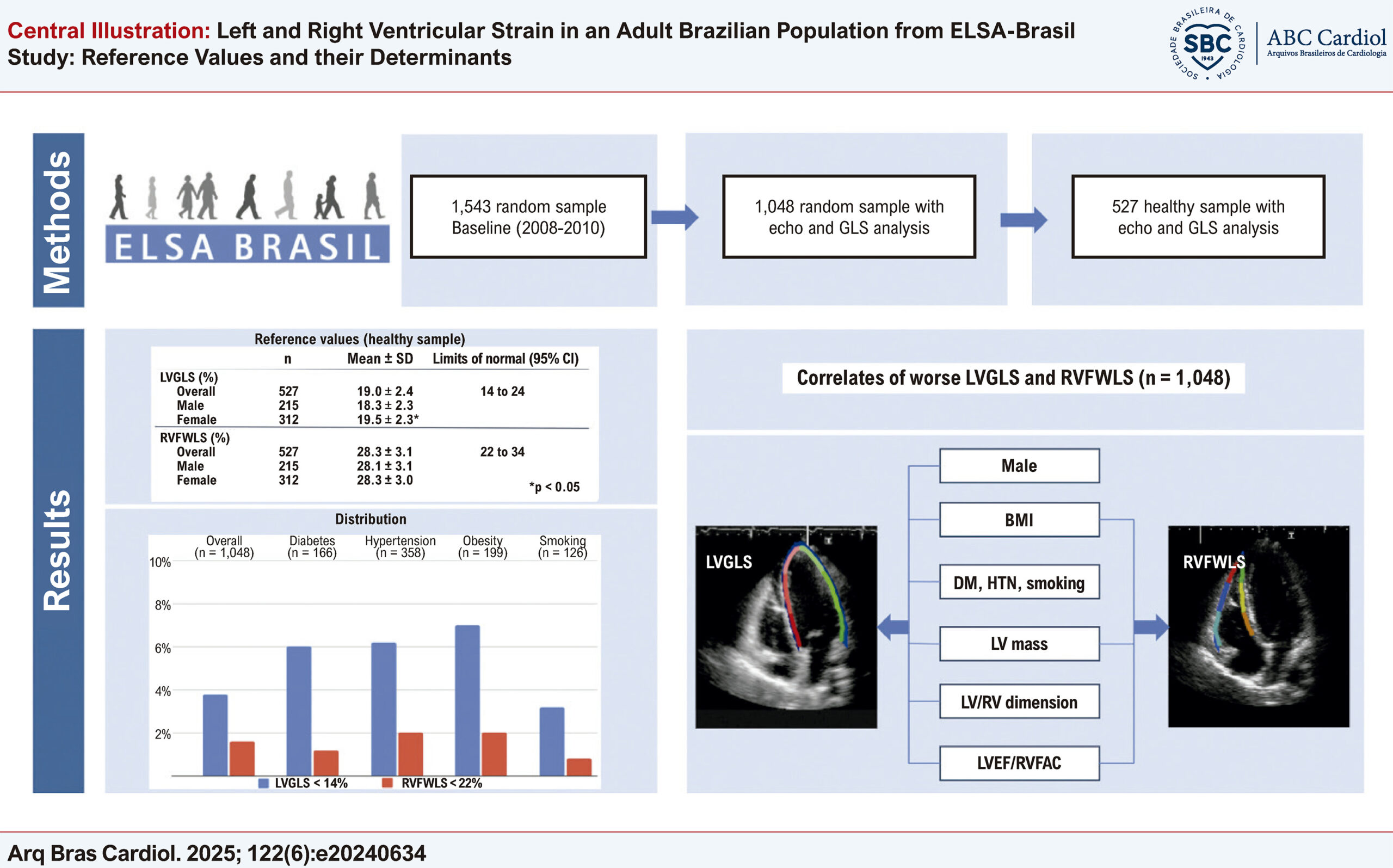
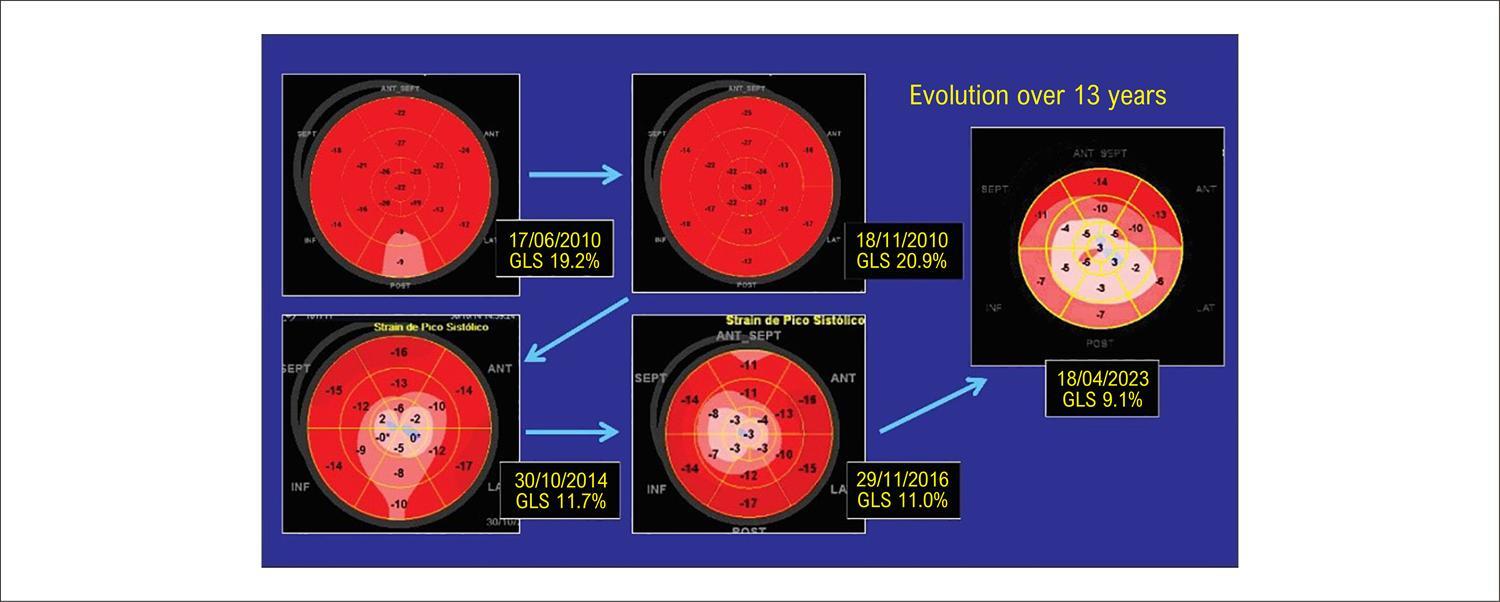
The analysis of myocardial deformation (strain) using speckle tracking technology in echocardiography is no longer something new and innovative. Several years have passed since its validation. Compared to left ventricular (LV) ejection fraction, global longitudinal strain (GLS) is a more accurate assessment of systolic function, important in several situations, such as cardio-oncology, valvular heart disease, cardiomyopathies, and ischemic heart disease. It is more accurate and reproducible data, is closely correlated with prognosis, and is currently available in most echocardiogram machines. On the other hand, this analysis requires an adequate acoustic window, and the issue of inter-vendor variability must still be taken into account.

A highly significant step that brought the analysis of myocardial deformation to our Brazilian reality was taken in 2023 with the publication of the “Position Statement on the Use of Myocardial Strain in Cardiology Routines by the Brazilian Society of Cardiology’s Department Of Cardiovascular Imaging – 2023” in the Arquivos Brasileiros de Cardiologia journal. Several experts joined in the production of this important document that increased cardiologists’ knowledge about GLS. Published in the current issue of the Arquivos Brasileiros de Cardiologia, another step was taken through the ELSA-Brasil study. ELSA-Brasil is a broad epidemiological study with more than 15,000 public servants from universities and research institutions on cardiovascular diseases and diabetes, carried out in 6 Brazilian cities (São Paulo, Rio de Janeiro, Belo Horizonte, Vitória, Salvador, and Porto Alegre), with several publications in the literature. This time, the focus was on determining normal values for LVGLS and right ventricular free wall longitudinal strain (RVFWLS), that is, an investigation of myocardial deformation in our population.

[…]

  • Echocardiography
  • Longitudinal Studies
  • Ventricular Function
Read Full Article
Creative Commons Attribution (CC BY) Creative Commons license
20/06/2025 DOI: 10.36660/abc.20250265i Views 1
Left and Right Ventricular Strain in an Adult Brazilian Population from ELSA-Brasil Study: Reference Values and their Determinants
ABC
Original Article

Left and Right Ventricular Strain in an Adult Brazilian Population from ELSA-Brasil Study: Reference Values and their Determinants

Eduardo Gatti Pianca et al. Murilo Foppa Giulia Bevilacqua Schmitz Wilson Cañon-Montañez … Bruce Bartholow Duncan Angela Barreto Santiago Santos

Abstract

Background

Impaired left ventricular (LV) and right ventricular functions are important predictors of cardiovascular risk. Global longitudinal strain (GLS) provides superior sensitivity for assessing systolic function compared to traditional parameters, enhancing diagnostic accuracy in various cardiac conditions. However, GLS reference values in diverse populations are lacking.

Objectives

To establish reference values for LVGLS and right ventricular free wall longitudinal strain (RVFWLS) in a Brazilian multiethnic population without cardiovascular risk factors or disease. We also explore how clinical and echocardiographic factors influence GLS distribution, addressing a gap in global guidelines that often rely on data from homogeneous or geographically distant populations.

Methods

We included 1,048 participants from the ELSA-Brasil cohort who underwent echocardiography with GLS analysis. A healthy subsample (n = 527) was defined by excluding individuals with cardiovascular or renal disease, hypertension, or diabetes to establish GLS reference ranges. The prevalence of abnormal GLS was assessed, and factors associated with reduced LVGLS and RVFWLS were identified. Statistical significance was defined as p < 0.05.

Results

In the healthy subsample (mean age 50.2 years, 59% female), mean LVGLS was 19.0% (95% confidence interval: 14.3 to 23.8) and RVFWLS was 28.3% (95% confidence interval: 22.3 to 34.3). Females exhibited higher LVGLS and RVFWLS values than males, with no significant age-related differences. Abnormal LVGLS and RVFWLS were observed in 3.8% and 1.6% of participants, respectively. Lower LVGLS was associated with obesity, hypertension, and diabetes; reduced RVFWLS correlated with higher body mass index and LV mass.

Conclusions

We propose reference values for LVGLS and RVFWLS in a large Brazilian cohort, highlighting associations with cardiovascular comorbidities and ventricular structure.

  • Cohort Studies
  • Echocardiography
  • Global Longitudinal Strain
  • Heart Ventricles
  • Ventricular Function
Read Full Article
Creative Commons Attribution (CC BY) Creative Commons license
16/06/2025 DOI: 10.36660/abc.20240634i Views 1
Impact of Valvular Event Time Assessment Methods on Myocardial Work Calculation by Echocardiography
ABCIC
Original Article

Impact of Valvular Event Time Assessment Methods on Myocardial Work Calculation by Echocardiography

Marcio Mendes Pereira et al. Francisco das Chagas Monteiro Júnior Sayuri Yamamura

Abstract

Background:

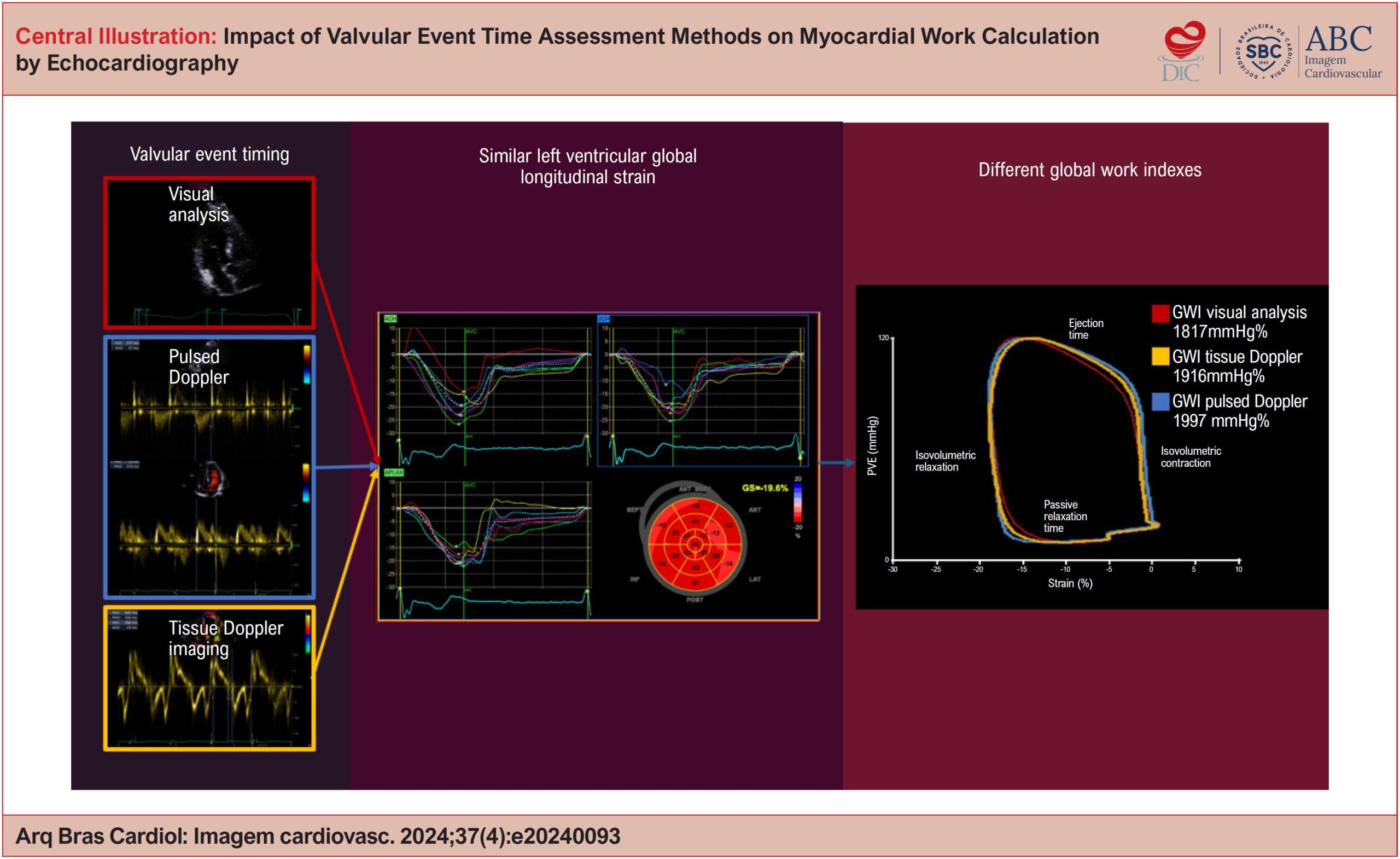
Left ventricular ejection fraction (LVEF) is the traditional parameter for assessing cardiac function, but it has limitations in the early detection of dysfunction. Global Longitudinal Strain (GLS) and myocardial work (MW) are promising alternatives, the latter being less influenced by load conditions. The calculation of MW depends on valvular event times (VET), obtained by methods that may generate divergent results.

Objectives:

To compare the impact of different methods of VET measurement – visual analysis (VA), tissue Doppler imaging (TDI) and pulsed Doppler (PD) – on global work index (GWI), global constructive work (GCW), global wasted work (GWW) and global work efficiency (GWE).

Method:

Cross-sectional clinical study with 35 healthy patients who underwent echocardiography between October 2022 and March 2023. MW was assessed using the EchoPAC software, with VET measurement by VA, TDI and DP. The results were analyzed by ANOVA and Friedman tests, followed by post-hoc analyses with a significance level of 5%.

Results:

GWI showed significant variation between the VET measurement methods, being lower by AV compared to TDI and DP (p<0.001). The GCW, GWW and GWE indexes did not demonstrate statistically significant differences between the methods.

Conclusions:

The VET measurement method significantly impacts the GWI, but does not affect the other MW indexes.

  • Echocardiography
  • Left Ventricular Dysfunction
  • Ventricular Function
Read Full Article
Creative Commons Attribution (CC BY) Creative Commons license
05/12/2024 DOI: 10.36660/abcimg.20240093i Views 1
Central Illustration
ABC
Original Article

Comparative Analysis of Conventional and Speckle Tracking Echocardiographic Variables between Patients with Unrejected Heart Transplants and Healthy Individuals

Aline Oliveira Martins Campo Dall’Orto et al. Maria Estefania Otto Simone Ferreira Leite Marco Antônio Freitas de Queiroz Maurício Filho … Natália Taveira Martins Samuel Rabelo Araújo Soraya Vasconcelos Almeida Luiza Valle Oliveira Brizida Fernando Antibas Atik

Abstract

Background

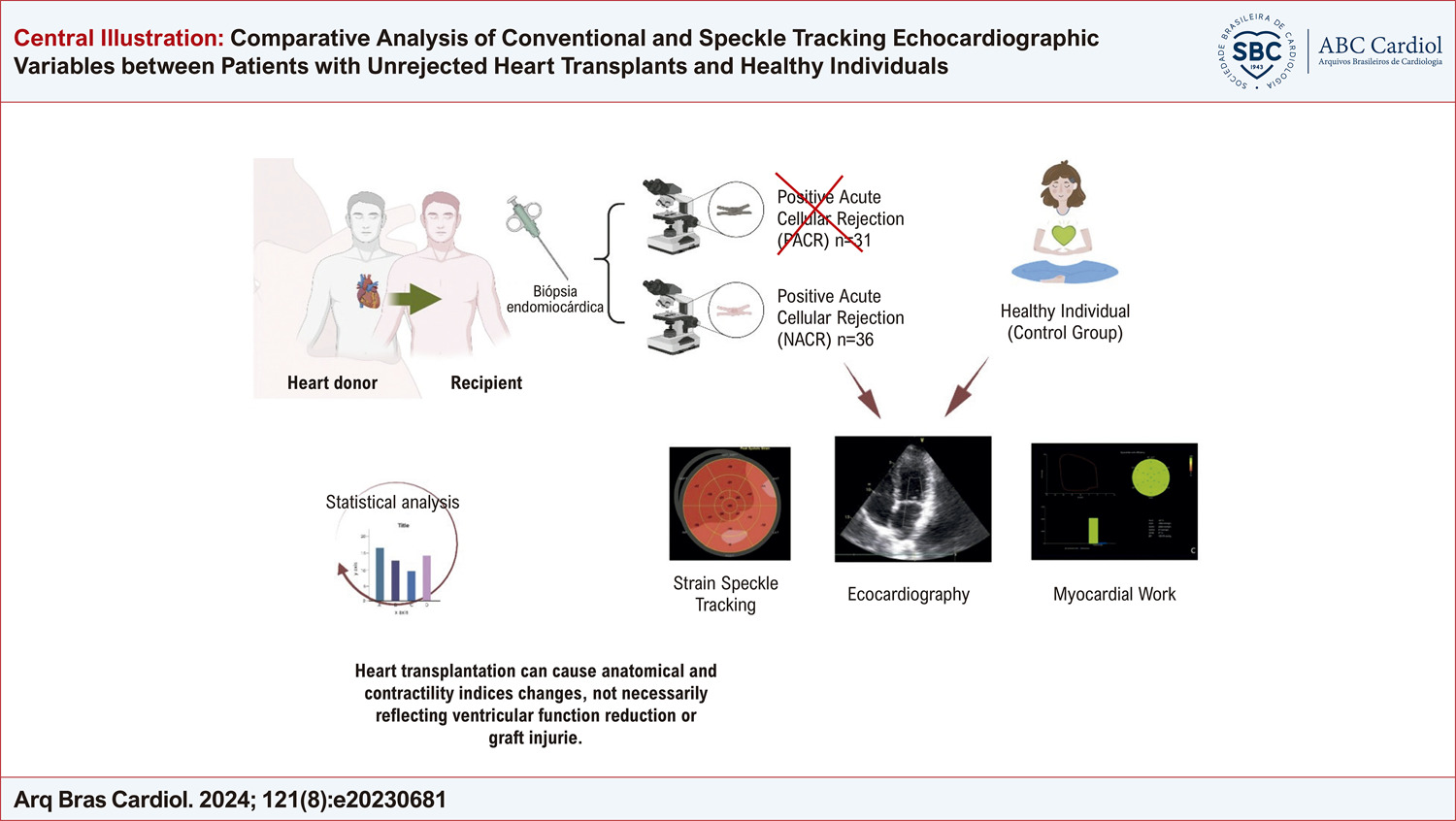
Echocardiography is essential for the assessment of patients with heart transplants. However, normal values in such individuals are not clearly defined.

Objectives

To compare conventional echocardiographic and speckle tracking variables between patients with unrejected heart transplants and healthy individuals.

Methods

: A prospective study was conducted with adult patients having undergone heart transplantation at least one year earlier and submitted to endomyocardial biopsy followed by transthoracic echocardiogram (TTE). Conventional TTE measures and mechanical heart strain assessments using speckle tracking were performed and the results were compared to those of a group of healthy volunteers. Statistical significance was set at 5% (p < 0.05).

Results

Thirty-six transplant patients without rejection were analyzed and compared to 30 healthy individuals. Chagas disease was the main reason for transplantation. Lower left ventricular global longitudinal strain expressed in absolute values was found (11.99% in transplant patients vs. 20.60% in controls; p <0.0001), right ventricular free wall longitudinal strain (16.67% in transplant patients vs. 25.50% in controls; p <0.0001) and myocardial work indices (p < 0.0001) as well as a larger size of the left atrium (38.17 ml/m2 in transplant patients vs. 18.98 ml/m2 in controls; p <0.0001) and greater mass and relative wall thickness (p <0.0001).

Conclusion

Stable patients having undergone heart transplants without rejection have differences concerning echocardiographic variables compared to healthy individuals. These findings indicate that conventional echocardiographic measures and heart mechanics are altered in transplant patients even in the absence of rejection. Such findings are relevant to the clinical context and follow-up of the patient.

  • Echocardiography
  • Heart Transplantation
  • Ventricular Function
Read Full Article
Creative Commons Attribution (CC BY) Creative Commons license
26/08/2024 DOI: 10.36660/abc.20230681i
ABCIC
Review Article

Use of Three-Dimensional Echocardiography in the Analysis of Ventricular Function in Chagas Disease

Antonio Carlos Leite de Barros Filho et al. Minna Moreira Dias Romano

Abstract

Chagas disease is recognized as a global public health issue, capable of causing cardiac, digestive, and neurological manifestations. The echocardiogram is a fundamental complementary exam in assessing patients with cardiac involvement, evaluating various parameters of cardiac structure and function. This review aims to discuss the use of three-dimensional echocardiography in analyzing ventricular function in patients with Chagas disease.

  • Chagas Cardiomyopathy
  • Echocardiography, Three-Dimensional
  • Ventricular Function
Read Full Article
Creative Commons Attribution (CC BY) Creative Commons license
18/12/2023 DOI: 10.36660/abcimg.20230093i Views 1
Right Ventricular Function and Exercise Tolerance in Patients with ST-Elevation Myocardial Infarction
ABC
Original Article

Right Ventricular Function and Exercise Tolerance in Patients with ST-Elevation Myocardial Infarction

Denisse Guzman-Ramirez et al. Anival Trujillo-Garcia Meredith Lopez-Rincon Roxella Botello Lopez

Introduction

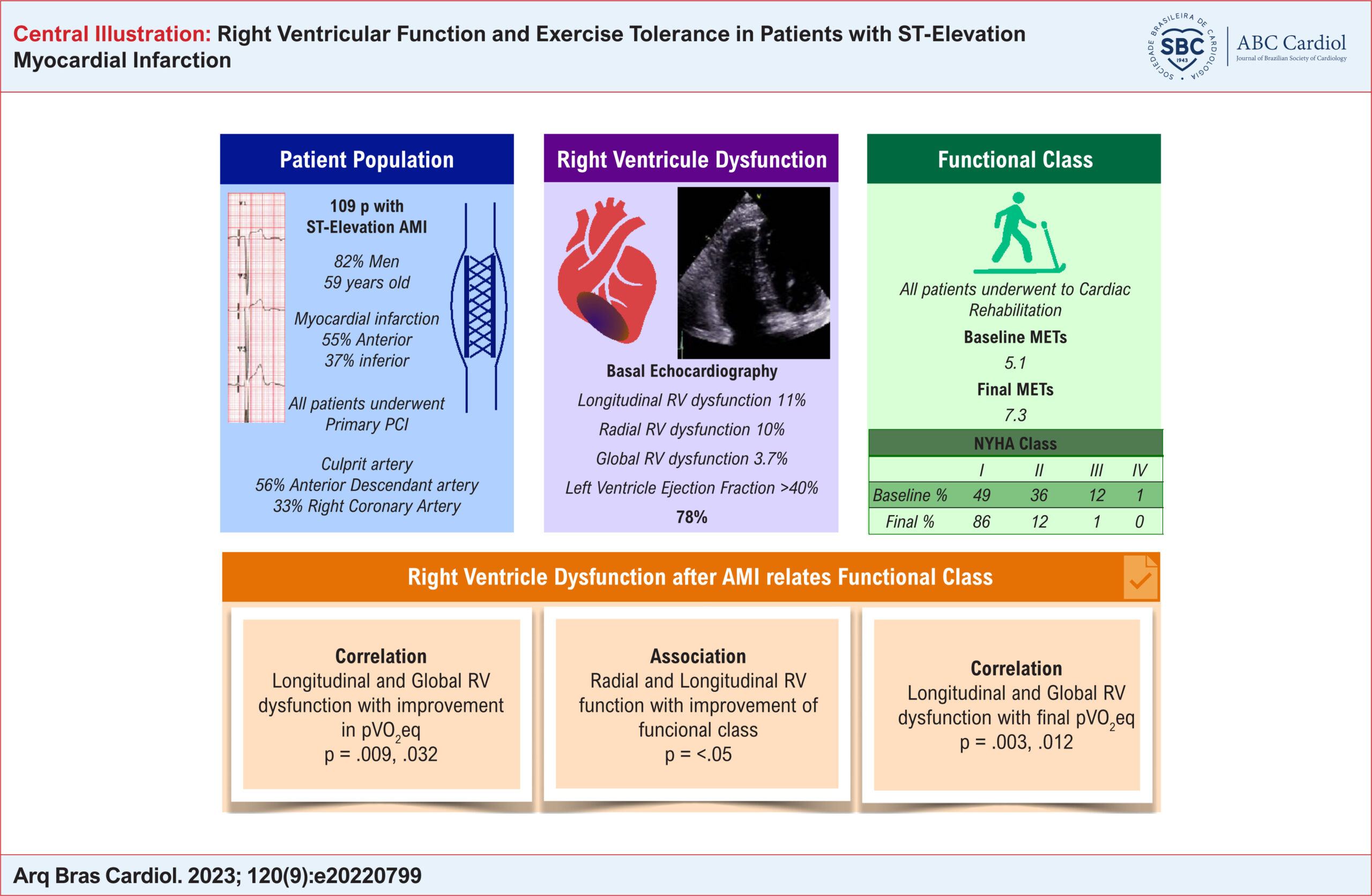
Right ventricle (RV) dysfunction has a prevalence of 10% in anterior myocardial infarctions and up to 50% in inferior myocardial infarctions. It is responsible for cardiogenic shock secondary to an acute coronary syndrome in 5% of cases. In patients with myocardial infarction, RV dysfunction is an important prognostic factor of mortality, ventricular arrhythmias, mechanical complications, cardiogenic shock, stent thrombosis, and increased in-hospital and one-year mortality. –

Tajima et al. investigated RV function, its association with exercise tolerance, and the efficacy of phase II cardiac rehabilitation (CR) in patients with ischemic heart disease. RV dysfunction was significantly associated with reduced exercise tolerance by 9% before rehabilitation. However, CR was effective in these patients.

[…]

  • Cardiac Rehabilitation
  • Echocardiography
  • ST Elevation Myocardial Infarction
  • Ventricular Function
Read Full Article
Creative Commons Attribution (CC BY) Creative Commons license
08/09/2023 DOI: 10.36660/abc.20220799 Views 1
Figure 1
ABCIC
Editorial

Is Echocardiographic Assessment of Strain Essential In Daily Routine?

Carlos Eduardo Suaide Silva

The assessment of myocardial fiber deformation (myocardial strain) during the cardiac cycle is a direct measurement of muscle function, which can be obtained by conventional imaging tests such as echocardiography and magnetic resonance.

Strain measurement can identify abnormalities in global and regional myocardial function, in addition to making the differential diagnosis of some cardiomyopathies. Furthermore, it is an earlier marker of ventricular dysfunction than ejection fraction and may predict adverse cardiovascular events.

[…]

  • Cardiac Imaging Techniques
  • Echocardiography
  • Ventricular Function
Read Full Article
Creative Commons Attribution (CC BY) Creative Commons license
06/09/2023 DOI: 10.36660/abcimg.20230063i Views 2
Figure 1
ABCIC
Review Article

Role of Strain in the Early Diagnosis of Diabetic Cardiomyopathy

Thais Rossoni Weber da Silva et al. Roberto Léo da Silva Adriana Ferraz Martins Jefferson Luiz Brum Marques

Abstract

Background

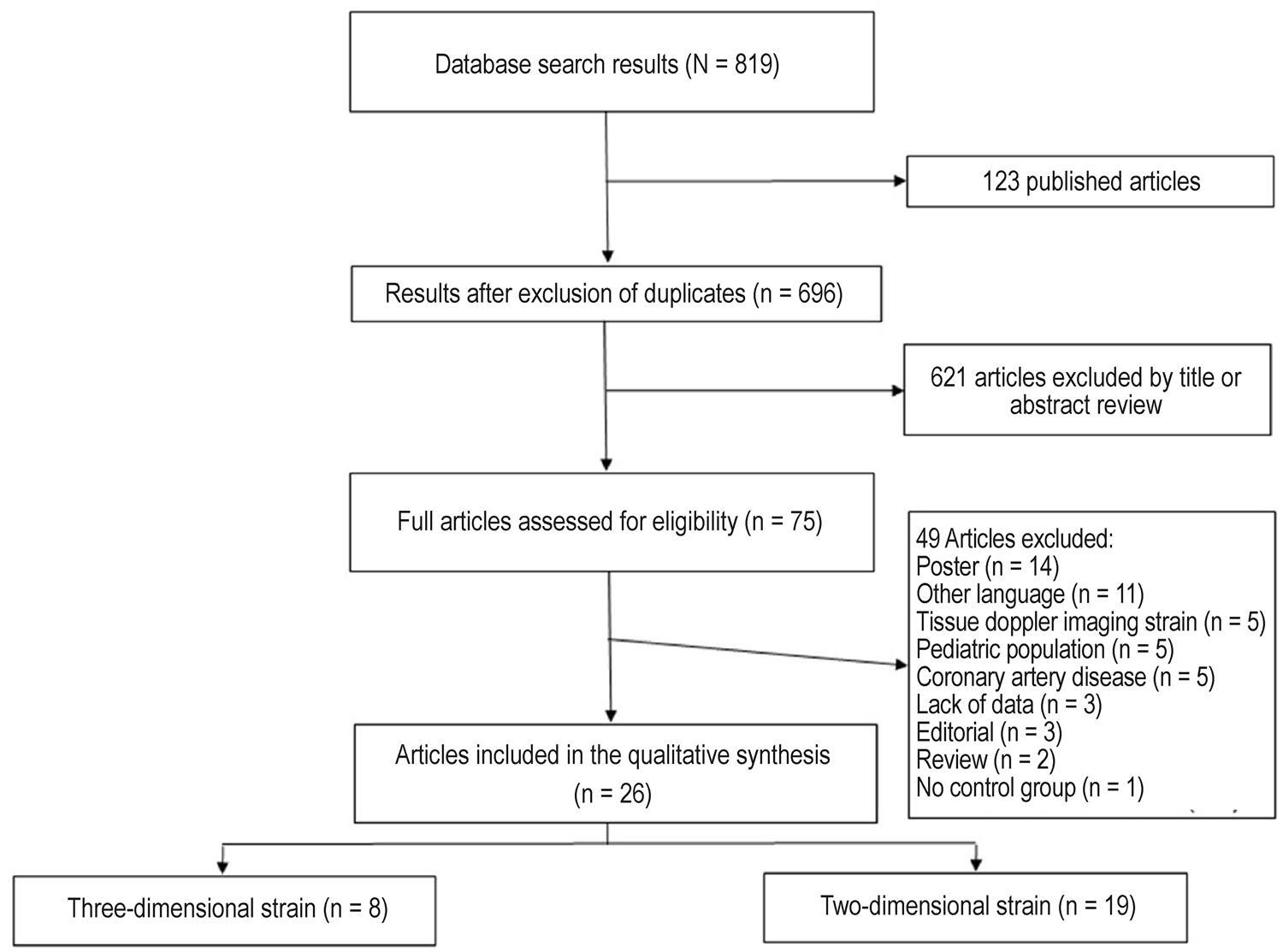
Advanced echocardiography using two- and three-dimensional myocardial strain proposes to identify subclinical systolic dysfunction in different clinical conditions. Strain assessment plays an important role in the early diagnosis of diabetic cardiomyopathy in diabetes mellitus (DM). However, the findings of published articles are heterogeneous. Here we conducted a systematic review to analyze the current role of strain assessment in patients with DM.

Methods

This systematic review of five databases identified 19 studies that used two-dimensional strain and 8 studies that used three-dimensional strain.

Results

The studies of two-dimensional strain included 1,774 DM patients (mean age, 57.1 years; median age, 55 years; 47.5% women), while those of three-dimensional strain included 488 DM patients (mean age, 55.7 years; median age, 63 years; 51% women). Global longitudinal strain was the myocardial deformation marker that differed most frequently between the DM and control groups.

Conclusion

Myocardial strain imaging by two- and three-dimensional speckle tracking echocardiography allows the identification of subclinical systolic dysfunction in DM patients, and differences become more marked when associated with risk factors and ventricular remodeling.

  • Diabetes Mellitus
  • Echocardiography
  • Ventricular Function
Read Full Article
Creative Commons Attribution (CC BY) Creative Commons license
28/05/2022 DOI: 10.47593/2675-312X/20223502eabc293 Views 3
ABC
Editorial

Lung Function and Inspiratory Muscle Strength in Heart Failure: Can They be Considered Potential Prognostic Markers?

Filipe Ferrari

Heart failure (HF) is a complex syndrome considered a major public health problem. Different HF subtypes are classically defined based on left ventricular ejection fraction (LVEF). Although its prognosis has improved in recent decades – explained, in part, by major therapeutic advances – HF persists with a high burden of mortality and negatively influences the quality of life., In this sense, common symptoms experienced in this disease, such as shortness of breath and exercise intolerance, contribute greatly to this sharp decline in the quality of life of individuals. Another condition, considered an important risk factor, which usually accompanies HF, is pulmonary dysfunction. The respiratory impairments observed in HF may be related to several reasons, such as an impairment of lung mechanics and gas diffusion, as well as respiratory muscle weakness – aggravating increased dyspnea, being a major limitation on physical exercise.

Spirometry is a widely used test that allows the analysis of lung function – by measuring the amount of air inhaled and exhaled to the maximum. Because chronic obstructive pulmonary disease shares similar signs and symptoms with HF, its identification in individuals with HF can be challenging; in this sense, spirometry can help confirm the diagnosis. About evaluating the potential severity of some lung diseases, the exercise test can also be useful, observing a series of parameters, such as the ratio of forced expiratory volume in 1 second/forced vital capacity (FEV1/FVC). The severity of the disease can still be classified based on FEV1 when it is below the lower limit of normal (ranging from mild when ≥70% of predicted to very severe when <35% of predicted). Although HF itself can lead to a decrease in FEV1 and FVC of about 20% of predicted, in addition to the fact that a worse FEV1 may predict higher mortality, convincing evidence examining the prognostic role of FEV1 in the HF setting still needs further investigation.

[…]

  • Exercise Tolerance
  • Heart Failure
  • Respiratory Insufficiency
  • Respiratory Muscles
  • Risk Assessment
  • Ventricular Function
Read Full Article
Creative Commons Attribution (CC BY) Creative Commons license
29/04/2022 DOI: 10.36660/abc.20211060 Views 2
ABC
Original Article

Relationship of Lung Function and Inspiratory Strength with Exercise Capacity and Prognosis in Heart Failure

Sergio Henrique Rodolpho Ramalho et al. Alexandra Correa Gervazoni Balbuena de Lima Fabiola Maria Ferreira da Silva Fausto Stauffer Junqueira de Souza … Lawrence Patrick Cahalin Graziella França Bernardelli Cipriano Gerson Cipriano Junior

Abstract

Background

Spirometry is underused in heart failure (HF) and the extent to which each defect associates with exercise capacity and prognosis is unclear.

Objective

To determine the distinct relationship of continuous %predicted FVC (ppFVC) and FEV1/FVC with: 1) maximal inspiratory pressure (MIP), left ventricular ejection fraction (LVEF), exercise performance; and 2) prognosis for the composite of cardiovascular death, heart transplantation or left ventricular assist device implant.

Methods

A cohort of 111 HF participants (AHA stages C/D) without diagnosed pneumopathy, spirometry, manovacuometry and maximum cardiopulmonary test. The association magnitudes were verified by linear and Cox (HR; 95% CI) regressions, age/sex adjusted. A p<0.05 was considered significant.

Results

Age was 57±12 years, 60% men, 64% in NYHA III. Every 10%-point increase in FEV1/FVC [β 7% (95% CI: 3–10)] and ppFVC [4% (2-6)] associated with ventilatory reserve (VRes), however only ppFVC associated with MIP [3.8 cmH2O (0.3-7.3)], LVEF [2.1% (0.5-3.8)] and VO2peak [0.5 mL/kg/min (0.1–1.0)], accounting for age/sex. In 2.2 years (mean), 22 events occurred, and neither FEV1/FVC (HR 1.44; 95% CI: 0.97–2.13) nor ppFVC (HR 1.13; 0.89–1.43) was significantly associated with the outcome. Only in the LVEF ≤50% subgroup (n=87, 20 events), FEV1/FVC (HR 1.50; 1.01–2.23), but not ppFVC, was associated with greater risk.

Conclusions

In chronic HF, reduced ppFVC associated with lower MIP, LVEF, VRes and VO2peak, but no distinct poorer prognosis over 2.2 years of follow-up. Distinctively, FEV1/FVC was associated only with VRes, and, in participants with LVEF ≤50%, FEV1/FVC reduction proportionally worsened prognosis. Therefore, FEV1/FVC and ppFVC add supplementary information regarding HF phenotyping.

  • Exercise Tolerance
  • Respiratory Insufficiency
  • Respiratory Muscles
  • Risk Assessment
  • Ventricular Function
Read Full Article
Creative Commons Attribution (CC BY) Creative Commons license
07/02/2022 DOI: 10.36660/abc.20201130
Figure 1
ABCIC
Editorial

My Approach to Assessing Right Ventricular Function After Tetralogy of Fallot Repair

Laura Mercer-Rosa

Qualitative assessment

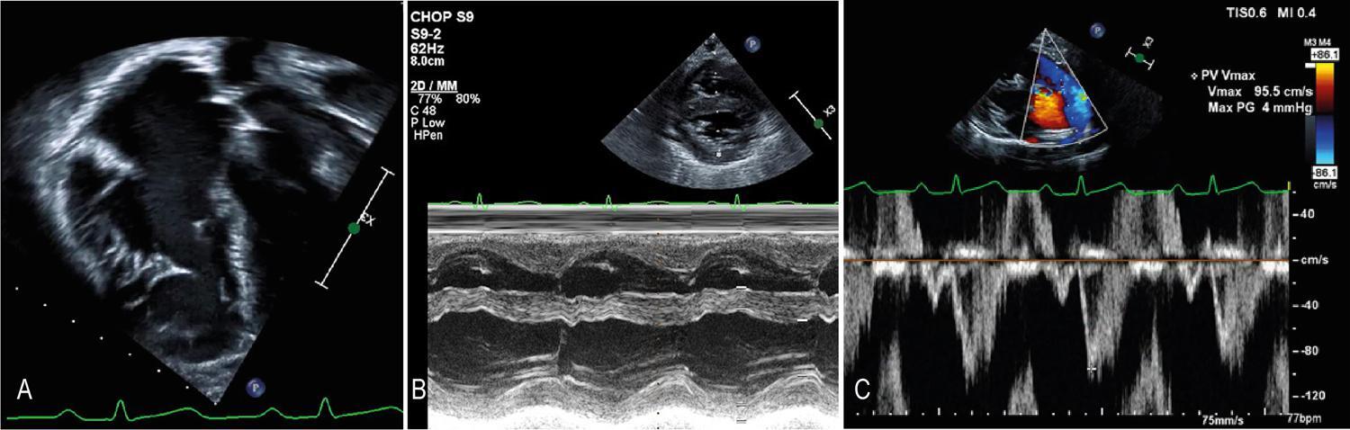
On the apical four-chamber view, I examine the motion of the tricuspid valve toward the apex (measured as the tricuspid annular plane systolic excursion [TAPSE] as detailed below) and the motion of the RV wall toward the septum versus data of prior studies. Likewise, parasternal long- and short-axis views are important. In those with favorable acoustic windows, RV wall shortening can be assessed using M-mode ( ). It is worth noting that the qualitative assessment of RV function should not be the sole parameter. ,

[…]

  • Resonance, Magnetic
  • Tetralogy of Fallot
  • Ventricular Function
Read Full Article
Creative Commons Attribution (CC BY) Creative Commons license
16/12/2021 DOI: 10.47593/2675-312X/20213404ecom17
Figure 1
ABCHF
Viewpoint

Cardiac Magnetic Resonance and Amyloidosis: How can it Assist Clinical Reasoning?

Ibraim Masciarelli F. Pinto

Evaluation of cardiac morphology

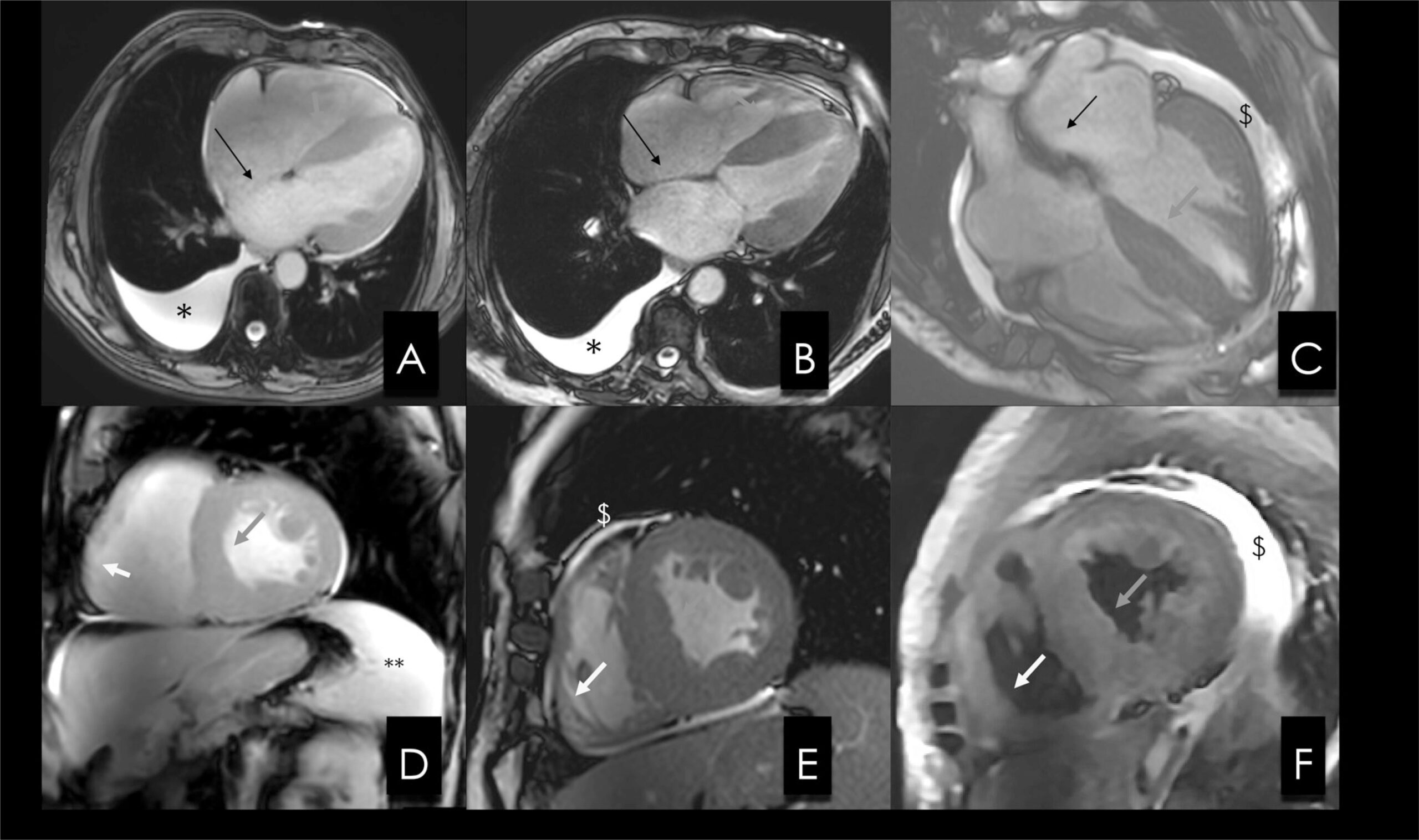
CA leads to changes in cardiac structure. In the atria, dilatation and increased thickness of the septum usually occur, while, in more advanced phases, there may be slow flow and thrombi, which, when arrhythmias are present, may be difficult to identify via CMR.

The ventricular myocardium usually shows increased thickness, typically at levels higher than those observed in patients with arterial hypertension; the levels are typically greater in forms of transthyretin amyloidosis than in light chain amyloidosis, and they may involve both ventricles. ,

[…]

  • Amyloidosis
  • Heart Failure
  • Ventricular Function
Read Full Article
Creative Commons Attribution (CC BY) Creative Commons license
10/12/2021 DOI: 10.36660/abchf.20210016
ABC
Editorial

Atrioventricular Conduction System Disorders and Potential Risks of Arrhythmic Events in Endurance Athletes

Carlos Alberto Cordeiro Hossri

Short Editorial related to the article: Assessing Dynamic Atrioventricular Conduction Time to RR-interval Coupling in Athletes and Sedentary Subjects

Some disorders found on electrocardiograms are commonly seen in endurance or high-performance athletes and often have characteristics that are similar to those observed in elderly individuals or in those with cardiovascular disease. –

[…]

  • Arrhythmias Cardiac
  • Eletrocardiography/methods
  • Keywords: Athletes
  • physical fitness
  • Resistance Training
  • Sedentarism
  • Ventricular Function
  • Ventricular Remodeling
Read Full Article
Creative Commons Attribution (CC BY) Creative Commons license
07/08/2020 DOI: 10.36660/abc.20200668
Figure 1
ABC
Original Article

Assessing Dynamic Atrioventricular Conduction Time to RR-interval Coupling in Athletes and Sedentary Subjects

Paulo Roberto Benchimol-Barbosa et al. Olivassé Nasario Junior Jurandir Nadal

Abstract

Background

Atrioventricular conduction time ( AVCT ) is influenced by autonomic input and subject to physiological remodeling.

Objective

To evaluate beat-by-beat AVCT and RR-interval variability in athletes and healthy sedentary subjects.

Methods

Twenty adults, including 10 healthy sedentary (Controls) and 10 elite long-distance runners (Athletes), age, weight and height-adjusted, underwent maximal metabolic equivalent (MET) assessment, and 15-min supine resting ECG recording seven days later. The interval between P-wave and R-wave peaks defined the AVCT . Mean (M) and standard deviation (SD) of consecutive RR-intervals (RR) and coupled AVCT were calculated, as well as regression lines of RR vs. AVCT (RR-AVCT) . Concordant AV conduction was defined as positive RR-AVCT slope and discordant otherwise. A multivariate linear regression model was developed to explain MET based on AVCT and RR-interval variability parameters. Significance-level: 5 %.

Results

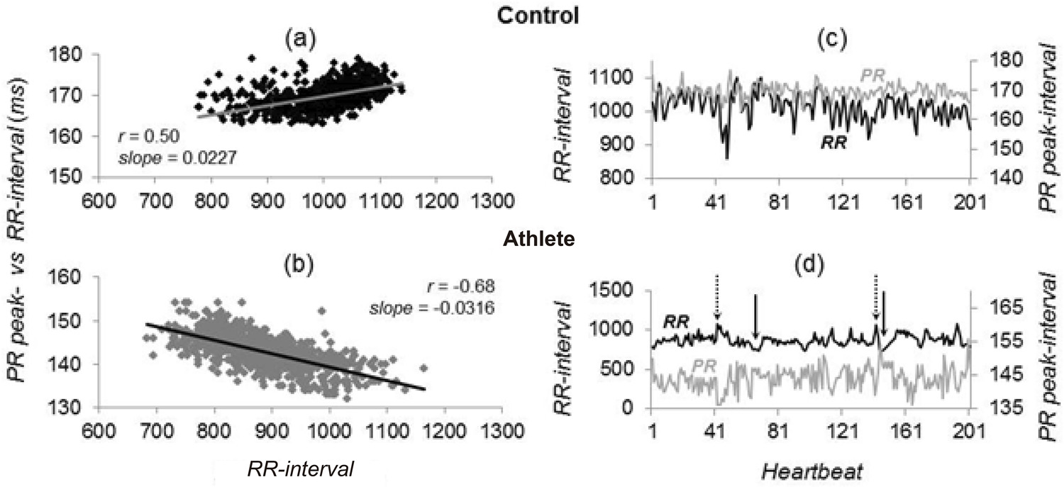
In Athletes, M-RR and SD-RR values were higher than in Controls, whereas M-AVCT and SD-AVCT were not. RR-AVCT slopes were, respectively, 0.038 ± 0.022 and 0.0034 ± 0.017 (p < 0.05). Using a cut-off value of 0.0044 (AUC 0.92 ± 0.07; p < 0.001), RR-AVCT slope showed 100% specificity and 80% sensitivity. In a multivariate model, SD-RR and RR-AVCT slope were independent explanatory variables of MET (F-ratio: 17.2; p < 0.001), showing 100% specificity and 90% sensitivity (AUC 0.99 ± 0.02; p < 0.001).

Conclusion

In elite runners, AVCT to RR -interval dynamic coupling shows spontaneous discordant AV conduction, characterized by negative AVCT vs. RR -interval regression line slope. RR -intervals standard deviation and AVCT vs. RR -interval regression line slope are independent explanatory variables of MET (Arq Bras Cardiol. 2020; [online].ahead print, PP.0-0)

  • Adults
  • Athletes
  • Electrocardiography/methods
  • Heart Ventricles
  • physical fitness
  • Resistance Training
  • Sedentarism
  • Ventricular Function
  • Ventricular Remodeling
Read Full Article
Creative Commons Attribution (CC BY) Creative Commons license
08/05/2020 DOI: 10.36660/abc.20190281
Figure 1
ABC
Original Article

Three-Dimensional Volumetric Assessment of Diastolic Function by Cardiac Magnetic Resonance Imaging: The Multi-Ethnic Study of Atherosclerosis (MESA)

Marcelo S Nacif et al. Andre L. C. Almeida Alistair A Young Brett R Cowan … Anderson C Armstrong Eunice Yang Christopher T Sibley W. Gregory Hundley Songtao Liu Joao AC Lima David A Bluemke

Abstract

Background:

Cardiac Magnetic Resonance is in need of a simple and robust method for diastolic function assessment that can be done with routine protocol sequences.

Objective:

To develop and validate a three-dimensional (3D) model-based volumetric assessment of diastolic function using cardiac magnetic resonance (CMR) imaging and compare the results obtained with the model with those obtained by echocardiography.

Methods:

The study participants provided written informed consent and were included if having undergone both echocardiography and cine steady-state free precession (SSFP) CMR on the same day. Guide points at the septal and lateral mitral annulus were used to define the early longitudinal relaxation rate (E’), while a time-volume curve from the 3D model was used to assess diastolic filling parameters. We determined the correlation between 3D CMR and echocardiography and the accuracy of CMR in classifying the diastolic function grade.

Results:

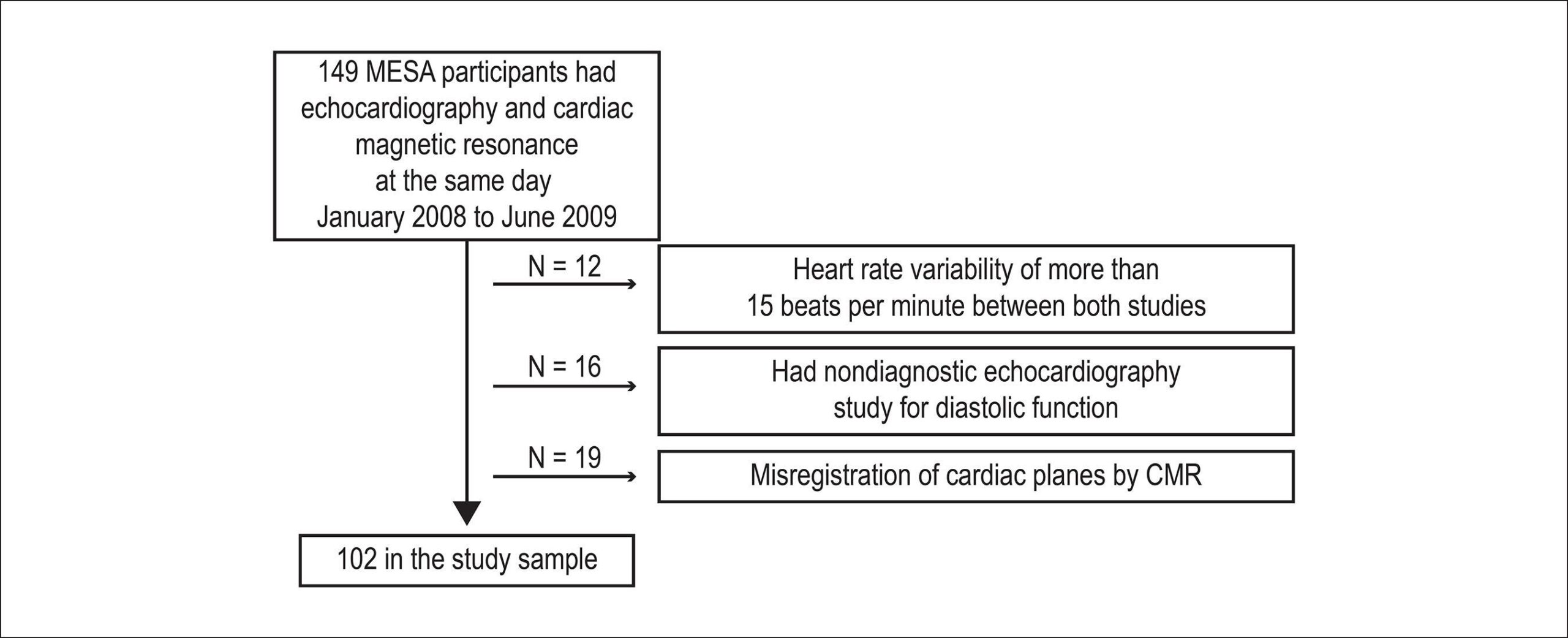
The study included 102 subjects. The E/A ratio by CMR was positively associated with the E/A ratio by echocardiography (r = 0.71, p < 0.0001). The early diastolic relaxation velocity by tissue Doppler and longitudinal relaxation rate for the lateral mitral annulus displacement were positively associated (p = 0.007), as were the ratio between Doppler E/e' and CMR E/E' (p = 0.01). CMR-determined normalized peak E (NE) and deceleration time (DT) were able to predict diastolic dysfunction (areas under the curve [AUCs] = 0.70 and 0.72, respectively). In addition, the lateral E/E' ratio showed good utility in identifying diastolic dysfunction (AUC = 0.80). Overall, echocardiography and CMR interobserver and intraobserver agreements were excellent (intraclass correlation coefficient range 0.72 – 0.97).

Conclusion:

3D modeling of standard cine CMR images was able to identify study subjects with reduced diastolic function and showed good reproducibility, suggesting a potential for a routine diastolic function assessment by CMR.

  • Echocardiography, Three-Dimensional
  • Evaluation
  • Imaging, Three-Dimensional
  • Magnetic Resonance
  • Ventricular Function
Read Full Article
Creative Commons Attribution (CC BY) Creative Commons license
25/05/2017 DOI: 10.5935/abc.20170063 Views 1
Figure 1
ABC
Original Article

Papillary Muscle Free Strain in Patients with Severe Degenerative and Functional Mitral Regurgitation

Alev Kılıcgedik et al. Gokhan Kahveci Ahmet Seyfeddin Gurbuz Can Yucel Karabay … Ahmet Guler Suleyman Cagan Efe Soe Moe Aung Ugur Arslantas Serdar Demir Ibrahim Akin Izgi Cevat Kirma

Abstract

Fundamento:

The role of papillary muscle function in severe mitral regurgitation with preserved and reduced left ventricular ejection fraction and the method of choice to evaluate PM have still been the subjects of controversy.

Objectives:

To evaluate and compare papillary muscle function in and between patients with severe degenerative and functional mitral regurgitation by using the free strain method.

Methods:

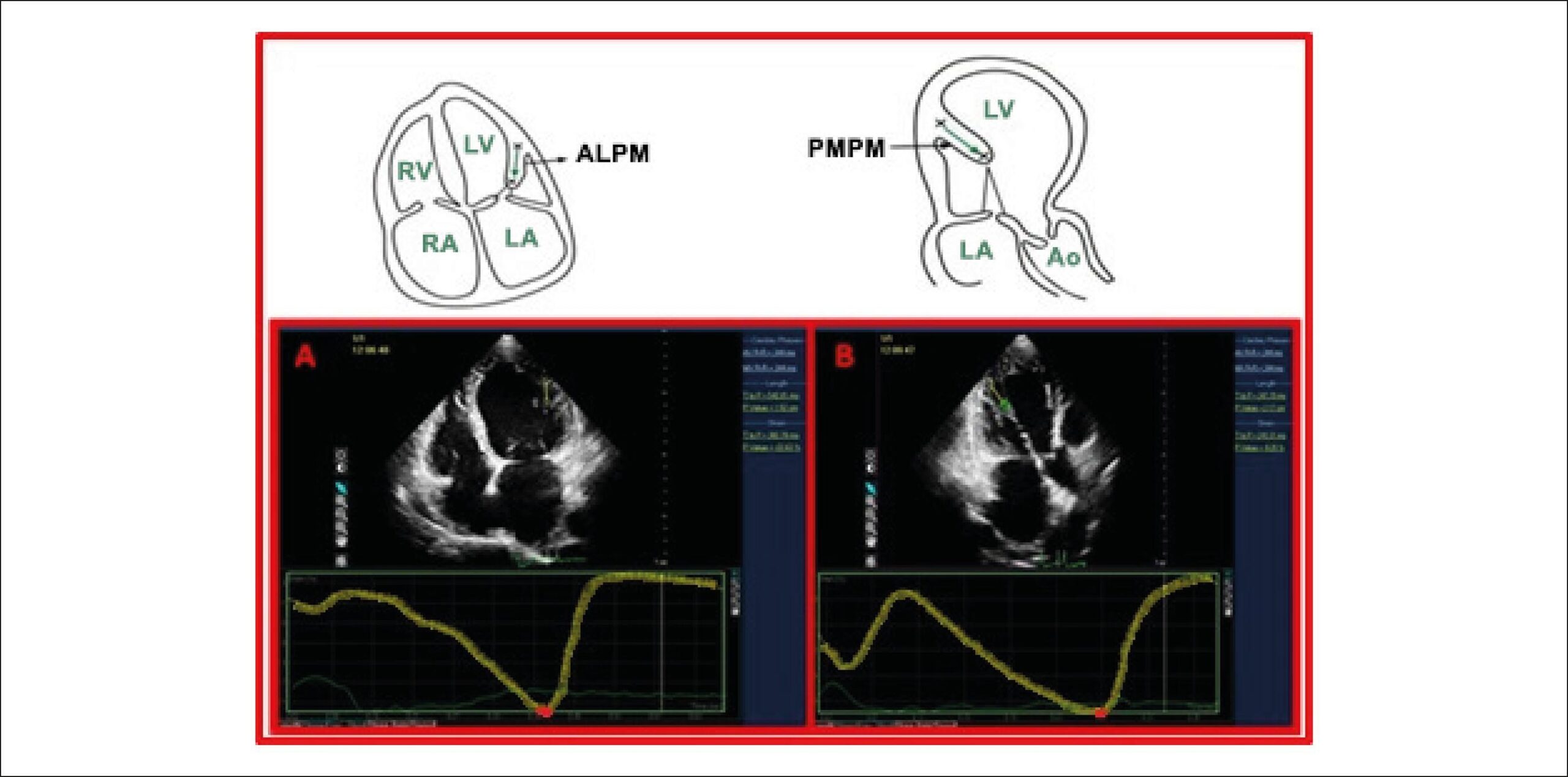
64 patients with severe mitral regurgitation – 39 patients with degenerative mitral regurgitation (DMR group) and 25 patients with severe functional mitral regurgitation (FMR group) – and 30 control subjects (control group) were included in the study. Papillary muscle function was evaluated through the free strain method from apical four chamber images of the anterolateral papillary muscle (APM) and from apical three chamber images of the posteromedial papillary muscle (PPM). Global left ventricular longitudinal and circumferential strains were evaluated by applying 2D speckle tracking imaging.

Results:

Global left ventricular longitudinal strain (DMR group, -17 [-14.2/-20]; FMR group, -9 [-7/-10.7]; control group, -20 [-18/-21] p < 0.001), global left ventricular circumferential strain (DMR group, -20 [-14.5/-22.7]; FMR group, -10 [-7/-12]; control group, -23 [-21/-27.5] p < 0.001) and papillary musle strains (PPMS; DMR group, -30.5 [-24/-46.7]; FMR group, -18 [-12/-30]; control group; -43 [-34.5/-39.5] p < 0.001; APMS; DMR group, (-35 [-23.5/-43]; FMR group, -20 [-13.5/-26]; control group, -40 [-32.5/-48] p < 0.001) were significantly different among all groups. APMS and PPMS were highly correlated with LVEF (p < 0.001, p < 0.001; respectively), GLS (p < 0.001, p < 0.001; respectively) and GCS (p < 0.001, p < 0.00; respectively) of LV among all groups. No correlation was found between papillary muscle strains and effective orifice area (EOA) in both groups of severe mitral regurgitation.

Conclusions:

Measuring papillary muscle longitudinal strain by the free strain method is practical and applicable. Papillary muscle dysfunction plays a small role in severe MR due to degenerative or functional causes and papillary muscle functions in general seems to follow left ventricular function. PPM is the most affected PM in severe mitral regurgitation in both groups of DMR and FMR.

  • Diagnostic Imaging
  • Echocardiography/methods
  • Mitral Valve Insufficiency / diagnostic
  • Mitral Valve Insufficiency / physiopathology
  • Papillary Muscles / physiopathology
  • Ventricular Function
  • Ventricular Remodeling
Read Full Article
Creative Commons Attribution (CC BY) Creative Commons license
01/04/2017 DOI: 10.5935/abc.20170035
Figure 1
IJCS
Original Article

Gallic Acid and Cyclosporine Mixture and their Effects on Cardiac Dysfunction Induced by Ischemia/Reperfusion and eNOS/iNOS Expression

Mohammad Badavi et al. Najmeh Sadeghi Mahin Dianat Alireza Samarbafzadeh

Abstract

Background:

Although many researches have been conducted on either a certain antioxidant or mPTP individually, little attention has been drawn to the effects of co-administration of an antioxidant and mPTP inhibitor together on cardiac dysfunction after I/R injury.

Objectives:

This study aims at determining the effects of gallic acid (as Antioxidant) combined with cyclosporine A (CsA) (as mPTP inhibitor) on I/R induced cardiac and endothelial (role of NO) dysfunction.

Methods:

Male Wistar rats were pretreated with gallic acid (7.5, 15, or 30 mg.kg-1 body weight, daily) for a period of 10 days. Then, the heart was isolated and exposed to 30-minute ischemia and perfused by CsA (0.2 µM) 20 min during reperfusion period.

Results:

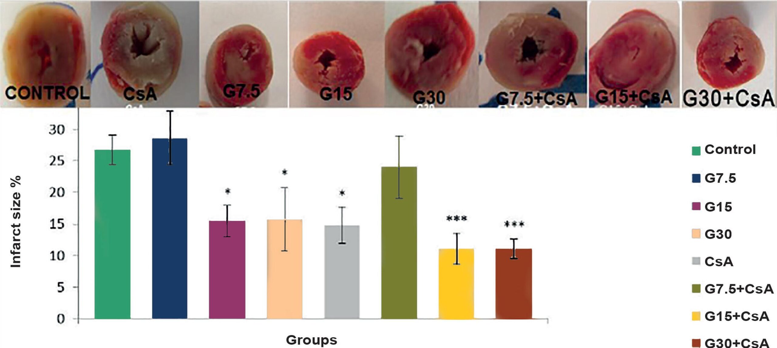
The data have shown that infarct size was decreased significantly by CsA and gallic acid alone (p < 0.05, one way ANOVA followed by LSD test), however the combination of both drugs had more significant improving effects (p < 0.001). The combination of these two drugs improved more significantly maximum rate of rise and fall of ventricular pressure (±dp.dt-1 max), rate pressure product (RPP), left ventricular developed pressure (LVDP), heart rate and coronary flow rather than applying each one alone (p < 0.05, repeated measurement ANOVA followed by LSD test).

Conclusions:

In conclusion, benefiting from an antioxidant concomitant with an mPTP inhibitor could have more improving effects on the cardiac dysfunction induced by I/R injury.

  • Antioxidants
  • cyclosporine
  • Endothelium
  • Gallic Acid
  • nitric oxide Synthase
  • Rats
  • Reperfusion
  • Ventricular Function
Read Full Article
Creative Commons Attribution (CC BY) Creative Commons license
01/01/2017 DOI: 10.5935/2359-4802.20170047 Views 1
Figure 1
ABC
Original Article

Impact of Atrial Fibrillation Ablation on Left Ventricular Filling Pressure and Left Atrial Remodeling

Simone Nascimento dos Santos et al. Benhur Davi Henz André Rodrigues Zanatta José Roberto Barreto … Kelly Bianca Loureiro Clarissa Novakoski Marcus Vinícius Nascimento dos Santos Fabio F. Giuseppin Edna Maria Oliveira Luiz Roberto Leite

Background:

Left ventricular (LV) diastolic dysfunction is associated with new-onset atrial fibrillation (AF), and the estimation of elevated LV filling pressures by E/e’ ratio is related to worse outcomes in patients with AF. However, it is unknown if restoring sinus rhythm reverses this process.

Objective:

To evaluate the impact of AF ablation on estimated LV filling pressure.

Methods:

A total of 141 patients underwent radiofrequency (RF) ablation to treat drug-refractory AF. Transthoracic echocardiography was performed 30 days before and 12 months after ablation. LV functional parameters, left atrial volume index (LAVind), and transmitral pulsed and mitral annulus tissue Doppler (e’ and E/e’) were assessed. Paroxysmal AF was present in 18 patients, persistent AF was present in 102 patients, and long-standing persistent AF in 21 patients. Follow-up included electrocardiographic examination and 24-h Holter monitoring at 3, 6, and 12 months after ablation.

Results:

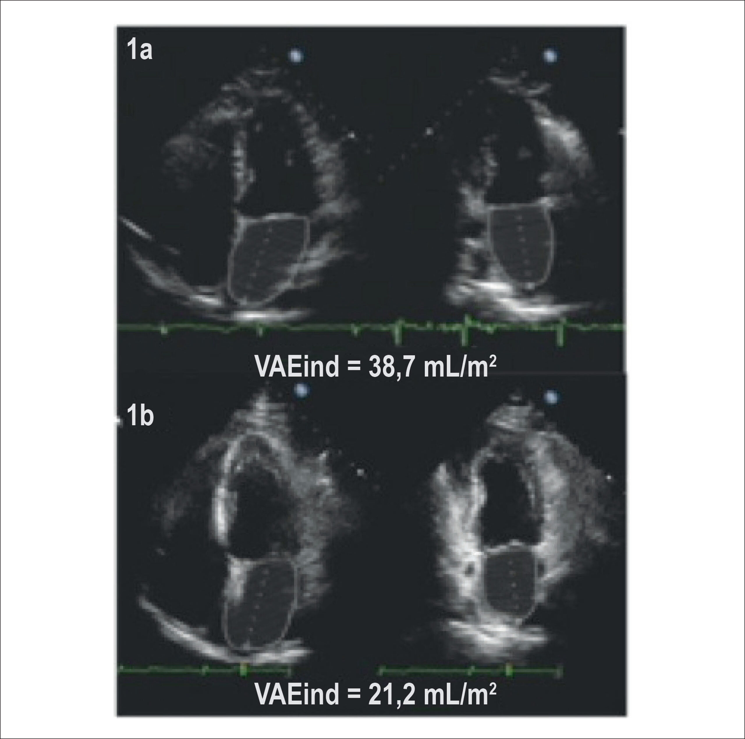
One hundred seventeen patients (82.9%) were free of AF during the follow-up (average, 18 ± 5 months). LAVind reduced in the successful group (30.2 mL/m2 ± 10.6 mL/m2 to 22.6 mL/m2 ± 1.1 mL/m2, p < 0.001) compared to the non-successful group (37.7 mL/m2 ± 14.3 mL/m2 to 37.5 mL/m2 ± 14.5 mL/m2, p = ns). Improvement of LV filling pressure assessed by a reduction in the E/e’ ratio was observed only after successful ablation (11.5 ± 4.5 vs. 7.1 ± 3.7, p < 0.001) but not in patients with recurrent AF (12.7 ± 4.4 vs. 12 ± 3.3, p = ns). The success rate was lower in the long-standing persistent AF patient group (57% vs. 87%, p = 0.001).

Conclusion:

Successful AF ablation is associated with LA reverse remodeling and an improvement in LV filling pressure.

  • Atrial Fibrillation
  • Atrial Remodeling
  • Catheter Ablation
  • Echocardiography
  • Stroke Volume
  • Ventricular Function
Read Full Article
Creative Commons Attribution (CC BY) Creative Commons license
14/10/2014 DOI: 10.5935/abc.20140152 Views 1
Figure 1
ABC
Image

Ablation of Premature Ventricular Contraction with the Help of a Notch

Firat Özcan et al. Serkan Topaloğ lu Osman Turak Serkan Çay Dursun Aras

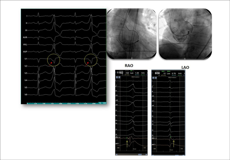
A 43-year-old male patient presented with a complaint of palpitation. Electrocardiography revealed premature ventricular contraction (PVC) that appeared to be originating from the left ventricular outflow tract. Tall positive R waves were observed in leads II, III, and aVF, and the R transition was observed at V2 in precordial leads. A notch was realized on the downward deflection of QS, which was only at lead V1. () This finding indicates that the region between the right and left coronary commissures is the origin of VPS. In addition, 24-h Holter monitoring revealed 32000 PVCs. Therefore, an electrophysiology study was planned. During the procedure, the catheter was moved between the right coronary and left coronary commissures, and the ablation catheter was placed at the ventricular side of the commissures where the earliest signal recorded was at 40 ms. At this site, pacing produced 12/12 morphology with PVC. In addition, at this site, we demonstrated a deflection prior to the intracardiac ventricular electrogram of PVCs corresponding to the notch. This deflection was after the intracardiac ventricular electrogram during sinus beats (). Ablation was performed at 20 watt 45 degrees, and it terminated all PVCs at this site.

PVCs can be very disturbing in some patients, and medical treatment may be tried. Long-term impacts of PVC may be deleterious for systolic ventricular function. Ablation of culprit source of PVC is an effective treatment and may improve systolic function of the left ventricle. Our patient was on a long-term medical treatment that failed to terminate PVCs. We preferred ablation for treatment. In this case, we would like to mention that ECG is an important, simple, cheap, and widely available tool that can be used for localization.

[…]

  • Catheter Ablation
  • Myocardial Contraction
  • Ventricular Function
Read Full Article
Creative Commons Attribution (CC BY) Creative Commons license
01/10/2014 DOI: 10.5935/abc.20140157 Views 1
Figure 1
ABC
Original Article

Resistance Training After Myocardial Infarction in Rats: Its Role on Cardiac and Autonomic Function

Camilla Figueiredo Grans et al. Daniele Jardim Feriani Marcos Elias Vergilino Abssamra Leandro Yanase Rocha … Nicolle Martins Carrozzi Cristiano Mostarda Diego Mendrot Figueroa Kátia De Angelis Maria Cláudia Irigoyen Bruno Rodrigues

Background:

Although resistance exercise training is part of cardiovascular rehabilitation programs, little is known about its role on the cardiac and autonomic function after myocardial infarction.

Objective:

To evaluate the effects of resistance exercise training, started early after myocardial infarction, on cardiac function, hemodynamic profile, and autonomic modulation in rats.

Methods:

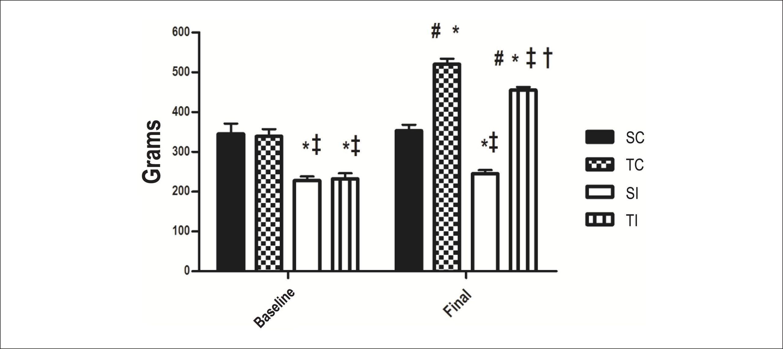
Male Wistar rats were divided into four groups: sedentary control, trained control, sedentary infarcted and trained infarcted rats. Each group with n = 9 rats. The animals underwent maximum load test and echocardiography at the beginning and at the end of the resistance exercise training (in an adapted ladder, 40% to 60% of the maximum load test, 3 months, 5 days/week). At the end, hemodynamic, baroreflex sensitivity and autonomic modulation assessments were made.

Results:

The maximum load test increased in groups trained control (+32%) and trained infarcted (+46%) in relation to groups sedentary control and sedentary infarcted. Although no change occurred regarding the myocardial infarction size and systolic function, the E/A ratio (-23%), myocardial performance index (-39%) and systolic blood pressure (+6%) improved with resistance exercise training in group trained infarcted. Concomitantly, the training provided additional benefits in the high frequency bands of the pulse interval (+45%), as well as in the low frequency band of systolic blood pressure (-46%) in rats from group trained infarcted in relation to group sedentary infarcted.

Conclusion:

Resistance exercise training alone may be an important and safe tool in the management of patients after myocardial infarction, considering that it does not lead to significant changes in the ventricular function, reduces the global cardiac stress, and significantly improves the vascular and cardiac autonomic modulation in infarcted rats.

  • Autonomic Nervous System
  • Exercise
  • Myocardial Infarction
  • Rats
  • Rehabilitation
  • Resistance Training
  • Ventricular Function
Read Full Article
Creative Commons Attribution (CC BY) Creative Commons license
09/07/2014 DOI: 10.5935/abc.20140093 Views 2
Morphological and functional measurements of the heart obtained by magnetic resonance imaging in Brazilians
ABC
Artigo Original

Morphological and functional measurements of the heart obtained by magnetic resonance imaging in Brazilians

Robson Macedo et al. Juliano Lara Fernandes Solange Souza Andrade Carlos Eduardo Rochitte … Kênio Costa Lima Álvaro Campos Cavalcanti Maciel Fernanda Cunha Maciel Geraldo Souza Pinho Alves Otávio Rizzi Coelho Rosiane Viana Zuza Diniz

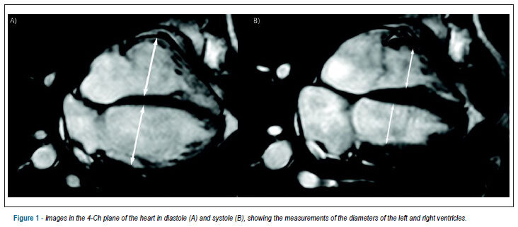
BACKGROUND: Still today, measurements used as a reference in the cardiac magnetic resonance imaging have been obtained mainly from studies carried out in North-American and European populations. OBJECTIVE: To obtain measurements of the diastolic diameter, systolic diameter, end diastolic volume, end systolic volume, ejection fraction, and myocardial mass of the left and right ventricles in Brazilians. METHODS: 54 men and 53 women, with mean age of 43.4 ± 13.1 years, asymptomatic, with no cardiomyopathies, have been subjected to the cardiac magnetic resonance imaging, using a balanced steady state free precession technique. RESULTS: The averages and the standard deviations of the parameters for the left ventricle have been: diastolic diameter = 4.8 ± 0.5 cm; systolic diameter = 3.0 ± 0.6 cm; end diastolic volume = 128.4 ± 29.6 mL; end systolic volume = 45.2 ± 16.6 mL; ejection fraction = 65.5 ± 6.3%; mass = 95.2 ± 30.8 g. For the right ventricle, they have been: diastolic diameter = 3.9 ± 1.3 cm; systolic diameter = 2.5 ± 0.5 cm; end diastolic volume = 126.5 ± 30.7 mL; end systolic volume = 53.6 ± 18.4 mL; ejection fraction = 58.3 ± 8.0%, and mass = 26.1 ± 6.1 g. The masses and the volumes were significantly greater in the men, except for the end systolic volume of the left ventricle. The ejection fraction of the right ventricle has been significantly greater in the women. There has been a significant and inverted correlation of the systolic volume of the right volume with the progression of the age. CONCLUSION: This study has described, for the first time, cardiac measurements obtained through the cardiac magnetic resonance imaging in Brazilians, asymptomatic, with no cardiomyopathies, showing differences in accordance with gender and age.

  • Genetics, Population
  • Heart Ventricle
  • Magnetic Resonance Imaging
  • Reference Values
  • Ventricular Function
Read Full Article
Creative Commons Attribution (CC BY) Creative Commons license
11/06/2013 DOI: 10.5935/abc.20130113 Views 1
ABC
Artigo Original

Impact of ventricular geometric pattern on cardiac remodeling after myocardial infarction

Elaine Farah et al. Daniéliso R. Fusco Paulo R. R. Okumoto Marcos F. Minicucci … Paula S. Azevedo Beatriz B. Matsubara Katashi Okoshi Siméia G. Zanati Sergio A. R. Paiva Leonardo A. M. Zornoff

BACKGROUND: The relevance of left ventricular (LV) geometric pattern after myocardial infarction is not known. OBJECTIVES: To analyze the presence of different LV geometric patterns and teir impact as a predictor of remodeling in patients with myocardial infarction. METHODS: Patients with anterior acute myocardial infarction (n = 80) were divided according to the geometric pattern: normal (normal left ventricular mass index [LVMI] and normal relative wall thickness [RWT]), concentric remodeling (normal LVMI and increased RWT), concentric hypertrophy (increased LVMI and RWT) and eccentric hypertrophy (increased LVMI and normal RWT). After six months, echocardiographic assessment was repeated. RESULTS: Four patients died. Of the survivors, 41 showed remodeling (R +), whereas 39 did not (R-). Considering the geometric pattern, the cases were distributed as follows: 24 patients with normal pattern, 13 with concentric remodeling, 29 with concentric hypertrophy and 14 with eccentric hypertrophy. Patients who showed remodeling had larger infarction sizes analyzed by peak CPK (R + = 4,610 (1,688-7,970), R- = 1,442 (775-4247), p <0.001) and CK-MB (R + = 441 (246 – 666), R- = 183 (101-465), p <0.001), trend towards higher prevalence of concentric remodeling (R+ = 10, R- = 3, p = 0.08) and lower prevalence of eccentric hypertrophy (R + = 2 R- = 12, p = 0.006). In the multivariate regression analysis, infarction size was a predictor (OR = 1.01, p = 0.020) and eccentric hypertrophy was a protective factor (OR = 0.189, p = 0.046) of ventricular remodeling after coronary occlusion. CONCLUSION: The LV geometric pattern of can have an impact on the remodeling process in patients with myocardial infarction.

  • Hypertension
  • left
  • Myocardial Infarction
  • Ventricular Function
  • Ventricular Remodeling
Read Full Article
Creative Commons Attribution (CC BY) Creative Commons license
14/05/2013 DOI: 10.5935/abc.20130104 Views 1
Echocardiography in thalassemic patients on blood transfusions and chelation without heart failure
ABC
Original Article

Echocardiography in thalassemic patients on blood transfusions and chelation without heart failure

Alexandre Rodrigues et al. Fábio Villaça Guimarães-Filho João Carlos Ferreira Braga Cássia Spínola Custódio Rodrigues … Paulo Waib Antonio Fabron-Junior Doralice Marvulle Tan Ana Cristina C. V. França Marina Politi Okoshi Katashi Okoshi

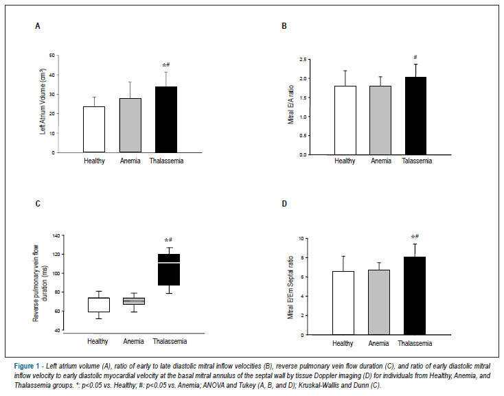
BACKGROUND: Patients with thalassemia major present chronic hemolysis and require regular blood transfusions which may cause iron overload cardiomyopathy and chronic heart failure. Hemochromatosis is characterized by excessive iron accumulation in tissues, and heart involvement is the main cause of death in patients with thalassemia. OBJECTIVE: The aim of this study was to evaluate cardiac structure and function by conventional Doppler echocardiography and tissue Doppler imaging in patients with TM and no clinical evidence of heart failure. METHODS: This is a prospective observational study including 18 patients with thalassemia major (TM) receiving regular blood transfusion. To separately evaluate anemia and blood transfusion effects, two gender, age, weight, and height-matched control groups were included: one with healthy individuals (Healthy, n=18) and one with iron deficient anemia patients (Anemia, n=18). Statistical analysis was performed using ANOVA followed by Tukey’s test or Kruskal-Wallis’s and Dunn’s test. RESULTS: The following echocardiographic variables presented significantly higher values in TM than the Anemia and Healthy groups: left atrium volume index (Healthy: 16.4±6.08; Anemia: 17.9±7.02; TM: 24.1±8.30 cm³/m²); mitral septal E/Em ratio (Healthy: 6.55±1.60; Anemia: 6.74±0.74; TM: 8.10±1.31); and duration of reverse pulmonary vein flow [Healthy: 74.0 (59.0-74.0); Anemia: 70.5 (67.0-74.0); TM: 111 (87.0-120) ms]. The mitral E/A ratio was higher in TM than Anemia (Healthy: 1.80±0.40; Anemia: 1.80±0.24; TM: 2.03±0.34). No differences were found in left ventricular structures and systolic function indexes. CONCLUSION: Conventional Doppler echocardiography and tissue Doppler allow changes in left ventricular diastolic function to be identified in asymptomatic patients with thalassemia major.

  • beta-Thalassemia
  • blood transfusion
  • Echocardiography, Doppler
  • Iron Chelating Agents
  • Ventricular Function
Read Full Article
Creative Commons Attribution (CC BY) Creative Commons license
08/01/2013 DOI: 10.1590/S0066-782X2013005000001
Role of echocardiography in the ventricular assessment of the transplanted heart versus heart rejection
ABC
Artigo Original

Role of echocardiography in the ventricular assessment of the transplanted heart versus heart rejection

Gabriel Antonio Stanisci Miguel et al. Salomón Soriano Ordinola Rojas Reinaldo Wilson Vieira José Pedro da Silva Henry Abensur

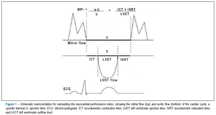
BACKGROUND: Heart transplantation is an alternative for individuals with end-stage heart disease. However, episodes of heart rejection (HR) are frequent and increase morbidity and mortality, requiring the use of an accurate non-invasive exam for their diagnosis, since endomyocardial biopsy (EMB) is not a complication-free procedure. OBJECTIVE: To compare the parameters obtained by use of Doppler echocardiography in a group of transplanted patients with HR (TX1) and another group of transplanted patients without rejection (TX0), having as reference a control group (CG) and observing the behavior of the left ventricular systo-diastolic function expressed as the myocardial performance index (MPI) METHODS: Transthoracic echocardiographies were performed from January 2006 to January 2008 to prospectively assess 47 patients divided into three groups: CG (36.2%); TX0 (38.3%); and TX1 (25.5%). The MPI was compared between the groups, and data were analyzed by use of Fisher exact test and nonparametric Kruskal-Wallis test, both with significance level of 5%. RESULTS: The groups did not differ regarding age, weight, height, and body surface. When compared to GC, TX0 and TX1 showed a change in the left ventricular systo-diastolic function, expressed as an increase in MPI, which was greater in TX1 [0.38 (0.29 – 0.44); 0.47 (0.43 – 0.56); 0.58 (0.52 – 0.74), respectively; p < 0.001]. CONCLUSION: Echocardiography was a very accurate test to detect changes in the systo-diastolic function of the transplanted heart; however, it did not prove to be reliable to replace BEM in the safe diagnosis of HR.

  • Echocardiography
  • Graft Rejection
  • Heart Transplantation
  • Ventricular Function
Read Full Article
Creative Commons Attribution (CC BY) Creative Commons license
09/11/2012 DOI: 10.1590/S0066-782X2012005000099 Views 2

Could not load more articles. Please try again.

How to cite this article

EndNote RIS

Electronic Document Format (Vancouver)

Lima MSM. The ELSA-Brasil Study and Our Myocardial Deformation. Arquivos Brasileiros de Cardiologia 2025;122(6):e20250265.

Electronic Document Format (ABNT)

Lima, Márcio S. M.. The ELSA-Brasil Study and Our Myocardial Deformation. Arquivos Brasileiros de Cardiologia, v. 122, n. 6, e20250265, Jun. 2025.

Electronic Document Format (APA)

Lima, M. S. M. (2025). The ELSA-Brasil Study and Our Myocardial Deformation. Arquivos Brasileiros de Cardiologia, 122(6), e20250265.

Electronic Document Format (ISO)

Lima, Márcio S. M.. The ELSA-Brasil Study and Our Myocardial Deformation. Arquivos Brasileiros de Cardiologia [online]. 2025, vol. 122, n. 6, [cited 2026-10-14], e20250265. Available from: <https://journals.cardiol.br/en/article/the-elsa-brasil-study-and-our-myocardial-deformation/>. ISSN 0066-782X

EndNote RIS

Electronic Document Format (Vancouver)

Pianca EG, Foppa M, Schmitz GB, Cañon-Montañez W, Duncan BB, Santos ABS. Left and Right Ventricular Strain in an Adult Brazilian Population from ELSA-Brasil Study: Reference Values and their Determinants. Arquivos Brasileiros de Cardiologia 2025;122(6):e20240634.

Electronic Document Format (ABNT)

Pianca, Eduardo Gatti; Foppa, Murilo; Schmitz, Giulia Bevilacqua; Cañon-Montañez, Wilson; Duncan, Bruce Bartholow; Santos, Angela Barreto Santiago. Left and Right Ventricular Strain in an Adult Brazilian Population from ELSA-Brasil Study: Reference Values and their Determinants. Arquivos Brasileiros de Cardiologia, v. 122, n. 6, e20240634, Jun. 2025.

Electronic Document Format (APA)

Pianca, E. G., Foppa, M., Schmitz, G. B., Cañon-Montañez, W., Duncan, B. B., & Santos, A. B. S. (2025). Left and Right Ventricular Strain in an Adult Brazilian Population from ELSA-Brasil Study: Reference Values and their Determinants. Arquivos Brasileiros de Cardiologia, 122(6), e20240634.

Electronic Document Format (ISO)

Pianca, Eduardo Gatti and Foppa, Murilo and Schmitz, Giulia Bevilacqua and Cañon-Montañez, Wilson and Duncan, Bruce Bartholow and Santos, Angela Barreto Santiago. Left and Right Ventricular Strain in an Adult Brazilian Population from ELSA-Brasil Study: Reference Values and their Determinants. Arquivos Brasileiros de Cardiologia [online]. 2025, vol. 122, n. 6, [cited 2026-10-14], e20240634. Available from: <https://journals.cardiol.br/en/article/left-and-right-ventricular-strain-in-an-adult-brazilian-population-from-elsa-brasil-study-reference-values-and-their-determinants/>. ISSN 0066-782X

EndNote RIS

Electronic Document Format (Vancouver)

Pereira MM, Monteiro Júnior FC, Yamamura S. Impact of Valvular Event Time Assessment Methods on Myocardial Work Calculation by Echocardiography. ABC Imagem Cardiovascular 2024;37(4):e20240093.

Electronic Document Format (ABNT)

Pereira, Marcio Mendes; Monteiro Júnior, Francisco das Chagas; Yamamura, Sayuri. Impact of Valvular Event Time Assessment Methods on Myocardial Work Calculation by Echocardiography. ABC Imagem Cardiovascular, v. 37, n. 4, e20240093, Dec. 2024.

Electronic Document Format (APA)

Pereira, M. M., Monteiro Júnior, F. C., & Yamamura, S. (2024). Impact of Valvular Event Time Assessment Methods on Myocardial Work Calculation by Echocardiography. ABC Imagem Cardiovascular, 37(4), e20240093.

Electronic Document Format (ISO)

Pereira, Marcio Mendes and Monteiro Júnior, Francisco das Chagas and Yamamura, Sayuri. Impact of Valvular Event Time Assessment Methods on Myocardial Work Calculation by Echocardiography. ABC Imagem Cardiovascular [online]. 2024, vol. 37, n. 4, [cited 2026-10-14], e20240093. Available from: <https://journals.cardiol.br/en/article/impact-of-valvular-event-time-assessment-methods-on-myocardial-work-calculation-by-echocardiography/>. ISSN 2318-8219

EndNote RIS

Electronic Document Format (Vancouver)

Dall’Orto AOMC, Otto ME, Leite SF, Maurício Filho MAFQ, Martins NT, Araújo SR, Almeida SV, et al. Comparative Analysis of Conventional and Speckle Tracking Echocardiographic Variables between Patients with Unrejected Heart Transplants and Healthy Individuals. Arquivos Brasileiros de Cardiologia 2024;121(8):e20230681.

Electronic Document Format (ABNT)

Dall’Orto, Aline Oliveira Martins Campo; Otto, Maria Estefania; Leite, Simone Ferreira; Maurício Filho, Marco Antônio Freitas de Queiroz; Martins, Natália Taveira; Araújo, Samuel Rabelo; Almeida, Soraya Vasconcelos; Brizida, Luiza Valle Oliveira; Atik, Fernando Antibas. Comparative Analysis of Conventional and Speckle Tracking Echocardiographic Variables between Patients with Unrejected Heart Transplants and Healthy Individuals. Arquivos Brasileiros de Cardiologia, v. 121, n. 8, e20230681, Aug. 2024.

Electronic Document Format (APA)

Dall’Orto, A. O. M. C., Otto, M. E., Leite, S. F., Maurício Filho, M. A. F. Q., Martins, N. T., Araújo, S. R., Almeida, S. V., Brizida, L. V. O., & Atik, F. A. (2024). Comparative Analysis of Conventional and Speckle Tracking Echocardiographic Variables between Patients with Unrejected Heart Transplants and Healthy Individuals. Arquivos Brasileiros de Cardiologia, 121(8), e20230681.

Electronic Document Format (ISO)

Dall’Orto, Aline Oliveira Martins Campo and Otto, Maria Estefania and Leite, Simone Ferreira and Maurício Filho, Marco Antônio Freitas de Queiroz and Martins, Natália Taveira and Araújo, Samuel Rabelo and Almeida, Soraya Vasconcelos and Brizida, Luiza Valle Oliveira and Atik, Fernando Antibas. Comparative Analysis of Conventional and Speckle Tracking Echocardiographic Variables between Patients with Unrejected Heart Transplants and Healthy Individuals. Arquivos Brasileiros de Cardiologia [online]. 2024, vol. 121, n. 8, [cited 2026-10-14], e20230681. Available from: <https://journals.cardiol.br/en/article/comparative-analysis-of-conventional-and-speckle-tracking-echocardiographic-variables-between-patients-with-unrejected-heart-transplants-and-healthy-individuals/>. ISSN 0066-782X

EndNote RIS

Electronic Document Format (Vancouver)

Barros Filho ACL, Romano MMD. Use of Three-Dimensional Echocardiography in the Analysis of Ventricular Function in Chagas Disease. ABC Imagem Cardiovascular 2023;36(4):e20230093.

Electronic Document Format (ABNT)

Barros Filho, Antonio Carlos Leite de; Romano, Minna Moreira Dias. Use of Three-Dimensional Echocardiography in the Analysis of Ventricular Function in Chagas Disease. ABC Imagem Cardiovascular, v. 36, n. 4, e20230093, Dec. 2023.

Electronic Document Format (APA)

Barros Filho, A. C. L. , & Romano, M. M. D. (2023). Use of Three-Dimensional Echocardiography in the Analysis of Ventricular Function in Chagas Disease. ABC Imagem Cardiovascular, 36(4), e20230093.

Electronic Document Format (ISO)

Barros Filho, Antonio Carlos Leite de and Romano, Minna Moreira Dias. Use of Three-Dimensional Echocardiography in the Analysis of Ventricular Function in Chagas Disease. ABC Imagem Cardiovascular [online]. 2023, vol. 36, n. 4, [cited 2026-10-14], e20230093. Available from: <https://journals.cardiol.br/en/article/use-of-three-dimensional-echocardiography-in-the-analysis-of-ventricular-function-in-chagas-disease/>. ISSN 2318-8219

EndNote RIS

Electronic Document Format (Vancouver)

Guzman-Ramirez D, Trujillo-Garcia A, Lopez-Rincon M, Lopez RB. Right Ventricular Function and Exercise Tolerance in Patients with ST-Elevation Myocardial Infarction. Arquivos Brasileiros de Cardiologia 2023;120(9):e20220799.

Electronic Document Format (ABNT)

Guzman-Ramirez, Denisse; Trujillo-Garcia, Anival; Lopez-Rincon, Meredith; Lopez, Roxella Botello. Right Ventricular Function and Exercise Tolerance in Patients with ST-Elevation Myocardial Infarction. Arquivos Brasileiros de Cardiologia, v. 120, n. 9, e20220799, Sep. 2023.

Electronic Document Format (APA)

Guzman-Ramirez, D., Trujillo-Garcia, A., Lopez-Rincon, M., & Lopez, R. B. (2023). Right Ventricular Function and Exercise Tolerance in Patients with ST-Elevation Myocardial Infarction. Arquivos Brasileiros de Cardiologia, 120(9), e20220799.

Electronic Document Format (ISO)

Guzman-Ramirez, Denisse and Trujillo-Garcia, Anival and Lopez-Rincon, Meredith and Lopez, Roxella Botello. Right Ventricular Function and Exercise Tolerance in Patients with ST-Elevation Myocardial Infarction. Arquivos Brasileiros de Cardiologia [online]. 2023, vol. 120, n. 9, [cited 2026-10-14], e20220799. Available from: <https://journals.cardiol.br/en/article/right-ventricular-function-and-exercise-tolerance-in-patients-with-st-elevation-myocardial-infarction/>. ISSN 0066-782X

EndNote RIS

Electronic Document Format (Vancouver)

Silva CES. Is Echocardiographic Assessment of Strain Essential In Daily Routine?. ABC Imagem Cardiovascular 2023;36(3):e20230063.

Electronic Document Format (ABNT)

Silva, Carlos Eduardo Suaide. Is Echocardiographic Assessment of Strain Essential In Daily Routine?. ABC Imagem Cardiovascular, v. 36, n. 3, e20230063, Sep. 2023.

Electronic Document Format (APA)

Silva, C. E. S. (2023). Is Echocardiographic Assessment of Strain Essential In Daily Routine?. ABC Imagem Cardiovascular, 36(3), e20230063.

Electronic Document Format (ISO)

Silva, Carlos Eduardo Suaide. Is Echocardiographic Assessment of Strain Essential In Daily Routine?. ABC Imagem Cardiovascular [online]. 2023, vol. 36, n. 3, [cited 2026-10-14], e20230063. Available from: <https://journals.cardiol.br/en/article/is-echocardiographic-assessment-of-strain-essential-in-daily-routine/>. ISSN 2318-8219

EndNote RIS

Electronic Document Format (Vancouver)

Silva TRW, Silva RL, Martins AF, Marques JLB. Role of Strain in the Early Diagnosis of Diabetic Cardiomyopathy. Arquivos Brasileiros de Cardiologia Imagem cardiovascular 2022;35(2):eabc293.

Electronic Document Format (ABNT)

Silva, Thais Rossoni Weber da; Silva, Roberto Léo da; Martins, Adriana Ferraz; Marques, Jefferson Luiz Brum. Role of Strain in the Early Diagnosis of Diabetic Cardiomyopathy. Arquivos Brasileiros de Cardiologia Imagem cardiovascular, v. 35, n. 2, eabc293, May. 2022.

Electronic Document Format (APA)

Silva, T. R. W. , Silva, R. L. , Martins, A. F., & Marques, J. L. B. (2022). Role of Strain in the Early Diagnosis of Diabetic Cardiomyopathy. Arquivos Brasileiros de Cardiologia Imagem cardiovascular, 35(2), eabc293.

Electronic Document Format (ISO)

Silva, Thais Rossoni Weber da and Silva, Roberto Léo da and Martins, Adriana Ferraz and Marques, Jefferson Luiz Brum. Role of Strain in the Early Diagnosis of Diabetic Cardiomyopathy. Arquivos Brasileiros de Cardiologia Imagem cardiovascular [online]. 2022, vol. 35, n. 2, [cited 2026-10-14], eabc293. Available from: <https://journals.cardiol.br/en/article/role-of-strain-in-the-early-diagnosis-of-diabetic-cardiomyopathy/>. ISSN 2318-8219

EndNote RIS

Electronic Document Format (Vancouver)

Ferrari F. Lung Function and Inspiratory Muscle Strength in Heart Failure: Can They be Considered Potential Prognostic Markers?. Arquivos Brasileiros de Cardiologia 2022;118(4):692-3.

Electronic Document Format (ABNT)

Ferrari, Filipe. Lung Function and Inspiratory Muscle Strength in Heart Failure: Can They be Considered Potential Prognostic Markers?. Arquivos Brasileiros de Cardiologia, v. 118, n. 4, p. 692-693, Apr. 2022.

Electronic Document Format (APA)

Ferrari, F. (2022). Lung Function and Inspiratory Muscle Strength in Heart Failure: Can They be Considered Potential Prognostic Markers?. Arquivos Brasileiros de Cardiologia, 118(4), 692-693.

Electronic Document Format (ISO)

Ferrari, Filipe. Lung Function and Inspiratory Muscle Strength in Heart Failure: Can They be Considered Potential Prognostic Markers?. Arquivos Brasileiros de Cardiologia [online]. 2022, vol. 118, n. 4, [cited 2026-10-14], pp. 692-693. Available from: <https://journals.cardiol.br/en/article/lung-function-and-inspiratory-muscle-strength-in-heart-failure-can-they-be-considered-potential-prognostic-markers/>. ISSN 0066-782X

EndNote RIS

Electronic Document Format (Vancouver)

Ramalho SHR, Lima ACGB, Silva FMF, Souza FSJ, Cahalin LP, Cipriano GFB, Cipriano Junior G, et al. Relationship of Lung Function and Inspiratory Strength with Exercise Capacity and Prognosis in Heart Failure. Arquivos Brasileiros de Cardiologia 2022;118(4):680-91.

Electronic Document Format (ABNT)

Ramalho, Sergio Henrique Rodolpho; Lima, Alexandra Correa Gervazoni Balbuena de; Silva, Fabiola Maria Ferreira da; Souza, Fausto Stauffer Junqueira de; Cahalin, Lawrence Patrick; Cipriano, Graziella França Bernardelli; Cipriano Junior, Gerson. Relationship of Lung Function and Inspiratory Strength with Exercise Capacity and Prognosis in Heart Failure. Arquivos Brasileiros de Cardiologia, v. 118, n. 4, p. 680-691, Feb. 2022.

Electronic Document Format (APA)

Ramalho, S. H. R., Lima, A. C. G. B. , Silva, F. M. F. , Souza, F. S. J. , Cahalin, L. P., Cipriano, G. F. B., & Cipriano Junior, G. (2022). Relationship of Lung Function and Inspiratory Strength with Exercise Capacity and Prognosis in Heart Failure. Arquivos Brasileiros de Cardiologia, 118(4), 680-691.

Electronic Document Format (ISO)

Ramalho, Sergio Henrique Rodolpho and Lima, Alexandra Correa Gervazoni Balbuena de and Silva, Fabiola Maria Ferreira da and Souza, Fausto Stauffer Junqueira de and Cahalin, Lawrence Patrick and Cipriano, Graziella França Bernardelli and Cipriano Junior, Gerson. Relationship of Lung Function and Inspiratory Strength with Exercise Capacity and Prognosis in Heart Failure. Arquivos Brasileiros de Cardiologia [online]. 2022, vol. 118, n. 4, [cited 2026-10-14], pp. 680-691. Available from: <https://journals.cardiol.br/en/article/relationship-of-lung-function-and-inspiratory-strength-with-exercise-capacity-and-prognosis-in-heart-failure/>. ISSN 0066-782X

EndNote RIS

Electronic Document Format (Vancouver)

Mercer-Rosa L. My Approach to Assessing Right Ventricular Function After Tetralogy of Fallot Repair. Arquivos Brasileiros de Cardiologia Imagem cardiovascular 2021;34(4):ecom17.

Electronic Document Format (ABNT)

Mercer-Rosa, Laura. My Approach to Assessing Right Ventricular Function After Tetralogy of Fallot Repair. Arquivos Brasileiros de Cardiologia Imagem cardiovascular, v. 34, n. 4, ecom17, Dec. 2021.

Electronic Document Format (APA)

Mercer-Rosa, L. (2021). My Approach to Assessing Right Ventricular Function After Tetralogy of Fallot Repair. Arquivos Brasileiros de Cardiologia Imagem cardiovascular, 34(4), ecom17.

Electronic Document Format (ISO)

Mercer-Rosa, Laura. My Approach to Assessing Right Ventricular Function After Tetralogy of Fallot Repair. Arquivos Brasileiros de Cardiologia Imagem cardiovascular [online]. 2021, vol. 34, n. 4, [cited 2026-10-14], ecom17. Available from: <https://journals.cardiol.br/en/article/my-approach-to-assessing-right-ventricular-function-after-tetralogy-of-fallot-repair/>. ISSN 2318-8219

EndNote RIS

Electronic Document Format (Vancouver)

Pinto IMF. Cardiac Magnetic Resonance and Amyloidosis: How can it Assist Clinical Reasoning?. Phenomenology, Humanities and Sciences – Fenomenologia, Humanidades e Ciências 2021;1(2):115-9.

Electronic Document Format (ABNT)

Pinto, Ibraim Masciarelli F.. Cardiac Magnetic Resonance and Amyloidosis: How can it Assist Clinical Reasoning?. Phenomenology, Humanities and Sciences – Fenomenologia, Humanidades e Ciências, v. 1, n. 2, p. 115-119, Dec. 2021.

Electronic Document Format (APA)

Pinto, I. M. F. (2021). Cardiac Magnetic Resonance and Amyloidosis: How can it Assist Clinical Reasoning?. Phenomenology, Humanities and Sciences – Fenomenologia, Humanidades e Ciências, 1(2), 115-119.

Electronic Document Format (ISO)

Pinto, Ibraim Masciarelli F.. Cardiac Magnetic Resonance and Amyloidosis: How can it Assist Clinical Reasoning?. Phenomenology, Humanities and Sciences – Fenomenologia, Humanidades e Ciências [online]. 2021, vol. 1, n. 2, [cited 2026-10-14], pp. 115-119. Available from: <https://journals.cardiol.br/en/article/cardiac-magnetic-resonance-and-amyloidosis-how-can-it-assist-clinical-reasoning/>

EndNote RIS

Electronic Document Format (Vancouver)

Hossri CAC. Atrioventricular Conduction System Disorders and Potential Risks of Arrhythmic Events in Endurance Athletes. Arquivos Brasileiros de Cardiologia 2020;115(1):78-9.

Electronic Document Format (ABNT)

Hossri, Carlos Alberto Cordeiro. Atrioventricular Conduction System Disorders and Potential Risks of Arrhythmic Events in Endurance Athletes. Arquivos Brasileiros de Cardiologia, v. 115, n. 1, p. 78-79, Aug. 2020.

Electronic Document Format (APA)

Hossri, C. A. C. (2020). Atrioventricular Conduction System Disorders and Potential Risks of Arrhythmic Events in Endurance Athletes. Arquivos Brasileiros de Cardiologia, 115(1), 78-79.

Electronic Document Format (ISO)

Hossri, Carlos Alberto Cordeiro. Atrioventricular Conduction System Disorders and Potential Risks of Arrhythmic Events in Endurance Athletes. Arquivos Brasileiros de Cardiologia [online]. 2020, vol. 115, n. 1, [cited 2026-10-14], pp. 78-79. Available from: <https://journals.cardiol.br/en/article/atrioventricular-conduction-system-disorders-and-potential-risks-of-arrhythmic-events-in-endurance-athletes/>. ISSN 0066-782X

EndNote RIS

Electronic Document Format (Vancouver)

Benchimol-Barbosa PR, Nasario Junior O, Nadal J. Assessing Dynamic Atrioventricular Conduction Time to RR-interval Coupling in Athletes and Sedentary Subjects. Arquivos Brasileiros de Cardiologia 2020;115(1):71-7.

Electronic Document Format (ABNT)

Benchimol-Barbosa, Paulo Roberto; Nasario Junior, Olivassé; Nadal, Jurandir. Assessing Dynamic Atrioventricular Conduction Time to RR-interval Coupling in Athletes and Sedentary Subjects. Arquivos Brasileiros de Cardiologia, v. 115, n. 1, p. 71-77, May. 2020.

Electronic Document Format (APA)

Benchimol-Barbosa, P. R., Nasario Junior, O., & Nadal, J. (2020). Assessing Dynamic Atrioventricular Conduction Time to RR-interval Coupling in Athletes and Sedentary Subjects. Arquivos Brasileiros de Cardiologia, 115(1), 71-77.

Electronic Document Format (ISO)

Benchimol-Barbosa, Paulo Roberto and Nasario Junior, Olivassé and Nadal, Jurandir. Assessing Dynamic Atrioventricular Conduction Time to RR-interval Coupling in Athletes and Sedentary Subjects. Arquivos Brasileiros de Cardiologia [online]. 2020, vol. 115, n. 1, [cited 2026-10-14], pp. 71-77. Available from: <https://journals.cardiol.br/en/article/assessing-dynamic-atrioventricular-conduction-time-to-rr-interval-coupling-in-athletes-and-sedentary-subjects/>. ISSN 0066-782X

EndNote RIS

Electronic Document Format (Vancouver)

Nacif MS, Almeida ALC, Young AA, Cowan BR, Armstrong AC, Yang E, Sibley CT, et al. Three-Dimensional Volumetric Assessment of Diastolic Function by Cardiac Magnetic Resonance Imaging: The Multi-Ethnic Study of Atherosclerosis (MESA). Arquivos Brasileiros de Cardiologia 2017;108(6):552-63.

Electronic Document Format (ABNT)

Nacif, Marcelo S; Almeida, Andre L. C.; Young, Alistair A; Cowan, Brett R; Armstrong, Anderson C; Yang, Eunice; Sibley, Christopher T; Hundley, W. Gregory; Liu, Songtao; Lima, Joao AC; Bluemke, David A. Three-Dimensional Volumetric Assessment of Diastolic Function by Cardiac Magnetic Resonance Imaging: The Multi-Ethnic Study of Atherosclerosis (MESA). Arquivos Brasileiros de Cardiologia, v. 108, n. 6, p. 552-563, May. 2017.

Electronic Document Format (APA)

Nacif, M. S., Almeida, A. L. C., Young, A. A., Cowan, B. R., Armstrong, A. C., Yang, E., Sibley, C. T., Hundley, W. G., Liu, S., Lima, J. A., & Bluemke, D. A. (2017). Three-Dimensional Volumetric Assessment of Diastolic Function by Cardiac Magnetic Resonance Imaging: The Multi-Ethnic Study of Atherosclerosis (MESA). Arquivos Brasileiros de Cardiologia, 108(6), 552-563.

Electronic Document Format (ISO)

Nacif, Marcelo S and Almeida, Andre L. C. and Young, Alistair A and Cowan, Brett R and Armstrong, Anderson C and Yang, Eunice and Sibley, Christopher T and Hundley, W. Gregory and Liu, Songtao and Lima, Joao AC and Bluemke, David A. Three-Dimensional Volumetric Assessment of Diastolic Function by Cardiac Magnetic Resonance Imaging: The Multi-Ethnic Study of Atherosclerosis (MESA). Arquivos Brasileiros de Cardiologia [online]. 2017, vol. 108, n. 6, [cited 2026-10-14], pp. 552-563. Available from: <https://journals.cardiol.br/en/article/three-dimensional-volumetric-assessment-of-diastolic-function-by-cardiac-magnetic-resonance-imaging-the-multi-ethnic-study-of-atherosclerosis-mesa/>. ISSN 0066-782X

EndNote RIS

Electronic Document Format (Vancouver)

Kılıcgedik A, Kahveci G, Gurbuz AS, Karabay CY, Guler A, Efe SC, Aung SM, et al. Papillary Muscle Free Strain in Patients with Severe Degenerative and Functional Mitral Regurgitation. Arquivos Brasileiros de Cardiologia 2017;108(4):339-46.

Electronic Document Format (ABNT)

Kılıcgedik, Alev; Kahveci, Gokhan; Gurbuz, Ahmet Seyfeddin; Karabay, Can Yucel; Guler, Ahmet; Efe, Suleyman Cagan; Aung, Soe Moe; Arslantas, Ugur; Demir, Serdar; Izgi, Ibrahim Akin; Kirma, Cevat. Papillary Muscle Free Strain in Patients with Severe Degenerative and Functional Mitral Regurgitation. Arquivos Brasileiros de Cardiologia, v. 108, n. 4, p. 339-346, Mar. 2017.

Electronic Document Format (APA)

Kılıcgedik, A., Kahveci, G., Gurbuz, A. S., Karabay, C. Y., Guler, A., Efe, S. C., Aung, S. M., Arslantas, U., Demir, S., Izgi, I. A., & Kirma, C. (2017). Papillary Muscle Free Strain in Patients with Severe Degenerative and Functional Mitral Regurgitation. Arquivos Brasileiros de Cardiologia, 108(4), 339-346.

Electronic Document Format (ISO)

Kılıcgedik, Alev and Kahveci, Gokhan and Gurbuz, Ahmet Seyfeddin and Karabay, Can Yucel and Guler, Ahmet and Efe, Suleyman Cagan and Aung, Soe Moe and Arslantas, Ugur and Demir, Serdar and Izgi, Ibrahim Akin and Kirma, Cevat. Papillary Muscle Free Strain in Patients with Severe Degenerative and Functional Mitral Regurgitation. Arquivos Brasileiros de Cardiologia [online]. 2017, vol. 108, n. 4, [cited 2026-10-14], pp. 339-346. Available from: <https://journals.cardiol.br/en/article/papillary-muscle-free-strain-in-patients-with-severe-degenerative-and-functional-mitral-regurgitation/>. ISSN 0066-782X

EndNote RIS

Electronic Document Format (Vancouver)

Badavi M, Sadeghi N, Dianat M, Samarbafzadeh A. Gallic Acid and Cyclosporine Mixture and their Effects on Cardiac Dysfunction Induced by Ischemia/Reperfusion and eNOS/iNOS Expression. International Journal of Cardiovascular Sciences 2017;30(3):207-18.

Electronic Document Format (ABNT)

Badavi, Mohammad; Sadeghi, Najmeh; Dianat, Mahin; Samarbafzadeh, Alireza. Gallic Acid and Cyclosporine Mixture and their Effects on Cardiac Dysfunction Induced by Ischemia/Reperfusion and eNOS/iNOS Expression. International Journal of Cardiovascular Sciences, v. 30, n. 3, p. 207-218, Dec. 2016.

Electronic Document Format (APA)

Badavi, M., Sadeghi, N., Dianat, M., & Samarbafzadeh, A. (2017). Gallic Acid and Cyclosporine Mixture and their Effects on Cardiac Dysfunction Induced by Ischemia/Reperfusion and eNOS/iNOS Expression. International Journal of Cardiovascular Sciences, 30(3), 207-218.

Electronic Document Format (ISO)

Badavi, Mohammad and Sadeghi, Najmeh and Dianat, Mahin and Samarbafzadeh, Alireza. Gallic Acid and Cyclosporine Mixture and their Effects on Cardiac Dysfunction Induced by Ischemia/Reperfusion and eNOS/iNOS Expression. International Journal of Cardiovascular Sciences [online]. 2017, vol. 30, n. 3, [cited 2026-10-14], pp. 207-218. Available from: <https://journals.cardiol.br/en/article/gallic-acid-and-cyclosporine-mixture-and-their-effects-on-cardiac-dysfunction-induced-by-ischemia-reperfusion-and-enos-inos-expression/>. ISSN 2359-4802

EndNote RIS

Electronic Document Format (Vancouver)

Santos SN, Henz BD, Zanatta AR, Barreto JR, Loureiro KB, Novakoski C, Santos MVN, et al. Impact of Atrial Fibrillation Ablation on Left Ventricular Filling Pressure and Left Atrial Remodeling. Arquivos Brasileiros de Cardiologia 2014;103(6):485-92.

Electronic Document Format (ABNT)

Santos, Simone Nascimento dos; Henz, Benhur Davi; Zanatta, André Rodrigues; Barreto, José Roberto; Loureiro, Kelly Bianca; Novakoski, Clarissa; Santos, Marcus Vinícius Nascimento dos; Giuseppin, Fabio F.; Oliveira, Edna Maria; Leite, Luiz Roberto. Impact of Atrial Fibrillation Ablation on Left Ventricular Filling Pressure and Left Atrial Remodeling. Arquivos Brasileiros de Cardiologia, v. 103, n. 6, p. 485-492, Oct. 2014.

Electronic Document Format (APA)

Santos, S. N. , Henz, B. D., Zanatta, A. R., Barreto, J. R., Loureiro, K. B., Novakoski, C., Santos, M. V. N. , Giuseppin, F. F., Oliveira, E. M., & Leite, L. R. (2014). Impact of Atrial Fibrillation Ablation on Left Ventricular Filling Pressure and Left Atrial Remodeling. Arquivos Brasileiros de Cardiologia, 103(6), 485-492.

Electronic Document Format (ISO)

Santos, Simone Nascimento dos and Henz, Benhur Davi and Zanatta, André Rodrigues and Barreto, José Roberto and Loureiro, Kelly Bianca and Novakoski, Clarissa and Santos, Marcus Vinícius Nascimento dos and Giuseppin, Fabio F. and Oliveira, Edna Maria and Leite, Luiz Roberto. Impact of Atrial Fibrillation Ablation on Left Ventricular Filling Pressure and Left Atrial Remodeling. Arquivos Brasileiros de Cardiologia [online]. 2014, vol. 103, n. 6, [cited 2026-10-14], pp. 485-492. Available from: <https://journals.cardiol.br/en/article/impact-of-atrial-fibrillation-ablation-on-left-ventricular-filling-pressure-and-left-atrial-remodeling/>. ISSN 0066-782X

EndNote RIS

Electronic Document Format (Vancouver)

Özcan F, lu ST, Turak O, Çay S, Aras D. Ablation of Premature Ventricular Contraction with the Help of a Notch. Arquivos Brasileiros de Cardiologia 2014;103(4):e55-6.

Electronic Document Format (ABNT)

Özcan, Firat; lu, Serkan Topaloğ; Turak, Osman; Çay, Serkan; Aras, Dursun. Ablation of Premature Ventricular Contraction with the Help of a Notch. Arquivos Brasileiros de Cardiologia, v. 103, n. 4, p. e55-e56, Sep. 2014.

Electronic Document Format (APA)

Özcan, F., lu, S. T., Turak, O., Çay, S., & Aras, D. (2014). Ablation of Premature Ventricular Contraction with the Help of a Notch. Arquivos Brasileiros de Cardiologia, 103(4), e55-e56.

Electronic Document Format (ISO)

Özcan, Firat and lu, Serkan Topaloğ and Turak, Osman and Çay, Serkan and Aras, Dursun. Ablation of Premature Ventricular Contraction with the Help of a Notch. Arquivos Brasileiros de Cardiologia [online]. 2014, vol. 103, n. 4, [cited 2026-10-14], pp. e55-e56. Available from: <https://journals.cardiol.br/en/article/ablation-of-premature-ventricular-contraction-with-the-help-of-a-notch/>. ISSN 0066-782X

EndNote RIS

Electronic Document Format (Vancouver)

Grans CF, Feriani DJ, Abssamra MEV, Rocha LY, Carrozzi NM, Mostarda C, Figueroa DM, et al. Resistance Training After Myocardial Infarction in Rats: Its Role on Cardiac and Autonomic Function. Arquivos Brasileiros de Cardiologia 2014;103(1):60-8.

Electronic Document Format (ABNT)

Grans, Camilla Figueiredo; Feriani, Daniele Jardim; Abssamra, Marcos Elias Vergilino; Rocha, Leandro Yanase; Carrozzi, Nicolle Martins; Mostarda, Cristiano; Figueroa, Diego Mendrot; Angelis, Kátia De; Irigoyen, Maria Cláudia; Rodrigues, Bruno. Resistance Training After Myocardial Infarction in Rats: Its Role on Cardiac and Autonomic Function. Arquivos Brasileiros de Cardiologia, v. 103, n. 1, p. 60-68, Jul. 2014.

Electronic Document Format (APA)

Grans, C. F., Feriani, D. J., Abssamra, M. E. V., Rocha, L. Y., Carrozzi, N. M., Mostarda, C., Figueroa, D. M., Angelis, K. D., Irigoyen, M. C., & Rodrigues, B. (2014). Resistance Training After Myocardial Infarction in Rats: Its Role on Cardiac and Autonomic Function. Arquivos Brasileiros de Cardiologia, 103(1), 60-68.

Electronic Document Format (ISO)

Grans, Camilla Figueiredo and Feriani, Daniele Jardim and Abssamra, Marcos Elias Vergilino and Rocha, Leandro Yanase and Carrozzi, Nicolle Martins and Mostarda, Cristiano and Figueroa, Diego Mendrot and Angelis, Kátia De and Irigoyen, Maria Cláudia and Rodrigues, Bruno. Resistance Training After Myocardial Infarction in Rats: Its Role on Cardiac and Autonomic Function. Arquivos Brasileiros de Cardiologia [online]. 2014, vol. 103, n. 1, [cited 2026-10-14], pp. 60-68. Available from: <https://journals.cardiol.br/en/article/resistance-training-after-myocardial-infarction-in-rats-its-role-on-cardiac-and-autonomic-function/>. ISSN 0066-782X

EndNote RIS

Electronic Document Format (Vancouver)

Macedo R, Fernandes JL, Andrade SS, Rochitte CE, Lima KC, Maciel CC, Maciel FC, et al. Morphological and functional measurements of the heart obtained by magnetic resonance imaging in Brazilians. Arquivos Brasileiros de Cardiologia 2013;101(1):68-77.

Electronic Document Format (ABNT)

Macedo, Robson; Fernandes, Juliano Lara; Andrade, Solange Souza; Rochitte, Carlos Eduardo; Lima, Kênio Costa; Maciel, Álvaro Campos Cavalcanti; Maciel, Fernanda Cunha; Alves, Geraldo Souza Pinho; Coelho, Otávio Rizzi; Diniz, Rosiane Viana Zuza. Morphological and functional measurements of the heart obtained by magnetic resonance imaging in Brazilians. Arquivos Brasileiros de Cardiologia, v. 101, n. 1, p. 68-77, Jun. 2013.

Electronic Document Format (APA)

Macedo, R., Fernandes, J. L., Andrade, S. S., Rochitte, C. E., Lima, K. C., Maciel, C. C., Maciel, F. C., Alves, G. S. P., Coelho, O. R., & Diniz, R. V. Z. (2013). Morphological and functional measurements of the heart obtained by magnetic resonance imaging in Brazilians. Arquivos Brasileiros de Cardiologia, 101(1), 68-77.

Electronic Document Format (ISO)

Macedo, Robson and Fernandes, Juliano Lara and Andrade, Solange Souza and Rochitte, Carlos Eduardo and Lima, Kênio Costa and Maciel, Álvaro Campos Cavalcanti and Maciel, Fernanda Cunha and Alves, Geraldo Souza Pinho and Coelho, Otávio Rizzi and Diniz, Rosiane Viana Zuza. Morphological and functional measurements of the heart obtained by magnetic resonance imaging in Brazilians. Arquivos Brasileiros de Cardiologia [online]. 2013, vol. 101, n. 1, [cited 2026-10-14], pp. 68-77. Available from: <https://journals.cardiol.br/en/article/morphological-and-functional-measurements-of-the-heart-obtained-by-magnetic-resonance-imaging-in-brazilians/>. ISSN 0066-782X

EndNote RIS

Electronic Document Format (Vancouver)

Farah E, Fusco DR, Okumoto PRR, Minicucci MF, Azevedo PS, Matsubara BB, Okoshi K, et al. Impact of ventricular geometric pattern on cardiac remodeling after myocardial infarction. Arquivos Brasileiros de Cardiologia 2013;100(6):518-23.

Electronic Document Format (ABNT)

Farah, Elaine; Fusco, Daniéliso R.; Okumoto, Paulo R. R.; Minicucci, Marcos F.; Azevedo, Paula S.; Matsubara, Beatriz B.; Okoshi, Katashi; Zanati, Siméia G.; Paiva, Sergio A. R.; Zornoff, Leonardo A. M.. Impact of ventricular geometric pattern on cardiac remodeling after myocardial infarction. Arquivos Brasileiros de Cardiologia, v. 100, n. 6, p. 518-523, May. 2013.

Electronic Document Format (APA)

Farah, E., Fusco, D. R., Okumoto, P. R. R., Minicucci, M. F., Azevedo, P. S., Matsubara, B. B., Okoshi, K., Zanati, S. G., Paiva, S. A. R., & Zornoff, L. A. M. (2013). Impact of ventricular geometric pattern on cardiac remodeling after myocardial infarction. Arquivos Brasileiros de Cardiologia, 100(6), 518-523.

Electronic Document Format (ISO)

Farah, Elaine and Fusco, Daniéliso R. and Okumoto, Paulo R. R. and Minicucci, Marcos F. and Azevedo, Paula S. and Matsubara, Beatriz B. and Okoshi, Katashi and Zanati, Siméia G. and Paiva, Sergio A. R. and Zornoff, Leonardo A. M.. Impact of ventricular geometric pattern on cardiac remodeling after myocardial infarction. Arquivos Brasileiros de Cardiologia [online]. 2013, vol. 100, n. 6, [cited 2026-10-14], pp. 518-523. Available from: <https://journals.cardiol.br/en/article/impact-of-ventricular-geometric-pattern-on-cardiac-remodeling-after-myocardial-infarction/>. ISSN 0066-782X

EndNote RIS

Electronic Document Format (Vancouver)

Rodrigues A, Guimarães-Filho FV, Braga JCF, Rodrigues CSC, Waib P, Fabron-Junior A, Tan DM, et al. Echocardiography in thalassemic patients on blood transfusions and chelation without heart failure. Arquivos Brasileiros de Cardiologia 2013;100(1):75-81.

Electronic Document Format (ABNT)

Rodrigues, Alexandre; Guimarães-Filho, Fábio Villaça; Braga, João Carlos Ferreira; Rodrigues, Cássia Spínola Custódio; Waib, Paulo; Fabron-Junior, Antonio; Tan, Doralice Marvulle; França, Ana Cristina C. V.; Okoshi, Marina Politi; Okoshi, Katashi. Echocardiography in thalassemic patients on blood transfusions and chelation without heart failure. Arquivos Brasileiros de Cardiologia, v. 100, n. 1, p. 75-81, Jan. 2013.

Electronic Document Format (APA)

Rodrigues, A., Guimarães-Filho, F. V., Braga, J. C. F., Rodrigues, C. S. C., Waib, P., Fabron-Junior, A., Tan, D. M., França, A. C. C. V., Okoshi, M. P., & Okoshi, K. (2013). Echocardiography in thalassemic patients on blood transfusions and chelation without heart failure. Arquivos Brasileiros de Cardiologia, 100(1), 75-81.

Electronic Document Format (ISO)

Rodrigues, Alexandre and Guimarães-Filho, Fábio Villaça and Braga, João Carlos Ferreira and Rodrigues, Cássia Spínola Custódio and Waib, Paulo and Fabron-Junior, Antonio and Tan, Doralice Marvulle and França, Ana Cristina C. V. and Okoshi, Marina Politi and Okoshi, Katashi. Echocardiography in thalassemic patients on blood transfusions and chelation without heart failure. Arquivos Brasileiros de Cardiologia [online]. 2013, vol. 100, n. 1, [cited 2026-10-14], pp. 75-81. Available from: <https://journals.cardiol.br/en/article/echocardiography-in-thalassemic-patients-on-blood-transfusions-and-chelation-without-heart-failure/>. ISSN 0066-782X

EndNote RIS

Electronic Document Format (Vancouver)

Miguel GAS, Rojas SSO, Vieira RW, Silva JP, Abensur H. Role of echocardiography in the ventricular assessment of the transplanted heart versus heart rejection. Arquivos Brasileiros de Cardiologia 2012;99(5):1031-9.

Electronic Document Format (ABNT)

Miguel, Gabriel Antonio Stanisci; Rojas, Salomón Soriano Ordinola; Vieira, Reinaldo Wilson; Silva, José Pedro da; Abensur, Henry. Role of echocardiography in the ventricular assessment of the transplanted heart versus heart rejection. Arquivos Brasileiros de Cardiologia, v. 99, n. 5, p. 1031-1039, Nov. 2012.

Electronic Document Format (APA)

Miguel, G. A. S., Rojas, S. S. O., Vieira, R. W., Silva, J. P. , & Abensur, H. (2012). Role of echocardiography in the ventricular assessment of the transplanted heart versus heart rejection. Arquivos Brasileiros de Cardiologia, 99(5), 1031-1039.

Electronic Document Format (ISO)

Miguel, Gabriel Antonio Stanisci and Rojas, Salomón Soriano Ordinola and Vieira, Reinaldo Wilson and Silva, José Pedro da and Abensur, Henry. Role of echocardiography in the ventricular assessment of the transplanted heart versus heart rejection. Arquivos Brasileiros de Cardiologia [online]. 2012, vol. 99, n. 5, [cited 2026-10-14], pp. 1031-1039. Available from: <https://journals.cardiol.br/en/article/role-of-echocardiography-in-the-ventricular-assessment-of-the-transplanted-heart-versus-heart-rejection/>. ISSN 0066-782X

  • About ABC Family
  • Internal Regulation
  • Editorial Board
  • Brazilian Society of Cardiology (SBC)

Issues

SBC Guidelines

  • Videos
  • Podcasts
  • News