ABC Family Arquivos Brasileiros de Cardiologia International Journal of Cardiovascular Sciences ABC Heart Failure & Cardiomyopathy ABC Imagem Cardiovascular
Português English
ABC Family
Português English
  • About
    • About ABC Family
    • Internal Regulation
    • Editorial Board
    • Brazilian Society of Cardiology (SBC)
  • Issues
  • SBC Guidelines
  • Content
    • Videos
    • Podcasts
    • News
  • About
    • About ABC Family
    • Internal Regulation
    • Editorial Board
    • Brazilian Society of Cardiology (SBC)
  • Issues
  • SBC Guidelines
  • Content
    • Videos
    • Podcasts
    • News

ABC Family

  • ABC Family
  • Arquivos Brasileiros de Cardiologia
  • International Journal of Cardiovascular Sciences
  • ABC Heart Failure & Cardiomyopathy
  • ABC Imagem Cardiovascular
Search in

No content found.

Journals
Ordering
Article types
Others

  1. Home
  2. Keyword: Magnetic Resonance
Figure 1
ABCIC
Review Article

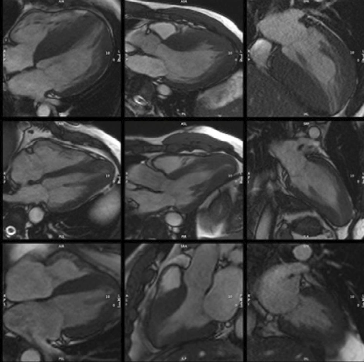
Hypertrophic Cardiomyopathy: A Review Using Magnetic Resonance Imaging

Raul Serra Valério et al. Marcio Sommer Bittencourt Marly M. Uellendahl

Abstract

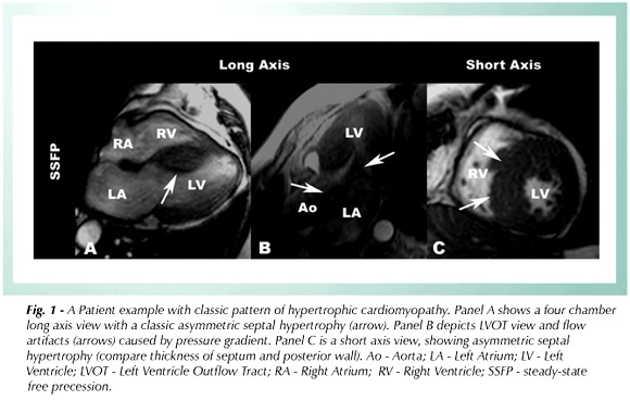
Hypertrophic cardiomyopathy, the most common genetic cardiopathy in the general population, is characterized by asymmetric left ventricular hypertrophy. However, the phenotypic changes in this cardiomyopathy extend beyond ventricular hypertrophy and include changes in the mitral valve apparatus, papillary muscles, and right ventricle. Due to the difficult differential diagnosis among multiple causes of hypertrophy, cardiac magnetic resonance has played a fundamental role in its diagnostic and prognostic evaluation; magnetic cine-resonance in defining the location and extent of hypertrophy; late enhancement, in the detection of areas of myocardial fibrosis; more recent techniques such as T1 mapping that assesses interstitial fibrosis and extracellular volume; and finally tissue tracking in the analysis of myocardial deformation.

  • Cardiac Imaging Techniques
  • Cardiomyopathy, Hypertrophic
  • Magnetic Resonance
Read Full Article
Creative Commons Attribution (CC BY) Creative Commons license
21/12/2022 DOI: 10.47593/2675-312X/20223504erer_15i
Figure 1
ABCHF
Review Article

Critical Analysis and Applicability of Imaging Methods in Monitoring and Diagnosing Ventricular Dysfunction in Patients with Cancer

Claudio Tinoco Mesquita et al. Marcelo Dantas Tavares de Melo Ariane Binoti Pacheco Leal André Luiz Cerqueira de Almeida

Abstract

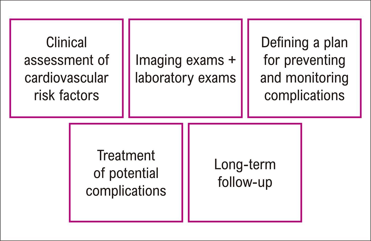
Cardio-oncology is a subspecialty of cardiology that has become necessary as a consequence of the favorable impact of cancer treatment, which increases patients’ survival rates, but makes them more prone to the cardiovascular side effects of cancer therapies in the short and long term. The presence of predisposing factors such as pre-existing cardiovascular disease, cardiovascular risk factors, genetic predisposition, previous antineoplastic therapies, and increased patient age is associated with a higher risk of cardiotoxicity in cancer treatment and may aggravate its complications. The use of imaging methods is fundamental in the management and detection of complications in cancer treatment. Echocardiography is considered the standard method for assessing left ventricular function and should be used in all patients. Magnetic resonance imaging is the best alternative for evaluation in patients with other associated conditions, especially advanced coronary disease, and in cases where it is difficult to obtain adequate echocardiographic images. Nuclear medicine offers options for patients for whom the use of echocardiography and magnetic resonance imaging is limited and for patients whose clinical and laboratory assessments conflict. Judicious use of imaging techniques leads to better patient outcomes during cancer treatment.

  • Cardiotoxicity
  • Computed Tomography
  • Echocardiography
  • Heart Failure
  • Magnetic Resonance
  • Scintigraphy
Read Full Article
Creative Commons Attribution (CC BY) Creative Commons license
18/12/2022 DOI: 10.36660/abchf.20220087 Views 2
Figure 1
ABCIC
Original Article

Left Ventricular Longitudinal Linear Displacement Versus Global Longitudinal Strain on Cardiovascular Magnetic Resonance

Rafael Almeida Fonseca et al. Sergio Marrone Ribeiro Clerio Francisco de Azevedo Filho Roney Sampaio … Flávio Tarasoutchi Carlos Eduardo Rochitte

Abstract

Background

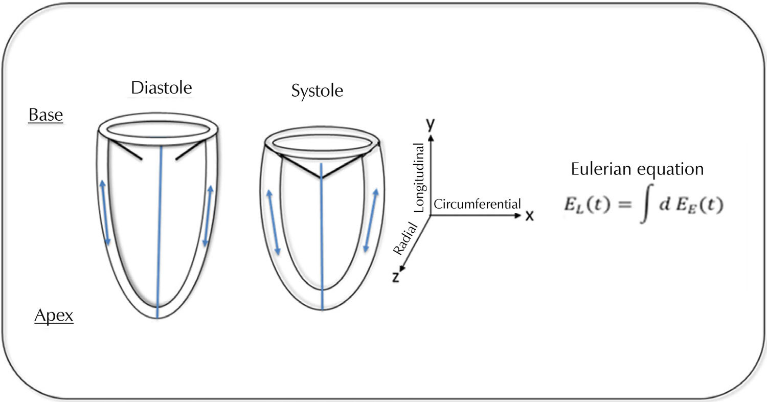
Left ventricular (LV) systolic diastolic function is prognostic in cardiovascular diseases and can be assessed via global longitudinal strain (GLS) on echocardiography and cardiac magnetic resonance (CMR). However, GLS by CMR requires the use of expensive software. Longitudinal linear displacement (LLD) may be a simple and inexpensive alternative to GLS, but the two have not been systematically compared.

Objective

To compare LLD with GLS and LV ejection fraction (LVEF) in aortic valve disease patients and controls.

Methods

We included 44 participants (26 with aortic valve disease, 19 controls). GLS was determined using CVI42 software (Circle Cardiovascular Imaging), while the LLD linear measurements of the distance between the base/apex of the LV included maximum displacement (MD), maximum velocity in early diastole (MVED), atrioventricular junction velocity in diastasis (VDS), and VDS/MVED ratio.

Results

DM and MVED were correlated with GLS (r=0.69 and r=0.65, respectively) and LVEF (r=0.47 and r=0.57, p<0.001 for both). DM and MVED showed areas under the receiver operating characteristic curve (AUC) of 0.88 and 0.91, and at the best cut-off point (-0.13 and 0.66), sensitivities of 72.43% and 57.14% and specificities of 80.65% and 87.10%, respectively, compared to GLS. Using LVEF as a reference, we obtained AUC of 0.70 and 0.82, and at the best cut-off point (-0.11 and 0.61), sensitivities of 75.00% and 50.00% and specificities of 72.97% and 78.38%, respectively.

Conclusion

LLD demonstrated similar performance to that of GLS. MD derived from LLD was the best parameter during systole, while MVED was the best during diastole. Our findings demonstrate the routine, quick, and inexpensive assessment of diastolic function on CMR.

  • Aortic Valve Insufficiency
  • Aortic Valve Stenosis
  • Diastole
  • Magnetic Resonance
  • Myocardial Contraction
Read Full Article
Creative Commons Attribution (CC BY) Creative Commons license
30/06/2022 DOI: 10.47593/2675-312X/20223502eabc304 Views 2
ABCIC
Editorial

What does the Cardiologist Expect from the Echocardiogram of a Transcatheter Aortic Valve Implant?

Gláucia Maria Moraes de Oliveira et al. Marcelo Antônio Cartaxo Queiroga Lopes

[…]

  • Aortic Valve Stenosis
  • Echocardiography
  • Magnetic Resonance
  • Tomography
Read Full Article
Creative Commons Attribution (CC BY) Creative Commons license
14/01/2022 DOI: 10.5935/2318-8219.20200024 Views 1
ABCIC
Editorial

My Approach to Assessment of Right Ventricle Function

Claudia Regina Pinheiro de Castro Grau

[…]

  • Echocardiography
  • Magnetic Resonance
  • Ventricular Dysfunction Right
Read Full Article
Creative Commons Attribution (CC BY) Creative Commons license
14/01/2022 DOI: 10.5935/2318-8219.20200045 Views 2
Figure 1
ABCIC
Original Article

Diagnostic yield of magnetic resonance imaging in heart failure with left ventricular dysfunction?

Ana Terra Fonseca Barreto et al. Mariana Lins Baptista Guedes Bezerra Rodrigo Morel Vieira de Melo Bruno Oliveira Isabella … Vanessa de Assis Reis Luiz Carlos Santana Passos

Abstract

Background:

Heart failure with reduced ejection fraction is responsible for half of heart failure cases worldwide and implicates in substantial morbidity and mortality. However, even with clinical history and physical examination associated with conventional complementary exams, many patients remain without etiological diagnosis. Cardiac magnetic resonance has offered the possibility to clarify a variable proportion of these cases.

Objective:

To verify how much cardiac magnetic resonance contributes to etiologic diagnosis of heart failure with left ventricular ejection fraction <50% in a specialized service.

Methods:

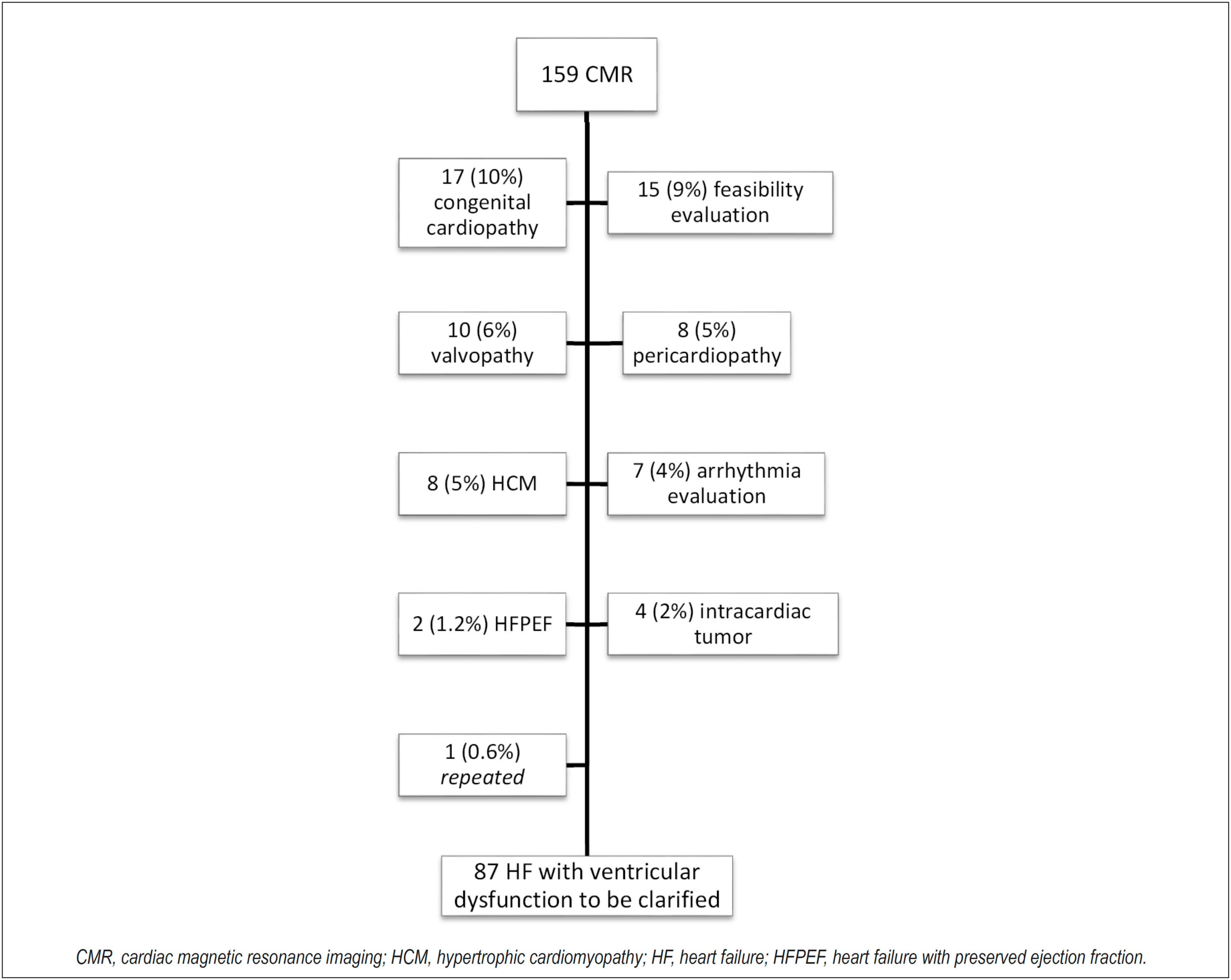
We included individuals referred to cardiac magnetic resonance with heart failure and left ventricular ejection fraction <50% by transthoracic echocardiogram, without defined etiology, from January, 2017 to June, 2018 in a tertiary hospital.

Results:

The sample consisted of 87 patients, with average age of 45±16 years, 49% male and left ventricular ejection fraction 32%±13. Of the patients, 55,3% had etiological diagnosis through cardiac magnetic resonance: 33,4% myocarditis, 11.5% non-compaction cardiomyopathy, 6.8% Chagas disease, and for hypertensive heart disease, amyloidosis and arrhythmogenic right ventricle dysplasia, 1,2% each. Late gadolinium enhancement was positive in 61% and non-ischemic pattern predominated (50,5%). Reverse remodeling occurred with normalization of ventricular function in 13% of patients.

Conclusion:

The performance of cardiac magnetic resonance in patients without etiologic diagnosis of HF with left ventricle dysfunction is clinically significant, since it contributed more than 50% of the time to the etiology and prognosis of patients. This positive impact occurred in a tertiary cardiology teaching service, so it is possible that in other circumstances the role of the cardiac magnetic resonance may be even greater than that here presented.

  • Cardiomyopathy, Dilated
  • Diagnostic Imaging
  • Heart Failure
  • Magnetic Resonance
Read Full Article
Creative Commons Attribution (CC BY) Creative Commons license
16/06/2021 DOI: 10.47593/2675-312X/20213401eabc108 Views 2
Figure 1
ABC
Image

Mitral Valve Mass in a Patient Suspected of Systemic Lupus: Tumor, Endocarditis or Both?

Thiago Sant’Anna Coutinho et al. Bárbara Cristina Rodrigues de Almeida Guilherme Dalcol Torres de Amorim Monica Zappa … Clara Weksler Cristiane da Cruz Lamas

Abstract

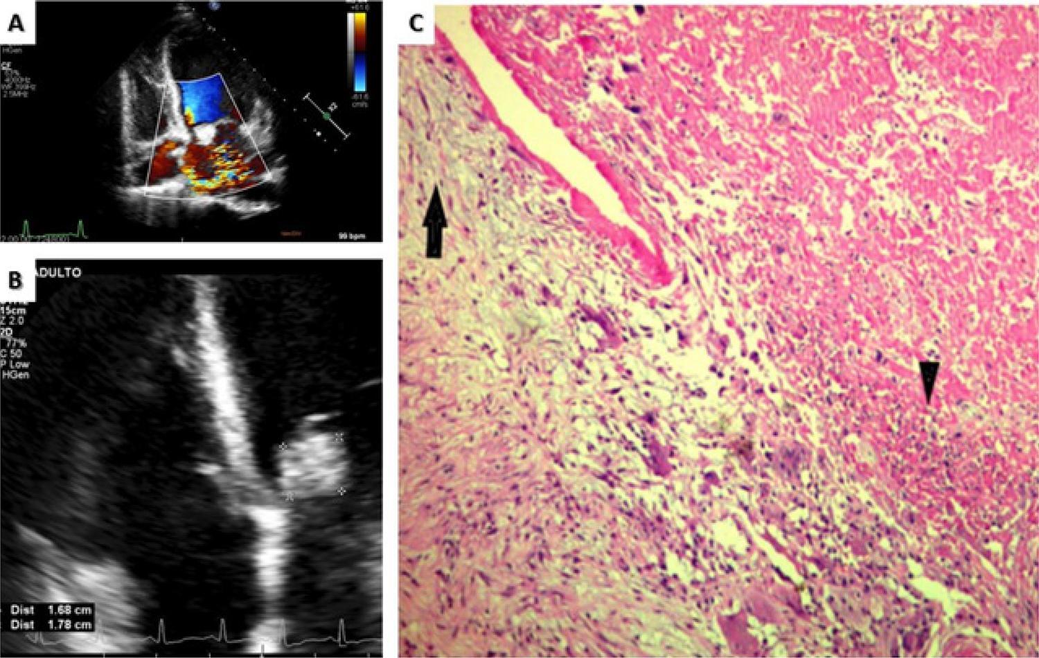
We present a case report of a patient with an infected mitral valve myxoma and a literature review on the subject. A 33-year-old female presented with a history of fever and dyspnea evolving over a few days. On admission, she had a lupus-like syndrome with positive blood cultures for Haemophilus species . Echocardiogram revealed a giant mass involving both mitral leaflets causing severe regurgitation, requiring biological mitral valve replacement. Microscopy showed an infected myxoma and the patient was discharged asymptomatic upon completion of antibiotics. She did well on follow-up. This is the sixth case of an infected mitral valve myxoma reported in the literature and the third case of a cardiac myxoma infected by the HACEK group. Exceedingly high incidence of embolic events makes prompt imaging, antibiotic therapy and surgery crucial for better outcomes. Time to diagnosis was much briefer than usually reported in other cases of HACEK endocarditis. Valve replacement was the most common surgical procedure and all patients from previous reports did well on follow-up.

  • Cardiac Neoplasms
  • Diagnostic Imaging
  • Echocardiography
  • Endocarditis
  • Lupus Erythematosus
  • Magnetic Resonance
  • Mitral Valve/pathology
  • Mitral Valve/surgery
Read Full Article
Creative Commons Attribution (CC BY) Creative Commons license
18/01/2021 DOI: 10.36660/abc.20200206
Figure 1
ABC
Original Article

Three-Dimensional Volumetric Assessment of Diastolic Function by Cardiac Magnetic Resonance Imaging: The Multi-Ethnic Study of Atherosclerosis (MESA)

Marcelo S Nacif et al. Andre L. C. Almeida Alistair A Young Brett R Cowan … Anderson C Armstrong Eunice Yang Christopher T Sibley W. Gregory Hundley Songtao Liu Joao AC Lima David A Bluemke

Abstract

Background:

Cardiac Magnetic Resonance is in need of a simple and robust method for diastolic function assessment that can be done with routine protocol sequences.

Objective:

To develop and validate a three-dimensional (3D) model-based volumetric assessment of diastolic function using cardiac magnetic resonance (CMR) imaging and compare the results obtained with the model with those obtained by echocardiography.

Methods:

The study participants provided written informed consent and were included if having undergone both echocardiography and cine steady-state free precession (SSFP) CMR on the same day. Guide points at the septal and lateral mitral annulus were used to define the early longitudinal relaxation rate (E’), while a time-volume curve from the 3D model was used to assess diastolic filling parameters. We determined the correlation between 3D CMR and echocardiography and the accuracy of CMR in classifying the diastolic function grade.

Results:

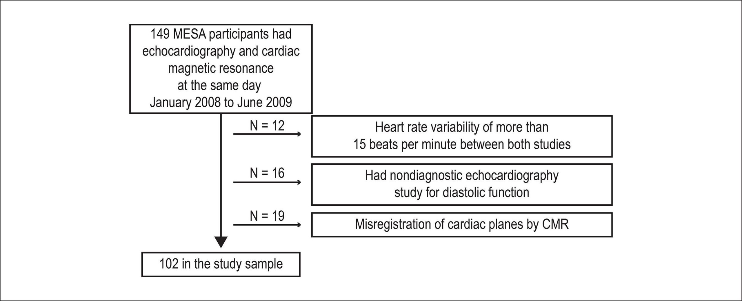
The study included 102 subjects. The E/A ratio by CMR was positively associated with the E/A ratio by echocardiography (r = 0.71, p < 0.0001). The early diastolic relaxation velocity by tissue Doppler and longitudinal relaxation rate for the lateral mitral annulus displacement were positively associated (p = 0.007), as were the ratio between Doppler E/e' and CMR E/E' (p = 0.01). CMR-determined normalized peak E (NE) and deceleration time (DT) were able to predict diastolic dysfunction (areas under the curve [AUCs] = 0.70 and 0.72, respectively). In addition, the lateral E/E' ratio showed good utility in identifying diastolic dysfunction (AUC = 0.80). Overall, echocardiography and CMR interobserver and intraobserver agreements were excellent (intraclass correlation coefficient range 0.72 – 0.97).

Conclusion:

3D modeling of standard cine CMR images was able to identify study subjects with reduced diastolic function and showed good reproducibility, suggesting a potential for a routine diastolic function assessment by CMR.

  • Echocardiography, Three-Dimensional
  • Evaluation
  • Imaging, Three-Dimensional
  • Magnetic Resonance
  • Ventricular Function
Read Full Article
Creative Commons Attribution (CC BY) Creative Commons license
25/05/2017 DOI: 10.5935/abc.20170063 Views 1
The diagnosis of left ventricular outflow tract obstruction in hypertrophic cardiomyopathy
ABC
Review Article

The diagnosis of left ventricular outflow tract obstruction in hypertrophic cardiomyopathy

Beatriz Piva e Mattos et al. Marco Antonio Rodrigues Torres Taiane Francieli Rebelatto Melina Silva de Loreto Fernando Luís Scolari

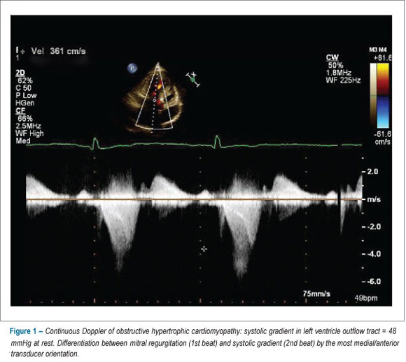
Hypertrophic cardiomyopathy is a prevalent genetic disease characterized by left ventricular hypertrophy, presenting dynamic obstruction of outflow tract with subaortic gradient happening at rest in 30% of the cases. It is attributed to the intricate interaction between the anterior mitral leaflet, the interventricular septum and altered flow vectors generated in left ventricle along with changes in outflow tract geometry. Mitral regurgitation in varying degrees is found with or without association with structural deformities of the valve apparatus. The exercise echocardiogram evidences latent obstruction easily induced by exercise in 60 to 75% of non-obstructive forms. The determination of the gradient under this condition must be considered in routine investigation of patients with mild or no obstruction at rest. The evaluation of hypertrophic cardiomyopathy incorporates methods based on the ultrasound image, which, along with MRI, allow recognizing ventricular obstruction generating mechanisms, thus facilitating the diagnosis and management of obstructive and latent obstructive forms.

  • Cardiomiopathy, hypertrophic
  • Echocardiography
  • Hypertrophy Left Ventricular
  • Magnetic Resonance
Read Full Article
Creative Commons Attribution (CC BY) Creative Commons license
04/09/2012 DOI: 10.1590/S0066-782X2012001000013 Views 1
Delayed enhancement cardiac magnetic resonance Imaging can identify the risk for ventricular tachycardia in chronic Chagas' heart disease
ABC
Artigo Original

Delayed enhancement cardiac magnetic resonance Imaging can identify the risk for ventricular tachycardia in chronic Chagas' heart disease

Ronaldo Peixoto de Mello et al. Gilberto Szarf Paulo Roberto Schvartzman Edson Minoru Nakano … Mariano Martinez Espinosa Denis Szejnfeld Verônica Fernandes João A. C. Lima Claudio Cirenza Angelo A.V. De Paola

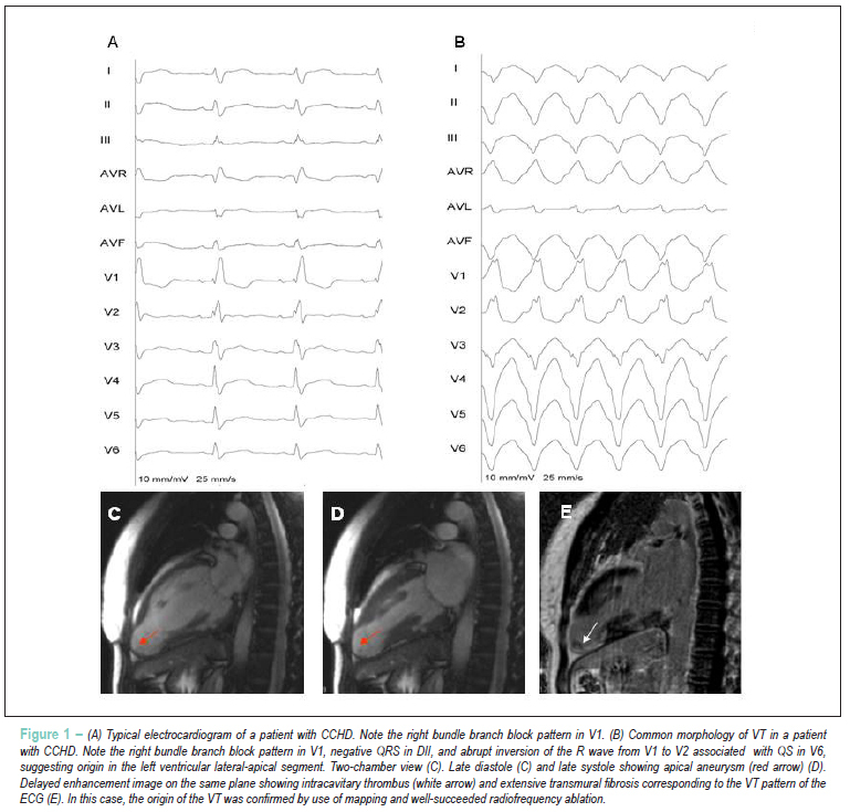
BACKGROUND: Invasive and non-invasive tests have been used to identify the risk of ventricular tachycardia (VT) in patients with chronic Chagas’ heart disease (CCHD). Cardiac magnetic resonance imaging (CMRI) using the delayed enhancement (DE) technique can be useful to select patients with global or segmentary ventricular dysfunction, with high degree of fibrosis and at higher risk for clinical VT. OBJECTIVE: To improve the identification of predictors of VT in patients with CCHD. METHOD: This study assessed 41 patients with CCHD [30 (72%) males; mean age, 55.1 ± 11.9 years]. Twenty-six patients had history of VT (VT group), and 15 had no VT (NVT group). All patients enrolled had DE and segmentary ventricular dysfunction. In each case, the following variables were determined: left ventricular volume; percentage of ventricular wall thickness impairment in each segment; and DE distribution. RESULTS: No statistical difference regarding the DE volume between both groups was observed: VT group = 30.0 ± 16.2%; NVT group = 21.7 ± 15.7%; p = 0.118. The probability of VT was greater in the presence of two or more contiguous transmural fibrosis areas, and that was a predictive factor of clinical VT (RR 4.1; p = 0,04). Agreement between observers was 100% regarding that criterion (p < 0.001). CONCLUSION: The identification of two or more segments of transmural DE by use of CMRI is associated with the occurrence of clinical VT in patients with CCHD. Thus, CMRI improved risk stratification in the population studied. (Arq Bras Cardiol. 2012; [online].ahead print, PP.0-0)

  • Chagas Cardiomyopathy
  • Magnetic Resonance
  • spectroscopy
  • Tachycardia
  • Ventricular
Read Full Article
Creative Commons Attribution (CC BY) Creative Commons license
29/03/2012 DOI: 10.1590/S0066-782X2012005000031
Cardiovascular magnetic resonance in hypertrophic cardiomyopathy
ABC
Revisão

Cardiovascular magnetic resonance in hypertrophic cardiomyopathy

Afonso Akio Shiozaki et al. Raymond J. Kim José Rodrigues Parga Eduardo Marinho Tassi … Edmundo Arteaga Carlos Eduardo Rochitte

REVIEW ARTICLE

Cardiovascular magnetic resonance in hypertrophic cardiomyopathy

[…]

  • Diagnosis
  • Hypertrophic Cardiomyopathy
  • Magnetic Resonance
  • Prognosis
Read Full Article
Creative Commons Attribution (CC BY) Creative Commons license
22/03/2007 DOI: 10.1590/S0066-782X2007000200018

How to cite this article

EndNote RIS

Electronic Document Format (Vancouver)

Valério RS, Bittencourt MS, Uellendahl MM. Hypertrophic Cardiomyopathy: A Review Using Magnetic Resonance Imaging. ABC Imagem Cardiovascular 2022;35(4):erer_15.

Electronic Document Format (ABNT)

Valério, Raul Serra; Bittencourt, Marcio Sommer; Uellendahl, Marly M.. Hypertrophic Cardiomyopathy: A Review Using Magnetic Resonance Imaging. ABC Imagem Cardiovascular, v. 35, n. 4, erer_15, Dec. 2022.

Electronic Document Format (APA)

Valério, R. S., Bittencourt, M. S., & Uellendahl, M. M. (2022). Hypertrophic Cardiomyopathy: A Review Using Magnetic Resonance Imaging. ABC Imagem Cardiovascular, 35(4), erer_15.

Electronic Document Format (ISO)

Valério, Raul Serra and Bittencourt, Marcio Sommer and Uellendahl, Marly M.. Hypertrophic Cardiomyopathy: A Review Using Magnetic Resonance Imaging. ABC Imagem Cardiovascular [online]. 2022, vol. 35, n. 4, [cited 2026-10-14], erer_15. Available from: <https://journals.cardiol.br/en/article/hypertrophic-cardiomyopathy-a-review-using-magnetic-resonance-imaging/>. ISSN 2318-8219

EndNote RIS

Electronic Document Format (Vancouver)

Mesquita CT, Melo MDT, Leal ABP, Almeida ALC. Critical Analysis and Applicability of Imaging Methods in Monitoring and Diagnosing Ventricular Dysfunction in Patients with Cancer. ABC: Heart Failure & Cardiomyopathy 2022;2(4):386-94.

Electronic Document Format (ABNT)

Mesquita, Claudio Tinoco; Melo, Marcelo Dantas Tavares de; Leal, Ariane Binoti Pacheco; Almeida, André Luiz Cerqueira de. Critical Analysis and Applicability of Imaging Methods in Monitoring and Diagnosing Ventricular Dysfunction in Patients with Cancer. ABC: Heart Failure & Cardiomyopathy, v. 2, n. 4, p. 386-394, Dec. 2022.

Electronic Document Format (APA)

Mesquita, C. T., Melo, M. D. T. , Leal, A. B. P., & Almeida, A. L. C. (2022). Critical Analysis and Applicability of Imaging Methods in Monitoring and Diagnosing Ventricular Dysfunction in Patients with Cancer. ABC: Heart Failure & Cardiomyopathy, 2(4), 386-394.

Electronic Document Format (ISO)

Mesquita, Claudio Tinoco and Melo, Marcelo Dantas Tavares de and Leal, Ariane Binoti Pacheco and Almeida, André Luiz Cerqueira de. Critical Analysis and Applicability of Imaging Methods in Monitoring and Diagnosing Ventricular Dysfunction in Patients with Cancer. ABC: Heart Failure & Cardiomyopathy [online]. 2022, vol. 2, n. 4, [cited 2026-10-14], pp. 386-394. Available from: <https://journals.cardiol.br/en/article/critical-analysis-and-applicability-of-imaging-methods-in-monitoring-and-diagnosing-ventricular-dysfunction-in-patients-with-cancer/>

EndNote RIS

Electronic Document Format (Vancouver)

Fonseca RA, Ribeiro SM, Azevedo Filho CF, Sampaio R, Tarasoutchi F, Rochitte CE. Left Ventricular Longitudinal Linear Displacement Versus Global Longitudinal Strain on Cardiovascular Magnetic Resonance. ABC Imagem Cardiovascular 2022;35(2):eabc304.

Electronic Document Format (ABNT)

Fonseca, Rafael Almeida; Ribeiro, Sergio Marrone; Azevedo Filho, Clerio Francisco de; Sampaio, Roney; Tarasoutchi, Flávio; Rochitte, Carlos Eduardo. Left Ventricular Longitudinal Linear Displacement Versus Global Longitudinal Strain on Cardiovascular Magnetic Resonance. ABC Imagem Cardiovascular, v. 35, n. 2, eabc304, Jun. 2022.

Electronic Document Format (APA)

Fonseca, R. A., Ribeiro, S. M., Azevedo Filho, C. F. , Sampaio, R., Tarasoutchi, F., & Rochitte, C. E. (2022). Left Ventricular Longitudinal Linear Displacement Versus Global Longitudinal Strain on Cardiovascular Magnetic Resonance. ABC Imagem Cardiovascular, 35(2), eabc304.

Electronic Document Format (ISO)

Fonseca, Rafael Almeida and Ribeiro, Sergio Marrone and Azevedo Filho, Clerio Francisco de and Sampaio, Roney and Tarasoutchi, Flávio and Rochitte, Carlos Eduardo. Left Ventricular Longitudinal Linear Displacement Versus Global Longitudinal Strain on Cardiovascular Magnetic Resonance. ABC Imagem Cardiovascular [online]. 2022, vol. 35, n. 2, [cited 2026-10-14], eabc304. Available from: <https://journals.cardiol.br/en/article/left-ventricular-longitudinal-linear-displacement-versus-global-longitudinal-strain-on-cardiovascular-magnetic-resonance/>. ISSN 2318-8219

EndNote RIS

Electronic Document Format (Vancouver)

Oliveira GMM, Lopes MACQ. What does the Cardiologist Expect from the Echocardiogram of a Transcatheter Aortic Valve Implant?. Arquivos Brasileiros de Cardiologia Imagem cardiovascular 2022;33(2):1-3.

Electronic Document Format (ABNT)

Oliveira, Gláucia Maria Moraes de; Lopes, Marcelo Antônio Cartaxo Queiroga. What does the Cardiologist Expect from the Echocardiogram of a Transcatheter Aortic Valve Implant?. Arquivos Brasileiros de Cardiologia Imagem cardiovascular, v. 33, n. 2, p. 1-3, Jan. 2022.

Electronic Document Format (APA)

Oliveira, G. M. M. , & Lopes, M. A. C. Q. (2022). What does the Cardiologist Expect from the Echocardiogram of a Transcatheter Aortic Valve Implant?. Arquivos Brasileiros de Cardiologia Imagem cardiovascular, 33(2), 1-3.

Electronic Document Format (ISO)

Oliveira, Gláucia Maria Moraes de and Lopes, Marcelo Antônio Cartaxo Queiroga. What does the Cardiologist Expect from the Echocardiogram of a Transcatheter Aortic Valve Implant?. Arquivos Brasileiros de Cardiologia Imagem cardiovascular [online]. 2022, vol. 33, n. 2, [cited 2026-10-14], pp. 1-3. Available from: <https://journals.cardiol.br/en/article/what-does-the-cardiologist-expect-from-the-echocardiogram-of-a-transcatheter-aortic-valve-implant/>. ISSN 2318-8219

EndNote RIS

Electronic Document Format (Vancouver)

Grau CRPC. My Approach to Assessment of Right Ventricle Function. Arquivos Brasileiros de Cardiologia Imagem cardiovascular 2022;33(3):1-5.

Electronic Document Format (ABNT)

Grau, Claudia Regina Pinheiro de Castro. My Approach to Assessment of Right Ventricle Function. Arquivos Brasileiros de Cardiologia Imagem cardiovascular, v. 33, n. 3, p. 1-5, Jan. 2022.

Electronic Document Format (APA)

Grau, C. R. P. C. (2022). My Approach to Assessment of Right Ventricle Function. Arquivos Brasileiros de Cardiologia Imagem cardiovascular, 33(3), 1-5.

Electronic Document Format (ISO)

Grau, Claudia Regina Pinheiro de Castro. My Approach to Assessment of Right Ventricle Function. Arquivos Brasileiros de Cardiologia Imagem cardiovascular [online]. 2022, vol. 33, n. 3, [cited 2026-10-14], pp. 1-5. Available from: <https://journals.cardiol.br/en/article/my-approach-to-assessment-of-right-ventricle-function/>. ISSN 2318-8219

EndNote RIS

Electronic Document Format (Vancouver)

Barreto ATF, Bezerra MLBG, Melo RMV, Isabella BO, Reis VA, Passos LCS. Diagnostic yield of magnetic resonance imaging in heart failure with left ventricular dysfunction?. ABC Imagem Cardiovascular 2021;34(1):eabc108.

Electronic Document Format (ABNT)

Barreto, Ana Terra Fonseca; Bezerra, Mariana Lins Baptista Guedes; Melo, Rodrigo Morel Vieira de; Isabella, Bruno Oliveira; Reis, Vanessa de Assis; Passos, Luiz Carlos Santana. Diagnostic yield of magnetic resonance imaging in heart failure with left ventricular dysfunction?. ABC Imagem Cardiovascular, v. 34, n. 1, eabc108, Jun. 2021.

Electronic Document Format (APA)

Barreto, A. T. F., Bezerra, M. L. B. G., Melo, R. M. V. , Isabella, B. O., Reis, V. A., & Passos, L. C. S. (2021). Diagnostic yield of magnetic resonance imaging in heart failure with left ventricular dysfunction?. ABC Imagem Cardiovascular, 34(1), eabc108.

Electronic Document Format (ISO)

Barreto, Ana Terra Fonseca and Bezerra, Mariana Lins Baptista Guedes and Melo, Rodrigo Morel Vieira de and Isabella, Bruno Oliveira and Reis, Vanessa de Assis and Passos, Luiz Carlos Santana. Diagnostic yield of magnetic resonance imaging in heart failure with left ventricular dysfunction?. ABC Imagem Cardiovascular [online]. 2021, vol. 34, n. 1, [cited 2026-10-14], eabc108. Available from: <https://journals.cardiol.br/en/article/diagnostic-yield-of-magnetic-resonance-imaging-in-heart-failure-with-left-ventricular-dysfunction/>. ISSN 2318-8219

EndNote RIS

Electronic Document Format (Vancouver)

Coutinho TS, Almeida BCR, Amorim GDT, Zappa M, Weksler C, Lamas CC. Mitral Valve Mass in a Patient Suspected of Systemic Lupus: Tumor, Endocarditis or Both?. Arquivos Brasileiros de Cardiologia 2021;115(6):1201-4.

Electronic Document Format (ABNT)

Coutinho, Thiago Sant’Anna; Almeida, Bárbara Cristina Rodrigues de; Amorim, Guilherme Dalcol Torres de; Zappa, Monica; Weksler, Clara; Lamas, Cristiane da Cruz. Mitral Valve Mass in a Patient Suspected of Systemic Lupus: Tumor, Endocarditis or Both?. Arquivos Brasileiros de Cardiologia, v. 115, n. 6, p. 1201-1204, Jan. 2021.

Electronic Document Format (APA)

Coutinho, T. S., Almeida, B. C. R. , Amorim, G. D. T. , Zappa, M., Weksler, C., & Lamas, C. C. (2021). Mitral Valve Mass in a Patient Suspected of Systemic Lupus: Tumor, Endocarditis or Both?. Arquivos Brasileiros de Cardiologia, 115(6), 1201-1204.

Electronic Document Format (ISO)

Coutinho, Thiago Sant’Anna and Almeida, Bárbara Cristina Rodrigues de and Amorim, Guilherme Dalcol Torres de and Zappa, Monica and Weksler, Clara and Lamas, Cristiane da Cruz. Mitral Valve Mass in a Patient Suspected of Systemic Lupus: Tumor, Endocarditis or Both?. Arquivos Brasileiros de Cardiologia [online]. 2021, vol. 115, n. 6, [cited 2026-10-14], pp. 1201-1204. Available from: <https://journals.cardiol.br/en/article/mitral-valve-mass-in-a-patient-suspected-of-systemic-lupus-tumor-endocarditis-or-both/>. ISSN 0066-782X

EndNote RIS

Electronic Document Format (Vancouver)

Nacif MS, Almeida ALC, Young AA, Cowan BR, Armstrong AC, Yang E, Sibley CT, et al. Three-Dimensional Volumetric Assessment of Diastolic Function by Cardiac Magnetic Resonance Imaging: The Multi-Ethnic Study of Atherosclerosis (MESA). Arquivos Brasileiros de Cardiologia 2017;108(6):552-63.

Electronic Document Format (ABNT)

Nacif, Marcelo S; Almeida, Andre L. C.; Young, Alistair A; Cowan, Brett R; Armstrong, Anderson C; Yang, Eunice; Sibley, Christopher T; Hundley, W. Gregory; Liu, Songtao; Lima, Joao AC; Bluemke, David A. Three-Dimensional Volumetric Assessment of Diastolic Function by Cardiac Magnetic Resonance Imaging: The Multi-Ethnic Study of Atherosclerosis (MESA). Arquivos Brasileiros de Cardiologia, v. 108, n. 6, p. 552-563, May. 2017.

Electronic Document Format (APA)

Nacif, M. S., Almeida, A. L. C., Young, A. A., Cowan, B. R., Armstrong, A. C., Yang, E., Sibley, C. T., Hundley, W. G., Liu, S., Lima, J. A., & Bluemke, D. A. (2017). Three-Dimensional Volumetric Assessment of Diastolic Function by Cardiac Magnetic Resonance Imaging: The Multi-Ethnic Study of Atherosclerosis (MESA). Arquivos Brasileiros de Cardiologia, 108(6), 552-563.

Electronic Document Format (ISO)

Nacif, Marcelo S and Almeida, Andre L. C. and Young, Alistair A and Cowan, Brett R and Armstrong, Anderson C and Yang, Eunice and Sibley, Christopher T and Hundley, W. Gregory and Liu, Songtao and Lima, Joao AC and Bluemke, David A. Three-Dimensional Volumetric Assessment of Diastolic Function by Cardiac Magnetic Resonance Imaging: The Multi-Ethnic Study of Atherosclerosis (MESA). Arquivos Brasileiros de Cardiologia [online]. 2017, vol. 108, n. 6, [cited 2026-10-14], pp. 552-563. Available from: <https://journals.cardiol.br/en/article/three-dimensional-volumetric-assessment-of-diastolic-function-by-cardiac-magnetic-resonance-imaging-the-multi-ethnic-study-of-atherosclerosis-mesa/>. ISSN 0066-782X

EndNote RIS

Electronic Document Format (Vancouver)

Mattos BP, Torres MAR, Rebelatto TF, Loreto MS, Scolari FL. The diagnosis of left ventricular outflow tract obstruction in hypertrophic cardiomyopathy. Arquivos Brasileiros de Cardiologia 2012;99(1):665-7.

Electronic Document Format (ABNT)

Mattos, Beatriz Piva e; Torres, Marco Antonio Rodrigues; Rebelatto, Taiane Francieli; Loreto, Melina Silva de; Scolari, Fernando Luís. The diagnosis of left ventricular outflow tract obstruction in hypertrophic cardiomyopathy. Arquivos Brasileiros de Cardiologia, v. 99, n. 1, p. 665-675, Sep. 2012.

Electronic Document Format (APA)

Mattos, B. P. , Torres, M. A. R., Rebelatto, T. F., Loreto, M. S. , & Scolari, F. L. (2012). The diagnosis of left ventricular outflow tract obstruction in hypertrophic cardiomyopathy. Arquivos Brasileiros de Cardiologia, 99(1), 665-675.

Electronic Document Format (ISO)

Mattos, Beatriz Piva e and Torres, Marco Antonio Rodrigues and Rebelatto, Taiane Francieli and Loreto, Melina Silva de and Scolari, Fernando Luís. The diagnosis of left ventricular outflow tract obstruction in hypertrophic cardiomyopathy. Arquivos Brasileiros de Cardiologia [online]. 2012, vol. 99, n. 1, [cited 2026-10-14], pp. 665-675. Available from: <https://journals.cardiol.br/en/article/the-diagnosis-of-left-ventricular-outflow-tract-obstruction-in-hypertrophic-cardiomyopathy/>. ISSN 0066-782X

EndNote RIS

Electronic Document Format (Vancouver)

Mello RP, Szarf G, Schvartzman PR, Nakano EM, Espinosa MM, Szejnfeld D, Fernandes V, et al. Delayed enhancement cardiac magnetic resonance Imaging can identify the risk for ventricular tachycardia in chronic Chagas’ heart disease. Arquivos Brasileiros de Cardiologia 2012;98(5):421-30.

Electronic Document Format (ABNT)

Mello, Ronaldo Peixoto de; Szarf, Gilberto; Schvartzman, Paulo Roberto; Nakano, Edson Minoru; Espinosa, Mariano Martinez; Szejnfeld, Denis; Fernandes, Verônica; Lima, João A. C.; Cirenza, Claudio; Paola, Angelo A.V. De. Delayed enhancement cardiac magnetic resonance Imaging can identify the risk for ventricular tachycardia in chronic Chagas’ heart disease. Arquivos Brasileiros de Cardiologia, v. 98, n. 5, p. 421-430, Mar. 2012.

Electronic Document Format (APA)

Mello, R. P. , Szarf, G., Schvartzman, P. R., Nakano, E. M., Espinosa, M. M., Szejnfeld, D., Fernandes, V., Lima, J. A. C., Cirenza, C., & Paola, A. A. D. (2012). Delayed enhancement cardiac magnetic resonance Imaging can identify the risk for ventricular tachycardia in chronic Chagas’ heart disease. Arquivos Brasileiros de Cardiologia, 98(5), 421-430.

Electronic Document Format (ISO)

Mello, Ronaldo Peixoto de and Szarf, Gilberto and Schvartzman, Paulo Roberto and Nakano, Edson Minoru and Espinosa, Mariano Martinez and Szejnfeld, Denis and Fernandes, Verônica and Lima, João A. C. and Cirenza, Claudio and Paola, Angelo A.V. De. Delayed enhancement cardiac magnetic resonance Imaging can identify the risk for ventricular tachycardia in chronic Chagas’ heart disease. Arquivos Brasileiros de Cardiologia [online]. 2012, vol. 98, n. 5, [cited 2026-10-14], pp. 421-430. Available from: <https://journals.cardiol.br/en/article/delayed-enhancement-cardiac-magnetic-resonance-imaging-can-identify-the-risk-for-ventricular-tachycardia-in-chronic-chagas-heart-disease/>. ISSN 0066-782X

EndNote RIS

Electronic Document Format (Vancouver)

Shiozaki AA, Kim RJ, Parga JR, Tassi EM, Arteaga E, Rochitte CE. Cardiovascular magnetic resonance in hypertrophic cardiomyopathy. Arquivos Brasileiros de Cardiologia 2007;88(2):243-8.

Electronic Document Format (ABNT)

Shiozaki, Afonso Akio; Kim, Raymond J.; Parga, José Rodrigues; Tassi, Eduardo Marinho; Arteaga, Edmundo; Rochitte, Carlos Eduardo. Cardiovascular magnetic resonance in hypertrophic cardiomyopathy. Arquivos Brasileiros de Cardiologia, v. 88, n. 2, p. 243-248, Mar. 2007.

Electronic Document Format (APA)

Shiozaki, A. A., Kim, R. J., Parga, J. R., Tassi, E. M., Arteaga, E., & Rochitte, C. E. (2007). Cardiovascular magnetic resonance in hypertrophic cardiomyopathy. Arquivos Brasileiros de Cardiologia, 88(2), 243-248.

Electronic Document Format (ISO)

Shiozaki, Afonso Akio and Kim, Raymond J. and Parga, José Rodrigues and Tassi, Eduardo Marinho and Arteaga, Edmundo and Rochitte, Carlos Eduardo. Cardiovascular magnetic resonance in hypertrophic cardiomyopathy. Arquivos Brasileiros de Cardiologia [online]. 2007, vol. 88, n. 2, [cited 2026-10-14], pp. 243-248. Available from: <https://journals.cardiol.br/en/article/cardiovascular-magnetic-resonance-in-hypertrophic-cardiomyopathy/>. ISSN 0066-782X

  • About ABC Family
  • Internal Regulation
  • Editorial Board
  • Brazilian Society of Cardiology (SBC)

Issues

SBC Guidelines

  • Videos
  • Podcasts
  • News