ABC Family Arquivos Brasileiros de Cardiologia International Journal of Cardiovascular Sciences ABC Heart Failure & Cardiomyopathy ABC Imagem Cardiovascular
Português English
ABC Family
Português English
  • About
    • About ABC Family
    • Internal Regulation
    • Editorial Board
    • Brazilian Society of Cardiology (SBC)
  • Issues
  • SBC Guidelines
  • Content
    • Videos
    • Podcasts
    • News
  • About
    • About ABC Family
    • Internal Regulation
    • Editorial Board
    • Brazilian Society of Cardiology (SBC)
  • Issues
  • SBC Guidelines
  • Content
    • Videos
    • Podcasts
    • News

ABC Family

  • ABC Family
  • Arquivos Brasileiros de Cardiologia
  • International Journal of Cardiovascular Sciences
  • ABC Heart Failure & Cardiomyopathy
  • ABC Imagem Cardiovascular
Search in

No content found.

Journals
Ordering
Article types
Others

  1. Home
  2. Keyword: Imaging, Three-Dimensional
Figure 1
ABCIC
Review Article

Stress Echocardiography Using Real-Time 3D Imaging

José Roberto Matos-Souza et al. Guilherme De Rossi

Abstract

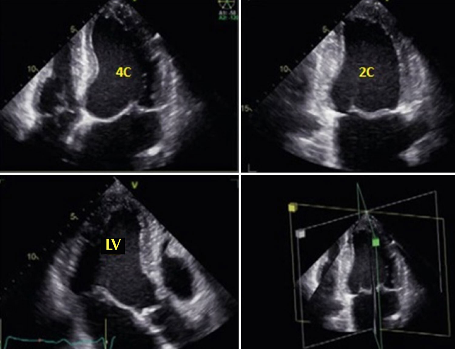
The need to examine the heart using a three-dimensional (3D) tool is not new. This complex and dynamic organ has always required 3D and real-time understanding. Without this feature, the examiner has to transform two-dimensional images to understand its volume, which requires complex knowledge and approximation interactions. Echocardiography was invented three decades ago, and its improvements resulted in commercial products at the beginning of the century. Some studies demonstrate 3D equivalence with gains in handling the necessary time. We use triplane modalities in our routine, with time gain and less stress on the examiner’s upper limb. Thus, 3D examinations can answer more complex questions and provide a more geometric approach to contraction, with thickening being analyzed in the background.

  • Echocardiography, Stress
  • Echocardiography, Three-Dimensional
  • Imaging, Three-Dimensional
Read Full Article
Creative Commons Attribution (CC BY) Creative Commons license
07/07/2022 DOI: 10.47593/2675-312X/20223503erer_07
Figure 1
ABC
Original Article

Three-Dimensional Volumetric Assessment of Diastolic Function by Cardiac Magnetic Resonance Imaging: The Multi-Ethnic Study of Atherosclerosis (MESA)

Marcelo S Nacif et al. Andre L. C. Almeida Alistair A Young Brett R Cowan … Anderson C Armstrong Eunice Yang Christopher T Sibley W. Gregory Hundley Songtao Liu Joao AC Lima David A Bluemke

Abstract

Background:

Cardiac Magnetic Resonance is in need of a simple and robust method for diastolic function assessment that can be done with routine protocol sequences.

Objective:

To develop and validate a three-dimensional (3D) model-based volumetric assessment of diastolic function using cardiac magnetic resonance (CMR) imaging and compare the results obtained with the model with those obtained by echocardiography.

Methods:

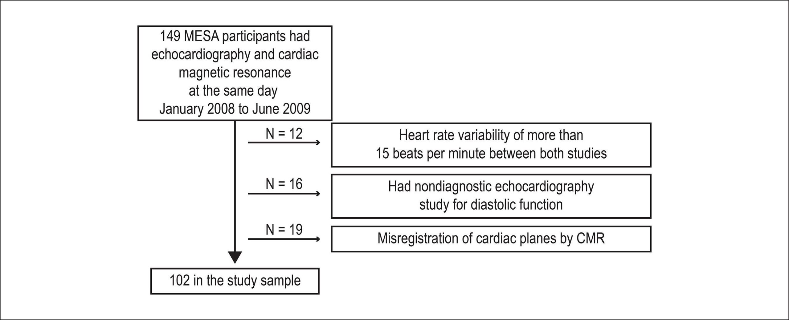
The study participants provided written informed consent and were included if having undergone both echocardiography and cine steady-state free precession (SSFP) CMR on the same day. Guide points at the septal and lateral mitral annulus were used to define the early longitudinal relaxation rate (E’), while a time-volume curve from the 3D model was used to assess diastolic filling parameters. We determined the correlation between 3D CMR and echocardiography and the accuracy of CMR in classifying the diastolic function grade.

Results:

The study included 102 subjects. The E/A ratio by CMR was positively associated with the E/A ratio by echocardiography (r = 0.71, p < 0.0001). The early diastolic relaxation velocity by tissue Doppler and longitudinal relaxation rate for the lateral mitral annulus displacement were positively associated (p = 0.007), as were the ratio between Doppler E/e' and CMR E/E' (p = 0.01). CMR-determined normalized peak E (NE) and deceleration time (DT) were able to predict diastolic dysfunction (areas under the curve [AUCs] = 0.70 and 0.72, respectively). In addition, the lateral E/E' ratio showed good utility in identifying diastolic dysfunction (AUC = 0.80). Overall, echocardiography and CMR interobserver and intraobserver agreements were excellent (intraclass correlation coefficient range 0.72 – 0.97).

Conclusion:

3D modeling of standard cine CMR images was able to identify study subjects with reduced diastolic function and showed good reproducibility, suggesting a potential for a routine diastolic function assessment by CMR.

  • Echocardiography, Three-Dimensional
  • Evaluation
  • Imaging, Three-Dimensional
  • Magnetic Resonance
  • Ventricular Function
Read Full Article
Creative Commons Attribution (CC BY) Creative Commons license
25/05/2017 DOI: 10.5935/abc.20170063 Views 1
Figure 1
ABC
Image

Three-Dimensional Printing Model-Guided Percutaneous Closure of Atrial Septal Defect

Hongxing Luo et al. Yu Xu Zhongmin Wang Yuhao Liu Chuanyu Gao

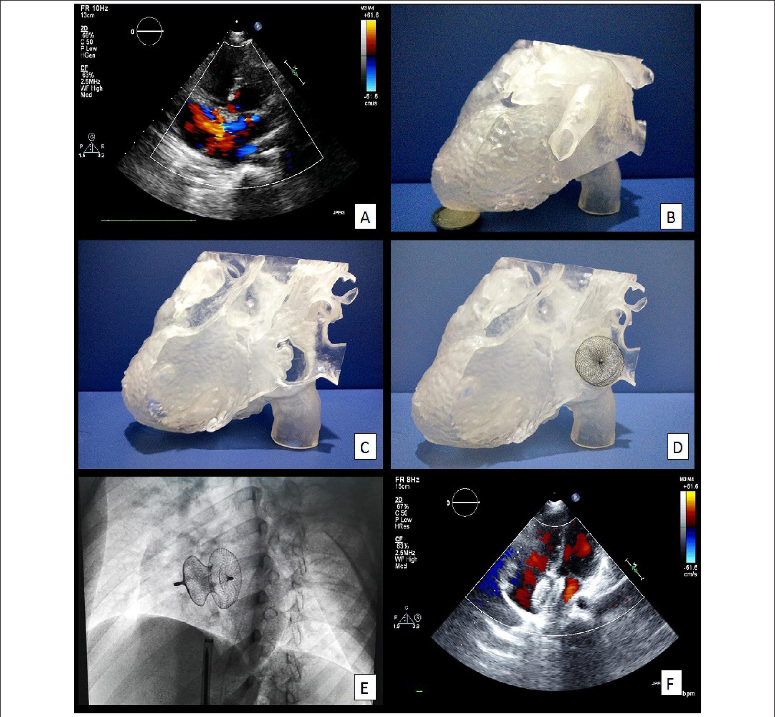
A 32-year-old female with a 2-year history of chest distress was admitted to our department due to exacerbation for 3 days. On physical examination, we found fixed splitting second heart sounds on the patient’s pulmonic area. An echocardiography was performed and showed a 15-mm atrial septal defect of the inferior vena cava type. After obtaining the patient’s consent, a three-dimensional printing cardiac model was printed out. We tried various sizes of ASD occluders on the model to completely cover the defect, which indicated that a 28-mm occluder was appropriate. Thus, we placed a 28-mm ASD occluder during the operation and succeeded after only one attempt. The patient was re-assessed by echocardiography, which showed a favorable position of the ASD occluder without any left-to-right shunt.

Three-dimensional printing (3D printing) is a new technology that converts two-dimensional medical images into a tangible object, allowing not only a comprehensive view of the cardiac anatomical structures but also preoperative simulation to choose the optimal size of ASD occluder. Although it has been applied to orthopedics, general surgery and so on, the use of 3D printing in cardiology is still at its infancy. Our case showed the feasibility of using a 3D printing cardiac model to guide the percutaneous closure of ASD. It is likely to increase the success rate and reduce the operation time for interventional cardiology, especially the complex ASD cases, and more studies should be carried out to extend its fields of application.

[…]

  • Echocardiography/methods
  • Heart Septal Defects, Atrial / surgery
  • Imaging, Three-Dimensional
Read Full Article
Creative Commons Attribution (CC BY) Creative Commons license
01/05/2017 DOI: 10.5935/abc.20170051
Reference curve of the fetal ventricular septum area by the STIC method: preliminary study
ABC
Artigo Original

Reference curve of the fetal ventricular septum area by the STIC method: preliminary study

Liliam Cristine Rolo et al. Luciano Marcondes Machado Nardozza Edward Araujo Júnior Christiane Simioni … Marina Maccagnano Zamith Antonio Fernandes Moron

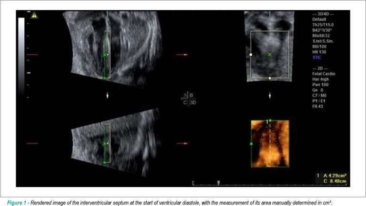
BACKGROUND: Early detection of septal changes such as septal hypertrophy commonly present in fetuses of diabetic mothers would help reduce the high rates of infant mortality. OBJECTIVE: Determine reference ranges for the fetal ventricular septal area through three-dimensional ultrasound (US3D) using the STIC method (Spatio-Temporal Image Correlation). METHODS: We conducted a cross-sectional study with 69 pregnant women between the 18th and 33rd weeks of pregnancy. We used as a reference the four-chamber plane with the ROI (Region of Interest) positioned from the ventricles; the septum area were manually marked. To assess the correlation of the interventricular septum area with gestational age (GA), we constructed scatter plots and calculated Pearson’s correlation coefficient (r), and the adjustment was performed by the coefficient of determination (R²). We calculated averages, medians, standard deviations (sd), as well as maximum and minimum values. To calculate the intraobserver reproducibility, we used the intraclass correlation coefficient (ICC). The interventricular septum thickness was measured and it was correlated with gestational age and the septal area rendered in 52 patients using the ICC. RESULTS: The interventricular septum area was highly correlated with gestational age (r = 0.81), and the average increased from 0.47 cm² in the 18th week to 2.42 cm² in the 33rd of gestation. The intraobserver reproducibility was excellent with ICC = 0.994. No significant correlation was observed between the interventricular septum measurement and the GA (R² = 0.200), as well as there was no correlation with the septal area rendered with ICC = 0.150. CONCLUSION: Reference intervals for the interventricular septum area between the 18th and the 33rd pregnancy week were determined to be highly reproducible.

  • fetal heart
  • Imaging, Three-Dimensional
  • Pregnancy
  • Reference Values
  • ventricular septum
Read Full Article
Creative Commons Attribution (CC BY) Creative Commons license
01/04/2011 DOI: 10.1590/S0066-782X2011005000036 Views 1

How to cite this article

EndNote RIS

Electronic Document Format (Vancouver)

Matos-Souza JR, Rossi GD. Stress Echocardiography Using Real-Time 3D Imaging. ABC Imagem Cardiovascular 2022;35(3):erer_07.

Electronic Document Format (ABNT)

Matos-Souza, José Roberto; Rossi, Guilherme De. Stress Echocardiography Using Real-Time 3D Imaging. ABC Imagem Cardiovascular, v. 35, n. 3, erer_07, Jul. 2022.

Electronic Document Format (APA)

Matos-Souza, J. R., & Rossi, G. D. (2022). Stress Echocardiography Using Real-Time 3D Imaging. ABC Imagem Cardiovascular, 35(3), erer_07.

Electronic Document Format (ISO)

Matos-Souza, José Roberto and Rossi, Guilherme De. Stress Echocardiography Using Real-Time 3D Imaging. ABC Imagem Cardiovascular [online]. 2022, vol. 35, n. 3, [cited 2026-10-14], erer_07. Available from: <https://journals.cardiol.br/en/article/stress-echocardiography-using-real-time-3d-imaging/>. ISSN 2318-8219

EndNote RIS

Electronic Document Format (Vancouver)

Nacif MS, Almeida ALC, Young AA, Cowan BR, Armstrong AC, Yang E, Sibley CT, et al. Three-Dimensional Volumetric Assessment of Diastolic Function by Cardiac Magnetic Resonance Imaging: The Multi-Ethnic Study of Atherosclerosis (MESA). Arquivos Brasileiros de Cardiologia 2017;108(6):552-63.

Electronic Document Format (ABNT)

Nacif, Marcelo S; Almeida, Andre L. C.; Young, Alistair A; Cowan, Brett R; Armstrong, Anderson C; Yang, Eunice; Sibley, Christopher T; Hundley, W. Gregory; Liu, Songtao; Lima, Joao AC; Bluemke, David A. Three-Dimensional Volumetric Assessment of Diastolic Function by Cardiac Magnetic Resonance Imaging: The Multi-Ethnic Study of Atherosclerosis (MESA). Arquivos Brasileiros de Cardiologia, v. 108, n. 6, p. 552-563, May. 2017.

Electronic Document Format (APA)

Nacif, M. S., Almeida, A. L. C., Young, A. A., Cowan, B. R., Armstrong, A. C., Yang, E., Sibley, C. T., Hundley, W. G., Liu, S., Lima, J. A., & Bluemke, D. A. (2017). Three-Dimensional Volumetric Assessment of Diastolic Function by Cardiac Magnetic Resonance Imaging: The Multi-Ethnic Study of Atherosclerosis (MESA). Arquivos Brasileiros de Cardiologia, 108(6), 552-563.

Electronic Document Format (ISO)

Nacif, Marcelo S and Almeida, Andre L. C. and Young, Alistair A and Cowan, Brett R and Armstrong, Anderson C and Yang, Eunice and Sibley, Christopher T and Hundley, W. Gregory and Liu, Songtao and Lima, Joao AC and Bluemke, David A. Three-Dimensional Volumetric Assessment of Diastolic Function by Cardiac Magnetic Resonance Imaging: The Multi-Ethnic Study of Atherosclerosis (MESA). Arquivos Brasileiros de Cardiologia [online]. 2017, vol. 108, n. 6, [cited 2026-10-14], pp. 552-563. Available from: <https://journals.cardiol.br/en/article/three-dimensional-volumetric-assessment-of-diastolic-function-by-cardiac-magnetic-resonance-imaging-the-multi-ethnic-study-of-atherosclerosis-mesa/>. ISSN 0066-782X

EndNote RIS

Electronic Document Format (Vancouver)

Luo H, Xu Y, Wang Z, Liu Y, Gao C. Three-Dimensional Printing Model-Guided Percutaneous Closure of Atrial Septal Defect. Arquivos Brasileiros de Cardiologia 2017;108(5):484-5.

Electronic Document Format (ABNT)

Luo, Hongxing; Xu, Yu; Wang, Zhongmin; Liu, Yuhao; Gao, Chuanyu. Three-Dimensional Printing Model-Guided Percutaneous Closure of Atrial Septal Defect. Arquivos Brasileiros de Cardiologia, v. 108, n. 5, p. 484-485, Apr. 2017.

Electronic Document Format (APA)

Luo, H., Xu, Y., Wang, Z., Liu, Y., & Gao, C. (2017). Three-Dimensional Printing Model-Guided Percutaneous Closure of Atrial Septal Defect. Arquivos Brasileiros de Cardiologia, 108(5), 484-485.

Electronic Document Format (ISO)

Luo, Hongxing and Xu, Yu and Wang, Zhongmin and Liu, Yuhao and Gao, Chuanyu. Three-Dimensional Printing Model-Guided Percutaneous Closure of Atrial Septal Defect. Arquivos Brasileiros de Cardiologia [online]. 2017, vol. 108, n. 5, [cited 2026-10-14], pp. 484-485. Available from: <https://journals.cardiol.br/en/article/three-dimensional-printing-model-guided-percutaneous-closure-of-atrial-septal-defect/>. ISSN 0066-782X

EndNote RIS

Electronic Document Format (Vancouver)

Rolo LC, Nardozza LMM, Araujo Júnior E, Simioni C, Zamith MM, Moron AF. Reference curve of the fetal ventricular septum area by the STIC method: preliminary study. Arquivos Brasileiros de Cardiologia 2011;96(5):386-92.

Electronic Document Format (ABNT)

Rolo, Liliam Cristine; Nardozza, Luciano Marcondes Machado; Araujo Júnior, Edward; Simioni, Christiane; Zamith, Marina Maccagnano; Moron, Antonio Fernandes. Reference curve of the fetal ventricular septum area by the STIC method: preliminary study. Arquivos Brasileiros de Cardiologia, v. 96, n. 5, p. 386-392, Mar. 2011.

Electronic Document Format (APA)

Rolo, L. C., Nardozza, L. M. M., Araujo Júnior, E., Simioni, C., Zamith, M. M., & Moron, A. F. (2011). Reference curve of the fetal ventricular septum area by the STIC method: preliminary study. Arquivos Brasileiros de Cardiologia, 96(5), 386-392.

Electronic Document Format (ISO)

Rolo, Liliam Cristine and Nardozza, Luciano Marcondes Machado and Araujo Júnior, Edward and Simioni, Christiane and Zamith, Marina Maccagnano and Moron, Antonio Fernandes. Reference curve of the fetal ventricular septum area by the STIC method: preliminary study. Arquivos Brasileiros de Cardiologia [online]. 2011, vol. 96, n. 5, [cited 2026-10-14], pp. 386-392. Available from: <https://journals.cardiol.br/en/article/reference-curve-of-the-fetal-ventricular-septum-area-by-the-stic-method-preliminary-study/>. ISSN 0066-782X

  • About ABC Family
  • Internal Regulation
  • Editorial Board
  • Brazilian Society of Cardiology (SBC)

Issues

SBC Guidelines

  • Videos
  • Podcasts
  • News