ABC Family Arquivos Brasileiros de Cardiologia International Journal of Cardiovascular Sciences ABC Heart Failure & Cardiomyopathy ABC Imagem Cardiovascular
Português English
ABC Family
Português English
  • About
    • About ABC Family
    • Internal Regulation
    • Editorial Board
    • Brazilian Society of Cardiology (SBC)
  • Issues
  • SBC Guidelines
  • Content
    • Videos
    • Podcasts
    • News
  • About
    • About ABC Family
    • Internal Regulation
    • Editorial Board
    • Brazilian Society of Cardiology (SBC)
  • Issues
  • SBC Guidelines
  • Content
    • Videos
    • Podcasts
    • News

ABC Family

  • ABC Family
  • Arquivos Brasileiros de Cardiologia
  • International Journal of Cardiovascular Sciences
  • ABC Heart Failure & Cardiomyopathy
  • ABC Imagem Cardiovascular
Search in

No content found.

Journals
Ordering
Article types
Others

  1. Home
  2. Keyword: Cardiac Imaging Techniques
Convolutional Neural Network Applications In Cardiac Imaging
ABCIC
Review Article

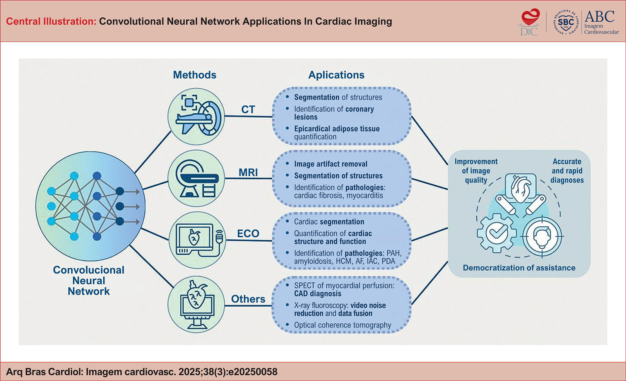
Convolutional Neural Network Applications In Cardiac Imaging

Luiz Henrique Cartaxo Fernandes et al. José Lucas Formiga Dantas Marilia Graziela Vieira de Macena Lima Glaudir Donato Marcelo Dantas Tavares de Melo

Abstract

Neural networks are computer models that mimic the workings of the human brain, learning from large volumes of data to perform increasingly complex tasks. This is done by means of interconnected artificial neurons that propagate information from raw layers to refined layers, sometimes of a weighted nature, weaving in the classification or identification of non-linear patterns. The Convolutional Neural Network (CNN) is a specialized deep learning architecture designed to extract and analyze spatial patterns within images, enabling applications in diagnostic decision-making and longitudinal clinical monitoring. Its use in Computed Tomography and Magnetic Resonance Imaging through cardiac image segmentation makes it possible to quickly and efficiently define cardiac structures, playing an important role in the reconstruction of three-dimensional images and in supporting preoperative procedures. In addition, compared to other forms of conventional analysis, it guarantees objective interpretations and superior image quality, based on the removal of artifacts from the studied segment. Echocardiograms have shown good results in identifying pathologies such as pulmonary hypertension, amyloidosis, hypertrophic cardiomyopathy and congenital diseases, and studies on the automation of these exams have shown an opening for what could be a dissemination framework in more remote areas where manual operators are not available. Consequently, CNN-based approaches have the potential to streamline cardiovascular imaging workflows, minimize inter-observer variability, and expand diagnostic capabilities to underserved and remote regions with limited access to specialized care.
  • Artificial Intelligence
  • Cardiac Imaging Techniques
  • Computer Neural Networks
Read Full Article
Creative Commons Attribution (CC BY) Creative Commons license
17/11/2025 DOI: 10.36660/abcimg.20250058i Views 3
The Complex Puzzle of Hypertrophic Phenotype: A Practical Approach for the Clinician
ABC
Review Article

The Complex Puzzle of Hypertrophic Phenotype: A Practical Approach for the Clinician

Alex dos Santos Felix et al. Silvio Henrique Barberato Marcelo Dantas Tavares de Melo Sílvia Aguiar Rosa Nuno Cardim

Abstract

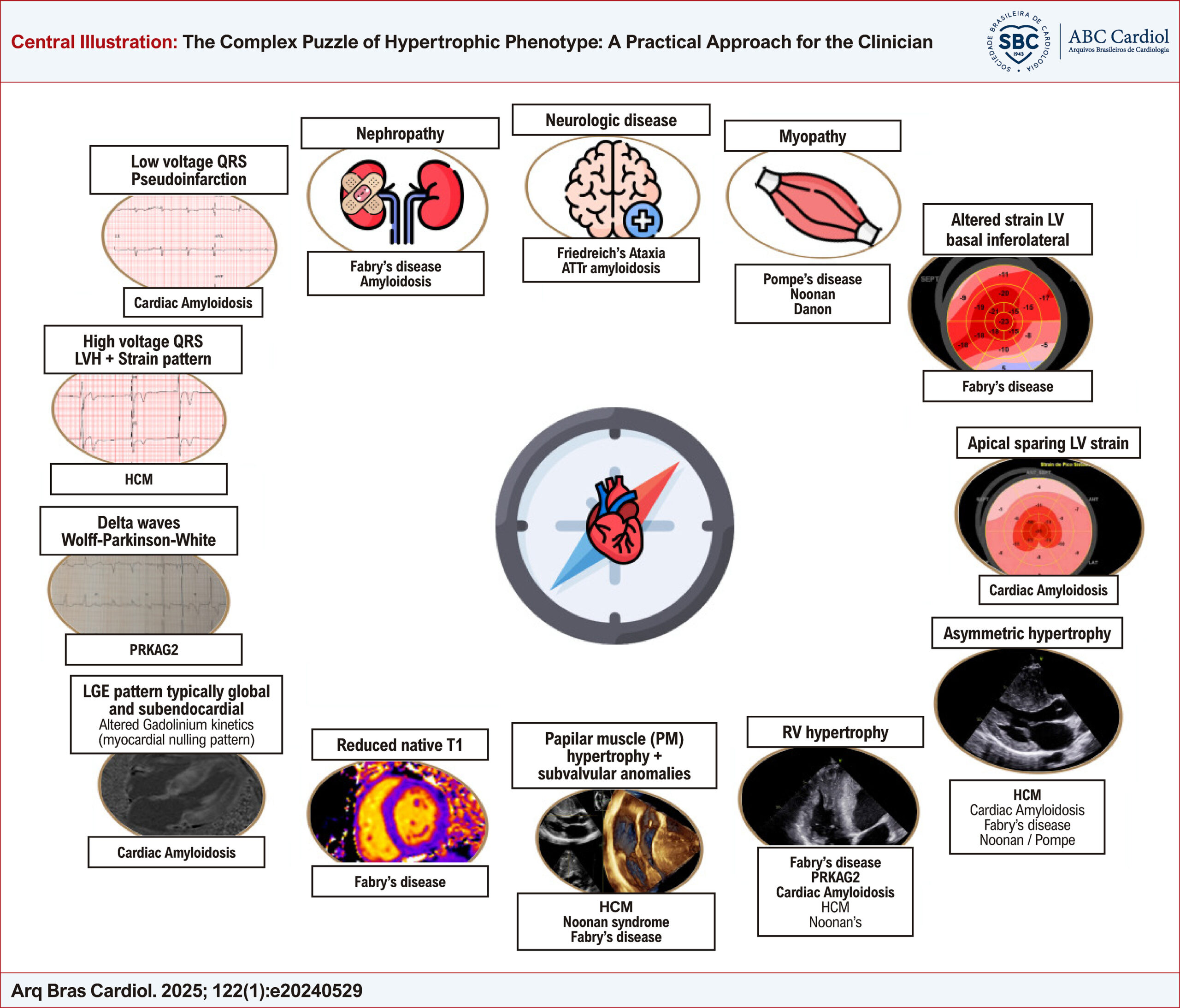
Left ventricular hypertrophy (LVH) represents a frequent observation in clinical practice. Nonetheless, the hypertrophic phenotype emerges as a common manifestation of diverse conditions, thereby presenting a diagnostic conundrum for clinicians. Differentiation among the etiologies of LVH is imperative for therapy decision-making, as different approaches must be implemented for distinct conditions, such as LVH secondary to loading changes, hypertrophic cardiomyopathy (HCM), or HCM mimics.

In some instances, an erroneous or late diagnosis may lead to a progression of the underlying disease with worsening functional capacity, high morbidity and mortality.

The rational use of cardiovascular multimodality imaging is of great importance when carried out in addition to a thorough clinical assessment and correlated with electrocardiographic findings, providing clues to fill the gaps, being, most of the time, the missing piece to solve this challenging puzzle.

An integrative approach is of paramount importance for the evaluation of these patients, as they are often followed by several specialties, with varied systemic manifestations. Although a multidisciplinary team is needed for an optimized follow-up of these patients, the most important player in this journey is the clinician, whose mission is to bring together all the red flags and coordinate all the data for an assertive diagnosis.

The objective of this review is to provide a pragmatic methodology, highlighting important clues for discriminating among the diverse conditions that result in LVH.

  • Cardiac Imaging Techniques
  • Diagnosis
  • Differential Diagnosis
  • Heart Function Tests
  • Left Ventricular Hypertrophy
Read Full Article
Creative Commons Attribution (CC BY) Creative Commons license
04/04/2025 DOI: 10.36660/abc.20240529i Views 1
Artificial intelligence for nuclear cardiology: Perspectives and challenges
IJCS
Review Article

Artificial intelligence for nuclear cardiology: Perspectives and challenges

Caitlyn Gilbert et al. Alec Chunta Robert Miller

Abstract

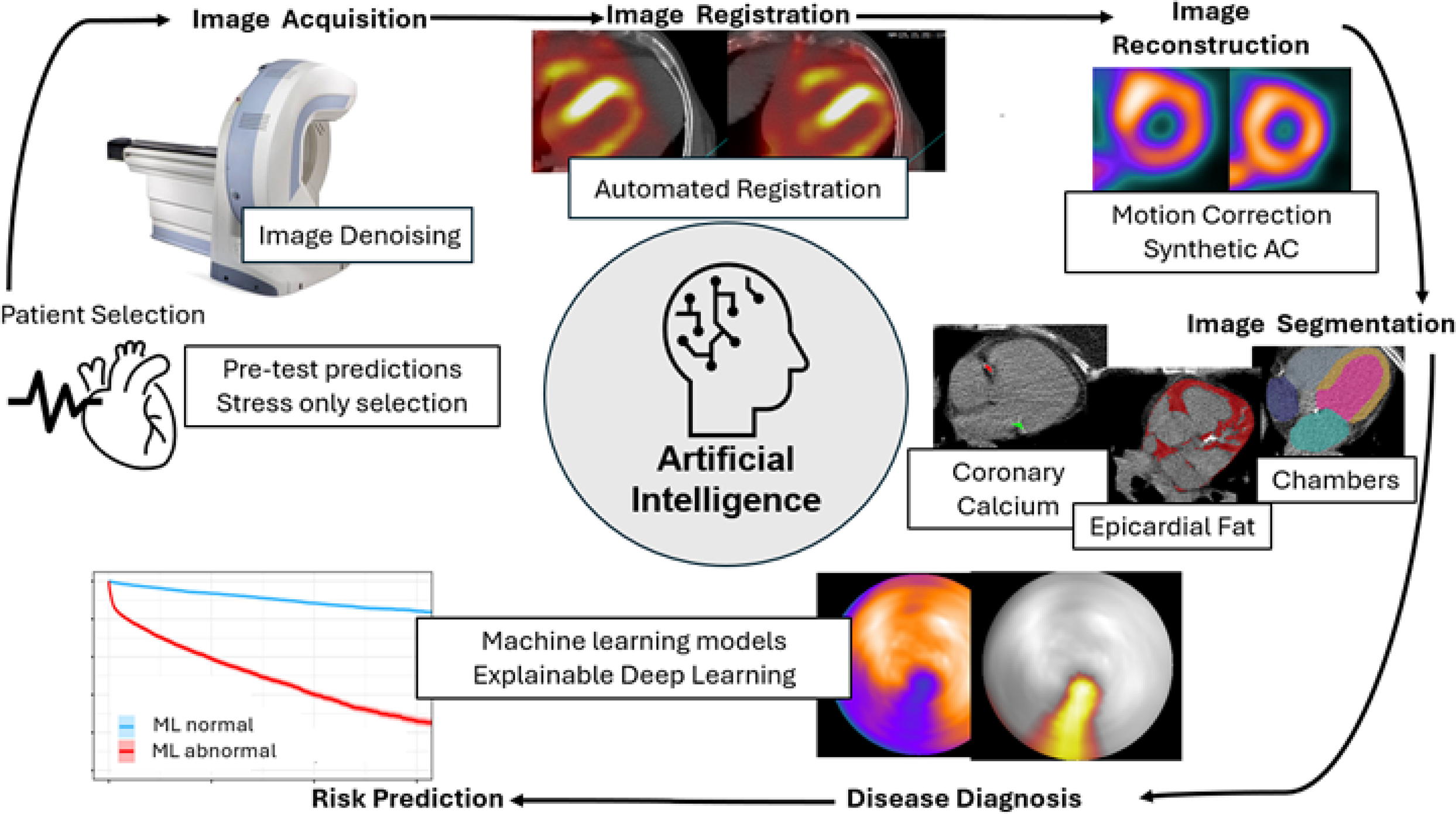
Recent advancements in artificial intelligence (AI) have significantly enhanced the efficiency and quality of nuclear cardiology imaging. AI algorithms, such as generative adversarial networks (GAN) and deep learning (DL) models, are improving image acquisition by reducing noise and correcting motion to minimize radiation exposure while maintaining diagnostic accuracy. AI has also demonstrated the potential to create synthetic attenuation correction (AC) imaging, accounting for soft-tissue attenuation without added computed tomography (CT) imaging. In patients with CT attenuation imaging, AI can automate the segmentation of coronary artery calcification (CAC), epicardial adipose tissue (EAT), and other structures to maximize the clinical information obtained from these added studies. Furthermore, machine learning (ML) models that combine clinical, imaging, and stress test data are advancing risk prediction for coronary artery disease (CAD) and ischemia, assisting clinicians in decision-making and improving patient outcomes. While many of these applications have been developed using myocardial perfusion imaging (MPI), they are increasingly applied to other nuclear cardiology studies. This review explores these AI applications and their impact on diagnostic accuracy, workflow efficiency, and risk stratification while also highlighting potential future directions and challenges.

  • Artificial Intelligence
  • Cardiac Imaging Techniques
  • Deep Learning
  • Machine Learning
  • Nuclear Medicine
Read Full Article
Creative Commons Attribution (CC BY) Creative Commons license
03/03/2025 DOI: 10.36660/ijcs.20240225 Views 2
Figure 1
ABC
Case Report

A Rare Encounter: Case of Cardiac Angioma in an Adult Patient

Inês Conde et al. Rodrigo Silva Nuno Antunes Vitor Hugo Pereira Catarina Quina-Rodrigues

Introduction

Cardiac masses encompass a wide spectrum of etiologies, including primary tumors, both benign and malignant, metastatic tumors, and non-neoplastic lesions., The differential diagnosis of cardiac masses is challenging due to overlapping clinical presentations, diverse imaging characteristics, heterogeneity among different mass types, limited clinical experience, and the rarity of some of these conditions., This clinical case presents an intriguing scenario involving a cardiac mass, highlighting the complexities in its diagnosis and management.

[…]

  • Cardiac Imaging Techniques
  • Echocardiography
  • Hemangioma
Read Full Article
Creative Commons Attribution (CC BY) Creative Commons license
13/01/2025 DOI: 10.36660/abc.20240150i Views 1
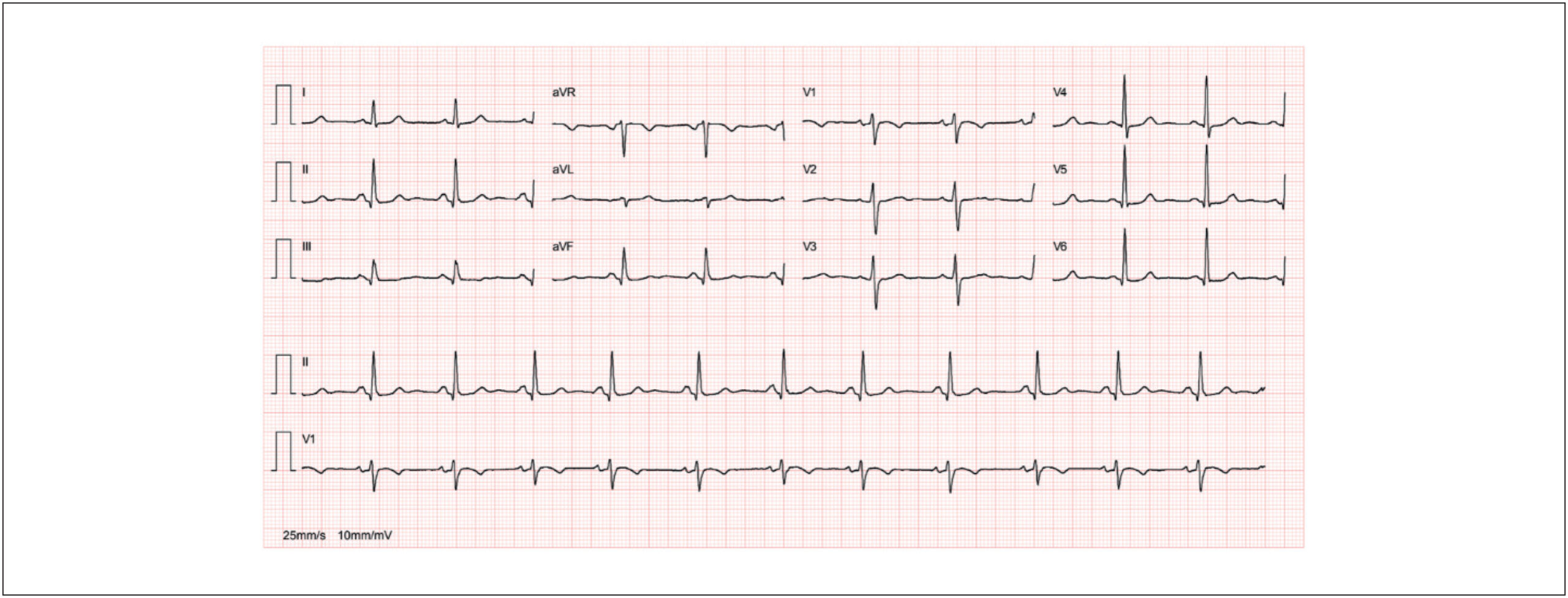
Figure 1
ABCIC
Editorial

The Use of X (Formerly Twitter) in Cardiovascular Imaging: Opportunities and Risks

Alex dos Santos Felix

Introduction

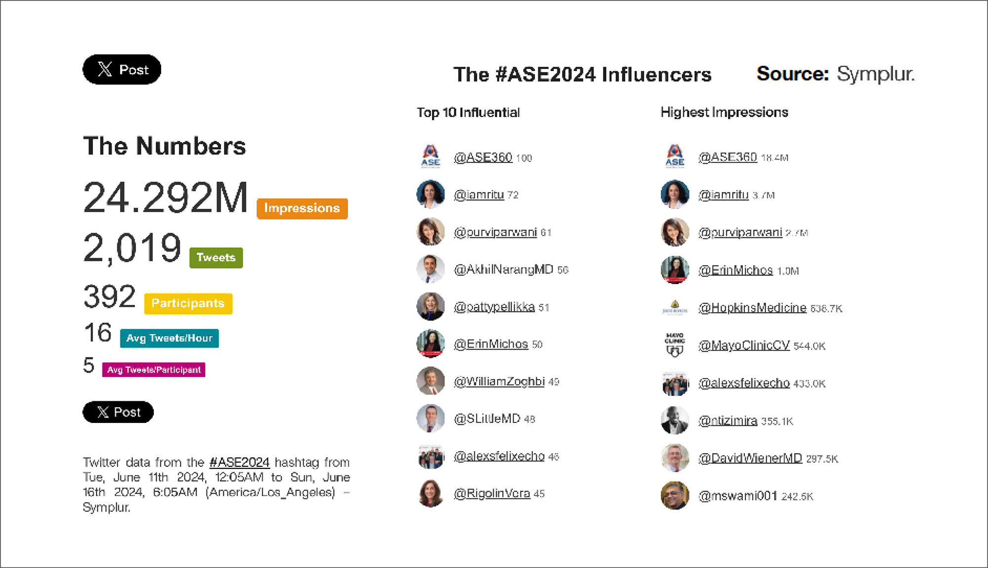
Social media has decisively transformed the way we disseminate medical information and knowledge, launching new digital tools to boost scientific debate and help redesign healthcare education. Globally, X, formerly Twitter, stands out in the medical field as a more scientific platform, with great interaction between peers, helping doctors and other healthcare professionals from all continents connect to discuss publications, debate scientific evidence, and share clinical experiences. X is also highly important as a channel for the dissemination of scientific events, courses and conferences. In a recent study involving healthcare professionals from 35 countries, Guerra et al. identified that X was the second most accessed social network (55.1%), second only to LinkedIn (60.8%). The speed of dissemination of information and interactivity are strong points of X, with the possibility of bringing together younger doctors, or those who work in institutions with lower complexity, with renowned professionals who work in large centers around the world. This valuable exchange of experiences creates a large virtual “grand round,” bringing other professionals into the debate and allowing for the discussion of controversial technical concepts and challenging clinical cases.

X was founded in 2006, still as Twitter, as an environment for the exchange of short messages, initially limited to 140 characters, having been extended to 280 characters in 2017, and recently relaxed to 25,000 characters for Premium users only. According to the Digital Report 2024, Brazil is the second country in the world where users spend the most time online. There are 245 million users on X, with more than 500 million posts per day, making Brazil the fourth largest user base of this platform in the world.

[…]

  • Cardiac Imaging Techniques
  • Echocardiography
  • Social Media
Read Full Article
Creative Commons Attribution (CC BY) Creative Commons license
09/12/2024 DOI: 10.36660/abcimg.20240109i
Figura 1
ABCIC
Case Report

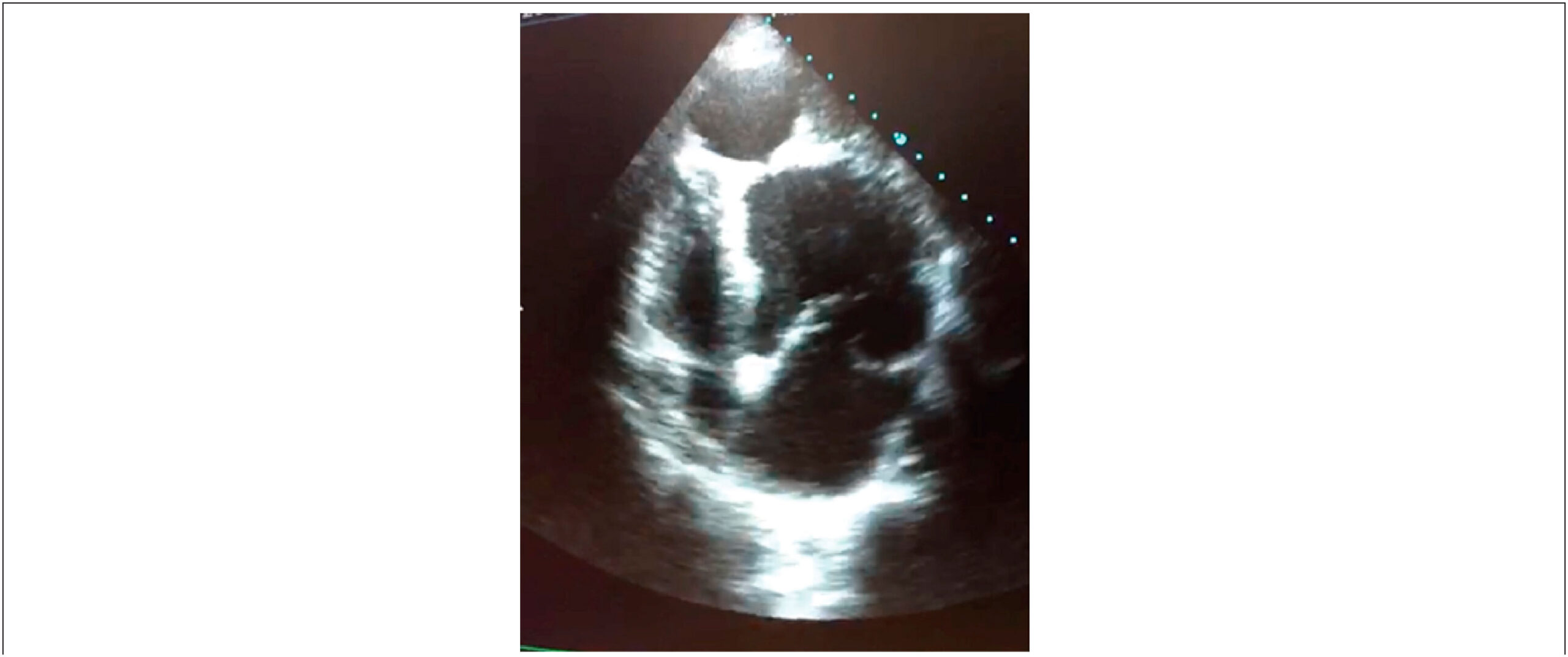
Not Everything is as it Seems at First Glance: Multimodal Imaging in Heart Cyst Evaluation

Carlos Guaman-Valdivieso et al. Julia Aramburu Natalia Lluberas Natalia Huart Gabriel Parma

Abstract

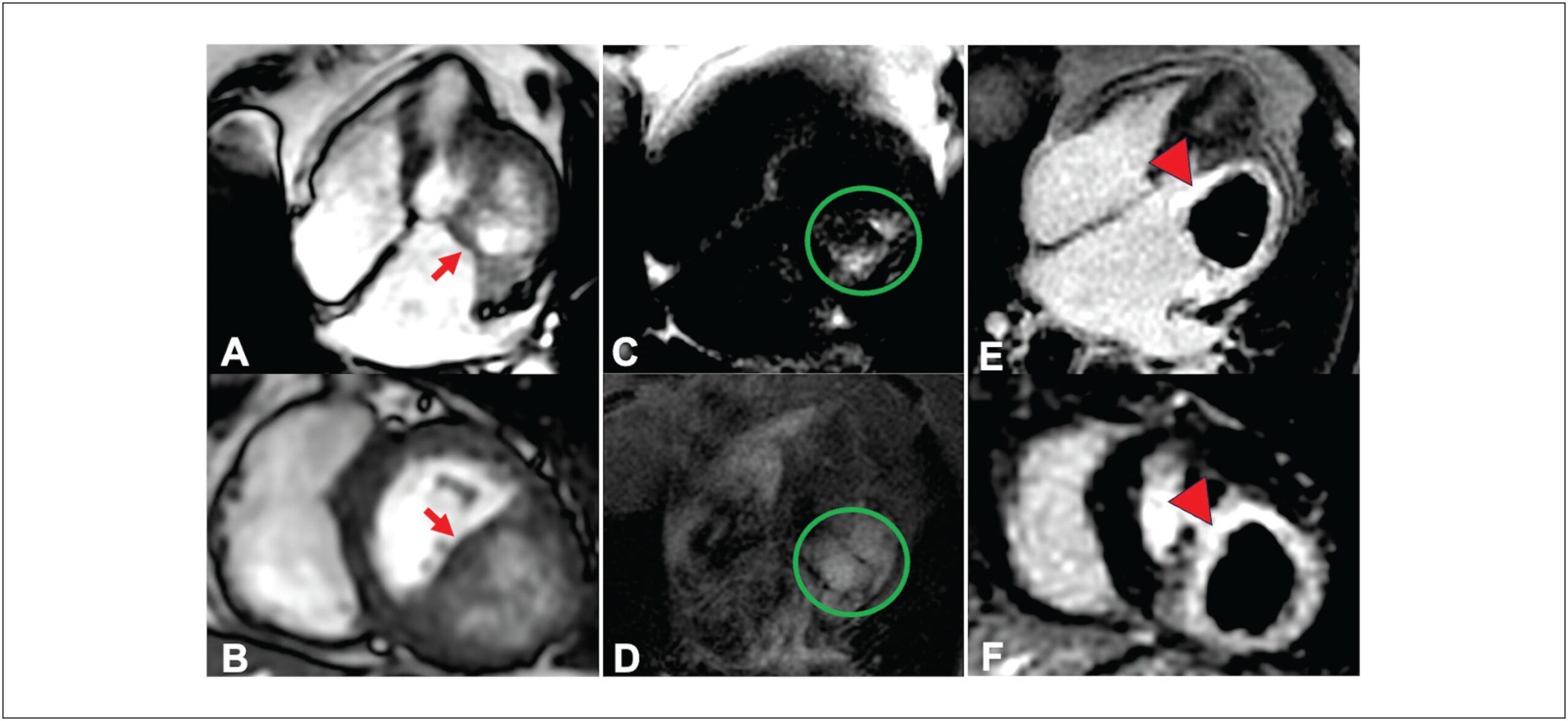
Cardiac masses, though rare, can lead to significant hemodynamic and arrhythmic issues, even if benign. Improved multimodal imaging has refined the diagnosis and understanding of these masses. We present an 85-year-old woman with New York Heart Association class II-III dyspnea for six months. Echocardiography showed a mass in the basal inferolateral wall of the left ventricle, along with severe left atrial dilation and moderate mitral regurgitation. Initially thought to be a pericardial cyst, further cardiac magnetic resonance (CMR) imaging identified an intramyocardial mass, and computed tomography (CT) suggested a hydatid cyst. Given the non-limiting symptoms and high surgical risk, the decision was made for expectant management. This case illustrates the rarity of cardiac cysts and the essential role of multimodal imaging in diagnosis and treatment planning.

  • Cardiac Imaging Techniques
  • Echinococcosis
  • Heart Neoplasms
Read Full Article
Creative Commons Attribution (CC BY) Creative Commons license
05/12/2024 DOI: 10.36660/abcimg.20240086i Views 1
Central Illustration
ABCIC
Original Article

The Women of the Cardiovascular Imaging Department of the Brazilian Society of Cardiology: Career and Family Challenges

Marcia Ferreira Alves Barberato et al. Daniela do Carmo Rassi Adenalva Lima de Souza Beck Marly Uellendahl … Samira Saady Morhy Silvio Henrique Barberato

Abstract

Introduction

The “DIC Mulheres” working group was created to research and discuss the role of women working in cardiovascular imaging in Brazil. Objectives: to map the challenges faced by women in the Cardiovascular Imaging Department of the Brazilian Society of Cardiology (DIC-SBC) in managing their careers and families.

Methods

A questionnaire with 29 questions on careers and family was made available between March and April 2024, inviting women from DIC-SBC to participate anonymously. The questions addressed age, medical training, career, family relationships, and mental health status. Data obtained were described in the form of categorical variables in percentages.

Results

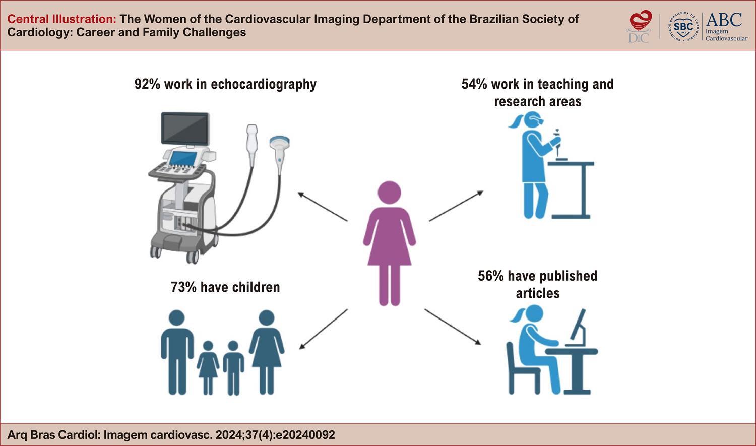
A total of 321 women responded to the questionnaire, representing 25% of DIC members. Most respondents are between 30 and 50 years old and work with echocardiography. Approximately 66% combine imaging activities with clinical cardiology, and 47% report a workload of more than 40 hours per week. Only 39% hold management positions, but more than 50% have published scientific production. Regarding motherhood, 73% have children and almost half of them did not stop working after the birth of their child. Regarding mental health, 63% reported symptoms, the most common being burnout (22%), anxiety (17%) and depression (17%).

Conclusion

Women face challenges in balancing their careers with family responsibilities, which can take a toll on their mental health. This study highlights the need for institutional policies that support motherhood, provide mentorship, and develop mental health programs specifically for those working in cardiovascular imaging.

  • Cardiac Imaging Techniques
  • Mental Health
  • Women
Read Full Article
Creative Commons Attribution (CC BY) Creative Commons license
21/11/2024 DOI: 10.36660/abcimg.20240092i Views 2
Figure 1
IJCS
Review Article

Multimodality Imaging of Chagas Heart Disease: Review and Case Presentation

Amalia Peix et al. Raul Araujo Veronica Gomez Aylen Perez Barreda … Maurizio Dondi Anita Brink Erika Minoshima Diana Paez

Abstract

Chagas disease (CD) is a neglected tropical disease (NTD) caused by Trypanosoma cruzi, a hemoflagellate protozoa. CD is diagnosed by epidemiologic history and two positive serological tests. The infection has two main clinical phases: acute, early after acquiring the infection, and chronic. The electrocardiogram (ECG) is the first tool to diagnose chronic Chagas heart disease after CD serological diagnosis. In addition, the different cardiac imaging modalities, including chest X-ray, echocardiography, single photon emission computed tomography (SPECT), computed tomography (CT), and magnetic resonance, play a crucial role in detecting the cardiac involvement, establishing the prognosis, stratifying patient risk, and addressing the management of CD patients. In this review of the current literature, we summarize the epidemiology, pathophysiology, and clinical presentation of CD. We also discuss the indications, advantages, and limitations of the different cardiac imaging modalities. We also present some case examples, where cardiac imaging proved helpful in cardiac CD (CCD). The early mortality and substantial disability caused by CD represents a health, social and economic burden, particularly in the Latin America. To harness the full potential of medical imaging for the effective management of patients with CD, a comprehensive approach is essential. This involves a concerted effort to invest in the education of medical providers and to facilitate widespread access to diagnostic tools. Accessible and timely diagnosis is crucial for initiating prompt and targeted interventions. Finally, continuous investment in research is vital to advance our understanding of CD and develop innovative solutions for improved patient care.

  • Cardiac Imaging Techniques
  • Chagas disease
  • Radionuclide Imaging
Read Full Article
Creative Commons Attribution (CC BY) Creative Commons license
19/08/2024 DOI: 10.36660/ijcs.20240114 Views 1
ABCIC
Editorial

One-Dimensional Echocardiography: Still Needed or Already Outdated?

Fernando Antonio de Portugal Morcerf

There is a widespread lack of knowledge about one-dimensional echocardiography (M-mode) by the current generation of echocardiographers. Has two-dimensional echocardiography (B-mode) and Doppler made M-mode outdated? M-mode provides information that cannot be obtained by using B-mode or even Doppler—be it spectral, color, or tissue Doppler—for two reasons:

Now let us consider some conditions that can only be detected by M-mode and not by B-mode or Doppler, which can aid in the diagnosis via B-mode or in the understanding of pathologies.

[…]

  • Cardiac Imaging Techniques
  • Cardiovascular Diseases
  • Echocardiography
Read Full Article
Creative Commons Attribution (CC BY) Creative Commons license
28/06/2024 DOI: 10.36660/abcimg.20240038i Views 1
Figure 1
ABCIC
Editorial

Is Echocardiographic Assessment of Strain Essential In Daily Routine?

Carlos Eduardo Suaide Silva

The assessment of myocardial fiber deformation (myocardial strain) during the cardiac cycle is a direct measurement of muscle function, which can be obtained by conventional imaging tests such as echocardiography and magnetic resonance.

Strain measurement can identify abnormalities in global and regional myocardial function, in addition to making the differential diagnosis of some cardiomyopathies. Furthermore, it is an earlier marker of ventricular dysfunction than ejection fraction and may predict adverse cardiovascular events.

[…]

  • Cardiac Imaging Techniques
  • Echocardiography
  • Ventricular Function
Read Full Article
Creative Commons Attribution (CC BY) Creative Commons license
06/09/2023 DOI: 10.36660/abcimg.20230063i Views 2
ABC
Editorial

The “Value of 0”: Is There an Added Value in Using the Calcium Score in the Stratification of Symptomatic Individuals with a Zero Score?

Catarina Ferreira

Coronary calcium score (CCS) is a good marker of cardiovascular risk, with an independent prognostic value that is incremental over traditional clinical risk factors.– It is performed using non-contrast computed tomography with very low radiation exposure to patients.

Its use is now recommended in selected asymptomatic individuals to refine risk assessment and help initiate or prolong preventive pharmacotherapies.

[…]

  • Cardiac Imaging Techniques
  • Chest Pain
  • Computed Tomography
  • Coronary Calcium
  • Coronary Disease
  • X-Ray
Read Full Article
Creative Commons Attribution (CC BY) Creative Commons license
07/04/2023 DOI: 10.36660/abc.20230105 Views 3
Figure 1
ABCIC
Review Article

Hypertrophic Cardiomyopathy: A Review Using Magnetic Resonance Imaging

Raul Serra Valério et al. Marcio Sommer Bittencourt Marly M. Uellendahl

Abstract

Hypertrophic cardiomyopathy, the most common genetic cardiopathy in the general population, is characterized by asymmetric left ventricular hypertrophy. However, the phenotypic changes in this cardiomyopathy extend beyond ventricular hypertrophy and include changes in the mitral valve apparatus, papillary muscles, and right ventricle. Due to the difficult differential diagnosis among multiple causes of hypertrophy, cardiac magnetic resonance has played a fundamental role in its diagnostic and prognostic evaluation; magnetic cine-resonance in defining the location and extent of hypertrophy; late enhancement, in the detection of areas of myocardial fibrosis; more recent techniques such as T1 mapping that assesses interstitial fibrosis and extracellular volume; and finally tissue tracking in the analysis of myocardial deformation.

  • Cardiac Imaging Techniques
  • Cardiomyopathy, Hypertrophic
  • Magnetic Resonance
Read Full Article
Creative Commons Attribution (CC BY) Creative Commons license
21/12/2022 DOI: 10.47593/2675-312X/20223504erer_15i Views 1
Figure 1
ABC
Image

Bulky Mitral Annulus Caseous Calcification in an Atypical Location

Joana Laranjeira Correia et al. Miguel Correia

Abstract

Mitral annular calcification (MAC) is a commonly observed chronic and degenerative fibrotic process of the base of the mitral valve, usually deemed as an incidental finding. Although initially, MAC was thought to be a consequence of an age-related degenerative process, recent findings suggest other independent contributive mechanisms, such as atherosclerosis and abnormal calcium-phosphorus metabolism.

Caseous calcification of the mitral annulus (cMAC) is a rarely described variant of MAC, characterized by an ovoid, focal mass with internal caseous fluid-like calcifications and debris.

Differentiating a cMAC from other cardiac masses attached to the mitral annulus may be challenging. A single imaging modality, such as transthoracic echocardiography, may not be sufficient for a clear diagnosis. Therefore, a multimodal imaging approach is necessary, including cardiac tomography computerized imaging and cardiac magnetic resonance (CMR).

MAC and cMAC typically affect the posterior mitral annulus, with very few cases in the literature describing the involvement of the anterior annulus. We present a rare case of an anterior mitral annulus caseous calcification found in a CMR performed to evaluate a left atrial mass identified on a transthoracic echocardiogram.

  • Cardiac Imaging Techniques
  • Stroke
  • Tomography
  • X-Ray Computed
Read Full Article
Creative Commons Attribution (CC BY) Creative Commons license
19/12/2022 DOI: 10.36660/abc.20210949 Views 1
Figure 1
ABC
Original Article

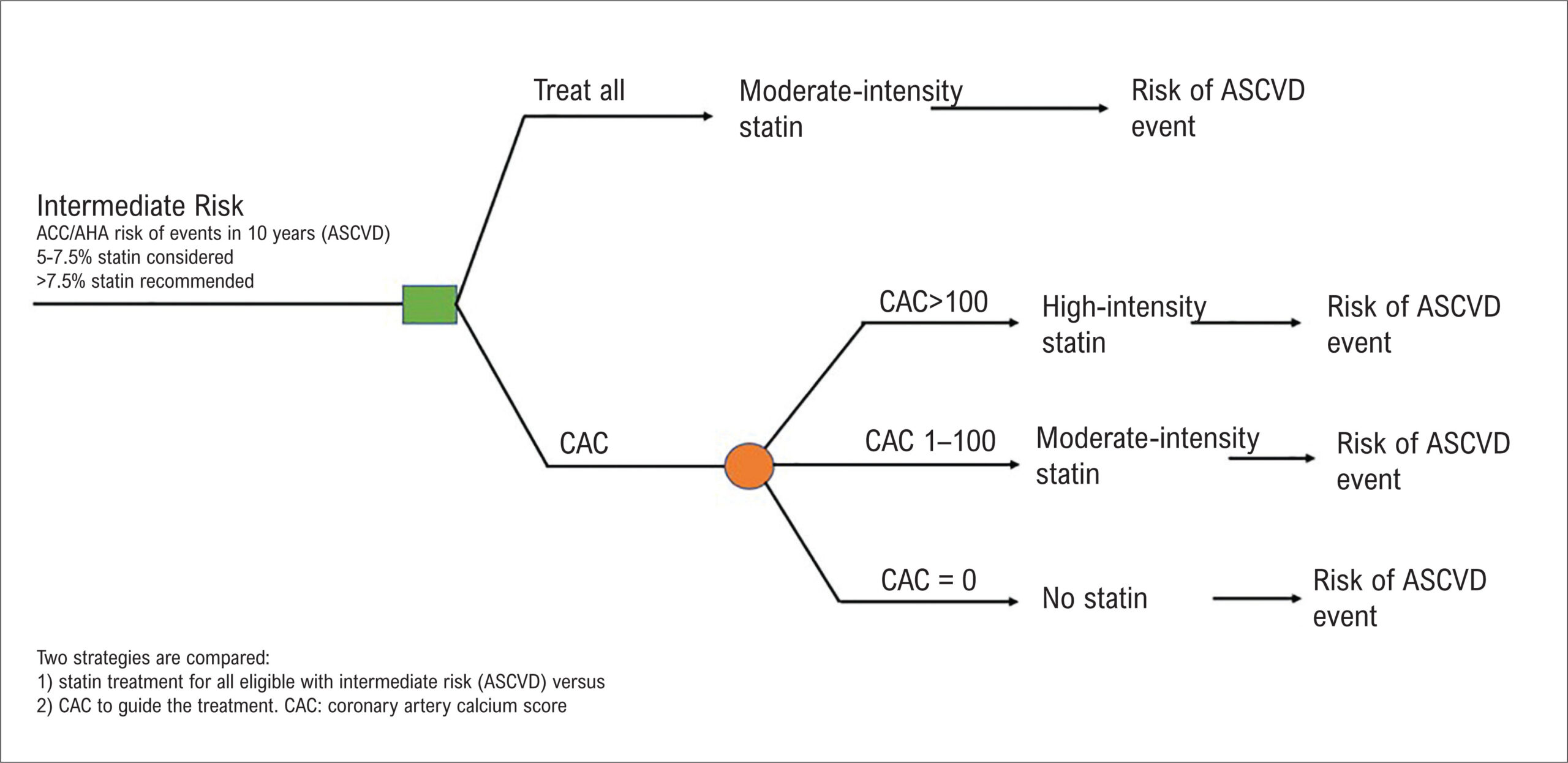
Cost-Effectiveness of Using the Coronary Artery Calcium Score in Guiding Therapeutic Decisions in Primary Prevention in the Brazilian Population

Raul Serra Valério et al. Giuliano Generoso Juliano Lara Fernandes Khurram Nasir … Jonathan C. Hong Marcio Sommer Bittencourt

Abstract

Background:

The use of the coronary artery calcium score to aid cardiovascular risk stratification may be a more cost-effective tool than the conventional strategy.

Objectives:

Evaluation of the cost-effectiveness of the use of the calcium score in therapeutic guidance for primary cardiovascular prevention.

Methods:

A microsimulation model to assess the clinical and economic consequences of atherosclerotic cardiovascular disease, comparing the prevention strategy using the calcium score and the conventional strategy.

Results:

The results obtained demonstrated a better cost-effectiveness of the therapeutic strategy guided by the calcium score, by reducing incremental costs and increasing quality-adjusted life years (QALY), which corresponds, in number, to improving the quality of life of the individual.

Conclusions:

The use of the coronary artery calcium score proved to be more cost-effective than the conventional strategy, both in terms of cost and QALY, in most of the scenarios studied.

  • Cardiac Imaging Techniques
  • Coronary Artery Calcium
  • Cost-Benefit Analysis
  • Primary Prevention
Read Full Article
Creative Commons Attribution (CC BY) Creative Commons license
10/06/2022 DOI: 10.36660/abc.20210347 Views 1
Figure 1
ABC
Research Letter

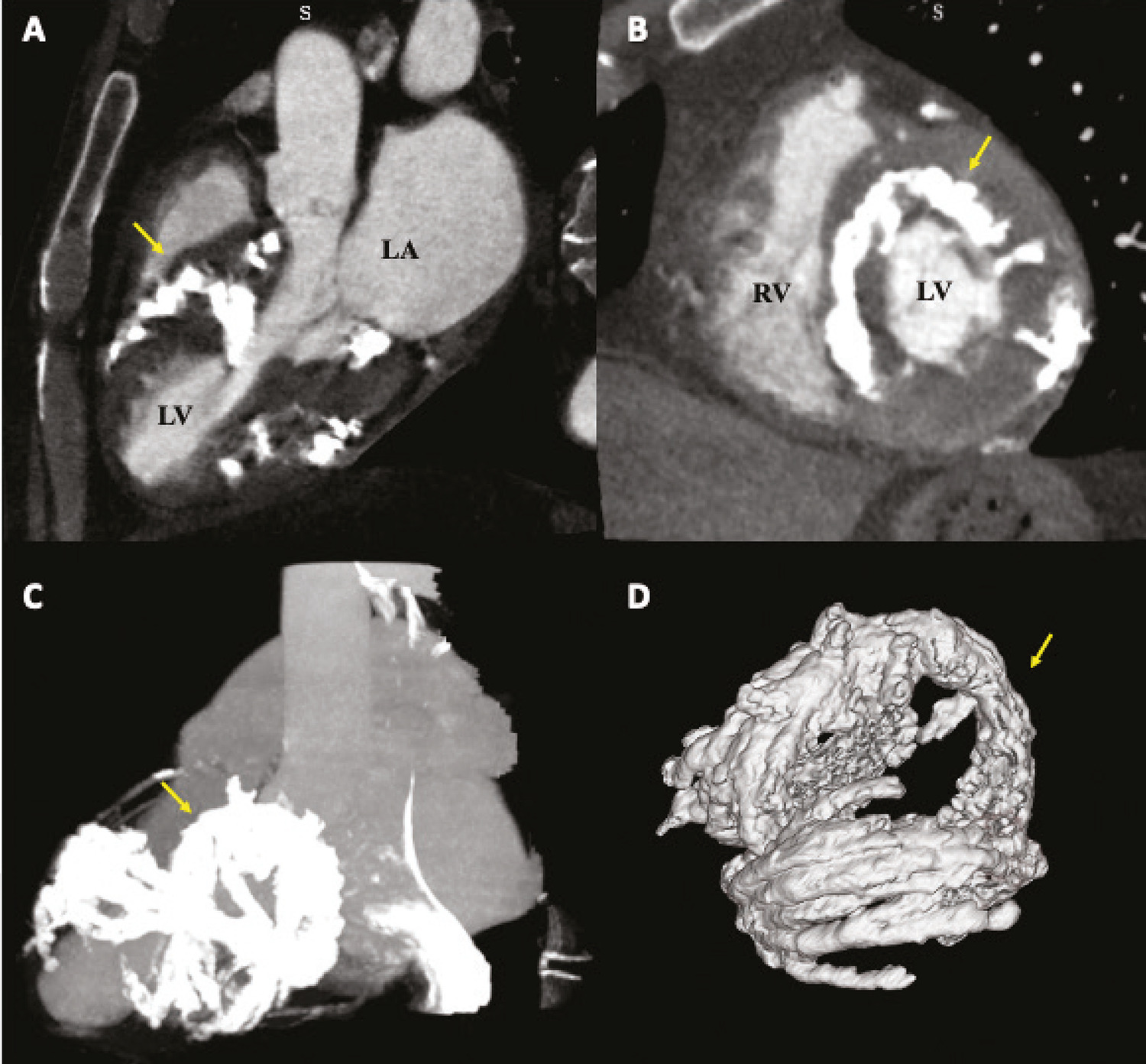
Torrent Guasp’s Helicoid Pattern Myocardial Calcification

Maria Marta Abraham-Foscolo et al. Rocío Blanco Juan Guido Chiabrando María Clara Llamedo … Diego Pérez de Arenaza Mariano L Falconi

Clinical case

72-year-old male patient with a past medical history of arterial hypertension, non-insulin-dependent diabetes, active tobacco consumption and chronic stable angina (coronary angiography showed severe stenosis in small size obtuse marginal branch). He also had a history of alcohol consumption, pancreatic calcification and renal lithiasis, without a history of Cancer, Sepsis or Tuberculosis to justify these diffuse calcifications.

He presented to the outpatient cardiology clinic complaining of angina and dyspnea New York Heart Association (NYHA) class II. His electrocardiogram (ECG) showed sinus rhythm and suggested left ventricular hypertrophy (LVH) without signs of ischemia.

[…]

  • Cardiac Imaging Techniques
  • Heart Failure
  • Vascular Calcification
Read Full Article
Creative Commons Attribution (CC BY) Creative Commons license
13/05/2022 DOI: 10.36660/abc.20210370 Views 1
Figure 1
ABCIC
Case Report

Left Ventricular Free-Wall Rupture (Pseudoaneurysm) after Acute Myocardial Infarction in an Asymptomatic Patient

Raul Serra Valério et al. Fernanda Sayuri Oshiro Alfredo Augusto Eyer Rodrigues Maria Eduarda Menezes de Siqueira Marly M. Uellendahl

Introduction

Left ventricular (LV) pseudoaneurysm after acute myocardial infarction may show nonspecific symptoms and be asymptomatic in up to 10% of cases. It demands investigation and a suspected diagnosis, with imaging methods playing a fundamental role in its diagnosis. This case report describes a pseudoaneurysm detected on a routine examination.

[…]

  • Cardiac Imaging Techniques
  • Diagnostic Imaging
  • False Aneurysm
Read Full Article
Creative Commons Attribution (CC BY) Creative Commons license
16/06/2021 DOI: 10.47593/2675-312X/20213401eabc134 Views 1

How to cite this article

EndNote RIS

Electronic Document Format (Vancouver)

Fernandes LHC, Dantas JLF, Lima MGVM, Donato G, Melo MDT. Convolutional Neural Network Applications In Cardiac Imaging. ABC Imagem Cardiovascular 2025;38(3):e20250058.

Electronic Document Format (ABNT)

Fernandes, Luiz Henrique Cartaxo; Dantas, José Lucas Formiga; Lima, Marilia Graziela Vieira de Macena; Donato, Glaudir; Melo, Marcelo Dantas Tavares de. Convolutional Neural Network Applications In Cardiac Imaging. ABC Imagem Cardiovascular, v. 38, n. 3, e20250058, Nov. 2025.

Electronic Document Format (APA)

Fernandes, L. H. C., Dantas, J. L. F., Lima, M. G. V. M., Donato, G., & Melo, M. D. T. (2025). Convolutional Neural Network Applications In Cardiac Imaging. ABC Imagem Cardiovascular, 38(3), e20250058.

Electronic Document Format (ISO)

Fernandes, Luiz Henrique Cartaxo and Dantas, José Lucas Formiga and Lima, Marilia Graziela Vieira de Macena and Donato, Glaudir and Melo, Marcelo Dantas Tavares de. Convolutional Neural Network Applications In Cardiac Imaging. ABC Imagem Cardiovascular [online]. 2025, vol. 38, n. 3, [cited 2026-10-15], e20250058. Available from: <https://journals.cardiol.br/en/article/convolutional-neural-network-applications-in-cardiac-imaging/>. ISSN 2318-8219

EndNote RIS

Electronic Document Format (Vancouver)

Felix AS, Barberato SH, Melo MDT, Rosa SA, Cardim N. The Complex Puzzle of Hypertrophic Phenotype: A Practical Approach for the Clinician. Arquivos Brasileiros de Cardiologia 2025;122(1):e20240529.

Electronic Document Format (ABNT)

Felix, Alex dos Santos; Barberato, Silvio Henrique; Melo, Marcelo Dantas Tavares de; Rosa, Sílvia Aguiar; Cardim, Nuno. The Complex Puzzle of Hypertrophic Phenotype: A Practical Approach for the Clinician. Arquivos Brasileiros de Cardiologia, v. 122, n. 1, e20240529, Apr. 2025.

Electronic Document Format (APA)

Felix, A. S., Barberato, S. H., Melo, M. D. T. , Rosa, S. A., & Cardim, N. (2025). The Complex Puzzle of Hypertrophic Phenotype: A Practical Approach for the Clinician. Arquivos Brasileiros de Cardiologia, 122(1), e20240529.

Electronic Document Format (ISO)

Felix, Alex dos Santos and Barberato, Silvio Henrique and Melo, Marcelo Dantas Tavares de and Rosa, Sílvia Aguiar and Cardim, Nuno. The Complex Puzzle of Hypertrophic Phenotype: A Practical Approach for the Clinician. Arquivos Brasileiros de Cardiologia [online]. 2025, vol. 122, n. 1, [cited 2026-10-15], e20240529. Available from: <https://journals.cardiol.br/en/article/the-complex-puzzle-of-hypertrophic-phenotype-a-practical-approach-for-the-clinician/>. ISSN 0066-782X

EndNote RIS

Electronic Document Format (Vancouver)

Gilbert C, Chunta A, Miller R. Artificial intelligence for nuclear cardiology: Perspectives and challenges. International Journal of Cardiovascular Sciences 2025;38:e20240225.

Electronic Document Format (ABNT)

Gilbert, Caitlyn; Chunta, Alec; Miller, Robert. Artificial intelligence for nuclear cardiology: Perspectives and challenges. International Journal of Cardiovascular Sciences, v. 38, e20240225, Mar. 2025.

Electronic Document Format (APA)

Gilbert, C., Chunta, A., & Miller, R. (2025). Artificial intelligence for nuclear cardiology: Perspectives and challenges. International Journal of Cardiovascular Sciences, 38, e20240225.

Electronic Document Format (ISO)

Gilbert, Caitlyn and Chunta, Alec and Miller, Robert. Artificial intelligence for nuclear cardiology: Perspectives and challenges. International Journal of Cardiovascular Sciences [online]. 2025, vol. 38, [cited 2026-10-15], e20240225. Available from: <https://journals.cardiol.br/en/article/artificial-intelligence-for-nuclear-cardiology-perspectives-and-challenges/>. ISSN 2359-4802

EndNote RIS

Electronic Document Format (Vancouver)

Conde I, Silva R, Antunes N, Pereira VH, Quina-Rodrigues C. A Rare Encounter: Case of Cardiac Angioma in an Adult Patient. Arquivos Brasileiros de Cardiologia 2025;121(12):e20240150.

Electronic Document Format (ABNT)

Conde, Inês; Silva, Rodrigo; Antunes, Nuno; Pereira, Vitor Hugo; Quina-Rodrigues, Catarina. A Rare Encounter: Case of Cardiac Angioma in an Adult Patient. Arquivos Brasileiros de Cardiologia, v. 121, n. 12, e20240150, Jan. 2025.

Electronic Document Format (APA)

Conde, I., Silva, R., Antunes, N., Pereira, V. H., & Quina-Rodrigues, C. (2025). A Rare Encounter: Case of Cardiac Angioma in an Adult Patient. Arquivos Brasileiros de Cardiologia, 121(12), e20240150.

Electronic Document Format (ISO)

Conde, Inês and Silva, Rodrigo and Antunes, Nuno and Pereira, Vitor Hugo and Quina-Rodrigues, Catarina. A Rare Encounter: Case of Cardiac Angioma in an Adult Patient. Arquivos Brasileiros de Cardiologia [online]. 2025, vol. 121, n. 12, [cited 2026-10-15], e20240150. Available from: <https://journals.cardiol.br/en/article/a-rare-encounter-case-of-cardiac-angioma-in-an-adult-patient/>. ISSN 0066-782X

EndNote RIS

Electronic Document Format (Vancouver)

Felix AS. The Use of X (Formerly Twitter) in Cardiovascular Imaging: Opportunities and Risks. ABC Imagem Cardiovascular 2024;37(4):e20240109.

Electronic Document Format (ABNT)

Felix, Alex dos Santos. The Use of X (Formerly Twitter) in Cardiovascular Imaging: Opportunities and Risks. ABC Imagem Cardiovascular, v. 37, n. 4, e20240109, Dec. 2024.

Electronic Document Format (APA)

Felix, A. S. (2024). The Use of X (Formerly Twitter) in Cardiovascular Imaging: Opportunities and Risks. ABC Imagem Cardiovascular, 37(4), e20240109.

Electronic Document Format (ISO)

Felix, Alex dos Santos. The Use of X (Formerly Twitter) in Cardiovascular Imaging: Opportunities and Risks. ABC Imagem Cardiovascular [online]. 2024, vol. 37, n. 4, [cited 2026-10-15], e20240109. Available from: <https://journals.cardiol.br/en/article/the-use-of-x-formerly-twitter-in-cardiovascular-imaging-opportunities-and-risks/>. ISSN 2318-8219

EndNote RIS

Electronic Document Format (Vancouver)

Guaman-Valdivieso C, Aramburu J, Lluberas N, Huart N, Parma G. Not Everything is as it Seems at First Glance: Multimodal Imaging in Heart Cyst Evaluation. ABC Imagem Cardiovascular 2024;37(4):e20240086.

Electronic Document Format (ABNT)

Guaman-Valdivieso, Carlos; Aramburu, Julia; Lluberas, Natalia; Huart, Natalia; Parma, Gabriel. Not Everything is as it Seems at First Glance: Multimodal Imaging in Heart Cyst Evaluation. ABC Imagem Cardiovascular, v. 37, n. 4, e20240086, Dec. 2024.

Electronic Document Format (APA)

Guaman-Valdivieso, C., Aramburu, J., Lluberas, N., Huart, N., & Parma, G. (2024). Not Everything is as it Seems at First Glance: Multimodal Imaging in Heart Cyst Evaluation. ABC Imagem Cardiovascular, 37(4), e20240086.

Electronic Document Format (ISO)

Guaman-Valdivieso, Carlos and Aramburu, Julia and Lluberas, Natalia and Huart, Natalia and Parma, Gabriel. Not Everything is as it Seems at First Glance: Multimodal Imaging in Heart Cyst Evaluation. ABC Imagem Cardiovascular [online]. 2024, vol. 37, n. 4, [cited 2026-10-15], e20240086. Available from: <https://journals.cardiol.br/en/article/not-everything-is-as-it-seems-at-first-glance-multimodal-imaging-in-heart-cyst-evaluation/>. ISSN 2318-8219

EndNote RIS

Electronic Document Format (Vancouver)

Barberato MFA, Rassi DC, Beck ALS, Uellendahl M, Morhy SS, Barberato SH. The Women of the Cardiovascular Imaging Department of the Brazilian Society of Cardiology: Career and Family Challenges. ABC Imagem Cardiovascular 2024;37(4):e20240092.

Electronic Document Format (ABNT)

Barberato, Marcia Ferreira Alves; Rassi, Daniela do Carmo; Beck, Adenalva Lima de Souza; Uellendahl, Marly; Morhy, Samira Saady; Barberato, Silvio Henrique. The Women of the Cardiovascular Imaging Department of the Brazilian Society of Cardiology: Career and Family Challenges. ABC Imagem Cardiovascular, v. 37, n. 4, e20240092, Nov. 2024.

Electronic Document Format (APA)

Barberato, M. F. A., Rassi, D. C., Beck, A. L. S., Uellendahl, M., Morhy, S. S., & Barberato, S. H. (2024). The Women of the Cardiovascular Imaging Department of the Brazilian Society of Cardiology: Career and Family Challenges. ABC Imagem Cardiovascular, 37(4), e20240092.

Electronic Document Format (ISO)

Barberato, Marcia Ferreira Alves and Rassi, Daniela do Carmo and Beck, Adenalva Lima de Souza and Uellendahl, Marly and Morhy, Samira Saady and Barberato, Silvio Henrique. The Women of the Cardiovascular Imaging Department of the Brazilian Society of Cardiology: Career and Family Challenges. ABC Imagem Cardiovascular [online]. 2024, vol. 37, n. 4, [cited 2026-10-15], e20240092. Available from: <https://journals.cardiol.br/en/article/the-women-of-the-cardiovascular-imaging-department-of-the-brazilian-society-of-cardiology-career-and-family-challenges/>. ISSN 2318-8219

EndNote RIS

Electronic Document Format (Vancouver)

Peix A, Araujo R, Gomez V, Barreda AP, Dondi M, Brink A, Minoshima E, et al. Multimodality Imaging of Chagas Heart Disease: Review and Case Presentation. International Journal of Cardiovascular Sciences 2024;37:e20240114.

Electronic Document Format (ABNT)

Peix, Amalia; Araujo, Raul; Gomez, Veronica; Barreda, Aylen Perez; Dondi, Maurizio; Brink, Anita; Minoshima, Erika; Paez, Diana. Multimodality Imaging of Chagas Heart Disease: Review and Case Presentation. International Journal of Cardiovascular Sciences, v. 37, e20240114, Aug. 2024.

Electronic Document Format (APA)

Peix, A., Araujo, R., Gomez, V., Barreda, A. P., Dondi, M., Brink, A., Minoshima, E., & Paez, D. (2024). Multimodality Imaging of Chagas Heart Disease: Review and Case Presentation. International Journal of Cardiovascular Sciences, 37, e20240114.

Electronic Document Format (ISO)

Peix, Amalia and Araujo, Raul and Gomez, Veronica and Barreda, Aylen Perez and Dondi, Maurizio and Brink, Anita and Minoshima, Erika and Paez, Diana. Multimodality Imaging of Chagas Heart Disease: Review and Case Presentation. International Journal of Cardiovascular Sciences [online]. 2024, vol. 37, [cited 2026-10-15], e20240114. Available from: <https://journals.cardiol.br/en/article/multimodality-imaging-of-chagas-heart-disease-review-and-case-presentation/>. ISSN 2359-4802

EndNote RIS

Electronic Document Format (Vancouver)

Morcerf FAP. One-Dimensional Echocardiography: Still Needed or Already Outdated?. ABC Imagem Cardiovascular 2024;37(2):e20240038.

Electronic Document Format (ABNT)

Morcerf, Fernando Antonio de Portugal. One-Dimensional Echocardiography: Still Needed or Already Outdated?. ABC Imagem Cardiovascular, v. 37, n. 2, e20240038, Jun. 2024.

Electronic Document Format (APA)

Morcerf, F. A. P. (2024). One-Dimensional Echocardiography: Still Needed or Already Outdated?. ABC Imagem Cardiovascular, 37(2), e20240038.

Electronic Document Format (ISO)

Morcerf, Fernando Antonio de Portugal. One-Dimensional Echocardiography: Still Needed or Already Outdated?. ABC Imagem Cardiovascular [online]. 2024, vol. 37, n. 2, [cited 2026-10-15], e20240038. Available from: <https://journals.cardiol.br/en/article/one-dimensional-echocardiography-still-needed-or-already-outdated/>. ISSN 2318-8219

EndNote RIS

Electronic Document Format (Vancouver)

Silva CES. Is Echocardiographic Assessment of Strain Essential In Daily Routine?. ABC Imagem Cardiovascular 2023;36(3):e20230063.

Electronic Document Format (ABNT)

Silva, Carlos Eduardo Suaide. Is Echocardiographic Assessment of Strain Essential In Daily Routine?. ABC Imagem Cardiovascular, v. 36, n. 3, e20230063, Sep. 2023.

Electronic Document Format (APA)

Silva, C. E. S. (2023). Is Echocardiographic Assessment of Strain Essential In Daily Routine?. ABC Imagem Cardiovascular, 36(3), e20230063.

Electronic Document Format (ISO)

Silva, Carlos Eduardo Suaide. Is Echocardiographic Assessment of Strain Essential In Daily Routine?. ABC Imagem Cardiovascular [online]. 2023, vol. 36, n. 3, [cited 2026-10-15], e20230063. Available from: <https://journals.cardiol.br/en/article/is-echocardiographic-assessment-of-strain-essential-in-daily-routine/>. ISSN 2318-8219

EndNote RIS

Electronic Document Format (Vancouver)

Ferreira C. The “Value of 0”: Is There an Added Value in Using the Calcium Score in the Stratification of Symptomatic Individuals with a Zero Score?. Arquivos Brasileiros de Cardiologia 2023;120(3):e20230105.

Electronic Document Format (ABNT)

Ferreira, Catarina. The “Value of 0”: Is There an Added Value in Using the Calcium Score in the Stratification of Symptomatic Individuals with a Zero Score?. Arquivos Brasileiros de Cardiologia, v. 120, n. 3, e20230105, Apr. 2023.

Electronic Document Format (APA)

Ferreira, C. (2023). The “Value of 0”: Is There an Added Value in Using the Calcium Score in the Stratification of Symptomatic Individuals with a Zero Score?. Arquivos Brasileiros de Cardiologia, 120(3), e20230105.

Electronic Document Format (ISO)

Ferreira, Catarina. The “Value of 0”: Is There an Added Value in Using the Calcium Score in the Stratification of Symptomatic Individuals with a Zero Score?. Arquivos Brasileiros de Cardiologia [online]. 2023, vol. 120, n. 3, [cited 2026-10-15], e20230105. Available from: <https://journals.cardiol.br/en/article/the-value-of-0-is-there-an-added-value-in-using-the-calcium-score-in-the-stratification-of-symptomatic-individuals-with-a-zero-score/>. ISSN 0066-782X

EndNote RIS

Electronic Document Format (Vancouver)

Valério RS, Bittencourt MS, Uellendahl MM. Hypertrophic Cardiomyopathy: A Review Using Magnetic Resonance Imaging. ABC Imagem Cardiovascular 2022;35(4):erer_15.

Electronic Document Format (ABNT)

Valério, Raul Serra; Bittencourt, Marcio Sommer; Uellendahl, Marly M.. Hypertrophic Cardiomyopathy: A Review Using Magnetic Resonance Imaging. ABC Imagem Cardiovascular, v. 35, n. 4, erer_15, Dec. 2022.

Electronic Document Format (APA)

Valério, R. S., Bittencourt, M. S., & Uellendahl, M. M. (2022). Hypertrophic Cardiomyopathy: A Review Using Magnetic Resonance Imaging. ABC Imagem Cardiovascular, 35(4), erer_15.

Electronic Document Format (ISO)

Valério, Raul Serra and Bittencourt, Marcio Sommer and Uellendahl, Marly M.. Hypertrophic Cardiomyopathy: A Review Using Magnetic Resonance Imaging. ABC Imagem Cardiovascular [online]. 2022, vol. 35, n. 4, [cited 2026-10-15], erer_15. Available from: <https://journals.cardiol.br/en/article/hypertrophic-cardiomyopathy-a-review-using-magnetic-resonance-imaging/>. ISSN 2318-8219

EndNote RIS

Electronic Document Format (Vancouver)

Correia JL, Correia M. Bulky Mitral Annulus Caseous Calcification in an Atypical Location. Arquivos Brasileiros de Cardiologia 2022;119(6):1002-5.

Electronic Document Format (ABNT)

Correia, Joana Laranjeira; Correia, Miguel. Bulky Mitral Annulus Caseous Calcification in an Atypical Location. Arquivos Brasileiros de Cardiologia, v. 119, n. 6, p. 1002-1005, Dec. 2022.

Electronic Document Format (APA)

Correia, J. L., & Correia, M. (2022). Bulky Mitral Annulus Caseous Calcification in an Atypical Location. Arquivos Brasileiros de Cardiologia, 119(6), 1002-1005.

Electronic Document Format (ISO)

Correia, Joana Laranjeira and Correia, Miguel. Bulky Mitral Annulus Caseous Calcification in an Atypical Location. Arquivos Brasileiros de Cardiologia [online]. 2022, vol. 119, n. 6, [cited 2026-10-15], pp. 1002-1005. Available from: <https://journals.cardiol.br/en/article/bulky-mitral-annulus-caseous-calcification-in-an-atypical-location/>. ISSN 0066-782X

EndNote RIS

Electronic Document Format (Vancouver)

Valério RS, Generoso G, Fernandes JL, Nasir K, Hong JC, Bittencourt MS. Cost-Effectiveness of Using the Coronary Artery Calcium Score in Guiding Therapeutic Decisions in Primary Prevention in the Brazilian Population. Arquivos Brasileiros de Cardiologia 2022;118(6):1126-31.

Electronic Document Format (ABNT)

Valério, Raul Serra; Generoso, Giuliano; Fernandes, Juliano Lara; Nasir, Khurram; Hong, Jonathan C.; Bittencourt, Marcio Sommer. Cost-Effectiveness of Using the Coronary Artery Calcium Score in Guiding Therapeutic Decisions in Primary Prevention in the Brazilian Population. Arquivos Brasileiros de Cardiologia, v. 118, n. 6, p. 1126-1131, Jun. 2022.

Electronic Document Format (APA)

Valério, R. S., Generoso, G., Fernandes, J. L., Nasir, K., Hong, J. C., & Bittencourt, M. S. (2022). Cost-Effectiveness of Using the Coronary Artery Calcium Score in Guiding Therapeutic Decisions in Primary Prevention in the Brazilian Population. Arquivos Brasileiros de Cardiologia, 118(6), 1126-1131.

Electronic Document Format (ISO)

Valério, Raul Serra and Generoso, Giuliano and Fernandes, Juliano Lara and Nasir, Khurram and Hong, Jonathan C. and Bittencourt, Marcio Sommer. Cost-Effectiveness of Using the Coronary Artery Calcium Score in Guiding Therapeutic Decisions in Primary Prevention in the Brazilian Population. Arquivos Brasileiros de Cardiologia [online]. 2022, vol. 118, n. 6, [cited 2026-10-15], pp. 1126-1131. Available from: <https://journals.cardiol.br/en/article/cost-effectiveness-of-using-the-coronary-artery-calcium-score-in-guiding-therapeutic-decisions-in-primary-prevention-in-the-brazilian-population/>. ISSN 0066-782X

EndNote RIS

Electronic Document Format (Vancouver)

Abraham-Foscolo MM, Blanco R, Chiabrando JG, Llamedo MC, Arenaza DP, Falconi ML. Torrent Guasp’s Helicoid Pattern Myocardial Calcification. Arquivos Brasileiros de Cardiologia 2022;118(5):992-5.

Electronic Document Format (ABNT)

Abraham-Foscolo, Maria Marta; Blanco, Rocío; Chiabrando, Juan Guido; Llamedo, María Clara; Arenaza, Diego Pérez de; Falconi, Mariano L. Torrent Guasp’s Helicoid Pattern Myocardial Calcification. Arquivos Brasileiros de Cardiologia, v. 118, n. 5, p. 992-995, May. 2022.

Electronic Document Format (APA)

Abraham-Foscolo, M. M., Blanco, R., Chiabrando, J. G., Llamedo, M. C., Arenaza, D. P. , & Falconi, M. L. (2022). Torrent Guasp’s Helicoid Pattern Myocardial Calcification. Arquivos Brasileiros de Cardiologia, 118(5), 992-995.

Electronic Document Format (ISO)

Abraham-Foscolo, Maria Marta and Blanco, Rocío and Chiabrando, Juan Guido and Llamedo, María Clara and Arenaza, Diego Pérez de and Falconi, Mariano L. Torrent Guasp’s Helicoid Pattern Myocardial Calcification. Arquivos Brasileiros de Cardiologia [online]. 2022, vol. 118, n. 5, [cited 2026-10-15], pp. 992-995. Available from: <https://journals.cardiol.br/en/article/torrent-guasps-helicoid-pattern-myocardial-calcification/>. ISSN 0066-782X

EndNote RIS

Electronic Document Format (Vancouver)

Valério RS, Oshiro FS, Rodrigues AAE, Siqueira MEM, Uellendahl MM. Left Ventricular Free-Wall Rupture (Pseudoaneurysm) after Acute Myocardial Infarction in an Asymptomatic Patient. ABC Imagem Cardiovascular 2021;34(1):eabc134.

Electronic Document Format (ABNT)

Valério, Raul Serra; Oshiro, Fernanda Sayuri; Rodrigues, Alfredo Augusto Eyer; Siqueira, Maria Eduarda Menezes de; Uellendahl, Marly M.. Left Ventricular Free-Wall Rupture (Pseudoaneurysm) after Acute Myocardial Infarction in an Asymptomatic Patient. ABC Imagem Cardiovascular, v. 34, n. 1, eabc134, Jun. 2021.

Electronic Document Format (APA)

Valério, R. S., Oshiro, F. S., Rodrigues, A. A. E., Siqueira, M. E. M. , & Uellendahl, M. M. (2021). Left Ventricular Free-Wall Rupture (Pseudoaneurysm) after Acute Myocardial Infarction in an Asymptomatic Patient. ABC Imagem Cardiovascular, 34(1), eabc134.

Electronic Document Format (ISO)

Valério, Raul Serra and Oshiro, Fernanda Sayuri and Rodrigues, Alfredo Augusto Eyer and Siqueira, Maria Eduarda Menezes de and Uellendahl, Marly M.. Left Ventricular Free-Wall Rupture (Pseudoaneurysm) after Acute Myocardial Infarction in an Asymptomatic Patient. ABC Imagem Cardiovascular [online]. 2021, vol. 34, n. 1, [cited 2026-10-15], eabc134. Available from: <https://journals.cardiol.br/en/article/left-ventricular-free-wall-rupture-pseudoaneurysm-after-acute-myocardial-infarction-in-an-asymptomatic-patient/>. ISSN 2318-8219

  • About ABC Family
  • Internal Regulation
  • Editorial Board
  • Brazilian Society of Cardiology (SBC)

Issues

SBC Guidelines

  • Videos
  • Podcasts
  • News