ABC Family Arquivos Brasileiros de Cardiologia International Journal of Cardiovascular Sciences ABC Heart Failure & Cardiomyopathy ABC Imagem Cardiovascular
Português English
ABC Family
Português English
  • About
    • About ABC Family
    • Internal Regulation
    • Editorial Board
    • Brazilian Society of Cardiology (SBC)
  • Issues
  • SBC Guidelines
  • Content
    • Videos
    • Podcasts
    • News
  • About
    • About ABC Family
    • Internal Regulation
    • Editorial Board
    • Brazilian Society of Cardiology (SBC)
  • Issues
  • SBC Guidelines
  • Content
    • Videos
    • Podcasts
    • News

ABC Family

  • ABC Family
  • Arquivos Brasileiros de Cardiologia
  • International Journal of Cardiovascular Sciences
  • ABC Heart Failure & Cardiomyopathy
  • ABC Imagem Cardiovascular
Search in

No content found.

Journals
Ordering
Article types
Others

  1. Home
  2. Keyword: Heart Ventricles
Figure 1
IJCS
Editorial

The "Right" Side of Myocardial Infarction: Sinister Outcomes?

Monica Verdoia

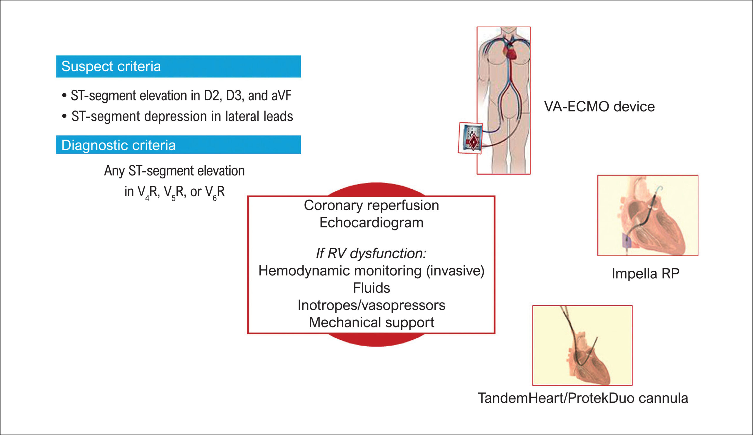
Right ventricular myocardial infarction (RVI) is rarely an isolated event; rather, it generally complicates 10% to 50% of cases of acute inferior ST-segment elevation myocardial infarction (STEMI)., Proximal right coronary artery occlusion, impairing right ventricular (RV) branch flow, is most frequently the culprit vessel, although it depends on coronary tree anatomy.

Previous studies underlined that RVI is often unrecognized, since RV ischemic dysfunction is generally transient, due to the different composition and higher resistance to hypoxia of the RV wall. In addition, the diagnostic criteria for RVI, which are mainly based on electrocardiogram (), can lead to underestimation, unless RV involvement becomes clinically manifest with hemodynamic complications. In fact, acute RV dysfunction can lead to reduced output, insufficient left heart filling, and cardiogenic shock, despite preserved left contractility. Moreover, intrapericardial ventricular interaction can translate into an abrupt RV dilation and further compression of the left chambers.

[…]

  • Heart Ventricles
  • Myocardial Infarction
  • Percutaneous Coronary Intervention
Read Full Article
Creative Commons Attribution (CC BY) Creative Commons license
13/03/2026 DOI: 10.36660/ijcs.20250249
Figure 1
IJCS
Case Report

Transcatheter Correction of Aortic-Right Ventricular Shunt Following Aortic Valve Replacement: A Case Report

Adnaldo da Silveira Maia et al. Mauro Henrique Batista Camacho Andresa Fernandes Perego Dayara Hoffmann Mayer José Honório Palma da Fonseca

Abstract

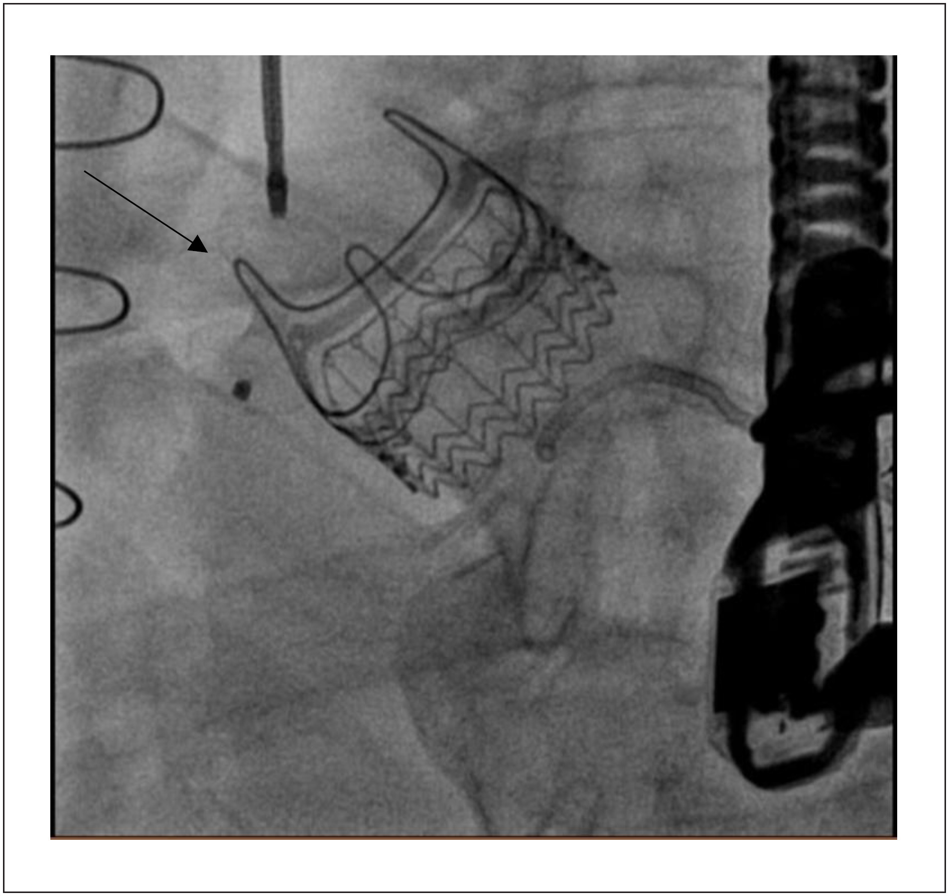
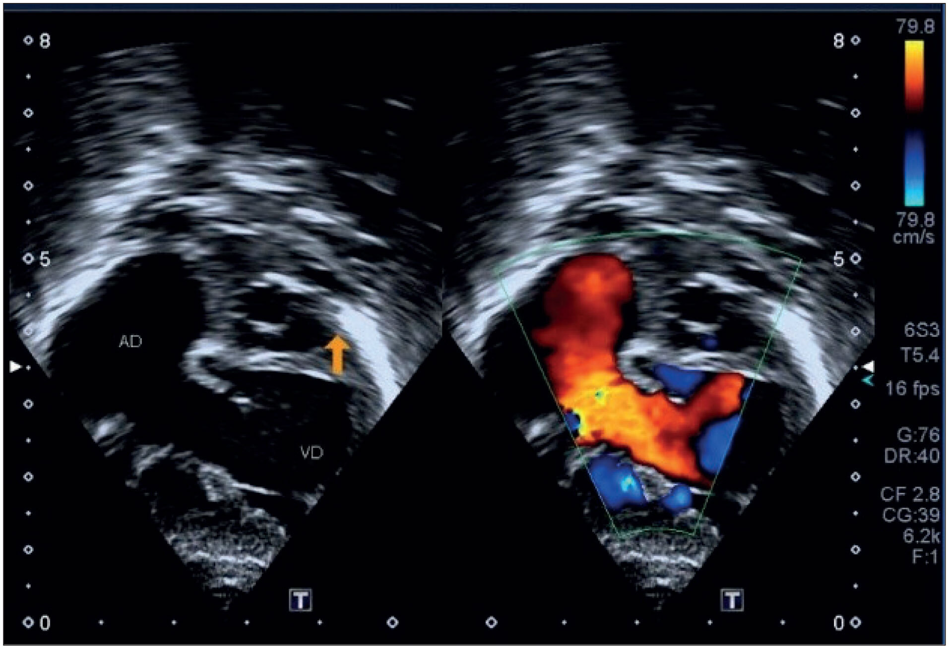
We describe the first case in the literature of transcatheter correction of aortic-right ventricular (Ao-RV) shunt after aortic valve replacement (AVR) with an Intuity® prosthesis (Edwards Lifesciences). A 79-year-old patient with a history of coronary artery bypass grafting and aortic regurgitation underwent AVR using a rapid-release 25-mm Intuity® prosthesis. After 45 days, he presented with severe heart failure, and an Ao-RV shunt was identified on transthoracic echocardiography. Given his history of previous cardiac surgeries, percutaneous correction was attempted. Initially, an attempt was made to catheterize the defect via the transapical approach, which was unsuccessful. The right femoral artery was punctured, and the shunt was catheterized via the aorta, with the implantation of a MemoPart ventricular septal defects (VSD) occluder, resulting in a significant reduction of the shunt. The patient was then transferred to the intensive care unit. However, there was no satisfactory clinical improvement, and transthoracic echocardiography revealed persistent Ao-RV shunt. Therefore, the surgical team opted for a new approach through the femoral artery and a second MemoPart VSD occluder was implanted. A transesophageal echocardiogram showed no leak, and the patient progressed satisfactorily and was discharged from the hospital. Shunts after AVR are rare. In these conditions, percutaneous management is a safe and feasible technical option. This is the first case reported in the literature of this type of complication after Intuity prosthesis implantation.

  • Aorta
  • Heart Ventricles
  • Transcatheter Aortic Valve Replacement
Read Full Article
Creative Commons Attribution (CC BY) Creative Commons license
09/01/2026 DOI: 10.36660/ijcs.20250060
Central Illustration
ABCHF
Review Article

Advanced Management of Right Heart Failure: An Illustrative Case

Pedro Castilhos de Freitas Crivelaro et al. Guilherme Pinheiro Machado Marcelo Basso Gazzana William Lorenzi … Igor Gorski Benedetto Felipe Homem Valle Rodrigo Vugman Wainstein

Abstract

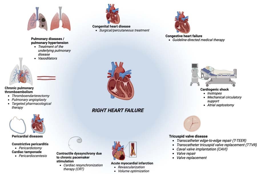
Right heart failure (RHF) is a serious condition characterized by the inability of the right ventricle (RV) to generate sufficient output for pulmonary circulation, leading to systemic congestion and hypoperfusion. Proper management of RHF is essential to improve prognosis and quality of life, requiring individualized treatment based on hemodynamic assessment.

This article reviews pathophysiology, diagnosis, and therapeutic options for RHF, including strategies for preload optimization, afterload reduction, and advanced circulatory support. Additionally, it presents a clinical case of a patient with severe RHF secondary to chronic pulmonary thromboembolism and primary tricuspid regurgitation, treated with a hybrid approach involving balloon pulmonary angioplasty and pulmonary thromboendarterectomy combined with surgical valve repair, resulting in significant functional improvement. This review highlights the importance of personalized strategies and combined therapies in the management of advanced RHF.

  • Heart Failure
  • Heart Ventricles
  • Pulmonary Hypertension
Read Full Article
Creative Commons Attribution (CC BY) Creative Commons license
03/11/2025 DOI: 10.36660/abchf.20250017i
Left and Right Ventricular Strain in an Adult Brazilian Population from ELSA-Brasil Study: Reference Values and their Determinants
ABC
Original Article

Left and Right Ventricular Strain in an Adult Brazilian Population from ELSA-Brasil Study: Reference Values and their Determinants

Eduardo Gatti Pianca et al. Murilo Foppa Giulia Bevilacqua Schmitz Wilson Cañon-Montañez … Bruce Bartholow Duncan Angela Barreto Santiago Santos

Abstract

Background

Impaired left ventricular (LV) and right ventricular functions are important predictors of cardiovascular risk. Global longitudinal strain (GLS) provides superior sensitivity for assessing systolic function compared to traditional parameters, enhancing diagnostic accuracy in various cardiac conditions. However, GLS reference values in diverse populations are lacking.

Objectives

To establish reference values for LVGLS and right ventricular free wall longitudinal strain (RVFWLS) in a Brazilian multiethnic population without cardiovascular risk factors or disease. We also explore how clinical and echocardiographic factors influence GLS distribution, addressing a gap in global guidelines that often rely on data from homogeneous or geographically distant populations.

Methods

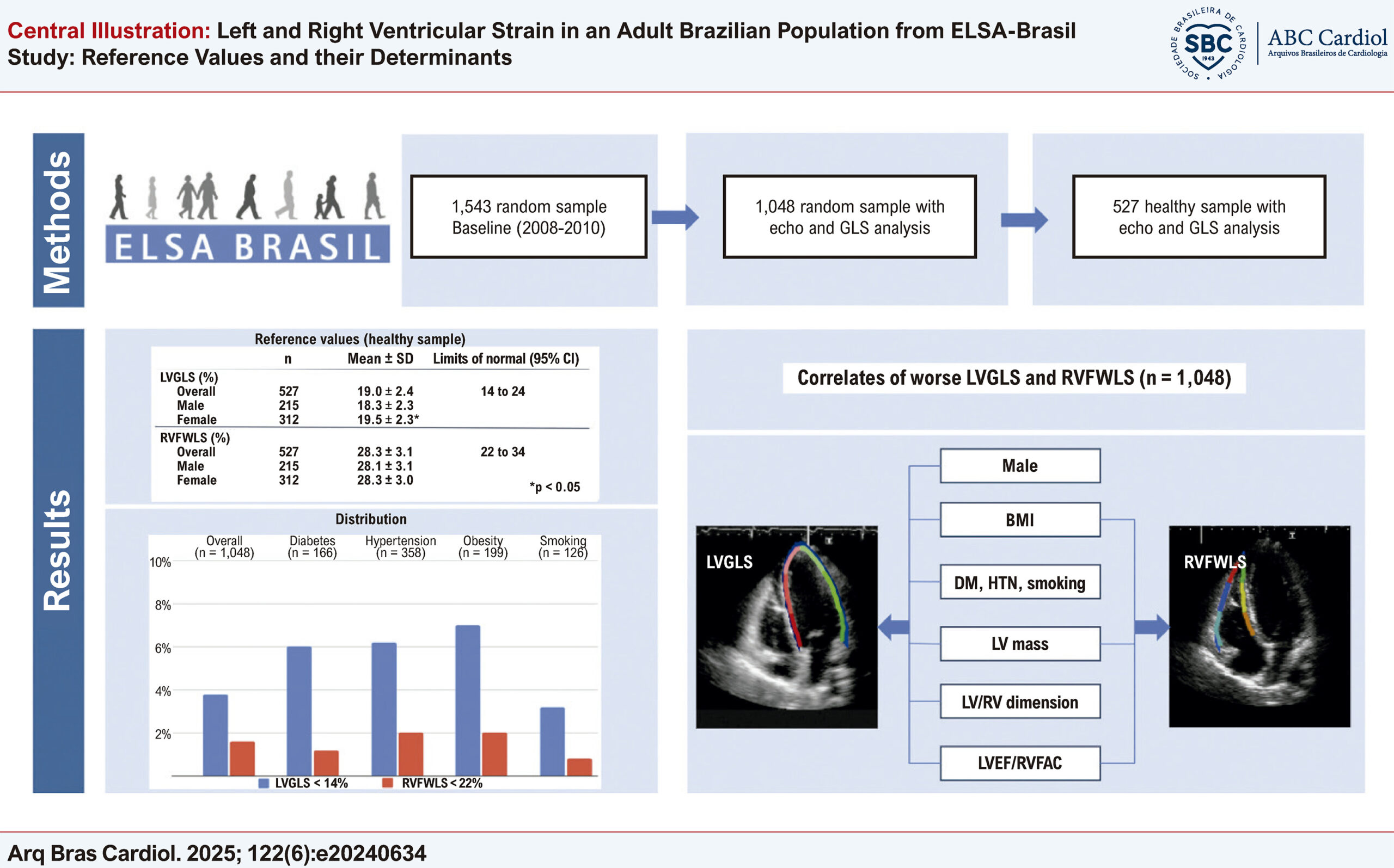
We included 1,048 participants from the ELSA-Brasil cohort who underwent echocardiography with GLS analysis. A healthy subsample (n = 527) was defined by excluding individuals with cardiovascular or renal disease, hypertension, or diabetes to establish GLS reference ranges. The prevalence of abnormal GLS was assessed, and factors associated with reduced LVGLS and RVFWLS were identified. Statistical significance was defined as p < 0.05.

Results

In the healthy subsample (mean age 50.2 years, 59% female), mean LVGLS was 19.0% (95% confidence interval: 14.3 to 23.8) and RVFWLS was 28.3% (95% confidence interval: 22.3 to 34.3). Females exhibited higher LVGLS and RVFWLS values than males, with no significant age-related differences. Abnormal LVGLS and RVFWLS were observed in 3.8% and 1.6% of participants, respectively. Lower LVGLS was associated with obesity, hypertension, and diabetes; reduced RVFWLS correlated with higher body mass index and LV mass.

Conclusions

We propose reference values for LVGLS and RVFWLS in a large Brazilian cohort, highlighting associations with cardiovascular comorbidities and ventricular structure.

  • Cohort Studies
  • Echocardiography
  • Global Longitudinal Strain
  • Heart Ventricles
  • Ventricular Function
Read Full Article
Creative Commons Attribution (CC BY) Creative Commons license
16/06/2025 DOI: 10.36660/abc.20240634i Views 1
Figure 1
ABCIC
Review Article

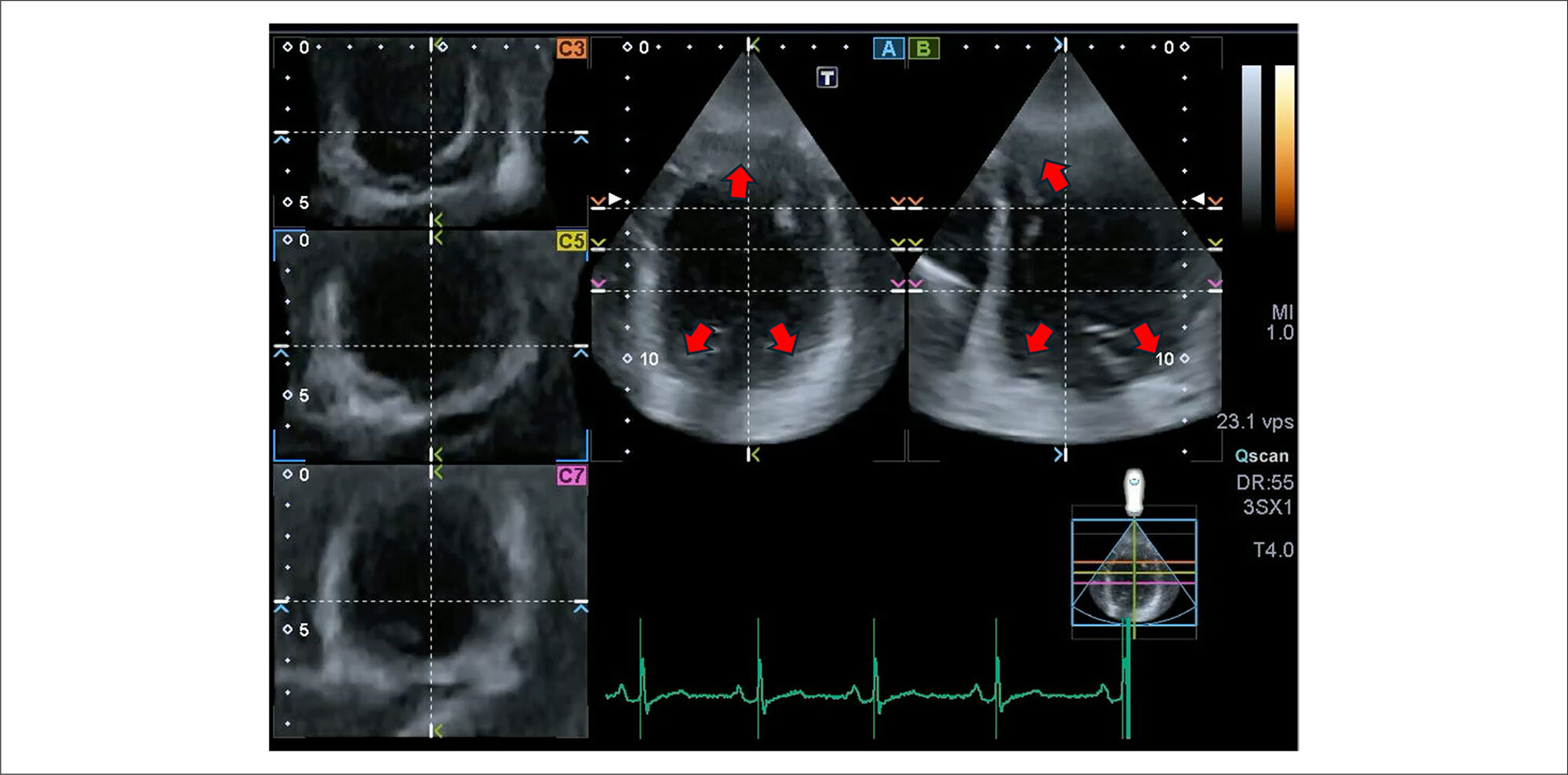
My Approach to 3-Dimensional Left Ventricular Strain

Rafael Bonafim Piveta et al. Miguel Osman Dias Aguiar Liria Lima Maria da Silva Marcelo Luiz Campos Vieira

Abstract

The role of strain analysis using 2-dimensional speckle tracking (2DST) has been well documented, and it represents an important complementary tool in the assessment of cardiac mechanics, adding value in the identification of incipient and subclinical myocardial injury in different clinical scenarios. A significant advance related to echocardiography was the development of 3-dimensional speckle tracking (3DST), which has the potential to overcome many limitations intrinsic to 2-dimensional technology, offering additional parameters of myocardial deformation (such as area strain) and a more comprehensive quantification of the geometry and function of the left ventricular myocardium, based on a single image acquisition. Among its main limitations, the low temporal and spatial resolution stands out, in addition to the dependence on high-quality images and a well-trained operator. Although it is a relatively recent technique that is still under development, several experimental studies and some clinical investigations have already demonstrated the reproducibility and potential applicability of 3DST. The objective of this article is to add information about adequate analysis of 3-dimensional left ventricular strain and explore its main points of vulnerability, discussing important variables to increase the accuracy and reproducibility of this technology.

  • Echocardiography
  • Heart Ventricles
  • Three-Dimensional Imaging
Read Full Article
Creative Commons Attribution (CC BY) Creative Commons license
28/05/2024 DOI: 10.36660/abcimg.20240028i Views 2
Body Surface Potential Mapping during Ventricular Depolarization in Athletes with Prolonged PQ Interval after Exercise
ABC
Original Article

Body Surface Potential Mapping during Ventricular Depolarization in Athletes with Prolonged PQ Interval after Exercise

Natalya I. Ivonina et al. Alexey G. Ivonin Irina M. Roshchevskaya

Abstract

Background:

Prolongation of the PQ interval, generally associated with an atrioventricular conduction delay, may be related to changes in intraventricular impulse spreading.

Objective:

To assess, using body surface potential mapping (BSPM), the process of ventricular depolarization in athletes with prolonged PQ intervals at rest and after exercise.

Methods:

The study included 7 cross-country skiers with a PQ interval of more than 200 ms (Prolonged-PQ group) and 7 with a PQ interval of less than 200 ms (Normal-PQ group). The BSPM from 64 unipolar torso leads was performed before (Pre-Ex) and after the bicycle exercise test (Post-Ex). Body surface equipotential maps were analyzed during ventricular depolarization. The significance level was 5%.

Results:

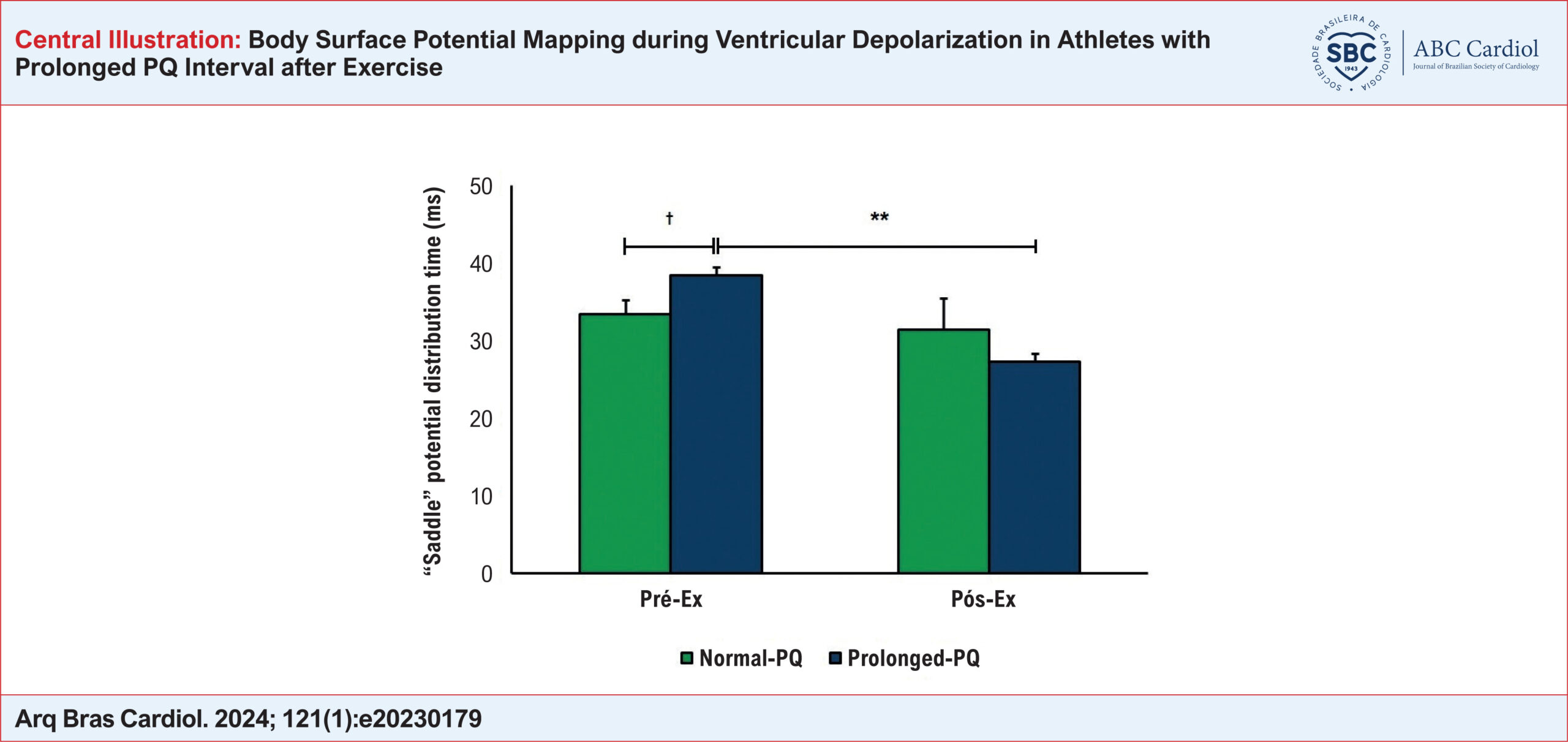
Compared to Normal-PQ athletes, the first and second periods of the stable position of cardiac potentials on the torso surface were longer, and the formation of the “saddle” potential distribution occurred later, at Pre-Ex, in Prolonged-PQ athletes. At Post-Ex, the Prolonged-PQ group showed a shortening of the first and second periods of stable potential distributions and a decrease in appearance time of the “saddle” phenomenon relative to Pre-Ex (to the values near to those of the Normal-PQ group). Additionally, at Post-Ex, the first inversion of potential distributions and the total duration of ventricular depolarization in Prolonged-PQ athletes decreased compared to Pre-Ex and with similar values in Normal-PQ athletes. Compared to Normal-PQ athletes, the second inversion was longer at Pre-Ex and Post-Ex in Prolonged-PQ athletes.

Conclusion:

Prolonged-PQ athletes had significant differences in the temporal characteristics of BSPM during ventricular depolarization both at rest and after exercise as compared to Normal-PQ athletes.

  • Athletes
  • Atrioventricular block
  • body surface potential mapping
  • Exercise Test
  • Heart Ventricles
Read Full Article
Creative Commons Attribution (CC BY) Creative Commons license
04/03/2024 DOI: 10.36660/abc.20230179 Views 1
Is Right Ventricular Cardiogenic Shock Important To You?
ABCHF
Review Article

Is Right Ventricular Cardiogenic Shock Important To You?

Marcus Vinícius Santos Andrade et al. Carlos Aurélio Santos Aragão Renato Moraes Pereira Figueiredo Breno Macedo de Almeida Lucas Guimarães da Rocha

Introduction

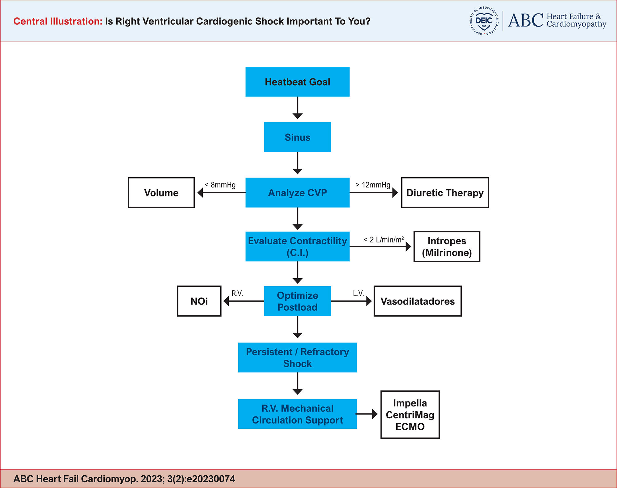
In the scenario of cardiogenic shock, attention was almost exclusively focused on the left ventricle (LV). Currently the concept that the circulatory system involves the interdependence between the LV and the right ventricle (RV) has been reinforced, and researchers seek to achieve a clearer understanding of the role of the right ventricle in cardiogenic shock.

In the 1940s, in search of a more in-depth understanding of the role of the RV, the American doctor and researcher Isaac Starr and his team carried out a series of experiments on animals, in which they caused severe damage to the right ventricle. Observing only a small increase in peripheral venous congestion, they concluded that “the weakness on the right side of the heart… appears to be less important” in the establishment of heart failure (HF).

[…]

  • Cardiogenic shock
  • Diagnosis
  • Heart Ventricles
  • Therapeutics
Read Full Article
Creative Commons Attribution (CC BY) Creative Commons license
31/12/2023 DOI: 10.36660/abchf.20230074 Views 1
Figure 1
ABCIC
Case Report

Uhl's Anomaly: Case Report and Differential Diagnosis in an Adult

Isaura Elaine Goncalves Moreira Rocha et al. Paulo Henrique Benevides Siqueira Beatriz Gonçalves Rocha Bruno Farias Oliveira … George Elias Luna Martins Simone Cristina Soares Brandão

Introduction

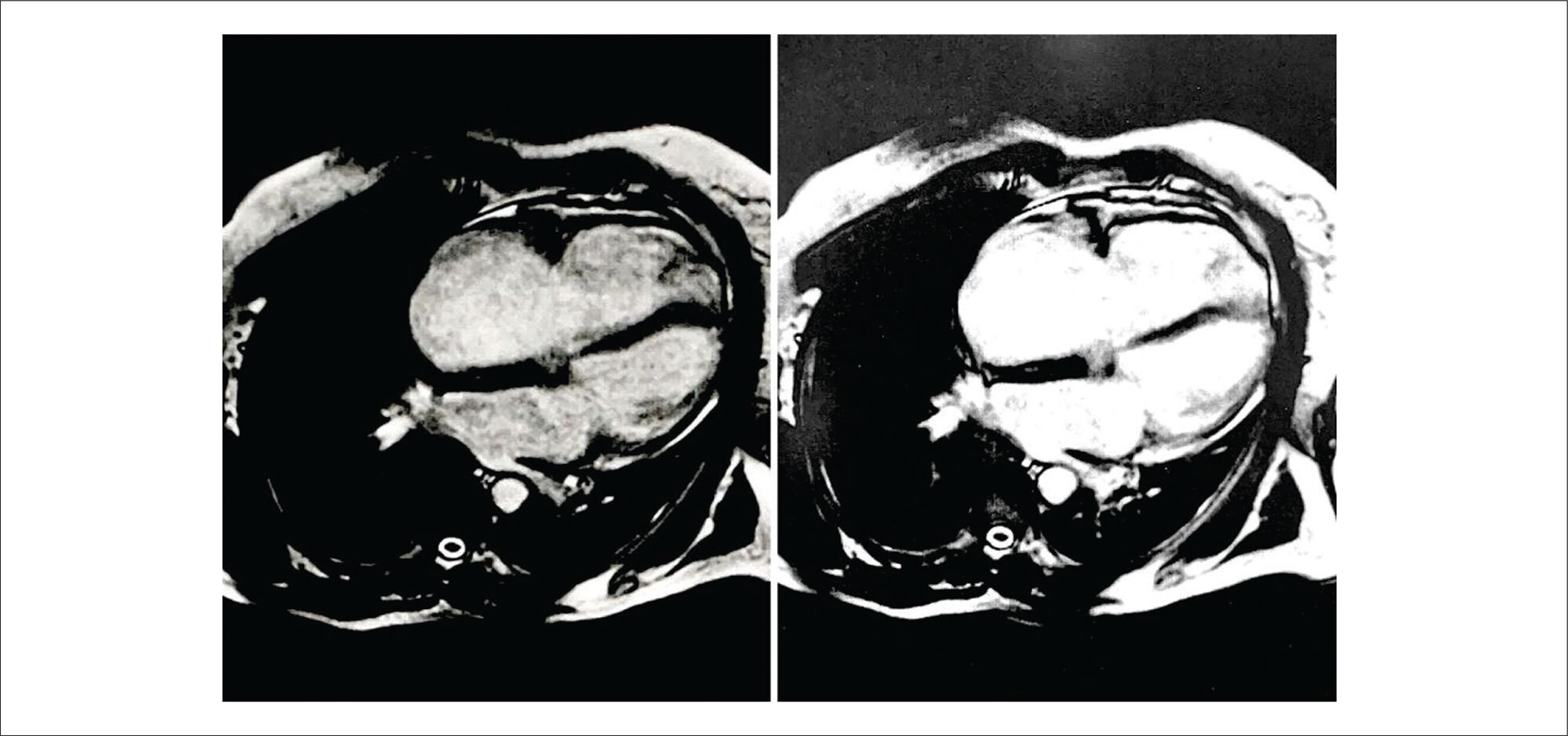
Uhl’s anomaly (UA) is a disorder exclusive to the right ventricle (RV) characterized by the absence of total or partial myocardium, in such a way that the ventricular wall is comprised of the overlapping of the endocardium and the epicardium, with no fatty tissue, or inflammatory or infiltrative process. The muscles of the atria, the interventricular septum and the left ventricle (LV) are not involved in the process.,

The clinical manifestations are due to right ventricular failure (RVF). Forms with partial involvement may be mildly symptomatic and remain undiagnosed for long periods, and may be confused with other more frequent pathologies that affect the right heart in adult life.,

[…]

  • Cardiovascular Abnormalities
  • Diagnosis, Differential
  • Heart Ventricles
Read Full Article
Creative Commons Attribution (CC BY) Creative Commons license
02/10/2023 DOI: 10.36660/abcimg.20230039i Views 1
Figure 1
ABCIC
Original Article

Right Ventricle Diastolic Function: Correlation with Age

Arthur Nascimento de Moura et al. Ana Cristina Camarozano Cintia Rocha Fortes de Sá Daniela de Castro Carmo … Jerônimo Antonio Fortunato Rubens Zenóbio Darwich Liz Andréa Villela Baroncini

Abstract

Introduction

Right ventricle (RV) elasticity tends to decrease with aging and may alter its diastolic function.

Objective

To evaluate the correlation between echocardiographic changes in RV diastolic function and advancing age in individuals with normal biventricular systolic function.

Methods

This retrospective observational cohort study included 110 patients: 66 (60%) women (62 [SD, 13.7] years) and 44 (40%) men (60.6 [SD, 10.8] years). The transthoracic echocardiography data included: spectral Doppler of tricuspid inflow E wave, A wave, E/A ratio, and E wave deceleration time; tissue Doppler of lateral tricuspid annulus e’ wave, a’ wave, and E/e’ ratio. Correlations were calculated using Pearson’s and Spearman’s correlation coefficients.

Results

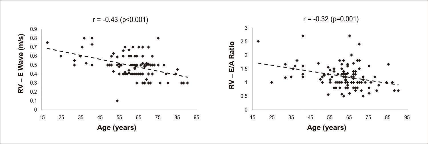
Only 35.2% of the sample had RV diastolic dysfunction. Of the evaluated echocardiographic variables, the E wave was negatively correlated with age in both women (r 0.473; p < 0.001) and men (r -0.37, p = 0.015). The E/A ratio was negatively correlated with age only in women (r -0.32; p = 0.001), and E wave deceleration time was positively correlated with age only in women (r 0.31; p = 0.014). The other variables were not significantly correlated with age in men or women.

Conclusions

We found a significant correlation between age and echocardiographic parameters for RV diastolic dysfunction, which was more evident in women.

  • Echocardiography
  • Heart Failure, Diastolic
  • Heart Ventricles
Read Full Article
Creative Commons Attribution (CC BY) Creative Commons license
27/06/2023 DOI: 10.36660/abcimg.2023333i Views 1
Figure 1
ABCIC
Case Report

Criss-Cross Heart: A Case Report

Mateus Oliveira Potratz et al. Lorrayne Zonatele Garbo Kamila Pompermaier Pessimilio André Silveira Loss … Crislane Belisario Ambrozim Ana Luiza Teixeira Albert Lima Danielle Lopes Rocha

Abstract

Criss-cross heart was first described in 1974. It is a rare congenital heart malformation that occurs in 8 cases per 1,000,000 children, and represents only 0.1% of congenital malformations. The diagnostic methods of choice are transthoracic echocardiography, cardiac magnetic resonance (CMR), computed tomography angiography (CT) and, sometimes, cardiac catheterization. This report describes the case of a newborn with a criss-cross heart in addition to double-outlet right ventricle (RV), with poorly positioned vessels, in addition to atrial septal defect (ASD), interventricular septal defect, tricuspid valve dysplasia and persistent left superior vena cava. The exact etiology of this malformation is not known, but it seems to occur due to rotation of the ventricles in their longitudinal axis, not accompanied by rotation of the atrial and atrioventricular (AV) valves. This movement produces abnormal ventricular inlets, determining that the RV be positioned on a superior plane and the left ventricle on an inferior plane. Although the exact cause of this anomaly is still unknown, it is believed that a genetic abnormality may be leading to these cases: mutation of the Cx43 gene. Diagnosis of the case concerned was given by transthoracic echocardiography and computed CT of the aorta and pulmonary arteries, which showed, in addition to the criss-cross heart, other abnormalities, such as double-outlet RV, large ASD and ventricular septal defect (VSD).

  • Congenital Heart Defects
  • Crisscross Heart
  • Heart Ventricles
Read Full Article
Creative Commons Attribution (CC BY) Creative Commons license
31/03/2023 DOI: 10.36660/abcimg.2023282i Views 1
Figure 1
ABC
Original Article

Body Surface Potential Mapping during Ventricular Depolarization in Rats after Acute Exhaustive Exercise

Alexey G. Ivonin et al. Svetlana L. Smirnova Irina M. Roshchevskaya

Abstract

Background

Exhaustive physical exercise can cause substantial changes in the electrical properties of the myocardium.

Objective

To evaluate, using body surface potential mapping, the electrical activity of the heart in rats during ventricular depolarization after acute exhaustive exercise.

Methods

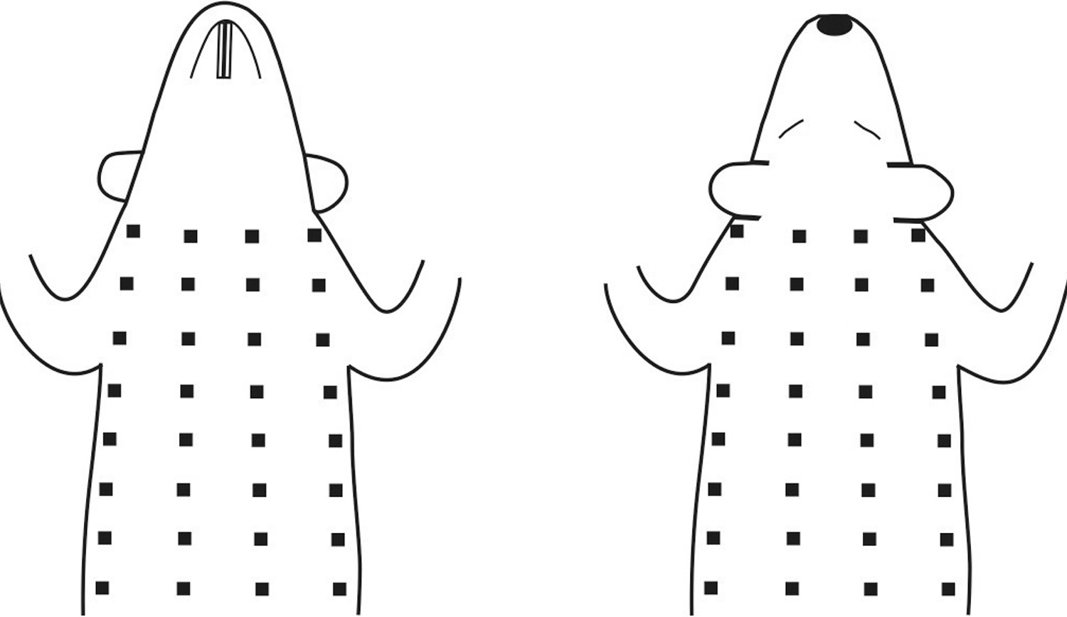
Twelve-week-old male rats were submitted to acute treadmill exercise at 36 m/min until exhaustion. Unipolar electrocardiograms (ECGs) from the torso surface were recorded in zoletil-anesthetized rats three to five days before (Pre-Ex), 5 and 10 minutes after exhaustive exercise (Post-Ex 5 and Post-Ex 10, respectively) simultaneously with ECGs in limb leads. The instantaneous body surface potential maps (BSPMs) were analyzed during ventricular depolarization. P values <0.05 were considered statistically significant.

Results

Compared with Pre-Ex, an early completion of the second inversion of potential distributions, an early completion of ventricular depolarization, as well as a decrease in the duration of the middle phase and the total duration of ventricular depolarization on BSPMs were revealed at Post-Ex 5. Also, compared with Pre-Ex, an increase in the amplitude of negative BSPM extremum at the R-wave peak on the ECG in lead II (RII-peak) and a decrease in the amplitude of negative BSPM extremum at 3 and 4 ms after RII-peak were showed at Post-Ex 5. At Post-Ex 10, parameters of BSPMs did not differ from those at Pre-Ex.

Conclusion

In rats, acute exhaustive exercise causes reversible changes in the temporal and amplitude characteristics of BSPMs during ventricular depolarization, most likely related to alterations in the excitation of the main mass of the ventricular myocardium.

  • acute
  • body surface potential mapping
  • Exercises
  • Heart Ventricles
  • Myocardium
  • Rats
Read Full Article
Creative Commons Attribution (CC BY) Creative Commons license
06/09/2022 DOI: 10.36660/abc.20211058
Figure 1
IJCS
Original Article

Effects of Exercise Training on Left Ventricular Diastolic Function Markers in Patients with Obstructive Sleep Apnea: A Randomized Study

Bruno G. Durante et al. Rosyvaldo Ferreira-Silva Thiago T. Goya Marta F. Lima … Ana Clara T. Rodrigues Luciano F. Drager Camila P. Jordão Amanda G. Rodrigues Maria Janieire de Nazare N. Alves Geraldo Lorenzi-Filho Carlos E. Negrão Linda M. Ueno-Pardi

Abstract

Background

Exercise training (ET) is an adjunctive treatment for obstructive sleep apnea (OSA) and its consequences. However, the effects of exercise on heart remodeling are unknown in the population with OSA.

Objective

We investigated the effect of ET on markers of diastolic function, sleep parameters, and functional capacity in patients with OSA.

Methods

Sedentary patients with OSA (apnea-hypopnea index, AHI ≥15 events/hr) were randomly assigned to untrained (n=18) and trained (n=20) strategies. Polysomnography, cardiopulmonary exercise test, and echocardiography were evaluated at the beginning and end of the study. ET consisted of 3 weekly sessions of aerobic exercise, resistance exercises, and flexibility training (72 sessions, completed in 11.65±0.86 months). A two-way analysis of variance (ANOVA) was used, followed by Tukey’s post-hoc test. The level of statistical significance was set at p<0.05 for all analyses.

Result

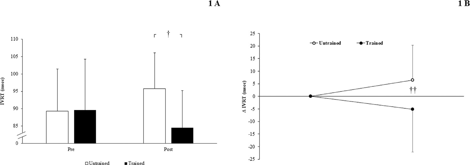
Thirty-eight patients were included (AHI:45±29 events/hr, age:52±7 y, body mass index: 30±4 kg/m2). They had similar baseline parameters. ET caused a significant change in OSA severity (AHI:4.5±18 versus -5.7±13 events/hr; arousal index:1.5±8 versus -6.1±13 events/hr, in untrained and trained groups respectively, p<0.05). The trained patients had an increase in functional capacity after intervention. ET improved isovolumetric relaxation time (IVRT, untrained=6.5±17.3 versus trained=-5.1±17.1 msec, p<0.05). There was a significant correlation between changes in IVRT and arousal index in the trained group (r =-0.54, p<0.05). No difference occurred in the other diastolic function parameters evaluated.

Conclusion

ET promotes modest but significant improvement in AHI, functional capacity, and cardiac IVRT, a validated parameter of diastolic function.

  • Exercise
  • Heart Ventricles
  • Obstructive
  • Sleep Apnea
Read Full Article
Creative Commons Attribution (CC BY) Creative Commons license
22/06/2022 DOI: 10.36660/ijcs.20210146 Views 1
ABCIC
Original Article

Echocardiographic Assessment of Right Chambers by Gender and Body Surface Area

Rafael Yared Forte et al. Ana Cristina Camarozano Daniela de Castro Carmo Jerônimo Antonio Fortunato … Rubens Zenóbio Darwich Júlia Ventura Niclewicz Liz Andréa Villela Baroncini

Abstract

Introduction

Assessment of right chambers dimensions and function according to gender, age, body surface area and body mass index is not uniformly performed.

Objective

To evaluate, by transthoracic echocardiography dimensions and function of right chambers, according to gender, body surface area and body mass index in an outpatient population.

Method

Cross-sectional study. Eighty-one patients were selected, 60.4 ± 13.5 years (57 women, 70%). Parameters assessed were the following: proximal, basal, medium and longitudinal, right ventricle diameter; right atrium area and right atrium volume; right ventricle wall thickness; tricuspid annular plane systolic excursion; and tricuspid annulus tissue Doppler and S’ wave. Results: Female sex presented lower diameters (Student’s t-test) of proximal right ventricle diameter (20.6 mm ± 2.4 vs. 22.7 mm ± 2.2; p = 0.001), basal right ventricle diameter (34,4 mm ± 3,5 vs. 38,2 mm ± 4,8; p < 0.001), medium right ventricle diameter (27 mm ± 3.3 vs. 32.4 mm ± 4.9; p < 0.001), right atrium area (13.7 cm² ± 2.7 vs. 16.6 cm² ± 3.9; p = 0.002) and right atrium volume (37 mL ± 10.6 vs. 50.7 mL ± 15.6 ; p = 0.002). Body mass index and body surface area correlated with proximal right ventricle diameter (correlation coefficient – CC 0.24; p = 0.03), basal right ventricle diameter (CC 0.22; p = 0.04), medium right ventricle diameter (CC 0.23; p = 0.04), longitudinal right ventricle diameter (CC 0.28; p = 0.01), right atrium area (CC 0.40; p = 0.001), and right atrium volume (CC 0.24; p = 0.0006).

Conclusion

Right ventricular diameters, right atrial area and volume were lower in females. A correlation was found with body mass index and body surface area. Tricuspid annular plane systolic excursion and S’ were not influenced by sex, body mass index and body surface area.

  • Echocardiography
  • Heart Ventricles
  • Organ Size
Read Full Article
Creative Commons Attribution (CC BY) Creative Commons license
14/01/2022 DOI: 10.5935/2318-8219.20200008
Figure 1
ABC
Original Article

Assessing Dynamic Atrioventricular Conduction Time to RR-interval Coupling in Athletes and Sedentary Subjects

Paulo Roberto Benchimol-Barbosa et al. Olivassé Nasario Junior Jurandir Nadal

Abstract

Background

Atrioventricular conduction time ( AVCT ) is influenced by autonomic input and subject to physiological remodeling.

Objective

To evaluate beat-by-beat AVCT and RR-interval variability in athletes and healthy sedentary subjects.

Methods

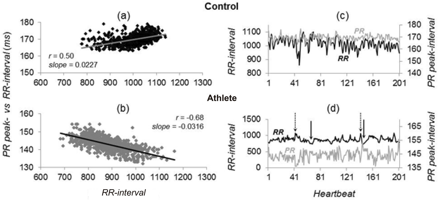
Twenty adults, including 10 healthy sedentary (Controls) and 10 elite long-distance runners (Athletes), age, weight and height-adjusted, underwent maximal metabolic equivalent (MET) assessment, and 15-min supine resting ECG recording seven days later. The interval between P-wave and R-wave peaks defined the AVCT . Mean (M) and standard deviation (SD) of consecutive RR-intervals (RR) and coupled AVCT were calculated, as well as regression lines of RR vs. AVCT (RR-AVCT) . Concordant AV conduction was defined as positive RR-AVCT slope and discordant otherwise. A multivariate linear regression model was developed to explain MET based on AVCT and RR-interval variability parameters. Significance-level: 5 %.

Results

In Athletes, M-RR and SD-RR values were higher than in Controls, whereas M-AVCT and SD-AVCT were not. RR-AVCT slopes were, respectively, 0.038 ± 0.022 and 0.0034 ± 0.017 (p < 0.05). Using a cut-off value of 0.0044 (AUC 0.92 ± 0.07; p < 0.001), RR-AVCT slope showed 100% specificity and 80% sensitivity. In a multivariate model, SD-RR and RR-AVCT slope were independent explanatory variables of MET (F-ratio: 17.2; p < 0.001), showing 100% specificity and 90% sensitivity (AUC 0.99 ± 0.02; p < 0.001).

Conclusion

In elite runners, AVCT to RR -interval dynamic coupling shows spontaneous discordant AV conduction, characterized by negative AVCT vs. RR -interval regression line slope. RR -intervals standard deviation and AVCT vs. RR -interval regression line slope are independent explanatory variables of MET (Arq Bras Cardiol. 2020; [online].ahead print, PP.0-0)

  • Adults
  • Athletes
  • Electrocardiography/methods
  • Heart Ventricles
  • physical fitness
  • Resistance Training
  • Sedentarism
  • Ventricular Function
  • Ventricular Remodeling
Read Full Article
Creative Commons Attribution (CC BY) Creative Commons license
08/05/2020 DOI: 10.36660/abc.20190281
Coronary fistula to the left ventricle: assessed by computed tomography
ABC
Case Report

Coronary fistula to the left ventricle: assessed by computed tomography

Roberto Moreno Mendonça et al. Rodrigo Lima Bandeira Fredson J. S Fonseca Robson Macedo Filho

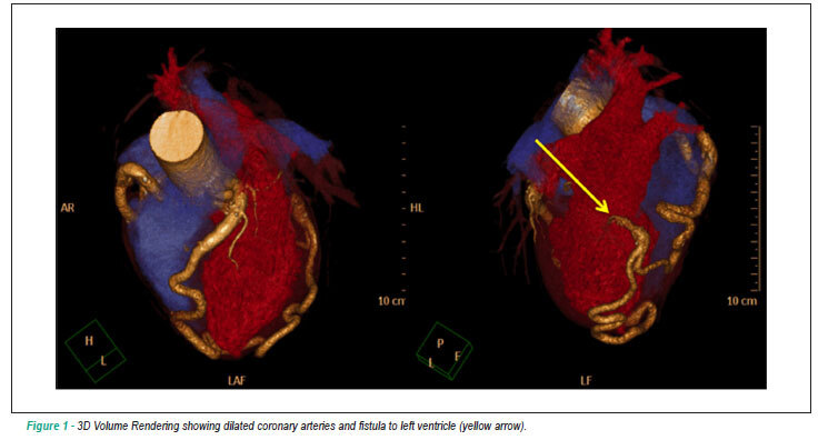
Coronary artery fistulas are rare and are most often diagnosed by echocardiography or by cine-angiocardiography. However, the computed tomography angiography (CTA) of coronary arteries has been gaining ground. The incidence of this disease is very low, with a more frequent occurrence of fistulas originating in the right coronary artery. There is a higher incidence of coronary artery fistulas to right heart chambers, with coronary artery fistulas to the left ventricle (LV) being rare. Treatment can be surgical or percutaneous. This report describes a case of coronary fistula to left ventricle diagnosed by CT angiography of coronary arteries in a hypertensive and asymptomatic 46-year-old male, who was tested positive for ischemia in an exercise test. The CT angiography ruled out coronary obstructive disease, but it revealed a coronary fistula to the left ventricular cavity.

  • Arterio-Arterial Fistula
  • Cardiovascular Diseases
  • Diagnosis
  • Heart Ventricles
  • Tomography Computed
Read Full Article
Creative Commons Attribution (CC BY) Creative Commons license
30/11/2011 DOI: 10.1590/S0066-782X2011001300017
Dysfunction of the pulmonary homograft used in the reconstruction of the right ventricle exit tract
ABC
Artigo Original

Dysfunction of the pulmonary homograft used in the reconstruction of the right ventricle exit tract

Andréa Weirich Lenzi et al. Márcia Olandoski Wanderley Saviolo Ferreira Fabio Said Sallum Nelson Itiro Miyague

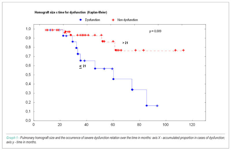
BACKGROUND: The pulmonary homograft has been used as an option in the correction of congenital cardiopathy with obstruction of the right ventricle exit tract. The long term results, however, are little satisfactory. OBJECTIVE: Identify the risk factors associated to the dysfunction and the pulmonary homograft failure. METHODS: Study with children submitted to the enlargement of the exit tract of the right ventricle with pulmonary homograft. The clinical, surgical, evolutional and morphological aspects of the prosthesis variables were analyzed as risk factors. RESULTS: The final sample of 75 patients with median age at the surgery of 22 months, varying from 1-157 months, presented 13 patients (17.0%) who developed dysfunction of the homograft, characterized by stenosis or severe pulmonary insufficiency. The occurrence time between the homograft implant and the detection of the dysfunction was of 45 ± 20 months. When the size of the homograft was smaller than 21 mm and the Z score of the pulmonary valva is lower than zero, or higher than three, the risk factors were considered for the dysfunction occurrence. CONCLUSION: The pulmonary homograft smaller than 21 mm and the inadequate pulmonary valva for the age and weight of the patient are determining factors for the prosthesis dysfunction.

  • Congenital
  • Heart Defects
  • Heart Ventricles
  • Transplantation, homologous
Read Full Article
Creative Commons Attribution (CC BY) Creative Commons license
10/12/2010 DOI: 10.1590/S0066-782X2010005000158 Views 1
Biventricular repair in unbalanced atrioventricular septal defect
ABC
Case Report

Biventricular repair in unbalanced atrioventricular septal defect

Edmar Atik et al. Patrícia O. Marques Rogério A. Miranda Vitor C. Guerra … Lucília Santana Faria Marcelo Jatene

We present the case of a 2.5 year-old child with unbalanced atrioventricular septal defect due to a small left ventricle (LV) (mitral annulus of 10mm and a 0.4 ratio in relation to the tricuspid annulus, LVDD: 17 mm, LV Vd2: 15 ml/m² and LV/RV long-axis ratio of 0.71); he had a favorable outcome after biventricular surgical repair. Normal LV development was observed three months after the operation (mitral annulus of 22 mm, with a 0.84 ratio in relation to the tricuspid annulus, and LVDD of 30 mm). Current parameters for utilization of the hypoplastic ventricle are discussed.

  • Heart Defects, Congenital
  • Heart Septal Defects, Ventricular
  • Heart Ventricles
Read Full Article
Creative Commons Attribution (CC BY) Creative Commons license
29/06/2009 DOI: 10.1590/S0066-782X2009000400015 Views 1
Right ventricular tumor in a patient with melanoma
ABC
Relato de Caso

Right ventricular tumor in a patient with melanoma

Vinícius Daudt Morais et al. Flávio Dalbem Krás Borges Vicente Restelli

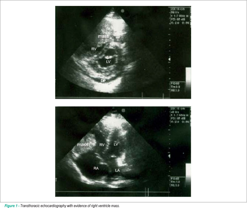
Primary tumors of the heart are not common. However, metastatic neoplasms affecting the heart are more commonly found. Postmortem studies have shown that some tumors have reported secondary cardiac implants to be over 50%. Such involvement is to be taken into account for patients with a history of neoplasm and who present conduction disorders, murmur, cardiomegaly or arrhythmia. This is to report the case of a 39-year-old man who had been referred due to fatigue and dyspnea on effort. Echocardiogram evidence showed large tumoral mass in right ventricle. Medical history showed previous melanoma; further evaluation showed metastasis to lungs, heart and brain. Outcome was death. The uniqueness in this case relies on the large metastatic mass in right ventricle, thus illustrating a rare clinical condition of guarded prognosis.

  • Heart Ventricles
  • melanoma
  • Neoplasm Metastasis
  • Neoplasms
Read Full Article
Creative Commons Attribution (CC BY) Creative Commons license
01/10/2008 DOI: 10.1590/S0066-782X2008001500012
Cardiac ventricular weights recorded at the autopsy of healthy subjects who died of external causes
ABC
Artigo Original

Cardiac ventricular weights recorded at the autopsy of healthy subjects who died of external causes

Sérgio Lamêgo Rodrigues et al. Enildo Broetto Pimentel José Geraldo Mill

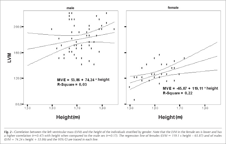
OBJECTIVE:To establish cardiac ventricular weights recorded during the autopsy of healthy individuals who died of external causes, aiming at determining normality patterns in our population. METHODS:A total of 94 hearts were evaluated at the Forensics Department of the city of Vitória, Espírito Santo. After the heart removal and resection of the atria and epicardial fat, the right ventricle (RV) and the left ventricle (LV), including the septum, were separated and weighed and the mass was indexed by the height. The Kolmogorov-Smirnov test was used to test the normality of the distribution. Data are presented as means± SD. RESULTS:After the exclusion of 12 hearts (possible cardiovascular disease detected post-mortem) 82 hearts were examined (52 males and 30 females, aged 16-68 yrs, 31±12 yrs). The weight of the LV was 181±25 g and 125±15 g, and the weight of the RV was 54±7 g and 38±6 g; the LV mass indexed by height was 105±14 g/m and 78±8 g/m, for males and females, respectively. The P95 of the LV weight was 218 g and 128 g/m in males and 148 g and 88 g/m in females. No significant correlation between ventricular mass and age was observed. CONCLUSION:The weight of the LV in the males from our sample was higher than that reported in the contemporary literature. Our results suggest that the presence of LV hypertrophy can be inferred in the presence of LV mass > 218 g or 128 g/m in males and 148 g or 88 g/m in females.

  • Heart Ventricles
  • Hypertrophy Left Ventricular
  • Organ Size
Read Full Article
Creative Commons Attribution (CC BY) Creative Commons license
07/12/2007 DOI: 10.1590/S0066-782X2007001700001
Radiofrequency catheter ablation of premature ventricular contractions originating in the right ventricular outflow tract
ABC
Artigo Original

Radiofrequency catheter ablation of premature ventricular contractions originating in the right ventricular outflow tract

Francisco C. C. Darrieux et al. Maurício I. Scanavacca Denise T. Hachul Sissy L. Melo … André B. D'Ávilla César J. Gruppi Paulo J. Moffa Eduardo A. Sosa

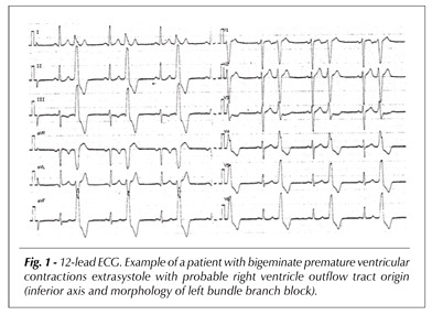
OBJECTIVES: To evaluate if radiofrequency catheter ablation is an effective procedure for the treatment of right ventricular outflow tract premature ventricular contractions (RVOT-PVC) and ascertain if it results in an improvement of symptoms. METHODS: A prospective study with 30 consecutive patients (mean age 40 ± 13 years, 25 females), with no apparent structural cardiopathy, with very frequent (mean density of 1,263 ± 593/h) RVOT-PVC, symptomatic for more than one year (mean = 74 months) and resistant to antiarrhythmic drugs (3 ± 1.7, including beta-blockers), who underwent radiofrequency catheter ablation. RESULTS: After the first procedure, there were 23 initial successful cases (76.6%) and 7 initial failures (23.4%). Four patients experienced relapses, two of whom did not undergo the second procedure. The second procedure was carried out in 9 patients (7 initial failures and 2 relapses), and there was success in 5 additional patients, one of them by epicardial access. The final success rate was 80% (24/30), and there were no major complications. After a mean follow-up of 14 ± 6 months, in the successful group there was a reduction greater than 90% in density of premature ventricular contractions (PVC) (24/24; p<0.0001) and a resulting absence of symptoms in the majority of patients (23/24; p<0.001). CONCLUSION: Radiofrequency catheter ablation is a safe and effective treatment for patients with persistent and symptomatic PVC with RVOT morphology.

  • Arrhythmia
  • Catheter Ablation
  • Heart Ventricles
  • premature ventricular contractions
Read Full Article
Creative Commons Attribution (CC BY) Creative Commons license
21/05/2007 DOI: 10.1590/S0066-782X2007000300003 Views 2

How to cite this article

EndNote RIS

Electronic Document Format (Vancouver)

Verdoia M. The “Right” Side of Myocardial Infarction: Sinister Outcomes?. International Journal of Cardiovascular Sciences 2026;39:e20250249.

Electronic Document Format (ABNT)

Verdoia, Monica. The “Right” Side of Myocardial Infarction: Sinister Outcomes?. International Journal of Cardiovascular Sciences, v. 39, e20250249, Mar. 2026.

Electronic Document Format (APA)

Verdoia, M. (2026). The “Right” Side of Myocardial Infarction: Sinister Outcomes?. International Journal of Cardiovascular Sciences, 39, e20250249.

Electronic Document Format (ISO)

Verdoia, Monica. The “Right” Side of Myocardial Infarction: Sinister Outcomes?. International Journal of Cardiovascular Sciences [online]. 2026, vol. 39, [cited 2026-10-14], e20250249. Available from: <https://journals.cardiol.br/en/article/the-right-side-of-myocardial-infarction-sinister-outcomes/>. ISSN 2359-4802

EndNote RIS

Electronic Document Format (Vancouver)

Maia AS, Camacho MHB, Perego AF, Mayer DH, Fonseca JHP. Transcatheter Correction of Aortic-Right Ventricular Shunt Following Aortic Valve Replacement: A Case Report. International Journal of Cardiovascular Sciences 2026;38:e20250060.

Electronic Document Format (ABNT)

Maia, Adnaldo da Silveira; Camacho, Mauro Henrique Batista; Perego, Andresa Fernandes; Mayer, Dayara Hoffmann; Fonseca, José Honório Palma da. Transcatheter Correction of Aortic-Right Ventricular Shunt Following Aortic Valve Replacement: A Case Report. International Journal of Cardiovascular Sciences, v. 38, e20250060, Jan. 2026.

Electronic Document Format (APA)

Maia, A. S., Camacho, M. H. B., Perego, A. F., Mayer, D. H., & Fonseca, J. H. P. (2026). Transcatheter Correction of Aortic-Right Ventricular Shunt Following Aortic Valve Replacement: A Case Report. International Journal of Cardiovascular Sciences, 38, e20250060.

Electronic Document Format (ISO)

Maia, Adnaldo da Silveira and Camacho, Mauro Henrique Batista and Perego, Andresa Fernandes and Mayer, Dayara Hoffmann and Fonseca, José Honório Palma da. Transcatheter Correction of Aortic-Right Ventricular Shunt Following Aortic Valve Replacement: A Case Report. International Journal of Cardiovascular Sciences [online]. 2026, vol. 38, [cited 2026-10-14], e20250060. Available from: <https://journals.cardiol.br/en/article/transcatheter-correction-of-aortic-right-ventricular-shunt-following-aortic-valve-replacement-a-case-report/>. ISSN 2359-4802

EndNote RIS

Electronic Document Format (Vancouver)

Crivelaro PCF, Machado GP, Gazzana MB, Lorenzi W, Benedetto IG, Valle FH, Wainstein RV, et al. Advanced Management of Right Heart Failure: An Illustrative Case. ABC Heart Fail Cardiomyop 2025;5(2):e20250017.

Electronic Document Format (ABNT)

Crivelaro, Pedro Castilhos de Freitas; Machado, Guilherme Pinheiro; Gazzana, Marcelo Basso; Lorenzi, William; Benedetto, Igor Gorski; Valle, Felipe Homem; Wainstein, Rodrigo Vugman. Advanced Management of Right Heart Failure: An Illustrative Case. ABC Heart Fail Cardiomyop, v. 5, n. 2, e20250017, Nov. 2025.

Electronic Document Format (APA)

Crivelaro, P. C. F., Machado, G. P., Gazzana, M. B., Lorenzi, W., Benedetto, I. G., Valle, F. H., & Wainstein, R. V. (2025). Advanced Management of Right Heart Failure: An Illustrative Case. ABC Heart Fail Cardiomyop, 5(2), e20250017.

Electronic Document Format (ISO)

Crivelaro, Pedro Castilhos de Freitas and Machado, Guilherme Pinheiro and Gazzana, Marcelo Basso and Lorenzi, William and Benedetto, Igor Gorski and Valle, Felipe Homem and Wainstein, Rodrigo Vugman. Advanced Management of Right Heart Failure: An Illustrative Case. ABC Heart Fail Cardiomyop [online]. 2025, vol. 5, n. 2, [cited 2026-10-14], e20250017. Available from: <https://journals.cardiol.br/en/article/advanced-management-of-right-heart-failure-an-illustrative-case/>

EndNote RIS

Electronic Document Format (Vancouver)

Pianca EG, Foppa M, Schmitz GB, Cañon-Montañez W, Duncan BB, Santos ABS. Left and Right Ventricular Strain in an Adult Brazilian Population from ELSA-Brasil Study: Reference Values and their Determinants. Arquivos Brasileiros de Cardiologia 2025;122(6):e20240634.

Electronic Document Format (ABNT)

Pianca, Eduardo Gatti; Foppa, Murilo; Schmitz, Giulia Bevilacqua; Cañon-Montañez, Wilson; Duncan, Bruce Bartholow; Santos, Angela Barreto Santiago. Left and Right Ventricular Strain in an Adult Brazilian Population from ELSA-Brasil Study: Reference Values and their Determinants. Arquivos Brasileiros de Cardiologia, v. 122, n. 6, e20240634, Jun. 2025.

Electronic Document Format (APA)

Pianca, E. G., Foppa, M., Schmitz, G. B., Cañon-Montañez, W., Duncan, B. B., & Santos, A. B. S. (2025). Left and Right Ventricular Strain in an Adult Brazilian Population from ELSA-Brasil Study: Reference Values and their Determinants. Arquivos Brasileiros de Cardiologia, 122(6), e20240634.

Electronic Document Format (ISO)

Pianca, Eduardo Gatti and Foppa, Murilo and Schmitz, Giulia Bevilacqua and Cañon-Montañez, Wilson and Duncan, Bruce Bartholow and Santos, Angela Barreto Santiago. Left and Right Ventricular Strain in an Adult Brazilian Population from ELSA-Brasil Study: Reference Values and their Determinants. Arquivos Brasileiros de Cardiologia [online]. 2025, vol. 122, n. 6, [cited 2026-10-14], e20240634. Available from: <https://journals.cardiol.br/en/article/left-and-right-ventricular-strain-in-an-adult-brazilian-population-from-elsa-brasil-study-reference-values-and-their-determinants/>. ISSN 0066-782X

EndNote RIS

Electronic Document Format (Vancouver)

Piveta RB, Aguiar MOD, Silva LLM, Vieira MLC. My Approach to 3-Dimensional Left Ventricular Strain. ABC Imagem Cardiovascular 2024;37(2):e20240028.

Electronic Document Format (ABNT)

Piveta, Rafael Bonafim; Aguiar, Miguel Osman Dias; Silva, Liria Lima Maria da; Vieira, Marcelo Luiz Campos. My Approach to 3-Dimensional Left Ventricular Strain. ABC Imagem Cardiovascular, v. 37, n. 2, e20240028, May. 2024.

Electronic Document Format (APA)

Piveta, R. B., Aguiar, M. O. D., Silva, L. L. M. , & Vieira, M. L. C. (2024). My Approach to 3-Dimensional Left Ventricular Strain. ABC Imagem Cardiovascular, 37(2), e20240028.

Electronic Document Format (ISO)

Piveta, Rafael Bonafim and Aguiar, Miguel Osman Dias and Silva, Liria Lima Maria da and Vieira, Marcelo Luiz Campos. My Approach to 3-Dimensional Left Ventricular Strain. ABC Imagem Cardiovascular [online]. 2024, vol. 37, n. 2, [cited 2026-10-14], e20240028. Available from: <https://journals.cardiol.br/en/article/my-approach-to-3-dimensional-left-ventricular-strain/>. ISSN 2318-8219

EndNote RIS

Electronic Document Format (Vancouver)

Ivonina NI, Ivonin AG, Roshchevskaya IM. Body Surface Potential Mapping during Ventricular Depolarization in Athletes with Prolonged PQ Interval after Exercise. Arquivos Brasileiros de Cardiologia 2024;121(1):e20230179.

Electronic Document Format (ABNT)

Ivonina, Natalya I.; Ivonin, Alexey G.; Roshchevskaya, Irina M.. Body Surface Potential Mapping during Ventricular Depolarization in Athletes with Prolonged PQ Interval after Exercise. Arquivos Brasileiros de Cardiologia, v. 121, n. 1, e20230179, Mar. 2024.

Electronic Document Format (APA)

Ivonina, N. I., Ivonin, A. G., & Roshchevskaya, I. M. (2024). Body Surface Potential Mapping during Ventricular Depolarization in Athletes with Prolonged PQ Interval after Exercise. Arquivos Brasileiros de Cardiologia, 121(1), e20230179.

Electronic Document Format (ISO)

Ivonina, Natalya I. and Ivonin, Alexey G. and Roshchevskaya, Irina M.. Body Surface Potential Mapping during Ventricular Depolarization in Athletes with Prolonged PQ Interval after Exercise. Arquivos Brasileiros de Cardiologia [online]. 2024, vol. 121, n. 1, [cited 2026-10-14], e20230179. Available from: <https://journals.cardiol.br/en/article/body-surface-potential-mapping-during-ventricular-depolarization-in-athletes-with-prolonged-pq-interval-after-exercise/>. ISSN 0066-782X

EndNote RIS

Electronic Document Format (Vancouver)

Andrade MVS, Aragão CAS, Figueiredo RMP, Almeida BM, Rocha LG. Is Right Ventricular Cardiogenic Shock Important To You?. ABC Heart Fail Cardiomyop 2023;3(2):e20230074.

Electronic Document Format (ABNT)

Andrade, Marcus Vinícius Santos; Aragão, Carlos Aurélio Santos; Figueiredo, Renato Moraes Pereira; Almeida, Breno Macedo de; Rocha, Lucas Guimarães da. Is Right Ventricular Cardiogenic Shock Important To You?. ABC Heart Fail Cardiomyop, v. 3, n. 2, e20230074, Dec. 2023.

Electronic Document Format (APA)

Andrade, M. V. S., Aragão, C. A. S., Figueiredo, R. M. P., Almeida, B. M. , & Rocha, L. G. (2023). Is Right Ventricular Cardiogenic Shock Important To You?. ABC Heart Fail Cardiomyop, 3(2), e20230074.

Electronic Document Format (ISO)

Andrade, Marcus Vinícius Santos and Aragão, Carlos Aurélio Santos and Figueiredo, Renato Moraes Pereira and Almeida, Breno Macedo de and Rocha, Lucas Guimarães da. Is Right Ventricular Cardiogenic Shock Important To You?. ABC Heart Fail Cardiomyop [online]. 2023, vol. 3, n. 2, [cited 2026-10-14], e20230074. Available from: <https://journals.cardiol.br/en/article/is-right-ventricular-cardiogenic-shock-important-to-you/>

EndNote RIS

Electronic Document Format (Vancouver)

Rocha IEGM, Siqueira PHB, Rocha BG, Oliveira BF, Martins GEL, Brandão SCS. Uhl’s Anomaly: Case Report and Differential Diagnosis in an Adult. ABC Imagem Cardiovascular 2023;36(3):e20230039.

Electronic Document Format (ABNT)

Rocha, Isaura Elaine Goncalves Moreira; Siqueira, Paulo Henrique Benevides; Rocha, Beatriz Gonçalves; Oliveira, Bruno Farias; Martins, George Elias Luna; Brandão, Simone Cristina Soares. Uhl’s Anomaly: Case Report and Differential Diagnosis in an Adult. ABC Imagem Cardiovascular, v. 36, n. 3, e20230039, Oct. 2023.

Electronic Document Format (APA)

Rocha, I. E. G. M., Siqueira, P. H. B., Rocha, B. G., Oliveira, B. F., Martins, G. E. L., & Brandão, S. C. S. (2023). Uhl’s Anomaly: Case Report and Differential Diagnosis in an Adult. ABC Imagem Cardiovascular, 36(3), e20230039.

Electronic Document Format (ISO)

Rocha, Isaura Elaine Goncalves Moreira and Siqueira, Paulo Henrique Benevides and Rocha, Beatriz Gonçalves and Oliveira, Bruno Farias and Martins, George Elias Luna and Brandão, Simone Cristina Soares. Uhl’s Anomaly: Case Report and Differential Diagnosis in an Adult. ABC Imagem Cardiovascular [online]. 2023, vol. 36, n. 3, [cited 2026-10-14], e20230039. Available from: <https://journals.cardiol.br/en/article/uhls-anomaly-case-report-and-differential-diagnosis-in-an-adult/>. ISSN 2318-8219

EndNote RIS

Electronic Document Format (Vancouver)

Moura AN, Camarozano AC, Sá CRF, Carmo DC, Fortunato JA, Darwich RZ, Baroncini LAV, et al. Right Ventricle Diastolic Function: Correlation with Age. ABC Imagem Cardiovascular 2023;36(2):e333.

Electronic Document Format (ABNT)

Moura, Arthur Nascimento de; Camarozano, Ana Cristina; Sá, Cintia Rocha Fortes de; Carmo, Daniela de Castro; Fortunato, Jerônimo Antonio; Darwich, Rubens Zenóbio; Baroncini, Liz Andréa Villela. Right Ventricle Diastolic Function: Correlation with Age. ABC Imagem Cardiovascular, v. 36, n. 2, e333, Jun. 2023.

Electronic Document Format (APA)

Moura, A. N. , Camarozano, A. C., Sá, C. R. F. , Carmo, D. C., Fortunato, J. A., Darwich, R. Z., & Baroncini, L. A. V. (2023). Right Ventricle Diastolic Function: Correlation with Age. ABC Imagem Cardiovascular, 36(2), e333.

Electronic Document Format (ISO)

Moura, Arthur Nascimento de and Camarozano, Ana Cristina and Sá, Cintia Rocha Fortes de and Carmo, Daniela de Castro and Fortunato, Jerônimo Antonio and Darwich, Rubens Zenóbio and Baroncini, Liz Andréa Villela. Right Ventricle Diastolic Function: Correlation with Age. ABC Imagem Cardiovascular [online]. 2023, vol. 36, n. 2, [cited 2026-10-14], e333. Available from: <https://journals.cardiol.br/en/article/right-ventricle-diastolic-function-correlation-with-age/>. ISSN 2318-8219

EndNote RIS

Electronic Document Format (Vancouver)

Potratz MO, Garbo LZ, Pessimilio KP, Loss AS, Ambrozim CB, Lima ALTA, Rocha DL, et al. Criss-Cross Heart: A Case Report. ABC Imagem Cardiovascular 2023;36(1):e282.

Electronic Document Format (ABNT)

Potratz, Mateus Oliveira; Garbo, Lorrayne Zonatele; Pessimilio, Kamila Pompermaier; Loss, André Silveira; Ambrozim, Crislane Belisario; Lima, Ana Luiza Teixeira Albert; Rocha, Danielle Lopes. Criss-Cross Heart: A Case Report. ABC Imagem Cardiovascular, v. 36, n. 1, e282, Mar. 2023.

Electronic Document Format (APA)

Potratz, M. O., Garbo, L. Z., Pessimilio, K. P., Loss, A. S., Ambrozim, C. B., Lima, A. L. T. A., & Rocha, D. L. (2023). Criss-Cross Heart: A Case Report. ABC Imagem Cardiovascular, 36(1), e282.

Electronic Document Format (ISO)

Potratz, Mateus Oliveira and Garbo, Lorrayne Zonatele and Pessimilio, Kamila Pompermaier and Loss, André Silveira and Ambrozim, Crislane Belisario and Lima, Ana Luiza Teixeira Albert and Rocha, Danielle Lopes. Criss-Cross Heart: A Case Report. ABC Imagem Cardiovascular [online]. 2023, vol. 36, n. 1, [cited 2026-10-14], e282. Available from: <https://journals.cardiol.br/en/article/criss-cross-heart-a-case-report/>. ISSN 2318-8219

EndNote RIS

Electronic Document Format (Vancouver)

Ivonin AG, Smirnova SL, Roshchevskaya IM. Body Surface Potential Mapping during Ventricular Depolarization in Rats after Acute Exhaustive Exercise. Arquivos Brasileiros de Cardiologia 2022;119(5):766-75.

Electronic Document Format (ABNT)

Ivonin, Alexey G.; Smirnova, Svetlana L.; Roshchevskaya, Irina M.. Body Surface Potential Mapping during Ventricular Depolarization in Rats after Acute Exhaustive Exercise. Arquivos Brasileiros de Cardiologia, v. 119, n. 5, p. 766-775, Sep. 2022.

Electronic Document Format (APA)

Ivonin, A. G., Smirnova, S. L., & Roshchevskaya, I. M. (2022). Body Surface Potential Mapping during Ventricular Depolarization in Rats after Acute Exhaustive Exercise. Arquivos Brasileiros de Cardiologia, 119(5), 766-775.

Electronic Document Format (ISO)

Ivonin, Alexey G. and Smirnova, Svetlana L. and Roshchevskaya, Irina M.. Body Surface Potential Mapping during Ventricular Depolarization in Rats after Acute Exhaustive Exercise. Arquivos Brasileiros de Cardiologia [online]. 2022, vol. 119, n. 5, [cited 2026-10-14], pp. 766-775. Available from: <https://journals.cardiol.br/en/article/body-surface-potential-mapping-during-ventricular-depolarization-in-rats-after-acute-exhaustive-exercise/>. ISSN 0066-782X

EndNote RIS

Electronic Document Format (Vancouver)

Durante BG, Ferreira-Silva R, Goya TT, Lima MF, Rodrigues ACT, Drager LF, Jordão CP, et al. Effects of Exercise Training on Left Ventricular Diastolic Function Markers in Patients with Obstructive Sleep Apnea: A Randomized Study. International Journal of Cardiovascular Sciences 2022;35(5):646-5.

Electronic Document Format (ABNT)

Durante, Bruno G.; Ferreira-Silva, Rosyvaldo; Goya, Thiago T.; Lima, Marta F.; Rodrigues, Ana Clara T.; Drager, Luciano F.; Jordão, Camila P.; Rodrigues, Amanda G.; Alves, Maria Janieire de Nazare N.; Lorenzi-Filho, Geraldo; Negrão, Carlos E.; Ueno-Pardi, Linda M.. Effects of Exercise Training on Left Ventricular Diastolic Function Markers in Patients with Obstructive Sleep Apnea: A Randomized Study. International Journal of Cardiovascular Sciences, v. 35, n. 5, p. 646-656, Jun. 2022.

Electronic Document Format (APA)

Durante, B. G., Ferreira-Silva, R., Goya, T. T., Lima, M. F., Rodrigues, A. C. T., Drager, L. F., Jordão, C. P., Rodrigues, A. G., Alves, M. J. N. N., Lorenzi-Filho, G., Negrão, C. E., & Ueno-Pardi, L. M. (2022). Effects of Exercise Training on Left Ventricular Diastolic Function Markers in Patients with Obstructive Sleep Apnea: A Randomized Study. International Journal of Cardiovascular Sciences, 35(5), 646-656.

Electronic Document Format (ISO)

Durante, Bruno G. and Ferreira-Silva, Rosyvaldo and Goya, Thiago T. and Lima, Marta F. and Rodrigues, Ana Clara T. and Drager, Luciano F. and Jordão, Camila P. and Rodrigues, Amanda G. and Alves, Maria Janieire de Nazare N. and Lorenzi-Filho, Geraldo and Negrão, Carlos E. and Ueno-Pardi, Linda M.. Effects of Exercise Training on Left Ventricular Diastolic Function Markers in Patients with Obstructive Sleep Apnea: A Randomized Study. International Journal of Cardiovascular Sciences [online]. 2022, vol. 35, n. 5, [cited 2026-10-14], pp. 646-656. Available from: <https://journals.cardiol.br/en/article/effects-of-exercise-training-on-left-ventricular-diastolic-function-markers-in-patients-with-obstructive-sleep-apnea-a-randomized-study/>. ISSN 2359-4802

EndNote RIS

Electronic Document Format (Vancouver)

Forte RY, Camarozano AC, Carmo DC, Fortunato JA, Darwich RZ, Niclewicz JV, Baroncini LAV, et al. Echocardiographic Assessment of Right Chambers by Gender and Body Surface Area. Arquivos Brasileiros de Cardiologia Imagem cardiovascular 2022;33(1):1-7.

Electronic Document Format (ABNT)

Forte, Rafael Yared; Camarozano, Ana Cristina; Carmo, Daniela de Castro; Fortunato, Jerônimo Antonio; Darwich, Rubens Zenóbio; Niclewicz, Júlia Ventura; Baroncini, Liz Andréa Villela. Echocardiographic Assessment of Right Chambers by Gender and Body Surface Area. Arquivos Brasileiros de Cardiologia Imagem cardiovascular, v. 33, n. 1, p. 1-7, Jan. 2022.

Electronic Document Format (APA)

Forte, R. Y., Camarozano, A. C., Carmo, D. C., Fortunato, J. A., Darwich, R. Z., Niclewicz, J. V., & Baroncini, L. A. V. (2022). Echocardiographic Assessment of Right Chambers by Gender and Body Surface Area. Arquivos Brasileiros de Cardiologia Imagem cardiovascular, 33(1), 1-7.

Electronic Document Format (ISO)

Forte, Rafael Yared and Camarozano, Ana Cristina and Carmo, Daniela de Castro and Fortunato, Jerônimo Antonio and Darwich, Rubens Zenóbio and Niclewicz, Júlia Ventura and Baroncini, Liz Andréa Villela. Echocardiographic Assessment of Right Chambers by Gender and Body Surface Area. Arquivos Brasileiros de Cardiologia Imagem cardiovascular [online]. 2022, vol. 33, n. 1, [cited 2026-10-14], pp. 1-7. Available from: <https://journals.cardiol.br/en/article/echocardiographic-assessment-of-right-chambers-by-gender-and-body-surface-area/>. ISSN 2318-8219

EndNote RIS

Electronic Document Format (Vancouver)

Benchimol-Barbosa PR, Nasario Junior O, Nadal J. Assessing Dynamic Atrioventricular Conduction Time to RR-interval Coupling in Athletes and Sedentary Subjects. Arquivos Brasileiros de Cardiologia 2020;115(1):71-7.

Electronic Document Format (ABNT)

Benchimol-Barbosa, Paulo Roberto; Nasario Junior, Olivassé; Nadal, Jurandir. Assessing Dynamic Atrioventricular Conduction Time to RR-interval Coupling in Athletes and Sedentary Subjects. Arquivos Brasileiros de Cardiologia, v. 115, n. 1, p. 71-77, May. 2020.

Electronic Document Format (APA)

Benchimol-Barbosa, P. R., Nasario Junior, O., & Nadal, J. (2020). Assessing Dynamic Atrioventricular Conduction Time to RR-interval Coupling in Athletes and Sedentary Subjects. Arquivos Brasileiros de Cardiologia, 115(1), 71-77.

Electronic Document Format (ISO)

Benchimol-Barbosa, Paulo Roberto and Nasario Junior, Olivassé and Nadal, Jurandir. Assessing Dynamic Atrioventricular Conduction Time to RR-interval Coupling in Athletes and Sedentary Subjects. Arquivos Brasileiros de Cardiologia [online]. 2020, vol. 115, n. 1, [cited 2026-10-14], pp. 71-77. Available from: <https://journals.cardiol.br/en/article/assessing-dynamic-atrioventricular-conduction-time-to-rr-interval-coupling-in-athletes-and-sedentary-subjects/>. ISSN 0066-782X

EndNote RIS

Electronic Document Format (Vancouver)

Mendonça RM, Bandeira RL, Fonseca FJS, Macedo Filho R. Coronary fistula to the left ventricle: assessed by computed tomography. Arquivos Brasileiros de Cardiologia 2011;97(4):e82-5.

Electronic Document Format (ABNT)

Mendonça, Roberto Moreno; Bandeira, Rodrigo Lima; Fonseca, Fredson J. S; Macedo Filho, Robson. Coronary fistula to the left ventricle: assessed by computed tomography. Arquivos Brasileiros de Cardiologia, v. 97, n. 4, p. e82-e85, Nov. 2011.

Electronic Document Format (APA)

Mendonça, R. M., Bandeira, R. L., Fonseca, F. J. S., & Macedo Filho, R. (2011). Coronary fistula to the left ventricle: assessed by computed tomography. Arquivos Brasileiros de Cardiologia, 97(4), e82-e85.

Electronic Document Format (ISO)

Mendonça, Roberto Moreno and Bandeira, Rodrigo Lima and Fonseca, Fredson J. S and Macedo Filho, Robson. Coronary fistula to the left ventricle: assessed by computed tomography. Arquivos Brasileiros de Cardiologia [online]. 2011, vol. 97, n. 4, [cited 2026-10-14], pp. e82-e85. Available from: <https://journals.cardiol.br/en/article/coronary-fistula-to-the-left-ventricle-assessed-by-computed-tomography/>. ISSN 0066-782X

EndNote RIS

Electronic Document Format (Vancouver)

Lenzi AW, Olandoski M, Ferreira WS, Sallum FS, Miyague NI. Dysfunction of the pulmonary homograft used in the reconstruction of the right ventricle exit tract. Arquivos Brasileiros de Cardiologia 2010;96(1):02-7.

Electronic Document Format (ABNT)

Lenzi, Andréa Weirich; Olandoski, Márcia; Ferreira, Wanderley Saviolo; Sallum, Fabio Said; Miyague, Nelson Itiro. Dysfunction of the pulmonary homograft used in the reconstruction of the right ventricle exit tract. Arquivos Brasileiros de Cardiologia, v. 96, n. 1, p. 02-07, Dec. 2010.

Electronic Document Format (APA)

Lenzi, A. W., Olandoski, M., Ferreira, W. S., Sallum, F. S., & Miyague, N. I. (2010). Dysfunction of the pulmonary homograft used in the reconstruction of the right ventricle exit tract. Arquivos Brasileiros de Cardiologia, 96(1), 02-07.

Electronic Document Format (ISO)

Lenzi, Andréa Weirich and Olandoski, Márcia and Ferreira, Wanderley Saviolo and Sallum, Fabio Said and Miyague, Nelson Itiro. Dysfunction of the pulmonary homograft used in the reconstruction of the right ventricle exit tract. Arquivos Brasileiros de Cardiologia [online]. 2010, vol. 96, n. 1, [cited 2026-10-14], pp. 02-07. Available from: <https://journals.cardiol.br/en/article/dysfunction-of-the-pulmonary-homograft-used-in-the-reconstruction-of-the-right-ventricle-exit-tract/>. ISSN 0066-782X

EndNote RIS

Electronic Document Format (Vancouver)

Atik E, Marques PO, Miranda RA, Guerra VC, Faria LS, Jatene M. Biventricular repair in unbalanced atrioventricular septal defect. Arquivos Brasileiros de Cardiologia 2009;92(4):e46-9.

Electronic Document Format (ABNT)

Atik, Edmar; Marques, Patrícia O.; Miranda, Rogério A.; Guerra, Vitor C.; Faria, Lucília Santana; Jatene, Marcelo. Biventricular repair in unbalanced atrioventricular septal defect. Arquivos Brasileiros de Cardiologia, v. 92, n. 4, p. e46-e49, Jun. 2009.

Electronic Document Format (APA)

Atik, E., Marques, P. O., Miranda, R. A., Guerra, V. C., Faria, L. S., & Jatene, M. (2009). Biventricular repair in unbalanced atrioventricular septal defect. Arquivos Brasileiros de Cardiologia, 92(4), e46-e49.

Electronic Document Format (ISO)

Atik, Edmar and Marques, Patrícia O. and Miranda, Rogério A. and Guerra, Vitor C. and Faria, Lucília Santana and Jatene, Marcelo. Biventricular repair in unbalanced atrioventricular septal defect. Arquivos Brasileiros de Cardiologia [online]. 2009, vol. 92, n. 4, [cited 2026-10-14], pp. e46-e49. Available from: <https://journals.cardiol.br/en/article/biventricular-repair-in-unbalanced-atrioventricular-septal-defect/>. ISSN 0066-782X

EndNote RIS

Electronic Document Format (Vancouver)

Morais VD, Dalbem F, Borges K, Restelli V. Right ventricular tumor in a patient with melanoma. Arquivos Brasileiros de Cardiologia 2008;91(3):e21-4.

Electronic Document Format (ABNT)

Morais, Vinícius Daudt; Dalbem, Flávio; Borges, Krás; Restelli, Vicente. Right ventricular tumor in a patient with melanoma. Arquivos Brasileiros de Cardiologia, v. 91, n. 3, p. e21-e24, Sep. 2008.

Electronic Document Format (APA)

Morais, V. D., Dalbem, F., Borges, K., & Restelli, V. (2008). Right ventricular tumor in a patient with melanoma. Arquivos Brasileiros de Cardiologia, 91(3), e21-e24.

Electronic Document Format (ISO)

Morais, Vinícius Daudt and Dalbem, Flávio and Borges, Krás and Restelli, Vicente. Right ventricular tumor in a patient with melanoma. Arquivos Brasileiros de Cardiologia [online]. 2008, vol. 91, n. 3, [cited 2026-10-14], pp. e21-e24. Available from: <https://journals.cardiol.br/en/article/right-ventricular-tumor-in-a-patient-with-melanoma/>. ISSN 0066-782X

EndNote RIS

Electronic Document Format (Vancouver)

Rodrigues SL, Pimentel EB, Mill JG. Cardiac ventricular weights recorded at the autopsy of healthy subjects who died of external causes. Arquivos Brasileiros de Cardiologia 2007;89(5):279-84.

Electronic Document Format (ABNT)

Rodrigues, Sérgio Lamêgo; Pimentel, Enildo Broetto; Mill, José Geraldo. Cardiac ventricular weights recorded at the autopsy of healthy subjects who died of external causes. Arquivos Brasileiros de Cardiologia, v. 89, n. 5, p. 279-284, Dec. 2007.

Electronic Document Format (APA)

Rodrigues, S. L., Pimentel, E. B., & Mill, J. G. (2007). Cardiac ventricular weights recorded at the autopsy of healthy subjects who died of external causes. Arquivos Brasileiros de Cardiologia, 89(5), 279-284.

Electronic Document Format (ISO)

Rodrigues, Sérgio Lamêgo and Pimentel, Enildo Broetto and Mill, José Geraldo. Cardiac ventricular weights recorded at the autopsy of healthy subjects who died of external causes. Arquivos Brasileiros de Cardiologia [online]. 2007, vol. 89, n. 5, [cited 2026-10-14], pp. 279-284. Available from: <https://journals.cardiol.br/en/article/cardiac-ventricular-weights-recorded-at-the-autopsy-of-healthy-subjects-who-died-of-external-causes/>. ISSN 0066-782X

EndNote RIS

Electronic Document Format (Vancouver)

Darrieux FCC, Scanavacca MI, Hachul DT, Melo SL, D’Ávilla AB, Gruppi CJ, Moffa PJ, et al. Radiofrequency catheter ablation of premature ventricular contractions originating in the right ventricular outflow tract. Arquivos Brasileiros de Cardiologia 2007;88(3):265-72.

Electronic Document Format (ABNT)

Darrieux, Francisco C. C.; Scanavacca, Maurício I.; Hachul, Denise T.; Melo, Sissy L.; D’Ávilla, André B.; Gruppi, César J.; Moffa, Paulo J.; Sosa, Eduardo A.. Radiofrequency catheter ablation of premature ventricular contractions originating in the right ventricular outflow tract. Arquivos Brasileiros de Cardiologia, v. 88, n. 3, p. 265-272, May. 2007.

Electronic Document Format (APA)

Darrieux, F. C. C., Scanavacca, M. I., Hachul, D. T., Melo, S. L., D’Ávilla, A. B., Gruppi, C. J., Moffa, P. J., & Sosa, E. A. (2007). Radiofrequency catheter ablation of premature ventricular contractions originating in the right ventricular outflow tract. Arquivos Brasileiros de Cardiologia, 88(3), 265-272.

Electronic Document Format (ISO)

Darrieux, Francisco C. C. and Scanavacca, Maurício I. and Hachul, Denise T. and Melo, Sissy L. and D’Ávilla, André B. and Gruppi, César J. and Moffa, Paulo J. and Sosa, Eduardo A.. Radiofrequency catheter ablation of premature ventricular contractions originating in the right ventricular outflow tract. Arquivos Brasileiros de Cardiologia [online]. 2007, vol. 88, n. 3, [cited 2026-10-14], pp. 265-272. Available from: <https://journals.cardiol.br/en/article/radiofrequency-catheter-ablation-of-premature-ventricular-contractions-originating-in-the-right-ventricular-outflow-tract/>. ISSN 0066-782X

  • About ABC Family
  • Internal Regulation
  • Editorial Board
  • Brazilian Society of Cardiology (SBC)

Issues

SBC Guidelines

  • Videos
  • Podcasts
  • News