| ABCIC
ABC Family Arquivos Brasileiros de Cardiologia International Journal of Cardiovascular Sciences ABC Heart Failure & Cardiomyopathy ABC Imagem Cardiovascular
Português English
ABC Family | ABCIC
Português English
  • About
    • About the Journal
    • Internal Regulation
    • Editors Section
    • Editorial Board
    • SBC Award
  • Instructions to Authors
    • Guidelines for Publication
    • Editorial Policy
    • Ethics Policy
    • Policy on the Use of Artificial Intelligence
    • Mandatory Publication Form
  • Issues
  • SBC Guidelines
  • About
    • About the Journal
    • Internal Regulation
    • Editors Section
    • Editorial Board
    • SBC Award
  • Instructions to Authors
    • Guidelines for Publication
    • Editorial Policy
    • Ethics Policy
    • Policy on the Use of Artificial Intelligence
    • Mandatory Publication Form
  • Issues
  • SBC Guidelines

ABC Family

  • ABC Family
  • Arquivos Brasileiros de Cardiologia
  • International Journal of Cardiovascular Sciences
  • ABC Heart Failure & Cardiomyopathy
  • ABC Imagem Cardiovascular
Search in

No content found.

Journals
Ordering
Article types
Others

  1. Home
  2. ABCIC
  3. Issues
  4. 2026, Volume 39, Number 2
Editorial

A New Frontier in Cardiovascular Prevention: Beyond Prohibition, Clinical Management of Anabolic Steroid Users

Fabio Roston et al. Alexandre Aby Azar Ribeiro Naiara Caroline Makiniks Luiz Felipe Branco Ribeiro Liliana Ludwing Ziegler

Subclinical Diagnosis: The Role of Advanced Echocardiography

Conventional cardiac assessment often fails to detect early damage, as Left Ventricular Ejection Fraction (LVEF) may remain within normal limits despite established myocardial injury. The literature highlights that AAS users exhibit alterations in cardiac geometry and function, including pathological hypertrophy, diastolic dysfunction, and biventricular impairment.,

In this setting, echocardiography incorporating advanced measurements such as myocardial strain has emerged as an essential diagnostic tool. Although LVEF may remain preserved during the early stages, myocardial deformation (strain) analysis enables the detection of incipient systolic dysfunction resulting from fibrosis and myofibrillar destruction caused by the direct toxic effects of androgens. Recent studies have demonstrated that both current and former AAS users may exhibit persistent biventricular cardiomyopathy, reinforcing the need for continuous monitoring.

[…]
  • Anabolic Androgenic Steroids
  • Cardiomyopathies
  • Myocardial Infarction
Read Full Article
Creative Commons Attribution (CC BY) Creative Commons license
31/07/2026 DOI: 10.36660/abcimg.20260076i Views 1
Figure 1
Brief Communication

Visualization of the Ascending Aorta by Transthoracic Echocardiography: Could a Modified Parasternal Long-Axis View Provide Additional Imaging of a Longer Aortic Segment?

Heba Farouk et al. Karim El-Chilali Axel Kloppe

Introduction

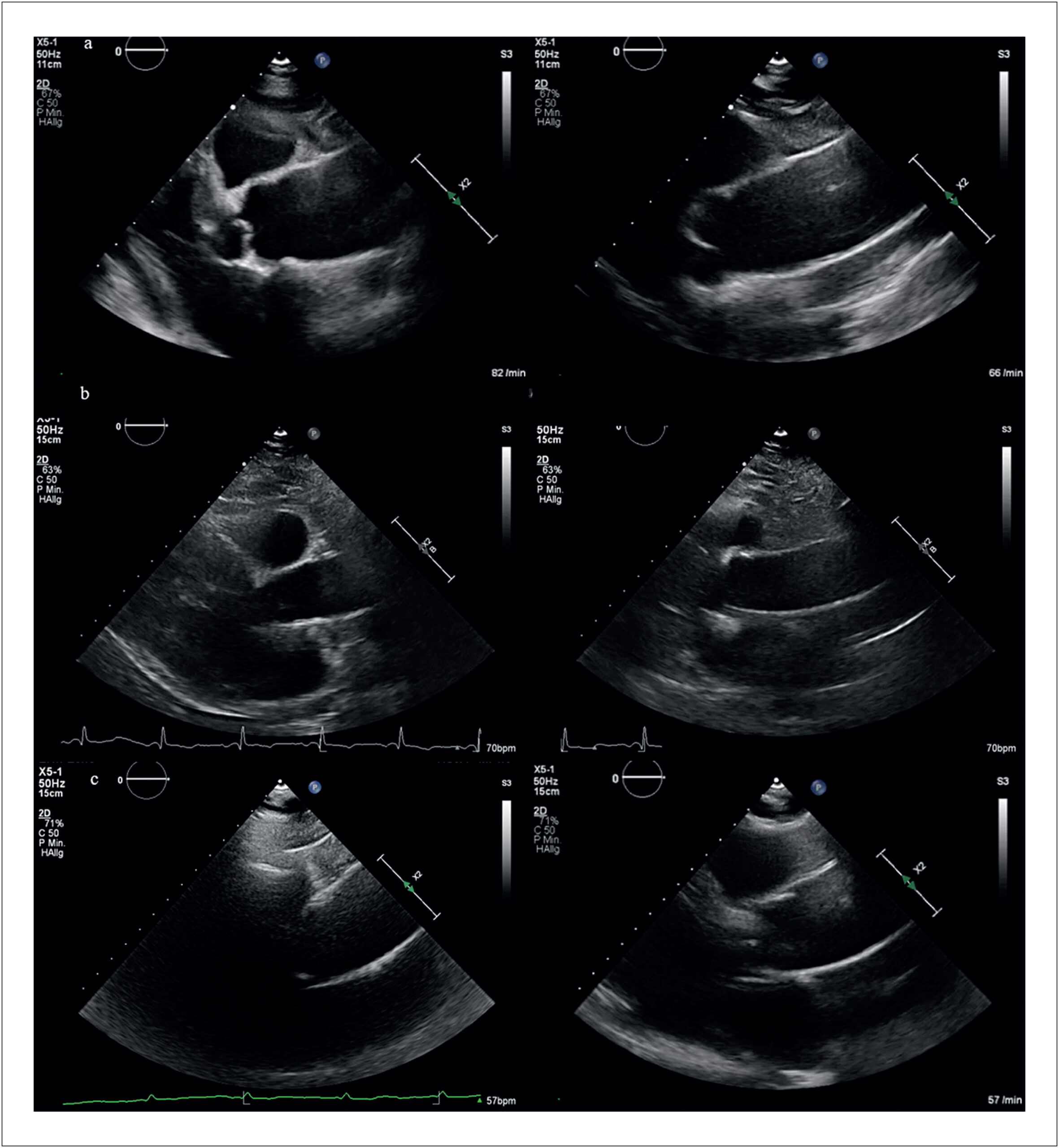
Assessment of the ascending aorta is an essential component of standard transthoracic echocardiography (TTE). While visualization of the aortic root and proximal ascending aorta is routinely achieved using the standard parasternal long-axis view (PLAX), imaging of the mid and distal ascending aorta remains technically challenging.– Current recommendations suggest moving the transducer to upper left intercostal spaces or alternatively using right parasternal windows to improve visualization of these segments in patients with adequate acoustic windows.

In clinical practice, experienced echocardiographers often obtain satisfactory visualization of the tubular ascending aorta using individualized modifications of conventional views. However, less experienced operators and general cardiologists may fail to consistently image these segments because the upper left parasternal long-axis view (uPLAX) is poorly described in the literature, whereas acquisition of the right parasternal view is more technically demanding and time-consuming.

[…]

  • Aortic imaging
  • Ascending aorta
  • Echocardiography
Read Full Article
Creative Commons Attribution (CC BY) Creative Commons license
31/07/2026 DOI: 10.36660/abcimg.20260068i Views 3
Recognition of Clinical Stages and Myocardial Damage in Aortic Stenosis: Beyond Diagnosis – To Op or Not To Op
Review Article

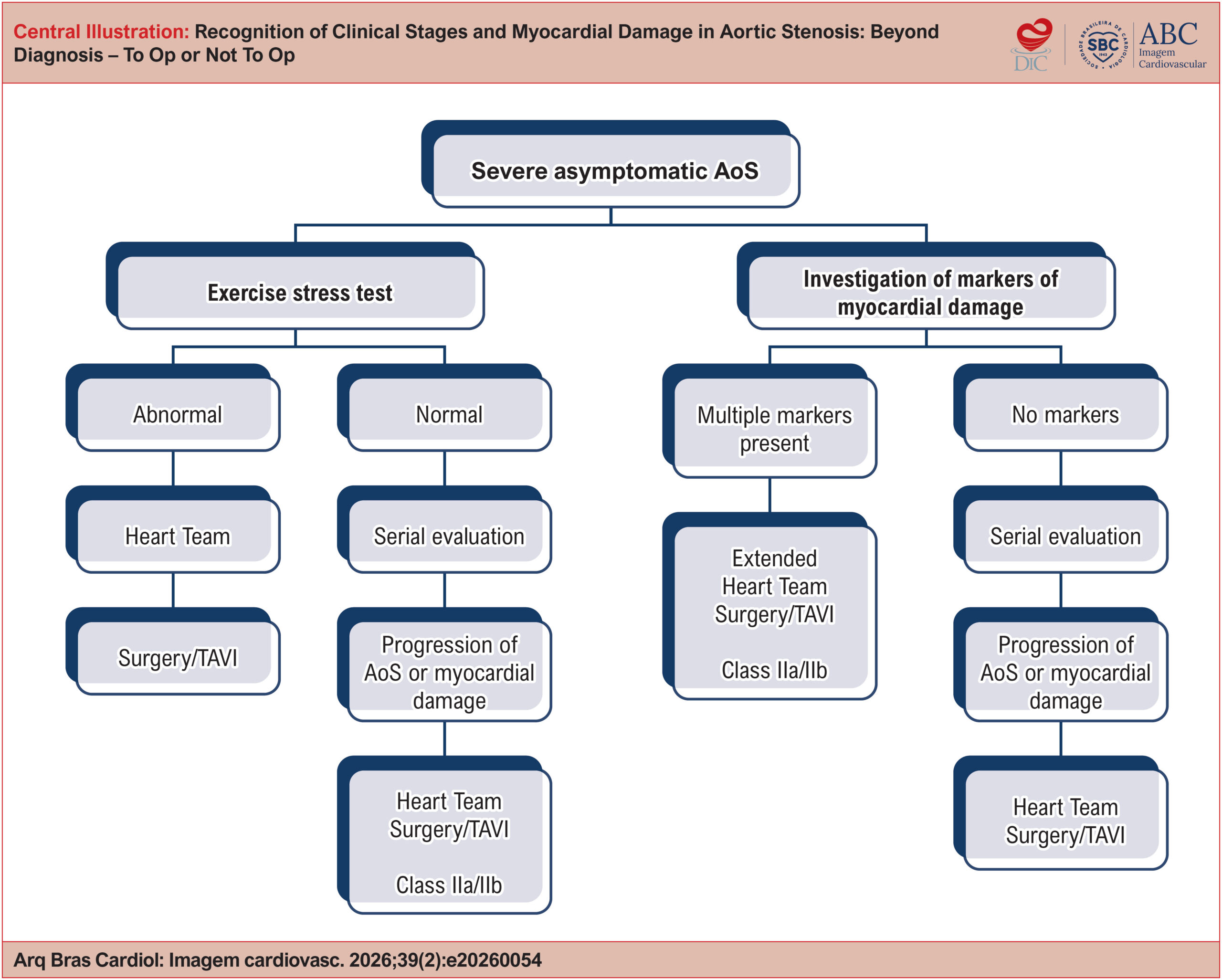
Recognition of Clinical Stages and Myocardial Damage in Aortic Stenosis: Beyond Diagnosis – To Op or Not To Op

Daniel Franca Vasconcelos

Abstract

Severe aortic stenosis is a progressive clinical entity that has historically developed alongside descriptions of pathological anatomy, cardiovascular semiology, heart surgery, and percutaneous cardiovascular interventions. Indication for valve replacement in patients with severe aortic stenosis is still based on symptom onset. With advances in surgical techniques, especially transcatheter implantation, the focus has shifted to identifying early markers of myocardial damage that may indicate irreversible myocardial injury. This review article seeks to provide a comprehensive analysis of methods for identifying myocardial damage in patients with severe aortic stenosis, including clinical evaluation, electrocardiography (ECG), echocardiography, cardiac magnetic resonance imaging (CMR), and plasma biomarkers. It also discusses the staging of myocardial damage and the optimal timing for therapeutic intervention based on clinical, functional, and structural findings. Recent studies designed to address how to manage asymptomatic patients with aortic stenosis have yielded divergent results. Accordingly, the aim is to assist clinicians in deciding the optimal timing for valve replacement, taking individual patient characteristics into account.

  • Aortic Valve Stenosis
  • Echocardiography
  • Magnetic Resonance Imaging
Read Full Article
Creative Commons Attribution (CC BY) Creative Commons license
31/07/2026 DOI: 10.36660/abcimg.20260054i Views 3
Editorial

Elevated Lipoprotein(a) in Patients Without Comorbidities: Which Imaging Tests Should be Ordered?

Eduardo Gomes Lima et al. Leticia Neves Solon Carvalho Tatiane Mascarenhas Santiago Emerich Eduardo Ferreira Amorim Fabiana Hanna Rached

The incorporation of lipoprotein(a) [Lp(a)] into contemporary cardiovascular risk assessment has introduced a practical question that is increasingly common in clinical practice: when faced with an elevated result, how should risk be reclassified and what management should be adopted, given that current guidelines recommend measuring Lp(a) at least once in adulthood and recognize it as a risk-modifying factor., However, the cardiovascular risk estimation proposed by these same guidelines — based on prognostic scores such as Predicting Risk of Cardiovascular Disease EVENTs – atherosclerotic cardiovascular disease (PREVENT-ASCVD), developed in 2023 by the American Heart Association — does not, a priori, account for the impact of elevated Lp(a) levels in its calculation. This creates a clinical scenario that is both concrete and challenging: the possibility that an asymptomatic patient, without traditional risk factors, may carry a biologically relevant risk factor that the score simply does not “see.” Thus arises the central question: what should be done when Lp(a) is elevated? In particular, should cardiovascular imaging be used to refine risk stratification?

A reasonable answer to these important questions requires an analysis of the biology and evidence that have propelled Lp(a) to its newly acquired prominence in the field of primary prevention in cardiology. Lp(a) is a particle similar to low-density lipoprotein (LDL), composed by an apolipoprotein B-100 molecule covalently bound to apolipoprotein(a), with levels predominantly determined by the LPA gene and relative stability throughout life., Its association with atherosclerotic cardiovascular disease is supported by epidemiological, genetic, and Mendelian randomization evidence, giving the particle a status stronger than that of a simple associative marker., Data from 450,000 patients demonstrate a strong linear correlation between elevated Lp(a) levels and atherosclerotic disease, with an approximate 11% increase in relative risk for every 50 nmol/L.

[…]

  • Atherosclerosis
  • Lipoprotein(a)
  • Primary Prevention
Read Full Article
Creative Commons Attribution (CC BY) Creative Commons license
31/07/2026 DOI: 10.36660/abcimg.20260053i Views 1
Figure 1
Case Report

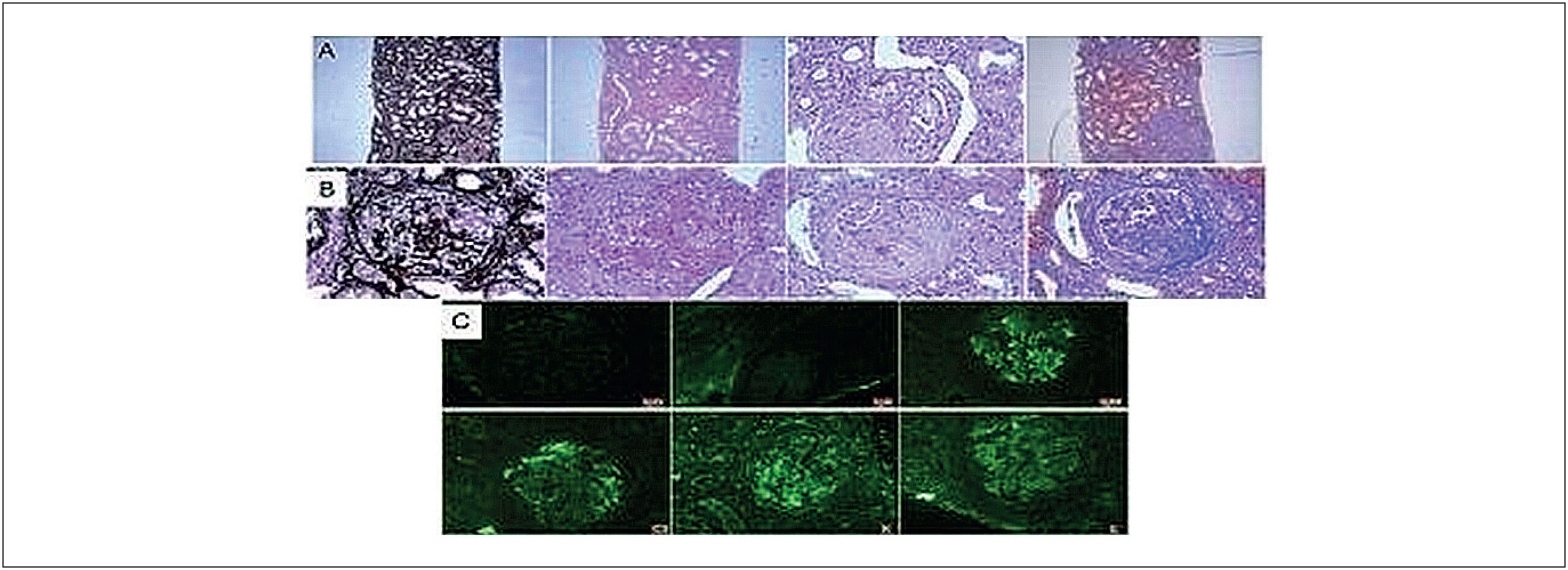
Dilated Cardiomyopathy as a Rare Initial Manifestation of ANCA-positive Microscopic Polyangiitis: Case Report

Karoline Gonzaga Costa et al. Maria Estefânia Bosco Otto André Felipe Lobão Fernandes Nathália de Macêdo Assunção … Mariana Ubaldo Barbosa Paiva Rosyane Luz Rufino de Lima Rita Mikelle Soares Dias

Introduction

Antineutrophil cytoplasmic antibody (ANCA)-associated vasculitis constitute a group of systemic small-vessel inflammatory diseases characterized by pauci-immune necrotizing vasculitis and multisystem involvement, including granulomatosis with polyangiitis (GPA), eosinophilic granulomatosis with polyangiitis (EGPA), and microscopic polyangiitis (MPA).– MPA is classically associated with rapidly progressive glomerulonephritis and alveolar hemorrhage, conditions that carry high morbidity and mortality when not promptly recognized and treated.,

In addition to predominant renal and pulmonary involvement, there is growing evidence that patients with ANCA-associated vasculitis have an increased cardiovascular risk, related not only to traditional factors but also to disease-specific mechanisms such as persistent inflammation, endothelial dysfunction, and accelerated atherosclerosis.– Studies suggest a higher incidence of major cardiovascular events (myocardial infarction, stroke, and heart failure) compared with the general population.

[…]

  • Anti-Neutrophil Cytoplasmic Antibody-Associated Vasculitis
  • Dilated Cardiomyopathy
  • Echocardiography
  • Microscopic Polyangiitis
Read Full Article
Creative Commons Attribution (CC BY) Creative Commons license
31/07/2026 DOI: 10.36660/abcimg.20260038i Views 1
Editorial

Beyond Ejection Fraction: Integrating Myocardial Strain and Cardiac Magnetic Resonance into the Contemporary Assessment of Left Ventricular Function

Marly Uellendahl

The assessment of left ventricular function is undergoing a profound transformation. For decades, left ventricular ejection fraction (LVEF) has occupied a central role in clinical practice, guiding diagnosis, risk stratification, and therapeutic decision-making across a broad spectrum of cardiovascular diseases. Its extensive clinical validation, ease of acquisition, and reproducibility have established two-dimensional echocardiography as the first-line modality for evaluating left ventricular systolic function.,

However, advances in cardiovascular imaging have demonstrated that myocardial function cannot be fully characterized by a single volumetric parameter. Although LVEF remains a robust marker of global left ventricular performance, its dependence on loading conditions and its limited sensitivity for detecting early contractile abnormalities underscore the need for a more comprehensive functional assessment.

[…]

  • Left Ventricular Dysfunction
  • Magnetic Resonance Imaging
  • Stroke Volume
Read Full Article
Creative Commons Attribution (CC BY) Creative Commons license
27/07/2026 DOI: 10.36660/abcimg.20260087i Views 1
Figure 1
Editorial

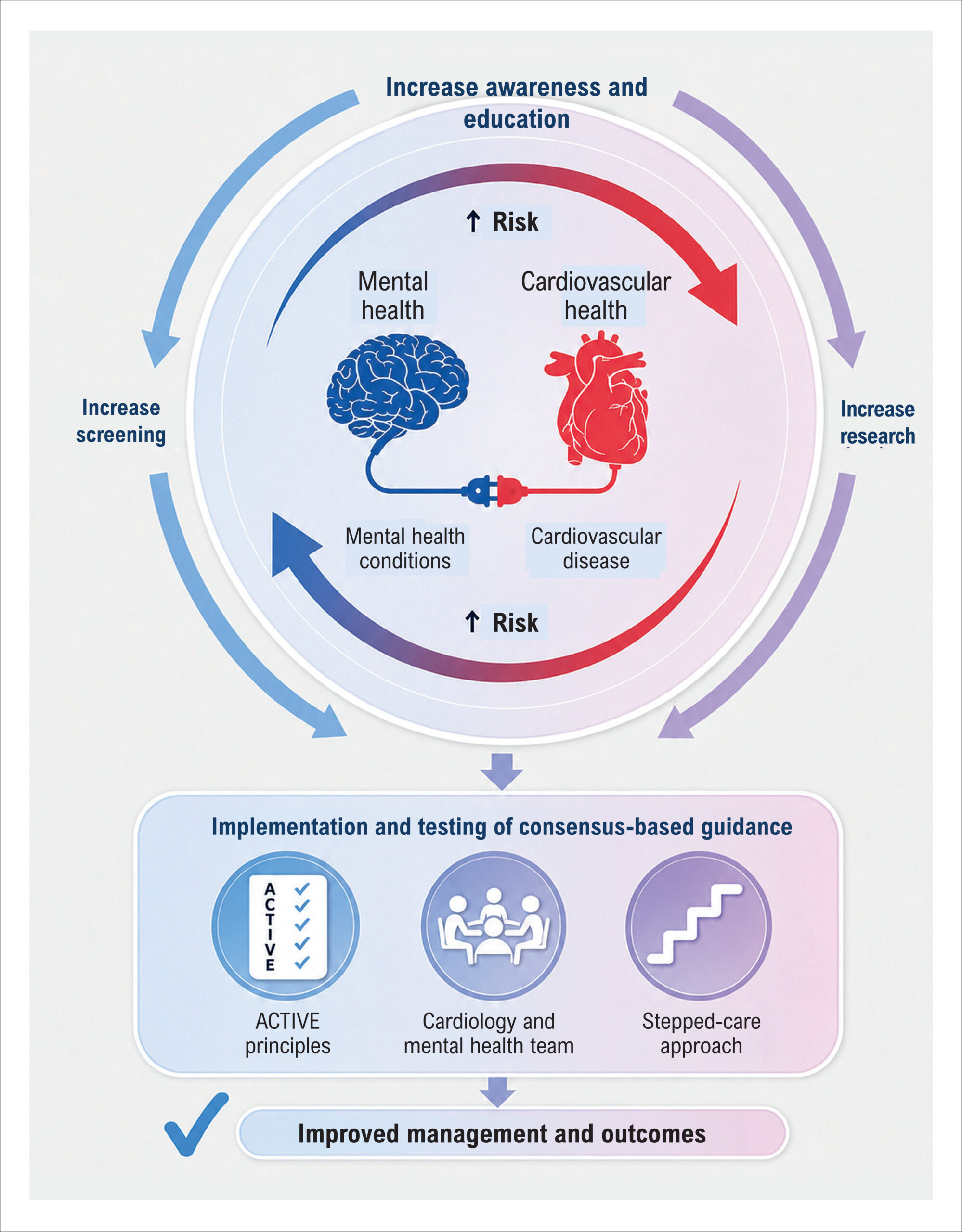
Beyond Imaging: The Silent Impact of Depression on Coronary Artery Disease

Priscila Cestari Quagliato

Coronary artery disease (CAD) remains the leading cause of global morbidity and mortality, and its intersection with mental health has received prominent attention in contemporary cardiology discussions. In 2025, the European Society of Cardiology published a guideline specifically addressing the multidirectional association between mental health and cardiovascular disease. Key issues were highlighted, including healthcare professionals’ limited awareness regarding the prevalence of mental disorders and their direct impacts on cardiovascular health, as well as inadequate recognition of severe mental illness and the stigmas associated with these conditions.

Mental disorders play a detrimental role in cardiovascular prevention, affecting treatment adherence and management of acute decompensations. The overlapping symptoms of the two conditions and the difficulties in monitoring chronic disease negatively impact patient prognosis.

[…]

  • Cardiology
  • Coronary Artery Disease
  • Depression
  • Mental Health
Read Full Article
Creative Commons Attribution (CC BY) Creative Commons license
27/07/2026 DOI: 10.36660/abcimg.20260074i Views 1
Editorial

Myocardial Work During Isometric Exercise: From Physiology to Clinical Practice

Rodrigo Bellio de Mattos Barretto et al. Carlos Eduardo Suaide Silva

Assessment of left ventricular function by echocardiography has advanced remarkably in its ability to characterize aspects that extend beyond traditional measures of volume and ejection fraction (EF). Among these advances, the evaluation of contractility through two-dimensional strain analysis has gained particular prominence. Technological developments now allow this measure to be adjusted for afterload through myocardial work (MW), providing greater potential to identify subtle changes in ventricular function.

Isometric exercise represents a strategy to increase afterload through handgrip, proving particularly useful in echocardiography because it enables the identification of functional changes in different clinical settings, including coronary artery disease, assessment of diastolic function, hypertension, athletes, and even atrial functional mitral regurgitation.–

[…]

  • Global Longitudinal Strain
  • Stress echocardiography
Read Full Article
Creative Commons Attribution (CC BY) Creative Commons license
27/07/2026 DOI: 10.36660/abcimg.20260073i
My Approach to Left Atrial Function: From Basic Assessment to Atrial Stiffness
Review Article

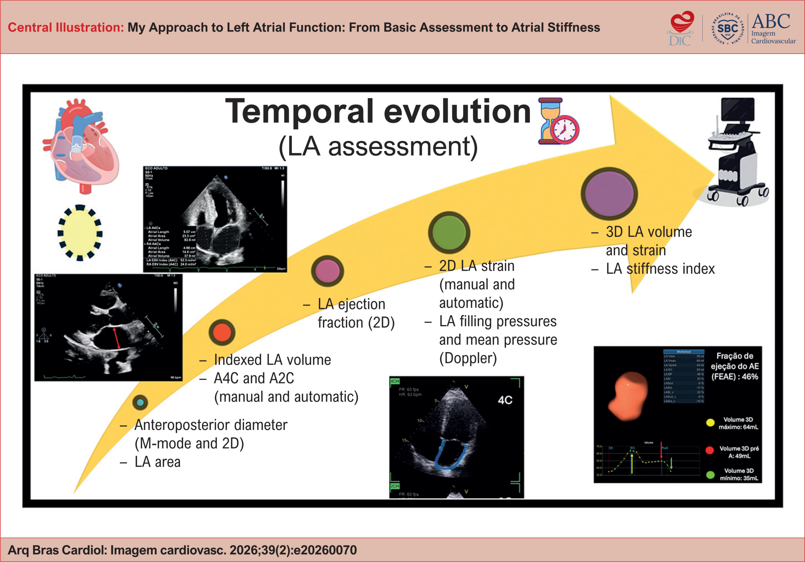
My Approach to Left Atrial Function: From Basic Assessment to Atrial Stiffness

Halsted Alarcão Gomes Pereira da Silva et al. Helder Moura Gomes Alexandre Costa Souza

Abstract

The left atrium (LA) has historically been considered a passive chamber that conducts blood flow between the pulmonary veins and the left ventricle (LV). Advances in cardiovascular physiology and imaging techniques have highlighted its active role in modulating cardiac output, ventricular diastolic function, and the stratification of various heart diseases. Assessment of the LA has evolved over the years into a multiparametric functional approach, consolidated as an essential component of modern echocardiography. The systematic incorporation of volumetric parameters, atrial strain, and atrial stiffness allows for a more precise characterization of cardiovascular pathophysiology and improves prognostic stratification, thus supporting more informed clinical decisions.

  • Ejection Fraction
  • Strain, Left atrium
Read Full Article
Creative Commons Attribution (CC BY) Creative Commons license
27/07/2026 DOI: 10.36660/abcimg.20260070i
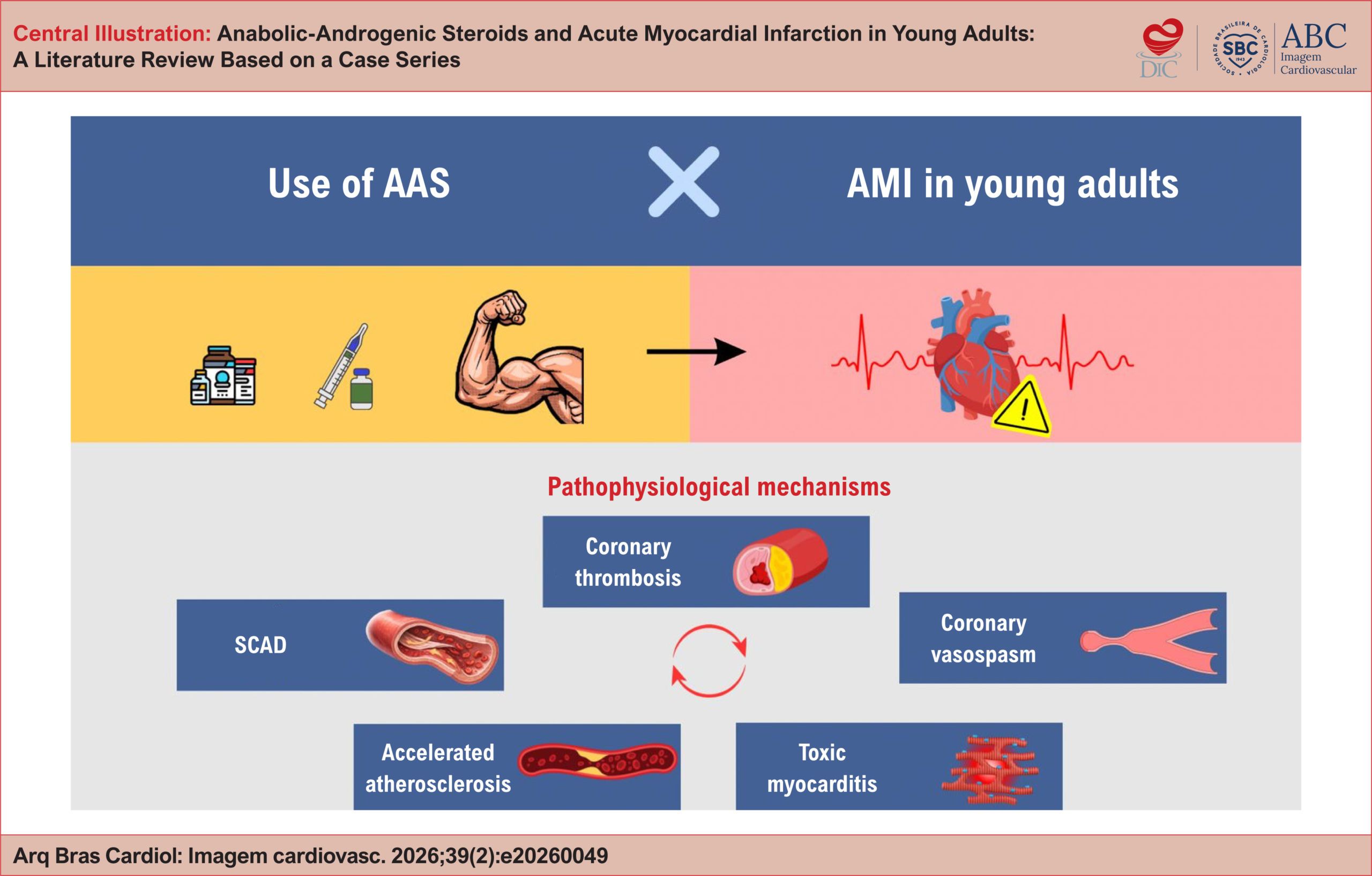
Anabolic-Androgenic Steroids and Acute Myocardial Infarction in Young Adults: A Literature Review Based on a Case Series
Original Article

Anabolic-Androgenic Steroids and Acute Myocardial Infarction in Young Adults: A Literature Review Based on a Case Series

Fabiana Rocha Botelho de Oliveira et al. Danielli Oliveira de Costa Lino Germano Freire Bezerra Filho Bruno Cavalcante Linhares … Leonardo Brito De Souza Luiz Filipe Torres de Alencar Matheus Rolim Santa Cruz Mateus Paiva Marques Feitosa

Abstract

Background:

The use of anabolic-androgenic steroids (AAS) has increased substantially, especially among young adults seeking aesthetic enhancement and improved physical performance. Scientific evidence demonstrates a significant association between the abuse of these substances and severe cardiovascular events, including acute myocardial infarction (AMI), often occurring in individuals without traditional cardiovascular risk factors.

Objective:

To describe the adverse effects of AAS on the cardiovascular system and the main pathophysiological mechanisms involved in the development of AMI through the analysis of a clinical case series combined with a review of medical literature.

Methods:

A systematic literature review was conducted using the PubMed and SciELO databases, complemented by the analysis of three clinical cases. Demographic variables and characteristics related to AAS use, including duration of exposure and route of administration, were evaluated, with emphasis on the pathophysiological mechanisms associated with AMI.

Results:

Analysis of the clinical cases identified different mechanisms related to AMI, including coronary thrombosis, atherosclerosis with plaque rupture, and spontaneous coronary artery dissection. The literature review also identified other relevant mechanisms, such as coronary vasospasm and toxic myocarditis. A predominance of male patients was observed, with the highest incidence occurring among individuals aged 20-40 years, and testosterone esters were the most frequently used AAS.

Conclusions:

AAS abuse represents a major threat to cardiovascular health and is associated with AMI through multiple pathophysiological mechanisms. These findings reinforce the need for public awareness as well as the development of preventive strategies and clinical guidelines aimed at managing this emerging condition.

  • Anabolic Androgenic Steroids
  • Atherosclerosis
  • Myocardial Infarction
  • Myocarditis
Read Full Article
Creative Commons Attribution (CC BY) Creative Commons license
27/07/2026 DOI: 10.36660/abcimg.20260049i Views 1
My Approach to Agitated Saline Contrast Echocardiography in Pediatric Patients
Review Article

My Approach to Agitated Saline Contrast Echocardiography in Pediatric Patients

Karen Saori Shiraishi Sawamura et al. Márcio Miranda Brito

Abstract

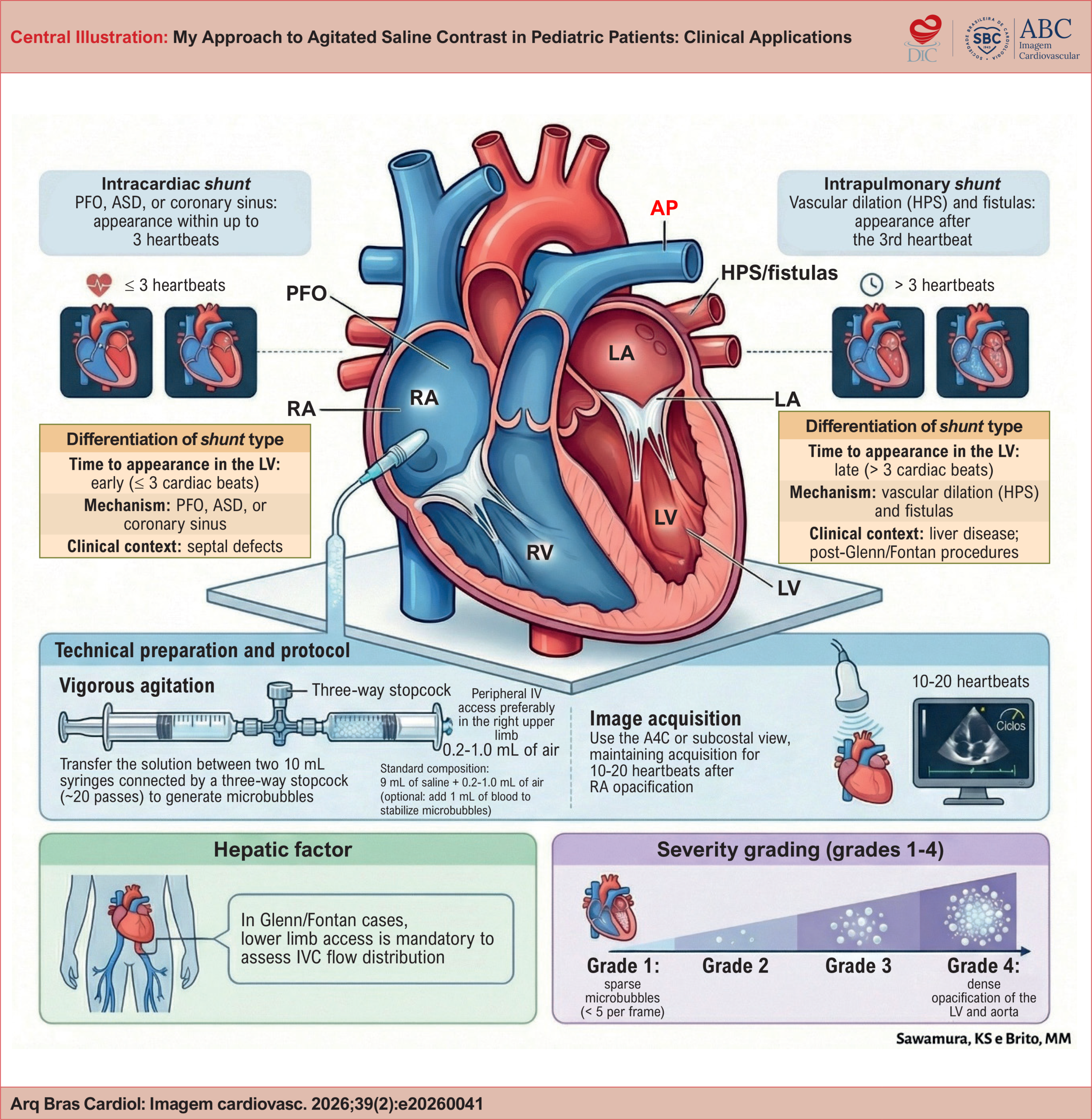
Agitated saline contrast is a simple, safe, widely available echocardiography technique. It is particularly useful for evaluating right-to-left shunts and pulmonary vascular abnormalities in the pediatric population. This article presents a practical approach to using this technique in pediatric echocardiography. It reviews the underlying physiological principles, the technical aspects of preparation and image acquisition, and the main clinical applications.

  • atrial septal defect
  • Contrast Echocardiography
  • ecocardiography
  • Hepatopulmonary Syndrome
  • Patent Foramen Ovale
Read Full Article
Creative Commons Attribution (CC BY) Creative Commons license
24/07/2026 DOI: 10.36660/abcimg.20260041i Views 1
Editorial

Cardiac Amyloidosis: Is It Truly a Hypertrophic Phenotype Cardiomyopathy?

Tonnison de Oliveira Silva

Cardiac amyloidosis is a classic example of an infiltrative disease whose predominant phenotype is characterized by a hypertrophic pattern of myocardial involvement., The most relevant studies—both those focused on the development of disease-modifying therapies and cohorts evaluating diagnostic tools—have used increased ventricular wall thickness as a key criterion for suspecting cardiac amyloidosis.– Based on this concept, several diagnostic algorithms have proposed ventricular wall thickening as one of the major red flags for investigating amyloid cardiomyopathy.,

However, increased ventricular wall thickness does not appear to be a universal requirement for diagnosis. Emerging evidence suggests that both Immunoglobulin Light Chain Amyloidosis (AL) and Transthyretin Amyloidosis (ATTR) amyloidosis can be diagnosed, including through noninvasive methods, even in the absence of the classic hypertrophic phenotype.

[…]

  • Amyloidosis without hypertrophy
  • Cardiac Amyloidosis
  • hypertrophic phenotype
Read Full Article
Creative Commons Attribution (CC BY) Creative Commons license
24/07/2026 DOI: 10.36660/abcimg.20260058i
Major Depressive Disorder and Quality of Life in Patients With Coronary Artery Disease Assessed by Myocardial Perfusion Imaging
Original Article

Major Depressive Disorder and Quality of Life in Patients With Coronary Artery Disease Assessed by Myocardial Perfusion Imaging

Guilherme Gonçais Lopes Almeida et al. Gustavo B. Barbirato Valéria de Queiroz Pagnin Daniel Pagnin Cláudio Tinoco Mesquita

Abstract

Background:

Major depressive disorder (MDD) may negatively influence cardiovascular prognosis, increasing the morbidity and mortality of patients with coronary artery disease (CAD). Thus, the psychometric assessment of these individuals may contribute to understanding how mental health impacts the pathophysiology of myocardial ischemia.

Objective:

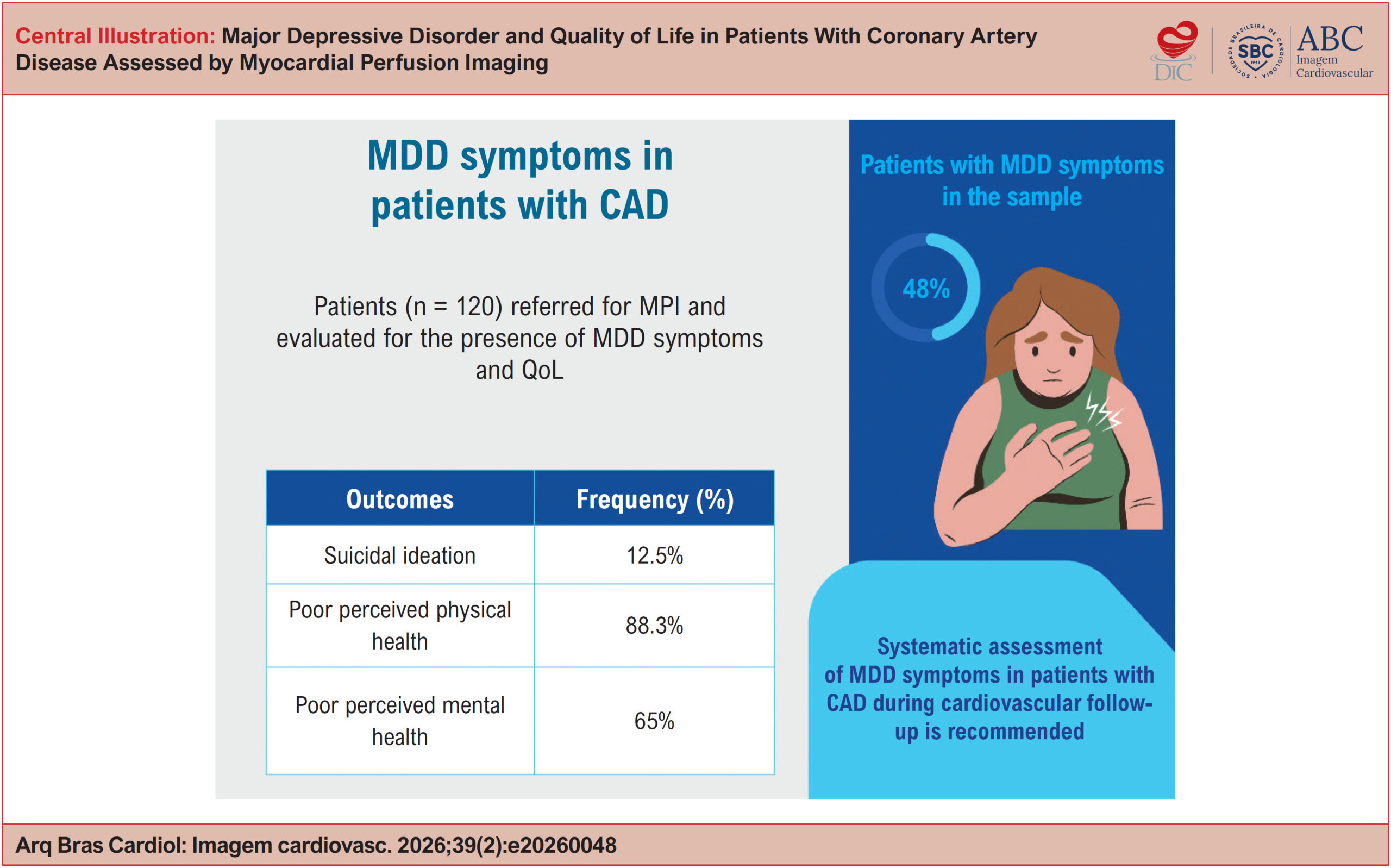
To evaluate the prevalence of MDD in patients with CAD undergoing stress and rest myocardial perfusion imaging (MPI) using the psychometric instrument Patient Health Questionnaire-9 (PHQ-9). As secondary objectives, to correlate quality of life (QoL) data obtained using the 12-Item Short Form Survey (SF-12) and Positive and Negative Affect Schedule (PANAS) instruments with the presence or absence of myocardial ischemia detected by MPI.

Methods:

The SF-12, PHQ-9, and PANAS questionnaires were administered to 120 consecutive patients referred for MPI for CAD evaluation. The prevalence of MDD was assessed, and the results were correlated with MPI findings and QoL scale scores.

Results:

A high prevalence of MDD was identified (58 cases; 48.3%), with no association with risk factors, age, or MPI findings. A significant rate of suicidal ideation was observed among the evaluated patients (15 cases; 12.5%), in addition to reduced QoL in 88.3% of patients (n = 106), with scores below 50 on the physical SF-12, and in 65% (n = 78), with scores below 50 on the mental SF-12, indicating poor perceived mental health.

Conclusion:

These findings reinforce the need for a multidisciplinary approach in the management of patients with suspected CAD, including systematic mental health assessment, given the opportunities to improve outcomes during patient interactions with the health care system.

  • Major Depressive Disorder
  • Myocardial Ischemia
  • Quality of life
Read Full Article
Creative Commons Attribution (CC BY) Creative Commons license
24/07/2026 DOI: 10.36660/abcimg.20260048i Views 1
Figure 1
Case Report

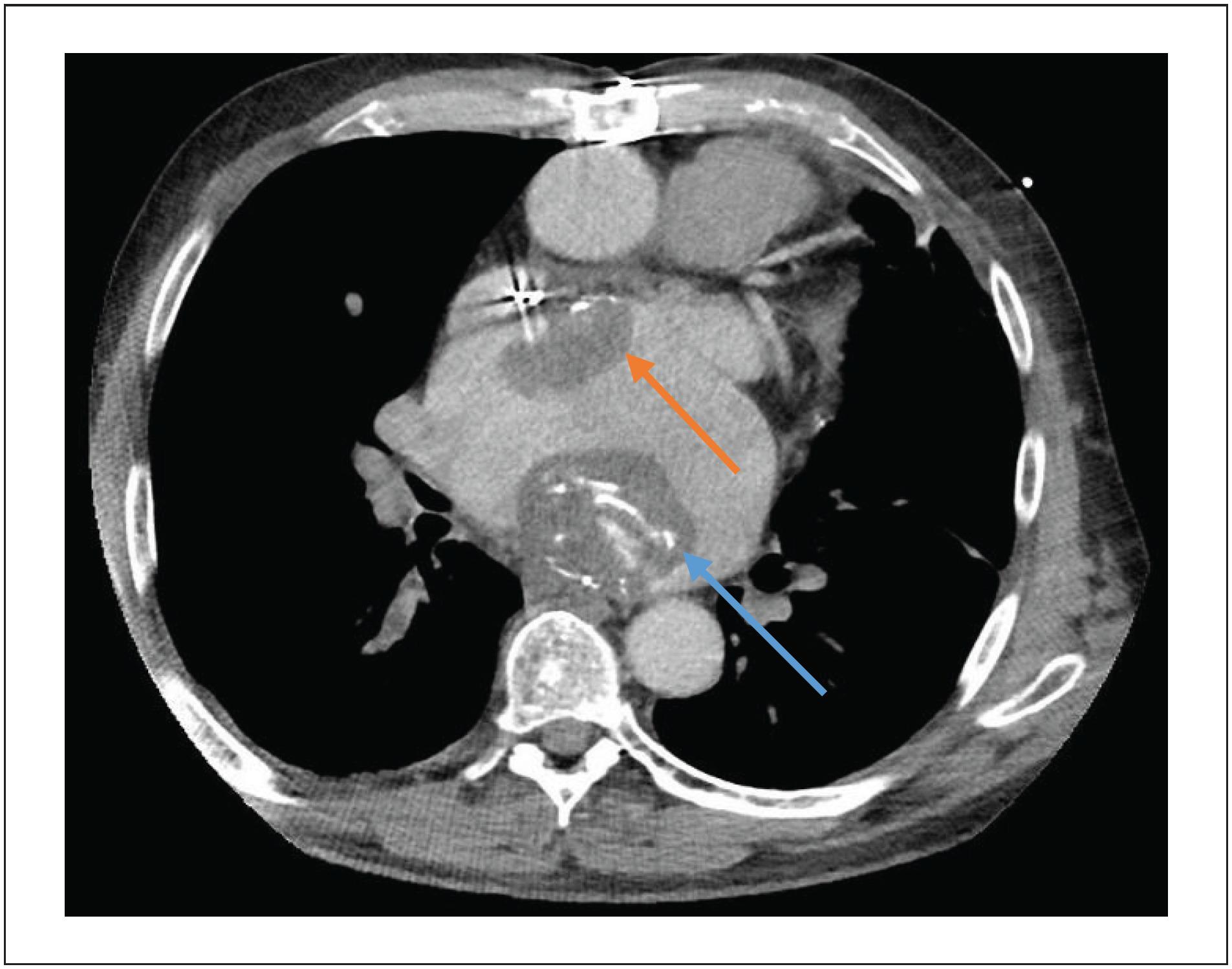
A Hidden Connection: Anomalous Left Circumflex Artery Arising From the Right Pulmonary Artery Unveiled by Cardiovascular Computed Tomography

Mubariz Ahmed Hassan et al. Ashraf Alzahrani Mohammad Mhanna Ola Abdelkarim … Rodrigo Bello Paulo Savoia Promporn Suksaranjit Kimberly Delcour

Introduction

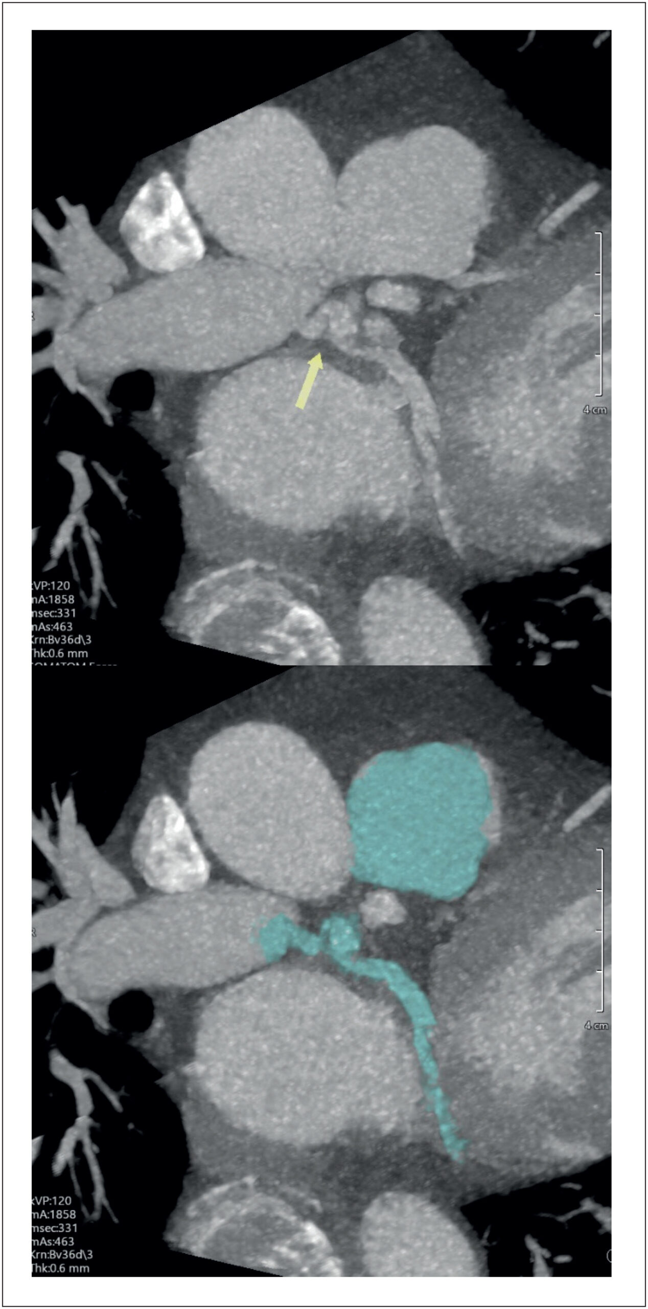
Congenital coronary artery anomalies are uncommon but clinically significant conditions that may present across a broad spectrum, ranging from incidental findings to myocardial ischemia, ventricular arrhythmias, heart failure (HF), and sudden cardiac death (SCD). In adults, anomalous coronary origins may be particularly challenging to recognize because symptoms are often nonspecific and may overlap with more prevalent causes of chest pain, dyspnea, or palpitations.

In rare cases, a coronary artery may originate from the pulmonary arterial circulation, resulting in myocardial perfusion that depends on collateral flow from the remaining coronary arteries arising from the aorta, thereby predisposing the myocardium to ischemia-related electrical instability. Noninvasive anatomical imaging plays a pivotal role in establishing a definitive diagnosis, particularly when conventional angiography is inconclusive.

[…]

  • Cardiac Catheterization
  • Coronary Vessels
  • Pulmonary Artery
  • X-Ray Computed Tomography
Read Full Article
Creative Commons Attribution (CC BY) Creative Commons license
24/07/2026 DOI: 10.36660/abcimg.20260040i Views 1
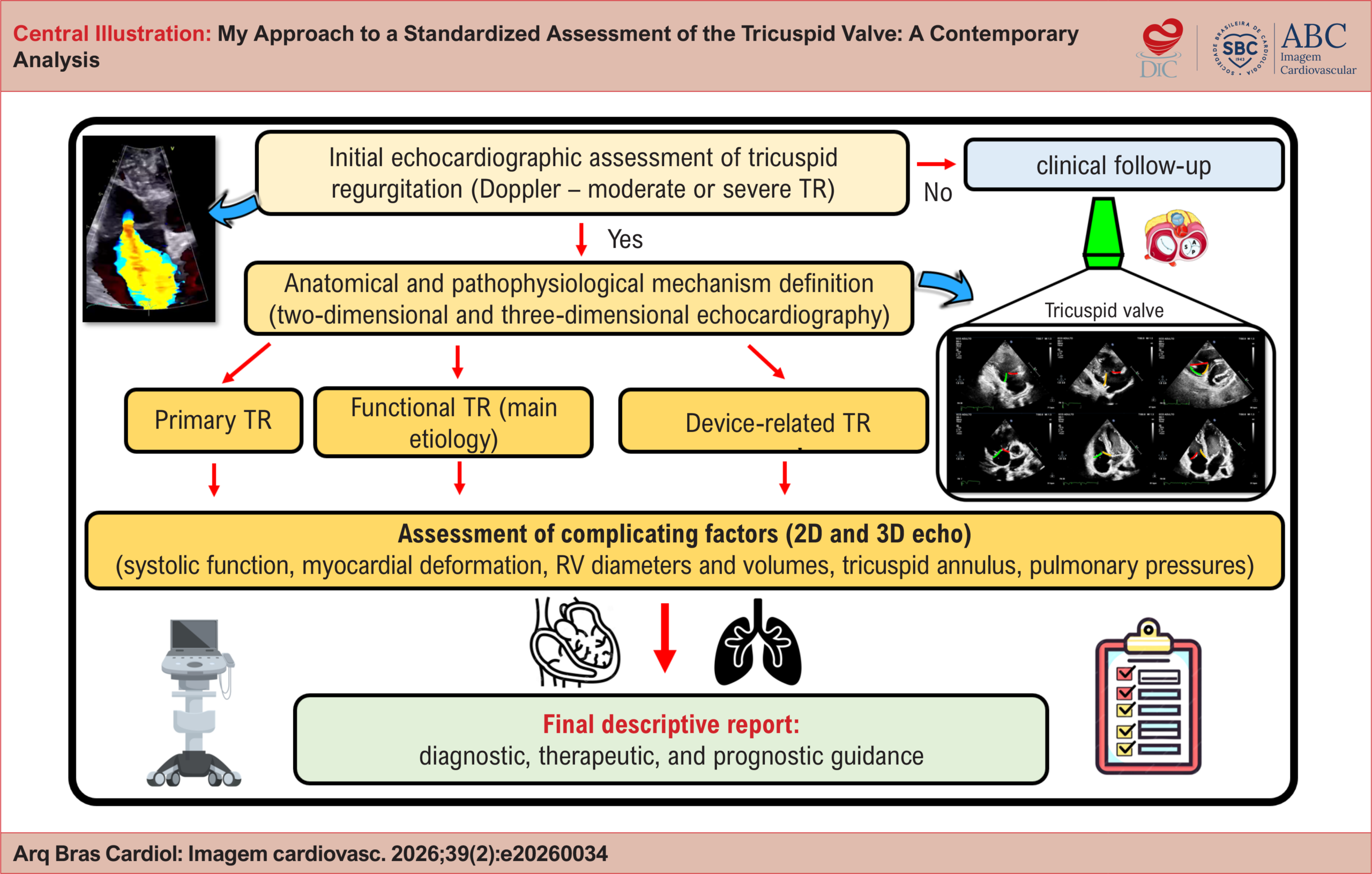
My Approach to a Standardized Assessment of the Tricuspid Valve: A Contemporary Analysis
Review Article

My Approach to a Standardized Assessment of the Tricuspid Valve: A Contemporary Analysis

Halsted Alarcão Gomes Pereira da Silva et al. Alexandre Costa Souza Adenalva Beck

Abstract

Tricuspid regurgitation (TR) remains one of the most frequent valvular diseases encountered in echocardiographic practice. Mild cases generally do not require further investigation or specific treatment. Moderate and severe valvular disease, however, require more detailed anatomical and etiological assessment to better understand the underlying pathophysiological mechanism.

Two-dimensional echocardiographic analysis is the initial examination of choice and should be performed in a standardized manner, with individual identification of the leaflets using the main transthoracic echocardiographic windows. The description of complementary parameters – such as right ventricular (RV) systolic function, myocardial deformation assessed by longitudinal strain, right-sided chamber diameters and volumes, and estimation of pulmonary pressures – guides the clinician regarding potential therapeutic strategies and provides relevant prognostic information.

In this context, the 2025 European Society of Cardiology (ESC) guideline reinforces the importance of a standardized and comprehensive evaluation of the tricuspid valve and its hemodynamic repercussions. Most of this assessment can be performed using widely available two-dimensional transthoracic echocardiography.

  • Pulmonary Hypertension
  • Tricuspid Valve
  • Tricuspid Valve Insufficiency
Read Full Article
Creative Commons Attribution (CC BY) Creative Commons license
24/07/2026 DOI: 10.36660/abcimg.20260034i Views 1
Figure 1
Case Report

Recurrent Intracardiac Masses in an Orthotopic Heart Transplant Recipient

Bilal Saeed et al. Natdanai Punnanithinont Shareef Mansour Paulo Savoia Promporn Suksaranjit

Introduction

As long-term survival improves among heart transplant recipients, rare post-transplant complications, including intracardiac masses, are being increasingly recognized.

Severe left atrial (LA) dilation and atrial arrhythmias may further contribute to blood stasis and thrombus formation in transplant recipients. LA thrombi in these patients may mimic neoplastic masses and represent important diagnostic and therapeutic challenges. Contributing factors include atrial dilation, arrhythmias, foreign material, and the components of Virchow’s triad, namely abnormal blood flow, endothelial injury, and hypercoagulability.,

[…]

  • Cardiac MRI
  • Heart Atria
  • Heart Transplant
  • Left atrial
  • X-Ray Computed Tomography
Read Full Article
Creative Commons Attribution (CC BY) Creative Commons license
24/07/2026 DOI: 10.36660/abcimg.20260031i Views 3
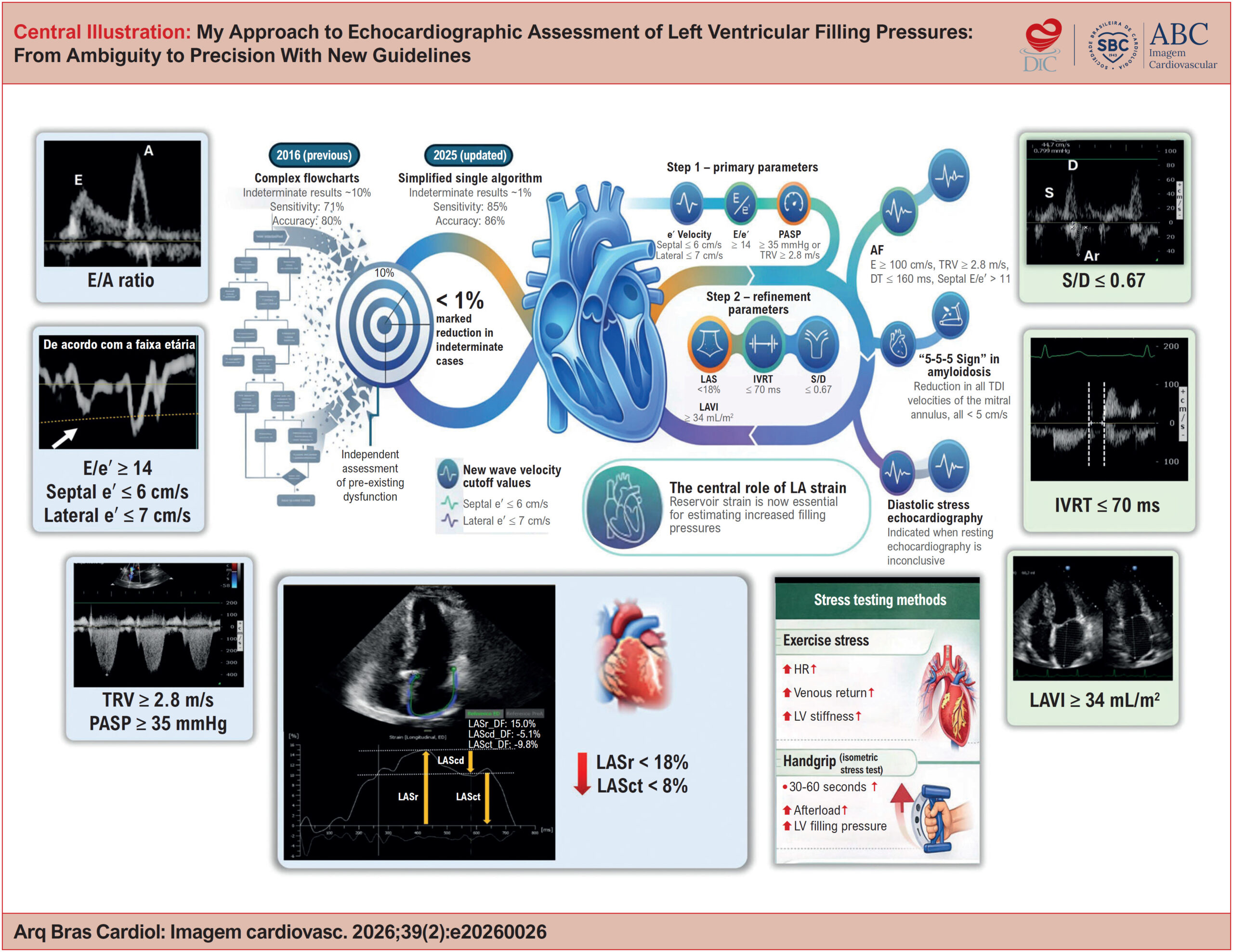
My Approach to Echocardiographic Assessment of Left Ventricular Filling Pressures: From Ambiguity to Precision With New Guidelines
Review Article

My Approach to Echocardiographic Assessment of Left Ventricular Filling Pressures: From Ambiguity to Precision With New Guidelines

Marco Stephan Lofrano Alves et al. Larissa Maria Vosgerau Marcelo Vitola Dreckmann Roberto D´Ávila Martins … Cláudia Biondo Zanlorensi Eduardo Henrique Bonotto

Abstract

The echocardiographic estimation of left ventricular (LV) filling pressures is a cornerstone in the assessment of heart failure, particularly in patients with heart failure with preserved ejection fraction (HFpEF). The 2016 American Society of Echocardiography/European Association of cardiovascular Imaging algorithm standardized this evaluation using key variables, but a substantial proportion of cases remained indeterminate in clinical practice. The recent update of the American guideline reorganized the approach into a hierarchical framework, incorporating age-related adjustments and formalizing the role of left atrial strain (LAS) as a tie-breaking parameter, while also providing a more detailed characterization of special clinical scenarios. In parallel, the 2024 British Society of Echocardiography guideline emphasizes the pathophysiological interpretation of ventricular filling, complementing the operational framework proposed by the American document. Multicenter studies with invasive validation support these updates, establishing LAS as a robust marker of increased filling pressures. In this article, we present My Approach to echocardiographic assessment of LV filling pressures, based on an initial morphofunctional evaluation, followed by structured screening (Step 1) and further refinement (Step 2) incorporating LAS and additional parameters. We also provide a comparison between guidelines, discuss common pitfalls and algorithm limitations, and include case-based videos for practical application.

  • Echocardiography
  • Heart Failure
  • Left Ventricular Function
Read Full Article
Creative Commons Attribution (CC BY) Creative Commons license
24/07/2026 DOI: 10.36660/abcimg.20260026i Views 1
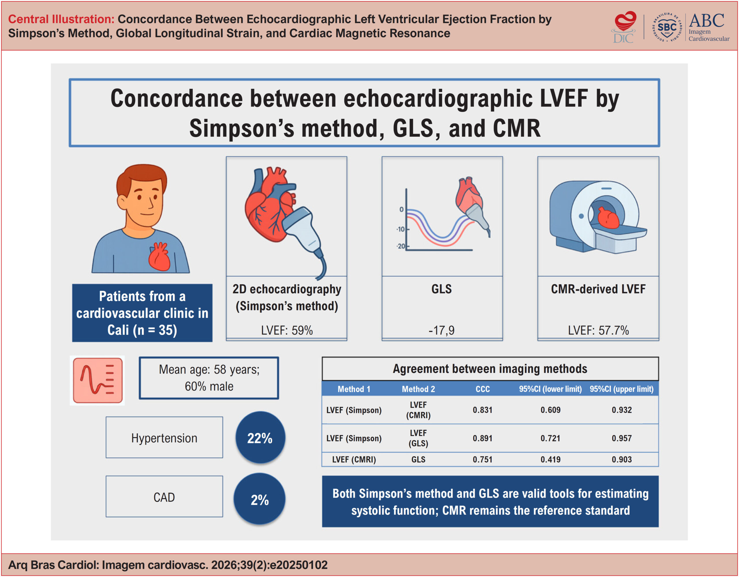
Concordance Between Echocardiographic Left Ventricular Ejection Fraction by Simpson's Method, Global Longitudinal Strain, and Cardiac Magnetic Resonance
Original Article

Concordance Between Echocardiographic Left Ventricular Ejection Fraction by Simpson's Method, Global Longitudinal Strain, and Cardiac Magnetic Resonance

Álvaro Herrera-Escandón et al. Juan Pablo Morales-Grisales Sebastián Ayala-Zapata Stephany Barbosa-Balaguera … Álvaro José Muriel-Ruiz Juan Felipe Bravo-Rueda José Eduardo Citelli-Ramírez Luis Fernando Osío-Jimenez Luis Miguel Benitez-Gómez Carlos Javier Ramírez-Estupiñán

Abstract

Background:

Left ventricular ejection fraction (LVEF) measured by echocardiography is a widely used parameter in clinical practice for the assessment of ventricular function. More recently, global longitudinal strain (GLS) has emerged as a complementary method, as has the assessment of LVEF by cardiac magnetic resonance (CMR). However, regional evidence evaluating the concordance among these three techniques remains limited.

Objectives:

To assess the concordance between echocardiographic and CMR measurements in patients treated at a cardiovascular clinic in the city of Cali, Colombia.

Methods:

This cross-sectional, analytical, descriptive study included 35 patients with confirmed or suspected heart disease, in whom all three methods were performed consecutively. Concordance was evaluated using Lin’s concordance correlation coefficient (CCC), Bland-Altman plots for LVEF, and linear and quadratic weighted κ coefficients for agreement between LVEF classifications.

Results:

The mean age was 58 years, and 60% of participants were male. The most common comorbidities were hypertension (22%) and dyslipidemia (11%). The mean LVEF was 59% by Simpson’s method and 57.7% by CMR, while the mean GLS was −17.7%. Concordance was as follows: Simpson’s LVEF vs. CMR (CCC, 0.831; 95%CI, 0.609-0.932); GLS vs. CMR-derived LVEF (CCC, 0.751; 95%CI, 0.419-0.903); and Simpson’s LVEF vs. GLS (CCC, 0.891; 95%CI, 0.721-0.957).

Conclusions:

Both Simpson’s method and GLS are valid tools for estimating systolic function. CMR remains the reference standard.

  • Echocardiography
  • Magnetic Resonance Spectroscopy
  • Stroke Volume
Read Full Article
Creative Commons Attribution (CC BY) Creative Commons license
24/07/2026 DOI: 10.36660/abcimg.20250102i Views 1
Editorial

Chemotherapy-Induced Cardiotoxicity in the Pediatric Population: What Are the Unique Aspects of Cardiovascular Imaging Follow-Up?

Jéssica Laureano Martins et al. Maria do Carmo Menezes Bezerra Duarte Fabiana Gomes Aragão Magalhães Feitosa Maria Verônica Câmara dos Santos

Special considerations for the pediatric population,

The pediatric perspective in cardio-oncology presents particular challenges, given that cardiotoxicity related to cancer treatment is not limited to an acute event, but represents a dynamic process that can interfere with myocardial growth and maturation over time. The vulnerability of the developing heart, associated with early exposure to potentially cardiotoxic therapies, confers a prolonged risk of progressive myocardial dysfunction. In this context, the use of more sensitive, reproducible, and integrated cardiovascular imaging strategies becomes fundamental. The multimodal approach, with emphasis on echocardiography (and cardiac magnetic resonance imaging in selected situations), increases diagnostic accuracy and contributes to better risk stratification. Additionally, structured longitudinal follow-up, with interpretation based on clinical individualization, is essential for adequate follow-up of these patients.

Finally, the incorporation of genetic, clinical, and therapeutic factors into risk models, coupled with continuous imaging monitoring, represents a promising perspective for precision medicine, with the goal of reducing cardiovascular morbidity and mortality and improving the quality of life of pediatric cancer survivors.

[…]

  • Cardiotoxicity
  • Child
  • Drug Therapy
  • Echocardiography
  • Neoplasms
Read Full Article
Creative Commons Attribution (CC BY) Creative Commons license
20/07/2026 DOI: 10.36660/abcimg.20260044i Views 1
Mitral Valve Leaflet Hypoplasia in Adults: Role of Cardiovascular Imaging
Review Article

Mitral Valve Leaflet Hypoplasia in Adults: Role of Cardiovascular Imaging

Fábio Luis de Jesus Soares

Abstract

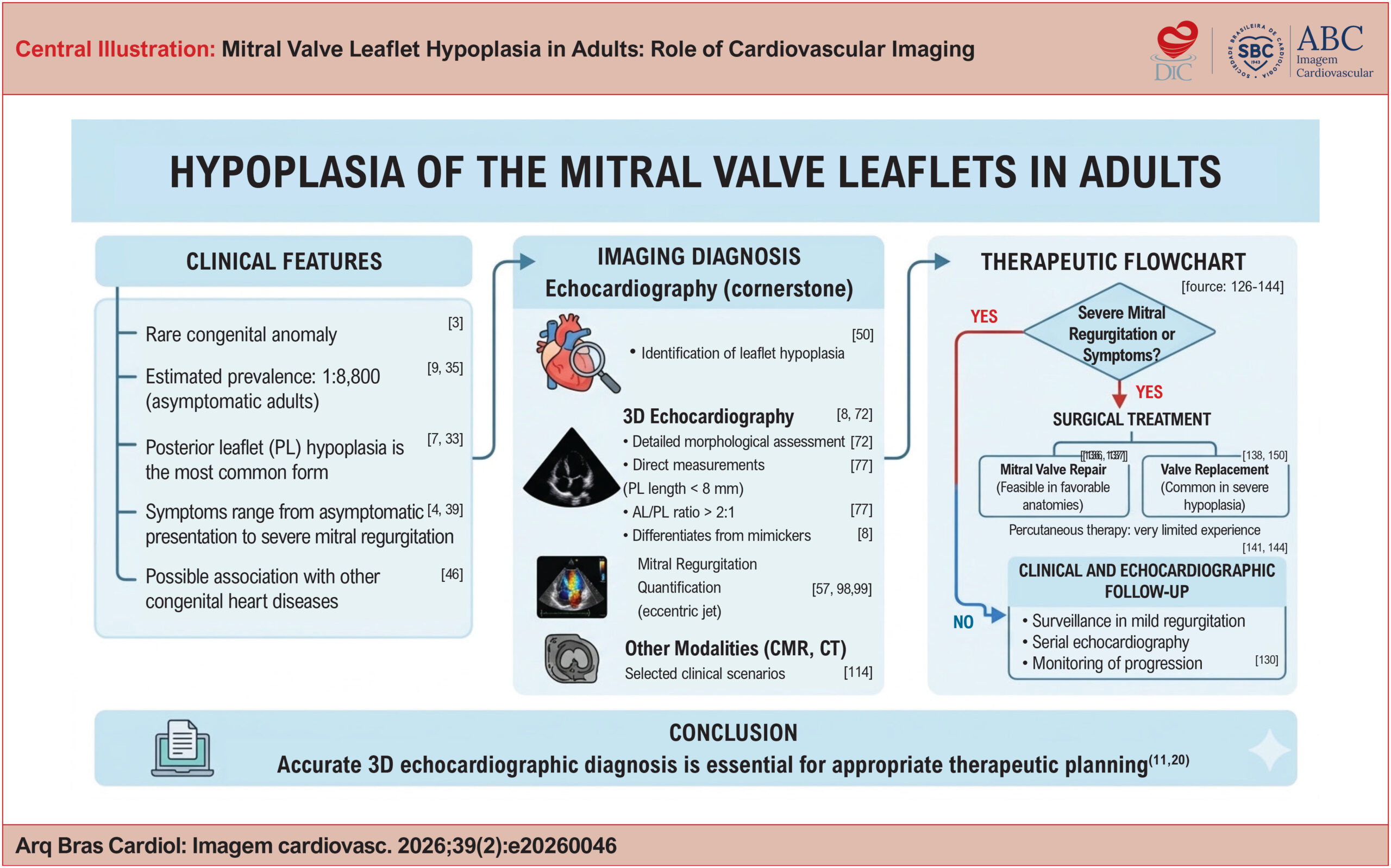
Mitral valve leaflet hypoplasia is a rare congenital anomaly, traditionally described in childhood but increasingly recognized in adults, often as an incidental finding or during the evaluation of mitral regurgitation. Clinical presentation is heterogeneous and depends on leaflet anatomy, the subvalvular apparatus, and the severity of regurgitation. In this narrative literature review, including case reports, case series, and review articles from nationally and internationally recognized journals, epidemiological and clinical aspects are discussed, with particular emphasis on echocardiographic findings. Posterior leaflet hypoplasia is the most common form and may be partial or complete. Three-dimensional echocardiography plays a central role in anatomical assessment, enabling direct measurements of leaflet area and length and helping differentiate true hypoplasia from mimicking entities such as mitral cleft, functional restriction, or subvalvular abnormalities. The estimated prevalence in asymptomatic adults is approximately 1:8,800. Therapeutic management is primarily determined by the severity of mitral regurgitation, with valve repair being feasible only in selected anatomical scenarios. Therefore, refined anatomical understanding, particularly through three-dimensional echocardiography, is essential for accurate diagnosis and appropriate therapeutic planning in this rare yet clinically relevant condition.

  • Congenital Abnormalities
  • Mitral Valve
  • Three-Dimensional Echocardiography
Read Full Article
Creative Commons Attribution (CC BY) Creative Commons license
20/07/2026 DOI: 10.36660/abcimg.20260046i Views 2
Impact of Isometric Exercise on Left Ventricular Mechanics Assessed by Global Longitudinal Strain and Myocardial Work in Healthy Adults
Original Article

Impact of Isometric Exercise on Left Ventricular Mechanics Assessed by Global Longitudinal Strain and Myocardial Work in Healthy Adults

Marcio Mendes Pereira et al. Maria Estefania Bosco Otto Juliana Lins da Paz Portela

Abstract

Background:

Traditional volumetric parameters have limitations in detecting subtle left ventricular (LV) systolic dysfunction. Global longitudinal strain (GLS) and myocardial work (MW) allow a more sensitive assessment of ventricular mechanics.

Objectives:

To evaluate changes in GLS and MW indices during isometric handgrip exercise compared with resting conditions.

Methods:

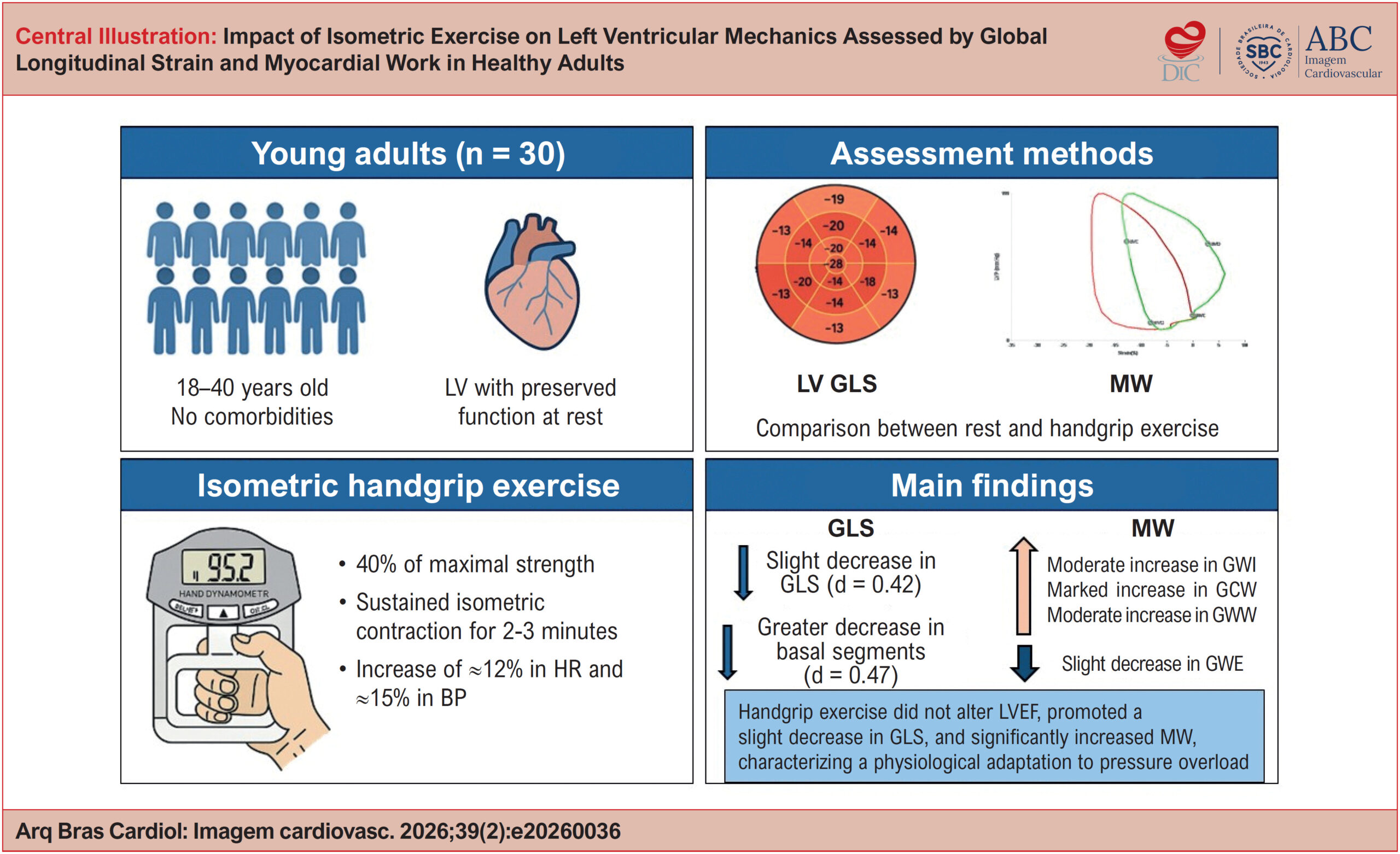
A total of 30 healthy individuals (29.3 ± 6.1 years; 50% male) were included in the sample. Echocardiography was performed at rest and during handgrip exercise (30%-40% of maximal strength). GLS, LV ejection fraction (LVEF), and MW indices were assessed: i) global work index (GWI), ii) global constructive work (GCW), iii) global wasted work (GWW), and iv) global work efficiency (GWE). Comparisons were performed using paired tests. Statistical significance was set at p < 0.05.

Results:

Handgrip exercise increased both systolic blood pressure (115 ± 16 vs 133 ± 18 mmHg; p < 0.0001) and diastolic blood pressure (69 ± 9 vs 79 ± 13 mmHg; p = 0.0002), without significant changes in LVEF (64.8% vs 64.4%; p = 0.62). A decrease in GLS was observed (20.38% ± 2.57% vs 19.60% ± 2.52%; p = 0.028), along with increases in GWI (+244 mmHg%; p = 0.0002), GCW (+313 mmHg%; p < 0.0001), and GWW (+52 mmHg%; p = 0.0008) as well as a decrease in GWE (94.8% ± 1.8% vs 93.6% ± 2.5%; p = 0.022).

Conclusions:

Handgrip exercise induces measurable ventricular mechanical changes in healthy individuals, reflecting a physiological response to acute pressure overload.

  • Echocardiography
  • Exercise
  • Left Ventricular Dysfunction
Read Full Article
Creative Commons Attribution (CC BY) Creative Commons license
20/07/2026 DOI: 10.36660/abcimg.20260036i Views 3
Figure 1
Case Report

Giant Atrial Myxoma in a Pregnant Patient: A Case Report

Roberto Ramos Barbosa et al. Caio Badiani Prando Victor Macedo Bianchini Lucas Crespo de Barros … Larissa Novaes Paganini Gracielly Barros Darlan Dadalt Sergio Luis Santos Guedes Vinicius Eduardo Araújo Costa Marcus Gustavo Tito Tiago Bernardo Nery Marcio Vinicius de Nardi de Angeli Maria Eduarda Vichi Gomes Viana Mariana Oliveira Roncato João Paulo Moulin Auad Guilherme Freitas Fernandes de Oliveira Renato Giestas Serpa Osmar Araujo Calil Luiz Fernando Machado Barbosa

Introduction

Atrial myxoma is the most prevalent primary heart tumor., Clinical manifestations of myxomas are usually nonspecific, and they may present with dyspnea, fatigue, reduced functional capacity, edema, and, eventually, cerebral embolic events with focal neurological deficits. Although they are classified as benign neoplasms, most commonly located in the left atrium, they can cause obstruction of intracardiac blood flow and, when friable, systemic embolism with consequent tissue ischemia.,

In spite of higher prevalence in women (65%), diagnosis during pregnancy is considered uncommon, with greater challenges in treatment and an increased risk of fetal death. We report the case of a pregnant patient with a complication of atrial myxoma that manifested during labor.

[…]

  • Heart Failure
  • Myxoma
  • Thoracic Surgery
Read Full Article
Creative Commons Attribution (CC BY) Creative Commons license
20/07/2026 DOI: 10.36660/abcimg.20260010i Views 1
Echocardiographic Assessment of Diastolic Dysfunction in Special Situations
Review Article

Echocardiographic Assessment of Diastolic Dysfunction in Special Situations

Antonio Amador Calvilho Júnior

Abstract

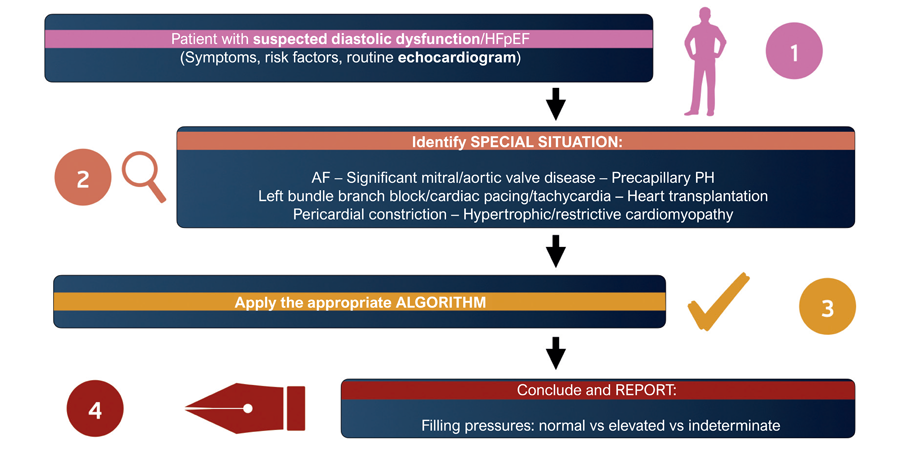
Echocardiographic assessment of diastolic function and left ventricular (LV) filling pressures is fundamental to the evaluation of dyspnea and the management of heart failure. However, conventional algorithms have limitations in several special clinical settings in which rhythm disturbances, valvular heart disease, pulmonary hypertension (PH), or structural cardiac abnormalities interfere with the interpretation of Doppler parameters. This article presents a practical approach to assessing diastolic function in conditions such as atrial fibrillation (AF), PH, mitral valve disease, mitral annular calcification, aortic valve disease, conduction disturbances, ventricular pacing, and restrictive cardiomyopathies. The main parameters applicable to each context, the technical aspects of image acquisition, and strategies for preparing a clear and clinically useful echocardiographic report are discussed.

  • Diastolic Heart Failure
  • Doppler Echocardiography
  • Stroke Volume
Read Full Article
Creative Commons Attribution (CC BY) Creative Commons license
17/07/2026 DOI: 10.36660/abcimg.20260029i Views 1
Figure 1
Case Report

Single Coronary Artery and Stress Cardiomyopathy: An Association Demonstrated by Multimodality Imaging

Marcus Vinicius Silva Ferreira et al. Julia Pereira Afonso dos Santos Tormin Roberto Nery Dantas Jr Roberto Vitor Almeida Torres … Renan Andreos Cordeiro José de Arimateia Batista Araújo Filho Luiz Francisco Cardoso

Case Report

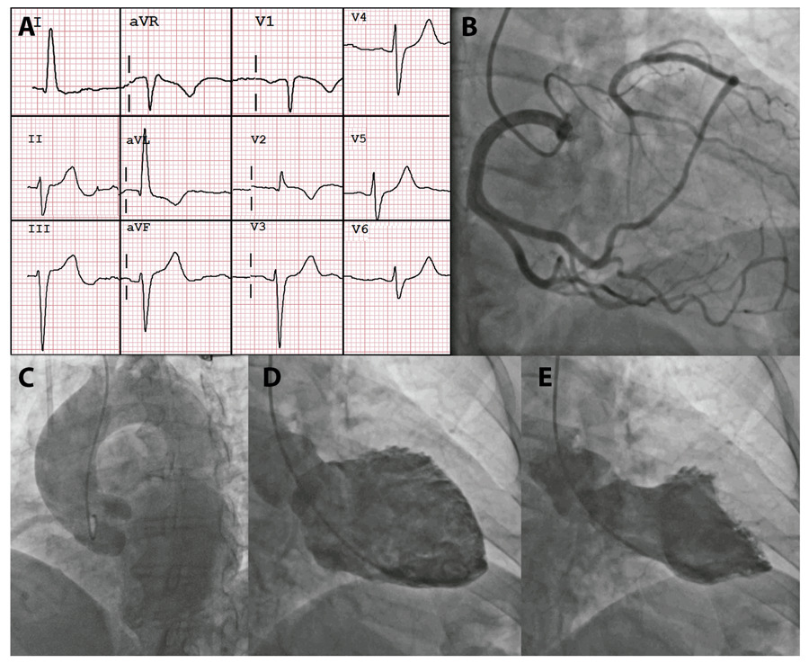
A 58-years-old female patient was admitted with retrosternal chest pain described as a continuous, oppressive tightness that had started two days earlier, after a tooth extraction procedure performed under ineffective local anesthesia. The patient’s past medical history included diabetes, dyslipidemia, and ovarian cancer, and she was receiving aspirin, rosuvastatin, metformin, dapagliflozin, and semaglutide. Physical examination was unremarkable. The chest pain protocol was initiated, and the initial electrocardiogram showed sinus rhythm with left anterior fascicular block, early repolarization in the inferior leads, and T-wave inversion in aVL, V1, and V2 (). The patient received 5 mg of sublingual isosorbide dinitrate, and blood samples were collected for laboratory testing. High-sensitivity troponin T (hs-cTnT) was 171 ng/L, confirming myocardial injury. An initial diagnosis of non–ST-elevation myocardial infarction (NSTEMI) was made, and the patient underwent coronary angiography (CA) for anatomic assessment ( / Video S1).

[…]

  • Coronary Angiography
  • Coronary Vessels
  • Echocardiography
  • Magnetic Resonance Imaging
  • Takotsubo Cardiomyopathy
Read Full Article
Creative Commons Attribution (CC BY) Creative Commons license
17/07/2026 DOI: 10.36660/abcimg.20260023i Views 1

Could not load more articles. Please try again.

How to cite this article

EndNote RIS

Electronic Document Format (Vancouver)

Roston F, Ribeiro AAA, Makiniks NC, Ribeiro LFB, Ziegler LL. A New Frontier in Cardiovascular Prevention: Beyond Prohibition, Clinical Management of Anabolic Steroid Users. ABC Imagem Cardiovascular 2026;39(2):e20260076.

Electronic Document Format (ABNT)

Roston, Fabio; Ribeiro, Alexandre Aby Azar; Makiniks, Naiara Caroline; Ribeiro, Luiz Felipe Branco; Ziegler, Liliana Ludwing. A New Frontier in Cardiovascular Prevention: Beyond Prohibition, Clinical Management of Anabolic Steroid Users. ABC Imagem Cardiovascular, v. 39, n. 2, e20260076, Jul. 2026.

Electronic Document Format (APA)

Roston, F., Ribeiro, A. A. A., Makiniks, N. C., Ribeiro, L. F. B., & Ziegler, L. L. (2026). A New Frontier in Cardiovascular Prevention: Beyond Prohibition, Clinical Management of Anabolic Steroid Users. ABC Imagem Cardiovascular, 39(2), e20260076.

Electronic Document Format (ISO)

Roston, Fabio and Ribeiro, Alexandre Aby Azar and Makiniks, Naiara Caroline and Ribeiro, Luiz Felipe Branco and Ziegler, Liliana Ludwing. A New Frontier in Cardiovascular Prevention: Beyond Prohibition, Clinical Management of Anabolic Steroid Users. ABC Imagem Cardiovascular [online]. 2026, vol. 39, n. 2, [cited 2026-10-14], e20260076. Available from: <https://journals.cardiol.br/en/article/a-new-frontier-in-cardiovascular-prevention-beyond-prohibition-clinical-management-of-anabolic-steroid-users/>. ISSN 2318-8219

EndNote RIS

Electronic Document Format (Vancouver)

Farouk H, El-Chilali K, Kloppe A. Visualization of the Ascending Aorta by Transthoracic Echocardiography: Could a Modified Parasternal Long-Axis View Provide Additional Imaging of a Longer Aortic Segment?. ABC Imagem Cardiovascular 2026;39(2):e20260068.

Electronic Document Format (ABNT)

Farouk, Heba; El-Chilali, Karim; Kloppe, Axel. Visualization of the Ascending Aorta by Transthoracic Echocardiography: Could a Modified Parasternal Long-Axis View Provide Additional Imaging of a Longer Aortic Segment?. ABC Imagem Cardiovascular, v. 39, n. 2, e20260068, Jul. 2026.

Electronic Document Format (APA)

Farouk, H., El-Chilali, K., & Kloppe, A. (2026). Visualization of the Ascending Aorta by Transthoracic Echocardiography: Could a Modified Parasternal Long-Axis View Provide Additional Imaging of a Longer Aortic Segment?. ABC Imagem Cardiovascular, 39(2), e20260068.

Electronic Document Format (ISO)

Farouk, Heba and El-Chilali, Karim and Kloppe, Axel. Visualization of the Ascending Aorta by Transthoracic Echocardiography: Could a Modified Parasternal Long-Axis View Provide Additional Imaging of a Longer Aortic Segment?. ABC Imagem Cardiovascular [online]. 2026, vol. 39, n. 2, [cited 2026-10-14], e20260068. Available from: <https://journals.cardiol.br/en/article/visualization-of-the-ascending-aorta-by-transthoracic-echocardiography-could-a-modified-parasternal-long-axis-view-provide-additional-imaging-of-a-longer-aortic-segment/>. ISSN 2318-8219

EndNote RIS

Electronic Document Format (Vancouver)

Vasconcelos DF. Recognition of Clinical Stages and Myocardial Damage in Aortic Stenosis: Beyond Diagnosis – To Op or Not To Op. ABC Imagem Cardiovascular 2026;39(2):e20260054.

Electronic Document Format (ABNT)

Vasconcelos, Daniel Franca. Recognition of Clinical Stages and Myocardial Damage in Aortic Stenosis: Beyond Diagnosis – To Op or Not To Op. ABC Imagem Cardiovascular, v. 39, n. 2, e20260054, Jul. 2026.

Electronic Document Format (APA)

Vasconcelos, D. F. (2026). Recognition of Clinical Stages and Myocardial Damage in Aortic Stenosis: Beyond Diagnosis – To Op or Not To Op. ABC Imagem Cardiovascular, 39(2), e20260054.

Electronic Document Format (ISO)

Vasconcelos, Daniel Franca. Recognition of Clinical Stages and Myocardial Damage in Aortic Stenosis: Beyond Diagnosis – To Op or Not To Op. ABC Imagem Cardiovascular [online]. 2026, vol. 39, n. 2, [cited 2026-10-14], e20260054. Available from: <https://journals.cardiol.br/en/article/recognition-of-clinical-stages-and-myocardial-damage-in-aortic-stenosis-beyond-diagnosis-to-op-or-not-to-op/>. ISSN 2318-8219

EndNote RIS

Electronic Document Format (Vancouver)

Lima EG, Carvalho LNS, Emerich TMS, Amorim EF, Rached FH. Elevated Lipoprotein(a) in Patients Without Comorbidities: Which Imaging Tests Should be Ordered?. ABC Imagem Cardiovascular 2026;39(2):e20260053.

Electronic Document Format (ABNT)

Lima, Eduardo Gomes; Carvalho, Leticia Neves Solon; Emerich, Tatiane Mascarenhas Santiago; Amorim, Eduardo Ferreira; Rached, Fabiana Hanna. Elevated Lipoprotein(a) in Patients Without Comorbidities: Which Imaging Tests Should be Ordered?. ABC Imagem Cardiovascular, v. 39, n. 2, e20260053, Jul. 2026.

Electronic Document Format (APA)

Lima, E. G., Carvalho, L. N. S., Emerich, T. M. S., Amorim, E. F., & Rached, F. H. (2026). Elevated Lipoprotein(a) in Patients Without Comorbidities: Which Imaging Tests Should be Ordered?. ABC Imagem Cardiovascular, 39(2), e20260053.

Electronic Document Format (ISO)

Lima, Eduardo Gomes and Carvalho, Leticia Neves Solon and Emerich, Tatiane Mascarenhas Santiago and Amorim, Eduardo Ferreira and Rached, Fabiana Hanna. Elevated Lipoprotein(a) in Patients Without Comorbidities: Which Imaging Tests Should be Ordered?. ABC Imagem Cardiovascular [online]. 2026, vol. 39, n. 2, [cited 2026-10-14], e20260053. Available from: <https://journals.cardiol.br/en/article/elevated-lipoproteina-in-patients-without-comorbidities-which-imaging-tests-should-be-ordered/>. ISSN 2318-8219

EndNote RIS

Electronic Document Format (Vancouver)

Costa KG, Otto MEB, Fernandes AFL, Assunção NM, Paiva MUB, Lima RLR, Dias RMS, et al. Dilated Cardiomyopathy as a Rare Initial Manifestation of ANCA-positive Microscopic Polyangiitis: Case Report. ABC Imagem Cardiovascular 2026;39(2):e20260038.

Electronic Document Format (ABNT)

Costa, Karoline Gonzaga; Otto, Maria Estefânia Bosco; Fernandes, André Felipe Lobão; Assunção, Nathália de Macêdo; Paiva, Mariana Ubaldo Barbosa; Lima, Rosyane Luz Rufino de; Dias, Rita Mikelle Soares. Dilated Cardiomyopathy as a Rare Initial Manifestation of ANCA-positive Microscopic Polyangiitis: Case Report. ABC Imagem Cardiovascular, v. 39, n. 2, e20260038, Jul. 2026.

Electronic Document Format (APA)

Costa, K. G., Otto, M. E. B., Fernandes, A. F. L., Assunção, N. M., Paiva, M. U. B., Lima, R. L. R. , & Dias, R. M. S. (2026). Dilated Cardiomyopathy as a Rare Initial Manifestation of ANCA-positive Microscopic Polyangiitis: Case Report. ABC Imagem Cardiovascular, 39(2), e20260038.

Electronic Document Format (ISO)

Costa, Karoline Gonzaga and Otto, Maria Estefânia Bosco and Fernandes, André Felipe Lobão and Assunção, Nathália de Macêdo and Paiva, Mariana Ubaldo Barbosa and Lima, Rosyane Luz Rufino de and Dias, Rita Mikelle Soares. Dilated Cardiomyopathy as a Rare Initial Manifestation of ANCA-positive Microscopic Polyangiitis: Case Report. ABC Imagem Cardiovascular [online]. 2026, vol. 39, n. 2, [cited 2026-10-14], e20260038. Available from: <https://journals.cardiol.br/en/article/dilated-cardiomyopathy-as-a-rare-initial-manifestation-of-anca-positive-microscopic-polyangiitis-case-report/>. ISSN 2318-8219

EndNote RIS

Electronic Document Format (Vancouver)

Uellendahl M. Beyond Ejection Fraction: Integrating Myocardial Strain and Cardiac Magnetic Resonance into the Contemporary Assessment of Left Ventricular Function. ABC Imagem Cardiovascular 2026;39(2):e20260087.

Electronic Document Format (ABNT)

Uellendahl, Marly. Beyond Ejection Fraction: Integrating Myocardial Strain and Cardiac Magnetic Resonance into the Contemporary Assessment of Left Ventricular Function. ABC Imagem Cardiovascular, v. 39, n. 2, e20260087, Jul. 2026.

Electronic Document Format (APA)

Uellendahl, M. (2026). Beyond Ejection Fraction: Integrating Myocardial Strain and Cardiac Magnetic Resonance into the Contemporary Assessment of Left Ventricular Function. ABC Imagem Cardiovascular, 39(2), e20260087.

Electronic Document Format (ISO)

Uellendahl, Marly. Beyond Ejection Fraction: Integrating Myocardial Strain and Cardiac Magnetic Resonance into the Contemporary Assessment of Left Ventricular Function. ABC Imagem Cardiovascular [online]. 2026, vol. 39, n. 2, [cited 2026-10-14], e20260087. Available from: <https://journals.cardiol.br/en/article/beyond-ejection-fraction-integrating-myocardial-strain-and-cardiac-magnetic-resonance-into-the-contemporary-assessment-of-left-ventricular-function/>. ISSN 2318-8219

EndNote RIS

Electronic Document Format (Vancouver)

Quagliato PC. Beyond Imaging: The Silent Impact of Depression on Coronary Artery Disease. ABC Imagem Cardiovascular 2026;39(2):e20260074.

Electronic Document Format (ABNT)

Quagliato, Priscila Cestari. Beyond Imaging: The Silent Impact of Depression on Coronary Artery Disease. ABC Imagem Cardiovascular, v. 39, n. 2, e20260074, Jul. 2026.

Electronic Document Format (APA)

Quagliato, P. C. (2026). Beyond Imaging: The Silent Impact of Depression on Coronary Artery Disease. ABC Imagem Cardiovascular, 39(2), e20260074.

Electronic Document Format (ISO)

Quagliato, Priscila Cestari. Beyond Imaging: The Silent Impact of Depression on Coronary Artery Disease. ABC Imagem Cardiovascular [online]. 2026, vol. 39, n. 2, [cited 2026-10-14], e20260074. Available from: <https://journals.cardiol.br/en/article/beyond-imaging-the-silent-impact-of-depression-on-coronary-artery-disease/>. ISSN 2318-8219

EndNote RIS

Electronic Document Format (Vancouver)

Barretto RBM, Silva CES. Myocardial Work During Isometric Exercise: From Physiology to Clinical Practice. ABC Imagem Cardiovascular 2026;39(2):e20260073.

Electronic Document Format (ABNT)

Barretto, Rodrigo Bellio de Mattos; Silva, Carlos Eduardo Suaide. Myocardial Work During Isometric Exercise: From Physiology to Clinical Practice. ABC Imagem Cardiovascular, v. 39, n. 2, e20260073, Jul. 2026.

Electronic Document Format (APA)

Barretto, R. B. M., & Silva, C. E. S. (2026). Myocardial Work During Isometric Exercise: From Physiology to Clinical Practice. ABC Imagem Cardiovascular, 39(2), e20260073.

Electronic Document Format (ISO)

Barretto, Rodrigo Bellio de Mattos and Silva, Carlos Eduardo Suaide. Myocardial Work During Isometric Exercise: From Physiology to Clinical Practice. ABC Imagem Cardiovascular [online]. 2026, vol. 39, n. 2, [cited 2026-10-14], e20260073. Available from: <https://journals.cardiol.br/en/article/myocardial-work-during-isometric-exercise-from-physiology-to-clinical-practice/>. ISSN 2318-8219

EndNote RIS

Electronic Document Format (Vancouver)

Silva HAGP, Gomes HM, Souza AC. My Approach to Left Atrial Function: From Basic Assessment to Atrial Stiffness. ABC Imagem Cardiovascular 2026;39(2):e20260070.

Electronic Document Format (ABNT)

Silva, Halsted Alarcão Gomes Pereira da; Gomes, Helder Moura; Souza, Alexandre Costa. My Approach to Left Atrial Function: From Basic Assessment to Atrial Stiffness. ABC Imagem Cardiovascular, v. 39, n. 2, e20260070, Jul. 2026.

Electronic Document Format (APA)

Silva, H. A. G. P. , Gomes, H. M., & Souza, A. C. (2026). My Approach to Left Atrial Function: From Basic Assessment to Atrial Stiffness. ABC Imagem Cardiovascular, 39(2), e20260070.

Electronic Document Format (ISO)

Silva, Halsted Alarcão Gomes Pereira da and Gomes, Helder Moura and Souza, Alexandre Costa. My Approach to Left Atrial Function: From Basic Assessment to Atrial Stiffness. ABC Imagem Cardiovascular [online]. 2026, vol. 39, n. 2, [cited 2026-10-14], e20260070. Available from: <https://journals.cardiol.br/en/article/my-approach-to-left-atrial-function-from-basic-assessment-to-atrial-stiffness/>. ISSN 2318-8219

EndNote RIS

Electronic Document Format (Vancouver)

Oliveira FRB, Lino DOC, Bezerra Filho GF, Linhares BC, Souza LBD, Alencar LFT, Cruz MRS, et al. Anabolic-Androgenic Steroids and Acute Myocardial Infarction in Young Adults: A Literature Review Based on a Case Series. ABC Imagem Cardiovascular 2026;39(2):e20260049.

Electronic Document Format (ABNT)

Oliveira, Fabiana Rocha Botelho de; Lino, Danielli Oliveira de Costa; Bezerra Filho, Germano Freire; Linhares, Bruno Cavalcante; Souza, Leonardo Brito De; Alencar, Luiz Filipe Torres de; Cruz, Matheus Rolim Santa; Feitosa, Mateus Paiva Marques. Anabolic-Androgenic Steroids and Acute Myocardial Infarction in Young Adults: A Literature Review Based on a Case Series. ABC Imagem Cardiovascular, v. 39, n. 2, e20260049, Jul. 2026.

Electronic Document Format (APA)

Oliveira, F. R. B. , Lino, D. O. C., Bezerra Filho, G. F., Linhares, B. C., Souza, L. B. D., Alencar, L. F. T. , Cruz, M. R. S., & Feitosa, M. P. M. (2026). Anabolic-Androgenic Steroids and Acute Myocardial Infarction in Young Adults: A Literature Review Based on a Case Series. ABC Imagem Cardiovascular, 39(2), e20260049.

Electronic Document Format (ISO)

Oliveira, Fabiana Rocha Botelho de and Lino, Danielli Oliveira de Costa and Bezerra Filho, Germano Freire and Linhares, Bruno Cavalcante and Souza, Leonardo Brito De and Alencar, Luiz Filipe Torres de and Cruz, Matheus Rolim Santa and Feitosa, Mateus Paiva Marques. Anabolic-Androgenic Steroids and Acute Myocardial Infarction in Young Adults: A Literature Review Based on a Case Series. ABC Imagem Cardiovascular [online]. 2026, vol. 39, n. 2, [cited 2026-10-14], e20260049. Available from: <https://journals.cardiol.br/en/article/anabolic-androgenic-steroids-and-acute-myocardial-infarction-in-young-adults-a-literature-review-based-on-a-case-series/>. ISSN 2318-8219

EndNote RIS

Electronic Document Format (Vancouver)

Sawamura KSS, Brito MM. My Approach to Agitated Saline Contrast Echocardiography in Pediatric Patients. ABC Imagem Cardiovascular 2026;39(2):e20260041.

Electronic Document Format (ABNT)

Sawamura, Karen Saori Shiraishi; Brito, Márcio Miranda. My Approach to Agitated Saline Contrast Echocardiography in Pediatric Patients. ABC Imagem Cardiovascular, v. 39, n. 2, e20260041, Jul. 2026.

Electronic Document Format (APA)

Sawamura, K. S. S., & Brito, M. M. (2026). My Approach to Agitated Saline Contrast Echocardiography in Pediatric Patients. ABC Imagem Cardiovascular, 39(2), e20260041.

Electronic Document Format (ISO)

Sawamura, Karen Saori Shiraishi and Brito, Márcio Miranda. My Approach to Agitated Saline Contrast Echocardiography in Pediatric Patients. ABC Imagem Cardiovascular [online]. 2026, vol. 39, n. 2, [cited 2026-10-14], e20260041. Available from: <https://journals.cardiol.br/en/article/my-approach-to-agitated-saline-contrast-echocardiography-in-pediatric-patients/>. ISSN 2318-8219

EndNote RIS

Electronic Document Format (Vancouver)

Silva TO. Cardiac Amyloidosis: Is It Truly a Hypertrophic Phenotype Cardiomyopathy?. ABC Imagem Cardiovascular 2026;39(2):e20260058.

Electronic Document Format (ABNT)

Silva, Tonnison de Oliveira. Cardiac Amyloidosis: Is It Truly a Hypertrophic Phenotype Cardiomyopathy?. ABC Imagem Cardiovascular, v. 39, n. 2, e20260058, Jul. 2026.

Electronic Document Format (APA)

Silva, T. O. (2026). Cardiac Amyloidosis: Is It Truly a Hypertrophic Phenotype Cardiomyopathy?. ABC Imagem Cardiovascular, 39(2), e20260058.

Electronic Document Format (ISO)

Silva, Tonnison de Oliveira. Cardiac Amyloidosis: Is It Truly a Hypertrophic Phenotype Cardiomyopathy?. ABC Imagem Cardiovascular [online]. 2026, vol. 39, n. 2, [cited 2026-10-14], e20260058. Available from: <https://journals.cardiol.br/en/article/cardiac-amyloidosis-is-it-truly-a-hypertrophic-phenotype-cardiomyopathy/>. ISSN 2318-8219

EndNote RIS

Electronic Document Format (Vancouver)

Almeida GGL, Barbirato GB, Pagnin VQ, Pagnin D, Mesquita CT. Major Depressive Disorder and Quality of Life in Patients With Coronary Artery Disease Assessed by Myocardial Perfusion Imaging. ABC Imagem Cardiovascular 2026;39(2):e20260048.

Electronic Document Format (ABNT)

Almeida, Guilherme Gonçais Lopes; Barbirato, Gustavo B.; Pagnin, Valéria de Queiroz; Pagnin, Daniel; Mesquita, Cláudio Tinoco. Major Depressive Disorder and Quality of Life in Patients With Coronary Artery Disease Assessed by Myocardial Perfusion Imaging. ABC Imagem Cardiovascular, v. 39, n. 2, e20260048, Jul. 2026.

Electronic Document Format (APA)

Almeida, G. G. L., Barbirato, G. B., Pagnin, V. Q., Pagnin, D., & Mesquita, C. T. (2026). Major Depressive Disorder and Quality of Life in Patients With Coronary Artery Disease Assessed by Myocardial Perfusion Imaging. ABC Imagem Cardiovascular, 39(2), e20260048.

Electronic Document Format (ISO)

Almeida, Guilherme Gonçais Lopes and Barbirato, Gustavo B. and Pagnin, Valéria de Queiroz and Pagnin, Daniel and Mesquita, Cláudio Tinoco. Major Depressive Disorder and Quality of Life in Patients With Coronary Artery Disease Assessed by Myocardial Perfusion Imaging. ABC Imagem Cardiovascular [online]. 2026, vol. 39, n. 2, [cited 2026-10-14], e20260048. Available from: <https://journals.cardiol.br/en/article/major-depressive-disorder-and-quality-of-life-in-patients-with-coronary-artery-disease-assessed-by-myocardial-perfusion-imaging/>. ISSN 2318-8219

EndNote RIS

Electronic Document Format (Vancouver)

Hassan MA, Alzahrani A, Mhanna M, Abdelkarim O, Bello R, Savoia P, Suksaranjit P, et al. A Hidden Connection: Anomalous Left Circumflex Artery Arising From the Right Pulmonary Artery Unveiled by Cardiovascular Computed Tomography. ABC Imagem Cardiovascular 2026;39(2):e20260040.

Electronic Document Format (ABNT)

Hassan, Mubariz Ahmed; Alzahrani, Ashraf; Mhanna, Mohammad; Abdelkarim, Ola; Bello, Rodrigo; Savoia, Paulo; Suksaranjit, Promporn; Delcour, Kimberly. A Hidden Connection: Anomalous Left Circumflex Artery Arising From the Right Pulmonary Artery Unveiled by Cardiovascular Computed Tomography. ABC Imagem Cardiovascular, v. 39, n. 2, e20260040, Jul. 2026.

Electronic Document Format (APA)

Hassan, M. A., Alzahrani, A., Mhanna, M., Abdelkarim, O., Bello, R., Savoia, P., Suksaranjit, P., & Delcour, K. (2026). A Hidden Connection: Anomalous Left Circumflex Artery Arising From the Right Pulmonary Artery Unveiled by Cardiovascular Computed Tomography. ABC Imagem Cardiovascular, 39(2), e20260040.

Electronic Document Format (ISO)

Hassan, Mubariz Ahmed and Alzahrani, Ashraf and Mhanna, Mohammad and Abdelkarim, Ola and Bello, Rodrigo and Savoia, Paulo and Suksaranjit, Promporn and Delcour, Kimberly. A Hidden Connection: Anomalous Left Circumflex Artery Arising From the Right Pulmonary Artery Unveiled by Cardiovascular Computed Tomography. ABC Imagem Cardiovascular [online]. 2026, vol. 39, n. 2, [cited 2026-10-14], e20260040. Available from: <https://journals.cardiol.br/en/article/a-hidden-connection-anomalous-left-circumflex-artery-arising-from-the-right-pulmonary-artery-unveiled-by-cardiovascular-computed-tomography/>. ISSN 2318-8219

EndNote RIS

Electronic Document Format (Vancouver)

Silva HAGP, Souza AC, Beck A. My Approach to a Standardized Assessment of the Tricuspid Valve: A Contemporary Analysis. ABC Imagem Cardiovascular 2026;39(2):e20260034.

Electronic Document Format (ABNT)

Silva, Halsted Alarcão Gomes Pereira da; Souza, Alexandre Costa; Beck, Adenalva. My Approach to a Standardized Assessment of the Tricuspid Valve: A Contemporary Analysis. ABC Imagem Cardiovascular, v. 39, n. 2, e20260034, Jul. 2026.

Electronic Document Format (APA)

Silva, H. A. G. P. , Souza, A. C., & Beck, A. (2026). My Approach to a Standardized Assessment of the Tricuspid Valve: A Contemporary Analysis. ABC Imagem Cardiovascular, 39(2), e20260034.

Electronic Document Format (ISO)

Silva, Halsted Alarcão Gomes Pereira da and Souza, Alexandre Costa and Beck, Adenalva. My Approach to a Standardized Assessment of the Tricuspid Valve: A Contemporary Analysis. ABC Imagem Cardiovascular [online]. 2026, vol. 39, n. 2, [cited 2026-10-14], e20260034. Available from: <https://journals.cardiol.br/en/article/my-approach-to-a-standardized-assessment-of-the-tricuspid-valve-a-contemporary-analysis/>. ISSN 2318-8219

EndNote RIS

Electronic Document Format (Vancouver)

Saeed B, Punnanithinont N, Mansour S, Savoia P, Suksaranjit P. Recurrent Intracardiac Masses in an Orthotopic Heart Transplant Recipient. ABC Imagem Cardiovascular 2026;39(2):e20260031.

Electronic Document Format (ABNT)

Saeed, Bilal; Punnanithinont, Natdanai; Mansour, Shareef; Savoia, Paulo; Suksaranjit, Promporn. Recurrent Intracardiac Masses in an Orthotopic Heart Transplant Recipient. ABC Imagem Cardiovascular, v. 39, n. 2, e20260031, Jul. 2026.

Electronic Document Format (APA)

Saeed, B., Punnanithinont, N., Mansour, S., Savoia, P., & Suksaranjit, P. (2026). Recurrent Intracardiac Masses in an Orthotopic Heart Transplant Recipient. ABC Imagem Cardiovascular, 39(2), e20260031.

Electronic Document Format (ISO)

Saeed, Bilal and Punnanithinont, Natdanai and Mansour, Shareef and Savoia, Paulo and Suksaranjit, Promporn. Recurrent Intracardiac Masses in an Orthotopic Heart Transplant Recipient. ABC Imagem Cardiovascular [online]. 2026, vol. 39, n. 2, [cited 2026-10-14], e20260031. Available from: <https://journals.cardiol.br/en/article/recurrent-intracardiac-masses-in-an-orthotopic-heart-transplant-recipient/>. ISSN 2318-8219

EndNote RIS

Electronic Document Format (Vancouver)

Alves MSL, Vosgerau LM, Dreckmann MV, Martins RD, Zanlorensi CB, Bonotto EH. My Approach to Echocardiographic Assessment of Left Ventricular Filling Pressures: From Ambiguity to Precision With New Guidelines. ABC Imagem Cardiovascular 2026;39(2):e20260026.

Electronic Document Format (ABNT)

Alves, Marco Stephan Lofrano; Vosgerau, Larissa Maria; Dreckmann, Marcelo Vitola; Martins, Roberto D´Ávila; Zanlorensi, Cláudia Biondo; Bonotto, Eduardo Henrique. My Approach to Echocardiographic Assessment of Left Ventricular Filling Pressures: From Ambiguity to Precision With New Guidelines. ABC Imagem Cardiovascular, v. 39, n. 2, e20260026, Jul. 2026.

Electronic Document Format (APA)

Alves, M. S. L., Vosgerau, L. M., Dreckmann, M. V., Martins, R. D., Zanlorensi, C. B., & Bonotto, E. H. (2026). My Approach to Echocardiographic Assessment of Left Ventricular Filling Pressures: From Ambiguity to Precision With New Guidelines. ABC Imagem Cardiovascular, 39(2), e20260026.

Electronic Document Format (ISO)

Alves, Marco Stephan Lofrano and Vosgerau, Larissa Maria and Dreckmann, Marcelo Vitola and Martins, Roberto D´Ávila and Zanlorensi, Cláudia Biondo and Bonotto, Eduardo Henrique. My Approach to Echocardiographic Assessment of Left Ventricular Filling Pressures: From Ambiguity to Precision With New Guidelines. ABC Imagem Cardiovascular [online]. 2026, vol. 39, n. 2, [cited 2026-10-14], e20260026. Available from: <https://journals.cardiol.br/en/article/my-approach-to-echocardiographic-assessment-of-left-ventricular-filling-pressures-from-ambiguity-to-precision-with-new-guidelines/>. ISSN 2318-8219

EndNote RIS

Electronic Document Format (Vancouver)

Herrera-Escandón , Morales-Grisales JP, Ayala-Zapata S, Barbosa-Balaguera S, Muriel-Ruiz J, Bravo-Rueda JF, Citelli-Ramírez JE, et al. Concordance Between Echocardiographic Left Ventricular Ejection Fraction by Simpson’s Method, Global Longitudinal Strain, and Cardiac Magnetic Resonance. ABC Imagem Cardiovascular 2026;39(2):e20250102.

Electronic Document Format (ABNT)

Herrera-Escandón, Álvaro; Morales-Grisales, Juan Pablo; Ayala-Zapata, Sebastián; Barbosa-Balaguera, Stephany; Muriel-Ruiz, Álvaro José; Bravo-Rueda, Juan Felipe; Citelli-Ramírez, José Eduardo; Osío-Jimenez, Luis Fernando; Benitez-Gómez, Luis Miguel; Ramírez-Estupiñán, Carlos Javier. Concordance Between Echocardiographic Left Ventricular Ejection Fraction by Simpson’s Method, Global Longitudinal Strain, and Cardiac Magnetic Resonance. ABC Imagem Cardiovascular, v. 39, n. 2, e20250102, Jul. 2026.

Electronic Document Format (APA)

Herrera-Escandón, , Morales-Grisales, J. P., Ayala-Zapata, S., Barbosa-Balaguera, S., Muriel-Ruiz, J., Bravo-Rueda, J. F., Citelli-Ramírez, J. E., Osío-Jimenez, L. F., Benitez-Gómez, L. M., & Ramírez-Estupiñán, C. J. (2026). Concordance Between Echocardiographic Left Ventricular Ejection Fraction by Simpson’s Method, Global Longitudinal Strain, and Cardiac Magnetic Resonance. ABC Imagem Cardiovascular, 39(2), e20250102.

Electronic Document Format (ISO)

Herrera-Escandón, Álvaro and Morales-Grisales, Juan Pablo and Ayala-Zapata, Sebastián and Barbosa-Balaguera, Stephany and Muriel-Ruiz, Álvaro José and Bravo-Rueda, Juan Felipe and Citelli-Ramírez, José Eduardo and Osío-Jimenez, Luis Fernando and Benitez-Gómez, Luis Miguel and Ramírez-Estupiñán, Carlos Javier. Concordance Between Echocardiographic Left Ventricular Ejection Fraction by Simpson’s Method, Global Longitudinal Strain, and Cardiac Magnetic Resonance. ABC Imagem Cardiovascular [online]. 2026, vol. 39, n. 2, [cited 2026-10-14], e20250102. Available from: <https://journals.cardiol.br/en/article/concordance-between-echocardiographic-left-ventricular-ejection-fraction-by-simpsons-method-global-longitudinal-strain-and-cardiac-magnetic-resonance/>. ISSN 2318-8219

EndNote RIS

Electronic Document Format (Vancouver)

Martins JL, Duarte MCMB, Feitosa FGAM, Santos MVC. Chemotherapy-Induced Cardiotoxicity in the Pediatric Population: What Are the Unique Aspects of Cardiovascular Imaging Follow-Up?. ABC Imagem Cardiovascular 2026;39(2):e20260044.

Electronic Document Format (ABNT)

Martins, Jéssica Laureano; Duarte, Maria do Carmo Menezes Bezerra; Feitosa, Fabiana Gomes Aragão Magalhães; Santos, Maria Verônica Câmara dos. Chemotherapy-Induced Cardiotoxicity in the Pediatric Population: What Are the Unique Aspects of Cardiovascular Imaging Follow-Up?. ABC Imagem Cardiovascular, v. 39, n. 2, e20260044, Jul. 2026.

Electronic Document Format (APA)

Martins, J. L., Duarte, M. C. M. B., Feitosa, F. G. A. M., & Santos, M. V. C. (2026). Chemotherapy-Induced Cardiotoxicity in the Pediatric Population: What Are the Unique Aspects of Cardiovascular Imaging Follow-Up?. ABC Imagem Cardiovascular, 39(2), e20260044.

Electronic Document Format (ISO)

Martins, Jéssica Laureano and Duarte, Maria do Carmo Menezes Bezerra and Feitosa, Fabiana Gomes Aragão Magalhães and Santos, Maria Verônica Câmara dos. Chemotherapy-Induced Cardiotoxicity in the Pediatric Population: What Are the Unique Aspects of Cardiovascular Imaging Follow-Up?. ABC Imagem Cardiovascular [online]. 2026, vol. 39, n. 2, [cited 2026-10-14], e20260044. Available from: <https://journals.cardiol.br/en/article/chemotherapy-induced-cardiotoxicity-in-the-pediatric-population-what-are-the-unique-aspects-of-cardiovascular-imaging-follow-up/>. ISSN 2318-8219

EndNote RIS

Electronic Document Format (Vancouver)

Soares FLJ. Mitral Valve Leaflet Hypoplasia in Adults: Role of Cardiovascular Imaging. ABC Imagem Cardiovascular 2026;39(2):e20260046.

Electronic Document Format (ABNT)

Soares, Fábio Luis de Jesus. Mitral Valve Leaflet Hypoplasia in Adults: Role of Cardiovascular Imaging. ABC Imagem Cardiovascular, v. 39, n. 2, e20260046, Jul. 2026.

Electronic Document Format (APA)

Soares, F. L. J. (2026). Mitral Valve Leaflet Hypoplasia in Adults: Role of Cardiovascular Imaging. ABC Imagem Cardiovascular, 39(2), e20260046.

Electronic Document Format (ISO)

Soares, Fábio Luis de Jesus. Mitral Valve Leaflet Hypoplasia in Adults: Role of Cardiovascular Imaging. ABC Imagem Cardiovascular [online]. 2026, vol. 39, n. 2, [cited 2026-10-14], e20260046. Available from: <https://journals.cardiol.br/en/article/mitral-valve-leaflet-hypoplasia-in-adults-role-of-cardiovascular-imaging/>. ISSN 2318-8219

EndNote RIS

Electronic Document Format (Vancouver)

Pereira MM, Otto MEB, Portela JLP. Impact of Isometric Exercise on Left Ventricular Mechanics Assessed by Global Longitudinal Strain and Myocardial Work in Healthy Adults. ABC Imagem Cardiovascular 2026;39(2):e20260036.

Electronic Document Format (ABNT)

Pereira, Marcio Mendes; Otto, Maria Estefania Bosco; Portela, Juliana Lins da Paz. Impact of Isometric Exercise on Left Ventricular Mechanics Assessed by Global Longitudinal Strain and Myocardial Work in Healthy Adults. ABC Imagem Cardiovascular, v. 39, n. 2, e20260036, Jul. 2026.

Electronic Document Format (APA)

Pereira, M. M., Otto, M. E. B., & Portela, J. L. P. (2026). Impact of Isometric Exercise on Left Ventricular Mechanics Assessed by Global Longitudinal Strain and Myocardial Work in Healthy Adults. ABC Imagem Cardiovascular, 39(2), e20260036.

Electronic Document Format (ISO)

Pereira, Marcio Mendes and Otto, Maria Estefania Bosco and Portela, Juliana Lins da Paz. Impact of Isometric Exercise on Left Ventricular Mechanics Assessed by Global Longitudinal Strain and Myocardial Work in Healthy Adults. ABC Imagem Cardiovascular [online]. 2026, vol. 39, n. 2, [cited 2026-10-14], e20260036. Available from: <https://journals.cardiol.br/en/article/impact-of-isometric-exercise-on-left-ventricular-mechanics-assessed-by-global-longitudinal-strain-and-myocardial-work-in-healthy-adults/>. ISSN 2318-8219

EndNote RIS

Electronic Document Format (Vancouver)

Barbosa RR, Prando CB, Bianchini VM, Barros LC, Paganini LN, Barros G, Dadalt D, et al. Giant Atrial Myxoma in a Pregnant Patient: A Case Report. ABC Imagem Cardiovascular 2026;39(2):e20260010.

Electronic Document Format (ABNT)

Barbosa, Roberto Ramos; Prando, Caio Badiani; Bianchini, Victor Macedo; Barros, Lucas Crespo de; Paganini, Larissa Novaes; Barros, Gracielly; Dadalt, Darlan; Guedes, Sergio Luis Santos; Costa, Vinicius Eduardo Araújo; Tito, Marcus Gustavo; Nery, Tiago Bernardo; Angeli, Marcio Vinicius de Nardi de; Viana, Maria Eduarda Vichi Gomes; Roncato, Mariana Oliveira; Auad, João Paulo Moulin; Oliveira, Guilherme Freitas Fernandes de; Serpa, Renato Giestas; Calil, Osmar Araujo; Barbosa, Luiz Fernando Machado. Giant Atrial Myxoma in a Pregnant Patient: A Case Report. ABC Imagem Cardiovascular, v. 39, n. 2, e20260010, Jul. 2026.

Electronic Document Format (APA)

Barbosa, R. R., Prando, C. B., Bianchini, V. M., Barros, L. C. , Paganini, L. N., Barros, G., Dadalt, D., Guedes, S. L. S., Costa, V. E. A., Tito, M. G., Nery, T. B., Angeli, M. V. N. , Viana, M. E. V. G., Roncato, M. O., Auad, J. P. M., Oliveira, G. F. F. , Serpa, R. G., Calil, O. A., & Barbosa, L. F. M. (2026). Giant Atrial Myxoma in a Pregnant Patient: A Case Report. ABC Imagem Cardiovascular, 39(2), e20260010.

Electronic Document Format (ISO)

Barbosa, Roberto Ramos and Prando, Caio Badiani and Bianchini, Victor Macedo and Barros, Lucas Crespo de and Paganini, Larissa Novaes and Barros, Gracielly and Dadalt, Darlan and Guedes, Sergio Luis Santos and Costa, Vinicius Eduardo Araújo and Tito, Marcus Gustavo and Nery, Tiago Bernardo and Angeli, Marcio Vinicius de Nardi de and Viana, Maria Eduarda Vichi Gomes and Roncato, Mariana Oliveira and Auad, João Paulo Moulin and Oliveira, Guilherme Freitas Fernandes de and Serpa, Renato Giestas and Calil, Osmar Araujo and Barbosa, Luiz Fernando Machado. Giant Atrial Myxoma in a Pregnant Patient: A Case Report. ABC Imagem Cardiovascular [online]. 2026, vol. 39, n. 2, [cited 2026-10-14], e20260010. Available from: <https://journals.cardiol.br/en/article/giant-atrial-myxoma-in-a-pregnant-patient-a-case-report/>. ISSN 2318-8219

EndNote RIS

Electronic Document Format (Vancouver)

Calvilho Júnior AA. Echocardiographic Assessment of Diastolic Dysfunction in Special Situations. ABC Imagem Cardiovascular 2026;39(2):e20260029.

Electronic Document Format (ABNT)

Calvilho Júnior, Antonio Amador. Echocardiographic Assessment of Diastolic Dysfunction in Special Situations. ABC Imagem Cardiovascular, v. 39, n. 2, e20260029, Jul. 2026.

Electronic Document Format (APA)

Calvilho Júnior, A. A. (2026). Echocardiographic Assessment of Diastolic Dysfunction in Special Situations. ABC Imagem Cardiovascular, 39(2), e20260029.

Electronic Document Format (ISO)

Calvilho Júnior, Antonio Amador. Echocardiographic Assessment of Diastolic Dysfunction in Special Situations. ABC Imagem Cardiovascular [online]. 2026, vol. 39, n. 2, [cited 2026-10-14], e20260029. Available from: <https://journals.cardiol.br/en/article/echocardiographic-assessment-of-diastolic-dysfunction-in-special-situations/>. ISSN 2318-8219

EndNote RIS

Electronic Document Format (Vancouver)

Ferreira MVS, Tormin JPAS, Dantas Jr RN, Torres RVA, Cordeiro RA, Araújo Filho JAB, Cardoso LF, et al. Single Coronary Artery and Stress Cardiomyopathy: An Association Demonstrated by Multimodality Imaging. ABC Imagem Cardiovascular 2026;39(2):e20260023.

Electronic Document Format (ABNT)

Ferreira, Marcus Vinicius Silva; Tormin, Julia Pereira Afonso dos Santos; Dantas Jr, Roberto Nery; Torres, Roberto Vitor Almeida; Cordeiro, Renan Andreos; Araújo Filho, José de Arimateia Batista; Cardoso, Luiz Francisco. Single Coronary Artery and Stress Cardiomyopathy: An Association Demonstrated by Multimodality Imaging. ABC Imagem Cardiovascular, v. 39, n. 2, e20260023, Jul. 2026.

Electronic Document Format (APA)

Ferreira, M. V. S., Tormin, J. P. A. S., Dantas Jr, R. N., Torres, R. V. A., Cordeiro, R. A., Araújo Filho, J. A. B., & Cardoso, L. F. (2026). Single Coronary Artery and Stress Cardiomyopathy: An Association Demonstrated by Multimodality Imaging. ABC Imagem Cardiovascular, 39(2), e20260023.

Electronic Document Format (ISO)

Ferreira, Marcus Vinicius Silva and Tormin, Julia Pereira Afonso dos Santos and Dantas Jr, Roberto Nery and Torres, Roberto Vitor Almeida and Cordeiro, Renan Andreos and Araújo Filho, José de Arimateia Batista and Cardoso, Luiz Francisco. Single Coronary Artery and Stress Cardiomyopathy: An Association Demonstrated by Multimodality Imaging. ABC Imagem Cardiovascular [online]. 2026, vol. 39, n. 2, [cited 2026-10-14], e20260023. Available from: <https://journals.cardiol.br/en/article/single-coronary-artery-and-stress-cardiomyopathy-an-association-demonstrated-by-multimodality-imaging/>. ISSN 2318-8219

Sede – São Paulo
Rua Barata Ribeiro, 380 cj 54 Bela Vista
São Paulo, Brasil – CEP: 01308-000
Tel.: +55 (11) 3120-3363
Telefax: +55 (11) 3259-2988
E-mail: depeco@cardiol.br

Sede – Rio de Janeiro
Av. Marechal Câmara, 160
3º andar – Sala: 330 – Centro
Rio de Janeiro – CEP: 20020-907
Tel.: +55 (21) 3478-2700
ou: +55 (21) 99386-3961
E-mail: abcimaging@cardiol.br

  • About the Journal
  • Internal Regulation
  • Editors Section
  • Editorial Board
  • SBC Award

  • Guidelines for Publication
  • Editorial Policy
  • Ethics Policy
  • Policy on the Use of Artificial Intelligence
  • Mandatory Publication Form

Issues

SBC Guidelines