ABC Family Arquivos Brasileiros de Cardiologia International Journal of Cardiovascular Sciences ABC Heart Failure & Cardiomyopathy ABC Imagem Cardiovascular
Português English
ABC Family
Português English
  • About
    • About ABC Family
    • Internal Regulation
    • Editorial Board
    • Brazilian Society of Cardiology (SBC)
  • Issues
  • SBC Guidelines
  • Content
    • Videos
    • Podcasts
    • News
  • About
    • About ABC Family
    • Internal Regulation
    • Editorial Board
    • Brazilian Society of Cardiology (SBC)
  • Issues
  • SBC Guidelines
  • Content
    • Videos
    • Podcasts
    • News

ABC Family

  • ABC Family
  • Arquivos Brasileiros de Cardiologia
  • International Journal of Cardiovascular Sciences
  • ABC Heart Failure & Cardiomyopathy
  • ABC Imagem Cardiovascular
Search in

No content found.

Journals
Ordering
Article types
Others

  1. Home
  2. Keyword: Left Ventricular Function
My Approach to Echocardiographic Assessment of Left Ventricular Filling Pressures: From Ambiguity to Precision With New Guidelines
ABCIC
Review Article

My Approach to Echocardiographic Assessment of Left Ventricular Filling Pressures: From Ambiguity to Precision With New Guidelines

Marco Stephan Lofrano Alves et al. Larissa Maria Vosgerau Marcelo Vitola Dreckmann Roberto D´Ávila Martins … Cláudia Biondo Zanlorensi Eduardo Henrique Bonotto

Abstract

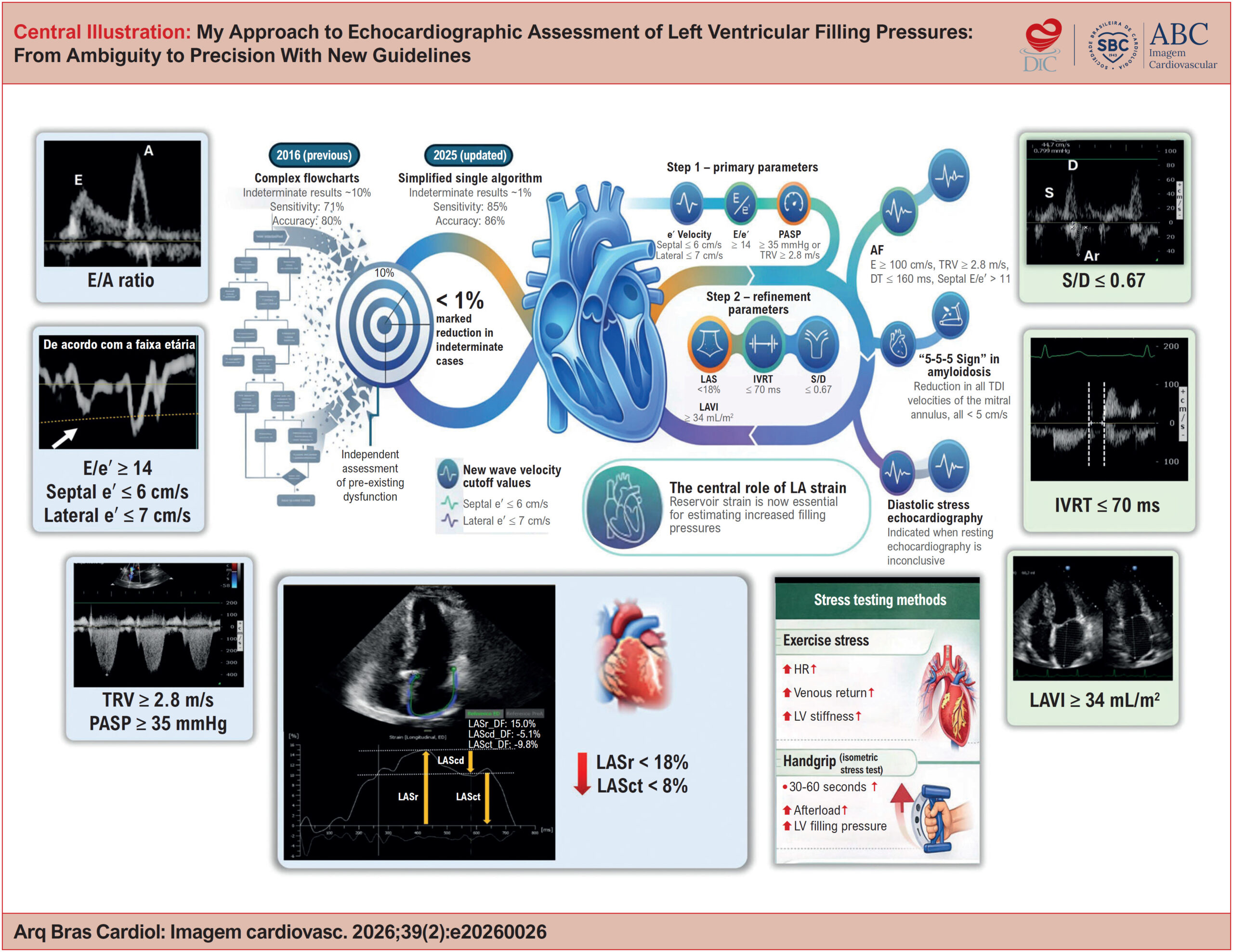
The echocardiographic estimation of left ventricular (LV) filling pressures is a cornerstone in the assessment of heart failure, particularly in patients with heart failure with preserved ejection fraction (HFpEF). The 2016 American Society of Echocardiography/European Association of cardiovascular Imaging algorithm standardized this evaluation using key variables, but a substantial proportion of cases remained indeterminate in clinical practice. The recent update of the American guideline reorganized the approach into a hierarchical framework, incorporating age-related adjustments and formalizing the role of left atrial strain (LAS) as a tie-breaking parameter, while also providing a more detailed characterization of special clinical scenarios. In parallel, the 2024 British Society of Echocardiography guideline emphasizes the pathophysiological interpretation of ventricular filling, complementing the operational framework proposed by the American document. Multicenter studies with invasive validation support these updates, establishing LAS as a robust marker of increased filling pressures. In this article, we present My Approach to echocardiographic assessment of LV filling pressures, based on an initial morphofunctional evaluation, followed by structured screening (Step 1) and further refinement (Step 2) incorporating LAS and additional parameters. We also provide a comparison between guidelines, discuss common pitfalls and algorithm limitations, and include case-based videos for practical application.
  • Echocardiography
  • Heart Failure
  • Left Ventricular Function
Read Full Article
Creative Commons Attribution (CC BY) Creative Commons license
24/07/2026 DOI: 10.36660/abcimg.20260026i Views 1
Figure 1
ABCIC
Review Article

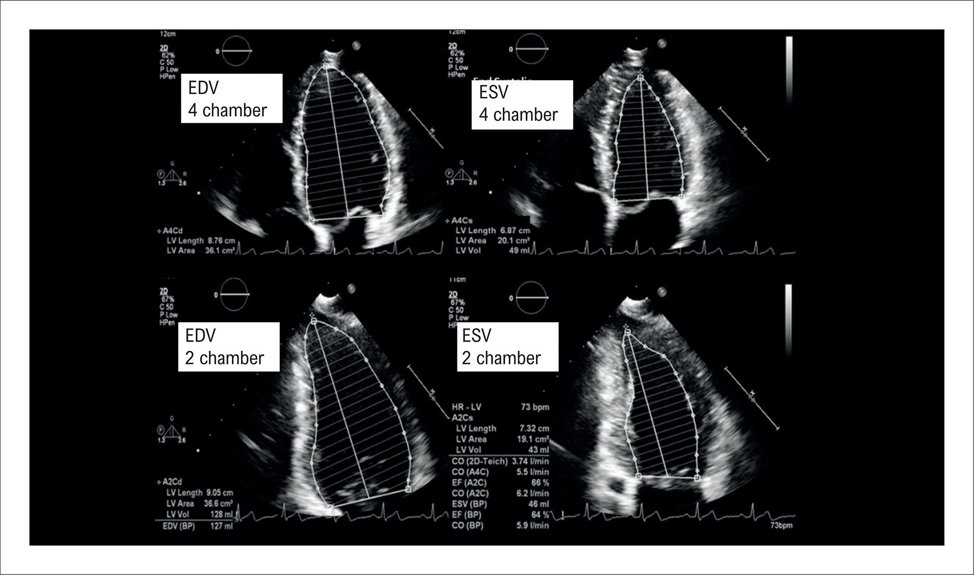
How I Assess Left Ventricular Systolic Function

Ana Clara Tude Rodrigues

Abstract

Assessment of left ventricular systolic function is central to transthoracic echocardiography and remains essential in clinical practice and research. Although left ventricular ejection fraction is still the most widely used parameter, advances in echocardiographic techniques have broadened diagnostic assessment. This review summarizes key methods for evaluating systolic function, from traditional linear measurements to modern two- and three-dimensional volumetric approaches. The roles and limitations of the Simpson method, contrast echocardiography, three-dimensional imaging, myocardial deformation analysis, and artificial intelligence are discussed, emphasizing the importance of an integrated and individualized approach.

  • Echocardiography
  • Heart Rate
  • Left Ventricular Function
Read Full Article
Creative Commons Attribution (CC BY) Creative Commons license
30/12/2025 DOI: 10.36660/abcimg.20250078i Views 3
Left Ventricular Function and Myocardial Longitudinal Strain Analysis in Patients With Chagas Disease: Case Series and Systematic Literature Review
ABCIC
Original Article

Left Ventricular Function and Myocardial Longitudinal Strain Analysis in Patients With Chagas Disease: Case Series and Systematic Literature Review

José Victor da Nóbrega Borges et al. Samira Abdel Correia Leila Manuella Guedes da Nóbrega Machado Kiran Sooraj Thirukonda Jegadeesh Babu … Karma Jashvantbhai Patel Kavya Hiteshkumar Shah Mahitha Reddy Takkasila Abdul Majid Shaik Muhammad Ahmed Shaikh Ninad Khandekar Sahithi Burra

Abstract

Introduction:

Left ventricular dysfunction as an independent predictor of mortality in Chagas disease. Global longitudinal strain (GLS) is an emerging echocardiographic modality with possible incremental value in early detection of cardiac involvement.

Objective:

Compare left ventricular function with multiple echocardiographic parameters between patients with the indeterminate and chronic forms of Chagas, and perform a literature review.

Methods:

In this observational study, 11 patients with a confirmed diagnosis of Chagas disease were evaluated. Individuals were distributed as follows: five with the indeterminate and six with the chronic forms. Conventional echocardiographic evaluation was followed by GLS measurement.

Results:

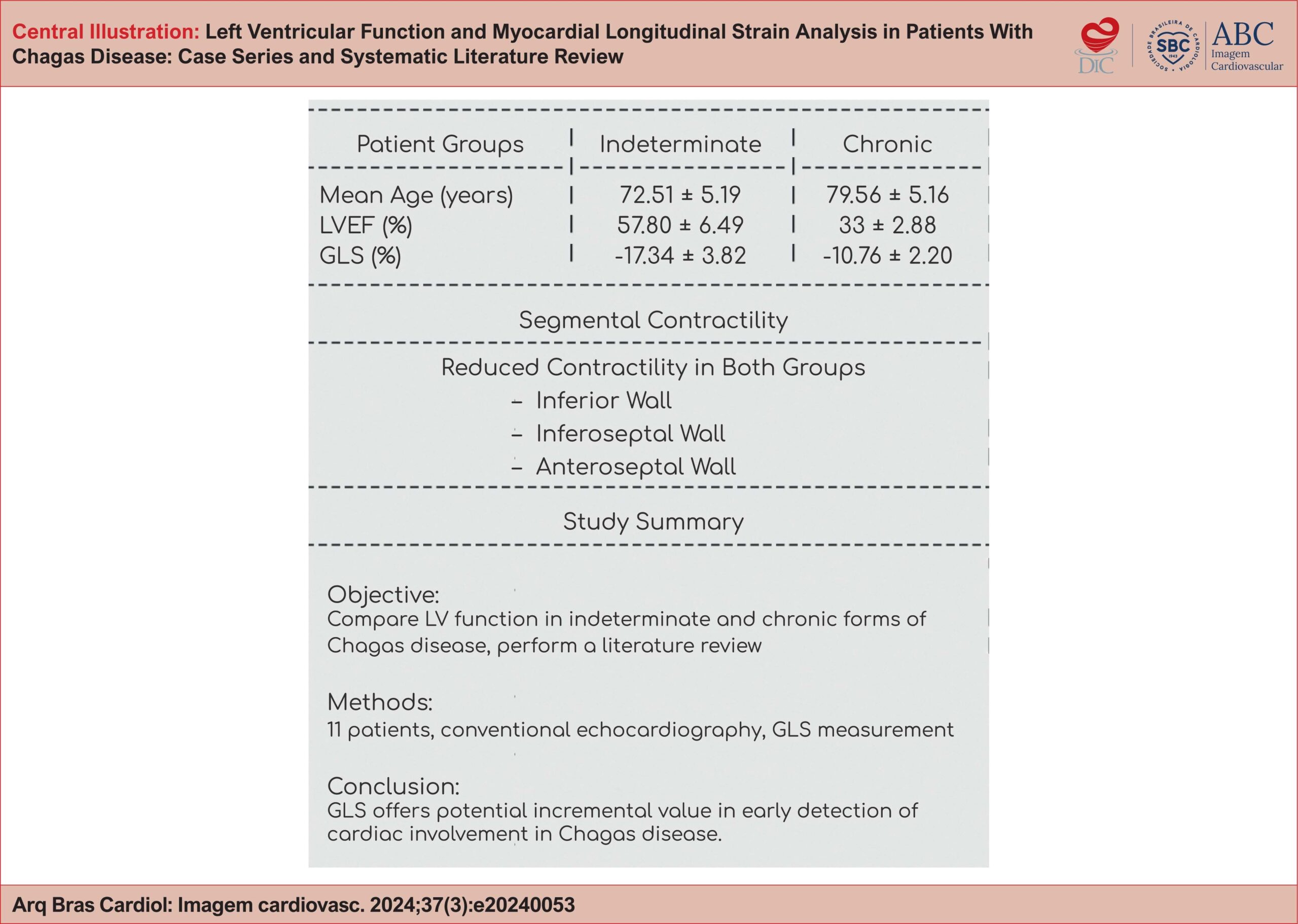
The mean left ventricle ejection fraction left ventricular ejection fraction (LVEF) was 33 ± 2.88, and the mean GLS of the left ventricle was −10.76 ± 2.20 in the chronic form as compared to 57.80 ± 6.49 and −17.34 ± 3.82, respectively in the indeterminate group. Contractility was reduced in segments of the inferior, inferoseptal and anteroseptal walls in both groups. These segmental alterations in the indeterminate group may be attributed to early myocardial damage from inflammation and fibrosis, even before significant left ventricular dysfunction is evident.

Conclusion:

Analysis of longitudinal strain in the indeterminate form has potential incremental value to the conventional analysis of ejection fraction when compared to patients with chronic Chagas cardiomyopathy, especially with individual segmental analysis, but the impact and clinical significance of these techniques are still unknown.

  • Chagas Cardiomyopathy
  • Global Longitudinal Strain
  • Left Ventricular Function
Read Full Article
Creative Commons Attribution (CC BY) Creative Commons license
01/10/2024 DOI: 10.36660/abcimg.20240053i Views 1
Central Illustration
ABCHF
Review Article

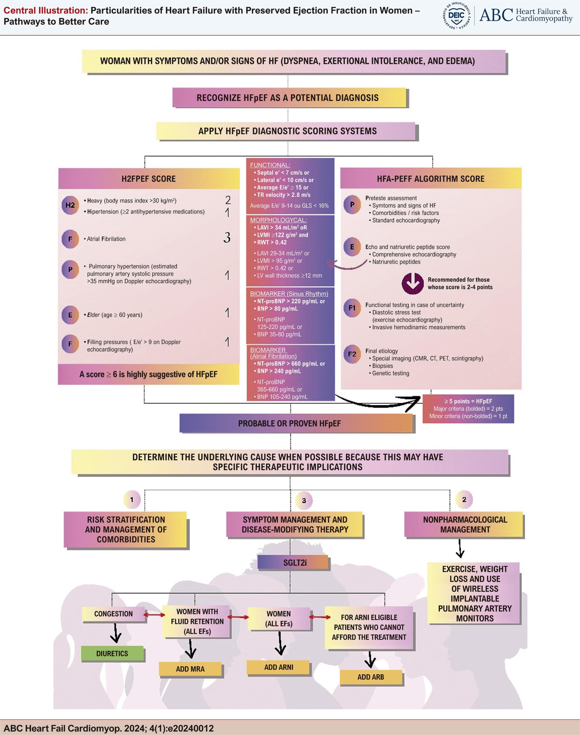
Particularities of Heart Failure with Preserved Ejection Fraction in Women – Pathways to Better Care

Larissa Neto Espíndola et al. Maria Cristina Costa de Almeida Marildes Luíza de Castro Cláudia Maria Vilas Freire Gláucia Maria Moraes de Oliveira

Abstract

While heart failure with reduced ejection fraction (HFrEF) is more prevalent in males, female predominance is a striking feature in heart failure with preserved ejection fraction (HFpEF).

The incidence of HFpEF has been gaining alarming proportions. It has exceeded HFrEF as the primary form of heart failure, especially in the female sex. Women tend to have smaller cardiac chambers with higher left ventricular ejection fraction; however, they have more severe symptoms and signs. Until now, there is no consensus about the exact mechanisms of these differences related to sex. Understanding these mechanisms is essential to mitigate the risks for HFpEF and direct efforts towards identifying novel preventive and disease-modifying treatments. Lifestyle, pharmacologic, and device-based approaches to reduce the medical and societal impact of HFpEF could improve patients’ quality of life.

The purpose of this review is to outline known biological sex differences in women with HFpEF with a specific focus on pathways to better care for the diagnosis and treatment of women with HFpEF.

  • Diagnosis
  • Heart Failure
  • Left Ventricular Function
  • Therapeutics
  • Women
Read Full Article
Creative Commons Attribution (CC BY) Creative Commons license
13/08/2024 DOI: 10.36660/abchf.20240012i Views 3
What do Cardiologists Expect Towards Myocardial Viability Assessment
ABCIC
Review Article

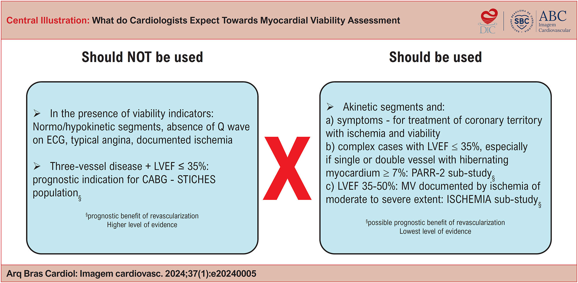
What do Cardiologists Expect Towards Myocardial Viability Assessment

Eduardo Gomes Lima et al. Eduardo Bello Martins Leticia Neves Solon Carvalho Diogo Freitas Cardoso de Azevedo

Within the clinical spectrum of chronic coronary syndrome (CCS), the presence of coronary artery disease (CAD) alongside ventricular dysfunction has long been a focal point due to the lower survival rates seen in this population. Improving survival in these patients, whether through medications or interventions, is a key concern for clinical cardiologists. Previous observational studies suggested that myocardial viability (MV) could influence the effectiveness of revascularization strategies in patients with ventricular dysfunction. Thus, viability assessment became a standard practice in cardiology. Different imaging methods assess viability through different parameters (such as cell membrane integrity, mitochondrial function, glycolytic metabolism, contractile reserve, or evidence of fibrosis), leading to differing sensitivities and specificities. However, recent evidence from large randomized studies does not support the notion that the presence of MV, as determined by various methods, is linked to the prognostic benefit of revascularization. Furthermore, the reduction in mortality observed in these studies was not attributed to the recovery of contractile function but rather to a decrease in fatal heart attacks. As a result, the current role of MV as a decision-making tool for revascularization in patients with CCS and left ventricular dysfunction has diminished. Nonetheless, there are still situations in specialist practice where its use may be warranted, particularly in the context of constructing a comprehensive functional revascularization strategy.

  • Left Ventricular Function
  • Myocardial Ischemia
  • Myocardial Stunning
Read Full Article
Creative Commons Attribution (CC BY) Creative Commons license
28/03/2024 DOI: 10.36660/abcimg.20240005i Views 2
Figure 1
IJCS
Original Article

Sociodemographic, Clinical Condition, and Functional Aerobic Capacity in Patients With Heart Failure With Varying Ventricular Ejection Fraction

Jhonatan Betancourt Peña et al. Iago Portela Pino Maria Jose Martinez Patino

Abstract

Background

Recently, a new heart failure (HF) classification was made considering the left ventricular ejection fraction (LVEF) phenotype. Comprehensive assessments of the groups are required to guide patient management.

Objective

To determine the differences in sociodemographic, clinical, functional aerobic capacity, and health-related quality of life (HRQOL) variables in patients with HF classified with different LVEFs and to explore the correlations between the variables.

Methods

This work is a cross-sectional descriptive and correlational study. Three groups of patients with HF (LVEF≥50%, LVEF<40%, and LVEF40-49%) were compared. Sociodemographic, clinical variables and functional aerobic capacity with Sit to Stand (STS), 6-minute walk test (6MWT), Duke Activity Status Index (DASI), Minnesota Living with HF Questionnaire (MLFHQ), and Patient Health Questionnaire 9 (PHQ-9) were considered. The Chi-square test, one-way analysis of variance (ANOVA) test, and Spearman's correlation were used for statistical analysis. The statistical significance level was set at 5%.

Results

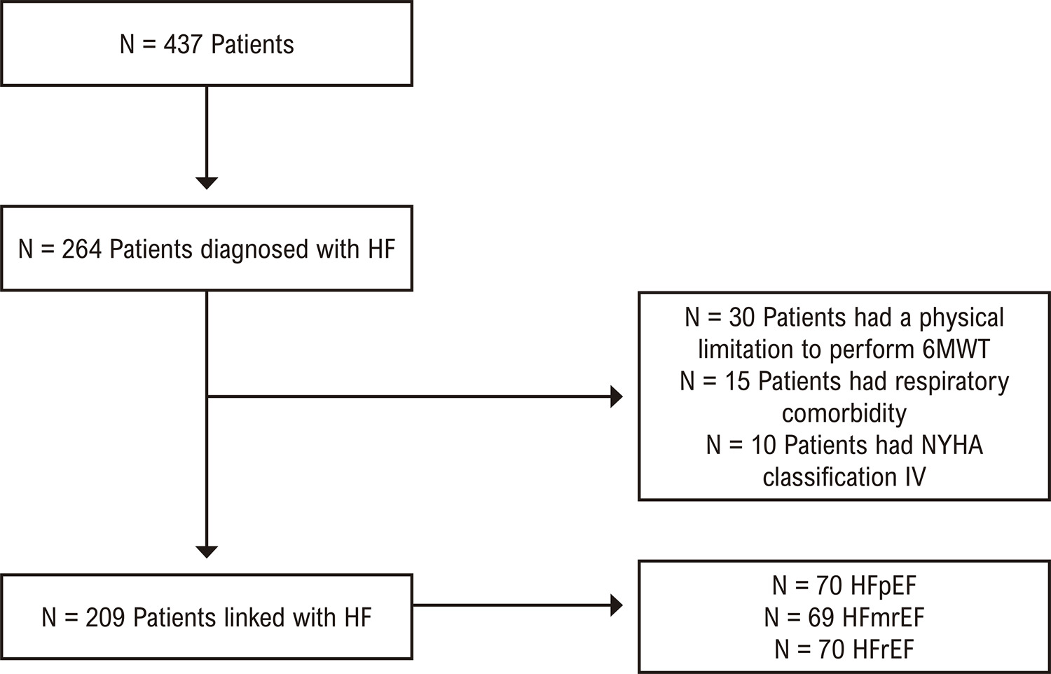
A total of 209 patients were admitted with a diagnosis of HF, with a more significant number of men. Marital status was a predominantly stable union in the HF with preserved ejection fraction (HFpEF) and HF with mid-range ejection fraction (HFmrEF) groups. A sedentary lifestyle was lower in the HF with reduced ejection fraction (HFrEF) group 59 (84.3%), p-value = 0.033, and the angina pectoris was higher in the HFpEF 30 (42.9%). Systolic blood pressure at the end of the 6MWT evidenced a higher score in HFpEF 132.0±17.25 concerning HFrEF 128.0±16.57, p-value=0.043. The fat percentage was higher in HFpEF 30.20±8.80 regarding the HFmrEF group 26.51±7.60, p-value = 0.028.

Conclusion

There were significant differences according to the LVEF classification in marital status, angina symptoms, fat percentage, and blood pressure at rest.

  • Exercise Tests
  • Heart Failure
  • Left Ventricular Function
  • Quality of life
  • Socioeconomic Factors
Read Full Article
Creative Commons Attribution (CC BY) Creative Commons license
08/03/2024 DOI: 10.36660/ijcs.20230079 Views 1
ABC
Editorial

Empowering the Imagers with 3D-Speckle Tracking Echocardiography to Detect Subclinical Cancer Therapy-Related Myocardial Dysfunction

Jennifer Mancio

Despite the expansion of the cardio-oncology field, defining cardiotoxicity to guide therapeutic decisions and impact prognosis is still a work in progress. The most widely recognized diagnosis of cardiotoxicity is based on serial changes in the left ventricular ejection fraction (LVEF). The ESC Position Paper recommends that if LVEF decreases by 10% to a value below the lower limit of normal (considered as an LVEF >50%), angiotensin-converting enzyme inhibitors (or angiotensin receptor blockers) in combination with beta-blockers are recommended to prevent further left ventricular (LV) dysfunction or the development of symptomatic heart failure. However, different LVEF thresholds have been proposed by different authors and guidelines,, and the 2-dimensional (2D)-echocardiography LVEF depends on loading conditions (fluid status varies significantly in cancer patients), has low sensitivity for detecting small LV function changes, is subject to intra- and interobserver variability and relies on geometrical assumptions. Hence, due to the limited availability of cardiac magnetic resonance imaging, 3D echocardiography is the preferred technique for monitoring cardiotoxicity in oncological patients, and the 3D-LVEF is the method of choice for cardiotoxicity surveillance.

Because any grade of cancer therapy-induced myocardial dysfunction matters, a growing body of literature has explored the role of myocardial deformation analysis in patients receiving cancer therapy for early detection of myocardial damage before LVEF starts declining. Speckle-tracking echocardiography (STE) allows quantitative assessment of global and segmental LV myocardial function by measuring strain in a manner largely independent of angle and ventricular geometry. Compared with 2D-strain, 3D-strain has the advantage of tracking the speckle patterns in any direction and out-of-the imaging plane, offering additional deformation parameters (such as area strain) and a more comprehensive quantitation of LV geometry and function.,

[…]

  • Cardiotoxicity
  • Left Ventricular Function
  • Myocardial deformation
  • Myocardium/diagnosis imaging
  • Tridensional Echocardiography/methods
Read Full Article
Creative Commons Attribution (CC BY) Creative Commons license
25/09/2023 DOI: 10.36660/abc.20230554 Views 1
Figura 1
ABCIC
Systematic Review

Myocardial Work: a Systematic Review of Novel Echocardiography Method and Clinical Applications

Sidhi Laksono et al. Hillary Kusharsamita Mefri Yanni Astri Astuti

Abstract

Background:

Myocardial work (MW) is a novel imaging modality that has emerged as a potential left ventricular (LV) function assessment in various clinical settings. MW calculates speckle-tracking echocardiography strain curves with an estimated LV pressure curve by non-invasively utilizing standard brachial blood pressure curves.

Objective:

This study aimed to provide a summary of current knowledge of non-invasive MW and its clinical applications, including in heart failure, coronary artery disease, cardiomyopathy, and hypertension. In addition, the limitations, and recommendations of MW in clinical practice are discussed.

Methods:

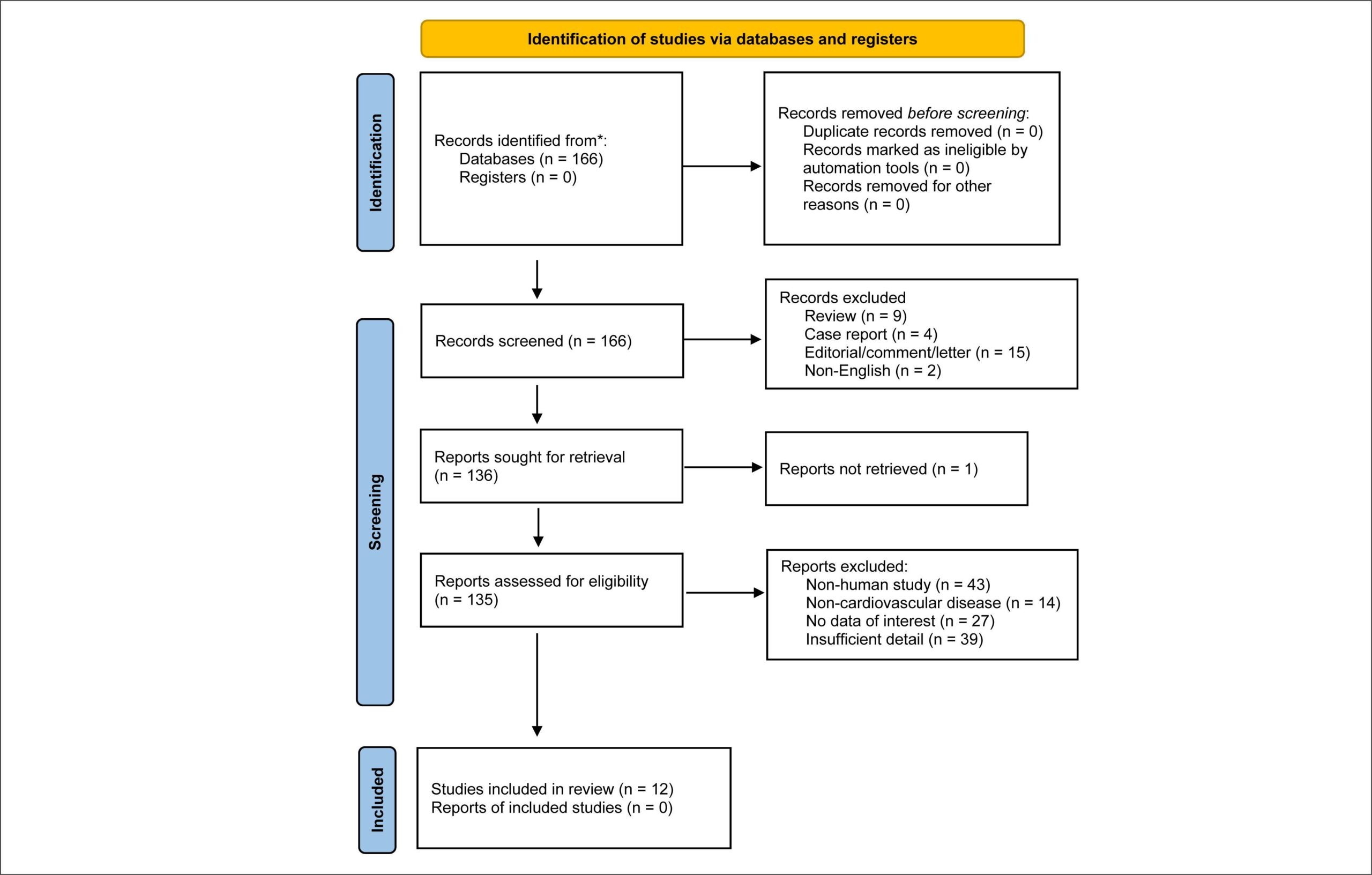
We searched the PubMed database using the following keywords: (myocardial constructive work) OR (wasted septal work) OR (global myocardial work) OR (myocardial work) OR (myocardial constructive work) OR (novel echocardiography). We further subjected 12 studies to full-text review and included them in this systematic review.

Results:

While MW indices, particularly global work index and global constructed work, have shown good correlations with ejection fraction (EF) and strain parameters, the opportunity of offering incremental information that is unaffected by loading conditions has made MW application particularly useful in a variety of clinical settings.

Conclusion:

Compared to EF and global longitudinal strain, MW is a promising test with higher sensitivity and accuracy for identifying individuals with cardiovascular disease. Clinicians should also evaluate symptoms and electrocardiographic findings until extensive multicenter studies validating this strategy are performed to establish the incremental value of MW in daily echocardiographic assessments.

  • Echocardiography
  • Left Ventricular Function
  • Myocardial Contraction
  • Noninvasive Method
Read Full Article
Creative Commons Attribution (CC BY) Creative Commons license
01/12/2022 DOI: 10.47593/2675-312X/20223504eabc299i Views 1
Figure 1
ABC
Original Article

3-Dimensional Echocardiography and 2-D Strain Analysis of Left Ventricular, Left Atrial and Right Ventricular Function in Healthy Brazilian Volunteers

Roberto M. Saraiva et al. Eliza Maria B. Scolin Nicole P. Pacheco Maria Eduarda Bouret … Mauro Felippe Felix Mediano Marcelo T. Holanda Andréa R. da Costa

Abstract

Background:

New echocardiographic techniques are used in the diagnosis and prognosis of many heart diseases. However, reference values in different populations are still needed for several of these new indexes. We studied these new echocardiographic parameters in a group of Brazilians with no known cardiovascular disease.

Objective:

To study values for new echocardiographic indexes in Brazilians without known cardiovascular disease and their correlation with age.

Methods:

Cross-sectional study that included healthy individuals who underwent three-dimensional echocardiography (3DE) and two-dimensional speckle tracking echocardiography (STE) strain (e) analysis. Left atrial (LA) and left ventricular (LV) function were analyzed by 3DE and STE, and right ventricular (RV) function by STE. P values < 0.05 were considered significant.

Results:

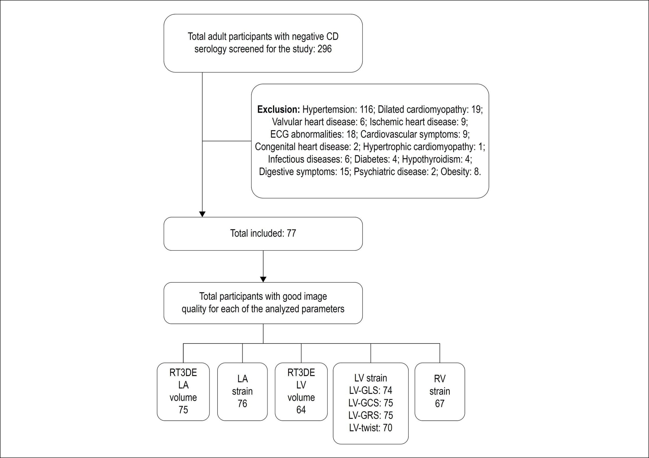
Seventy-seven subjects (46.7% men; 40.4 ± 10.4 years) were included. Maximum, minimum and pre-atrial contraction (pre-A) LA volumes (ml/m2) were 21.2 ± 5.5, 7.8 ± 2.5, and 11.0 ± 3.1, respectively. Peak positive global LA e (LAScd), peak negative global LA e and total global LA e (LASr) were 17.4 ± 5.2%, -13.2 ± 2.0% and 30.5 ± 5.9%, respectively. LV end-diastolic and end-systolic volumes (ml/m2) measured 57 ± 12 and 24 ± 6, and 3D LV ejection fraction measured 58 ± 6%. Global LV longitudinal, circumferential and radial e were -19 ± 2%, -19 ± 3%, and 46 ± 12%, respectively. LV torsion measured 1.6 ± 0.70 /cm. Global longitudinal RV e (RV-GLS) and RV free wall strain were -22 ± 3% and -24 ± 5%. Minimum LA and pre-A volumes, LV apical rotation, torsion and RV-GLS increased with age, while total and passive LA emptying fractions, LAScd, LASr, LV end-diastolic and end-systolic volumes decreased with age.

Conclusion:

Values for new echocardiographic indexes in Brazilians without known cardiovascular disease and their correlation with age are presented.

  • Cardiovascular Diseases
  • Echocardiography, Three-Dimensional
  • Left Ventricular Function
  • Reference Values
  • Right Ventricular Function
  • Speckle Tracking
  • Strain
Read Full Article
Creative Commons Attribution (CC BY) Creative Commons license
29/08/2019 DOI: 10.5935/abc.20190155
Cardiac resynchronization therapy improves the uptake of MIBI-99mTc and cardiac function
ABC
Relato de Caso

Cardiac resynchronization therapy improves the uptake of MIBI-99mTc and cardiac function

Simone Cristina Soares Brandão et al. Maria Clementina Giorgi Silvana D'Orio Nishioka Martino Martinelli Filho … José Soares Jr. José Cláudio Meneghetti

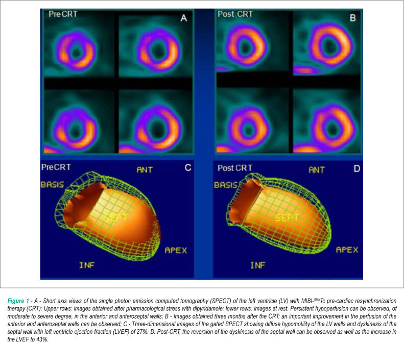
This case shows the improvement promoted by cardiac resynchronization therapy (CRT) on myocardial perfusion and left ventricular (LV) performance assessed by gated myocardial perfusion scintigraphy. The patient had idiopathic dilated cardiomyopathy, left bundle branch block and severe heart failure despite optimized medical treatment. After CRT, clinical improvement, QRS reduction and improvement of previously hypoperfused anterior and septal walls were observed. There was also decrease in LV end-diastolic and systolic volumes and increase in LV ejection fraction.

  • Dilated Cardiomyopathy
  • Left Ventricular Function
  • myocardial perfusion
  • Ventricular Function
Read Full Article
Creative Commons Attribution (CC BY) Creative Commons license
01/10/2008 DOI: 10.1590/S0066-782X2008001500013
Evolutive echocardiographic study of the structural and functional heart alterations in obese individuals after bariatric surgery
ABC
Artigo Original

Evolutive echocardiographic study of the structural and functional heart alterations in obese individuals after bariatric surgery

Luciana de Cerjat Bernardes P. Cunha et al. Cláudio L. Pereira da Cunha Admar Moraes de Souza Nelson Chiminacio Neto … Ricardo Soares Pereira Henrique Lacerda Suplicy

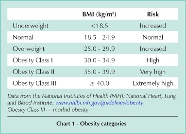
OBJECTIVE: To evaluate with Doppler echocardiography the reversibility of structural and hemodynamic changes in obeses after bariatric surgery. METHODS: Twenty-three patients (19 women = 82.6%) were studied. Mean age was 37.9 years. All subjects had Class III or Class II obesity with comorbidity and were submitted to bariatric surgery. Clinical and echocardiographic evaluation were performed preoperatively, in 6 months and 3 years after surgery. RESULTS: Preoperatively, the mean weight and blood pressure (BP) were respectively 128.7± 25.8 kg and 142.2 ± 16.2 / 92.2 ± 10.4 mmHg. Postoperatively, they showed important body weight reduction in 6 months (97.6 ± 18.3 kg) and 3 years (83.6 ± 13.5 kg), and BP reduction in 6 months (128.5 ± 16.1/80.7 ± 9.9 mmHg) that remain stable in 3rd year. On echocardiogram, preoperatively, there was hypertrophy of the septum and posterior wall associated with normal diastolic dimension; the predominant LV geometric pattern was concentric remodeling (74%). At six months, thinning of the ventricular septum and LV posterior wall, and increase in LV diastolic dimension were demonstrated. At 3rd year, the predominant LV pattern was normal (69%), with reduction of LV mass and LV mass/height² index. We noticed improved diastolic function, with an increased E/A ratio and a decreased LV isovolumic relaxation time. The Myocardial Performance Index was obtained retrospectively in 13 patients and improved in 6 months. There was an increase of the ejection time in 6 months and an elevation of the ejection fraction in 3rd year, suggesting improvement of the LV systolic function. CONCLUSION: The weight loss obtained with bariatric surgery promotes both structural and functional myocardial changes that improve cardiac performance.

  • bariatric surgery
  • Left Ventricular Function
  • Obesity
  • Transthoracic Echocardiography
Read Full Article
Creative Commons Attribution (CC BY) Creative Commons license
06/03/2007 DOI: 10.1590/S0066-782X2006001800011
Left ventricular systolic function assessed by echocardiography in children and adolescents with osteosarcoma treated with doxorubicin alone or in combination with dexrazoxane
ABC
Artigo Original

Left ventricular systolic function assessed by echocardiography in children and adolescents with osteosarcoma treated with doxorubicin alone or in combination with dexrazoxane

Ranulfo Pinheiro de Matos Neto et al. Antônio Sérgio Petrilli Célia Maria Campos Silva Orlando Campos Filho … Victor Manuel Oporto Lourdes de Fátima Gonçalves Gomes Marcelo Goulart Paiva Antônio Carlos Camargo Carvalho Valdir Ambrósio Moisés

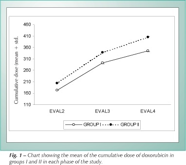
OBJECTIVE: To evaluate left ventricular (LV) systolic function by means of echocardiography in patients with osteosarcoma treated with doxorubicin alone or in combination with dexrazoxane. METHODS: The study analyzed 55 patients with osteosarcoma, with or without metastasis, undergoing a six-cycle chemotherapy regimen of doxorubicin, who were divided into two groups according to dexrazoxane use. Group I: Thirty-seven patients who did not receive dexrazoxane (28 males, average age 15.4 years). Group II: Eighteen patients who did receive dexrazoxane (15 males, average age 15.1 years). Four echocardiographic evaluations were performed: 1) before the beginning of the chemotherapy (initial evaluation); 2) up to two weeks after the third cycle; 3) up to two weeks after the fifth cycle; and 4) up to four weeks after the sixth cycle of chemotherapy (final evaluation). The left ventricular systolic function was assessed by the fractional percentage of systolic shortening (FS%) on echocardiography. Alterations in the contractile function or cardiac toxicity were defined as FS% values equal to or lower than 29%, and/or by a drop in FS% by an absolute value equal to or greater than 10 units of the baseline value of each patient. RESULTS: No significant difference as to age, gender, and race was observed between the groups. The cumulative dose of doxorubicin was significantly higher in group II throughout all phases of the treatment: 174 x 203 mg/m²; 292 x 338 mg/m² and 345 x 405 mg/² (p < 0.0001). According to previously established criteria, the incidence of LV systolic dysfunction was not significantly different (p=0.248) between patients in group I (18.92%) and patients in group II (11.1%). The variance analysis with repeated measurements did not show significant differences in the means of fractional percentage of systolic percentage (FS%) throughout the study (p=0.967). However, a significant difference (p=0.029) was observed between the FS% means in groups I and II at evaluations 2 (35.67 x 37.21%), 3 (34.95 x 38.47%) and 4 (35.26 x 38.22%). CONCLUSION: Data in this study show that in patients with osteosarcoma treated with doxorubicin alone or combined with dexrazoxane, the LV systolic function, as assessed by the fractional percentage of systolic shortening mean, showed a better performance in the group that received dexrazoxane. On the other hand, the occurrence of systolic dysfunction was similar in both groups.

  • dexrazoxane
  • Doxorubicin
  • Echocardiography
  • Left Ventricular Function
  • osteosarcoma
Read Full Article
Creative Commons Attribution (CC BY) Creative Commons license
18/01/2007 DOI: 10.1590/S0066-782X2006001900013 Views 1
Texture of mitral bioprosthesis, ventricular function and formation of thrombus: analysis through transesophageal echocardiography and use of bioscore
ABC
Artigo Original

Texture of mitral bioprosthesis, ventricular function and formation of thrombus: analysis through transesophageal echocardiography and use of bioscore

Henry Abensur et al. Max Grinberg José A. F. Ramires

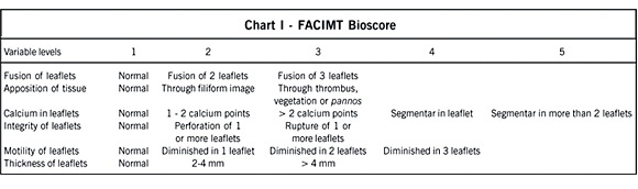
OBJECTIVE: Facing the hypothesis of participation of mechanical stress as a cause of mitral bioprosthesis dysfunction, we decided to assess the relation of preservation of the texture of the mitral bioprosthesis leaflets with left ventricular function, in addition to the casual formation of thrombus in left atrium in patients with left ventricular dysfunction from the implant of mitral bioprosthesis. METHODS: Forty 40 patients with mitral bioprosthesis through multiplane transesophageal echocardiogram were studied and divided in two groups: with left ventricular dysfunction (FE=0.40±0.09) since the bioprosthesis implant (20 patients: age 47.75±11.10 years old and surgery time 5.3±2.6 years) and with normal left ventricular function (FE=0.73±0.06) since the implant (20 patients: age 49.75±13.59 years old and surgery time 5.7±3 years). The texture of bioprosthesis leaflets was analyzed through a transesophageal echocardiographic score (FACIMT Bioscore): 1) Fusion of leaflets (score 1 to 3); 2) Apposition of tissues (score 1 to 3); 3) Calcium in leaflets (score 1 to 5); 4) Integrity of leaflets (score 1 to 3); 5) Motility of leaflets (score 1 to 4) and 6) Thickness of leaflets (score 1 to 3). The presence of thrombi in left atrium was assessed through multiplane scanning of the left atrium and left atrial appendage in the transesophageal study. RESULTS: There was no significant difference in the texture of bioprosthesis mitral position between the groups, for the total score (8.7±2.4 vs. 7.9±2.1, p=0.259), and for each analyzed item. A greater incidence of thrombi in left atrium and left atrial appendage was detected in patients with ventricular dysfunction (65% vs. 20%, p=0.004). CONCLUSION: The left ventricular dysfunction was not a protecting factor of the texture of bioprosthesis leaflets in mitral position in the tardive post-surgery period. The patients with left ventricular dysfunction showed a more favorable environment for the formation of thrombi in left atrium.

  • formation of thrombi
  • Left Ventricular Function
  • mitral bioprosthesis
  • Transesophageal echocardiography
Read Full Article
Creative Commons Attribution (CC BY) Creative Commons license
24/05/2005 DOI: 10.1590/S0066-782X2005000500011 Views 1
Mildly symptomatic chronic mitral regurgitation. Analysis of left ventricular systolic function and mitral regurgitant fraction under pharmacological influence: echocardiographic study
ABC
Original Article

Mildly symptomatic chronic mitral regurgitation. Analysis of left ventricular systolic function and mitral regurgitant fraction under pharmacological influence: echocardiographic study

Ivan Romero Rivera et al. Valdir Ambrósio Moisés Antonio Carlos Carvalho Angelo Amato V. de Paola

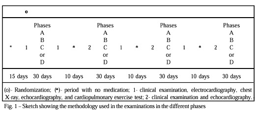
OBJECTIVE: To study echocardiographic parameters of left ventricular systolic function and valvar regurgitation under pharmacological influence in mildly symptomatic patients with chronic mitral regurgitation (MR). METHODS: We carried out a double-blind placebo controlled study in 12 patients with MR, mean aged 12.5 years old, who were randomized in 4 phases: A) digoxin; B) enalapril; C) digoxin + enalapril; D) placebo. The medication was administered for 30 days in each phase, and the following variables were analyzed: shortening and ejection fractions, wall stress index of left ventricle, left ventricular meridional end-systolic wall stress, Doppler-derived mean rate of left ventricular pressure rise (mean dP/dt), stroke volume and MR jet area. The clinical variables analysed were heart rate and systemic arterial pressure. RESULTS: No significant variation was observed in the clinical variables analysed. The shortening and ejection fraction, the mean dP/dt and stroke volume significantly increased and the wall stress index of left ventricle, the meridional left ventricular end systolic wall stress and the mitral regurgitation jet area decreased in the phases with medication as compared with that in the placebo phase. CONCLUSION: The parameters of left ventricular systolic function improved significantly and the degree of MR decreased with the isolated administration of digoxin or enalapril in mildly symptomatic patients with chronic MR. The combination of the drugs, however, did not show better results.

  • Echocardiography
  • Left Ventricular Function
  • mitral regurgitation
Read Full Article
Creative Commons Attribution (CC BY) Creative Commons license
02/04/2003 DOI: 10.1590/S0066-782X2003000200003 Views 1
ABC
Original Article

Aortic stenosis. Gender influence on left ventricular geometry and function in patients under 70 years of age

Antonio Sérgio Cordeiro da Rocha et al. Maria Auxiliadora Vellasco Pereira Nazareth de Novaes Rocha Rita de Cássia Villela Soares … Celso Garcia, Rosana Grandelle Paulo Roberto Dutra da Silva

OBJECTIVE: To verify if adaptive left ventricle (LV) characteristics are also present in individuals under 70 years of age with severe aortic stenosis (AS). METHODS: The study comprised 40 consecutive patients under 70 years of age with AS and no associated coronary artery disease, referred for valve surgery. Out of the 40 patients, 22 were men and 18 women, and the mean age was 49.8±14.3 years. Cardiac symptoms, presence of systemic hypertension (SH), functional class according to the New York Heart Association (NYHA), and valve lesion etiology were considered. LV cavity dimensions, ejection fraction (EF), fractional shortening (FS), mass (MS), and relative diastolic thickness (RDT) were examined by Doppler echocardiography. RESULTS: Fourteen (63.6%) men and 11 (61.6%) women were classified as NYHA class III/IV (p=0.70). There was no difference in the frequency of angina, syncope or dyspnea between genders. The incidence of SH was greater in women than in men (10 versus 2, p=0.0044). Women had a smaller LV end-diastolic diameter index (32.1±6.5 x 36.5±5.3mm/m², p=0.027), LV end-systolic diameter index (19.9±5.9 x 26.5±6.4mm/m², p=0.0022) and LV mass index (MS) (211.4±71.1 x 270.9±74.9g/m², p=0.017) when compared with men. EF (66.2±13.4 x 52.0±14.6%, p=0.0032), FS (37.6±10.7 x 27.9±9.6%, p=0.0046) and RDT (0.58±0.22 x 0.44±0.09, p=0.0095) were significantly greater in women than in men. CONCLUSION: It is the patient gender rather than age that influences left ventricular adaptive response to AS.

  • Aortic Stenosis
  • Gender
  • Left Ventricular Function
  • Ventricular Hypertrophy
Read Full Article
Creative Commons Attribution (CC BY) Creative Commons license
08/01/2002 DOI: 10.1590/S0066-782X1999000400007 Views 2
Anomalous origin of the left coronary artery from the pulmonary trunk. Clinical features and midterm results after surgical treatment
ABC
Original Article

Anomalous origin of the left coronary artery from the pulmonary trunk. Clinical features and midterm results after surgical treatment

Fernando Amaral et al. Julene S. Carvalho João A. Granzotti Elliot A. Shinebourne

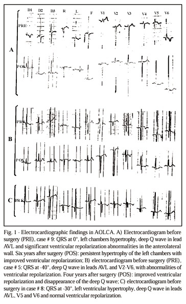
OBJECTIVE: To report the authors’ experience with the anomalous origin of the left coronary artery (AOLCA) from the pulmonary trunk, emphasizing preoperative data, surgical aspects and midterm results of the follow-up. METHODS: Retrospective analysis of 11 patients operated upon at the Royal Brompton Hospital from October, 84 to April, 97. RESULTS: Nine infants had heart failure (HF) and two other children presented with dyspnea and chest pain. All had ECG changes. The echocardiogram identified the anomalous origin of the coronary artery in 7 (64%) patients and hemodynamic studies were performed in 7 patients. All infants were operated upon between the 2nd and 10th month of life. Six patients were treated with aortic reimplantation of the left coronary artery, whereas five were operated upon according to the Takeuchi technique. All patients are alive, with clear improvement of the ECG changes and ventricular function, as evaluated by echocardiography. Two patients operated upon according to the Takeuchi technique required additional surgery due to severe supravalvular pulmonary stenosis. CONCLUSION: AOLCA is a rare disease. Most patients show early signs of severe HF associated with ECG findings. Surgical therapy must be instituted early in the disease, preferentially through aortic implantation of the anomalous coronary artery, with a high possibility of success. Shortly after surgery, clinical and ECG improvement, as well as normalization of left ventricular function, should be expected.

  • Anomalous Left Coronary Artery
  • Bland-White-Garland Syndrome
  • Left Ventricular Function
Read Full Article
Creative Commons Attribution (CC BY) Creative Commons license
08/01/2002 DOI: 10.1590/S0066-782X1999000300004 Views 1

How to cite this article

EndNote RIS

Electronic Document Format (Vancouver)

Alves MSL, Vosgerau LM, Dreckmann MV, Martins RD, Zanlorensi CB, Bonotto EH. My Approach to Echocardiographic Assessment of Left Ventricular Filling Pressures: From Ambiguity to Precision With New Guidelines. ABC Imagem Cardiovascular 2026;39(2):e20260026.

Electronic Document Format (ABNT)

Alves, Marco Stephan Lofrano; Vosgerau, Larissa Maria; Dreckmann, Marcelo Vitola; Martins, Roberto D´Ávila; Zanlorensi, Cláudia Biondo; Bonotto, Eduardo Henrique. My Approach to Echocardiographic Assessment of Left Ventricular Filling Pressures: From Ambiguity to Precision With New Guidelines. ABC Imagem Cardiovascular, v. 39, n. 2, e20260026, Jul. 2026.

Electronic Document Format (APA)

Alves, M. S. L., Vosgerau, L. M., Dreckmann, M. V., Martins, R. D., Zanlorensi, C. B., & Bonotto, E. H. (2026). My Approach to Echocardiographic Assessment of Left Ventricular Filling Pressures: From Ambiguity to Precision With New Guidelines. ABC Imagem Cardiovascular, 39(2), e20260026.

Electronic Document Format (ISO)

Alves, Marco Stephan Lofrano and Vosgerau, Larissa Maria and Dreckmann, Marcelo Vitola and Martins, Roberto D´Ávila and Zanlorensi, Cláudia Biondo and Bonotto, Eduardo Henrique. My Approach to Echocardiographic Assessment of Left Ventricular Filling Pressures: From Ambiguity to Precision With New Guidelines. ABC Imagem Cardiovascular [online]. 2026, vol. 39, n. 2, [cited 2026-10-14], e20260026. Available from: <https://journals.cardiol.br/en/article/my-approach-to-echocardiographic-assessment-of-left-ventricular-filling-pressures-from-ambiguity-to-precision-with-new-guidelines/>. ISSN 2318-8219

EndNote RIS

Electronic Document Format (Vancouver)

Rodrigues ACT. How I Assess Left Ventricular Systolic Function. ABC Imagem Cardiovascular 2025;38(4):e20250078.

Electronic Document Format (ABNT)

Rodrigues, Ana Clara Tude. How I Assess Left Ventricular Systolic Function. ABC Imagem Cardiovascular, v. 38, n. 4, e20250078, Dec. 2025.

Electronic Document Format (APA)

Rodrigues, A. C. T. (2025). How I Assess Left Ventricular Systolic Function. ABC Imagem Cardiovascular, 38(4), e20250078.

Electronic Document Format (ISO)

Rodrigues, Ana Clara Tude. How I Assess Left Ventricular Systolic Function. ABC Imagem Cardiovascular [online]. 2025, vol. 38, n. 4, [cited 2026-10-14], e20250078. Available from: <https://journals.cardiol.br/en/article/how-i-assess-left-ventricular-systolic-function/>. ISSN 2318-8219

EndNote RIS

Electronic Document Format (Vancouver)

Borges JVN, Leila SAC, Machado MGN, Babu KSTJ, Patel KJ, Shah KH, Takkasila MR, et al. Left Ventricular Function and Myocardial Longitudinal Strain Analysis in Patients With Chagas Disease: Case Series and Systematic Literature Review. ABC Imagem Cardiovascular 2024;37(3):e20240053.

Electronic Document Format (ABNT)

Borges, José Victor da Nóbrega; Leila, Samira Abdel Correia; Machado, Manuella Guedes da Nóbrega; Babu, Kiran Sooraj Thirukonda Jegadeesh; Patel, Karma Jashvantbhai; Shah, Kavya Hiteshkumar; Takkasila, Mahitha Reddy; Shaik, Abdul Majid; Shaikh, Muhammad Ahmed; Khandekar, Ninad; Burra, Sahithi. Left Ventricular Function and Myocardial Longitudinal Strain Analysis in Patients With Chagas Disease: Case Series and Systematic Literature Review. ABC Imagem Cardiovascular, v. 37, n. 3, e20240053, Sep. 2024.

Electronic Document Format (APA)

Borges, J. V. N., Leila, S. A. C., Machado, M. G. N., Babu, K. S. T. J., Patel, K. J., Shah, K. H., Takkasila, M. R., Shaik, A. M., Shaikh, M. A., Khandekar, N., & Burra, S. (2024). Left Ventricular Function and Myocardial Longitudinal Strain Analysis in Patients With Chagas Disease: Case Series and Systematic Literature Review. ABC Imagem Cardiovascular, 37(3), e20240053.

Electronic Document Format (ISO)

Borges, José Victor da Nóbrega and Leila, Samira Abdel Correia and Machado, Manuella Guedes da Nóbrega and Babu, Kiran Sooraj Thirukonda Jegadeesh and Patel, Karma Jashvantbhai and Shah, Kavya Hiteshkumar and Takkasila, Mahitha Reddy and Shaik, Abdul Majid and Shaikh, Muhammad Ahmed and Khandekar, Ninad and Burra, Sahithi. Left Ventricular Function and Myocardial Longitudinal Strain Analysis in Patients With Chagas Disease: Case Series and Systematic Literature Review. ABC Imagem Cardiovascular [online]. 2024, vol. 37, n. 3, [cited 2026-10-14], e20240053. Available from: <https://journals.cardiol.br/en/article/left-ventricular-function-and-myocardial-longitudinal-strain-analysis-in-patients-with-chagas-disease-case-series-and-systematic-literature-review/>. ISSN 2318-8219

EndNote RIS

Electronic Document Format (Vancouver)

Espíndola LN, Almeida MCC, Castro ML, Freire CMV, Oliveira GMM. Particularities of Heart Failure with Preserved Ejection Fraction in Women – Pathways to Better Care. ABC Heart Fail Cardiomyop 2024;4(1):e20240012.

Electronic Document Format (ABNT)

Espíndola, Larissa Neto; Almeida, Maria Cristina Costa de; Castro, Marildes Luíza de; Freire, Cláudia Maria Vilas; Oliveira, Gláucia Maria Moraes de. Particularities of Heart Failure with Preserved Ejection Fraction in Women – Pathways to Better Care. ABC Heart Fail Cardiomyop, v. 4, n. 1, e20240012, Aug. 2024.

Electronic Document Format (APA)

Espíndola, L. N., Almeida, M. C. C. , Castro, M. L. , Freire, C. M. V., & Oliveira, G. M. M. (2024). Particularities of Heart Failure with Preserved Ejection Fraction in Women – Pathways to Better Care. ABC Heart Fail Cardiomyop, 4(1), e20240012.

Electronic Document Format (ISO)

Espíndola, Larissa Neto and Almeida, Maria Cristina Costa de and Castro, Marildes Luíza de and Freire, Cláudia Maria Vilas and Oliveira, Gláucia Maria Moraes de. Particularities of Heart Failure with Preserved Ejection Fraction in Women – Pathways to Better Care. ABC Heart Fail Cardiomyop [online]. 2024, vol. 4, n. 1, [cited 2026-10-14], e20240012. Available from: <https://journals.cardiol.br/en/article/particularities-of-heart-failure-with-preserved-ejection-fraction-in-women-pathways-to-better-care/>

EndNote RIS

Electronic Document Format (Vancouver)

Lima EG, Martins EB, Carvalho LNS, Azevedo DFC. What do Cardiologists Expect Towards Myocardial Viability Assessment. ABC Imagem Cardiovascular 2024;37(1):e20240005.

Electronic Document Format (ABNT)

Lima, Eduardo Gomes; Martins, Eduardo Bello; Carvalho, Leticia Neves Solon; Azevedo, Diogo Freitas Cardoso de. What do Cardiologists Expect Towards Myocardial Viability Assessment. ABC Imagem Cardiovascular, v. 37, n. 1, e20240005, Mar. 2024.

Electronic Document Format (APA)

Lima, E. G., Martins, E. B., Carvalho, L. N. S., & Azevedo, D. F. C. (2024). What do Cardiologists Expect Towards Myocardial Viability Assessment. ABC Imagem Cardiovascular, 37(1), e20240005.

Electronic Document Format (ISO)

Lima, Eduardo Gomes and Martins, Eduardo Bello and Carvalho, Leticia Neves Solon and Azevedo, Diogo Freitas Cardoso de. What do Cardiologists Expect Towards Myocardial Viability Assessment. ABC Imagem Cardiovascular [online]. 2024, vol. 37, n. 1, [cited 2026-10-14], e20240005. Available from: <https://journals.cardiol.br/en/article/what-do-cardiologists-expect-towards-myocardial-viability-assessment/>. ISSN 2318-8219

EndNote RIS

Electronic Document Format (Vancouver)

Peña JB, Pino IP, Patino MJM. Sociodemographic, Clinical Condition, and Functional Aerobic Capacity in Patients With Heart Failure With Varying Ventricular Ejection Fraction. International Journal of Cardiovascular Sciences 2024;37:e20230079.

Electronic Document Format (ABNT)

Peña, Jhonatan Betancourt; Pino, Iago Portela; Patino, Maria Jose Martinez. Sociodemographic, Clinical Condition, and Functional Aerobic Capacity in Patients With Heart Failure With Varying Ventricular Ejection Fraction. International Journal of Cardiovascular Sciences, v. 37, e20230079, Mar. 2024.

Electronic Document Format (APA)

Peña, J. B., Pino, I. P., & Patino, M. J. M. (2024). Sociodemographic, Clinical Condition, and Functional Aerobic Capacity in Patients With Heart Failure With Varying Ventricular Ejection Fraction. International Journal of Cardiovascular Sciences, 37, e20230079.

Electronic Document Format (ISO)

Peña, Jhonatan Betancourt and Pino, Iago Portela and Patino, Maria Jose Martinez. Sociodemographic, Clinical Condition, and Functional Aerobic Capacity in Patients With Heart Failure With Varying Ventricular Ejection Fraction. International Journal of Cardiovascular Sciences [online]. 2024, vol. 37, [cited 2026-10-14], e20230079. Available from: <https://journals.cardiol.br/en/article/sociodemographic-clinical-condition-and-functional-aerobic-capacity-in-patients-with-heart-failure-with-varying-ventricular-ejection-fraction/>. ISSN 2359-4802

EndNote RIS

Electronic Document Format (Vancouver)

Mancio J. Empowering the Imagers with 3D-Speckle Tracking Echocardiography to Detect Subclinical Cancer Therapy-Related Myocardial Dysfunction. Arquivos Brasileiros de Cardiologia 2023;120(8):e20230554.

Electronic Document Format (ABNT)

Mancio, Jennifer. Empowering the Imagers with 3D-Speckle Tracking Echocardiography to Detect Subclinical Cancer Therapy-Related Myocardial Dysfunction. Arquivos Brasileiros de Cardiologia, v. 120, n. 8, e20230554, Sep. 2023.

Electronic Document Format (APA)

Mancio, J. (2023). Empowering the Imagers with 3D-Speckle Tracking Echocardiography to Detect Subclinical Cancer Therapy-Related Myocardial Dysfunction. Arquivos Brasileiros de Cardiologia, 120(8), e20230554.

Electronic Document Format (ISO)

Mancio, Jennifer. Empowering the Imagers with 3D-Speckle Tracking Echocardiography to Detect Subclinical Cancer Therapy-Related Myocardial Dysfunction. Arquivos Brasileiros de Cardiologia [online]. 2023, vol. 120, n. 8, [cited 2026-10-14], e20230554. Available from: <https://journals.cardiol.br/en/article/empowering-the-imagers-with-3d-speckle-tracking-echocardiography-to-detect-subclinical-cancer-therapy-related-myocardial-dysfunction/>. ISSN 0066-782X

EndNote RIS

Electronic Document Format (Vancouver)

Laksono S, Kusharsamita H, Yanni M, Astuti A. Myocardial Work: a Systematic Review of Novel Echocardiography Method and Clinical Applications. ABC Imagem Cardiovascular 2022;35(4):eabc299.

Electronic Document Format (ABNT)

Laksono, Sidhi; Kusharsamita, Hillary; Yanni, Mefri; Astuti, Astri. Myocardial Work: a Systematic Review of Novel Echocardiography Method and Clinical Applications. ABC Imagem Cardiovascular, v. 35, n. 4, eabc299, Nov. 2022.

Electronic Document Format (APA)

Laksono, S., Kusharsamita, H., Yanni, M., & Astuti, A. (2022). Myocardial Work: a Systematic Review of Novel Echocardiography Method and Clinical Applications. ABC Imagem Cardiovascular, 35(4), eabc299.

Electronic Document Format (ISO)

Laksono, Sidhi and Kusharsamita, Hillary and Yanni, Mefri and Astuti, Astri. Myocardial Work: a Systematic Review of Novel Echocardiography Method and Clinical Applications. ABC Imagem Cardiovascular [online]. 2022, vol. 35, n. 4, [cited 2026-10-14], eabc299. Available from: <https://journals.cardiol.br/en/article/myocardial-work-a-systematic-review-of-novel-echocardiography-method-and-clinical-applications/>. ISSN 2318-8219

EndNote RIS

Electronic Document Format (Vancouver)

Saraiva RM, Scolin EMB, Pacheco NP, Bouret ME, Mediano MFF, Holanda MT, Costa AR, et al. 3-Dimensional Echocardiography and 2-D Strain Analysis of Left Ventricular, Left Atrial and Right Ventricular Function in Healthy Brazilian Volunteers. Arquivos Brasileiros de Cardiologia 2019;113(5):935-4.

Electronic Document Format (ABNT)

Saraiva, Roberto M.; Scolin, Eliza Maria B.; Pacheco, Nicole P.; Bouret, Maria Eduarda; Mediano, Mauro Felippe Felix; Holanda, Marcelo T.; Costa, Andréa R. da. 3-Dimensional Echocardiography and 2-D Strain Analysis of Left Ventricular, Left Atrial and Right Ventricular Function in Healthy Brazilian Volunteers. Arquivos Brasileiros de Cardiologia, v. 113, n. 5, p. 935-945, Aug. 2019.

Electronic Document Format (APA)

Saraiva, R. M., Scolin, E. M. B., Pacheco, N. P., Bouret, M. E., Mediano, M. F. F., Holanda, M. T., & Costa, A. R. (2019). 3-Dimensional Echocardiography and 2-D Strain Analysis of Left Ventricular, Left Atrial and Right Ventricular Function in Healthy Brazilian Volunteers. Arquivos Brasileiros de Cardiologia, 113(5), 935-945.

Electronic Document Format (ISO)

Saraiva, Roberto M. and Scolin, Eliza Maria B. and Pacheco, Nicole P. and Bouret, Maria Eduarda and Mediano, Mauro Felippe Felix and Holanda, Marcelo T. and Costa, Andréa R. da. 3-Dimensional Echocardiography and 2-D Strain Analysis of Left Ventricular, Left Atrial and Right Ventricular Function in Healthy Brazilian Volunteers. Arquivos Brasileiros de Cardiologia [online]. 2019, vol. 113, n. 5, [cited 2026-10-14], pp. 935-945. Available from: <https://journals.cardiol.br/en/article/3-dimensional-echocardiography-and-2-d-strain-analysis-of-left-ventricular-left-atrial-and-right-ventricular-function-in-healthy-brazilian-volunteers/>. ISSN 0066-782X

EndNote RIS

Electronic Document Format (Vancouver)

Brandão SCS, Giorgi MC, Nishioka SD, Martinelli Filho M, Soares Jr. J, Meneghetti JC. Cardiac resynchronization therapy improves the uptake of MIBI-99mTc and cardiac function. Arquivos Brasileiros de Cardiologia 2008;91(3):e25-8.

Electronic Document Format (ABNT)

Brandão, Simone Cristina Soares; Giorgi, Maria Clementina; Nishioka, Silvana D’Orio; Martinelli Filho, Martino; Soares Jr., José; Meneghetti, José Cláudio. Cardiac resynchronization therapy improves the uptake of MIBI-99mTc and cardiac function. Arquivos Brasileiros de Cardiologia, v. 91, n. 3, p. e25-e28, Sep. 2008.

Electronic Document Format (APA)

Brandão, S. C. S., Giorgi, M. C., Nishioka, S. D., Martinelli Filho, M., Soares Jr., J., & Meneghetti, J. C. (2008). Cardiac resynchronization therapy improves the uptake of MIBI-99mTc and cardiac function. Arquivos Brasileiros de Cardiologia, 91(3), e25-e28.

Electronic Document Format (ISO)

Brandão, Simone Cristina Soares and Giorgi, Maria Clementina and Nishioka, Silvana D’Orio and Martinelli Filho, Martino and Soares Jr., José and Meneghetti, José Cláudio. Cardiac resynchronization therapy improves the uptake of MIBI-99mTc and cardiac function. Arquivos Brasileiros de Cardiologia [online]. 2008, vol. 91, n. 3, [cited 2026-10-14], pp. e25-e28. Available from: <https://journals.cardiol.br/en/article/cardiac-resynchronization-therapy-improves-the-uptake-of-mibi-99mtc-and-cardiac-function/>. ISSN 0066-782X

EndNote RIS

Electronic Document Format (Vancouver)

Cunha LCBP, Cunha CLP, Souza AM, Chiminacio Neto N, Pereira RS, Suplicy HL. Evolutive echocardiographic study of the structural and functional heart alterations in obese individuals after bariatric surgery. Arquivos Brasileiros de Cardiologia 2007;87(5):615-22.

Electronic Document Format (ABNT)

Cunha, Luciana de Cerjat Bernardes P.; Cunha, Cláudio L. Pereira da; Souza, Admar Moraes de; Chiminacio Neto, Nelson; Pereira, Ricardo Soares; Suplicy, Henrique Lacerda. Evolutive echocardiographic study of the structural and functional heart alterations in obese individuals after bariatric surgery. Arquivos Brasileiros de Cardiologia, v. 87, n. 5, p. 615-622, Mar. 2007.

Electronic Document Format (APA)

Cunha, L. C. B. P., Cunha, C. L. P. , Souza, A. M. , Chiminacio Neto, N., Pereira, R. S., & Suplicy, H. L. (2007). Evolutive echocardiographic study of the structural and functional heart alterations in obese individuals after bariatric surgery. Arquivos Brasileiros de Cardiologia, 87(5), 615-622.

Electronic Document Format (ISO)

Cunha, Luciana de Cerjat Bernardes P. and Cunha, Cláudio L. Pereira da and Souza, Admar Moraes de and Chiminacio Neto, Nelson and Pereira, Ricardo Soares and Suplicy, Henrique Lacerda. Evolutive echocardiographic study of the structural and functional heart alterations in obese individuals after bariatric surgery. Arquivos Brasileiros de Cardiologia [online]. 2007, vol. 87, n. 5, [cited 2026-10-14], pp. 615-622. Available from: <https://journals.cardiol.br/en/article/evolutive-echocardiographic-study-of-the-structural-and-functional-heart-alterations-in-obese-individuals-after-bariatric-surgery/>. ISSN 0066-782X

EndNote RIS

Electronic Document Format (Vancouver)

Matos Neto RP, Petrilli AS, Silva CMC, Campos Filho O, Oporto VM, Gomes LFG, Paiva MG, et al. Left ventricular systolic function assessed by echocardiography in children and adolescents with osteosarcoma treated with doxorubicin alone or in combination with dexrazoxane. Arquivos Brasileiros de Cardiologia 2007;87(6):763-71.

Electronic Document Format (ABNT)

Matos Neto, Ranulfo Pinheiro de; Petrilli, Antônio Sérgio; Silva, Célia Maria Campos; Campos Filho, Orlando; Oporto, Victor Manuel; Gomes, Lourdes de Fátima Gonçalves; Paiva, Marcelo Goulart; Carvalho, Antônio Carlos Camargo; Moisés, Valdir Ambrósio. Left ventricular systolic function assessed by echocardiography in children and adolescents with osteosarcoma treated with doxorubicin alone or in combination with dexrazoxane. Arquivos Brasileiros de Cardiologia, v. 87, n. 6, p. 763-771, Jan. 2007.

Electronic Document Format (APA)

Matos Neto, R. P. , Petrilli, A. S., Silva, C. M. C., Campos Filho, O., Oporto, V. M., Gomes, L. F. G., Paiva, M. G., Carvalho, A. C. C., & Moisés, V. A. (2007). Left ventricular systolic function assessed by echocardiography in children and adolescents with osteosarcoma treated with doxorubicin alone or in combination with dexrazoxane. Arquivos Brasileiros de Cardiologia, 87(6), 763-771.

Electronic Document Format (ISO)

Matos Neto, Ranulfo Pinheiro de and Petrilli, Antônio Sérgio and Silva, Célia Maria Campos and Campos Filho, Orlando and Oporto, Victor Manuel and Gomes, Lourdes de Fátima Gonçalves and Paiva, Marcelo Goulart and Carvalho, Antônio Carlos Camargo and Moisés, Valdir Ambrósio. Left ventricular systolic function assessed by echocardiography in children and adolescents with osteosarcoma treated with doxorubicin alone or in combination with dexrazoxane. Arquivos Brasileiros de Cardiologia [online]. 2007, vol. 87, n. 6, [cited 2026-10-14], pp. 763-771. Available from: <https://journals.cardiol.br/en/article/left-ventricular-systolic-function-assessed-by-echocardiography-in-children-and-adolescents-with-osteosarcoma-treated-with-doxorubicin-alone-or-in-combination-with-dexrazoxane/>. ISSN 0066-782X

EndNote RIS

Electronic Document Format (Vancouver)

Abensur H, Grinberg M, Ramires JAF. Texture of mitral bioprosthesis, ventricular function and formation of thrombus: analysis through transesophageal echocardiography and use of bioscore. Arquivos Brasileiros de Cardiologia 2005;84(5):410-5.

Electronic Document Format (ABNT)

Abensur, Henry; Grinberg, Max; Ramires, José A. F.. Texture of mitral bioprosthesis, ventricular function and formation of thrombus: analysis through transesophageal echocardiography and use of bioscore. Arquivos Brasileiros de Cardiologia, v. 84, n. 5, p. 410-415, May. 2005.

Electronic Document Format (APA)

Abensur, H., Grinberg, M., & Ramires, J. A. F. (2005). Texture of mitral bioprosthesis, ventricular function and formation of thrombus: analysis through transesophageal echocardiography and use of bioscore. Arquivos Brasileiros de Cardiologia, 84(5), 410-415.

Electronic Document Format (ISO)

Abensur, Henry and Grinberg, Max and Ramires, José A. F.. Texture of mitral bioprosthesis, ventricular function and formation of thrombus: analysis through transesophageal echocardiography and use of bioscore. Arquivos Brasileiros de Cardiologia [online]. 2005, vol. 84, n. 5, [cited 2026-10-14], pp. 410-415. Available from: <https://journals.cardiol.br/en/article/texture-of-mitral-bioprosthesis-ventricular-function-and-formation-of-thrombus-analysis-through-transesophageal-echocardiography-and-use-of-bioscore/>. ISSN 0066-782X

EndNote RIS

Electronic Document Format (Vancouver)

Rivera IR, Moisés VA, Carvalho AC, Paola AAV. Mildly symptomatic chronic mitral regurgitation. Analysis of left ventricular systolic function and mitral regurgitant fraction under pharmacological influence: echocardiographic study. Arquivos Brasileiros de Cardiologia 2003;80(2):144-9.

Electronic Document Format (ABNT)

Rivera, Ivan Romero; Moisés, Valdir Ambrósio; Carvalho, Antonio Carlos; Paola, Angelo Amato V. de. Mildly symptomatic chronic mitral regurgitation. Analysis of left ventricular systolic function and mitral regurgitant fraction under pharmacological influence: echocardiographic study. Arquivos Brasileiros de Cardiologia, v. 80, n. 2, p. 144-149, Apr. 2003.

Electronic Document Format (APA)

Rivera, I. R., Moisés, V. A., Carvalho, A. C., & Paola, A. A. V. (2003). Mildly symptomatic chronic mitral regurgitation. Analysis of left ventricular systolic function and mitral regurgitant fraction under pharmacological influence: echocardiographic study. Arquivos Brasileiros de Cardiologia, 80(2), 144-149.

Electronic Document Format (ISO)

Rivera, Ivan Romero and Moisés, Valdir Ambrósio and Carvalho, Antonio Carlos and Paola, Angelo Amato V. de. Mildly symptomatic chronic mitral regurgitation. Analysis of left ventricular systolic function and mitral regurgitant fraction under pharmacological influence: echocardiographic study. Arquivos Brasileiros de Cardiologia [online]. 2003, vol. 80, n. 2, [cited 2026-10-14], pp. 144-149. Available from: <https://journals.cardiol.br/en/article/mildly-symptomatic-chronic-mitral-regurgitation-analysis-of-left-ventricular-systolic-function-and-mitral-regurgitant-fraction-under-pharmacological-influence-echocardiographic-study/>. ISSN 0066-782X

EndNote RIS

Electronic Document Format (Vancouver)

Rocha ASC, Pereira MAV, Rocha NN, Soares RCV, Garcia C, Grandelle R, Silva PRD, et al. Aortic stenosis. Gender influence on left ventricular geometry and function in patients under 70 years of age. Arquivos Brasileiros de Cardiologia 2002;72(4):479-82.

Electronic Document Format (ABNT)

Rocha, Antonio Sérgio Cordeiro da; Pereira, Maria Auxiliadora Vellasco; Rocha, Nazareth de Novaes; Soares, Rita de Cássia Villela; Garcia, Celso; Grandelle, Rosana; Silva, Paulo Roberto Dutra da. Aortic stenosis. Gender influence on left ventricular geometry and function in patients under 70 years of age. Arquivos Brasileiros de Cardiologia, v. 72, n. 4, p. 479-482, Jan. 2002.

Electronic Document Format (APA)

Rocha, A. S. C. , Pereira, M. A. V., Rocha, N. N., Soares, R. C. V., Garcia, C., Grandelle, R., & Silva, P. R. D. (2002). Aortic stenosis. Gender influence on left ventricular geometry and function in patients under 70 years of age. Arquivos Brasileiros de Cardiologia, 72(4), 479-482.

Electronic Document Format (ISO)

Rocha, Antonio Sérgio Cordeiro da and Pereira, Maria Auxiliadora Vellasco and Rocha, Nazareth de Novaes and Soares, Rita de Cássia Villela and Garcia, Celso and Grandelle, Rosana and Silva, Paulo Roberto Dutra da. Aortic stenosis. Gender influence on left ventricular geometry and function in patients under 70 years of age. Arquivos Brasileiros de Cardiologia [online]. 2002, vol. 72, n. 4, [cited 2026-10-14], pp. 479-482. Available from: <https://journals.cardiol.br/en/article/aortic-stenosis-gender-influence-on-left-ventricular-geometry-and-function-in-patients-under-70-years-of-age/>. ISSN 0066-782X

EndNote RIS

Electronic Document Format (Vancouver)

Amaral F, Carvalho JS, Granzotti JA, Shinebourne EA. Anomalous origin of the left coronary artery from the pulmonary trunk. Clinical features and midterm results after surgical treatment. Arquivos Brasileiros de Cardiologia 2002;72(3):307-20.

Electronic Document Format (ABNT)

Amaral, Fernando; Carvalho, Julene S.; Granzotti, João A.; Shinebourne, Elliot A.. Anomalous origin of the left coronary artery from the pulmonary trunk. Clinical features and midterm results after surgical treatment. Arquivos Brasileiros de Cardiologia, v. 72, n. 3, p. 307-320, Jan. 2002.

Electronic Document Format (APA)

Amaral, F., Carvalho, J. S., Granzotti, J. A., & Shinebourne, E. A. (2002). Anomalous origin of the left coronary artery from the pulmonary trunk. Clinical features and midterm results after surgical treatment. Arquivos Brasileiros de Cardiologia, 72(3), 307-320.

Electronic Document Format (ISO)

Amaral, Fernando and Carvalho, Julene S. and Granzotti, João A. and Shinebourne, Elliot A.. Anomalous origin of the left coronary artery from the pulmonary trunk. Clinical features and midterm results after surgical treatment. Arquivos Brasileiros de Cardiologia [online]. 2002, vol. 72, n. 3, [cited 2026-10-14], pp. 307-320. Available from: <https://journals.cardiol.br/en/article/anomalous-origin-of-the-left-coronary-artery-from-the-pulmonary-trunk-clinical-features-and-midterm-results-after-surgical-treatment/>. ISSN 0066-782X

  • About ABC Family
  • Internal Regulation
  • Editorial Board
  • Brazilian Society of Cardiology (SBC)

Issues

SBC Guidelines

  • Videos
  • Podcasts
  • News