ABC Family Arquivos Brasileiros de Cardiologia International Journal of Cardiovascular Sciences ABC Heart Failure & Cardiomyopathy ABC Imagem Cardiovascular
Português English
ABC Family
Português English
  • About
    • About ABC Family
    • Internal Regulation
    • Editorial Board
    • Brazilian Society of Cardiology (SBC)
  • Issues
  • SBC Guidelines
  • Content
    • Videos
    • Podcasts
    • News
  • About
    • About ABC Family
    • Internal Regulation
    • Editorial Board
    • Brazilian Society of Cardiology (SBC)
  • Issues
  • SBC Guidelines
  • Content
    • Videos
    • Podcasts
    • News

ABC Family

  • ABC Family
  • Arquivos Brasileiros de Cardiologia
  • International Journal of Cardiovascular Sciences
  • ABC Heart Failure & Cardiomyopathy
  • ABC Imagem Cardiovascular
Search in

No content found.

Journals
Ordering
Article types
Others

  1. Home
  2. Keyword: Doppler Echocardiography
Assessment of Coronary Sinus Flow and Dimensions by Transthoracic Echocardiography in Patients with Atrioventricular Nodal Reentrant Tachycardia
ABC
Original Article

Assessment of Coronary Sinus Flow and Dimensions by Transthoracic Echocardiography in Patients with Atrioventricular Nodal Reentrant Tachycardia

Ali Duygu et al. Alkame Akgümüş Ahmet Balun Fahri Er

Abstract

Background:

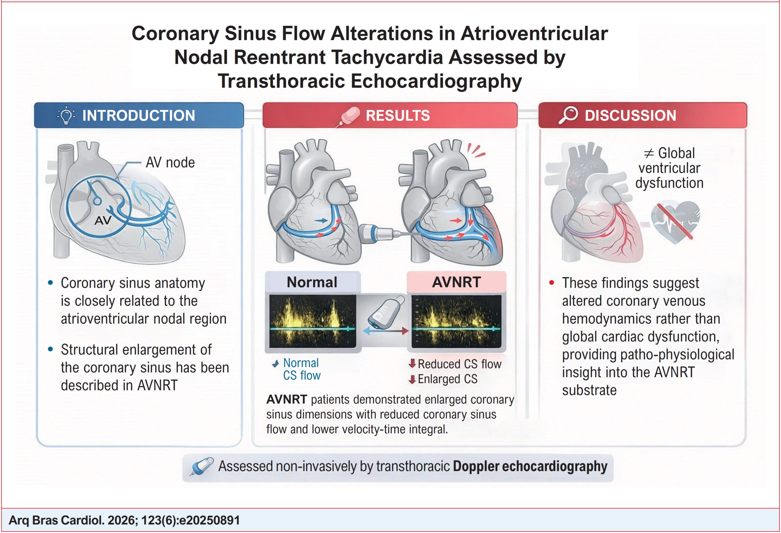
The coronary sinus (CS) plays a pivotal role in cardiac venous drainage and is anatomically adjacent to the atrioventricular nodal region. Although structural enlargement of the CS has been described in patients with atrioventricular nodal reentrant tachycardia (AVNRT), coronary sinus flow (CSF) assessed by transthoracic echocardiography (TTE) has not previously been investigated in this population.

Objective:

To evaluate the relationship between AVNRT and CSF.

Methods:

This retrospective single-center study included 35 patients with AVNRT who underwent successful slow-pathway ablation and 34 age- and sex-matched healthy controls. All participants underwent comprehensive transthoracic echocardiography. CS ostial diameter, CS diameter, and CSF were measured using pulsed-wave Doppler echocardiography. CSF was calculated using standard geometric and Doppler-derived parameters. A p-value <0.05 was considered statistically significant.

Results:

Global left and right ventricular systolic and diastolic function parameters were similar between groups. Patients with AVNRT exhibited significantly larger CS ostial and CS diameters compared with controls (both p<0.001). Despite this enlargement, CSF was significantly reduced in the AVNRT group (460.8 ± 83.3 vs. 523.8 ± 90.1 mL/min, p=0.004), accompanied by a lower CS velocity–time integral. Notably, CSF indexed to left ventricular mass also remained significantly lower in AVNRT patients, indicating that reduced flow was independent of myocardial mass.

Conclusion:

Patients with AVNRT demonstrate a distinct coronary sinus hemodynamic profile characterized by CS enlargement and reduced CSF as assessed by transthoracic echocardiography. These findings suggest altered coronary venous hemodynamics in AVNRT and provide novel pathophysiological insights using a non-invasive imaging modality.
  • Atrioventricular Nodal Reentry Tachycardia
  • Coronary sinus
  • Doppler Echocardiography
Read Full Article
Creative Commons Attribution (CC BY) Creative Commons license
24/07/2026 DOI: 10.36660/abc.20250891i Views 1
Echocardiographic Assessment of Diastolic Dysfunction in Special Situations
ABCIC
Review Article

Echocardiographic Assessment of Diastolic Dysfunction in Special Situations

Antonio Amador Calvilho Júnior

Abstract

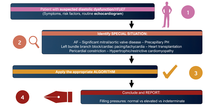
Echocardiographic assessment of diastolic function and left ventricular (LV) filling pressures is fundamental to the evaluation of dyspnea and the management of heart failure. However, conventional algorithms have limitations in several special clinical settings in which rhythm disturbances, valvular heart disease, pulmonary hypertension (PH), or structural cardiac abnormalities interfere with the interpretation of Doppler parameters. This article presents a practical approach to assessing diastolic function in conditions such as atrial fibrillation (AF), PH, mitral valve disease, mitral annular calcification, aortic valve disease, conduction disturbances, ventricular pacing, and restrictive cardiomyopathies. The main parameters applicable to each context, the technical aspects of image acquisition, and strategies for preparing a clear and clinically useful echocardiographic report are discussed.

  • Diastolic Heart Failure
  • Doppler Echocardiography
  • Stroke Volume
Read Full Article
Creative Commons Attribution (CC BY) Creative Commons license
17/07/2026 DOI: 10.36660/abcimg.20260029i Views 1
Step-by-Step Approach to the Evaluation of Constrictive Pericarditis
ABCIC
Review Article

Step-by-Step Approach to the Evaluation of Constrictive Pericarditis

Aline Travessa et al. Paulo Henrique Pereira Louise Moutinho Machado Natalia Sousa Esteves

Abstract

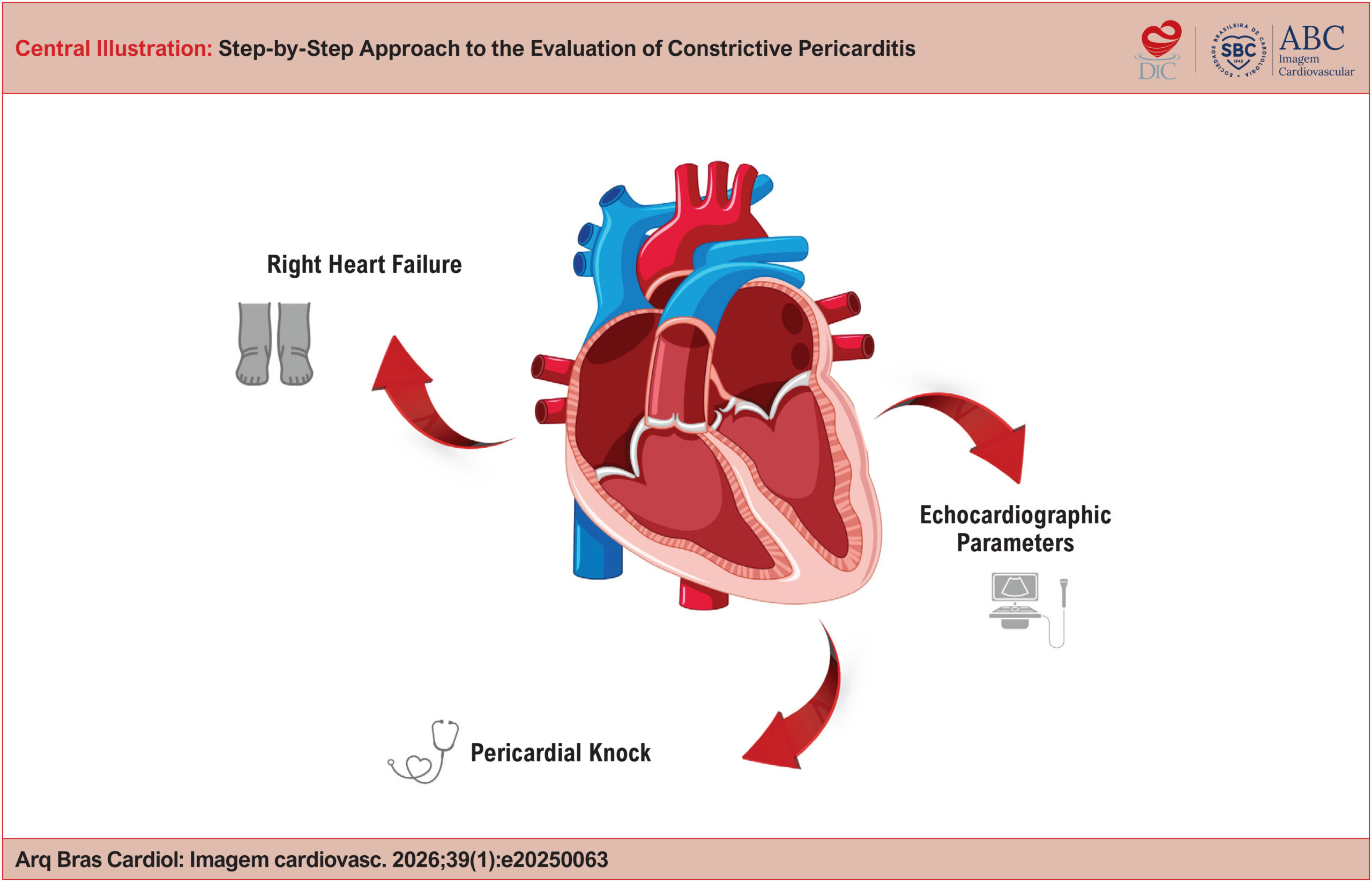
Constrictive Pericarditis (CP) is an uncommon but potentially curable condition, typically presenting with right-sided heart failure and impaired ventricular filling. Diagnosis can be challenging and requires a structured approach that integrates various cardiovascular imaging modalities. This article offers a practical guide, based on evidence and clinical experience, for the step-by-step recognition of Constrictive Pericarditis, highlighting echocardiographic findings.

  • Doppler Echocardiography
  • Echocardiography
  • Pericarditis
Read Full Article
Creative Commons Attribution (CC BY) Creative Commons license
25/05/2026 DOI: 10.36660/abcimg.20250063i
Figure 1
ABC
Brief Communication

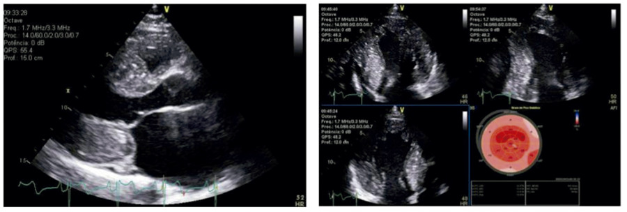
Apical Sparing and Granular Sparkling: Not Everything that Glitters Is Cardiac Amyloidosis

Ana Paula Morais Barros et al. Cristhian Espinoza Romero Juliano Novaes Cardoso Fábio Fernandes Viviane Tiemi Hotta

Introduction

With recent advances in the diagnosis and treatment of cardiac amyloidosis (CA), the role of complementary cardiac imaging methods for the early recognition of this disease has been widely discussed. In this context, myocardial hyperrefringence and preservation of left ventricular apex contractility (apical sparing; AS), evidenced by transthoracic echocardiography (TTE) with myocardial strain analysis (STE), are findings that frequently raise suspicion of CA.

However, despite this association, the heterogeneity of this finding in CA, depending on the population studied, in addition to its presentation in other conditions, leads to questions regarding the real accuracy of AS for the diagnosis of this disease. Through the report of three clinical cases of patients with different clinical conditions – hypertrophic cardiomyopathy (HCM), chronic kidney disease (CKD), and use of anabolic steroids – in which myocardial hyperrefringence and AS were described on echocardiography, a brief discussion is proposed regarding the pathophysiology of these findings and their limitations for the diagnosis of CA.

[…]

  • Amyloidosis
  • Doppler Echocardiography
  • Global Longitudinal Strain
Read Full Article
Creative Commons Attribution (CC BY) Creative Commons license
09/01/2026 DOI: 10.36660/abc.20250198i Views 3
Echocardiographic Parameters for Evaluating the Right Ventricle in Patients with Chronic Obstructive Pulmonary Disease: Literature Review
ABCHF
Review Article

Echocardiographic Parameters for Evaluating the Right Ventricle in Patients with Chronic Obstructive Pulmonary Disease: Literature Review

Irving Gabriel Araújo Bispo et al. Vitor Morais Brambila Gabriela Câmara Chagas Maria Luisa Ribeiro Gonçalves Pereira Maria Angélica Miqueri de Melo

Abstract

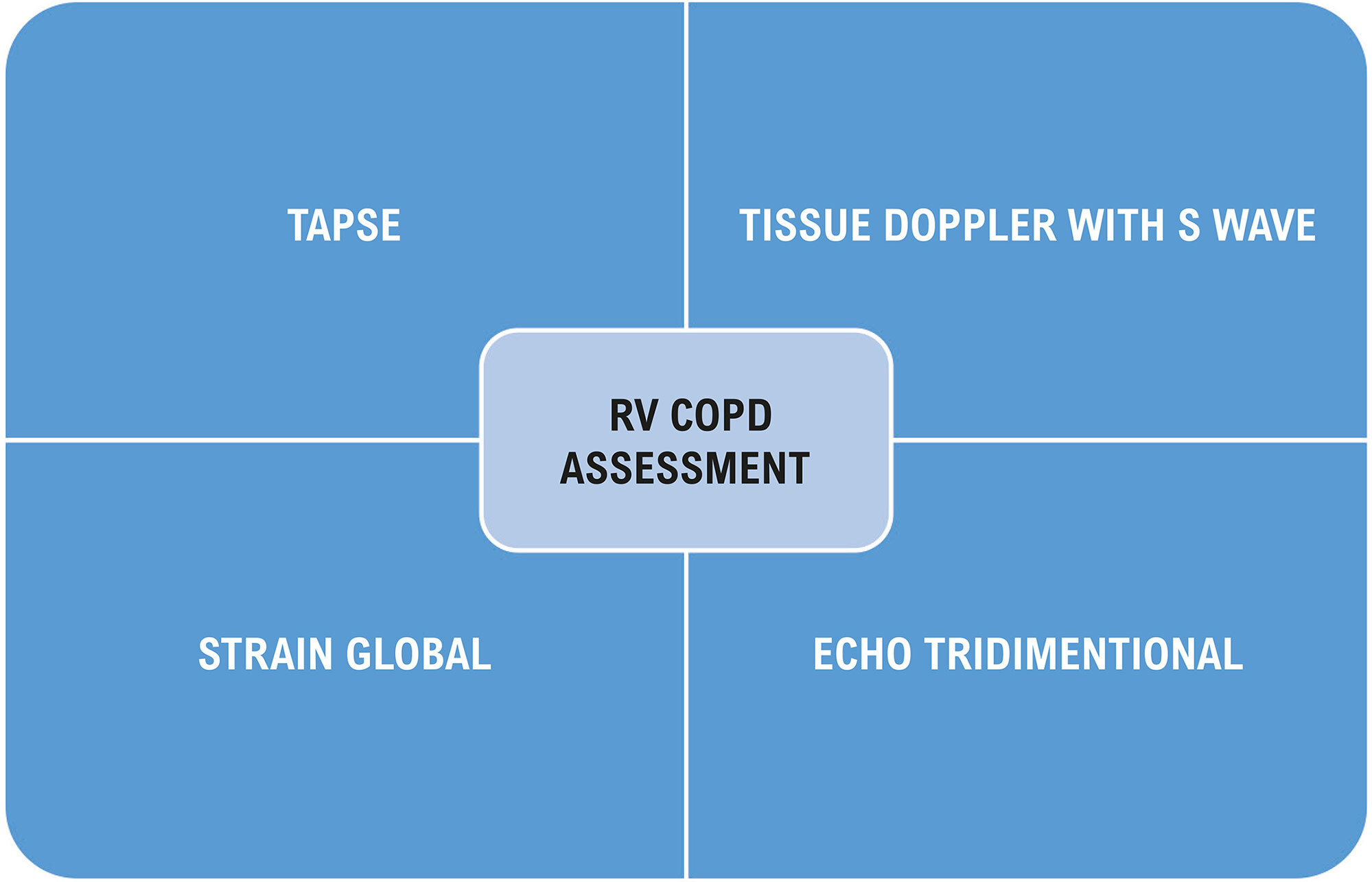
Chronic obstructive pulmonary disease (COPD) is a lung disease that often has an indolent course and systemic repercussions. Its main cause is constant and prolonged exposure to cigarette smoke or other gases. Such particles lead to reduced airflow and consequent lung hyperinflation with varying degrees of airway obstruction, also causing skeletal muscle dysfunction, respiratory failure, and decreased peripheral blood flow. COPD patients with concomitant cardiovascular disease (CVD) have a worse prognosis than simply the prognosis of each disease alone. However, it is difficult to establish what the risk is for a given group of patients due to the complex network of etiological and pathophysiological interactions underlying both diseases. Quantitative and qualitative echocardiographic assessment of right ventricular function is becoming of increasing interest in cardiac diseases that affect the right ventricle (RV), such as congenital heart disease and pulmonary hypertension (PH). However, it is still challenging due to the complex anatomy of the RV. The objective is to revisit the echocardiographic parameters that functionally assess the RV in patients with COPD, even if they do not present clinical signs of ventricular dysfunction.

  • Chronic Obstructive Pulmonary Disease
  • Doppler Echocardiography
  • Global Longitudinal Strain
  • Right Ventricular Dysfunction
Read Full Article
Creative Commons Attribution (CC BY) Creative Commons license
19/03/2025 DOI: 10.36660/abchf.20240046i Views 1
Echocardiographic Evaluation of Right Atrial Function in Patients with Precapillary Pulmonary Hypertension
ABCIC
Original Article

Echocardiographic Evaluation of Right Atrial Function in Patients with Precapillary Pulmonary Hypertension

Wanessa Alves de Carvalho et al. Andressa Alves de Carvalho Eliauria Rosa Martins Agostinho Hermes de Medeiros Neto … Fernando Bacal Marcelo Dantas Tavares de Melo

Abstract

Background:

Considering that the effect of right ventricular (RV) failure on the right atrium (RA) is an essential part of the pathophysiology of pulmonary hypertension (PH), the assessment of RA function seems to have prognostic value in the course of the disease, but there are still few studies on this subject.

Objective:

To evaluate myocardial strain of the RA in patients with precapillary PH.

Methods:

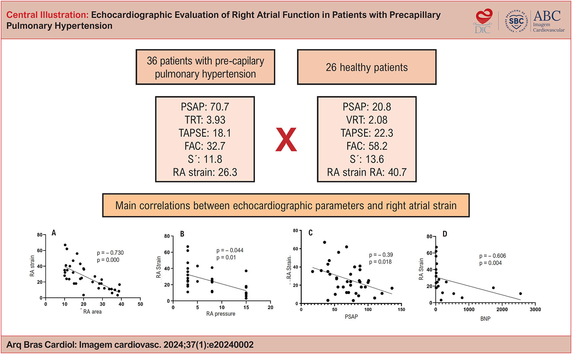
This is an observational, longitudinal, prospective study involving 36 patients with precapillary PH, in whom two-dimensional Doppler echocardiography was performed. A control group consisting of 26 healthy individuals was used.

Results:

There was a statistically significant difference in mean RA strain between the group with PH (26.3%) and the control group (40.7%). Regarding RA strain of the patients, there was a negative and moderate correlation with RA pressure, RV diameter, and RV hypokinesia; negative and weak correlation with pulmonary artery systolic pressure (PASP); negative and strong correlation with RA area; and positive and moderate correlation with, tricuspid annular plane systolic excursion (TAPSE), TAPSE/PASP, and RV strain. Patients with RA strain ≤ 23 had significantly reduced overall survival compared with patients with RA strain > 23, but this value was not a predictor of mortality.

Conclusion:

It was possible to demonstrate that the analysis of myocardial strain of the RA provides more accurate information about RA function and can be used as an additional parameter in the follow-up of patients with PH, given the early evaluation of atrial dysfunction.

  • Doppler Echocardiography
  • Pulmonary Hypertension
  • Right Atrial Function
Read Full Article
Creative Commons Attribution (CC BY) Creative Commons license
27/03/2024 DOI: 10.36660/abcimg.20240002i Views 1
ABCIC
Review Article

The Indiscriminate Use Of Androgenic Anabolic Steroids: The Contribution of Cardiovascular Imaging

Irving Gabriel Araöşjo Bispo et al. Isabella Montanher Zago

Abstract

Anabolic androgenic steroids (AAS) are natural or synthetic compounds similar to the male hormone testosterone. The literature also describes cases of AAS abuse, such as in bodybuilding, cosmetics, recreational use, and aesthetic enhancement. It was discovered that AAS alone directly induce myocardial injury, with the main pathological finding in autopsied hearts being left ventricle (LV) hypertrophy in frequent association with myocyte hypertrophy, increased collagen deposition in the matrix, increased activity of the cardiac angiotensin, and myocardial fibrosis.

Patients who used illicit AAS present decreased LV systolic function. In athletes who self-administer AAS, LV hypertrophy and elevated sympathetic modulation were observed, as well as elevated blood pressure (BP). High doses of AAS can cause the user to have ventricular arrhythmias and sudden death. Furthermore, AAS users demonstrated greater coronary artery plaque volume than non-users. The objective of this review is to revisit the main effects of the use of AAS on cardiac changes through cardiovascular imaging as well as to establish a difference in relation to athletic heart syndrome.

  • Doppler Echocardiography
  • Global Longitudinal Strain
  • Steroids
  • Tomography
Read Full Article
Creative Commons Attribution (CC BY) Creative Commons license
21/03/2024 DOI: 10.36660/abcimg.20230113i
ABC
Letter to the Editor

Association between Aortic Valve Sclerosis and Adverse Cardiovascular Events

Cristiane Machado Lucena et al. Rodrigo Pires dos Santos

Dear Editor,

The presence of aortic valve sclerosis (AVS) is associated with adverse cardiovascular events even when pre-existing risk factors are considered. In the Cardiovascular Health Study, which included more than 5,000 patients who were ≥ 65 years of age and were periodically evaluated for more than five years, AVS was associated with a 40% increase in the risk of myocardial infarction; in patients who did not initially present with heart disease, there were a trend toward an increased risk of angina, heart failure, and stroke. It is still unclear whether the mechanism of association between AVS and adverse clinical cardiovascular events is caused by diffuse atherosclerosis, endothelial dysfunction, altered calcium metabolism, lipid accumulation, genetic polymorphism, or other as yet undefined factors.

AVS is defined as thickening and increased echogenicity in the aortic leaflets with no reduction in mobility on an echocardiograph. The early lesion of AVS is an active process with some similarities to atherosclerosis, including the histopathological features and an association with risk for coronary artery disease. Patients are at greater risk for coronary artery disease if they are older, male, hypertensive, smokers, diabetics, or if their HDL and LDL levels are high. A cross-sectional study by Marmelo et al. evaluated 2,494 individuals who presented with AVS diagnosed via transthoracic Doppler echocardiography. These individuals also had a greater prevalence of hypertension, previous myocardial infarction, diabetes, a history of smoking, left ventricular systolic dysfunction, and mitral valve sclerosis. Multivariate analysis allows for a more complete study of the association between each variable and its outcome.

[…]

  • Cardiovascular Risk Factors
  • Coronary Artery Disease
  • Doppler Echocardiography
  • Heart Valve Diseases/physiopathology
Read Full Article
Creative Commons Attribution (CC BY) Creative Commons license
01/07/2015 DOI: 10.5935/abc.20150081
ABC
Artigo Original

Analysis of the sensitivity and specificity of noninvasive imaging tests for the diagnosis of renal artery stenosis

Flavio Antonio de Oliveira Borelli et al. Ibraim M. F. Pinto Celso Amodeo Paola E. P. Smanio … Antonio M. Kambara Ana Claudia G. Petisco Samuel M. Moreira Ricardo Calil Paiva Hugo Belotti Lopes Amanda G. M. R. Sousa

BACKGROUND: Aging and atherosclerosis are related to renovascular hypertension in elderly individuals. Regardless of comorbidities, renal artery stenosis is itself an important cause of cardiovascular morbidity and mortality. OBJECTIVE: To define the sensitivity, specificity, positive predictive value, and negative predictive value of noninvasive imaging tests used in the diagnosis of renal artery stenosis. METHODS: In a group of 61 patients recruited, 122 arteries were analized, thus permitting the definition of sensitivity, specificity, and the relative contribution of each imaging study performed (Doppler, scintigraphy and computed tomographic angiography in comparison to renal arteriography). RESULTS: The mean age was 65.43 years (standard deviation: 8.7). Of the variables related to the study population that were compared to arteriography, two correlated with renal artery stenosis, renal dysfunction and triglycerides. The median glomerular filtration rate was 52.8 mL/min/m². Doppler showed sensitivity of 82.90%, specificity of 70%, a positive predictive value of 85% and negative predictive value of 66.70%. For tomography, sensitivity was 66.70%, specificity 80%, positive predictive value 87.50% and negative predictive value 55.20%. With these findings, we could identify the imaging tests that best detected stenosis. CONCLUSION: Tomography and Doppler showed good quality and efficacy in the diagnosis of renal artery stenosis, with Doppler having the advantage of not requiring the use of contrast medium for the assessment of a disease that is common in diabetics and is associated with renal dysfunction and severe left ventricular dysfunction.

  • Doppler Echocardiography
  • hypertension, renovascular
  • Magnetic Resonance Imaging
  • Renal Artery Obstruction
Read Full Article
Creative Commons Attribution (CC BY) Creative Commons license
24/09/2013 DOI: 10.5935/abc.20130191
Cardiac morphofunctional injuries in patients with hepatitis C virus: doppler echocardiographic evaluation
ABC
Artigo Original

Cardiac morphofunctional injuries in patients with hepatitis C virus: doppler echocardiographic evaluation

Carlos Tosta et al. Ricardo Ladeira Betty Guz João Pimenta

OBJECTIVE: To evaluate the possible morphological and functional heart injuries in hepatitis C virus patients. METHODS: Control-case study analyzing Doppler echocardiographic aspects in 31 hepatitis C virus patients and 20 controls. RESULTS: There were no significant difference in relation to the myocardial thickening, left ventricular diameters, circumferential shortening, ejection fraction, mitral valve flow velocities, tissue sistolic and diastolic mitral annular ones. CONCLUSION: Individuals with hepatitis C virus in the initial phases of the disease did not show morpho-functional abnormalities of the heart when evaluated by doppler echocardiography.

  • cardiomyopathy
  • Doppler Echocardiography
  • hepatitis C virus
Read Full Article
Creative Commons Attribution (CC BY) Creative Commons license
06/03/2007 DOI: 10.1590/S0066-782X2006001800012 Views 1
Evaluation of asymptomatic patients with chronic Chagas’ disease through ambulatory electrocardiogram, echocardiogram and B-Type natriuretic peptide analyses
ABC
Artigo Original

Evaluation of asymptomatic patients with chronic Chagas’ disease through ambulatory electrocardiogram, echocardiogram and B-Type natriuretic peptide analyses

Divina Seila de Oliveira Marques et al. Manoel Fernandes Canesin Flavio Barutta Júnior Claudio José Fuganti Antonio Carlos Pereira Barretto

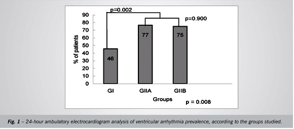
OBJECTIVE: To evaluate asymptomatic patients with chronic Chagas’ disease to determine prevalence of ventricular arrhythmias, left ventricular dysfunction, and B-type natriuretic peptide (BNP) plasma levels. METHODS: One hundred and six patients from the Chagas’ disease outpatient clinic underwent clinical evaluation, electrocardiogram (ECG), cardiothoracic index (CTI), ambulatory electrocardiogram (Holter monitoring), echocardiogram, and BNP measurement and then were distributed into three groups: GI, with normal ECG (n = 50); GIIA, with ECG changes characteristic of Chagas’ disease (n = 31); and GIIB, with other ECG changes (n = 25). RESULTS: The most common electrocardiographic changes were the following. GIIA: complete right bundle branch block (35%), left anterior hemiblock (35%), and electrically inactive areas (32%); GIIB: inferolateral repolarization change (28%), and left ventricular overload (24%). Mean CTI index values were similar (p = 0.383). Ventricular arrhythmia prevalence was higher in the GIIA (77%) and GIIB (75%) groups than in the GI group (46%) (p = 0.002). Ventricular dysfunction was more prevalent in the GIIA (52%) and GIIB (32%) groups than in the GI group (14%) (p = 0.001). Systolic dysfunction was more prevalent in the GIIA group (29%) than in the GIIB (20%) and GI groups (2%) (p < 0.001). Diastolic dysfunction was more prevalent in the GIIA (42%) and GIIB (28%) groups than in the GI group (12%) (p = 0.005). Mean B-type natriuretic peptide levels were 30 ± 88 pg/mL in the GI group, 66 ± 194 in the GIIA group and 24 ± 82 for the GIIB group (p = 0.121), respectively. CONCLUSION: Arrhythmias and left ventricular dysfunction are more prevalent in asymptomatic patients with chronic Chagas’ disease and abnormal ECG than in patients with normal ECG. Plasma BNP levels were similar among the groups.

  • Ambulatory Electrocardiography
  • Brain Natriuretic Peptide
  • Chagas disease
  • Doppler Echocardiography
Read Full Article
Creative Commons Attribution (CC BY) Creative Commons license
16/10/2006 DOI: 10.1590/S0066-782X2006001600017 Views 1
ABC
Artigo Original

Comparative study of clinical and Doppler echocardiographic evaluations of the progression of valve diseases in children and adolescents with rheumatic fever

Zilda Maria Alves Meira et al. Eugênio Marcos Andrade Goulart Cleonice de Carvalho Coelho Mota

OBJECTIVE: Compare clinical and Doppler echocardiographic evaluations in assessing valvular diseases in children and adolescents with rheumatic fever, as well as assess the progression of the disease in light of these assessments. METHODS: This is a longitudinal study of 258 children and adolescents diagnosed with rheumatic fever according to Jones’ criteria. The follow-up period ranged from 2-15 years. The presence and quantification of valve diseases were determined by means of clinical and Doppler echocardiographic evaluations performed during the acute and chronic phases. The Kappa statististics method was used to estimate the degree of agreement between clinical and Doppler echocardiographic evaluations. Comparisons between clinical and Doppler echocardiographic findings on the progress of carditis and valvulitis, respectively, were made using chi-square test or Fisher’s exact test, p< 0.05. RESULTS: Of the 109 patients who underwent Doppler echocardiographic evaluation during the acute phase, 31 did not present clinical evidence of carditis, but the Doppler echocardiograms of 17 (54.8%) of them showed valve lesions (subclinical valvulitis). During the chronic phase, 153 of the 258 patients had normal cardiovascular examination results; however, Doppler echocardiograms showed that 81 of them (52.9%) had valve lesions (subclinical chronic valvular diseases). Involution of the valvular lesions, as shown by Doppler echocardiographic evaluations, was less frequent and occurred in 10 (25.0%) patients with mild valvulitis, in only one (2.5%) patient with moderate valvulitis, and in none of the patients with severe valvulitis. CONCLUSION: The identification of rheumatic fever valve lesions can be enhanced when clinical evaluations are supplemented by Doppler echocardiographic examinations; also, clinical examinations are not as suitable to detect valvular lesion regression as the echocardiography. The diagnosis of subclinical valvulitis and valvulopathy influences the secondary prophylaxis of rheumatic fever and endocarditis.

  • Doppler Echocardiography
  • Rheumatic Fever
  • rheumatic valvulopathy
  • valvulitis
Read Full Article
Creative Commons Attribution (CC BY) Creative Commons license
13/02/2006 DOI: 10.1590/S0066-782X2006000100006 Views 1
Serial clinical and echocardiographic evaluation in children with Marfan syndrome
ABC
Artigo Original

Serial clinical and echocardiographic evaluation in children with Marfan syndrome

Victor Manuel Oporto Lopez et al. Ana Beatriz Alvarez Perez Valdir Ambrósio Moisés Lourdes Gomes … Patricia da Silveira Pedreira Célia C. Silva Orlando Campos Filho Antônio Carlos C. Carvalho

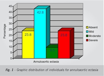
OBJECTIVE: To describe the clinical cardiac manifestations and temporal evolution of Marfan syndrome in children; to estimate the incidence of annuloaortic ectasia and mitral valve prolapse; and to evaluate tolerability and efficacy of beta-blockers in these patients. METHODS: During one year, 21children with Marfan syndrome underwent serial clinical and echocardiographic examinations. Echocardiograms assessed: the presence of mitral valve prolapse, aortic root diameter, mitral and aortic valves regurgitation, and aortic enlargement during beta-blocker therapy. Eleven patients had two measurements of the aortic root taken one year apart. RESULTS: The children were asymptomatic throughout the study. Mitral prolapse was found in 11 (52%) children. Annuloaortic ectasia occurred in 16 (76%) patients and found to be mild in 42.8%, moderate in 9.5%, and severe in 23.8%. One of these patients underwent aortic valve replacement and repair of the ascending aorta by the Bentall-De Bono technique, with good results. Heart rate decreased by 13.6% (from 85 to 73 bpm; p < 0.009) with the use of beta-blockers; however, aortic root diameter increased by 1.4 mm/year (p<0.02). One child could not be given beta-blockers due to bronchial asthma, and no significant side effects were observed in the remaining children, including one who also had bronchial asthma. CONCLUSION: The children remained asymptomatic throughout the study, the use of beta-blockers led to a significant decrease in heart rate, and no significant adverse effects were observed. Contrary to the literature, incidence of annuloaortic ectasia was high among the study population, greater than that of mitral valve prolapse, even during beta-blocker therapy.

  • Doppler Echocardiography
  • Marfan syndrome
  • Mitral Valve Prolapse
  • thoracic aortic aneurysm
Read Full Article
Creative Commons Attribution (CC BY) Creative Commons license
05/12/2005 DOI: 10.1590/S0066-782X2005001800003
Doppler echocardiographic variables and the type of surgery to be performed in rheumatic mitral valve regurgitation
ABC
Artigo Original

Doppler echocardiographic variables and the type of surgery to be performed in rheumatic mitral valve regurgitation

Jorge Eduardo Assef et al. Leopoldo Soares Piegas Sérgio Cunha Pontes Junior Rodrigo Bellio de Mattos Barretto … Mercedes Maldonado Mohamed Hassam Saleh David Le Bihan Vera Marcia Lopes Gimenes Zilda Machado Meneghelo Paulo Paredes Paulista

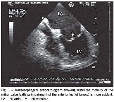
OBJECTIVE: To identify the Doppler echocardiographic variables associated with the type of surgery performed in rheumatic mitral valve regurgitation and to determine the relation between those variables and the medium-term results of valvuloplasty. METHODS: Doppler echocardiographic variables were assessed in 68 patients with severe rheumatic mitral valve regurgitation on the day preceding surgery. The patients were divided, according to the surgery performed, into 2 groups: 1) valvuloplasty group; and 2) valve replacement group. The valvuloplasty group also underwent Doppler echocardiography before hospital discharge and 6 months after that. The Doppler echocardiographic variables of the preoperative period were assessed considering the type of surgery performed and the degree of regurgitation detected 6 months later. RESULTS: The groups did not differ in regard to their demographic characteristics and ventricular function. The valve replacement group had a smaller mitral area (P=0.001). In univariate analysis, the variables related to valve replacement were as follows: restricted mobility of the anterior (P=0.01) and posterior (P=0.01) leaflets; calcification of the anterior leaflet (P=0.01); and fusion of the chordae tendineae (P=0.018). Restricted mobility of the anterior leaflet and area remained as independent determining factors of the prosthetic implantation after multivariate analysis. Of the 7 patients with mitral regurgitation greater than mild detected 6 months after valvuloplasty, 6 had, before surgery, restricted mobility of the anterior leaflet and 4 had fusion of the chordae tendineae. CONCLUSION: The chance of valve replacement is 3.8 times greater when restricted mobility of the mitral valve anterior leaflet exists and 2.2 times greater for each 0.5 cm² reduction in the mitral valve area. Restricted mobility of the anterior leaflet and fusion of the chordae tendineae are associated with regurgitation greater than mild, observed 6 months after valvuloplasty.

  • Doppler Echocardiography
  • rheumatic mitral valve regurgitation
  • Surgery
Read Full Article
Creative Commons Attribution (CC BY) Creative Commons license
13/12/2004 DOI: 10.1590/S0066-782X2004001900012
Cardiovascular assessment of patients with Ullrich-Turner's Syndrome on Doppler echocardiography and magnetic resonance imaging
ABC
Original Article

Cardiovascular assessment of patients with Ullrich-Turner's Syndrome on Doppler echocardiography and magnetic resonance imaging

Ana Valéria Barros de Castro et al. Katashi Okoshi Sérgio Marrone Ribeiro Maurício Fregonesi Barbosa … Patricia Ferreira Mattos Luciana Pagliare Nancy Figueiredo Bueno Débora Aparecida Rodrigueiro Adriana Lúcia Mendes Haddad

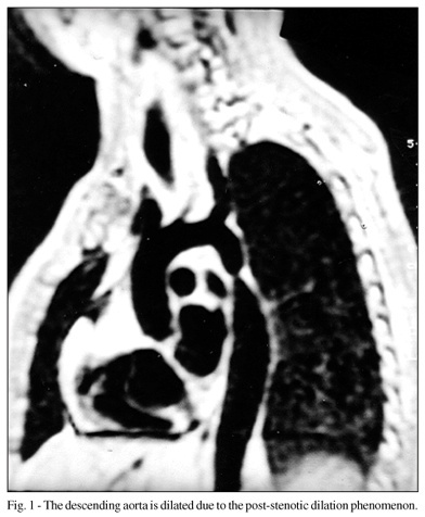
OBJECTIVE: To assess the cardiovascular features of Ullrich-Turner’s syndrome using echocardiography and magnetic resonance imaging, and to correlate them with the phenotype and karyotype of the patients. The diagnostic concordance between the 2 methods was also assessed. METHODS: Fifteen patients with the syndrome were assessed by echocardiography and magnetic resonance imaging (cardiac chambers, valves, and aorta). Their ages ranged from 10 to 28 (mean of 16.7) years. The karyotype was analyzed in 11 or 25 metaphases of peripheral blood lymphocytes, or both. RESULTS: The most common phenotypic changes were short stature and spontaneous absence of puberal development (100%); 1 patient had a cardiac murmur. The karyotypes detected were as follows: 45,X (n=7), mosaics (n=5), and deletions (n=3). No echocardiographic changes were observed. In regard to magnetic resonance imaging, coarctation and dilation of the aorta were found in 1 patient, and isolated dilation of the aorta was found in 4 patients. CONCLUSION: The frequencies of coarctation and dilation of the aorta detected on magnetic resonance imaging were similar to those reported in the literature (5.5% to 20%, and 6.3% to 29%, respectively). This confirmed the adjuvant role of magnetic resonance imaging to Doppler echocardiography for diagnosing cardiovascular alterations in patients with Ullrich-Turner’s syndrome.

  • Doppler Echocardiography
  • Magnetic Resonance Imaging
  • Turner's syndrome
Read Full Article
Creative Commons Attribution (CC BY) Creative Commons license
19/02/2002 DOI: 10.1590/S0066-782X2002000100005 Views 1
ABC
Artigo Original

The value of exercise test in the prognosis of asymptomatic patients with significant aortic valve stenosis

Marisa Campos Moraes Amato et al. Paulo Jorge Moffa

PURPOSE: To evaluate the exercise test as a prognostic factor for asymptomatic patients with aortic valve stenosis. METHODS: During 60 months 70 patients with aortic valve area <1cm² without any other heart lesion were studied. The patients underwent Dopplerechocardiograms exercise tests (considered positive in the presence of electrocardiographic changes or symptoms) and clinical follow-up in order to determine the onset of symptoms or sudden death. RESULTS: The event-free probability was 50% after 16 months of follow-up. There was no relationship between the occurence of events and the sex or to the transvalvar gradient. However, there was a statistically significant association between the incidence of events (p= 0.0124), valvar area (p= 0.0003) and exercise test results (p= 0.0001). CONCLUSION: Patients with significant aortic stenosis even without symptoms, have a low survival rate and the exercise test is a good prognostic factor during follow-up.

  • Aortic Valve Stenosis
  • Doppler Echocardiography
  • ergometric test
Read Full Article
Creative Commons Attribution (CC BY) Creative Commons license
08/01/2002 DOI: 10.1590/S0066-782X1998000400003
ABC
Artigo Original

Evaluation of left internal thoracic artery anastomosis with left anterior descending coronary artery by Doppler echocardiography

Adelaide Arruda et al. Orlando Campos Fº Expedito Ribeiro Antônia Petrizzo … José Lázaro Andrade Antônio Carlos C. Carvalho Enio Buffolo Eulógio Martinez Wilson Mathias Jr

PURPOSE: To study the value of Doppler echocardiography as a tool for the evaluation of left internal thoracic artery graft (LITAG) patency in patients who underwent coronary revascularization using minimally invasive bypass surgery without extracorporeal circulation. METHODS: The first 12 consecutive patients were studied after coronary artery bypass surgery using a 5MHz Doppler transducer. Doppler signals for the systolic and diastolic flow velocities were preferably obtained in the second intercostal space. All patients underwent coronary angiography while hospitalized. RESULTS: The exam was feasible in 93% of patients. Doppler flow pattern was predominantly diastolic (pattern A) in patients with patent anastomoses (6/7). In patients with occluded anastomoses (4/4) Doppler flow pattern was predominantly systolic (pattern B) (p=0.003). CONCLUSION: Internal thoracic artery flow pattern as measured by Doppler echocardiography after minimally invasive coronary artery bypass graft surgery is an accurate method for identifying LITAG patency.

  • bypass surgery
  • Doppler Echocardiography
  • thoracic artery
Read Full Article
Creative Commons Attribution (CC BY) Creative Commons license
23/01/2001 DOI: 10.1590/S0066-782X1997001200009 Views 1

How to cite this article

EndNote RIS

Electronic Document Format (Vancouver)

Duygu A, Akgümüş A, Balun A, Er F. Assessment of Coronary Sinus Flow and Dimensions by Transthoracic Echocardiography in Patients with Atrioventricular Nodal Reentrant Tachycardia. Arquivos Brasileiros de Cardiologia 2026;123(6):e20250891.

Electronic Document Format (ABNT)

Duygu, Ali; Akgümüş, Alkame; Balun, Ahmet; Er, Fahri. Assessment of Coronary Sinus Flow and Dimensions by Transthoracic Echocardiography in Patients with Atrioventricular Nodal Reentrant Tachycardia. Arquivos Brasileiros de Cardiologia, v. 123, n. 6, e20250891, Jul. 2026.

Electronic Document Format (APA)

Duygu, A., Akgümüş, A., Balun, A., & Er, F. (2026). Assessment of Coronary Sinus Flow and Dimensions by Transthoracic Echocardiography in Patients with Atrioventricular Nodal Reentrant Tachycardia. Arquivos Brasileiros de Cardiologia, 123(6), e20250891.

Electronic Document Format (ISO)

Duygu, Ali and Akgümüş, Alkame and Balun, Ahmet and Er, Fahri. Assessment of Coronary Sinus Flow and Dimensions by Transthoracic Echocardiography in Patients with Atrioventricular Nodal Reentrant Tachycardia. Arquivos Brasileiros de Cardiologia [online]. 2026, vol. 123, n. 6, [cited 2026-10-14], e20250891. Available from: <https://journals.cardiol.br/en/article/assessment-of-coronary-sinus-flow-and-dimensions-by-transthoracic-echocardiography-in-patients-with-atrioventricular-nodal-reentrant-tachycardia/>. ISSN 0066-782X

EndNote RIS

Electronic Document Format (Vancouver)

Calvilho Júnior AA. Echocardiographic Assessment of Diastolic Dysfunction in Special Situations. ABC Imagem Cardiovascular 2026;39(2):e20260029.

Electronic Document Format (ABNT)

Calvilho Júnior, Antonio Amador. Echocardiographic Assessment of Diastolic Dysfunction in Special Situations. ABC Imagem Cardiovascular, v. 39, n. 2, e20260029, Jul. 2026.

Electronic Document Format (APA)

Calvilho Júnior, A. A. (2026). Echocardiographic Assessment of Diastolic Dysfunction in Special Situations. ABC Imagem Cardiovascular, 39(2), e20260029.

Electronic Document Format (ISO)

Calvilho Júnior, Antonio Amador. Echocardiographic Assessment of Diastolic Dysfunction in Special Situations. ABC Imagem Cardiovascular [online]. 2026, vol. 39, n. 2, [cited 2026-10-14], e20260029. Available from: <https://journals.cardiol.br/en/article/echocardiographic-assessment-of-diastolic-dysfunction-in-special-situations/>. ISSN 2318-8219

EndNote RIS

Electronic Document Format (Vancouver)

Travessa A, Pereira PH, Machado LM, Esteves NS. Step-by-Step Approach to the Evaluation of Constrictive Pericarditis. ABC Imagem Cardiovascular 2026;39(1):e20250063.

Electronic Document Format (ABNT)

Travessa, Aline; Pereira, Paulo Henrique; Machado, Louise Moutinho; Esteves, Natalia Sousa. Step-by-Step Approach to the Evaluation of Constrictive Pericarditis. ABC Imagem Cardiovascular, v. 39, n. 1, e20250063, May. 2026.

Electronic Document Format (APA)

Travessa, A., Pereira, P. H., Machado, L. M., & Esteves, N. S. (2026). Step-by-Step Approach to the Evaluation of Constrictive Pericarditis. ABC Imagem Cardiovascular, 39(1), e20250063.

Electronic Document Format (ISO)

Travessa, Aline and Pereira, Paulo Henrique and Machado, Louise Moutinho and Esteves, Natalia Sousa. Step-by-Step Approach to the Evaluation of Constrictive Pericarditis. ABC Imagem Cardiovascular [online]. 2026, vol. 39, n. 1, [cited 2026-10-14], e20250063. Available from: <https://journals.cardiol.br/en/article/step-by-step-approach-to-the-evaluation-of-constrictive-pericarditis/>. ISSN 2318-8219

EndNote RIS

Electronic Document Format (Vancouver)

Barros APM, Romero CE, Cardoso JN, Fernandes F, Hotta VT. Apical Sparing and Granular Sparkling: Not Everything that Glitters Is Cardiac Amyloidosis. Arquivos Brasileiros de Cardiologia 2026;122(11):e20250198.

Electronic Document Format (ABNT)

Barros, Ana Paula Morais; Romero, Cristhian Espinoza; Cardoso, Juliano Novaes; Fernandes, Fábio; Hotta, Viviane Tiemi. Apical Sparing and Granular Sparkling: Not Everything that Glitters Is Cardiac Amyloidosis. Arquivos Brasileiros de Cardiologia, v. 122, n. 11, e20250198, Jan. 2026.

Electronic Document Format (APA)

Barros, A. P. M., Romero, C. E., Cardoso, J. N., Fernandes, F., & Hotta, V. T. (2026). Apical Sparing and Granular Sparkling: Not Everything that Glitters Is Cardiac Amyloidosis. Arquivos Brasileiros de Cardiologia, 122(11), e20250198.

Electronic Document Format (ISO)

Barros, Ana Paula Morais and Romero, Cristhian Espinoza and Cardoso, Juliano Novaes and Fernandes, Fábio and Hotta, Viviane Tiemi. Apical Sparing and Granular Sparkling: Not Everything that Glitters Is Cardiac Amyloidosis. Arquivos Brasileiros de Cardiologia [online]. 2026, vol. 122, n. 11, [cited 2026-10-14], e20250198. Available from: <https://journals.cardiol.br/en/article/apical-sparing-and-granular-sparkling-not-everything-that-glitters-is-cardiac-amyloidosis/>. ISSN 0066-782X

EndNote RIS

Electronic Document Format (Vancouver)

Bispo IGA, Brambila VM, Chagas GC, Pereira MLRG, Melo MAM. Echocardiographic Parameters for Evaluating the Right Ventricle in Patients with Chronic Obstructive Pulmonary Disease: Literature Review. ABC Heart Fail Cardiomyop 2025;5(1):e20240046.

Electronic Document Format (ABNT)

Bispo, Irving Gabriel Araújo; Brambila, Vitor Morais; Chagas, Gabriela Câmara; Pereira, Maria Luisa Ribeiro Gonçalves; Melo, Maria Angélica Miqueri de. Echocardiographic Parameters for Evaluating the Right Ventricle in Patients with Chronic Obstructive Pulmonary Disease: Literature Review. ABC Heart Fail Cardiomyop, v. 5, n. 1, e20240046, Mar. 2025.

Electronic Document Format (APA)

Bispo, I. G. A., Brambila, V. M., Chagas, G. C., Pereira, M. L. R. G., & Melo, M. A. M. (2025). Echocardiographic Parameters for Evaluating the Right Ventricle in Patients with Chronic Obstructive Pulmonary Disease: Literature Review. ABC Heart Fail Cardiomyop, 5(1), e20240046.

Electronic Document Format (ISO)

Bispo, Irving Gabriel Araújo and Brambila, Vitor Morais and Chagas, Gabriela Câmara and Pereira, Maria Luisa Ribeiro Gonçalves and Melo, Maria Angélica Miqueri de. Echocardiographic Parameters for Evaluating the Right Ventricle in Patients with Chronic Obstructive Pulmonary Disease: Literature Review. ABC Heart Fail Cardiomyop [online]. 2025, vol. 5, n. 1, [cited 2026-10-14], e20240046. Available from: <https://journals.cardiol.br/en/article/echocardiographic-parameters-for-evaluating-the-right-ventricle-in-patients-with-chronic-obstructive-pulmonary-disease-literature-review/>

EndNote RIS

Electronic Document Format (Vancouver)

Carvalho WA, Carvalho AA, Martins ER, Medeiros Neto AH, Bacal F, Melo MDT. Echocardiographic Evaluation of Right Atrial Function in Patients with Precapillary Pulmonary Hypertension. ABC Imagem Cardiovascular 2024;37(1):e20240002.

Electronic Document Format (ABNT)

Carvalho, Wanessa Alves de; Carvalho, Andressa Alves de; Martins, Eliauria Rosa; Medeiros Neto, Agostinho Hermes de; Bacal, Fernando; Melo, Marcelo Dantas Tavares de. Echocardiographic Evaluation of Right Atrial Function in Patients with Precapillary Pulmonary Hypertension. ABC Imagem Cardiovascular, v. 37, n. 1, e20240002, Mar. 2024.

Electronic Document Format (APA)

Carvalho, W. A. , Carvalho, A. A. , Martins, E. R., Medeiros Neto, A. H. , Bacal, F., & Melo, M. D. T. (2024). Echocardiographic Evaluation of Right Atrial Function in Patients with Precapillary Pulmonary Hypertension. ABC Imagem Cardiovascular, 37(1), e20240002.

Electronic Document Format (ISO)

Carvalho, Wanessa Alves de and Carvalho, Andressa Alves de and Martins, Eliauria Rosa and Medeiros Neto, Agostinho Hermes de and Bacal, Fernando and Melo, Marcelo Dantas Tavares de. Echocardiographic Evaluation of Right Atrial Function in Patients with Precapillary Pulmonary Hypertension. ABC Imagem Cardiovascular [online]. 2024, vol. 37, n. 1, [cited 2026-10-14], e20240002. Available from: <https://journals.cardiol.br/en/article/echocardiographic-evaluation-of-right-atrial-function-in-patients-with-precapillary-pulmonary-hypertension/>. ISSN 2318-8219

EndNote RIS

Electronic Document Format (Vancouver)

Bispo IGA, Zago IM. The Indiscriminate Use Of Androgenic Anabolic Steroids: The Contribution of Cardiovascular Imaging. ABC Imagem Cardiovascular 2024;37(1):e20230113.

Electronic Document Format (ABNT)

Bispo, Irving Gabriel Araöşjo; Zago, Isabella Montanher. The Indiscriminate Use Of Androgenic Anabolic Steroids: The Contribution of Cardiovascular Imaging. ABC Imagem Cardiovascular, v. 37, n. 1, e20230113, Mar. 2024.

Electronic Document Format (APA)

Bispo, I. G. A., & Zago, I. M. (2024). The Indiscriminate Use Of Androgenic Anabolic Steroids: The Contribution of Cardiovascular Imaging. ABC Imagem Cardiovascular, 37(1), e20230113.

Electronic Document Format (ISO)

Bispo, Irving Gabriel Araöşjo and Zago, Isabella Montanher. The Indiscriminate Use Of Androgenic Anabolic Steroids: The Contribution of Cardiovascular Imaging. ABC Imagem Cardiovascular [online]. 2024, vol. 37, n. 1, [cited 2026-10-14], e20230113. Available from: <https://journals.cardiol.br/en/article/the-indiscriminate-use-of-androgenic-anabolic-steroids-the-contribution-of-cardiovascular-imaging/>. ISSN 2318-8219

EndNote RIS

Electronic Document Format (Vancouver)

Lucena CM, Santos RP. Association between Aortic Valve Sclerosis and Adverse Cardiovascular Events. Arquivos Brasileiros de Cardiologia 2015;105(1):99-100.

Electronic Document Format (ABNT)

Lucena, Cristiane Machado; Santos, Rodrigo Pires dos. Association between Aortic Valve Sclerosis and Adverse Cardiovascular Events. Arquivos Brasileiros de Cardiologia, v. 105, n. 1, p. 99-100, Jun. 2015.

Electronic Document Format (APA)

Lucena, C. M., & Santos, R. P. (2015). Association between Aortic Valve Sclerosis and Adverse Cardiovascular Events. Arquivos Brasileiros de Cardiologia, 105(1), 99-100.

Electronic Document Format (ISO)

Lucena, Cristiane Machado and Santos, Rodrigo Pires dos. Association between Aortic Valve Sclerosis and Adverse Cardiovascular Events. Arquivos Brasileiros de Cardiologia [online]. 2015, vol. 105, n. 1, [cited 2026-10-14], pp. 99-100. Available from: <https://journals.cardiol.br/en/article/association-between-aortic-valve-sclerosis-and-adverse-cardiovascular-events/>. ISSN 0066-782X

EndNote RIS

Electronic Document Format (Vancouver)

Borelli FAO, Pinto IMF, Amodeo C, Smanio PEP, Kambara AM, Petisco ACG, Moreira SM, et al. Analysis of the sensitivity and specificity of noninvasive imaging tests for the diagnosis of renal artery stenosis. Arquivos Brasileiros de Cardiologia 2013;101(5):423-3.

Electronic Document Format (ABNT)

Borelli, Flavio Antonio de Oliveira; Pinto, Ibraim M. F.; Amodeo, Celso; Smanio, Paola E. P.; Kambara, Antonio M.; Petisco, Ana Claudia G.; Moreira, Samuel M.; Paiva, Ricardo Calil; Lopes, Hugo Belotti; Sousa, Amanda G. M. R.. Analysis of the sensitivity and specificity of noninvasive imaging tests for the diagnosis of renal artery stenosis. Arquivos Brasileiros de Cardiologia, v. 101, n. 5, p. 423-433, Sep. 2013.

Electronic Document Format (APA)

Borelli, F. A. O., Pinto, I. M. F., Amodeo, C., Smanio, P. E. P., Kambara, A. M., Petisco, A. C. G., Moreira, S. M., Paiva, R. C., Lopes, H. B., & Sousa, A. G. M. R. (2013). Analysis of the sensitivity and specificity of noninvasive imaging tests for the diagnosis of renal artery stenosis. Arquivos Brasileiros de Cardiologia, 101(5), 423-433.

Electronic Document Format (ISO)

Borelli, Flavio Antonio de Oliveira and Pinto, Ibraim M. F. and Amodeo, Celso and Smanio, Paola E. P. and Kambara, Antonio M. and Petisco, Ana Claudia G. and Moreira, Samuel M. and Paiva, Ricardo Calil and Lopes, Hugo Belotti and Sousa, Amanda G. M. R.. Analysis of the sensitivity and specificity of noninvasive imaging tests for the diagnosis of renal artery stenosis. Arquivos Brasileiros de Cardiologia [online]. 2013, vol. 101, n. 5, [cited 2026-10-14], pp. 423-433. Available from: <https://journals.cardiol.br/en/article/analysis-of-the-sensitivity-and-specificity-of-noninvasive-imaging-tests-for-the-diagnosis-of-renal-artery-stenosis/>. ISSN 0066-782X

EndNote RIS

Electronic Document Format (Vancouver)

Tosta C, Ladeira R, Guz B, Pimenta J. Cardiac morphofunctional injuries in patients with hepatitis C virus: doppler echocardiographic evaluation. Arquivos Brasileiros de Cardiologia 2007;87(5):623-7.

Electronic Document Format (ABNT)

Tosta, Carlos; Ladeira, Ricardo; Guz, Betty; Pimenta, João. Cardiac morphofunctional injuries in patients with hepatitis C virus: doppler echocardiographic evaluation. Arquivos Brasileiros de Cardiologia, v. 87, n. 5, p. 623-627, Mar. 2007.

Electronic Document Format (APA)

Tosta, C., Ladeira, R., Guz, B., & Pimenta, J. (2007). Cardiac morphofunctional injuries in patients with hepatitis C virus: doppler echocardiographic evaluation. Arquivos Brasileiros de Cardiologia, 87(5), 623-627.

Electronic Document Format (ISO)

Tosta, Carlos and Ladeira, Ricardo and Guz, Betty and Pimenta, João. Cardiac morphofunctional injuries in patients with hepatitis C virus: doppler echocardiographic evaluation. Arquivos Brasileiros de Cardiologia [online]. 2007, vol. 87, n. 5, [cited 2026-10-14], pp. 623-627. Available from: <https://journals.cardiol.br/en/article/cardiac-morphofunctional-injuries-in-patients-with-hepatitis-c-virus-doppler-echocardiographic-evaluation/>. ISSN 0066-782X

EndNote RIS

Electronic Document Format (Vancouver)

Marques DSO, Canesin MF, Barutta Júnior F, Fuganti CJ, Barretto ACP. Evaluation of asymptomatic patients with chronic Chagas’ disease through ambulatory electrocardiogram, echocardiogram and B-Type natriuretic peptide analyses. Arquivos Brasileiros de Cardiologia 2006;87(3):336-43.

Electronic Document Format (ABNT)

Marques, Divina Seila de Oliveira; Canesin, Manoel Fernandes; Barutta Júnior, Flavio; Fuganti, Claudio José; Barretto, Antonio Carlos Pereira. Evaluation of asymptomatic patients with chronic Chagas’ disease through ambulatory electrocardiogram, echocardiogram and B-Type natriuretic peptide analyses. Arquivos Brasileiros de Cardiologia, v. 87, n. 3, p. 336-343, Oct. 2006.

Electronic Document Format (APA)

Marques, D. S. O., Canesin, M. F., Barutta Júnior, F., Fuganti, C. J., & Barretto, A. C. P. (2006). Evaluation of asymptomatic patients with chronic Chagas’ disease through ambulatory electrocardiogram, echocardiogram and B-Type natriuretic peptide analyses. Arquivos Brasileiros de Cardiologia, 87(3), 336-343.

Electronic Document Format (ISO)

Marques, Divina Seila de Oliveira and Canesin, Manoel Fernandes and Barutta Júnior, Flavio and Fuganti, Claudio José and Barretto, Antonio Carlos Pereira. Evaluation of asymptomatic patients with chronic Chagas’ disease through ambulatory electrocardiogram, echocardiogram and B-Type natriuretic peptide analyses. Arquivos Brasileiros de Cardiologia [online]. 2006, vol. 87, n. 3, [cited 2026-10-14], pp. 336-343. Available from: <https://journals.cardiol.br/en/article/evaluation-of-asymptomatic-patients-with-chronic-chagas-disease-through-ambulatory-electrocardiogram-echocardiogram-and-b-type-natriuretic-peptide-analyses/>. ISSN 0066-782X

EndNote RIS

Electronic Document Format (Vancouver)

Meira ZMA, Goulart EMA, Mota CCC. Comparative study of clinical and Doppler echocardiographic evaluations of the progression of valve diseases in children and adolescents with rheumatic fever. Arquivos Brasileiros de Cardiologia 2006;86(1):32-8.

Electronic Document Format (ABNT)

Meira, Zilda Maria Alves; Goulart, Eugênio Marcos Andrade; Mota, Cleonice de Carvalho Coelho. Comparative study of clinical and Doppler echocardiographic evaluations of the progression of valve diseases in children and adolescents with rheumatic fever. Arquivos Brasileiros de Cardiologia, v. 86, n. 1, p. 32-38, Feb. 2006.

Electronic Document Format (APA)

Meira, Z. M. A., Goulart, E. M. A., & Mota, C. C. C. (2006). Comparative study of clinical and Doppler echocardiographic evaluations of the progression of valve diseases in children and adolescents with rheumatic fever. Arquivos Brasileiros de Cardiologia, 86(1), 32-38.

Electronic Document Format (ISO)

Meira, Zilda Maria Alves and Goulart, Eugênio Marcos Andrade and Mota, Cleonice de Carvalho Coelho. Comparative study of clinical and Doppler echocardiographic evaluations of the progression of valve diseases in children and adolescents with rheumatic fever. Arquivos Brasileiros de Cardiologia [online]. 2006, vol. 86, n. 1, [cited 2026-10-14], pp. 32-38. Available from: <https://journals.cardiol.br/en/article/comparative-study-of-clinical-and-doppler-echocardiographic-evaluations-of-the-progression-of-valve-diseases-in-children-and-adolescents-with-rheumatic-fever/>. ISSN 0066-782X

EndNote RIS

Electronic Document Format (Vancouver)

Lopez VMO, Perez ABA, Moisés VA, Gomes L, Pedreira PS, Silva CC, Campos Filho O, et al. Serial clinical and echocardiographic evaluation in children with Marfan syndrome. Arquivos Brasileiros de Cardiologia 2005;85(5):314-8.

Electronic Document Format (ABNT)

Lopez, Victor Manuel Oporto; Perez, Ana Beatriz Alvarez; Moisés, Valdir Ambrósio; Gomes, Lourdes; Pedreira, Patricia da Silveira; Silva, Célia C.; Campos Filho, Orlando; Carvalho, Antônio Carlos C.. Serial clinical and echocardiographic evaluation in children with Marfan syndrome. Arquivos Brasileiros de Cardiologia, v. 85, n. 5, p. 314-318, Dec. 2005.

Electronic Document Format (APA)

Lopez, V. M. O., Perez, A. B. A., Moisés, V. A., Gomes, L., Pedreira, P. S., Silva, C. C., Campos Filho, O., & Carvalho, A. C. C. (2005). Serial clinical and echocardiographic evaluation in children with Marfan syndrome. Arquivos Brasileiros de Cardiologia, 85(5), 314-318.

Electronic Document Format (ISO)

Lopez, Victor Manuel Oporto and Perez, Ana Beatriz Alvarez and Moisés, Valdir Ambrósio and Gomes, Lourdes and Pedreira, Patricia da Silveira and Silva, Célia C. and Campos Filho, Orlando and Carvalho, Antônio Carlos C.. Serial clinical and echocardiographic evaluation in children with Marfan syndrome. Arquivos Brasileiros de Cardiologia [online]. 2005, vol. 85, n. 5, [cited 2026-10-14], pp. 314-318. Available from: <https://journals.cardiol.br/en/article/serial-clinical-and-echocardiographic-evaluation-in-children-with-marfan-syndrome/>. ISSN 0066-782X

EndNote RIS

Electronic Document Format (Vancouver)

Assef JE, Piegas LS, Pontes Junior SC, Barretto RBM, Maldonado M, Saleh MH, Bihan DL, et al. Doppler echocardiographic variables and the type of surgery to be performed in rheumatic mitral valve regurgitation. Arquivos Brasileiros de Cardiologia 2004;83(spe):64-70.

Electronic Document Format (ABNT)

Assef, Jorge Eduardo; Piegas, Leopoldo Soares; Pontes Junior, Sérgio Cunha; Barretto, Rodrigo Bellio de Mattos; Maldonado, Mercedes; Saleh, Mohamed Hassam; Bihan, David Le; Gimenes, Vera Marcia Lopes; Meneghelo, Zilda Machado; Paulista, Paulo Paredes. Doppler echocardiographic variables and the type of surgery to be performed in rheumatic mitral valve regurgitation. Arquivos Brasileiros de Cardiologia, v. 83, n. spe, p. 64-70, Dec. 2004.

Electronic Document Format (APA)

Assef, J. E., Piegas, L. S., Pontes Junior, S. C., Barretto, R. B. M., Maldonado, M., Saleh, M. H., Bihan, D. L., Gimenes, V. M. L., Meneghelo, Z. M., & Paulista, P. P. (2004). Doppler echocardiographic variables and the type of surgery to be performed in rheumatic mitral valve regurgitation. Arquivos Brasileiros de Cardiologia, 83(spe), 64-70.

Electronic Document Format (ISO)

Assef, Jorge Eduardo and Piegas, Leopoldo Soares and Pontes Junior, Sérgio Cunha and Barretto, Rodrigo Bellio de Mattos and Maldonado, Mercedes and Saleh, Mohamed Hassam and Bihan, David Le and Gimenes, Vera Marcia Lopes and Meneghelo, Zilda Machado and Paulista, Paulo Paredes. Doppler echocardiographic variables and the type of surgery to be performed in rheumatic mitral valve regurgitation. Arquivos Brasileiros de Cardiologia [online]. 2004, vol. 83, n. spe, [cited 2026-10-14], pp. 64-70. Available from: <https://journals.cardiol.br/article/doppler-echocardiographic-variables-and-the-type-of-surgery-to-be-performed-in-rheumatic-mitral-valve-regurgitation/>. ISSN 0066-782X

EndNote RIS

Electronic Document Format (Vancouver)

Castro AVB, Okoshi K, Ribeiro SM, Barbosa MF, Mattos PF, Pagliare L, Bueno NF, et al. Cardiovascular assessment of patients with Ullrich-Turner’s Syndrome on Doppler echocardiography and magnetic resonance imaging. Arquivos Brasileiros de Cardiologia 2002;78(1):55-8.

Electronic Document Format (ABNT)

Castro, Ana Valéria Barros de; Okoshi, Katashi; Ribeiro, Sérgio Marrone; Barbosa, Maurício Fregonesi; Mattos, Patricia Ferreira; Pagliare, Luciana; Bueno, Nancy Figueiredo; Rodrigueiro, Débora Aparecida; Haddad, Adriana Lúcia Mendes. Cardiovascular assessment of patients with Ullrich-Turner’s Syndrome on Doppler echocardiography and magnetic resonance imaging. Arquivos Brasileiros de Cardiologia, v. 78, n. 1, p. 55-58, Feb. 2002.

Electronic Document Format (APA)

Castro, A. V. B. , Okoshi, K., Ribeiro, S. M., Barbosa, M. F., Mattos, P. F., Pagliare, L., Bueno, N. F., Rodrigueiro, D. A., & Haddad, A. L. M. (2002). Cardiovascular assessment of patients with Ullrich-Turner’s Syndrome on Doppler echocardiography and magnetic resonance imaging. Arquivos Brasileiros de Cardiologia, 78(1), 55-58.

Electronic Document Format (ISO)

Castro, Ana Valéria Barros de and Okoshi, Katashi and Ribeiro, Sérgio Marrone and Barbosa, Maurício Fregonesi and Mattos, Patricia Ferreira and Pagliare, Luciana and Bueno, Nancy Figueiredo and Rodrigueiro, Débora Aparecida and Haddad, Adriana Lúcia Mendes. Cardiovascular assessment of patients with Ullrich-Turner’s Syndrome on Doppler echocardiography and magnetic resonance imaging. Arquivos Brasileiros de Cardiologia [online]. 2002, vol. 78, n. 1, [cited 2026-10-14], pp. 55-58. Available from: <https://journals.cardiol.br/en/article/cardiovascular-assessment-of-patients-with-ullrich-turners-syndrome-on-doppler-echocardiography-and-magnetic-resonance-imaging/>. ISSN 0066-782X

EndNote RIS

Electronic Document Format (Vancouver)

Amato MCM, Moffa PJ. The value of exercise test in the prognosis of asymptomatic patients with significant aortic valve stenosis. Arquivos Brasileiros de Cardiologia 2002;70(4):251-5.

Electronic Document Format (ABNT)

Amato, Marisa Campos Moraes; Moffa, Paulo Jorge. The value of exercise test in the prognosis of asymptomatic patients with significant aortic valve stenosis. Arquivos Brasileiros de Cardiologia, v. 70, n. 4, p. 251-255, Jan. 2002.

Electronic Document Format (APA)

Amato, M. C. M., & Moffa, P. J. (2002). The value of exercise test in the prognosis of asymptomatic patients with significant aortic valve stenosis. Arquivos Brasileiros de Cardiologia, 70(4), 251-255.

Electronic Document Format (ISO)

Amato, Marisa Campos Moraes and Moffa, Paulo Jorge. The value of exercise test in the prognosis of asymptomatic patients with significant aortic valve stenosis. Arquivos Brasileiros de Cardiologia [online]. 2002, vol. 70, n. 4, [cited 2026-10-14], pp. 251-255. Available from: <https://journals.cardiol.br/en/article/the-value-of-exercise-test-in-the-prognosis-of-asymptomatic-patients-with-significant-aortic-valve-stenosis/>. ISSN 0066-782X

EndNote RIS

Electronic Document Format (Vancouver)

Arruda A, Fº OC, Ribeiro E, Petrizzo A, Andrade JL, Carvalho ACC, Buffolo E, et al. Evaluation of left internal thoracic artery anastomosis with left anterior descending coronary artery by Doppler echocardiography. Arquivos Brasileiros de Cardiologia 2001;69(6):413-9.

Electronic Document Format (ABNT)

Arruda, Adelaide; Fº, Orlando Campos; Ribeiro, Expedito; Petrizzo, Antônia; Andrade, José Lázaro; Carvalho, Antônio Carlos C.; Buffolo, Enio; Martinez, Eulógio; Mathias Jr, Wilson. Evaluation of left internal thoracic artery anastomosis with left anterior descending coronary artery by Doppler echocardiography. Arquivos Brasileiros de Cardiologia, v. 69, n. 6, p. 413-419, Jan. 2001.

Electronic Document Format (APA)

Arruda, A., Fº, O. C., Ribeiro, E., Petrizzo, A., Andrade, J. L., Carvalho, A. C. C., Buffolo, E., Martinez, E., & Mathias Jr, W. (2001). Evaluation of left internal thoracic artery anastomosis with left anterior descending coronary artery by Doppler echocardiography. Arquivos Brasileiros de Cardiologia, 69(6), 413-419.

Electronic Document Format (ISO)

Arruda, Adelaide and Fº, Orlando Campos and Ribeiro, Expedito and Petrizzo, Antônia and Andrade, José Lázaro and Carvalho, Antônio Carlos C. and Buffolo, Enio and Martinez, Eulógio and Mathias Jr, Wilson. Evaluation of left internal thoracic artery anastomosis with left anterior descending coronary artery by Doppler echocardiography. Arquivos Brasileiros de Cardiologia [online]. 2001, vol. 69, n. 6, [cited 2026-10-14], pp. 413-419. Available from: <https://journals.cardiol.br/en/article/evaluation-of-left-internal-thoracic-artery-anastomosis-with-left-anterior-descending-coronary-artery-by-doppler-echocardiography/>. ISSN 0066-782X

  • About ABC Family
  • Internal Regulation
  • Editorial Board
  • Brazilian Society of Cardiology (SBC)

Issues

SBC Guidelines

  • Videos
  • Podcasts
  • News