ABC Family Arquivos Brasileiros de Cardiologia International Journal of Cardiovascular Sciences ABC Heart Failure & Cardiomyopathy ABC Imagem Cardiovascular
Português English
ABC Family
Português English
  • About
    • About ABC Family
    • Internal Regulation
    • Editorial Board
    • Brazilian Society of Cardiology (SBC)
  • Issues
  • SBC Guidelines
  • Content
    • Videos
    • Podcasts
    • News
  • About
    • About ABC Family
    • Internal Regulation
    • Editorial Board
    • Brazilian Society of Cardiology (SBC)
  • Issues
  • SBC Guidelines
  • Content
    • Videos
    • Podcasts
    • News

ABC Family

  • ABC Family
  • Arquivos Brasileiros de Cardiologia
  • International Journal of Cardiovascular Sciences
  • ABC Heart Failure & Cardiomyopathy
  • ABC Imagem Cardiovascular
Search in

No content found.

Journals
Ordering
Article types
Others

  1. Home
  2. Keyword: Three-Dimensional Echocardiography
Effects of Pacemakers on Ventricular Functions and Early Variables Associated with Induced Cardiomyopathy
ABC
Original Article

Effects of Pacemakers on Ventricular Functions and Early Variables Associated with Induced Cardiomyopathy

Sanjeev Kumar et al. Chandra Bhan Meena Navida Babbar Anil Bhandari … Dhananjay Singh Shekhawat Ankita Meel

Abstract

Background

Right ventricular (RV) pacing can alter ventricular mechanics and may lead to ventricular dysfunction, even in patients with preserved baseline left ventricular ejection fraction (LVEF) and structurally normal hearts.

Objective

This study examines the hemodynamic effects of right ventricular pacing on left ventricular function and volume, and identifies early variables associated with pacemaker-induced left ventricular dysfunction and cardiomyopathy in patients with dual-chamber RV pacemakers.

Methods

The study enrolled patients undergoing dual-chamber RV pacemaker implantation without any structural abnormality of the heart. Data on biventricular functions, LV volumes, and global longitudinal strain (GLS) were collected with 2D, speckle tracking, and 3D echocardiography. Paired continuous variables were compared using two-sided tests with a significance level of 0.05.

Result

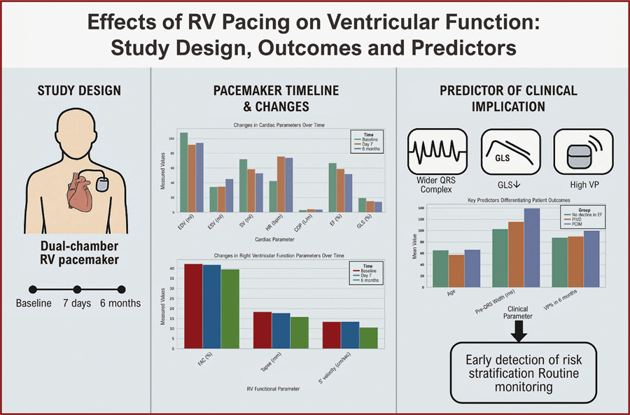
Out of 75 patients, 20 experienced LV systolic dysfunction, 15 developed PIVD, and five developed PICM. In comparison to the control group, patients with PICM had lower GLS (17.02 vs. 19.27; p<0.05), wider QRS width (139.57 vs. 102.75; p < 0.05), and greater ventricular pacing percentage (VP%; 94.32 vs. 84.03; p = 0.06).

Conclusion

RV pacing resulted in a substantial decline in LV systolic function in the short and long term. Concurrently, RV functions declined over the long term (six months). Diminished GLS at seven days, a broader pre-implantation QRS complex, and an elevation in VP% are early indicators of PIVD and PICM. Routine patient monitoring for these indicators can be beneficial for timely intervention on a case-by-case basis.
  • Artificial Pacemaker
  • Cardiomyopathies
  • Heart Failure
  • Three-Dimensional Echocardiography
Read Full Article
Creative Commons Attribution (CC BY) Creative Commons license
18/09/2026 DOI: 10.36660/abc.20250566i Views 1
Mitral Valve Leaflet Hypoplasia in Adults: Role of Cardiovascular Imaging
ABCIC
Review Article

Mitral Valve Leaflet Hypoplasia in Adults: Role of Cardiovascular Imaging

Fábio Luis de Jesus Soares

Abstract

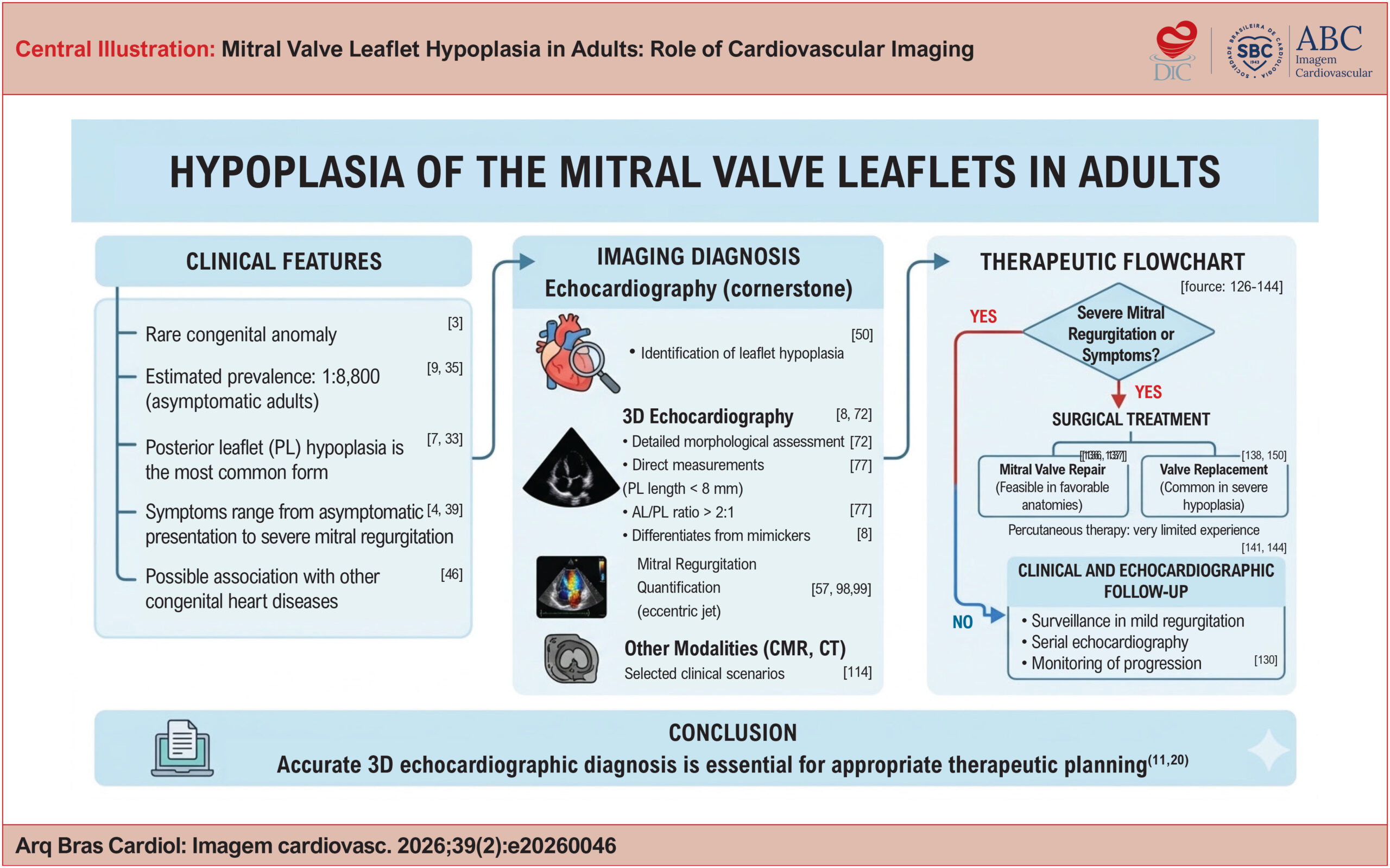
Mitral valve leaflet hypoplasia is a rare congenital anomaly, traditionally described in childhood but increasingly recognized in adults, often as an incidental finding or during the evaluation of mitral regurgitation. Clinical presentation is heterogeneous and depends on leaflet anatomy, the subvalvular apparatus, and the severity of regurgitation. In this narrative literature review, including case reports, case series, and review articles from nationally and internationally recognized journals, epidemiological and clinical aspects are discussed, with particular emphasis on echocardiographic findings. Posterior leaflet hypoplasia is the most common form and may be partial or complete. Three-dimensional echocardiography plays a central role in anatomical assessment, enabling direct measurements of leaflet area and length and helping differentiate true hypoplasia from mimicking entities such as mitral cleft, functional restriction, or subvalvular abnormalities. The estimated prevalence in asymptomatic adults is approximately 1:8,800. Therapeutic management is primarily determined by the severity of mitral regurgitation, with valve repair being feasible only in selected anatomical scenarios. Therefore, refined anatomical understanding, particularly through three-dimensional echocardiography, is essential for accurate diagnosis and appropriate therapeutic planning in this rare yet clinically relevant condition.

  • Congenital Abnormalities
  • Mitral Valve
  • Three-Dimensional Echocardiography
Read Full Article
Creative Commons Attribution (CC BY) Creative Commons license
20/07/2026 DOI: 10.36660/abcimg.20260046i Views 2
Three-Dimensional Echocardiographic Assessment of the Right Ventricle: Why Should We Use It
ABCIC
Review Article

Three-Dimensional Echocardiographic Assessment of the Right Ventricle: Why Should We Use It

Tiago R. Politi et al. Rodrigo B.M. Barretto João Cesar Nunes Sbano David Costa de Souza Le Bihan Wilson Mathias Jr.

Abstract

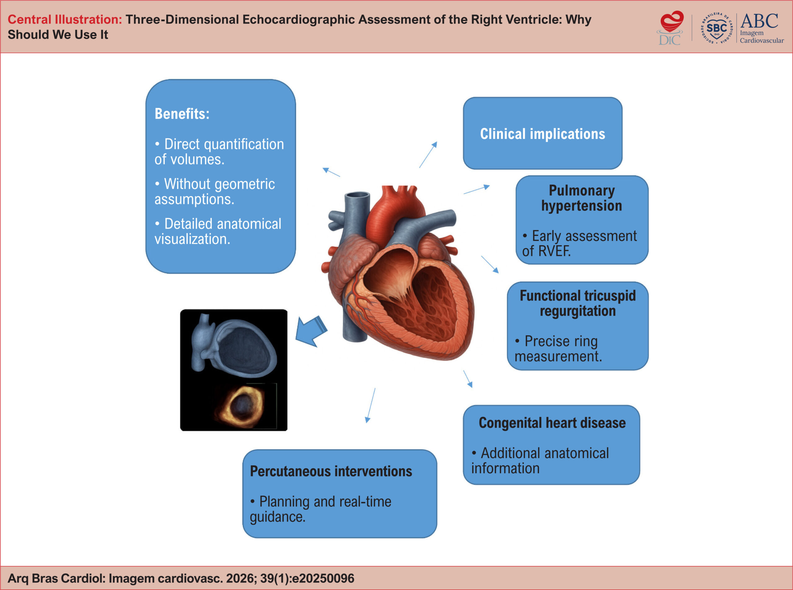
Right ventricular (RV) assessment using two-dimensional (2D) echocardiography has historically faced significant challenges due to the chamber’s complex and unique geometry and its thoracic orientation. In this context, three-dimensional (3D) echocardiography has emerged as a promising tool to overcome and illuminate these limitations, enabling accurate quantification of volumes and ejection fraction without relying on geometric assumptions. As a result, the routine incorporation of 3D echocardiography into RV evaluation may redefine diagnostic and prognostic paradigms, fostering a more precise and personalized approach in modern cardiology. To consolidate and highlight this technique, this review article explores the technical principles of 3D echocardiography for RV assessment, discusses its advantages over conventional 2D imaging, examines its validation against cardiac magnetic resonance (CMR), and reviews key clinical applications, including pulmonary hypertension, functional tricuspid regurgitation, congenital heart disease, and right-sided heart failure. Additionally, the article outlines current limitations of the technique, future perspectives, and practical recommendations based on contemporary literature.

  • Cardiac Magnetic Resonance Imaging
  • Ejection Fraction
  • Right ventricle
  • Three-Dimensional Echocardiography
Read Full Article
Creative Commons Attribution (CC BY) Creative Commons license
10/04/2026 DOI: 10.36660/abcimg.20250096i Views 1
Central Illustration
ABCIC
Review Article

Tricuspid Valve: Anatomical Basis and Patient Selection Criteria for Transcatheter Structural Interventions

Alexandre Costa Souza et al. Halsted Alarcão Gomes Pereira da Silva Alex dos Santos Felix

Abstract

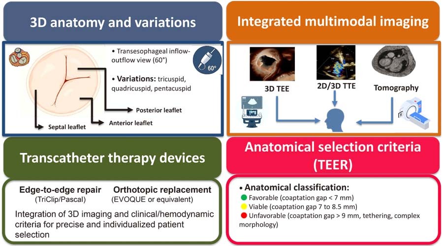
Tricuspid regurgitation has been recognized as a relevant clinical entity, with a direct impact on morbidity and mortality. Recent advances in multimodal cardiovascular imaging have promoted more detailed anatomical and functional assessment of the tricuspid apparatus, guiding the indication and planning of transcatheter interventions. To provide a review of the anatomical basis of the tricuspid valve and discuss echocardiographic and clinical criteria for selecting candidates for transcatheter therapies. This narrative review of the anatomical variability of the tricuspid valve and its subvalvular apparatus incorporates recent recommendations from echocardiography societies and clinical evidence. Special emphasis has been given to three-dimensional echocardiography, transesophageal echocardiography, and computed tomography for morphological and functional characterization of tricuspid regurgitation. Contemporary evidence has demonstrated that up to 45% of patients present with non-classical morphologies (quadricuspid or pentacuspid valves), which impact the viability and outcomes of edge-to-edge repair. The integration of three-dimensional echocardiography enhances accuracy in grading regurgitation severity, enables precise measurement of the vena contracta area, and assists in device selection. Clinical factors including right ventricular function, pulmonary artery systolic pressure, and the presence of intracardiac devices also influence patient selection. Appropriate selection of candidates for transcatheter tricuspid valve therapies requires a systematic approach with integrated application of imaging methods and clinical parameters. Detailed echocardiographic characterization of leaflet morphology, coaptation gap, and annulus geometry is crucial for planning the procedure, selecting the device, and predicting outcomes.

  • Three-Dimensional Echocardiography
  • Transesophageal echocardiography
  • Tricuspid Valve
  • Tricuspid Valve Insufficiency
Read Full Article
Creative Commons Attribution (CC BY) Creative Commons license
16/12/2025 DOI: 10.36660/abcimg.20250064i Views 2
Figure 1
ABCIC
Review Article

My Approach to Three-Dimensional Echocardiography in Mitral Stenosis: How and When?

Alexandre Costa Souza et al. Marcus Vinicius Silva Freire de Carvalho Marco André Moraes Sales

Abstract

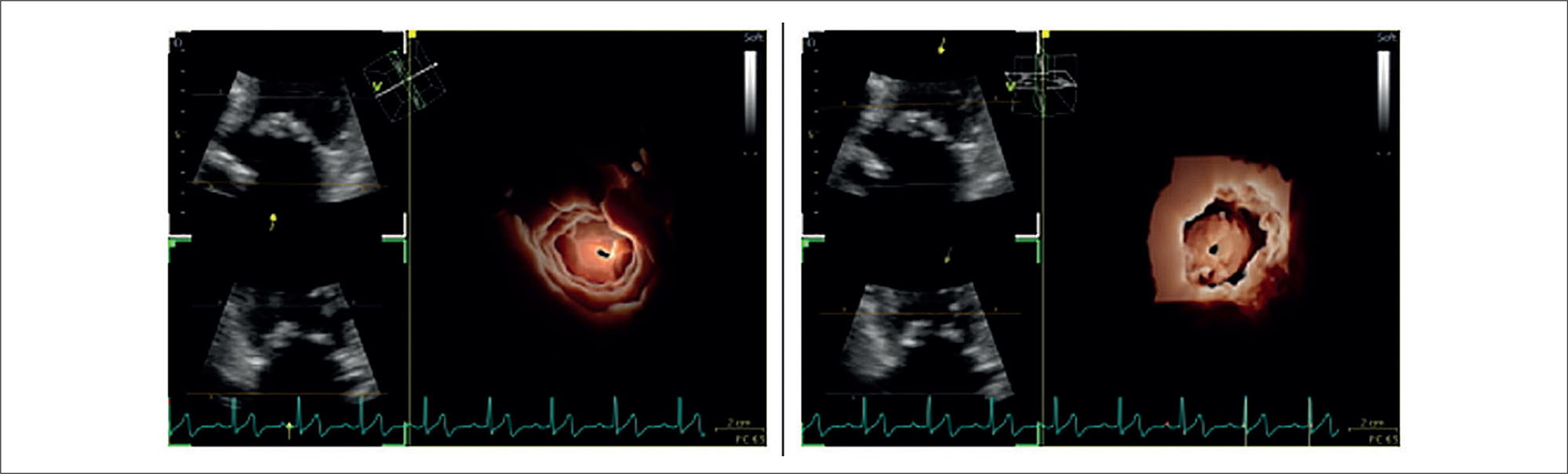
Three-dimensional echocardiography has become an essential tool in the assessment of mitral stenosis, allowing detailed anatomical analyses and reliable measurements, even in complex anatomies or extensive calcification. Based on daily clinical experience in an imaging laboratory, this article offers a practical guide for image acquisition and optimization, multiplanar reconstruction, and three-dimensional planimetry, focusing on technical adjustments, the use of tools such as rotation and cropping, and strategies for integrating the method into the routine workflow of cardiovascular imaging centers.

  • Mitral Valve Stenosis
  • Three-Dimensional Echocardiography
  • Three-Dimensional Imaging
Read Full Article
Creative Commons Attribution (CC BY) Creative Commons license
14/11/2025 DOI: 10.36660/abcimg.20250057i Views 2
Figure 1
ABCIC
Review Article

My Approach to Calculate the Left Ventricular Ejection Fraction: From a Two-Dimensional to a Three-Dimensional Technique

Lucas Arraes de França et al. Danilo Bora Moleta

Abstract

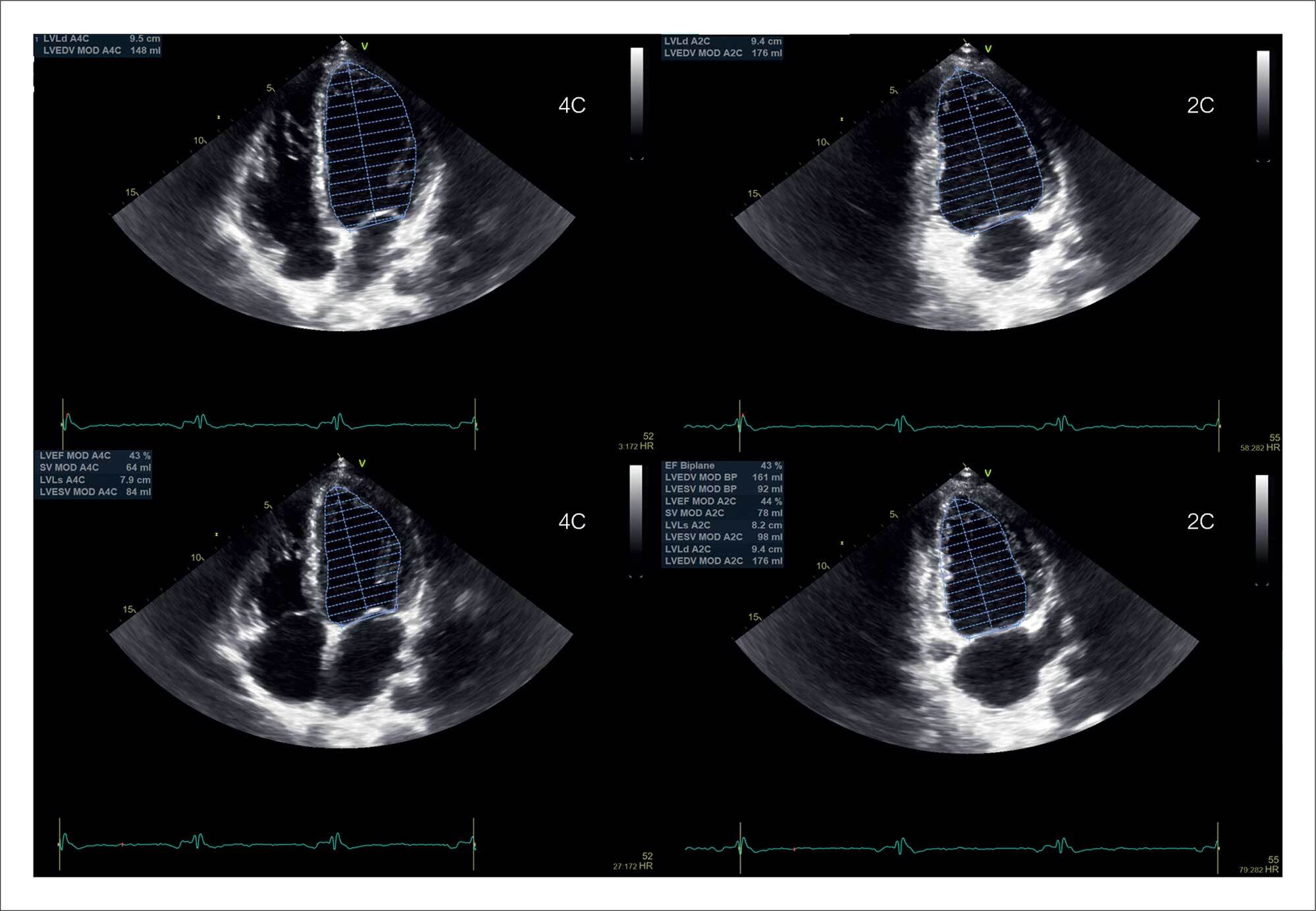
Echocardiography is the most used diagnostic method for measuring left ventricular systolic function indices. It is possible to estimate, in a quick, available, and non-invasive way, parameters such as ventricular volumes, stroke volume, and ejection fraction. Being an expert in the technique is quite important to estimate these parameters accurately. Being able to refine the technique constantly is also important, and this is closely associated with the examiner’s experience and knowledge of the current software solutions.

In this review article, we will discuss how to accurately measure left ventricular volumes and their ejection fraction using two-dimensional (2D) and three-dimensional (3D) echocardiographic techniques. We will also outline tips and suggestions based on the main North American and European guidelines for quantifying heart chambers and the most recent articles in the literature.

  • Echocardiography
  • Stroke Volume
  • Three-Dimensional Echocardiography
Read Full Article
Creative Commons Attribution (CC BY) Creative Commons license
29/08/2024 DOI: 10.36660/abcimg.20240059i
ABCIC
Review Article

Cardiac Tumors: Recognition Is Essential!

Eliza de Almeida Gripp et al. Bruna Zanforlin Jácome Anna Esther Araujo e Silva Fernanda Baptista Ribeiro Dias … Bruna Barbosa Chagas de Almeida Arnaldo Rabischoffsky

Abstract

Although cardiac tumors are rare and often found accidentally, they can be associated with constitutional, cardiac, or embolic symptoms and, thus, must always be considered in diagnostic hypotheses. Three-dimensional Doppler echocardiography and contrast enhancement are additional diagnostic resources and can help determine the most appropriate surgical approach, when necessary.

  • Echocardiography
  • Heart Neoplasms
  • Three-Dimensional Echocardiography
Read Full Article
Creative Commons Attribution (CC BY) Creative Commons license
26/06/2024 DOI: 10.36660/abcimg.20240032i Views 1
Central Illustration
ABCIC
Review Article

My Approach to 3D Echocardiography in Mitral Valve Insufficiency: How and When

Fábio Cañellas

Abstract

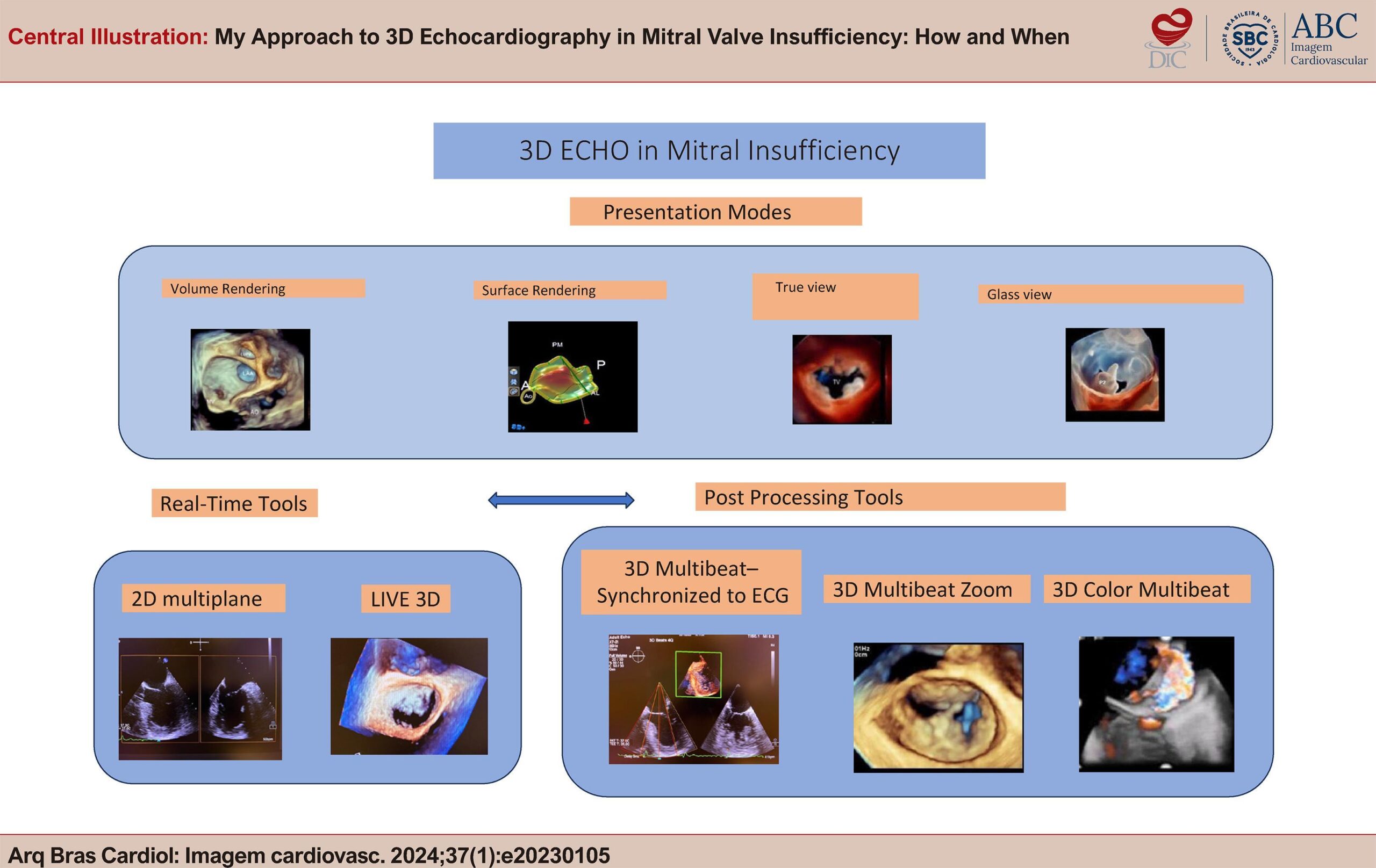
3D Echocardiogram is an indispensable tool in all stages of mitral valve disease, from its diagnosis to the moment of treatment, playing a fundamental role mainly in the decision for surgical plasty or percutaneous correction. In this article, we sought to present very important basic concepts in the use of the technique, from obtaining a 2D image, through the main tools for obtaining 3D images, to new solutions brought by manufacturers for presenting images and for post-processing software. This knowledge is fundamental and contributes to the demystification of 3D Echo, often labeled as difficult to perform and still little incorporated into our clinical practice.

  • Mitral Valve Insufficiency Diagnosis
  • Three-Dimensional Echocardiography
Read Full Article
Creative Commons Attribution (CC BY) Creative Commons license
28/02/2024 DOI: 10.36660/abcimg.20230105i
Dynamic three-dimensional reconstruction of the heart by transesophageal echocardiography
ABC
Original Article

Dynamic three-dimensional reconstruction of the heart by transesophageal echocardiography

Maria de Fátima Veiga et al. Mário G. Lopes Fausto J. Pinto

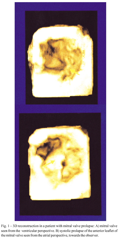
OBJECTIVE: To evaluate echocardiography accuracy in performing and obtaining images for dynamical three-dimensional (3D) reconstruction. METHODS: Three-dimensional (3D) image reconstruction was obtained in 20 consecutive patients who underwent transesophageal echocardiography. A multiplanar 5 MHz transducer was used for 3D reconstruction. RESULTS: Twenty patients were studied consecutively. The following cardiac diseases were present: valvar prostheses–6 (2 mitral, 2 aortic and 2 mitral and aortic); mitral valve prolapse– 3; mitral and aortic disease – 2; aortic valve disease– 5; congenital heart disease– 3 (2 atrial septal defect– ASD – and 1 transposition of the great arteries -TGA); arteriovenous fistula– 1. In 7 patients, color Doppler was also obtained and used for 3D flow reconstruction. Twenty five cardiac structures were acquired and 60 reconstructions generated (28 of mitral valves, 14 of aortic valves, 4 of mitral prostheses, 7 of aortic prostheses and 7 of the ASD). Fifty five of 60 (91.6%) reconstructions were considered of good quality by 2 independent observers. The 11 reconstructed mitral valves/prostheses and the 2 reconstructed ASDs provided more anatomical information than two dimensional echocardiography (2DE) alone. CONCLUSION: 3D echocardiography using a transesophageal transducer is a feasible technique, which improves detection of anatomical details of cardiac structures, particularly of the mitral valve and atrial septum.

  • Three-Dimensional Echocardiography
  • Ultrasonography
  • valvar disease
Read Full Article
Creative Commons Attribution (CC BY) Creative Commons license
27/10/2000 DOI: 10.1590/S0066-782X1999000500003 Views 1

How to cite this article

EndNote RIS

Electronic Document Format (Vancouver)

Kumar S, Meena CB, Babbar N, Bhandari A, Shekhawat DS, Meel A. Effects of Pacemakers on Ventricular Functions and Early Variables Associated with Induced Cardiomyopathy. Arquivos Brasileiros de Cardiologia 2026;123(7):e20250566.

Electronic Document Format (ABNT)

Kumar, Sanjeev; Meena, Chandra Bhan; Babbar, Navida; Bhandari, Anil; Shekhawat, Dhananjay Singh; Meel, Ankita. Effects of Pacemakers on Ventricular Functions and Early Variables Associated with Induced Cardiomyopathy. Arquivos Brasileiros de Cardiologia, v. 123, n. 7, e20250566, Sep. 2026.

Electronic Document Format (APA)

Kumar, S., Meena, C. B., Babbar, N., Bhandari, A., Shekhawat, D. S., & Meel, A. (2026). Effects of Pacemakers on Ventricular Functions and Early Variables Associated with Induced Cardiomyopathy. Arquivos Brasileiros de Cardiologia, 123(7), e20250566.

Electronic Document Format (ISO)

Kumar, Sanjeev and Meena, Chandra Bhan and Babbar, Navida and Bhandari, Anil and Shekhawat, Dhananjay Singh and Meel, Ankita. Effects of Pacemakers on Ventricular Functions and Early Variables Associated with Induced Cardiomyopathy. Arquivos Brasileiros de Cardiologia [online]. 2026, vol. 123, n. 7, [cited 2026-10-14], e20250566. Available from: <https://journals.cardiol.br/en/article/effects-of-pacemakers-on-ventricular-functions-and-early-variables-associated-with-induced-cardiomyopathy/>. ISSN 0066-782X

EndNote RIS

Electronic Document Format (Vancouver)

Soares FLJ. Mitral Valve Leaflet Hypoplasia in Adults: Role of Cardiovascular Imaging. ABC Imagem Cardiovascular 2026;39(2):e20260046.

Electronic Document Format (ABNT)

Soares, Fábio Luis de Jesus. Mitral Valve Leaflet Hypoplasia in Adults: Role of Cardiovascular Imaging. ABC Imagem Cardiovascular, v. 39, n. 2, e20260046, Jul. 2026.

Electronic Document Format (APA)

Soares, F. L. J. (2026). Mitral Valve Leaflet Hypoplasia in Adults: Role of Cardiovascular Imaging. ABC Imagem Cardiovascular, 39(2), e20260046.

Electronic Document Format (ISO)

Soares, Fábio Luis de Jesus. Mitral Valve Leaflet Hypoplasia in Adults: Role of Cardiovascular Imaging. ABC Imagem Cardiovascular [online]. 2026, vol. 39, n. 2, [cited 2026-10-14], e20260046. Available from: <https://journals.cardiol.br/en/article/mitral-valve-leaflet-hypoplasia-in-adults-role-of-cardiovascular-imaging/>. ISSN 2318-8219

EndNote RIS

Electronic Document Format (Vancouver)

Politi TR, Barretto RB, Sbano JCN, Bihan DCSL, Mathias Jr. W. Three-Dimensional Echocardiographic Assessment of the Right Ventricle: Why Should We Use It. ABC Imagem Cardiovascular 2026;39(1):e20250096.

Electronic Document Format (ABNT)

Politi, Tiago R.; Barretto, Rodrigo B.M.; Sbano, João Cesar Nunes; Bihan, David Costa de Souza Le; Mathias Jr., Wilson. Three-Dimensional Echocardiographic Assessment of the Right Ventricle: Why Should We Use It. ABC Imagem Cardiovascular, v. 39, n. 1, e20250096, Apr. 2026.

Electronic Document Format (APA)

Politi, T. R., Barretto, R. B., Sbano, J. C. N., Bihan, D. C. S. L., & Mathias Jr., W. (2026). Three-Dimensional Echocardiographic Assessment of the Right Ventricle: Why Should We Use It. ABC Imagem Cardiovascular, 39(1), e20250096.

Electronic Document Format (ISO)

Politi, Tiago R. and Barretto, Rodrigo B.M. and Sbano, João Cesar Nunes and Bihan, David Costa de Souza Le and Mathias Jr., Wilson. Three-Dimensional Echocardiographic Assessment of the Right Ventricle: Why Should We Use It. ABC Imagem Cardiovascular [online]. 2026, vol. 39, n. 1, [cited 2026-10-14], e20250096. Available from: <https://journals.cardiol.br/en/article/three-dimensional-echocardiographic-assessment-of-the-right-ventricle-why-should-we-use-it/>. ISSN 2318-8219

EndNote RIS

Electronic Document Format (Vancouver)

Souza AC, Silva HAGP, Felix AS. Tricuspid Valve: Anatomical Basis and Patient Selection Criteria for Transcatheter Structural Interventions. ABC Imagem Cardiovascular 2025;38(3):e20250064.

Electronic Document Format (ABNT)

Souza, Alexandre Costa; Silva, Halsted Alarcão Gomes Pereira da; Felix, Alex dos Santos. Tricuspid Valve: Anatomical Basis and Patient Selection Criteria for Transcatheter Structural Interventions. ABC Imagem Cardiovascular, v. 38, n. 3, e20250064, Dec. 2025.

Electronic Document Format (APA)

Souza, A. C., Silva, H. A. G. P. , & Felix, A. S. (2025). Tricuspid Valve: Anatomical Basis and Patient Selection Criteria for Transcatheter Structural Interventions. ABC Imagem Cardiovascular, 38(3), e20250064.

Electronic Document Format (ISO)

Souza, Alexandre Costa and Silva, Halsted Alarcão Gomes Pereira da and Felix, Alex dos Santos. Tricuspid Valve: Anatomical Basis and Patient Selection Criteria for Transcatheter Structural Interventions. ABC Imagem Cardiovascular [online]. 2025, vol. 38, n. 3, [cited 2026-10-14], e20250064. Available from: <https://journals.cardiol.br/en/article/tricuspid-valve-anatomical-basis-and-patient-selection-criteria-for-transcatheter-structural-interventions/>. ISSN 2318-8219

EndNote RIS

Electronic Document Format (Vancouver)

Souza AC, Carvalho MVSF, Sales MAM. My Approach to Three-Dimensional Echocardiography in Mitral Stenosis: How and When?. ABC Imagem Cardiovascular 2025;38(3):e20250057.

Electronic Document Format (ABNT)

Souza, Alexandre Costa; Carvalho, Marcus Vinicius Silva Freire de; Sales, Marco André Moraes. My Approach to Three-Dimensional Echocardiography in Mitral Stenosis: How and When?. ABC Imagem Cardiovascular, v. 38, n. 3, e20250057, Nov. 2025.

Electronic Document Format (APA)

Souza, A. C., Carvalho, M. V. S. F. , & Sales, M. A. M. (2025). My Approach to Three-Dimensional Echocardiography in Mitral Stenosis: How and When?. ABC Imagem Cardiovascular, 38(3), e20250057.

Electronic Document Format (ISO)

Souza, Alexandre Costa and Carvalho, Marcus Vinicius Silva Freire de and Sales, Marco André Moraes. My Approach to Three-Dimensional Echocardiography in Mitral Stenosis: How and When?. ABC Imagem Cardiovascular [online]. 2025, vol. 38, n. 3, [cited 2026-10-14], e20250057. Available from: <https://journals.cardiol.br/en/article/my-approach-to-three-dimensional-echocardiography-in-mitral-stenosis-how-and-when/>. ISSN 2318-8219

EndNote RIS

Electronic Document Format (Vancouver)

França LA, Moleta DB. My Approach to Calculate the Left Ventricular Ejection Fraction: From a Two-Dimensional to a Three-Dimensional Technique. ABC Imagem Cardiovascular 2024;37(3):e20240059.

Electronic Document Format (ABNT)

França, Lucas Arraes de; Moleta, Danilo Bora. My Approach to Calculate the Left Ventricular Ejection Fraction: From a Two-Dimensional to a Three-Dimensional Technique. ABC Imagem Cardiovascular, v. 37, n. 3, e20240059, Aug. 2024.

Electronic Document Format (APA)

França, L. A. , & Moleta, D. B. (2024). My Approach to Calculate the Left Ventricular Ejection Fraction: From a Two-Dimensional to a Three-Dimensional Technique. ABC Imagem Cardiovascular, 37(3), e20240059.

Electronic Document Format (ISO)

França, Lucas Arraes de and Moleta, Danilo Bora. My Approach to Calculate the Left Ventricular Ejection Fraction: From a Two-Dimensional to a Three-Dimensional Technique. ABC Imagem Cardiovascular [online]. 2024, vol. 37, n. 3, [cited 2026-10-14], e20240059. Available from: <https://journals.cardiol.br/en/article/my-approach-to-calculate-the-left-ventricular-ejection-fraction-from-a-two-dimensional-to-a-three-dimensional-technique/>. ISSN 2318-8219

EndNote RIS

Electronic Document Format (Vancouver)

Gripp EA, Jácome BZ, Silva AEA, Dias FBR, Almeida BBC, Rabischoffsky A. Cardiac Tumors: Recognition Is Essential!. ABC Imagem Cardiovascular 2024;37(2):e20240032.

Electronic Document Format (ABNT)

Gripp, Eliza de Almeida; Jácome, Bruna Zanforlin; Silva, Anna Esther Araujo e; Dias, Fernanda Baptista Ribeiro; Almeida, Bruna Barbosa Chagas de; Rabischoffsky, Arnaldo. Cardiac Tumors: Recognition Is Essential!. ABC Imagem Cardiovascular, v. 37, n. 2, e20240032, Jun. 2024.

Electronic Document Format (APA)

Gripp, E. A., Jácome, B. Z., Silva, A. E. A. , Dias, F. B. R., Almeida, B. B. C. , & Rabischoffsky, A. (2024). Cardiac Tumors: Recognition Is Essential!. ABC Imagem Cardiovascular, 37(2), e20240032.

Electronic Document Format (ISO)

Gripp, Eliza de Almeida and Jácome, Bruna Zanforlin and Silva, Anna Esther Araujo e and Dias, Fernanda Baptista Ribeiro and Almeida, Bruna Barbosa Chagas de and Rabischoffsky, Arnaldo. Cardiac Tumors: Recognition Is Essential!. ABC Imagem Cardiovascular [online]. 2024, vol. 37, n. 2, [cited 2026-10-14], e20240032. Available from: <https://journals.cardiol.br/en/article/cardiac-tumors-recognition-is-essential/>. ISSN 2318-8219

EndNote RIS

Electronic Document Format (Vancouver)

Cañellas F. My Approach to 3D Echocardiography in Mitral Valve Insufficiency: How and When. ABC Imagem Cardiovascular 2024;37(1):e20230105.

Electronic Document Format (ABNT)

Cañellas, Fábio. My Approach to 3D Echocardiography in Mitral Valve Insufficiency: How and When. ABC Imagem Cardiovascular, v. 37, n. 1, e20230105, Feb. 2024.

Electronic Document Format (APA)

Cañellas, F. (2024). My Approach to 3D Echocardiography in Mitral Valve Insufficiency: How and When. ABC Imagem Cardiovascular, 37(1), e20230105.

Electronic Document Format (ISO)

Cañellas, Fábio. My Approach to 3D Echocardiography in Mitral Valve Insufficiency: How and When. ABC Imagem Cardiovascular [online]. 2024, vol. 37, n. 1, [cited 2026-10-14], e20230105. Available from: <https://journals.cardiol.br/en/article/my-approach-to-3d-echocardiography-in-mitral-valve-insufficiency-how-and-when/>. ISSN 2318-8219

EndNote RIS

Electronic Document Format (Vancouver)

Veiga MF, Lopes MG, Pinto FJ. Dynamic three-dimensional reconstruction of the heart by transesophageal echocardiography. Arquivos Brasileiros de Cardiologia 2000;72(5):564-8.

Electronic Document Format (ABNT)

Veiga, Maria de Fátima; Lopes, Mário G.; Pinto, Fausto J.. Dynamic three-dimensional reconstruction of the heart by transesophageal echocardiography. Arquivos Brasileiros de Cardiologia, v. 72, n. 5, p. 564-568, Oct. 2000.

Electronic Document Format (APA)

Veiga, M. F., Lopes, M. G., & Pinto, F. J. (2000). Dynamic three-dimensional reconstruction of the heart by transesophageal echocardiography. Arquivos Brasileiros de Cardiologia, 72(5), 564-568.

Electronic Document Format (ISO)

Veiga, Maria de Fátima and Lopes, Mário G. and Pinto, Fausto J.. Dynamic three-dimensional reconstruction of the heart by transesophageal echocardiography. Arquivos Brasileiros de Cardiologia [online]. 2000, vol. 72, n. 5, [cited 2026-10-14], pp. 564-568. Available from: <https://journals.cardiol.br/en/article/dynamic-three-dimensional-reconstruction-of-the-heart-by-transesophageal-echocardiography/>. ISSN 0066-782X

  • About ABC Family
  • Internal Regulation
  • Editorial Board
  • Brazilian Society of Cardiology (SBC)

Issues

SBC Guidelines

  • Videos
  • Podcasts
  • News