ABC Family Arquivos Brasileiros de Cardiologia International Journal of Cardiovascular Sciences ABC Heart Failure & Cardiomyopathy ABC Imagem Cardiovascular
Português English
ABC Family
Português English
  • About
    • About ABC Family
    • Internal Regulation
    • Editorial Board
    • Brazilian Society of Cardiology (SBC)
  • Issues
  • SBC Guidelines
  • Content
    • Videos
    • Podcasts
    • News
  • About
    • About ABC Family
    • Internal Regulation
    • Editorial Board
    • Brazilian Society of Cardiology (SBC)
  • Issues
  • SBC Guidelines
  • Content
    • Videos
    • Podcasts
    • News

ABC Family

  • ABC Family
  • Arquivos Brasileiros de Cardiologia
  • International Journal of Cardiovascular Sciences
  • ABC Heart Failure & Cardiomyopathy
  • ABC Imagem Cardiovascular
Search in

No content found.

Journals
Ordering
Article types
Others

  1. Home
  2. Keyword: Ejection Fraction
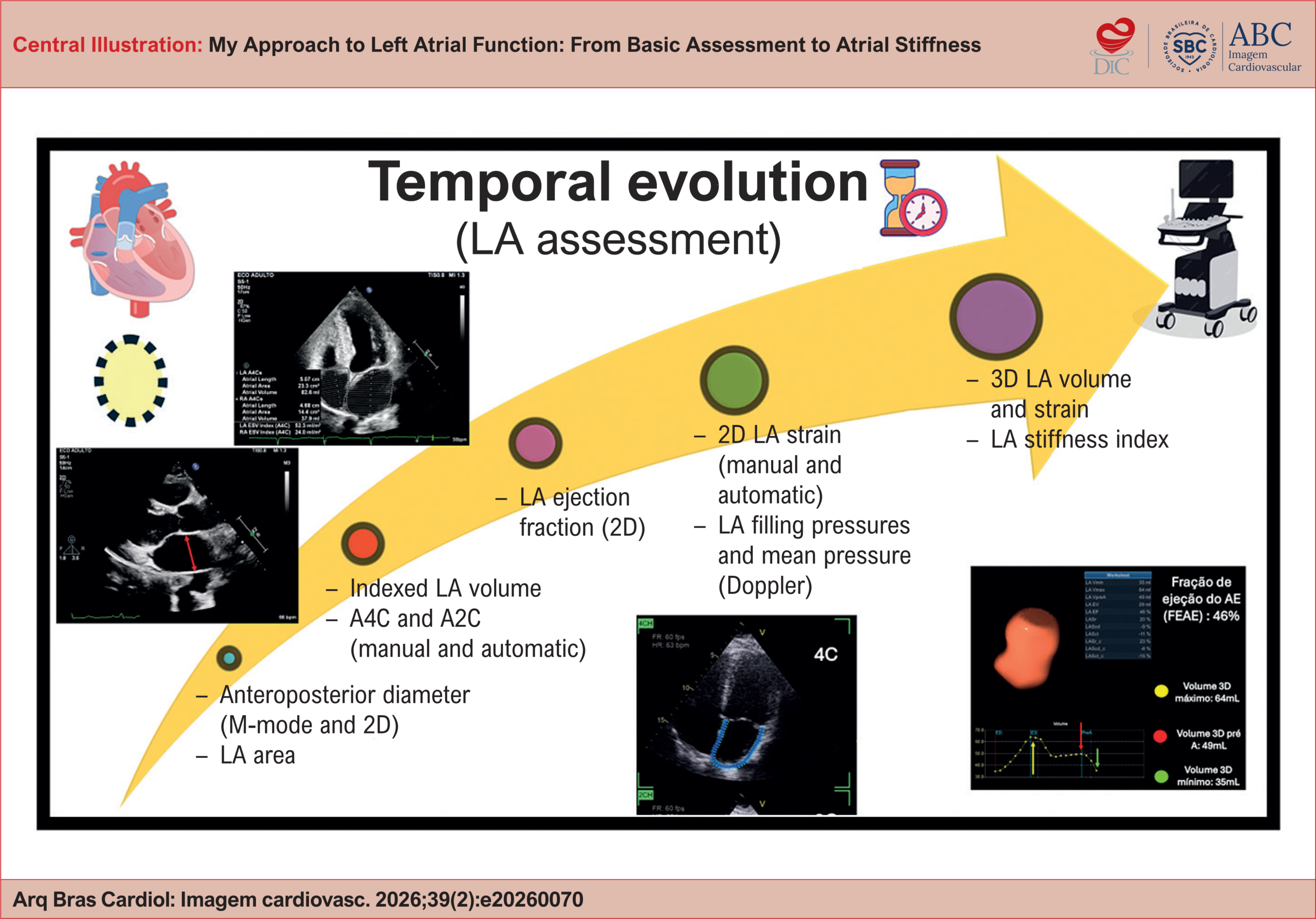
My Approach to Left Atrial Function: From Basic Assessment to Atrial Stiffness
ABCIC
Review Article

My Approach to Left Atrial Function: From Basic Assessment to Atrial Stiffness

Halsted Alarcão Gomes Pereira da Silva et al. Helder Moura Gomes Alexandre Costa Souza

Abstract

The left atrium (LA) has historically been considered a passive chamber that conducts blood flow between the pulmonary veins and the left ventricle (LV). Advances in cardiovascular physiology and imaging techniques have highlighted its active role in modulating cardiac output, ventricular diastolic function, and the stratification of various heart diseases. Assessment of the LA has evolved over the years into a multiparametric functional approach, consolidated as an essential component of modern echocardiography. The systematic incorporation of volumetric parameters, atrial strain, and atrial stiffness allows for a more precise characterization of cardiovascular pathophysiology and improves prognostic stratification, thus supporting more informed clinical decisions.

  • Ejection Fraction
  • Strain, Left atrium
Read Full Article
Creative Commons Attribution (CC BY) Creative Commons license
27/07/2026 DOI: 10.36660/abcimg.20260070i
Three-Dimensional Echocardiographic Assessment of the Right Ventricle: Why Should We Use It
ABCIC
Review Article

Three-Dimensional Echocardiographic Assessment of the Right Ventricle: Why Should We Use It

Tiago R. Politi et al. Rodrigo B.M. Barretto João Cesar Nunes Sbano David Costa de Souza Le Bihan Wilson Mathias Jr.

Abstract

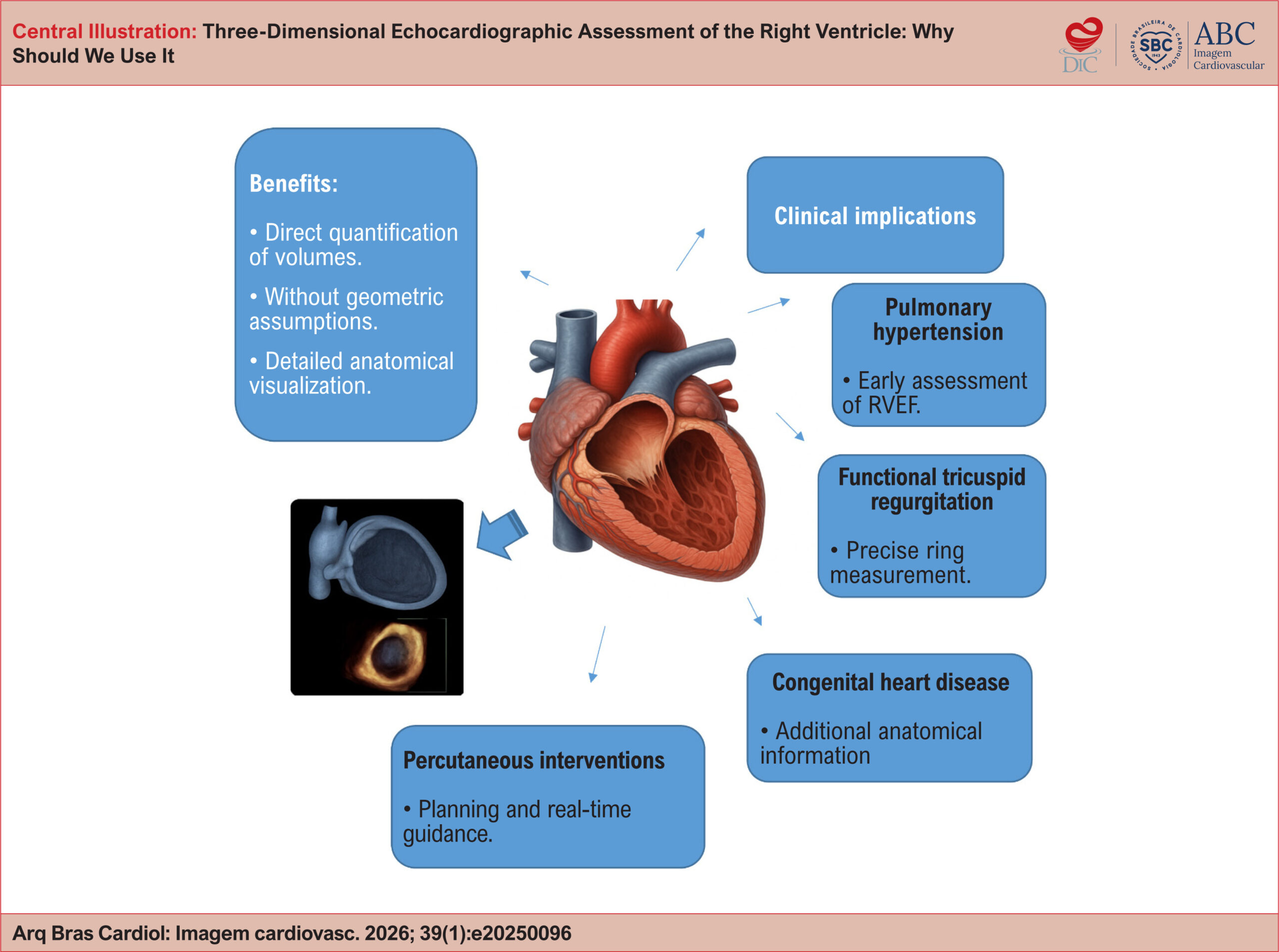
Right ventricular (RV) assessment using two-dimensional (2D) echocardiography has historically faced significant challenges due to the chamber’s complex and unique geometry and its thoracic orientation. In this context, three-dimensional (3D) echocardiography has emerged as a promising tool to overcome and illuminate these limitations, enabling accurate quantification of volumes and ejection fraction without relying on geometric assumptions. As a result, the routine incorporation of 3D echocardiography into RV evaluation may redefine diagnostic and prognostic paradigms, fostering a more precise and personalized approach in modern cardiology. To consolidate and highlight this technique, this review article explores the technical principles of 3D echocardiography for RV assessment, discusses its advantages over conventional 2D imaging, examines its validation against cardiac magnetic resonance (CMR), and reviews key clinical applications, including pulmonary hypertension, functional tricuspid regurgitation, congenital heart disease, and right-sided heart failure. Additionally, the article outlines current limitations of the technique, future perspectives, and practical recommendations based on contemporary literature.

  • Cardiac Magnetic Resonance Imaging
  • Ejection Fraction
  • Right ventricle
  • Three-Dimensional Echocardiography
Read Full Article
Creative Commons Attribution (CC BY) Creative Commons license
10/04/2026 DOI: 10.36660/abcimg.20250096i Views 1
Central Illustration
ABC
Original Article

Relationship between BUN/Cr and Prognosis of HF Across the Full Spectrum of Ejection Fraction

Yuan Kang et al. Conglin Wang Xiaojing Niu Zhijing Shi … Mingxue Li Jianli Tian

Abstract

Background

In patients with heart failure (HF), due to the relative deficiency of blood volume, neurohormone system activation leads to renal vasoconstriction, which affects the content of blood urea nitrogen (BUN) and creatinine (Cr) in the body, while BUN and Cr are easily affected by other factors. Therefore, BUN/Cr can be used as another marker for the prognosis of HF.

Objective

Explore the prognosis of adverse outcome of HF in the high BUN/Cr group compared with the low BUN/Cr group across the full spectrum of ejection fraction.

Methods

From 2014 to 2016, symptomatic hospitalized HF patients were recruited and followed up to observe adverse cardiovascular outcomes. Logistic analysis and COX analysis were performed to determine significance. p-values <0.05 were considered statistically significant.

Results

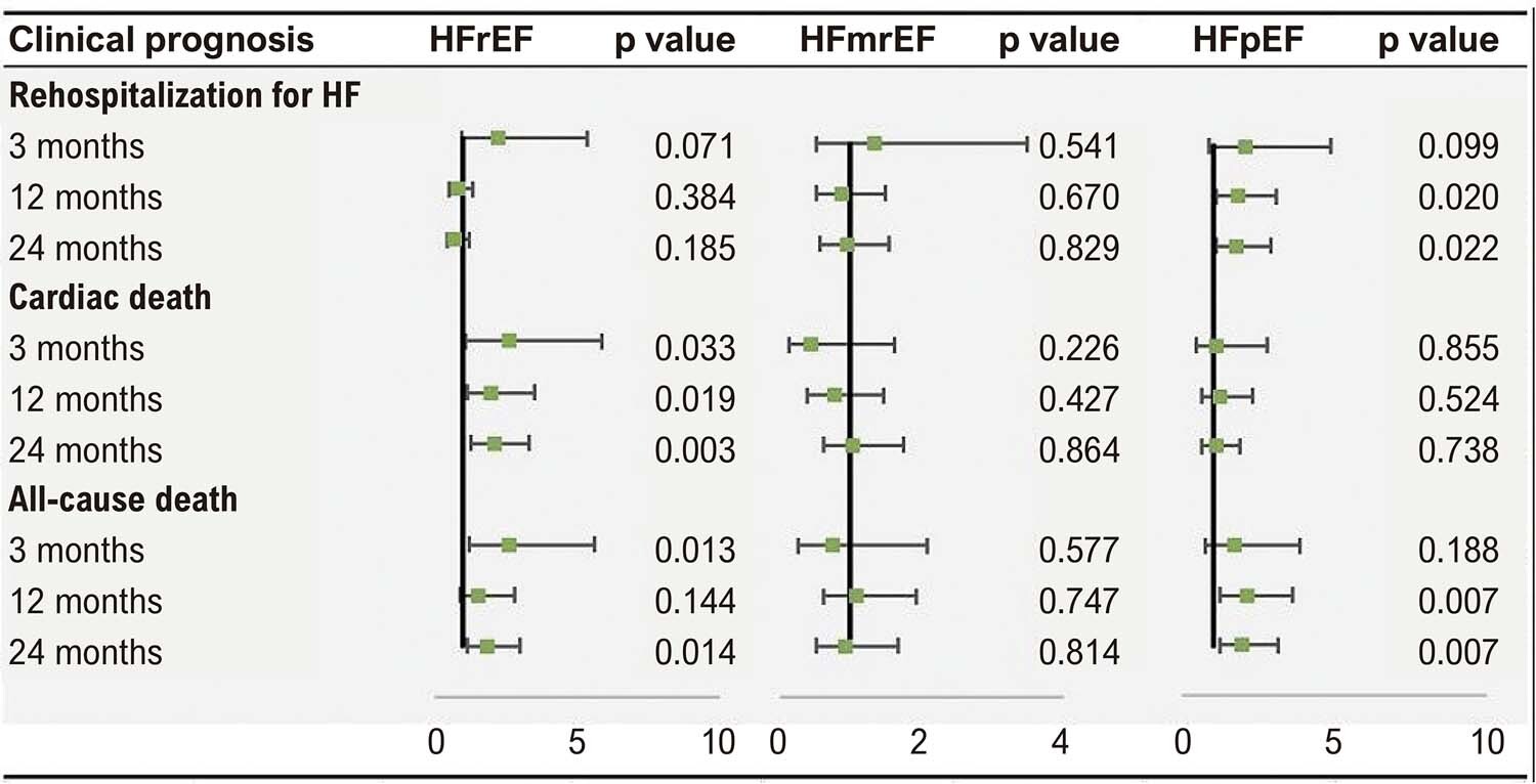
In the univariate logistic regression analysis, the high BUN/Cr group had a higher risk of adverse outcome in heart failure with reduced ejection fraction (HFrEF) and heart failure with preserved ejection fraction (HFpEF). Multivariate logistic regression analysis showed that the risk of cardiac death in the HFrEF group was higher than that in the low BUN/Cr group, while the risk of all-cause death was significant only in 3 months (p<0.05) (Central Illustration). The risk of all-cause death in the high BUN/Cr in the HFpEF group was significantly higher than that in the low BUN/Cr group at two years.

Conclusion

The high BUN/Cr group is related to the risk of poor prognosis of HFpEF, and is not lower than the predictive value of left ventricular ejection fraction (LVEF).

  • BUN/Cr
  • Ejection Fraction
  • Heart Failure
  • Prognosis
Read Full Article
Creative Commons Attribution (CC BY) Creative Commons license
03/04/2023 DOI: 10.36660/abc.20220427 Views 3
ABCHF
Research Letter

When to Suspect Advanced Heart Failure in Heart Failure with Preserved Ejection Fraction?

Miguel Morita Fernandes-Silva et al. Fabiana G. Marcondes-Braga

Author Contributions

Writing of the manuscript and Critical revision of the manuscript for intellectual content: Fernandes-Silva MM, Marcondes-Braga FG.

[…]

  • Advanced Heart Failure
  • Ejection Fraction
  • Heart Failure
Read Full Article
Creative Commons Attribution (CC BY) Creative Commons license
29/06/2022 DOI: 10.36660/abchf.20220037 Views 1
Figure 1
ABCHF
Viewpoint

SGLT2 Inhibitors and Sacubitril-Valsartan: How Trial Results will Revolutionize the Treatment of Heart Failure with Mildly Reduced Ejection Fraction

Miguel Morita Fernandes-Silva

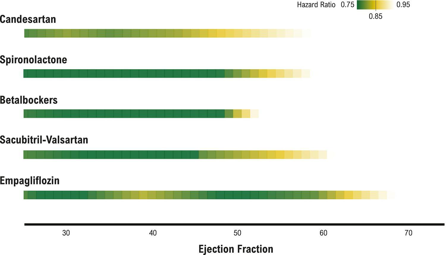
Left ventricular ejection fraction (EF) is a key parameter in the management of heart failure (HF) and a crucial biomarker for prognostic evaluation and therapeutic decision. Patients with HF are classified into different categories according to EF. Although the concept of “normal” EF was introduced four decades ago, the cutoff values for normal and abnormal EF have considerably varied over time. In 2016, the European Society of Cardiology HF guidelines defined HF with reduced EF (HFrEF) as patients with EF below 40%, while HF with preserved EF (HFpEF) was the category for patients with EF equal to 50% or above. To fill in the gap between the two categories, the term HF with mid-range EF was introduced. More recently, the guidelines considered more appropriate to rename this category to HF with mildly reduced EF (HFmrEF). , Why has the EF-based classification changed in recent years and how may this affect HF treatment?

In the 1980s, the first HF trials used in their design EF cutoffs to select patients with worse prognosis as an enrichment strategy, ie, they included patients based on a biomarker that improves design efficiency. Because patients with lower EF have worse prognosis and, therefore, higher rates of events, a relatively smaller sample size would be required to detect an effect. These trials used cutoff values for EF that ranged from < 45% to < 25% ( ) and were highly successful in finding effective therapies. In general, trials using a cutoff EF of < 40% consistently found drugs and devices for the treatment of HF that improved outcomes.

[…]

  • Angiotensin receptor-neprylisin Inhibitor
  • Angiotensin-converting Enzyme Inhibitor
  • Beta-blocker
  • Ejection Fraction
  • Heart Failure
  • Mineralocorticoid Receptor Antagonist
  • Sodium-glucose Cotransporter 2 Inhibitor
Read Full Article
Creative Commons Attribution (CC BY) Creative Commons license
12/04/2022 DOI: 10.36660/abchf.20220013 Views 1
Maximal functional capacity, ejection fraction, and functional class in Chagas cardiomyopathy: are these indices related?
ABC
Artigo Original

Maximal functional capacity, ejection fraction, and functional class in Chagas cardiomyopathy: are these indices related?

Charles Mady et al. Vera Maria Cury Salemi Barbara Maria Ianni Felix José Alvarez Ramires Edmundo Arteaga

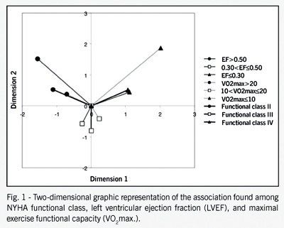
OBJECTIVE: Left ventricular ejection fraction (LVEF) and maximal functional capacity (VO2max) have both been shown to be related to a poor long-term survival in Chagas’ disease patients. The aim of this study was to estimate the potential association of VO2max, LVEF, and NYHA functional class in patients with Chagas’ disease cardiomyopathy. METHODS: One hundred four male patients, aged 40.3±9.0 years (range, 18 to 65), with a definite diagnosis of Chagas disease cardiomyopathy were studied. LVEF and VO2max were both classified into 3 degrees: LVEF <0.30, 0.30< LVEF 0.50 and VO2max <10, 10<VO2max 20 ml.kg-1.min-1, respectively. RESULTS: Thirty-one patients (29.8%) were in NYHA functional class II, 41 (39.4%) in functional class III, and 32 (30.8%) in functional class IV. The corresponding values of VO2max and LVEF for functional classes II, III, and IV were 21.5±4.0 ml.kg-1.min-1, 18.3±5.8 ml.kg-1.min-1, and 14.7±4.9 ml.kg-1.min-1 and 0.50±0.6, 0.35± 0.9, and 0.29±0.7, respectively. LVEF= <0.30 and VO2max = 0.50 as well as VO2max >20 ml.kg-1.min-1. CONCLUSION: A convincingly good association exists between NYHA functional class, functional capacity and LVEF in patients with Chagas’ disease cardiomyopathy. These data may be helpful in the management of heart failure in Chagas’ disease patients.

  • Ejection Fraction
  • functional class in Chagas cardiomyopathy
  • maximal functional capacity
Read Full Article
Creative Commons Attribution (CC BY) Creative Commons license
09/03/2005 DOI: 10.1590/S0066-782X2005000200011 Views 2
ABC
Original Article

Chagas' heart disease: evolutive evaluation of electrocardiographic and echocardiographic parameters in patients with the indeterminate form

Barbara Maria Ianni et al. Edmundo Arteaga Clovis de Carvalho Frimm Antonio Carlos Pereira Barretto Charles Mady

OBJECTIVE: To identify and associate potential electrocardiographic and echocardiographic changes in patients with the indeterminate form of Chagas’ disease during long-term follow-up. METHODS: One hundred sixty patients underwent standard electrocardiography and two-dimensional guided M-mode echocardiography for left ventricular ejection fraction determination. Patients were followed up for 98.6±30.4 months, undergoing repeat electrocardiographic studies at 6-month intervals and echocardiographic studies at 12-month intervals. RESULTS: Based on the electrocardiographic findings, the patients were divided into group I, 125 patients (78.6%) with normal electrocardiograms throughout follow-up, and group II, 34 patients (21.3%) who developed electrocardiographic changes. Group II was further divided into group IIA (9 patients, 5.6%) with permanent electrocardiographic changes, group IIB (14 patients, 8.8%) with transitory electrocardiographic changes, and group IIC (11 patients, 6.9%) with changes appearing only on the final electrocardiogram. Left ventricular ejection fractions remained normal in the entire population studied and did not differ among groups. CONCLUSION: The indeterminate form of Chagas’ disease clearly represents a benign condition with a favorable long-term prognosis. Although some patients develop electrocardiographic changes, left ventricular systolic function is well preserved.

  • Chagas' heart disease
  • Ejection Fraction
  • electrocardiographic changes
Read Full Article
Creative Commons Attribution (CC BY) Creative Commons license
30/07/2001 DOI: 10.1590/S0066-782X2001000700006

How to cite this article

EndNote RIS

Electronic Document Format (Vancouver)

Silva HAGP, Gomes HM, Souza AC. My Approach to Left Atrial Function: From Basic Assessment to Atrial Stiffness. ABC Imagem Cardiovascular 2026;39(2):e20260070.

Electronic Document Format (ABNT)

Silva, Halsted Alarcão Gomes Pereira da; Gomes, Helder Moura; Souza, Alexandre Costa. My Approach to Left Atrial Function: From Basic Assessment to Atrial Stiffness. ABC Imagem Cardiovascular, v. 39, n. 2, e20260070, Jul. 2026.

Electronic Document Format (APA)

Silva, H. A. G. P. , Gomes, H. M., & Souza, A. C. (2026). My Approach to Left Atrial Function: From Basic Assessment to Atrial Stiffness. ABC Imagem Cardiovascular, 39(2), e20260070.

Electronic Document Format (ISO)

Silva, Halsted Alarcão Gomes Pereira da and Gomes, Helder Moura and Souza, Alexandre Costa. My Approach to Left Atrial Function: From Basic Assessment to Atrial Stiffness. ABC Imagem Cardiovascular [online]. 2026, vol. 39, n. 2, [cited 2026-10-15], e20260070. Available from: <https://journals.cardiol.br/en/article/my-approach-to-left-atrial-function-from-basic-assessment-to-atrial-stiffness/>. ISSN 2318-8219

EndNote RIS

Electronic Document Format (Vancouver)

Politi TR, Barretto RB, Sbano JCN, Bihan DCSL, Mathias Jr. W. Three-Dimensional Echocardiographic Assessment of the Right Ventricle: Why Should We Use It. ABC Imagem Cardiovascular 2026;39(1):e20250096.

Electronic Document Format (ABNT)

Politi, Tiago R.; Barretto, Rodrigo B.M.; Sbano, João Cesar Nunes; Bihan, David Costa de Souza Le; Mathias Jr., Wilson. Three-Dimensional Echocardiographic Assessment of the Right Ventricle: Why Should We Use It. ABC Imagem Cardiovascular, v. 39, n. 1, e20250096, Apr. 2026.

Electronic Document Format (APA)

Politi, T. R., Barretto, R. B., Sbano, J. C. N., Bihan, D. C. S. L., & Mathias Jr., W. (2026). Three-Dimensional Echocardiographic Assessment of the Right Ventricle: Why Should We Use It. ABC Imagem Cardiovascular, 39(1), e20250096.

Electronic Document Format (ISO)

Politi, Tiago R. and Barretto, Rodrigo B.M. and Sbano, João Cesar Nunes and Bihan, David Costa de Souza Le and Mathias Jr., Wilson. Three-Dimensional Echocardiographic Assessment of the Right Ventricle: Why Should We Use It. ABC Imagem Cardiovascular [online]. 2026, vol. 39, n. 1, [cited 2026-10-15], e20250096. Available from: <https://journals.cardiol.br/en/article/three-dimensional-echocardiographic-assessment-of-the-right-ventricle-why-should-we-use-it/>. ISSN 2318-8219

EndNote RIS

Electronic Document Format (Vancouver)

Kang Y, Wang C, Niu X, Shi Z, Li M, Tian J. Relationship between BUN/Cr and Prognosis of HF Across the Full Spectrum of Ejection Fraction. Arquivos Brasileiros de Cardiologia 2023;120(3):e20220427.

Electronic Document Format (ABNT)

Kang, Yuan; Wang, Conglin; Niu, Xiaojing; Shi, Zhijing; Li, Mingxue; Tian, Jianli. Relationship between BUN/Cr and Prognosis of HF Across the Full Spectrum of Ejection Fraction. Arquivos Brasileiros de Cardiologia, v. 120, n. 3, e20220427, Apr. 2023.

Electronic Document Format (APA)

Kang, Y., Wang, C., Niu, X., Shi, Z., Li, M., & Tian, J. (2023). Relationship between BUN/Cr and Prognosis of HF Across the Full Spectrum of Ejection Fraction. Arquivos Brasileiros de Cardiologia, 120(3), e20220427.

Electronic Document Format (ISO)

Kang, Yuan and Wang, Conglin and Niu, Xiaojing and Shi, Zhijing and Li, Mingxue and Tian, Jianli. Relationship between BUN/Cr and Prognosis of HF Across the Full Spectrum of Ejection Fraction. Arquivos Brasileiros de Cardiologia [online]. 2023, vol. 120, n. 3, [cited 2026-10-15], e20220427. Available from: <https://journals.cardiol.br/en/article/relationship-between-bun-cr-and-prognosis-of-hf-across-the-full-spectrum-of-ejection-fraction/>. ISSN 0066-782X

EndNote RIS

Electronic Document Format (Vancouver)

Fernandes-Silva MM, Marcondes-Braga FG. When to Suspect Advanced Heart Failure in Heart Failure with Preserved Ejection Fraction?. ABC: Heart Failure & Cardiomyopathy 2022;2(2):214-7.

Electronic Document Format (ABNT)

Fernandes-Silva, Miguel Morita; Marcondes-Braga, Fabiana G.. When to Suspect Advanced Heart Failure in Heart Failure with Preserved Ejection Fraction?. ABC: Heart Failure & Cardiomyopathy, v. 2, n. 2, p. 214-217, Jun. 2022.

Electronic Document Format (APA)

Fernandes-Silva, M. M., & Marcondes-Braga, F. G. (2022). When to Suspect Advanced Heart Failure in Heart Failure with Preserved Ejection Fraction?. ABC: Heart Failure & Cardiomyopathy, 2(2), 214-217.

Electronic Document Format (ISO)

Fernandes-Silva, Miguel Morita and Marcondes-Braga, Fabiana G.. When to Suspect Advanced Heart Failure in Heart Failure with Preserved Ejection Fraction?. ABC: Heart Failure & Cardiomyopathy [online]. 2022, vol. 2, n. 2, [cited 2026-10-15], pp. 214-217. Available from: <https://journals.cardiol.br/en/article/when-to-suspect-advanced-heart-failure-in-heart-failure-with-preserved-ejection-fraction/>

EndNote RIS

Electronic Document Format (Vancouver)

Fernandes-Silva MM. SGLT2 Inhibitors and Sacubitril-Valsartan: How Trial Results will Revolutionize the Treatment of Heart Failure with Mildly Reduced Ejection Fraction. ABC Heart Fail Cardiomyop 2022;2(1):67-71.

Electronic Document Format (ABNT)

Fernandes-Silva, Miguel Morita. SGLT2 Inhibitors and Sacubitril-Valsartan: How Trial Results will Revolutionize the Treatment of Heart Failure with Mildly Reduced Ejection Fraction. ABC Heart Fail Cardiomyop, v. 2, n. 1, p. 67-71, Apr. 2022.

Electronic Document Format (APA)

Fernandes-Silva, M. M. (2022). SGLT2 Inhibitors and Sacubitril-Valsartan: How Trial Results will Revolutionize the Treatment of Heart Failure with Mildly Reduced Ejection Fraction. ABC Heart Fail Cardiomyop, 2(1), 67-71.

Electronic Document Format (ISO)

Fernandes-Silva, Miguel Morita. SGLT2 Inhibitors and Sacubitril-Valsartan: How Trial Results will Revolutionize the Treatment of Heart Failure with Mildly Reduced Ejection Fraction. ABC Heart Fail Cardiomyop [online]. 2022, vol. 2, n. 1, [cited 2026-10-15], pp. 67-71. Available from: <https://journals.cardiol.br/en/article/sglt2-inhibitors-and-sacubitril-valsartan-how-trial-results-will-revolutionize-the-treatment-of-heart-failure-with-mildly-reduced-ejection-fraction/>

EndNote RIS

Electronic Document Format (Vancouver)

Mady C, Salemi VMC, Ianni BM, Ramires FJA, Arteaga E. Maximal functional capacity, ejection fraction, and functional class in Chagas cardiomyopathy: are these indices related?. Arquivos Brasileiros de Cardiologia 2005;84(2):152-5.

Electronic Document Format (ABNT)

Mady, Charles; Salemi, Vera Maria Cury; Ianni, Barbara Maria; Ramires, Felix José Alvarez; Arteaga, Edmundo. Maximal functional capacity, ejection fraction, and functional class in Chagas cardiomyopathy: are these indices related?. Arquivos Brasileiros de Cardiologia, v. 84, n. 2, p. 152-155, Mar. 2005.

Electronic Document Format (APA)

Mady, C., Salemi, V. M. C., Ianni, B. M., Ramires, F. J. A., & Arteaga, E. (2005). Maximal functional capacity, ejection fraction, and functional class in Chagas cardiomyopathy: are these indices related?. Arquivos Brasileiros de Cardiologia, 84(2), 152-155.

Electronic Document Format (ISO)

Mady, Charles and Salemi, Vera Maria Cury and Ianni, Barbara Maria and Ramires, Felix José Alvarez and Arteaga, Edmundo. Maximal functional capacity, ejection fraction, and functional class in Chagas cardiomyopathy: are these indices related?. Arquivos Brasileiros de Cardiologia [online]. 2005, vol. 84, n. 2, [cited 2026-10-15], pp. 152-155. Available from: <https://journals.cardiol.br/en/article/maximal-functional-capacity-ejection-fraction-and-functional-class-in-chagas-cardiomyopathy-are-these-indices-related/>. ISSN 0066-782X

EndNote RIS

Electronic Document Format (Vancouver)

Ianni BM, Arteaga E, Frimm CC, Barretto ACP, Mady C. Chagas’ heart disease: evolutive evaluation of electrocardiographic and echocardiographic parameters in patients with the indeterminate form. Arquivos Brasileiros de Cardiologia 2001;77(1):59-62.

Electronic Document Format (ABNT)

Ianni, Barbara Maria; Arteaga, Edmundo; Frimm, Clovis de Carvalho; Barretto, Antonio Carlos Pereira; Mady, Charles. Chagas’ heart disease: evolutive evaluation of electrocardiographic and echocardiographic parameters in patients with the indeterminate form. Arquivos Brasileiros de Cardiologia, v. 77, n. 1, p. 59-62, Jul. 2001.

Electronic Document Format (APA)

Ianni, B. M., Arteaga, E., Frimm, C. C., Barretto, A. C. P., & Mady, C. (2001). Chagas’ heart disease: evolutive evaluation of electrocardiographic and echocardiographic parameters in patients with the indeterminate form. Arquivos Brasileiros de Cardiologia, 77(1), 59-62.

Electronic Document Format (ISO)

Ianni, Barbara Maria and Arteaga, Edmundo and Frimm, Clovis de Carvalho and Barretto, Antonio Carlos Pereira and Mady, Charles. Chagas’ heart disease: evolutive evaluation of electrocardiographic and echocardiographic parameters in patients with the indeterminate form. Arquivos Brasileiros de Cardiologia [online]. 2001, vol. 77, n. 1, [cited 2026-10-15], pp. 59-62. Available from: <https://journals.cardiol.br/en/article/chagas-heart-disease-evolutive-evaluation-of-electrocardiographic-and-echocardiographic-parameters-in-patients-with-the-indeterminate-form/>. ISSN 0066-782X

  • About ABC Family
  • Internal Regulation
  • Editorial Board
  • Brazilian Society of Cardiology (SBC)

Issues

SBC Guidelines

  • Videos
  • Podcasts
  • News