Visualization of the Ascending Aorta by Transthoracic Echocardiography: Could a Modified Parasternal Long-Axis View Provide Additional Imaging of a Longer Aortic Segment?
Introduction
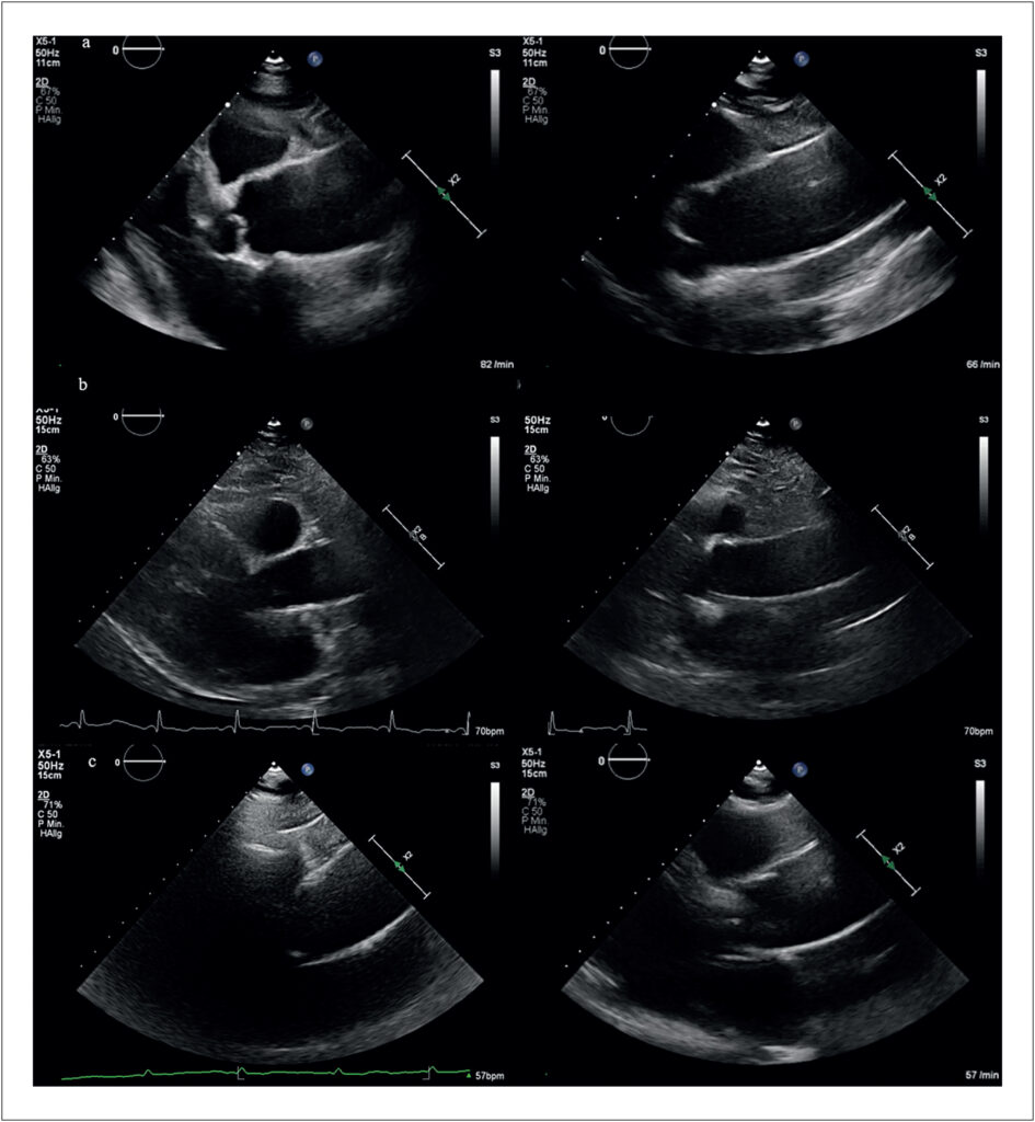
Assessment of the ascending aorta is an essential component of standard transthoracic echocardiography (TTE). While visualization of the aortic root and proximal ascending aorta is routinely achieved using the standard parasternal long-axis view (PLAX), imaging of the mid and distal ascending aorta remains technically challenging.– Current recommendations suggest moving the transducer to upper left intercostal spaces or alternatively using right parasternal windows to improve visualization of these segments in patients with adequate acoustic windows.
In clinical practice, experienced echocardiographers often obtain satisfactory visualization of the tubular ascending aorta using individualized modifications of conventional views. However, less experienced operators and general cardiologists may fail to consistently image these segments because the upper left parasternal long-axis view (uPLAX) is poorly described in the literature, whereas acquisition of the right parasternal view is more technically demanding and time-consuming.
[…]
Images
Image gallery fullscreen viewer
Comments are moderated by the editorial team before publication. Discussions should be evidence-based and use scientific language. Conflicts of interest should be declared.