ABC Family Arquivos Brasileiros de Cardiologia International Journal of Cardiovascular Sciences ABC Heart Failure & Cardiomyopathy ABC Imagem Cardiovascular
Português English
ABC Family
Português English
  • About
    • About ABC Family
    • Internal Regulation
    • Editorial Board
    • Brazilian Society of Cardiology (SBC)
  • Issues
  • SBC Guidelines
  • Content
    • Videos
    • Podcasts
    • News
  • About
    • About ABC Family
    • Internal Regulation
    • Editorial Board
    • Brazilian Society of Cardiology (SBC)
  • Issues
  • SBC Guidelines
  • Content
    • Videos
    • Podcasts
    • News

ABC Family

  • ABC Family
  • Arquivos Brasileiros de Cardiologia
  • International Journal of Cardiovascular Sciences
  • ABC Heart Failure & Cardiomyopathy
  • ABC Imagem Cardiovascular
Search in

No content found.

Journals
Ordering
Article types
Others

  1. Home
  2. Keyword: Coronary Vessels
Figure 1
ABCIC
Case Report

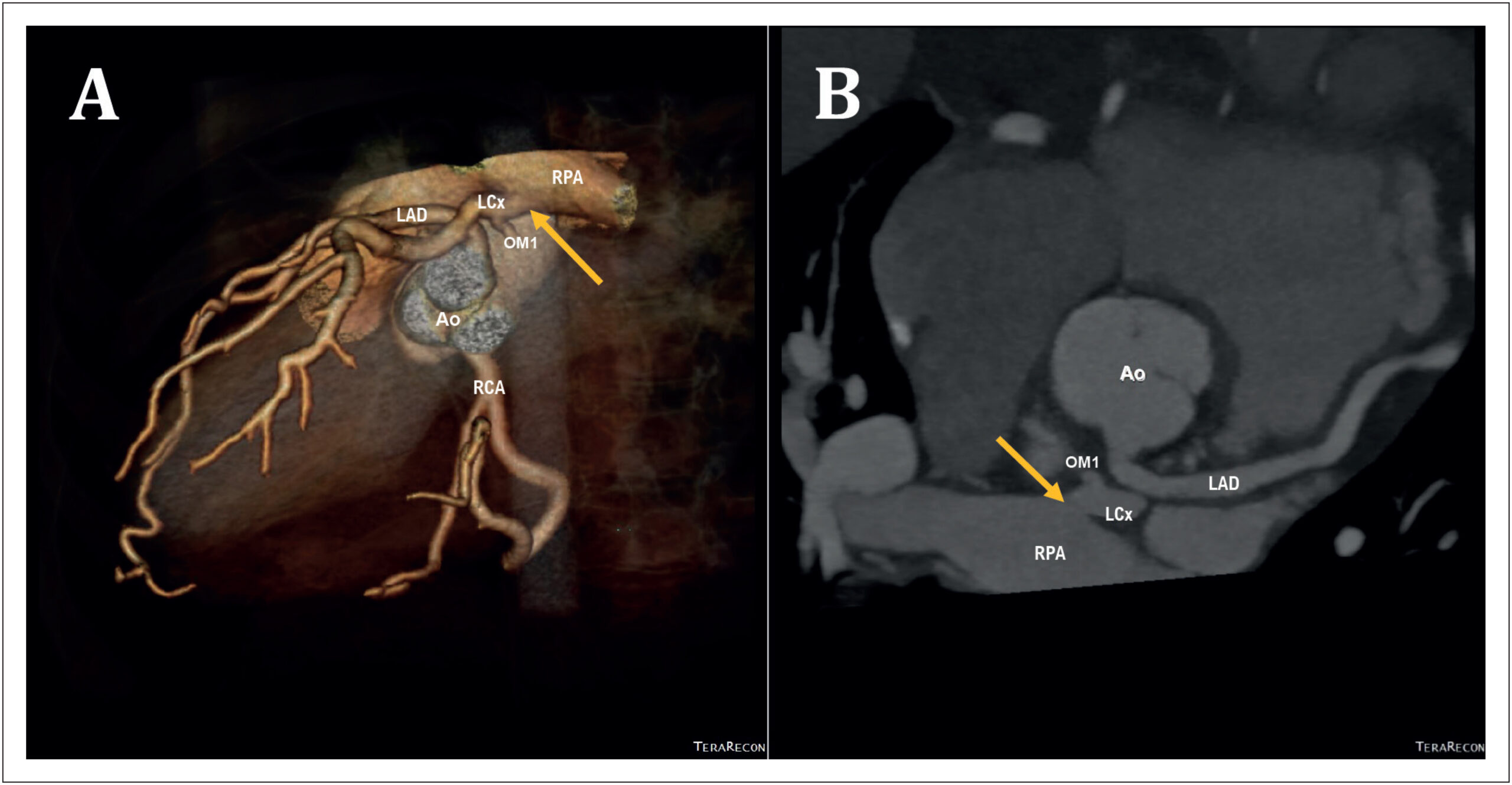
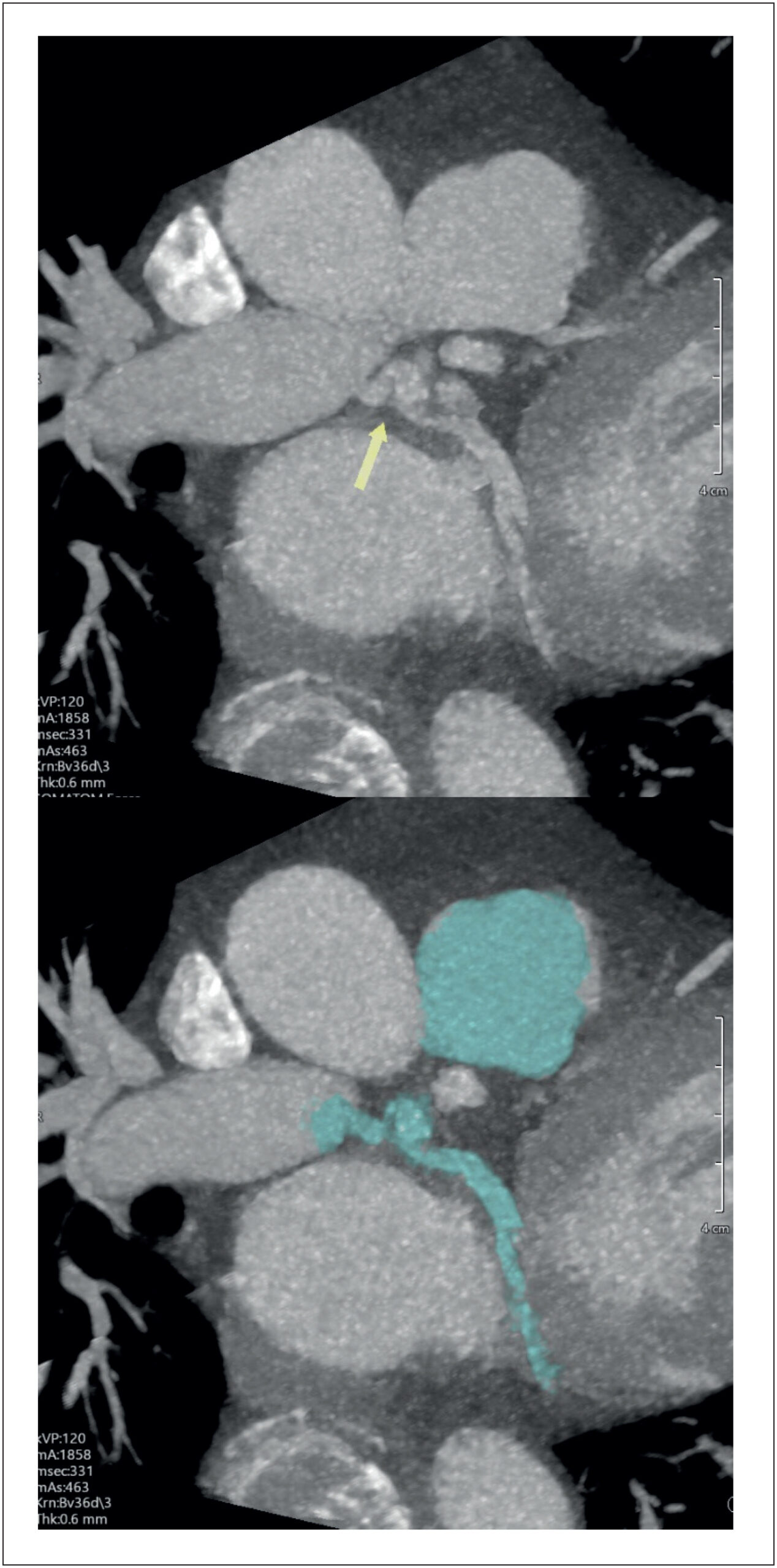
A Hidden Connection: Anomalous Left Circumflex Artery Arising From the Right Pulmonary Artery Unveiled by Cardiovascular Computed Tomography

Mubariz Ahmed Hassan et al. Ashraf Alzahrani Mohammad Mhanna Ola Abdelkarim … Rodrigo Bello Paulo Savoia Promporn Suksaranjit Kimberly Delcour

Introduction

Congenital coronary artery anomalies are uncommon but clinically significant conditions that may present across a broad spectrum, ranging from incidental findings to myocardial ischemia, ventricular arrhythmias, heart failure (HF), and sudden cardiac death (SCD). In adults, anomalous coronary origins may be particularly challenging to recognize because symptoms are often nonspecific and may overlap with more prevalent causes of chest pain, dyspnea, or palpitations.

In rare cases, a coronary artery may originate from the pulmonary arterial circulation, resulting in myocardial perfusion that depends on collateral flow from the remaining coronary arteries arising from the aorta, thereby predisposing the myocardium to ischemia-related electrical instability. Noninvasive anatomical imaging plays a pivotal role in establishing a definitive diagnosis, particularly when conventional angiography is inconclusive.

[…]
  • Cardiac Catheterization
  • Coronary Vessels
  • Pulmonary Artery
  • X-Ray Computed Tomography
Read Full Article
Creative Commons Attribution (CC BY) Creative Commons license
24/07/2026 DOI: 10.36660/abcimg.20260040i Views 1
Figure 1
ABCIC
Case Report

Single Coronary Artery and Stress Cardiomyopathy: An Association Demonstrated by Multimodality Imaging

Marcus Vinicius Silva Ferreira et al. Julia Pereira Afonso dos Santos Tormin Roberto Nery Dantas Jr Roberto Vitor Almeida Torres … Renan Andreos Cordeiro José de Arimateia Batista Araújo Filho Luiz Francisco Cardoso

Case Report

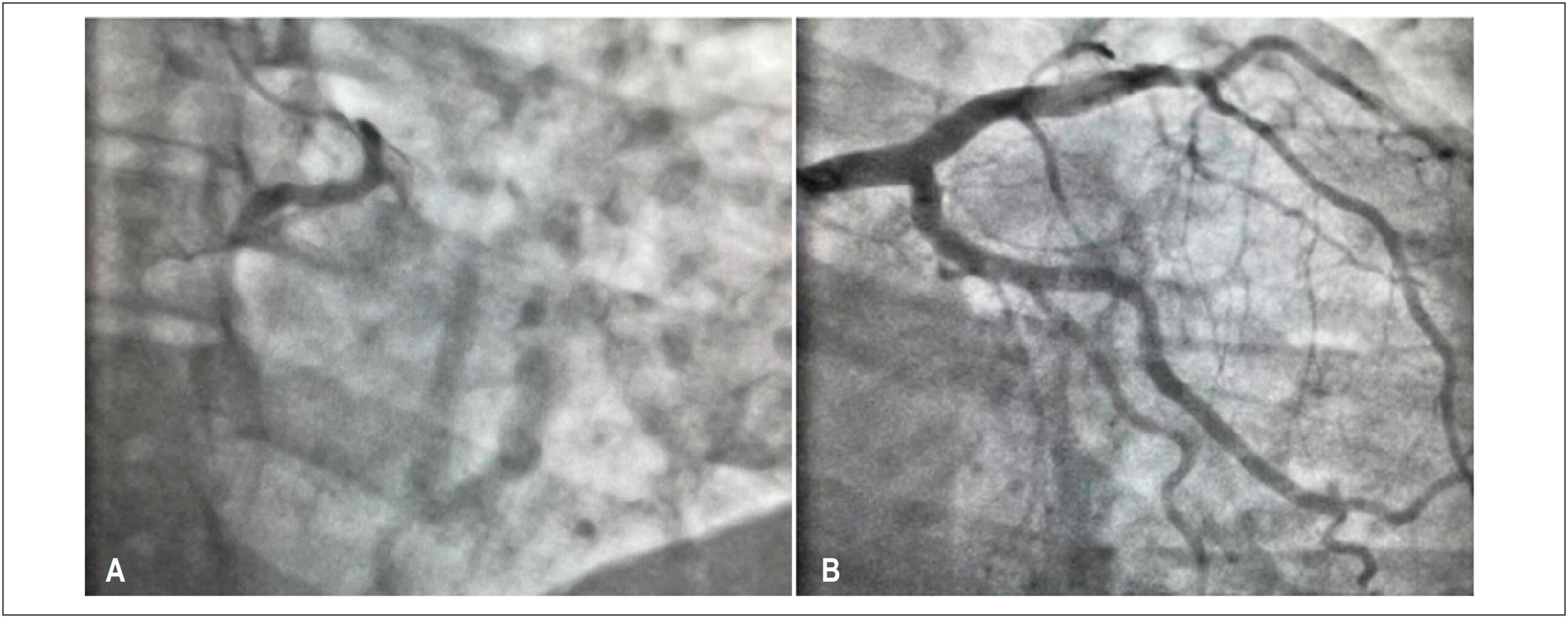
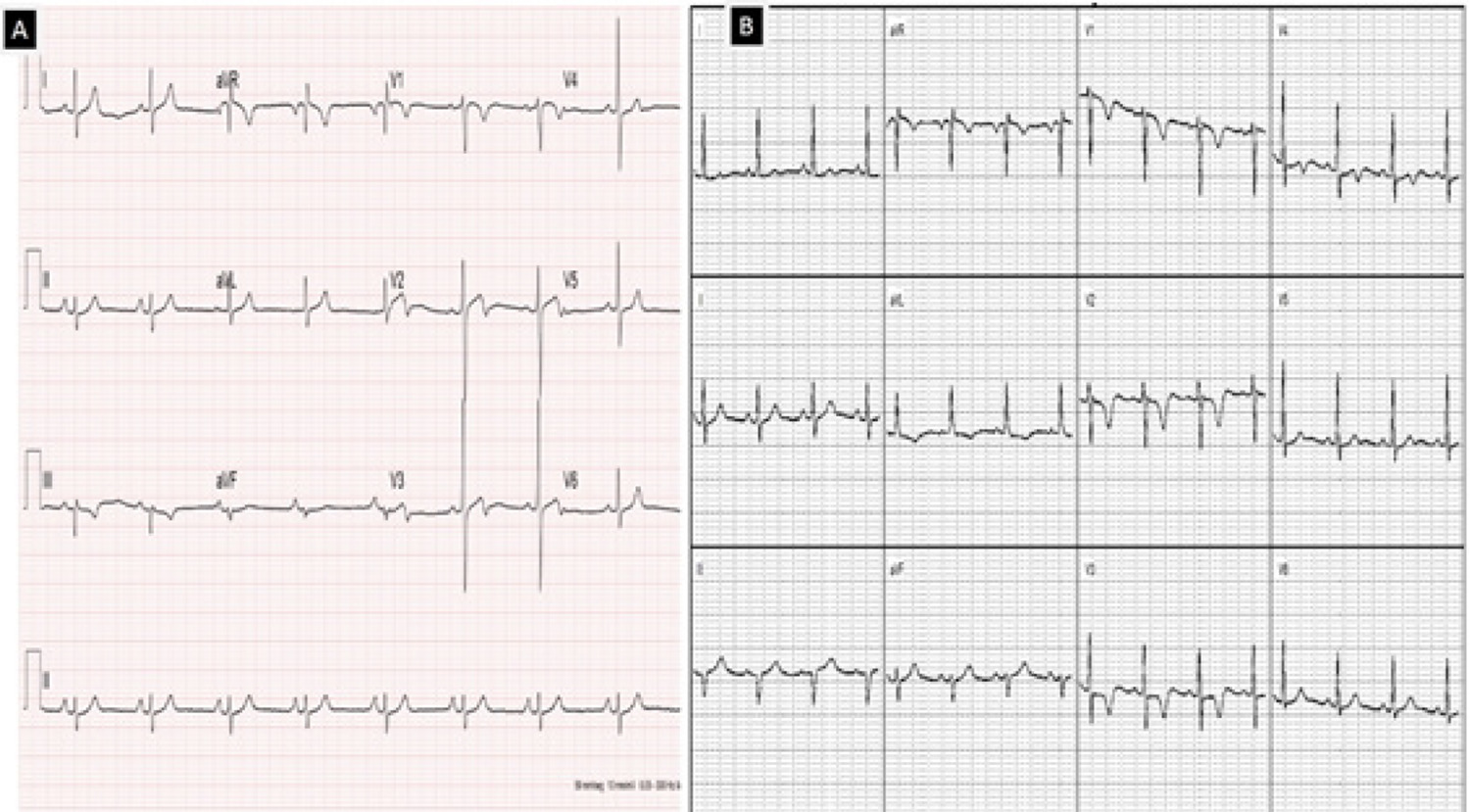
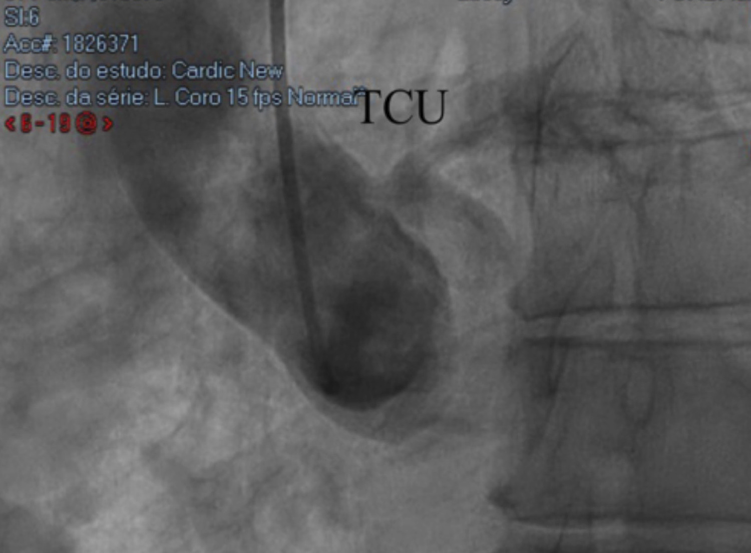
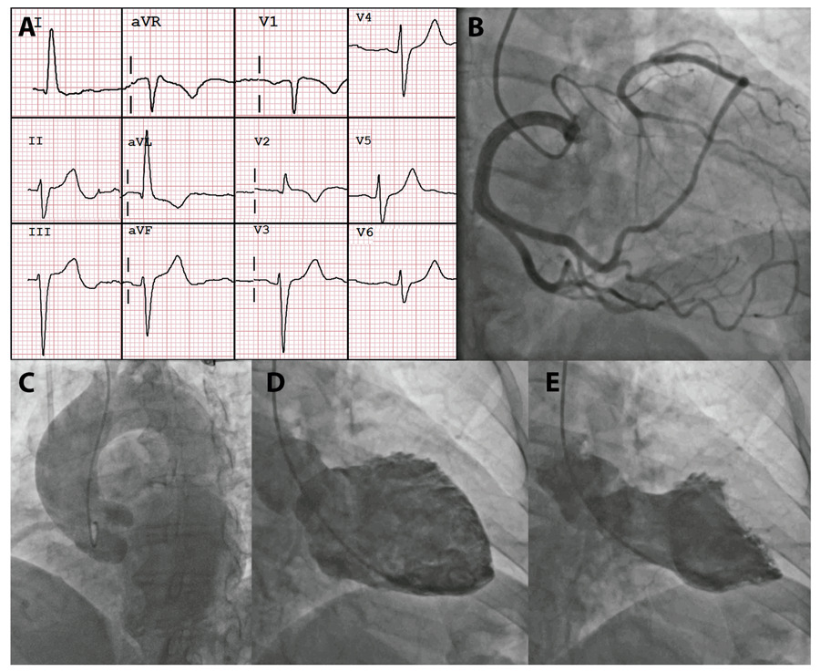
A 58-years-old female patient was admitted with retrosternal chest pain described as a continuous, oppressive tightness that had started two days earlier, after a tooth extraction procedure performed under ineffective local anesthesia. The patient’s past medical history included diabetes, dyslipidemia, and ovarian cancer, and she was receiving aspirin, rosuvastatin, metformin, dapagliflozin, and semaglutide. Physical examination was unremarkable. The chest pain protocol was initiated, and the initial electrocardiogram showed sinus rhythm with left anterior fascicular block, early repolarization in the inferior leads, and T-wave inversion in aVL, V1, and V2 (). The patient received 5 mg of sublingual isosorbide dinitrate, and blood samples were collected for laboratory testing. High-sensitivity troponin T (hs-cTnT) was 171 ng/L, confirming myocardial injury. An initial diagnosis of non–ST-elevation myocardial infarction (NSTEMI) was made, and the patient underwent coronary angiography (CA) for anatomic assessment ( / Video S1).

[…]

  • Coronary Angiography
  • Coronary Vessels
  • Echocardiography
  • Magnetic Resonance Imaging
  • Takotsubo Cardiomyopathy
Read Full Article
Creative Commons Attribution (CC BY) Creative Commons license
17/07/2026 DOI: 10.36660/abcimg.20260023i Views 1
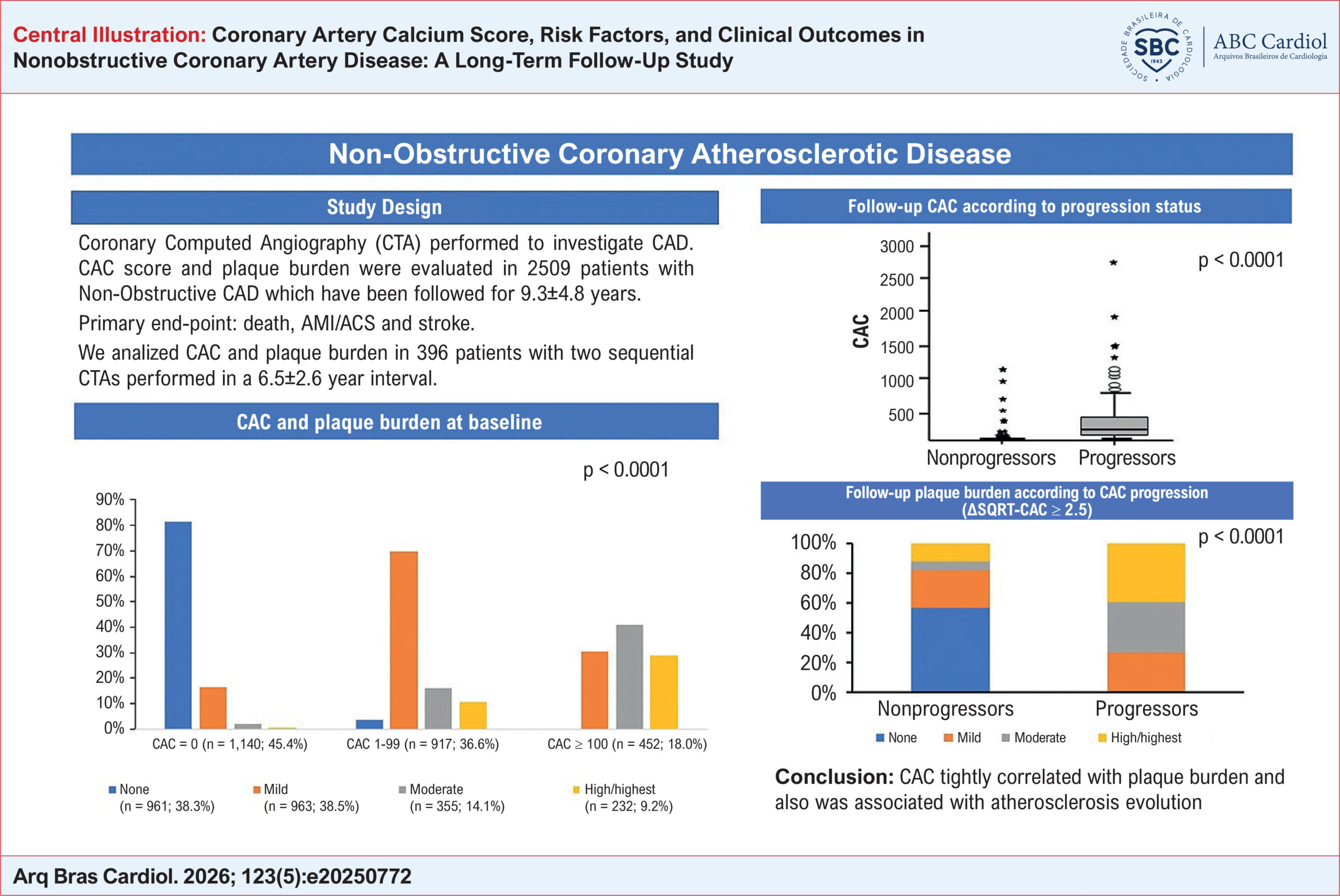
Coronary Artery Calcium Score, Risk Factors, and Clinical Outcomes in Nonobstructive Coronary Artery Disease: A Long-Term Follow-Up Study
ABC
Original Article

Coronary Artery Calcium Score, Risk Factors, and Clinical Outcomes in Nonobstructive Coronary Artery Disease: A Long-Term Follow-Up Study

Protásio Lemos Da Luz et al. Desiderio Favarato Alexandre A.C. Abizaid Luiz Antônio Machado Cesar … Carlos Vicente Serrano Jr. Antônio Carlos Palandri Chagas Carlos E. Rochitte Marco A. Gutierrez

Abstract

Background:

The prognostic value of coronary artery calcium (CAC) score in nonobstructive coronary artery disease (NObCAD; stenosis < 50%) remains insufficiently characterized.

Objectives:

To investigate the association between CAC score, cardiovascular risk factors, and clinical outcomes in patients with NObCAD.

Methods:

A total of 2,509 patients underwent coronary computed tomography angiography (CTA) and were followed for 8.9 ± 2.6 years. Plaque burden was classified according to CAD-RADS™ 2.0 into none, mild, moderate, and high/highest. The primary endpoint (PEP) was a composite of all-cause mortality, acute coronary syndrome/acute myocardial infarction, and stroke. Statistical significance was set at 5%.

Results:

CAC score was 0 in 45.4% of patients, 1-99 in 36.6%, and ≥ 100 in 18.0%. Correspondingly, 38.3% of patients had no coronary lesions, 38.5% had mild lesions, 14.1% had moderate lesions, and 9.2% had high/highest lesions. Among patients with CAC = 0, the absence of coronary lesions predominated (81.2%), whereas it was rare among those with CAC ≥ 100. The PEP occurred in 4.9% of patients, predominantly driven by all-cause mortality. Among the 396 patients with serial coronary CTA (mean interval: 6.5 ± 2.6 years), CAC progression (> 2.5 increase based on the square root method) was observed in 41.9%. Plaque burden increased in parallel. CAC score showed a positive association with plaque burden and cardiovascular risk factors.

Conclusions:

In NObCAD, CAC is present in more than half of patients and is associated with both plaque burden and cardiovascular risk factors. The incidence of PEP increases in proportion to risk factor burden and CAC levels. CAC and plaque burden progress concurrently over time, supporting the role of CAC as a surrogate marker of subclinical atherosclerosis progression.

  • Calcium
  • Coronary Artery Disease
  • Coronary Vessels
Read Full Article
Creative Commons Attribution (CC BY) Creative Commons license
17/07/2026 DOI: 10.36660/abc.20250772i Views 1
Figure 1
ABC
Editorial

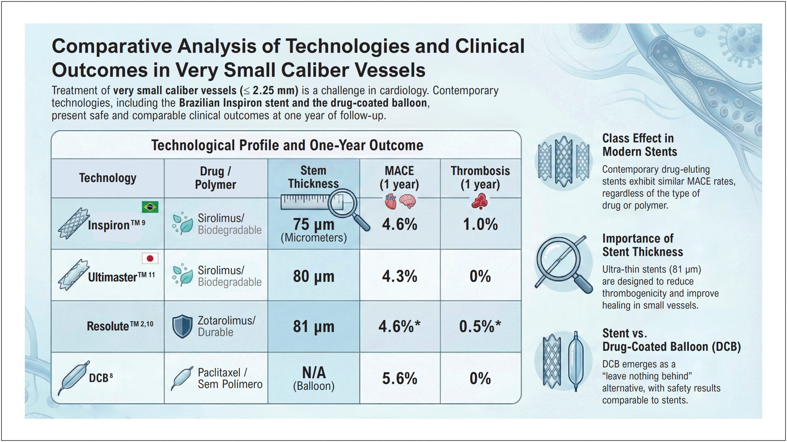
Small Vessels, Big Decisions

José Airton de Arruda et al. José de Ribamar Costa Junior

Percutaneous coronary intervention (PCI) in small-caliber vessels remains one of the most challenging scenarios in contemporary interventional cardiology.

Historically, a small vessel was considered to be one with a diameter ≤2.75 mm. With the technological evolution of devices and the greater refinement of interventional techniques, this concept has been progressively redefined. In contemporary practice, small vessels are standardized as those with a diameter <2.5 mm, while very small or extremely small vessels often include segments <2.25 mm.,

[…]

  • Coronary Restenosis
  • Coronary Vessels
  • Drug-Eluting Stents
  • Percutaneous Coronary Intervention
  • Sirolimus
Read Full Article
Creative Commons Attribution (CC BY) Creative Commons license
01/06/2026 DOI: 10.36660/abc.20260207i Views 2
Figure 1
ABCIC
Case Report

Coexistence of Partial Anomalous Pulmonary Venous Connection and Coronary Artery Fistulas: A Rare Case Report

Mourad Haj Abdo et al. Hussain Latsh Mathias Wagner George König Sebastian Barth

Abstract

Background

Dyspnoea is a common clinical symptom that frequently prompts hospital admission and is associated with significant morbidity. While it most often results from prevalent cardiopulmonary conditions, rare congenital cardiovascular anomalies can also manifest with dyspnoea. Partial Anomalous Pulmonary Venous Connection (PAPVC) and Coronary Artery Fistulas (CAFs) are uncommon congenital malformations of the cardiovascular system, and their simultaneous presence is exceedingly rare. Early recognition of such anomalies is critical to avoid progressive hemodynamic compromise and to guide appropriate management strategies.

Case Presentation

We present the case of a 55-year-old man who experienced an acute onset of dyspnoea lasting approximately two hours. Initial clinical assessment and routine investigations—including physical examination, echocardiography, electrocardiography, and right and left heart catheterization—raised suspicion of an underlying cardiac abnormality, prompting further evaluation. Subsequent cardiac Magnetic Resonance Imaging (MRI) and Multi-Detector Computed Tomography (MDCT) revealed the presence of a Partial Anomalous Pulmonary Venous Connection (PAPVC) accompanied by Coronary Artery Fistulas (CAFs). Given the non-complex characteristics of the shunt in this case, a shared decision was reached with the patient to proceed with conservative management.

Discussion

PAPVC and Coronary Artery Fistulas CAFs are rare entities that should be considered in the differential diagnosis. However, recommending their evaluation as initial diagnostic hypotheses may result in unnecessary investigations.

  • Coronary Vessels
  • Differential Diagnosis
  • Echocardiography
Read Full Article
Creative Commons Attribution (CC BY) Creative Commons license
25/05/2026 DOI: 10.36660/abcimg.20250030i Views 1
ABC
Original Article

Estimating Left Main Bifurcation Geometry: Are Geometric Models Reliable in Diseased Arteries?

Stefano Garzon et al. Lucas Pilotto José Mariani Felipe Bezerra … Guy Prado Willterson Bandeira Breno Almeida Pedro Lemos

Abstract

Background:

Accurate characterization of coronary bifurcation geometry is essential for planning and optimizing percutaneous coronary interventions. Although several geometric models have been proposed to predict coronary bifurcation dimensions, their performance in diseased vessels remains insufficiently explored.

Objective:

To assess the accuracy and precision of three geometric prediction models (Murray, Finet, and Huo–Kassab) in estimating left main coronary artery (LMCA) bifurcation diameters, using quantitative coronary angiography (QCA) and intravascular ultrasound (IVUS) as reference standards.

Methods:

This retrospective, single-center study included 13 patients undergoing IVUS evaluation of the LMCA, left anterior descending artery, and left circumflex artery. Bifurcation diameters predicted by each geometric model were compared with measurements obtained by QCA and IVUS. Accuracy, precision, and model ratios were calculated for each comparison. Agreement between predicted and measured values was further assessed using Bland–Altman analysis.

Results:

When compared with QCA, the Murray and Huo–Kassab models demonstrated higher accuracy (0.06 mm and −0.03 mm, respectively) and similar precision (0.95 mm for both) than the Finet model (accuracy −0.35 mm, precision 0.96 mm). In comparisons with IVUS, the Murray model showed the best performance, with an accuracy of −0.49 mm and a precision of 0.84 mm. Bland–Altman analysis indicated superior agreement for the Murray model, particularly when IVUS was used as the reference standard.

Conclusions:

In this population of patients with moderately diseased LMCA bifurcations, the Murray and Huo–Kassab models provided more accurate geometric predictions than the Finet model. Overall, the Murray model demonstrated the best performance, especially in IVUS-based assessments.

  • Coronary Angiography
  • Coronary Artery Disease
  • Coronary Vessels
Read Full Article
Creative Commons Attribution (CC BY) Creative Commons license
02/03/2026 DOI: 10.36660/abc.20250605i Views 1
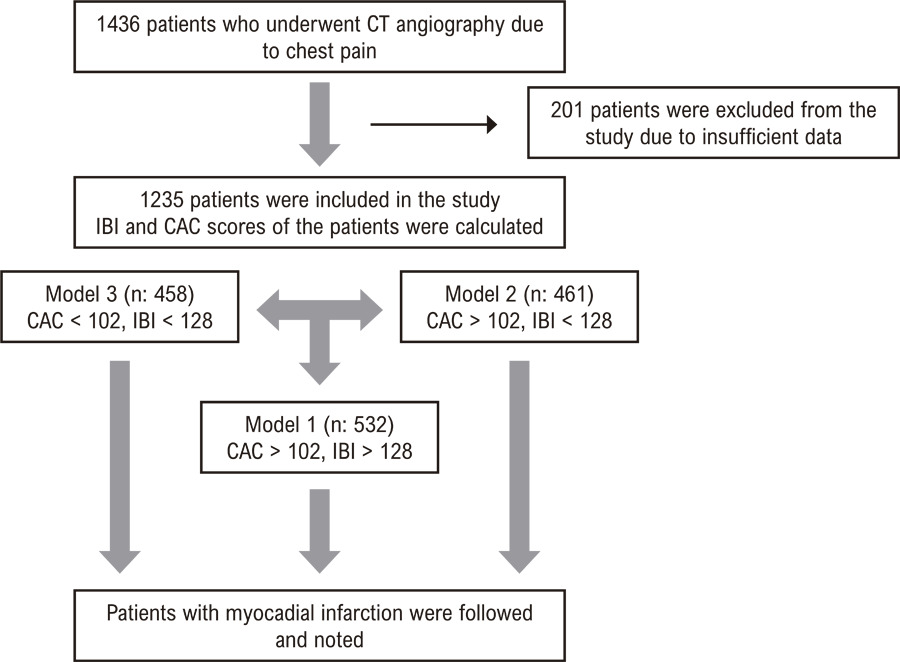
Cardiac CT-Detected Vulnerable Plaques and Risk of Future Acute Myocardial Infarction: Predictive Value of CAC Scores and Inflammatory Burden Index
ABC
Original Article

Cardiac CT-Detected Vulnerable Plaques and Risk of Future Acute Myocardial Infarction: Predictive Value of CAC Scores and Inflammatory Burden Index

Sefa Erdi Ömür et al. Emin Koyun Gökhan Cabri

Abstract

Background

Computed Tomography (CT) angiography is widely used as a basic non-invasive modality to evaluate coronary artery disease. Inflammatory burden index (IBI) is a systemic inflammation indicator reflecting the inflammatory status. It is still difficult to determine the high risk of future acute myocardial infarction (MI) in patients with plaque detected by CT angiography.

Objective

This study aimed to evaluate the effectiveness of CAC scores and IBI in predicting MI risk among patients undergoing CT angiography for chest pain. We sought to assess the individual and combined predictive value of these indices, determine their optimal cut-off values, and compare MI risk across patient groups with varying CAC and IBI scores. We also aimed to evaluate the potential role of these markers in identifying ‘vulnerable patients’ at high risk for future cardiovascular events.

Methods

This retrospective study included 1235 patients who underwent CT angiography due to chest pain and were followed for 3 years (2.8-3.4). Patients were categorized into three models based on their CAC and IBI scores. The primary outcome was the occurrence of MI during the follow-up period. Statistical significance was established at p < 0.05 for all analyses.

Results

Patients who experienced MI had significantly higher IBI and CAC scores compared to those without MI. Logistic regression analysis identified CAC score and IBI as independent predictors of MI. ROC analysis determined optimal cut-off values for IBI (128) and CAC (102) in predicting MI. Kaplan-Meier analysis revealed a significant gradient in MI risk across the three models, with the highest risk observed in patients with both high IBI and CAC scores.

Conclusion

The combination of IBI and CAC scores provides improved risk stratification for the prediction of future MI (vulnerable plaque) in patients with plaque detected by CT angiography. This approach may facilitate more specific treatment strategies for high-risk patients.

  • Computed Tomography Angiography
  • Coronary Vessels
  • Myocardial Infarction
Read Full Article
Creative Commons Attribution (CC BY) Creative Commons license
09/01/2026 DOI: 10.36660/abc.20250052i Views 1
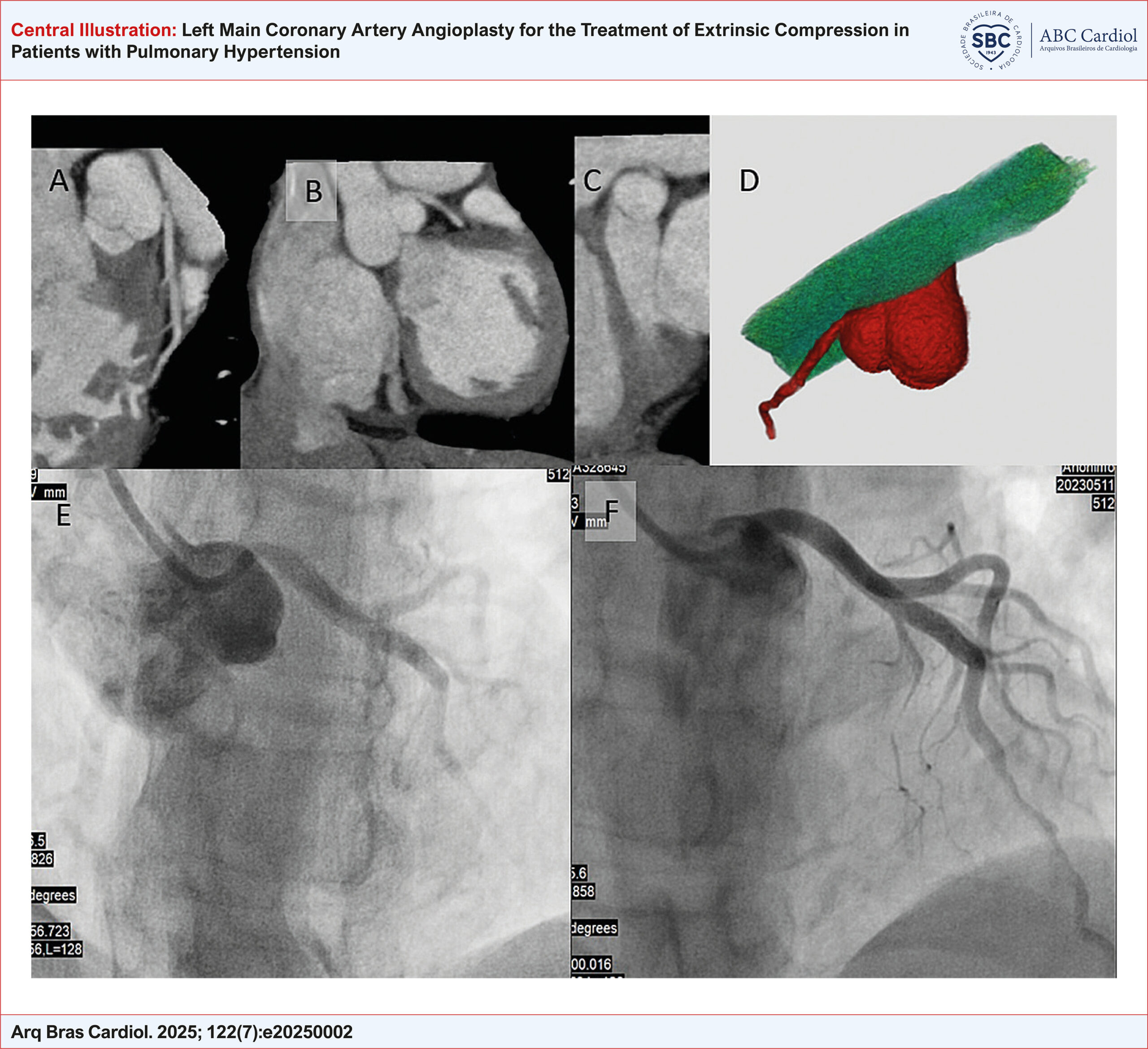
Left Main Coronary Artery Angioplasty for the Treatment of Extrinsic Compression in Patients with Pulmonary Hypertension
ABC
Original Article

Left Main Coronary Artery Angioplasty for the Treatment of Extrinsic Compression in Patients with Pulmonary Hypertension

Luciana Dornfeld Bichuette et al. Daniela Calderaro Pedro Alves Lemos Neto Eduardo Kaiser Ururahy Nunes Fonseca … Luiza Sarmento Tatagiba Tulio Martins Vieira Yally Priscila Pessoa Nascimento Marcela Araujo Castro Taysa Antônia Félix Silva Yuri de Deus Montalverne Parente Caio Julio Fernandes Francisco Akira Malta Cardozo Cesar Higa Nomura Pedro Henrique Almeida Marins Rogerio Souza Carlos Vianna Poyares Jardim

Abstract

Background

The most common clinical presentation of pulmonary hypertension (PH) includes exertional dyspnea, signs of systemic congestion, and syncope. Angina pectoris can also be a relevant manifestation, especially in cases where the left main coronary artery (LMCA) is externally compressed by a dilated pulmonary artery. However, significant gaps remain regarding the most appropriate diagnostic and therapeutic strategies for coronary obstruction in this clinical scenario.

Objectives

To assess the feasibility and impact of coronary angioplasty with stent implantation on symptom relief in patients with PH and extrinsic compression of LMCA.

Methods

This descriptive study included 12 patients with PH who were followed at the Pulmonary Circulation Outpatient Clinic of the Instituto do Coração, Hospital das Clínicas da Faculdade de Medicina da Universidade de São Paulo. All patients underwent coronary angioplasty with stent implantation to treat extrinsic compression of LMCA.

Results

A total of 12 patients were analyzed, with a mean age of 47.9 years, predominantly with group 1 PH and under specific therapy. All procedures achieved excellent immediate results, with angina relief observed at 30 days. During a mean follow-up of 33 months, no procedure-related complications were reported, and angina symptoms remained controlled. Four patients died due to progressive heart failure.

Conclusion

The findings support the feasibility of coronary angioplasty as a strategy for symptomatic relief of angina in patients with LMCA compression associated with PH. Further studies are needed to evaluate the impact of this intervention on hard clinical outcomes, as well as the role of screening in asymptomatic patients.

  • Angioplasty
  • Coronary Vessels
  • Pulmonary Hypertension
Read Full Article
Creative Commons Attribution (CC BY) Creative Commons license
01/08/2025 DOI: 10.36660/abc.20250002i Views 3
Figure 1
ABC
Image

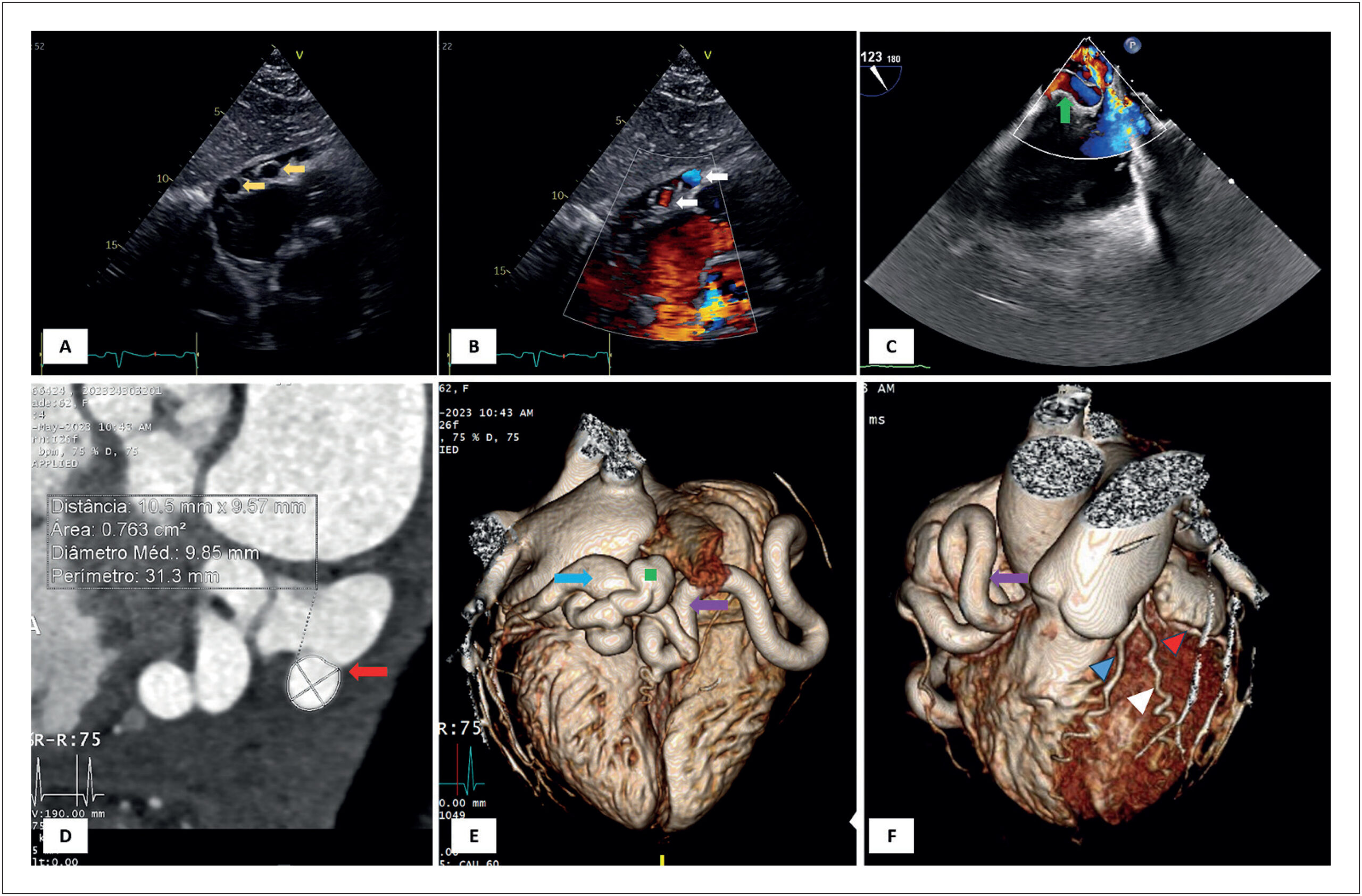
Silent Giant: Right Coronary Artery Ectasia with a Hidden Fistula to the Coronary Sinus in an Asymptomatic Patient

Inês Ferreira Neves et al. André Ferreira Inês Almeida Tânia Branco Mano Lídia de Sousa

A 62-year-old woman, asymptomatic and without significant medical history, was referred to a routine transthoracic echocardiogram (TTE). The TTE had no abnormal findings but showed, in subcostal view, three round images adjacent to the lateral wall of the right atrium. Color Doppler showed that these were apparently vascularized (, panels A and B, supplemental and ). A transesophageal echocardiogram showed a giant right coronary artery (RCA), apparently originating from the RCA ostium, with turbulent flow inside (, panel C, supplemental ). It was not possible to observe shunts or fistulae in this imaging modality. Given the suspicion of a fistula to the RCA, computed tomography coronary angiography was performed, showing an ectatic RCA with a normal origin, a diameter of 10,5 mm, and a tortuous trajectory, with fistulation to the coronary sinus in its final segment ( panels D to F). Right heart catheterization demonstrated a non-significant left-to-right shunt (Qp: Qs ratio of 1.60). Cardiac magnetic resonance imaging (MRI) had no evidence of perfusion defects during hyperemia, thus excluding the coronary “steal phenomenon”. After a multidisciplinary team discussion, since the patient was asymptomatic and the fistula had no hemodynamic significance, no invasive treatment was performed at this time.

Coronary arteriovenous fistulas are rare, with an incidence of 0.002%., The majority of these are congenital and asymptomatic at presentation. Coronary steal can lead to several complications, including ischemia., The treatment approach remains controversial. The presence of symptoms, significant left-right shunt, and large fistula are the most frequent indications for percutaneous or surgical approach.,

[…]

  • Arteriovenous Fistula
  • Congenital Heart Defects
  • Coronary sinus
  • Coronary Vessels
Read Full Article
Creative Commons Attribution (CC BY) Creative Commons license
28/07/2025 DOI: 10.36660/abc.20240574i
Figure 1
ABCIC
Case Report

Rare Presentation of an Anatomical Congenital Anomaly of the Coronary Arteries on Computed Tomography Angiography

Marcello Zapparoli et al. Alexandre Alessi Franco Camargo Zapparoli Mateus Rodrigues Alessi

Introduction

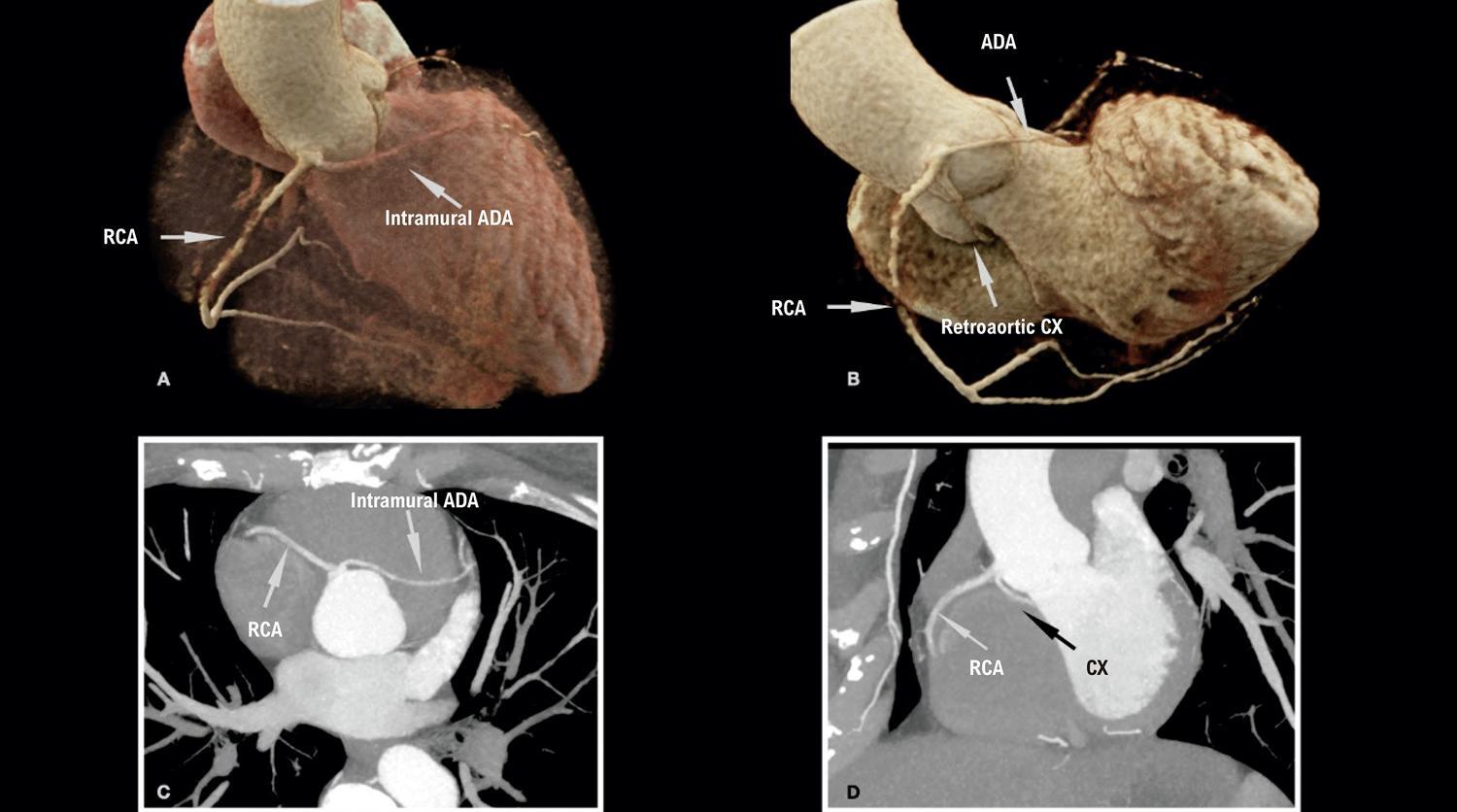
Anomalous origin of the coronary arteries represents a heterogeneous group of alterations in the origin and course of these vessels, ranging from the most common, asymptomatic and low risk, to the rarest and highest risk, which may eventually be symptomatic or have sudden death as their first manifestation., Their prevalence corresponds to 1.3% of patients undergoing coronary angiography, but this number may be higher depending on the diagnostic method and the population selected, representing the second leading cause of sudden death in young athletes, behind only hypertrophic cardiomyopathy.,

Within this group, coronary anomalies with a single ostium are present in a smaller proportion of the population, approximately 0.004% to 0.098%. They are most often asymptomatic, but they may manifest with chest pain, dyspnea, palpitation, myocardial infarction, and sudden death., The presentation of this anomaly has worse prognosis in the presence of an interarterial vessel course (between the aorta and pulmonary trunk), an acute angle of origin, and atherosclerotic plaque; the latter change has a higher incidence in anomalous vessels on account of the greater stress on the arterial wall., In addition to presenting in isolation, they may also be associated with other alterations such as cardiac fistulas or a bicuspid aortic valve.

[…]

  • Anatomic Variation
  • Chest Pain
  • Computed Tomography Angiography
  • Coronary Vessel Anomalies
  • Coronary Vessels
Read Full Article
Creative Commons Attribution (CC BY) Creative Commons license
21/11/2024 DOI: 10.36660/abcimg.20240075i Views 1
IJCS
Editorial

Venturing into Uncharted Territories: Unveiling Frontal QRS-T Angle in Patients with Coronary Artery Ectasia

Maqsood Alam et al. Eduardo Sanhueza Adrian Baranchuk

It is with profound interest that we read the manuscript by Karahan et al. titled “Evaluation of Frontal QRS-T Angle in Patients with Coronary Artery Ectasia” published in the recent volume of International Journal of Cardiovascular Sciences . Coronary artery ectasia (CAE), a condition marked by focal enlargement of coronary arteries, has long intrigued researchers and clinicians due to its enigmatic nature. Beyond its silence existence, CAE hides a spectrum of clinical ranges, from chest pain with coexisting coronary artery disease to life threatening arrhythmias and sudden cardiac death. , While previous research has delved into the multifaceted etiology of CAE, , this study took a unique path, exploring the electrocardiographic landscape to better understand ventricular heterogeneity in CAE.

The cornerstone of this study lies in the evaluation of frontal QRS-T angle, a marker of ventricular repolarization heterogeneity. Spatial QRS-T angles have received attention in previous research, often as an indicator of worse cardiovascular outcomes. This method is a little tiresome, as it requires a 3-dimensional computational model for interpretation, whereas the ease of measurement of the frontal QRS-T angle from routine 12-lead electrocardiograms makes it an attractive alternative candidate for exploring ventricular repolarization. The frontal QRS-T angle was calculated as the absolute value of difference between the QRS- and T-axis, yielding values between 0° and 180°, and it was categorized as normal (≤ 90° ) or abnormal (≥ 100°). However, its application in the context of CAE is a novel and compelling avenue of investigation.

[…]

  • Ambulatory Electrocardiography
  • Coronary Vessels
  • Pathologic Dilatation
Read Full Article
Creative Commons Attribution (CC BY) Creative Commons license
07/06/2024 DOI: 10.36660/ijcs.20240049
ABC
Editorial

Index of Systemic Immunoinflammation and Ischemia in Patients with Non-Obstructive Coronary Arteries

Henrique Tria Bianco

Cardiovascular disease is the leading cause of death worldwide and coronary artery disease (CAD) is the most common type of cardiovascular disease. However, a significant portion of patients who present with characteristic chest discomfort do not demonstrate obstructive CAD, defined as ≥50% stenosis in at least one coronary artery on angiography. These patients are generally diagnosed with non-cardiological conditions, such as gastrointestinal disorders or psychosomatic disorders. It is speculated that coronary vascular dysfunction appears to be the underlying cause of ischemia in a large proportion of these cases. Therefore, standardization of diagnostic criteria for ischemic symptoms due to coronary microvascular dysfunction (CMD) is necessary for further investigation of patients presenting with anginal chest pain consistent with “microvascular angina”.

Historically, the only practical methods available for assessment have been invasive, such as intracoronary Doppler or thermodilution. This probably hindered the objective assessment of CMD in patients presenting with chest pain without obstructive CAD. Thus, treatment has often been studied in imprecise clinical entities such as cardiac syndrome distinct., Furthermore, the lack of consensus on diagnostic criteria and nomenclature has further obscured the evidence that sought to objectively define microvascular angina as a distinct clinical entity.

[…]

  • Chest Pain
  • Coronary Artery Disease
  • Coronary Vessels
  • Myocardial Ischemia
Read Full Article
Creative Commons Attribution (CC BY) Creative Commons license
26/04/2024 DOI: 10.36660/abc.20240081 Views 2
Figure 1
IJCS
Viewpoint

Coronary Artery Microfistulas: Historical Description and Contemporary Clinical Relevance

Stans Murad Netto et al. Rachel Vianna O'Neill de Souza Victor Murad Evandro Tinoco Mesquita

Introduction

Coronary microfistula was initially described as an anatomical finding in the late 1960s by Murad and colleagues, and it was later recognized as a morphofunctional abnormality of the coronary arteries capable of causing clinical findings (). This often asymptomatic entity is still poorly recognized in clinical practice, although it can be readily identified in the catheterization laboratory, cineangiography studies, and more recently by coronary CT angiography.

Initially, this condition was not taken into consideration, and it was justified as a possible “error” in cineangiography technique. Different studies have demonstrated its existence and its potential as a nosological entity responsible for myocardial ischemia, cardiac chamber overload, arrhythmias, and even heart failure.,

[…]

  • Angina Pectoris
  • CF
  • Coronary Vessels
  • Myocardial Ischemia
Read Full Article
Creative Commons Attribution (CC BY) Creative Commons license
18/12/2023 DOI: 10.36660/ijcs.20230092
ABCIC
Original Article

Incidental Tomographic Findings of Coronary Artery Calcifications: A Prevalence Study in Southern Brazil

Karen Rafaela Okaseski Scopel et al. Tássia Machado Medeiros Bibiana Natalia Porto Maicá Maria Carbonari Velho … Mariana Motta Dias da Silva Juliane Nascimento Mattos Guilherme Galante Heuser Eliane Roseli Winkelmann

Abstract

Introduction

Coronary artery calcifications (CAC) are shown to be a predictive factor of cardiovascular diseases. Computed tomography (CT) of the chest with a low-dose acquisition protocol is accurate in identifying CAC and provides incidental findings of these calcifications, which are commonly overlooked. This study will analyze the prevalence of incidental findings of calcification in coronary arteries in non-cardiac individuals undergoing chest CT.

Methods

Consecutive cross-sectional study of an analytical and descriptive nature. Individuals of both genders who underwent chest CT by referral, over 18 years of age and without heart disease were included. Data collection was carried out using medical records and a self-applied anamnesis form. The variables referring to the CAC and the extension of the impairment were obtained from the reassessment of the chest CT images available in the institution’s system. The exams were anonymized and evaluated by two experienced radiologists. P≤0.05 was considered statistically significant.

Results

397 exams were analyzed. A prevalence of calcifications was found in 176 (44%) of the cases. The existence of these coronary calcifications is related to age (p<0.001). Calcifications are related to gender (p = 0.03) with a higher odds ratio of development in men (odds ratio [OR] = 1.55). Smoking (p<0.001), sedentary lifestyle (p<0.001), systemic arterial hypertension (p<0.001), Diabetes Mellitus (p = 0.04), and dyslipidemia (p<0.001) showed a positive association.

Conclusion

The prevalence of incidental CAC findings was 44%; vary in greater numbers between mild and severe; higher odds ratio in males and increased prevalence with age. Therefore, chest CT proves to be an effective method to assess CAC, and together with the patient’s clinical history, it can be used to measure risk factors for CVD and intervene in the outcome of the condition.

  • Coronary Vessels
  • Incidental Findings
  • Tomography, X-Ray Computed
  • Vascular Calcification
Read Full Article
Creative Commons Attribution (CC BY) Creative Commons license
24/03/2023 DOI: 10.36660/abcimg.2023368i Views 2
Figure 1
ABC
Original Article

Coronary Tortuosity as a New Phenotype for Ischemia without Coronary Artery Disease

André Estrada et al. André Silveira Sousa Claudio Tinoco Mesquita Humberto Villacorta

Abstract

Background

Coronary arteries tend to be more tortuous than other arteries and follow the repeated flexion and relaxation movements that occur during the cardiac cycle. Coronary tortuosity (CorT) leads to changes in coronary flow with a reduction in distal perfusion pressure, which could cause myocardial ischemia.

Objective

To assess the association between CorT and myocardial ischemia.

Methods

Between January 2015 and December 2017, 57 patients with angina and nonobstructive coronary artery disease detected by invasive coronary angiography (ICA) were retrospectively enrolled. Angiographic variables were analyzed to assess the presence and degree of tortuosity and correlated with their respective vascular territories on stress myocardial perfusion imaging (MPI). CorT was defined as coronary arteries with three or more bend angles ≤90°, measured during diastole. Statistical significance was determined at the 5% level.

Results

A total of 17 men and 40 women were enrolled (mean age 58.3 years). CorT was observed in 16 patients (28%) and in 24 of 171 arteries. There was a significant association between CorT and ischemia when analyzed per artery (p<0.0001). The angiographic factor most associated with ischemia was the number of bend angles in an epicardial artery measured at systole (p=0.021).

Conclusion

This study showed an association of CorT and myocardial ischemia in patients with unobstructed coronary arteries and angina. An increased number of coronary bend angles measured by angiography during systole was related to ischemia.

  • Coronary Vessels
  • Ischemia
  • Myocardial Ischemia
Read Full Article
Creative Commons Attribution (CC BY) Creative Commons license
23/09/2022 DOI: 10.36660/abc.20210787 Views 3
Figure 1
ABC
Original Article

Relationship between Systemic Immune-Inflammation Index and Coronary Collateral Circulation in Patients with Chronic Total Occlusion

Mehmet Koray Adali et al. Ipek Buber Gursel Sen Samet Yilmaz

Abstract

Background

Inflammation plays a key role in the initiation and progression of coronary artery disease (CAD). The systemic immune-inflammation index (SII) is a novel inflammatory parameter that has been shown to be associated with CAD.

Objective

This study aimed to investigate the relationship between SII and coronary collateral circulation (CCC) in patients with stable CAD and chronic total occlusion (CTO).

Methods

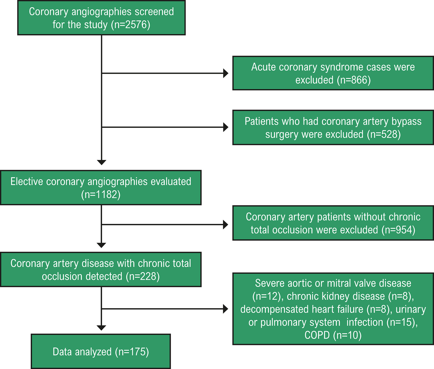
The patients were divided into two groups, with poor CCC and good CCC, according to the Rentrop Classification. Ninety-four patients had poor CCC, and 81 patients had good CCC. Inflammation parameters were calculated from the laboratory results. The statistical significance level applied was 0.05.

Results

High SII level (OR: 1.003, 95% CI: 1.001-1.004, p<0,001), absence of CTO in RCA (OR: 0.204, 95% CI: 0.096-0.436, p<0,001) and low Gensini score (OR: 0.980, 95% CI: 0.962-0.998, p=0,028) were significantly associated with poor CCC. The cutoff value of SII was 679.96 for the highest predictive power of poor CCC, with a sensitivity of 74.5% and specificity of 43.2%. Mortality rates were similar between the two groups during a mean follow-up of 21.5±10.8 months (p=0.107).

Conclusions

High SII level, the absence of CTO in the right coronary artery, and low Gensini score were significantly related to poor CCC. The rapid and cost-effective use of new inflammatory markers in clinical practice guides the prognosis of CAD.

  • Collateral Circulation
  • Coronary Occlusion
  • Coronary Vessels
Read Full Article
Creative Commons Attribution (CC BY) Creative Commons license
10/06/2022 DOI: 10.36660/abc.20210414 Views 2
Figure 1
ABC
Original Article

Systemic Immune-Inflammation Index Predicts Major Cardiovascular Adverse Events in Patients with ST-Segment Elevated Myocardial Infarction

Faysal Saylik et al. Tayyar Akbulut

Abstract

Background

The systemic immune-inflammation index (SII) has been reported as a new prognostic marker in tumors and cardiovascular diseases

Objective

To investigate the association of SII with adverse cardiovascular events in patients with ST-segment elevated myocardial infarction (STEMI).

Methods

A retrospective observational study was conducted on 843 patients with STEMI. Patients were divided into two groups based on the median value of SII. Major adverse cardiovascular events were compared between SII groups. Cox regression analysis was used for detecting independent predictors of cardiovascular adverse events. The improvement of discrimination ability by adding SII to the traditional risk factors such as age, hypertension, diabetes mellitus, and male gender for major adverse events was calculated by c-statistics, integrated discrimination improvement, and net reclassification improvement. A two-sided p-value <0.05 was considered significant.

Results

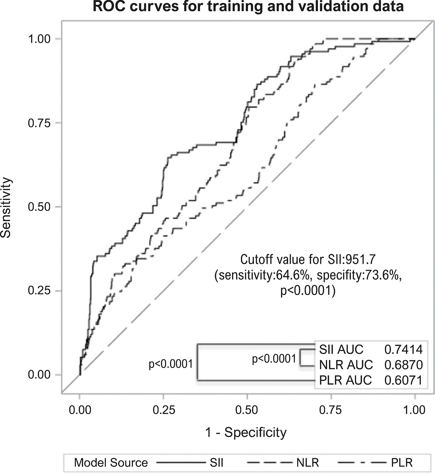
High SII group was older than the low SII group (61.2±11.2, 59.2±7.9, respectively, p=0.002). The high SII group had higher rates of cardiac death, nonfatal myocardial infarction, nonfatal stroke, hospitalization for heart failure, revascularization, and composite major adverse cardiovascular events than the low SII group. SII was an independent predictor of all events mentioned above. Adding SII to traditional risk factors improved their discrimination ability for cardiovascular events. SII was superior to the neutrophil-to-lymphocyte and platelet-to- lymphocyte ratios for predicting cardiovascular adverse events.

Conclusion

SII was an independent predictor of major adverse events in patients with STEMI and may be used to improve the prediction of adverse events, especially when combined with traditional risk factors.

  • Coronary Vessels
  • Heart Defects, Congenital
  • Myocardial Infarction
Read Full Article
Creative Commons Attribution (CC BY) Creative Commons license
02/05/2022 DOI: 10.36660/abc.20210412 Views 1
Figure 1
ABCIC
Case Report

Left Internal Thoracic Artery Fistula to the Bronchial Artery: a Rare Cause of Coronary Flow Theft

Marcela Gomes de Souza et al. Alice Mirane Malta Carrijo Vinicius Ferreira Aratani Flávia Bittar Britto Arantes

Introduction

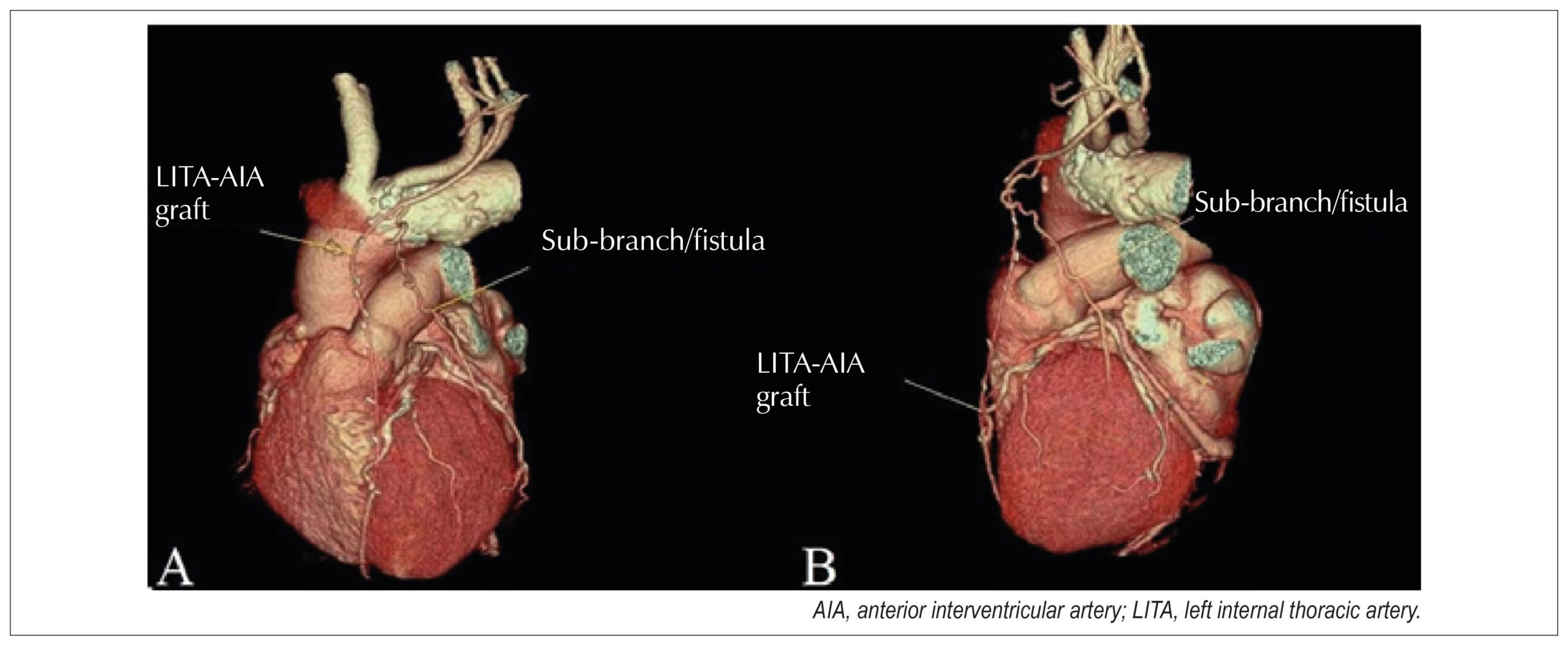
The left internal thoracic artery (LITA) fistula to the pulmonary artery (PA) and/or its branches is a rare condition that was first described in 1947 by Burchell and Clagett and can be acquired or congenital. Its etiologies include, trauma, infection, neoplasms, and mainly myocardial revascularization surgery (MRS)., The surgical manipulation of the intrathoracic vessels during MRS may predispose a patient to postoperative LITA fistulization to the PA, causing coronary flow leakage and myocardial ischemia. LITA should be considered an etiological diagnosis in cases of persistent angina after MRS., Its diagnosis requires coronary cineangiography,– and its treatment options include conservative drug therapy, surgical ligation, or endovascular intervention.

[…]

  • Arteries
  • Case report
  • Coronary Vessel Anomalies
  • Coronary Vessels
  • Diagnostic Imaging
  • Fistula
Read Full Article
Creative Commons Attribution (CC BY) Creative Commons license
26/11/2021 DOI: 10.47593/2675-312X/20213403eabc203
Figure 1
ABC
Case Report

Anomalous Origin of the Circumflex Coronary Artery from the Right Pulmonary Artery: Diagnosis Through Cardiac CT

Bebiana Faria et al. Lucy Calvo Sílvia Ribeiro Catarina Ruivo António Lourenço

Introduction

Anomalous origin of the left circumflex coronary artery (LCx) from the right pulmonary artery (PA) is an extremely rare coronary anomaly. Although the clinical course may be silent, the risk of sudden cardiac death is increased. The symptoms are related to collateralization and amount of myocardium that it supplies, and some patients need surgical treatment.,

[…]

  • Computed/methods
  • Congenital
  • Congenital Abnormalities
  • Coronary Artery Disease
  • Coronary Vessels
  • Heart Defects
  • Pulmonary Artery
  • Tomography
Read Full Article
Creative Commons Attribution (CC BY) Creative Commons license
19/07/2021 DOI: 10.36660/abc.20200060 Views 1
Figure 1
ABCIC
Case Report

Anomalous Origin of the Right Coronary from the Left Coronary Sinus: How to approach?

Danielle Campos de Almeida et al. Alice Mirane Malta Carrijo Marcela Gomes de Souza Fernando de Melo Martinelli … Fernando Roberto de Fazzio João Lucas O’Connell

Introduction

Coronary artery anomalies (CAAs) are characterized by a changed vessel origin, path, or structure. It is a rare condition, present in approximately 1% of the general population. These anomalies include the anomalous aortic origin of a coronary artery (AAOCA), which can travel retroaortic, subpulmonic, prepulmonic, and inter-arterial courses.

In general, these abnormalities remain asymptomatic until adulthood, being incidental findings on complementary tests or necropsy. However, they can also cause angina, syncope, ischemia, arrhythmias, or sudden death., Sudden death can be the first presentation of this pathology in young athletes, representing the second leading cause of death in this group.

[…]

  • Arteries
  • Coronary Vessel Anomalies
  • Coronary Vessels
  • Diagnostic Imaging
Read Full Article
Creative Commons Attribution (CC BY) Creative Commons license
16/06/2021 DOI: 10.47593/2675-312X/20213401eabc146 Views 1
Figure 1
ABC
Image

Radiologic-Electrocardiography Correlation in Wellens Syndrome

Eduardo Kaiser Ururahy Nunes Fonseca et al. Nevelton Heringer Filho Marcelo L. Montemor Luiz Francisco Rodrigues de Ávila Carlos Eduardo Rochitte

Wellens Syndrome, also known as “Anterior Descending Coronary T-wave syndrome”, was described in 1982 by Dr. Henrick Joan Joost (Hein) Wellens, a Dutch physician who also contributed to the characterization of the reentry mechanism in Wolff–Parkinson–White syndrome (WPWS).

Originally described during hospital admission (60% at admission and 40% at the follow-up) of patients with unstable angina, it was characterized by the occurrence of 2 electrocardiographic patterns, with pattern A in 25% of patients and B in 75% of patients.

[…]

  • Computed Tomography Angiography
  • Coronary Angiography
  • Coronary Vessels
  • Electrocardiography
  • Myocardial Infarction
Read Full Article
Creative Commons Attribution (CC BY) Creative Commons license
01/03/2021 DOI: 10.36660/abc.20200200 Views 1
ABC
Original Article

Safety, Efficacy, and Dose Protocol of Metoprolol for Heart Rate Reduction in Pediatric Outpatients Undergoing Cardiac CT Angiography

Mariana de Oliveira Nunes et al. Dawn R. Witt Susan A. Casey Larissa I. Stanberry … David J. Caye Bradford J.Chu B. Jana Lindberg John R. Lesser B. Kelly Han

Abstract

Background

Image quality and radiation dose are optimized with a slow, steady heart rate (HR) when imaging the coronary arteries during cardiac computed tomography angiography (CCTA). The safety, efficacy, and protocol for HR reduction with beta blocker medication is not well described in a pediatric patient population.

Objective

Provide a safe and efficient metoprolol dose protocol to be used in pediatric outpatients undergoing CCTA.

Methods

We conducted a retrospective review of all pediatric outpatients who received metoprolol during CCTA. Demographic and clinical characteristics were summarized and the average reduction in HR was estimated using a multivariate linear regression model. Images were evaluated on a 1-4 scale (1= optimal).

Results

Seventy-eight pediatric outpatients underwent a CCTA scan with the use of metoprolol. The median age was 13 years, median weight of 46 kg, and 36 (46%) were male. The median doses of metoprolol were 1.5 (IQR 1.1, 1.8) mg/kg and 0.4 (IQR 0.2, 0.7) mg/kg for oral and intravenous administrations, respectively. Procedural dose-length product was 57 (IQR 30, 119) mGy*cm. The average reduction in HR was 19 (IQR 12, 26) beats per minute, or 23%. No complications or adverse events were reported.

Conclusion

Use of metoprolol in a pediatric outpatient setting for HR reduction prior to CCTA is safe and effective. A metoprolol dose protocol can be reproduced when a slower HR is needed, ensuring faster acquisition times, clear images, and associated reduction in radiation exposure in this population. (Arq Bras Cardiol. 2021; 116(1):100-105)

  • Adrenergic, Antagonists
  • Computed Tomography
  • Coronary Vessels
  • Heart Defects, Congenital
  • Heart Rate
  • metoprolol
Read Full Article
Creative Commons Attribution (CC BY) Creative Commons license
03/02/2021 DOI: 10.36660/abc.20190892 Views 1
Figure 1
IJCS
Case Report

Acute Coronary Syndrome in a Patient With Single-Vessel Coronary Artery: Case Report

Bruno Dala Vedova Gomes Beato et al. Carolina de Souza Galvão Eduardo Belisário Falchetto Eduardo Kei Marquesini Washizu … Fábio Ávila Tofani Juliano Moreira Reis Filho

Introduction

Anomalies of coronary arteries affect 1-2% of the population. , They are associated with increased risk of sudden death and coronary artery injury during surgical procedures. , Among the coronary artery anomalies, the single-vessel coronary artery disease subtype has a low incidence – 0.03-0.04% in patients without other heart diseases and 0.024% in patients with other heart diseases. Concomitant presence of acute coronary syndrome in these patients may have an impact on their prognosis and survival.

Here we report a rare and challenging case of an 81-year-old woman with high-risk unstable angina undergoing emergency catheterization which indicated the presence of single-vessel coronary artery disease with lesions in multiple segments.

[…]

  • Acute coronary syndrome
  • Atherosclerosis
  • Cardiac Catheterization
  • Coronary Vessels
  • Myocardial Ischemia
  • stent
Read Full Article
Creative Commons Attribution (CC BY) Creative Commons license
15/07/2020 DOI: 10.36660/ijcs.20190211 Views 1
Figure 1
ABC
Case Report

Vasospasm during Exertion: New Pathophysiological Insights

Gonzalo Navarrete et al. Eduardo Pozo Fernando Rivero Teresa Bastante … Luis Jesús Jiménez-Borreguero Fernando Alfonso

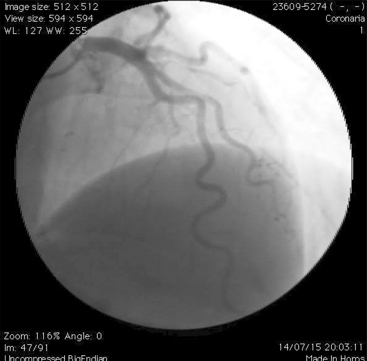
A 69-year-old male with hypertension, dyslipemia and no previous cardiovascular disease presented a transient ST-segment elevation on electrographic (ECG) monitoring during a planned ureteroscopy. The patient remained asymptomatic during the postoperative period. He only referred very sporadic episodes of effort chest pain in the last years without progression or events at rest. Given the lack of ECG registry, an exercise stress echocardiogram was indicated. The study was clinical and echocardiographically positive for inducible ischemia, with hypokinesia of the mid and apical anterior wall segments at peak exercise without ECG changes. With the documentation of ischemia in the left anterior descending coronary artery (LAD) territory, a coronary angiogram was performed. Only a mild lesion in the proximal LAD was demonstrated. However, a severe lesion in the proximal right coronary artery (RCA) ( and ), was treated with a bioabsorbable scaffold (), despite the absence of inducible ischemia demonstration in that territory. The patient was discharged on dual antiplatelet treatment, betablockers and statins and remained asymptomatic without new episodes of chest pain. Six months later, during another ureteroscopy, a new episode of transient ST-segment elevation was observed on ECG monitoring. At that time, a treadmill test was performed showing marked transient ST-segment elevation in V1-V4 leads (). Repeated coronary angiography demonstrated the good result of the previously implanted stent in RCA and persistence of a mild plaque in the proximal LAD. Due to the previously documented ECG changes in the right precordial leads, a pressure guide was performed in the proximal LAD, which ruled out the functional significance of this lesion (iFR 0.88; FFR 0.87 with 600 mcg Adenosine) (). In the light of the discordant ECG, echocardiographic and angiographic findings, a vasospasm test with intracoronary methylergonovin (20 mcg) was eventually carried out. This test was positive with angina reproduction and a significant reduction in LAD vessel diameter up to 90% diameter stenosis () with a concomitant Pd/Pa value of 0.60 (). Finally, the patient could be discharged under treatment with nitrates and calcium channel blockers, remaining asymptomatic at 1-year follow-up. An exercise stress echocardiogram was negative for inducible ischemia and repeated ureteroscopy procedures were uneventful.

[…]

  • Coronary Angiography
  • Coronary Vasoespasm/physiopathology
  • Coronary Vessels
  • Echocardiography
  • Electrocardiology
  • Myocardial Ischemia
Read Full Article
Creative Commons Attribution (CC BY) Creative Commons license
08/08/2019 DOI: 10.5935/abc.20190090 Views 1

Could not load more articles. Please try again.

How to cite this article

EndNote RIS

Electronic Document Format (Vancouver)

Hassan MA, Alzahrani A, Mhanna M, Abdelkarim O, Bello R, Savoia P, Suksaranjit P, et al. A Hidden Connection: Anomalous Left Circumflex Artery Arising From the Right Pulmonary Artery Unveiled by Cardiovascular Computed Tomography. ABC Imagem Cardiovascular 2026;39(2):e20260040.

Electronic Document Format (ABNT)

Hassan, Mubariz Ahmed; Alzahrani, Ashraf; Mhanna, Mohammad; Abdelkarim, Ola; Bello, Rodrigo; Savoia, Paulo; Suksaranjit, Promporn; Delcour, Kimberly. A Hidden Connection: Anomalous Left Circumflex Artery Arising From the Right Pulmonary Artery Unveiled by Cardiovascular Computed Tomography. ABC Imagem Cardiovascular, v. 39, n. 2, e20260040, Jul. 2026.

Electronic Document Format (APA)

Hassan, M. A., Alzahrani, A., Mhanna, M., Abdelkarim, O., Bello, R., Savoia, P., Suksaranjit, P., & Delcour, K. (2026). A Hidden Connection: Anomalous Left Circumflex Artery Arising From the Right Pulmonary Artery Unveiled by Cardiovascular Computed Tomography. ABC Imagem Cardiovascular, 39(2), e20260040.

Electronic Document Format (ISO)

Hassan, Mubariz Ahmed and Alzahrani, Ashraf and Mhanna, Mohammad and Abdelkarim, Ola and Bello, Rodrigo and Savoia, Paulo and Suksaranjit, Promporn and Delcour, Kimberly. A Hidden Connection: Anomalous Left Circumflex Artery Arising From the Right Pulmonary Artery Unveiled by Cardiovascular Computed Tomography. ABC Imagem Cardiovascular [online]. 2026, vol. 39, n. 2, [cited 2026-10-14], e20260040. Available from: <https://journals.cardiol.br/en/article/a-hidden-connection-anomalous-left-circumflex-artery-arising-from-the-right-pulmonary-artery-unveiled-by-cardiovascular-computed-tomography/>. ISSN 2318-8219

EndNote RIS

Electronic Document Format (Vancouver)

Ferreira MVS, Tormin JPAS, Dantas Jr RN, Torres RVA, Cordeiro RA, Araújo Filho JAB, Cardoso LF, et al. Single Coronary Artery and Stress Cardiomyopathy: An Association Demonstrated by Multimodality Imaging. ABC Imagem Cardiovascular 2026;39(2):e20260023.

Electronic Document Format (ABNT)

Ferreira, Marcus Vinicius Silva; Tormin, Julia Pereira Afonso dos Santos; Dantas Jr, Roberto Nery; Torres, Roberto Vitor Almeida; Cordeiro, Renan Andreos; Araújo Filho, José de Arimateia Batista; Cardoso, Luiz Francisco. Single Coronary Artery and Stress Cardiomyopathy: An Association Demonstrated by Multimodality Imaging. ABC Imagem Cardiovascular, v. 39, n. 2, e20260023, Jul. 2026.

Electronic Document Format (APA)

Ferreira, M. V. S., Tormin, J. P. A. S., Dantas Jr, R. N., Torres, R. V. A., Cordeiro, R. A., Araújo Filho, J. A. B., & Cardoso, L. F. (2026). Single Coronary Artery and Stress Cardiomyopathy: An Association Demonstrated by Multimodality Imaging. ABC Imagem Cardiovascular, 39(2), e20260023.

Electronic Document Format (ISO)

Ferreira, Marcus Vinicius Silva and Tormin, Julia Pereira Afonso dos Santos and Dantas Jr, Roberto Nery and Torres, Roberto Vitor Almeida and Cordeiro, Renan Andreos and Araújo Filho, José de Arimateia Batista and Cardoso, Luiz Francisco. Single Coronary Artery and Stress Cardiomyopathy: An Association Demonstrated by Multimodality Imaging. ABC Imagem Cardiovascular [online]. 2026, vol. 39, n. 2, [cited 2026-10-14], e20260023. Available from: <https://journals.cardiol.br/en/article/single-coronary-artery-and-stress-cardiomyopathy-an-association-demonstrated-by-multimodality-imaging/>. ISSN 2318-8219

EndNote RIS

Electronic Document Format (Vancouver)

Luz PLD, Favarato D, Abizaid AA, Cesar LAM, Serrano Jr. CV, Chagas ACP, Rochitte CE, et al. Coronary Artery Calcium Score, Risk Factors, and Clinical Outcomes in Nonobstructive Coronary Artery Disease: A Long-Term Follow-Up Study. Arquivos Brasileiros de Cardiologia 2026;123(5):e20250772.

Electronic Document Format (ABNT)

Luz, Protásio Lemos Da; Favarato, Desiderio; Abizaid, Alexandre A.C.; Cesar, Luiz Antônio Machado; Serrano Jr., Carlos Vicente; Chagas, Antônio Carlos Palandri; Rochitte, Carlos E.; Gutierrez, Marco A.. Coronary Artery Calcium Score, Risk Factors, and Clinical Outcomes in Nonobstructive Coronary Artery Disease: A Long-Term Follow-Up Study. Arquivos Brasileiros de Cardiologia, v. 123, n. 5, e20250772, Jul. 2026.

Electronic Document Format (APA)

Luz, P. L. D., Favarato, D., Abizaid, A. A., Cesar, L. A. M., Serrano Jr., C. V., Chagas, A. C. P., Rochitte, C. E., & Gutierrez, M. A. (2026). Coronary Artery Calcium Score, Risk Factors, and Clinical Outcomes in Nonobstructive Coronary Artery Disease: A Long-Term Follow-Up Study. Arquivos Brasileiros de Cardiologia, 123(5), e20250772.

Electronic Document Format (ISO)

Luz, Protásio Lemos Da and Favarato, Desiderio and Abizaid, Alexandre A.C. and Cesar, Luiz Antônio Machado and Serrano Jr., Carlos Vicente and Chagas, Antônio Carlos Palandri and Rochitte, Carlos E. and Gutierrez, Marco A.. Coronary Artery Calcium Score, Risk Factors, and Clinical Outcomes in Nonobstructive Coronary Artery Disease: A Long-Term Follow-Up Study. Arquivos Brasileiros de Cardiologia [online]. 2026, vol. 123, n. 5, [cited 2026-10-14], e20250772. Available from: <https://journals.cardiol.br/en/article/coronary-artery-calcium-score-risk-factors-and-clinical-outcomes-in-nonobstructive-coronary-artery-disease-a-long-term-follow-up-study/>. ISSN 0066-782X

EndNote RIS

Electronic Document Format (Vancouver)

Arruda JA, Costa Junior JR. Small Vessels, Big Decisions. Arquivos Brasileiros de Cardiologia 2026;123(2):e20260207.

Electronic Document Format (ABNT)

Arruda, José Airton de; Costa Junior, José de Ribamar. Small Vessels, Big Decisions. Arquivos Brasileiros de Cardiologia, v. 123, n. 2, e20260207, May. 2026.

Electronic Document Format (APA)

Arruda, J. A. , & Costa Junior, J. R. (2026). Small Vessels, Big Decisions. Arquivos Brasileiros de Cardiologia, 123(2), e20260207.

Electronic Document Format (ISO)

Arruda, José Airton de and Costa Junior, José de Ribamar. Small Vessels, Big Decisions. Arquivos Brasileiros de Cardiologia [online]. 2026, vol. 123, n. 2, [cited 2026-10-14], e20260207. Available from: <https://journals.cardiol.br/en/article/small-vessels-big-decisions/>. ISSN 0066-782X

EndNote RIS

Electronic Document Format (Vancouver)

Abdo MH, Latsh H, Wagner M, König G, Barth S. Coexistence of Partial Anomalous Pulmonary Venous Connection and Coronary Artery Fistulas: A Rare Case Report. ABC Imagem Cardiovascular 2026;39(1):e20250030.

Electronic Document Format (ABNT)

Abdo, Mourad Haj; Latsh, Hussain; Wagner, Mathias; König, George; Barth, Sebastian. Coexistence of Partial Anomalous Pulmonary Venous Connection and Coronary Artery Fistulas: A Rare Case Report. ABC Imagem Cardiovascular, v. 39, n. 1, e20250030, May. 2026.

Electronic Document Format (APA)

Abdo, M. H., Latsh, H., Wagner, M., König, G., & Barth, S. (2026). Coexistence of Partial Anomalous Pulmonary Venous Connection and Coronary Artery Fistulas: A Rare Case Report. ABC Imagem Cardiovascular, 39(1), e20250030.

Electronic Document Format (ISO)

Abdo, Mourad Haj and Latsh, Hussain and Wagner, Mathias and König, George and Barth, Sebastian. Coexistence of Partial Anomalous Pulmonary Venous Connection and Coronary Artery Fistulas: A Rare Case Report. ABC Imagem Cardiovascular [online]. 2026, vol. 39, n. 1, [cited 2026-10-14], e20250030. Available from: <https://journals.cardiol.br/en/article/coexistence-of-partial-anomalous-pulmonary-venous-connection-and-coronary-artery-fistulas-a-rare-case-report/>. ISSN 2318-8219

EndNote RIS

Electronic Document Format (Vancouver)

Garzon S, Pilotto L, Mariani J, Bezerra F, Prado G, Bandeira W, Almeida B, et al. Estimating Left Main Bifurcation Geometry: Are Geometric Models Reliable in Diseased Arteries?. Arquivos Brasileiros de Cardiologia 2026;123(1):e20250605.

Electronic Document Format (ABNT)

Garzon, Stefano; Pilotto, Lucas; Mariani, José; Bezerra, Felipe; Prado, Guy; Bandeira, Willterson; Almeida, Breno; Lemos, Pedro. Estimating Left Main Bifurcation Geometry: Are Geometric Models Reliable in Diseased Arteries?. Arquivos Brasileiros de Cardiologia, v. 123, n. 1, e20250605, Mar. 2026.

Electronic Document Format (APA)

Garzon, S., Pilotto, L., Mariani, J., Bezerra, F., Prado, G., Bandeira, W., Almeida, B., & Lemos, P. (2026). Estimating Left Main Bifurcation Geometry: Are Geometric Models Reliable in Diseased Arteries?. Arquivos Brasileiros de Cardiologia, 123(1), e20250605.

Electronic Document Format (ISO)

Garzon, Stefano and Pilotto, Lucas and Mariani, José and Bezerra, Felipe and Prado, Guy and Bandeira, Willterson and Almeida, Breno and Lemos, Pedro. Estimating Left Main Bifurcation Geometry: Are Geometric Models Reliable in Diseased Arteries?. Arquivos Brasileiros de Cardiologia [online]. 2026, vol. 123, n. 1, [cited 2026-10-14], e20250605. Available from: <https://journals.cardiol.br/en/article/estimating-left-main-bifurcation-geometry-are-geometric-models-reliable-in-diseased-arteries/>. ISSN 0066-782X

EndNote RIS

Electronic Document Format (Vancouver)

Ömür SE, Koyun E, Cabri G. Cardiac CT-Detected Vulnerable Plaques and Risk of Future Acute Myocardial Infarction: Predictive Value of CAC Scores and Inflammatory Burden Index. Arquivos Brasileiros de Cardiologia 2026;122(11):e20250052.

Electronic Document Format (ABNT)

Ömür, Sefa Erdi; Koyun, Emin; Cabri, Gökhan. Cardiac CT-Detected Vulnerable Plaques and Risk of Future Acute Myocardial Infarction: Predictive Value of CAC Scores and Inflammatory Burden Index. Arquivos Brasileiros de Cardiologia, v. 122, n. 11, e20250052, Jan. 2026.

Electronic Document Format (APA)

Ömür, S. E., Koyun, E., & Cabri, G. (2026). Cardiac CT-Detected Vulnerable Plaques and Risk of Future Acute Myocardial Infarction: Predictive Value of CAC Scores and Inflammatory Burden Index. Arquivos Brasileiros de Cardiologia, 122(11), e20250052.

Electronic Document Format (ISO)

Ömür, Sefa Erdi and Koyun, Emin and Cabri, Gökhan. Cardiac CT-Detected Vulnerable Plaques and Risk of Future Acute Myocardial Infarction: Predictive Value of CAC Scores and Inflammatory Burden Index. Arquivos Brasileiros de Cardiologia [online]. 2026, vol. 122, n. 11, [cited 2026-10-14], e20250052. Available from: <https://journals.cardiol.br/en/article/cardiac-ct-detected-vulnerable-plaques-and-risk-of-future-acute-myocardial-infarction-predictive-value-of-cac-scores-and-inflammatory-burden-index/>. ISSN 0066-782X

EndNote RIS

Electronic Document Format (Vancouver)

Bichuette LD, Calderaro D, Lemos Neto PA, Fonseca EKUN, Tatagiba LS, Vieira TM, Nascimento YPP, et al. Left Main Coronary Artery Angioplasty for the Treatment of Extrinsic Compression in Patients with Pulmonary Hypertension. Arquivos Brasileiros de Cardiologia 2025;122(7):e20250002.

Electronic Document Format (ABNT)

Bichuette, Luciana Dornfeld; Calderaro, Daniela; Lemos Neto, Pedro Alves; Fonseca, Eduardo Kaiser Ururahy Nunes; Tatagiba, Luiza Sarmento; Vieira, Tulio Martins; Nascimento, Yally Priscila Pessoa; Castro, Marcela Araujo; Silva, Taysa Antônia Félix; Parente, Yuri de Deus Montalverne; Fernandes, Caio Julio; Cardozo, Francisco Akira Malta; Nomura, Cesar Higa; Marins, Pedro Henrique Almeida; Souza, Rogerio; Jardim, Carlos Vianna Poyares. Left Main Coronary Artery Angioplasty for the Treatment of Extrinsic Compression in Patients with Pulmonary Hypertension. Arquivos Brasileiros de Cardiologia, v. 122, n. 7, e20250002, Jul. 2025.

Electronic Document Format (APA)

Bichuette, L. D., Calderaro, D., Lemos Neto, P. A., Fonseca, E. K. U. N., Tatagiba, L. S., Vieira, T. M., Nascimento, Y. P. P., Castro, M. A., Silva, T. A. F., Parente, Y. D. M., Fernandes, C. J., Cardozo, F. A. M., Nomura, C. H., Marins, P. H. A., Souza, R., & Jardim, C. V. P. (2025). Left Main Coronary Artery Angioplasty for the Treatment of Extrinsic Compression in Patients with Pulmonary Hypertension. Arquivos Brasileiros de Cardiologia, 122(7), e20250002.

Electronic Document Format (ISO)

Bichuette, Luciana Dornfeld and Calderaro, Daniela and Lemos Neto, Pedro Alves and Fonseca, Eduardo Kaiser Ururahy Nunes and Tatagiba, Luiza Sarmento and Vieira, Tulio Martins and Nascimento, Yally Priscila Pessoa and Castro, Marcela Araujo and Silva, Taysa Antônia Félix and Parente, Yuri de Deus Montalverne and Fernandes, Caio Julio and Cardozo, Francisco Akira Malta and Nomura, Cesar Higa and Marins, Pedro Henrique Almeida and Souza, Rogerio and Jardim, Carlos Vianna Poyares. Left Main Coronary Artery Angioplasty for the Treatment of Extrinsic Compression in Patients with Pulmonary Hypertension. Arquivos Brasileiros de Cardiologia [online]. 2025, vol. 122, n. 7, [cited 2026-10-14], e20250002. Available from: <https://journals.cardiol.br/en/article/left-main-coronary-artery-angioplasty-for-the-treatment-of-extrinsic-compression-in-patients-with-pulmonary-hypertension/>. ISSN 0066-782X

EndNote RIS

Electronic Document Format (Vancouver)

Neves IF, Ferreira A, Almeida I, Mano TB, Sousa L. Silent Giant: Right Coronary Artery Ectasia with a Hidden Fistula to the Coronary Sinus in an Asymptomatic Patient. Arquivos Brasileiros de Cardiologia 2025;122(6):e20240574.

Electronic Document Format (ABNT)

Neves, Inês Ferreira; Ferreira, André; Almeida, Inês; Mano, Tânia Branco; Sousa, Lídia de. Silent Giant: Right Coronary Artery Ectasia with a Hidden Fistula to the Coronary Sinus in an Asymptomatic Patient. Arquivos Brasileiros de Cardiologia, v. 122, n. 6, e20240574, Jul. 2025.

Electronic Document Format (APA)

Neves, I. F., Ferreira, A., Almeida, I., Mano, T. B., & Sousa, L. (2025). Silent Giant: Right Coronary Artery Ectasia with a Hidden Fistula to the Coronary Sinus in an Asymptomatic Patient. Arquivos Brasileiros de Cardiologia, 122(6), e20240574.

Electronic Document Format (ISO)

Neves, Inês Ferreira and Ferreira, André and Almeida, Inês and Mano, Tânia Branco and Sousa, Lídia de. Silent Giant: Right Coronary Artery Ectasia with a Hidden Fistula to the Coronary Sinus in an Asymptomatic Patient. Arquivos Brasileiros de Cardiologia [online]. 2025, vol. 122, n. 6, [cited 2026-10-14], e20240574. Available from: <https://journals.cardiol.br/en/article/silent-giant-right-coronary-artery-ectasia-with-a-hidden-fistula-to-the-coronary-sinus-in-an-asymptomatic-patient/>. ISSN 0066-782X

EndNote RIS

Electronic Document Format (Vancouver)

Zapparoli M, Alessi A, Zapparoli FC, Alessi MR. Rare Presentation of an Anatomical Congenital Anomaly of the Coronary Arteries on Computed Tomography Angiography. ABC Imagem Cardiovascular 2024;37(4):e20240075.

Electronic Document Format (ABNT)

Zapparoli, Marcello; Alessi, Alexandre; Zapparoli, Franco Camargo; Alessi, Mateus Rodrigues. Rare Presentation of an Anatomical Congenital Anomaly of the Coronary Arteries on Computed Tomography Angiography. ABC Imagem Cardiovascular, v. 37, n. 4, e20240075, Nov. 2024.

Electronic Document Format (APA)

Zapparoli, M., Alessi, A., Zapparoli, F. C., & Alessi, M. R. (2024). Rare Presentation of an Anatomical Congenital Anomaly of the Coronary Arteries on Computed Tomography Angiography. ABC Imagem Cardiovascular, 37(4), e20240075.

Electronic Document Format (ISO)

Zapparoli, Marcello and Alessi, Alexandre and Zapparoli, Franco Camargo and Alessi, Mateus Rodrigues. Rare Presentation of an Anatomical Congenital Anomaly of the Coronary Arteries on Computed Tomography Angiography. ABC Imagem Cardiovascular [online]. 2024, vol. 37, n. 4, [cited 2026-10-14], e20240075. Available from: <https://journals.cardiol.br/en/article/rare-presentation-of-an-anatomical-congenital-anomaly-of-the-coronary-arteries-on-computed-tomography-angiography/>. ISSN 2318-8219

EndNote RIS

Electronic Document Format (Vancouver)

Alam M, Sanhueza E, Baranchuk A. Venturing into Uncharted Territories: Unveiling Frontal QRS-T Angle in Patients with Coronary Artery Ectasia. International Journal of Cardiovascular Sciences 2024;37:e20240049.

Electronic Document Format (ABNT)

Alam, Maqsood; Sanhueza, Eduardo; Baranchuk, Adrian. Venturing into Uncharted Territories: Unveiling Frontal QRS-T Angle in Patients with Coronary Artery Ectasia. International Journal of Cardiovascular Sciences, v. 37, e20240049, Jun. 2024.

Electronic Document Format (APA)

Alam, M., Sanhueza, E., & Baranchuk, A. (2024). Venturing into Uncharted Territories: Unveiling Frontal QRS-T Angle in Patients with Coronary Artery Ectasia. International Journal of Cardiovascular Sciences, 37, e20240049.

Electronic Document Format (ISO)

Alam, Maqsood and Sanhueza, Eduardo and Baranchuk, Adrian. Venturing into Uncharted Territories: Unveiling Frontal QRS-T Angle in Patients with Coronary Artery Ectasia. International Journal of Cardiovascular Sciences [online]. 2024, vol. 37, [cited 2026-10-14], e20240049. Available from: <https://journals.cardiol.br/en/article/venturing-into-uncharted-territories-unveiling-frontal-qrs-t-angle-in-patients-with-coronary-artery-ectasia/>. ISSN 2359-4802

EndNote RIS

Electronic Document Format (Vancouver)

Bianco HT. Index of Systemic Immunoinflammation and Ischemia in Patients with Non-Obstructive Coronary Arteries. Arquivos Brasileiros de Cardiologia 2024;121(2):e20240081.

Electronic Document Format (ABNT)

Bianco, Henrique Tria. Index of Systemic Immunoinflammation and Ischemia in Patients with Non-Obstructive Coronary Arteries. Arquivos Brasileiros de Cardiologia, v. 121, n. 2, e20240081, Apr. 2024.

Electronic Document Format (APA)

Bianco, H. T. (2024). Index of Systemic Immunoinflammation and Ischemia in Patients with Non-Obstructive Coronary Arteries. Arquivos Brasileiros de Cardiologia, 121(2), e20240081.

Electronic Document Format (ISO)

Bianco, Henrique Tria. Index of Systemic Immunoinflammation and Ischemia in Patients with Non-Obstructive Coronary Arteries. Arquivos Brasileiros de Cardiologia [online]. 2024, vol. 121, n. 2, [cited 2026-10-14], e20240081. Available from: <https://journals.cardiol.br/en/article/index-of-systemic-immunoinflammation-and-ischemia-in-patients-with-non-obstructive-coronary-arteries/>. ISSN 0066-782X

EndNote RIS

Electronic Document Format (Vancouver)

Netto SM, Souza RVO, Murad V, Mesquita ET. Coronary Artery Microfistulas: Historical Description and Contemporary Clinical Relevance. International Journal of Cardiovascular Sciences 2023;36:e20230092.

Electronic Document Format (ABNT)

Netto, Stans Murad; Souza, Rachel Vianna O’Neill de; Murad, Victor; Mesquita, Evandro Tinoco. Coronary Artery Microfistulas: Historical Description and Contemporary Clinical Relevance. International Journal of Cardiovascular Sciences, v. 36, e20230092, Dec. 2023.

Electronic Document Format (APA)

Netto, S. M., Souza, R. V. O. , Murad, V., & Mesquita, E. T. (2023). Coronary Artery Microfistulas: Historical Description and Contemporary Clinical Relevance. International Journal of Cardiovascular Sciences, 36, e20230092.

Electronic Document Format (ISO)

Netto, Stans Murad and Souza, Rachel Vianna O’Neill de and Murad, Victor and Mesquita, Evandro Tinoco. Coronary Artery Microfistulas: Historical Description and Contemporary Clinical Relevance. International Journal of Cardiovascular Sciences [online]. 2023, vol. 36, [cited 2026-10-14], e20230092. Available from: <https://journals.cardiol.br/en/article/coronary-artery-microfistulas-historical-description-and-contemporary-clinical-relevance/>. ISSN 2359-4802

EndNote RIS

Electronic Document Format (Vancouver)

Scopel KRO, Medeiros TM, Maicá BNP, Velho MC, Silva MMD, Mattos JN, Heuser GG, et al. Incidental Tomographic Findings of Coronary Artery Calcifications: A Prevalence Study in Southern Brazil. Arquivos Brasileiros de Cardiologia Imagem cardiovascular 2023;36(1):e368.

Electronic Document Format (ABNT)

Scopel, Karen Rafaela Okaseski; Medeiros, Tássia Machado; Maicá, Bibiana Natalia Porto; Velho, Maria Carbonari; Silva, Mariana Motta Dias da; Mattos, Juliane Nascimento; Heuser, Guilherme Galante; Winkelmann, Eliane Roseli. Incidental Tomographic Findings of Coronary Artery Calcifications: A Prevalence Study in Southern Brazil. Arquivos Brasileiros de Cardiologia Imagem cardiovascular, v. 36, n. 1, e368, Mar. 2023.

Electronic Document Format (APA)

Scopel, K. R. O., Medeiros, T. M., Maicá, B. N. P., Velho, M. C., Silva, M. M. D. , Mattos, J. N., Heuser, G. G., & Winkelmann, E. R. (2023). Incidental Tomographic Findings of Coronary Artery Calcifications: A Prevalence Study in Southern Brazil. Arquivos Brasileiros de Cardiologia Imagem cardiovascular, 36(1), e368.

Electronic Document Format (ISO)

Scopel, Karen Rafaela Okaseski and Medeiros, Tássia Machado and Maicá, Bibiana Natalia Porto and Velho, Maria Carbonari and Silva, Mariana Motta Dias da and Mattos, Juliane Nascimento and Heuser, Guilherme Galante and Winkelmann, Eliane Roseli. Incidental Tomographic Findings of Coronary Artery Calcifications: A Prevalence Study in Southern Brazil. Arquivos Brasileiros de Cardiologia Imagem cardiovascular [online]. 2023, vol. 36, n. 1, [cited 2026-10-14], e368. Available from: <https://journals.cardiol.br/en/article/incidental-tomographic-findings-of-coronary-artery-calcifications-a-prevalence-study-in-southern-brazil/>. ISSN 2318-8219

EndNote RIS

Electronic Document Format (Vancouver)

Estrada A, Sousa AS, Mesquita CT, Villacorta H. Coronary Tortuosity as a New Phenotype for Ischemia without Coronary Artery Disease. Arquivos Brasileiros de Cardiologia 2022;119(6):883-90.

Electronic Document Format (ABNT)

Estrada, André; Sousa, André Silveira; Mesquita, Claudio Tinoco; Villacorta, Humberto. Coronary Tortuosity as a New Phenotype for Ischemia without Coronary Artery Disease. Arquivos Brasileiros de Cardiologia, v. 119, n. 6, p. 883-890, Sep. 2022.

Electronic Document Format (APA)

Estrada, A., Sousa, A. S., Mesquita, C. T., & Villacorta, H. (2022). Coronary Tortuosity as a New Phenotype for Ischemia without Coronary Artery Disease. Arquivos Brasileiros de Cardiologia, 119(6), 883-890.

Electronic Document Format (ISO)

Estrada, André and Sousa, André Silveira and Mesquita, Claudio Tinoco and Villacorta, Humberto. Coronary Tortuosity as a New Phenotype for Ischemia without Coronary Artery Disease. Arquivos Brasileiros de Cardiologia [online]. 2022, vol. 119, n. 6, [cited 2026-10-14], pp. 883-890. Available from: <https://journals.cardiol.br/en/article/coronary-tortuosity-as-a-new-phenotype-for-ischemia-without-coronary-artery-disease-2/>. ISSN 0066-782X

EndNote RIS

Electronic Document Format (Vancouver)

Adali MK, Buber I, Sen G, Yilmaz S. Relationship between Systemic Immune-Inflammation Index and Coronary Collateral Circulation in Patients with Chronic Total Occlusion. Arquivos Brasileiros de Cardiologia 2022;119(1):69-75.

Electronic Document Format (ABNT)

Adali, Mehmet Koray; Buber, Ipek; Sen, Gursel; Yilmaz, Samet. Relationship between Systemic Immune-Inflammation Index and Coronary Collateral Circulation in Patients with Chronic Total Occlusion. Arquivos Brasileiros de Cardiologia, v. 119, n. 1, p. 69-75, Jun. 2022.

Electronic Document Format (APA)

Adali, M. K., Buber, I., Sen, G., & Yilmaz, S. (2022). Relationship between Systemic Immune-Inflammation Index and Coronary Collateral Circulation in Patients with Chronic Total Occlusion. Arquivos Brasileiros de Cardiologia, 119(1), 69-75.

Electronic Document Format (ISO)

Adali, Mehmet Koray and Buber, Ipek and Sen, Gursel and Yilmaz, Samet. Relationship between Systemic Immune-Inflammation Index and Coronary Collateral Circulation in Patients with Chronic Total Occlusion. Arquivos Brasileiros de Cardiologia [online]. 2022, vol. 119, n. 1, [cited 2026-10-14], pp. 69-75. Available from: <https://journals.cardiol.br/en/article/relationship-between-systemic-immune-inflammation-index-and-coronary-collateral-circulation-in-patients-with-chronic-total-occlusion/>. ISSN 0066-782X

EndNote RIS

Electronic Document Format (Vancouver)

Saylik F, Akbulut T. Systemic Immune-Inflammation Index Predicts Major Cardiovascular Adverse Events in Patients with ST-Segment Elevated Myocardial Infarction. Arquivos Brasileiros de Cardiologia 2022;119(1):14-22.

Electronic Document Format (ABNT)

Saylik, Faysal; Akbulut, Tayyar. Systemic Immune-Inflammation Index Predicts Major Cardiovascular Adverse Events in Patients with ST-Segment Elevated Myocardial Infarction. Arquivos Brasileiros de Cardiologia, v. 119, n. 1, p. 14-22, May. 2022.

Electronic Document Format (APA)

Saylik, F., & Akbulut, T. (2022). Systemic Immune-Inflammation Index Predicts Major Cardiovascular Adverse Events in Patients with ST-Segment Elevated Myocardial Infarction. Arquivos Brasileiros de Cardiologia, 119(1), 14-22.

Electronic Document Format (ISO)

Saylik, Faysal and Akbulut, Tayyar. Systemic Immune-Inflammation Index Predicts Major Cardiovascular Adverse Events in Patients with ST-Segment Elevated Myocardial Infarction. Arquivos Brasileiros de Cardiologia [online]. 2022, vol. 119, n. 1, [cited 2026-10-14], pp. 14-22. Available from: <https://journals.cardiol.br/en/article/systemic-immune-inflammation-index-predicts-major-cardiovascular-adverse-events-in-patients-with-st-segment-elevated-myocardial-infarction/>. ISSN 0066-782X

EndNote RIS

Electronic Document Format (Vancouver)

Souza MG, Carrijo AMM, Aratani VF, Arantes FBB. Left Internal Thoracic Artery Fistula to the Bronchial Artery: a Rare Cause of Coronary Flow Theft. ABC Imagem Cardiovascular 2021;34(3):eabc203.

Electronic Document Format (ABNT)

Souza, Marcela Gomes de; Carrijo, Alice Mirane Malta; Aratani, Vinicius Ferreira; Arantes, Flávia Bittar Britto. Left Internal Thoracic Artery Fistula to the Bronchial Artery: a Rare Cause of Coronary Flow Theft. ABC Imagem Cardiovascular, v. 34, n. 3, eabc203, Nov. 2021.

Electronic Document Format (APA)

Souza, M. G. , Carrijo, A. M. M., Aratani, V. F., & Arantes, F. B. B. (2021). Left Internal Thoracic Artery Fistula to the Bronchial Artery: a Rare Cause of Coronary Flow Theft. ABC Imagem Cardiovascular, 34(3), eabc203.

Electronic Document Format (ISO)

Souza, Marcela Gomes de and Carrijo, Alice Mirane Malta and Aratani, Vinicius Ferreira and Arantes, Flávia Bittar Britto. Left Internal Thoracic Artery Fistula to the Bronchial Artery: a Rare Cause of Coronary Flow Theft. ABC Imagem Cardiovascular [online]. 2021, vol. 34, n. 3, [cited 2026-10-14], eabc203. Available from: <https://journals.cardiol.br/en/article/left-internal-thoracic-artery-fistula-to-the-bronchial-artery-a-rare-cause-of-coronary-flow-theft/>. ISSN 2318-8219

EndNote RIS

Electronic Document Format (Vancouver)

Faria B, Calvo L, Ribeiro S, Ruivo C, Lourenço A. Anomalous Origin of the Circumflex Coronary Artery from the Right Pulmonary Artery: Diagnosis Through Cardiac CT. Arquivos Brasileiros de Cardiologia 2021;117(1 suppl 1):8-11.

Electronic Document Format (ABNT)

Faria, Bebiana; Calvo, Lucy; Ribeiro, Sílvia; Ruivo, Catarina; Lourenço, António. Anomalous Origin of the Circumflex Coronary Artery from the Right Pulmonary Artery: Diagnosis Through Cardiac CT. Arquivos Brasileiros de Cardiologia, v. 117, n. 1 suppl 1, p. 8-11, Jul. 2021.

Electronic Document Format (APA)

Faria, B., Calvo, L., Ribeiro, S., Ruivo, C., & Lourenço, A. (2021). Anomalous Origin of the Circumflex Coronary Artery from the Right Pulmonary Artery: Diagnosis Through Cardiac CT. Arquivos Brasileiros de Cardiologia, 117(1 suppl 1), 8-11.

Electronic Document Format (ISO)

Faria, Bebiana and Calvo, Lucy and Ribeiro, Sílvia and Ruivo, Catarina and Lourenço, António. Anomalous Origin of the Circumflex Coronary Artery from the Right Pulmonary Artery: Diagnosis Through Cardiac CT. Arquivos Brasileiros de Cardiologia [online]. 2021, vol. 117, n. 1 suppl 1, [cited 2026-10-14], pp. 8-11. Available from: <https://journals.cardiol.br/en/article/anomalous-origin-of-the-circumflex-coronary-artery-from-the-right-pulmonary-artery-diagnosis-through-cardiac-ct/>. ISSN 0066-782X

EndNote RIS

Electronic Document Format (Vancouver)

Almeida DC, Carrijo AMM, Souza MG, Martinelli FM, Fazzio FR, O’Connell JL. Anomalous Origin of the Right Coronary from the Left Coronary Sinus: How to approach?. ABC Imagem Cardiovascular 2021;34(1):eabc146.

Electronic Document Format (ABNT)

Almeida, Danielle Campos de; Carrijo, Alice Mirane Malta; Souza, Marcela Gomes de; Martinelli, Fernando de Melo; Fazzio, Fernando Roberto de; O’Connell, João Lucas. Anomalous Origin of the Right Coronary from the Left Coronary Sinus: How to approach?. ABC Imagem Cardiovascular, v. 34, n. 1, eabc146, Jun. 2021.

Electronic Document Format (APA)

Almeida, D. C. , Carrijo, A. M. M., Souza, M. G. , Martinelli, F. M., Fazzio, F. R. , & O’Connell, J. L. (2021). Anomalous Origin of the Right Coronary from the Left Coronary Sinus: How to approach?. ABC Imagem Cardiovascular, 34(1), eabc146.

Electronic Document Format (ISO)

Almeida, Danielle Campos de and Carrijo, Alice Mirane Malta and Souza, Marcela Gomes de and Martinelli, Fernando de Melo and Fazzio, Fernando Roberto de and O’Connell, João Lucas. Anomalous Origin of the Right Coronary from the Left Coronary Sinus: How to approach?. ABC Imagem Cardiovascular [online]. 2021, vol. 34, n. 1, [cited 2026-10-14], eabc146. Available from: <https://journals.cardiol.br/en/article/anomalous-origin-of-the-right-coronary-from-the-left-coronary-sinus-how-to-approach/>. ISSN 2318-8219

EndNote RIS

Electronic Document Format (Vancouver)

Fonseca EKUN, Heringer Filho N, Montemor ML, Ávila LFR, Rochitte CE. Radiologic-Electrocardiography Correlation in Wellens Syndrome. Arquivos Brasileiros de Cardiologia 2021;116(2):363-6.

Electronic Document Format (ABNT)

Fonseca, Eduardo Kaiser Ururahy Nunes; Heringer Filho, Nevelton; Montemor, Marcelo L.; Ávila, Luiz Francisco Rodrigues de; Rochitte, Carlos Eduardo. Radiologic-Electrocardiography Correlation in Wellens Syndrome. Arquivos Brasileiros de Cardiologia, v. 116, n. 2, p. 363-366, Feb. 2021.

Electronic Document Format (APA)

Fonseca, E. K. U. N., Heringer Filho, N., Montemor, M. L., Ávila, L. F. R. , & Rochitte, C. E. (2021). Radiologic-Electrocardiography Correlation in Wellens Syndrome. Arquivos Brasileiros de Cardiologia, 116(2), 363-366.

Electronic Document Format (ISO)

Fonseca, Eduardo Kaiser Ururahy Nunes and Heringer Filho, Nevelton and Montemor, Marcelo L. and Ávila, Luiz Francisco Rodrigues de and Rochitte, Carlos Eduardo. Radiologic-Electrocardiography Correlation in Wellens Syndrome. Arquivos Brasileiros de Cardiologia [online]. 2021, vol. 116, n. 2, [cited 2026-10-14], pp. 363-366. Available from: <https://journals.cardiol.br/en/article/radiologic-electrocardiography-correlation-in-wellens-syndrome/>. ISSN 0066-782X

EndNote RIS

Electronic Document Format (Vancouver)

Nunes MO, Witt DR, Casey SA, Stanberry LI, Caye DJ, J.Chu B, Lindberg BJ, et al. Safety, Efficacy, and Dose Protocol of Metoprolol for Heart Rate Reduction in Pediatric Outpatients Undergoing Cardiac CT Angiography. Arquivos Brasileiros de Cardiologia 2021;116(1):100-5.

Electronic Document Format (ABNT)

Nunes, Mariana de Oliveira; Witt, Dawn R.; Casey, Susan A.; Stanberry, Larissa I.; Caye, David J.; J.Chu, Bradford; Lindberg, B. Jana; Lesser, John R.; Han, B. Kelly. Safety, Efficacy, and Dose Protocol of Metoprolol for Heart Rate Reduction in Pediatric Outpatients Undergoing Cardiac CT Angiography. Arquivos Brasileiros de Cardiologia, v. 116, n. 1, p. 100-105, Feb. 2021.

Electronic Document Format (APA)

Nunes, M. O., Witt, D. R., Casey, S. A., Stanberry, L. I., Caye, D. J., J.Chu, B., Lindberg, B. J., Lesser, J. R., & Han, B. K. (2021). Safety, Efficacy, and Dose Protocol of Metoprolol for Heart Rate Reduction in Pediatric Outpatients Undergoing Cardiac CT Angiography. Arquivos Brasileiros de Cardiologia, 116(1), 100-105.

Electronic Document Format (ISO)

Nunes, Mariana de Oliveira and Witt, Dawn R. and Casey, Susan A. and Stanberry, Larissa I. and Caye, David J. and J.Chu, Bradford and Lindberg, B. Jana and Lesser, John R. and Han, B. Kelly. Safety, Efficacy, and Dose Protocol of Metoprolol for Heart Rate Reduction in Pediatric Outpatients Undergoing Cardiac CT Angiography. Arquivos Brasileiros de Cardiologia [online]. 2021, vol. 116, n. 1, [cited 2026-10-14], pp. 100-105. Available from: <https://journals.cardiol.br/en/article/safety-efficacy-and-dose-protocol-of-metoprolol-for-heart-rate-reduction-in-pediatric-outpatients-undergoing-cardiac-ct-angiography/>. ISSN 0066-782X

EndNote RIS

Electronic Document Format (Vancouver)

Beato BDVG, Galvão CS, Falchetto EB, Washizu EKM, Tofani F, Reis Filho JM. Acute Coronary Syndrome in a Patient With Single-Vessel Coronary Artery: Case Report. International Journal of Cardiovascular Sciences 2020;34(3):326-9.

Electronic Document Format (ABNT)

Beato, Bruno Dala Vedova Gomes; Galvão, Carolina de Souza; Falchetto, Eduardo Belisário; Washizu, Eduardo Kei Marquesini; Tofani, Fábio Ávila; Reis Filho, Juliano Moreira. Acute Coronary Syndrome in a Patient With Single-Vessel Coronary Artery: Case Report. International Journal of Cardiovascular Sciences, v. 34, n. 3, p. 326-329, Jul. 2020.

Electronic Document Format (APA)

Beato, B. D. V. G., Galvão, C. S., Falchetto, E. B., Washizu, E. K. M., Tofani, F. , & Reis Filho, J. M. (2020). Acute Coronary Syndrome in a Patient With Single-Vessel Coronary Artery: Case Report. International Journal of Cardiovascular Sciences, 34(3), 326-329.

Electronic Document Format (ISO)

Beato, Bruno Dala Vedova Gomes and Galvão, Carolina de Souza and Falchetto, Eduardo Belisário and Washizu, Eduardo Kei Marquesini and Tofani, Fábio Ávila and Reis Filho, Juliano Moreira. Acute Coronary Syndrome in a Patient With Single-Vessel Coronary Artery: Case Report. International Journal of Cardiovascular Sciences [online]. 2020, vol. 34, n. 3, [cited 2026-10-14], pp. 326-329. Available from: <https://journals.cardiol.br/en/article/acute-coronary-syndrome-in-a-patient-with-single-vessel-coronary-artery-case-report/>. ISSN 2359-4802

EndNote RIS

Electronic Document Format (Vancouver)

Navarrete G, Pozo E, Rivero F, Bastante T, Jiménez-Borreguero LJ, Alfonso F. Vasospasm during Exertion: New Pathophysiological Insights. Arquivos Brasileiros de Cardiologia 2019;113(1):106-8.

Electronic Document Format (ABNT)

Navarrete, Gonzalo; Pozo, Eduardo; Rivero, Fernando; Bastante, Teresa; Jiménez-Borreguero, Luis Jesús; Alfonso, Fernando. Vasospasm during Exertion: New Pathophysiological Insights. Arquivos Brasileiros de Cardiologia, v. 113, n. 1, p. 106-108, Aug. 2019.

Electronic Document Format (APA)

Navarrete, G., Pozo, E., Rivero, F., Bastante, T., Jiménez-Borreguero, L. J., & Alfonso, F. (2019). Vasospasm during Exertion: New Pathophysiological Insights. Arquivos Brasileiros de Cardiologia, 113(1), 106-108.

Electronic Document Format (ISO)

Navarrete, Gonzalo and Pozo, Eduardo and Rivero, Fernando and Bastante, Teresa and Jiménez-Borreguero, Luis Jesús and Alfonso, Fernando. Vasospasm during Exertion: New Pathophysiological Insights. Arquivos Brasileiros de Cardiologia [online]. 2019, vol. 113, n. 1, [cited 2026-10-14], pp. 106-108. Available from: <https://journals.cardiol.br/en/article/vasospasm-during-exertion-new-pathophysiological-insights/>. ISSN 0066-782X

  • About ABC Family
  • Internal Regulation
  • Editorial Board
  • Brazilian Society of Cardiology (SBC)

Issues

SBC Guidelines

  • Videos
  • Podcasts
  • News