ABC Family Arquivos Brasileiros de Cardiologia International Journal of Cardiovascular Sciences ABC Heart Failure & Cardiomyopathy ABC Imagem Cardiovascular
Português English
ABC Family
Português English
  • About
    • About ABC Family
    • Internal Regulation
    • Editorial Board
    • Brazilian Society of Cardiology (SBC)
  • Issues
  • SBC Guidelines
  • Content
    • Videos
    • Podcasts
    • News
  • About
    • About ABC Family
    • Internal Regulation
    • Editorial Board
    • Brazilian Society of Cardiology (SBC)
  • Issues
  • SBC Guidelines
  • Content
    • Videos
    • Podcasts
    • News

ABC Family

  • ABC Family
  • Arquivos Brasileiros de Cardiologia
  • International Journal of Cardiovascular Sciences
  • ABC Heart Failure & Cardiomyopathy
  • ABC Imagem Cardiovascular
Search in

No content found.

Journals
Ordering
Article types
Others

  1. Home
  2. Keyword: Heart Atria
Figure 1
ABCIC
Case Report

Recurrent Intracardiac Masses in an Orthotopic Heart Transplant Recipient

Bilal Saeed et al. Natdanai Punnanithinont Shareef Mansour Paulo Savoia Promporn Suksaranjit

Introduction

As long-term survival improves among heart transplant recipients, rare post-transplant complications, including intracardiac masses, are being increasingly recognized.

Severe left atrial (LA) dilation and atrial arrhythmias may further contribute to blood stasis and thrombus formation in transplant recipients. LA thrombi in these patients may mimic neoplastic masses and represent important diagnostic and therapeutic challenges. Contributing factors include atrial dilation, arrhythmias, foreign material, and the components of Virchow’s triad, namely abnormal blood flow, endothelial injury, and hypercoagulability.,

[…]

  • Cardiac MRI
  • Heart Atria
  • Heart Transplant
  • Left atrial
  • X-Ray Computed Tomography
Read Full Article
Creative Commons Attribution (CC BY) Creative Commons license
24/07/2026 DOI: 10.36660/abcimg.20260031i Views 2
Figure 1
ABC
Letter to the Editor

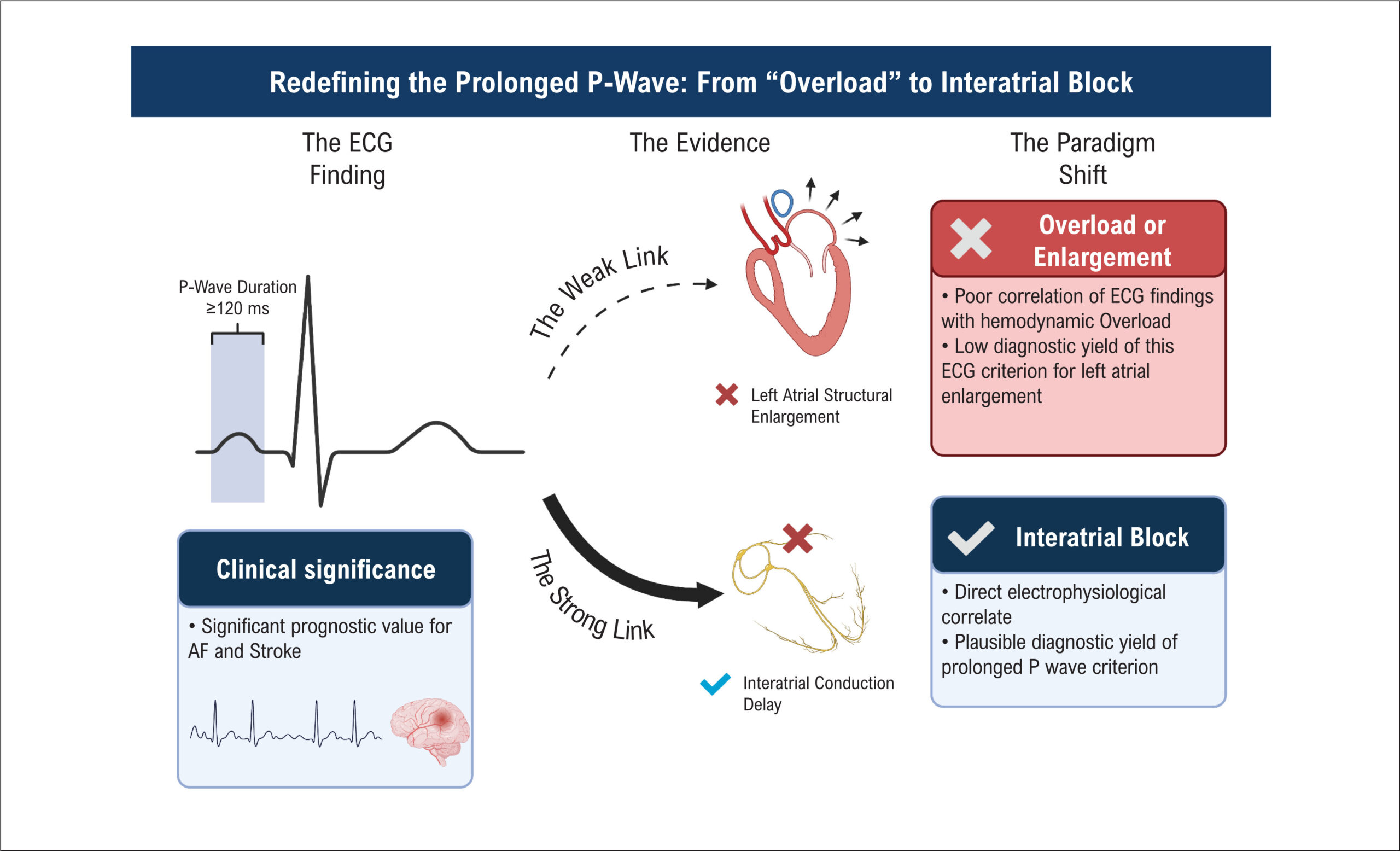
The End of "Left Atrial Overload"

José Nunes de Alencar et al. Daniel Teixeira de Campos e Barreiro Adrian Baranchuk

To the Editor,

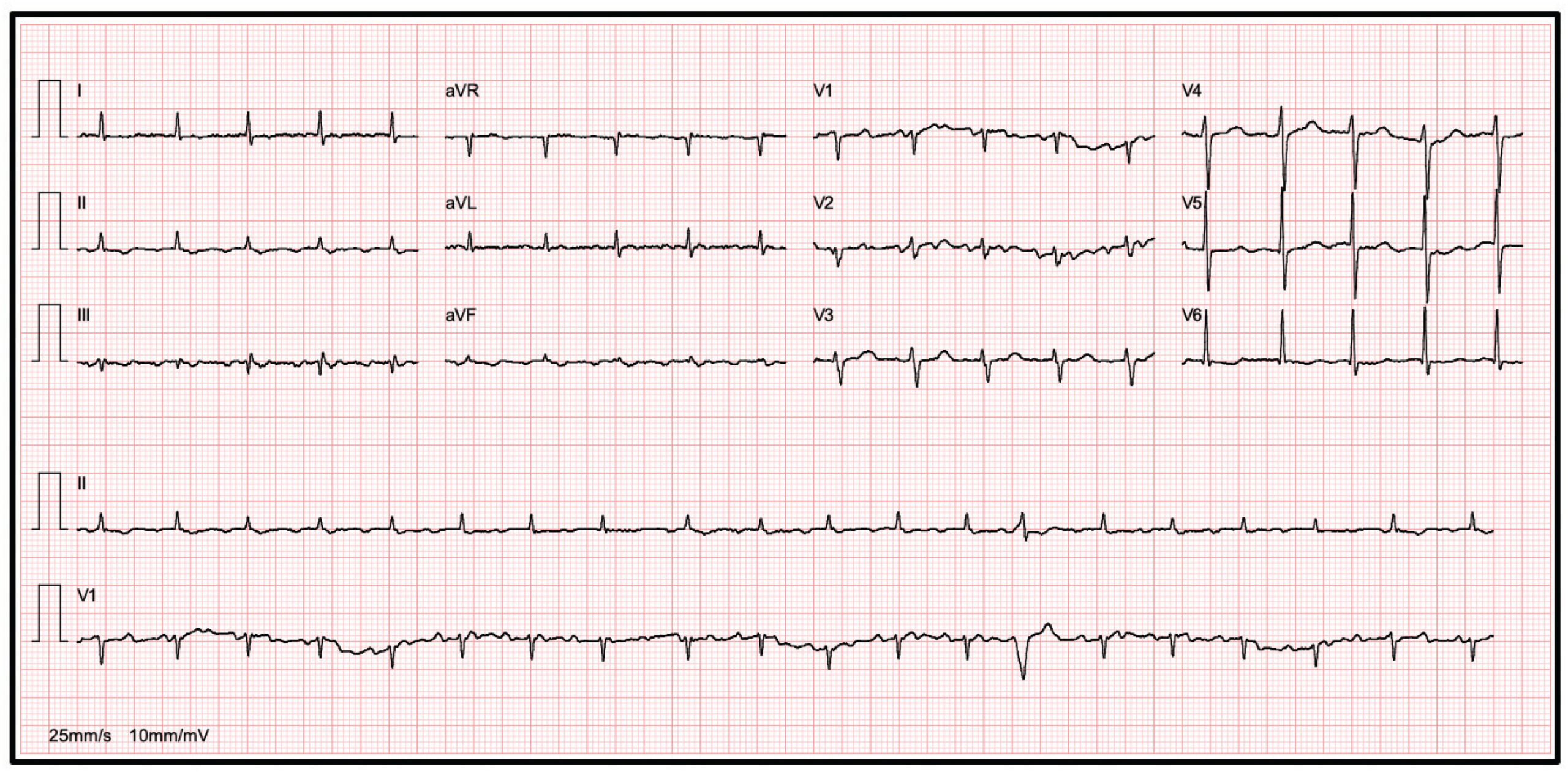
We read with great interest the article by Oliveira et al. The authors are to be commended for conducting a study of this magnitude and rigor. While previous literature relied on smaller sample sizes or non-standard/obsolete echocardiographic cutoffs for left atrial enlargement (LAE),– the ELSA-Brasil cohort provides the statistical power necessary to settle a longstanding debate in clinical cardiology. We believe this research is destined to become a reference point in standard electrocardiography textbooks.

[…]

  • Diagnosis
  • Electrocardiography
  • Heart Atria
Read Full Article
Creative Commons Attribution (CC BY) Creative Commons license
01/06/2026 DOI: 10.36660/abc.20250814i Views 1
Figure 1
ABCIC
Editorial

Left Atrium Reservoir Strain and Cardiovascular Hemodynamics: the Hidden Truths Reveal Far More Than What is Readily Apparent

Renato A. Hortegal et al. David Le Bihan Rodrigo Barretto Wilson Mathias Jr

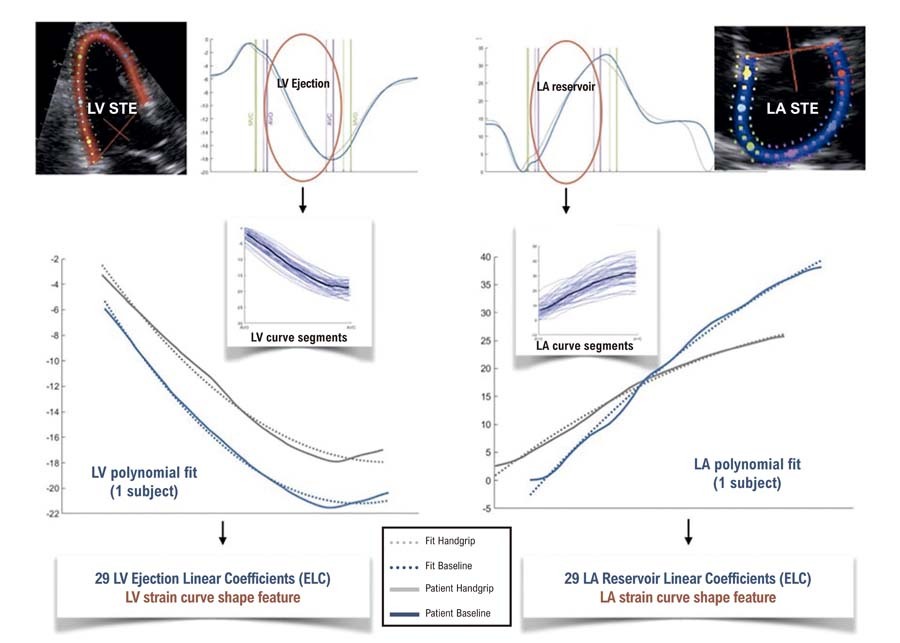
Over the past 15 years, Speckle-tracking Echocardiography has evolved into a robust technique for assessing both global and regional myocardial function. Among its expanding applications, Left Atrial (LA) strain, particularly in the reservoir phase, has emerged as a clinically relevant parameter. A reduced Left Atrial Strain reservoir (LASr) has been independently associated with elevated Left Ventricular (LV) filling pressures, recurrence of atrial fibrillation, heart failure hospitalizations, and overall cardiovascular mortality., Reflecting this growing body of evidence, LASr has been formally incorporated into clinical practice in the recently published “Recommendations for the Evaluation of Left Ventricular Diastolic Function by Echocardiography and for Heart Failure With Preserved Ejection Fraction Diagnosis: An Update From the American Society of Echocardiography.”

Despite this, the complexity of LA mechanics has largely followed an oversimplified approach: it is often treated as a surrogate marker of filling pressures, evaluated in isolation and interpreted using fixed cut-off values, without integrating the hemodynamic and mechanical context in which the atrium operates. While such an approach has facilitated its clinical adoption, it may obscure important physiological nuances and lead to misinterpretation in complex scenarios.

[…]

  • Atrial Function
  • Echocardiography
  • Heart Atria
  • Hemodynamics
Read Full Article
Creative Commons Attribution (CC BY) Creative Commons license
16/12/2025 DOI: 10.36660/abc.20250068i Views 2
Diagnostic Utility of P-Wave Duration in Identifying Left Atrial Enlargement: Insights from the ELSA-Brasil Cohort
ABC
Original Article

Diagnostic Utility of P-Wave Duration in Identifying Left Atrial Enlargement: Insights from the ELSA-Brasil Cohort

Ana Cecília de Sena Oliveira et al. Vinicius Tostes Carvalho Marcelo Martins Pinto-Filho Luisa Caldeira Brant … Sandhi Maria Barreto Murilo Foppa Antonio Luiz Pinho Ribeiro

Abstract

Background

Atrial cardiomyopathy encompasses various atrial abnormalities, including left atrial enlargement (LAE), which is associated with adverse cardiovascular and neurocognitive outcomes. P-wave duration ≥120 ms on 12-lead electrocardiogram (ECG) is a widely available, cost-effective tool and one of the main diagnostic criteria adopted. However, its diagnostic performance in large-scale studies remains underexplored.

Objectives

To evaluate the correlation between P-wave duration and left atrial volume (LAV) measured by transthoracic echocardiogram (TTE) and determine the diagnostic accuracy of P-wave duration ≥120 ms for identifying LAE.

Methods

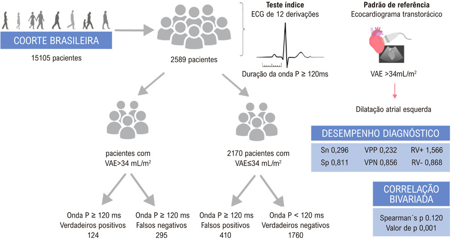
We conducted a cross-sectional analysis of baseline data from the ELSA-Brasil cohort participants in sinus rhythm. P-wave duration was automatically measured via standard 12-lead ECG, with ≥120 ms indicating enlargement. LAE was defined as LAV>34 mL/m2 by TTE. Spearman’s rank test assessed correlation. Diagnostic performance metrics – sensitivity (Sn), specificity (Sp), positive predictive value (PPV), and negative predictive value (NPV), positive and negative likelihood ratio (LR+ and LR-) – were calculated. Also, a Receiver Operating Characteristic (ROC) was constructed. Statistical significance was set at p-value<0.05 with 95% confidence interval.

Results

The study included 2589 participants, 419 (16,1%) with LAV index>34 mL/m2. P-wave duration correlated weakly with LAV (Spearman’s ρ = 0.120, p < 0.001). Diagnostic accuracy measurements were AUC = 0.557 ± 0.032; Sn = 0.296; Sp = 0.811; PPV = 0.232; NPV = 0.856; LR+ 1.566; LR- 0.868.

Conclusion

P-wave duration demonstrates limited diagnostic value for identifying LAE, suggesting against its recommendation as a standalone diagnostic criterion for this condition.

  • Cardiomyopathies
  • Echocardiography
  • Electrocardiography
  • Heart Atria
Read Full Article
Creative Commons Attribution (CC BY) Creative Commons license
15/12/2025 DOI: 10.36660/abc.20250151i Views 1
Figure 1
ABCIC
Case Report

Giant Left Atrium in Rheumatic Mitral Valve Disease

Alice Cunha Darzé et al. Antonio Tito Paladino Filho Amanda Marsiaj Rassi Carlos Eduardo Prazeres … Ibraim Masciarelli Francisco Pinto João Marcelo Caneschi Jose Roberto Tuma da Ponte Junior Paul Salvador Morales Rafael Castro Hendges Tiago Senra

Abstract

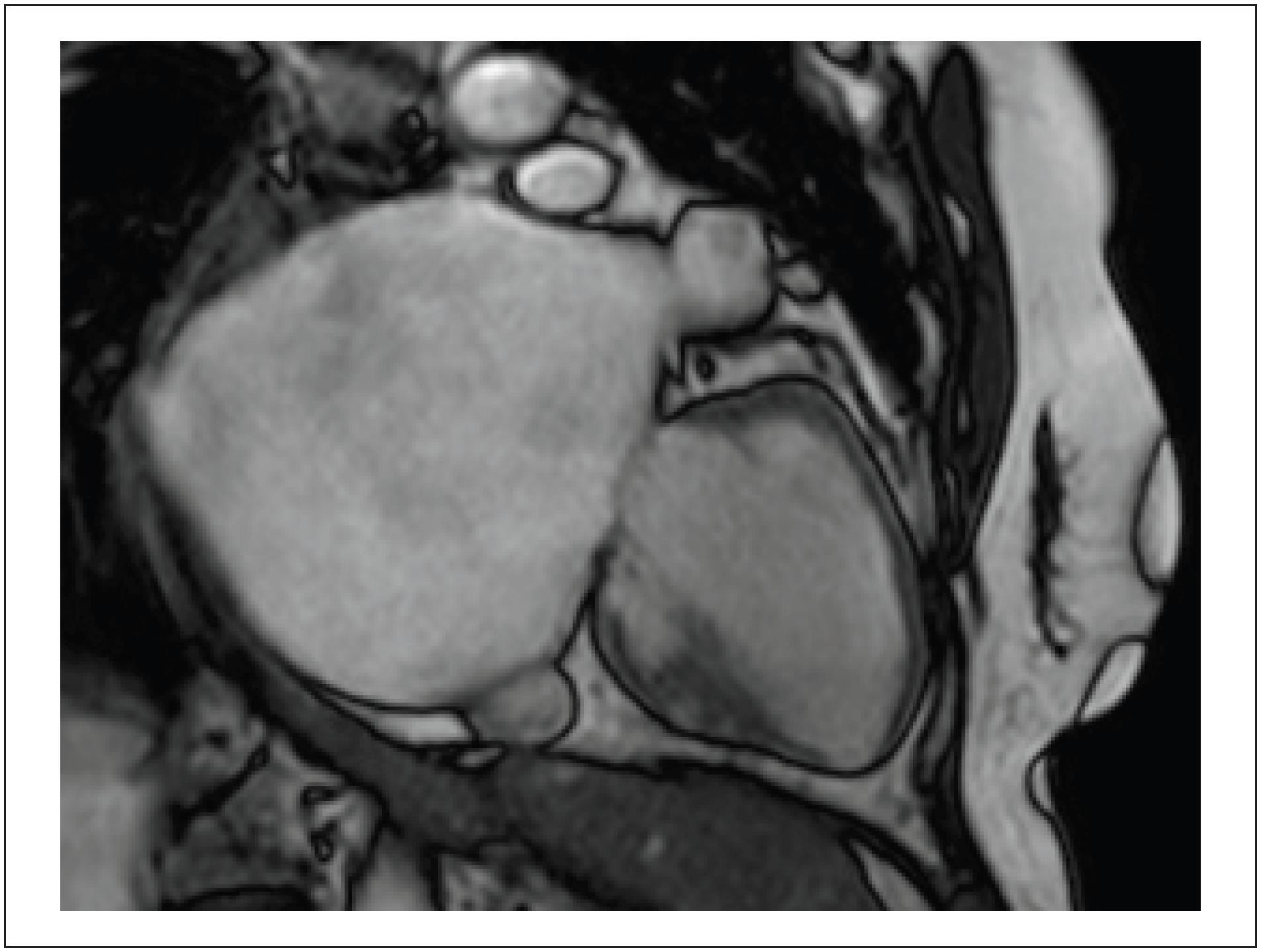
The Giant Left Atrium (GLA) is a rare condition, more commonly related to rheumatic mitral valve disease; it can be mistaken with a few possible differential diagnoses when less accurate methods are implemented, potentially leading to particularly dangerous and unnecessary procedures in this setting. Cardiovascular imaging methods, such as Cardiac Magnetic Resonance (CMR), can be valuable for this differentiation and provide high spatial resolution, supporting accurate diagnosis and surgical planning when indicated. Selecting the most precise diagnostic method, however, requires deep knowledge of the GLA condition and clinical data, leading to a correct suspicion. Furthermore, opting for using volumetry rather than the biplanar method in cases of significant deformity ensures greater accuracy in the values obtained.

  • Heart Atria
  • Heart Valve Diseases
  • Magnetic Resonance Imaging
Read Full Article
Creative Commons Attribution (CC BY) Creative Commons license
05/12/2024 DOI: 10.36660/abcimg.20240083i Views 1
Figure 1
IJCS
Case Report

Six-Year Follow-Up of an Octogenarian With a Left Atrial Myxoma: Case Report

Miguel Bernardino Antunes Vicente et al. Tânia Amaro Elsa Fernandes

Abstract

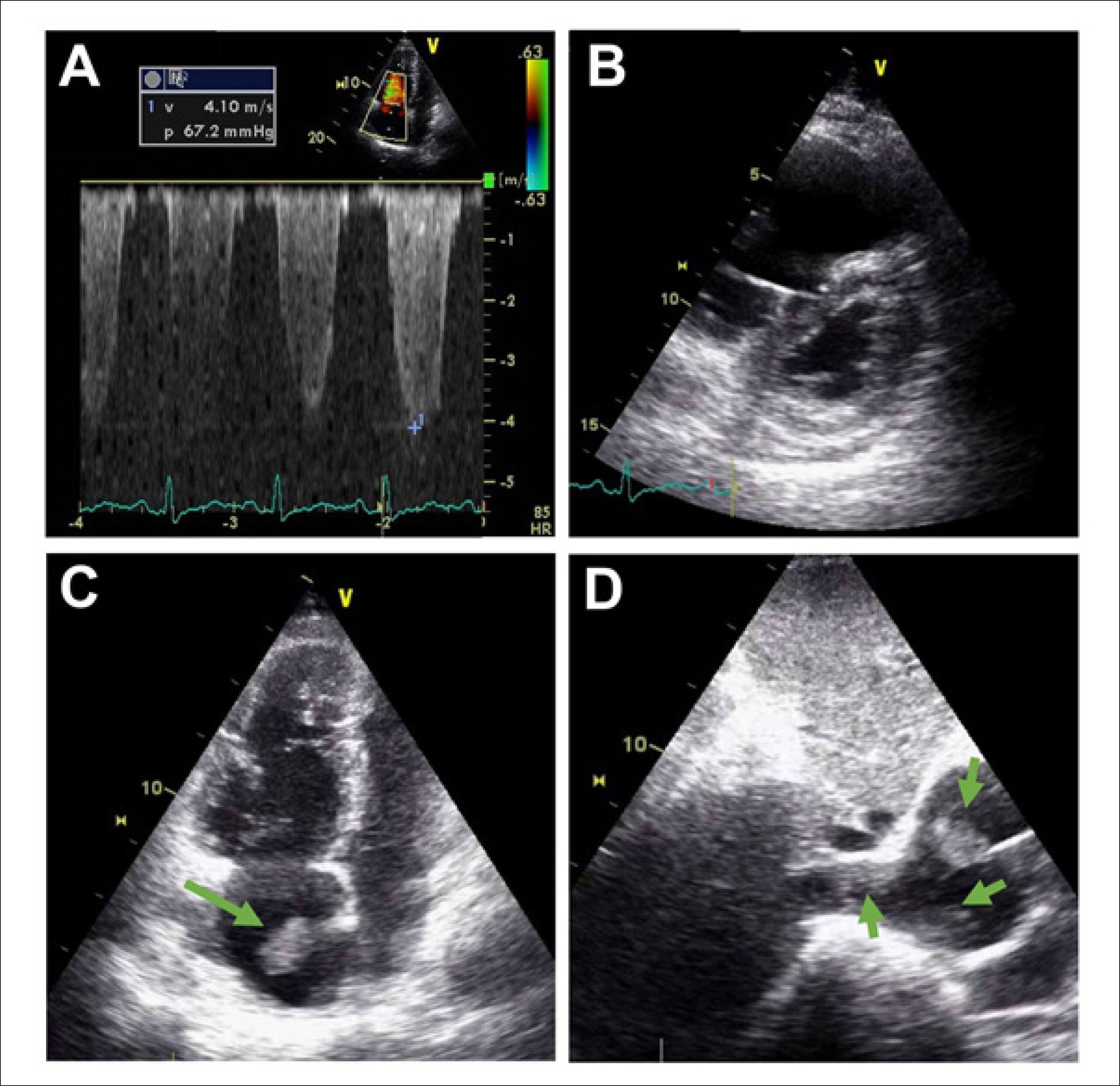
Myxoma is the most common primary cardiac tumour, especially in adults. However, reports on this nosological entity in patients over the age of 80 are scarce in the medical literature. Echocardiography is a useful diagnostic tool for detecting cardiac myxomas. These tumours are usually surgically removed soon after diagnosis, limiting knowledge of their natural evolution. In this report, we describe the case of an 86-year-old woman who had been followed irregularly for arterial hypertension since 2016, when an echocardiogram showed a left atrial myxoma adhered to the interatrial septum, initially without impairment of the mitral valve (MV) or obstruction of the pulmonary veins (PV). The patient refused surgical removal at the time. Six years later, she was hospitalized for urinary sepsis. A new echocardiogram revealed an increase in the size of the myxoma, which occupied around 80% of the left atrium (LA), obstructing the PV and MV. New suggestion of surgery was refused by the patient.

  • Case Reports
  • Heart Atria
  • Myxoma
Read Full Article
Creative Commons Attribution (CC BY) Creative Commons license
02/12/2024 DOI: 10.36660/ijcs.20240135 Views 2
ABCHF
Image

Cardiac Tumor in the Left Atrium

Henrique Iahnke Garbin et al. Matheus Piccoli Machado Klauberg Géris Mazzutti

A

[…]

  • Heart Atria
  • Heart Failure
  • Heart Neoplasms
  • Ultrasonography
Read Full Article
Creative Commons Attribution (CC BY) Creative Commons license
07/08/2024 DOI: 10.36660/abchf.20240004i
ABC
Editorial

Is Left Atrial Strain the Missing Part to Diagnose Diastolic Dysfunction in Children with Chronic Kidney Disease?

José Luiz Barros Pena

Echocardiography is the most critical imaging modality for assessing diastolic function, an essential part of the cardiac function evaluation.

Since its publication in 2016, the ASE/EACVI recommendations for evaluating diastolic function have been validated in different cohorts but have shown conflicting results, especially when considering patients with preserved ejection fraction (EF). When patients with preserved EF were studied in a separate subgroup, a poorer sensitivity was observed.

[…]

  • Child
  • Echocardiography/methods
  • Heart Atria
  • Kidney
Read Full Article
Creative Commons Attribution (CC BY) Creative Commons license
08/07/2024 DOI: 10.36660/abc.20240257i
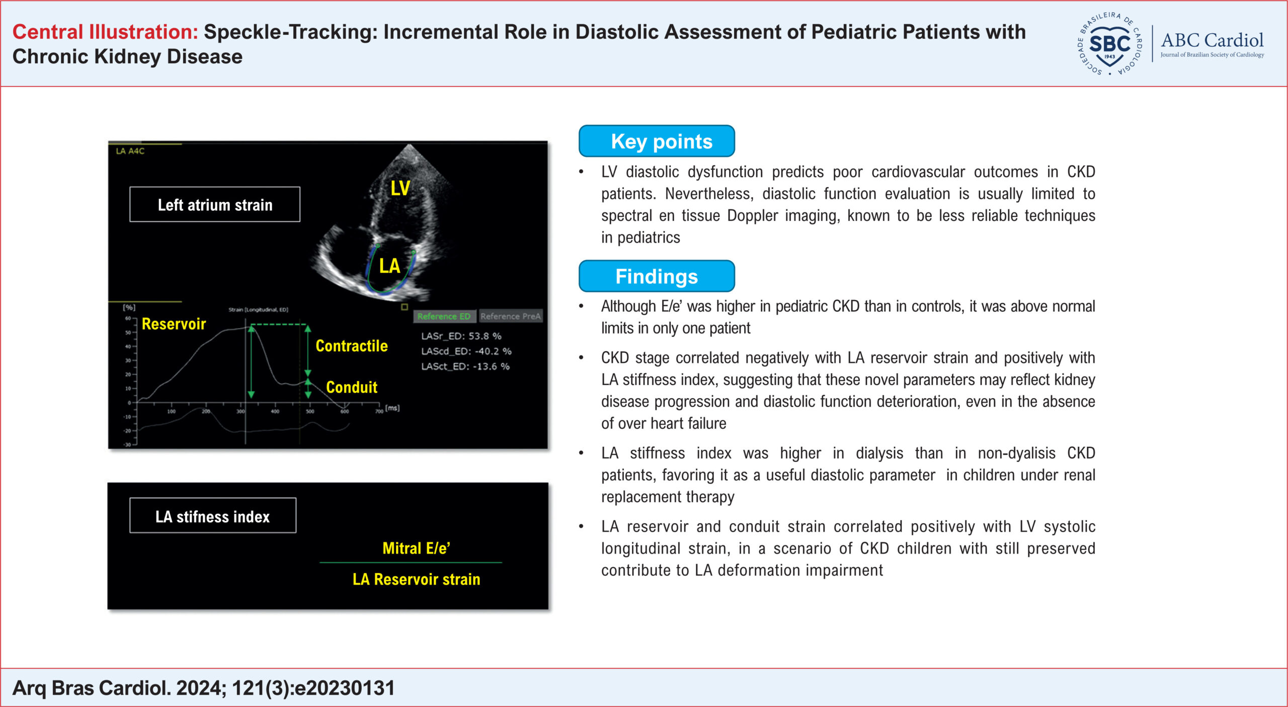
Speckle-Tracking: Incremental Role in Diastolic Assessment of Pediatric Patients with Chronic Kidney Disease
ABC
Original Article

Speckle-Tracking: Incremental Role in Diastolic Assessment of Pediatric Patients with Chronic Kidney Disease

Flora Maciel Penachio et al. Maria de Fátima Rodrigues Diniz Rosana Sbruzzi Prado Laurino Andreia Watanabe … Karen Saori Shiraishi Sawamura Alessandro Cavalcanti Lianza Carolina Rocha Brito Menezes Isabela de Sousa Lobo Silva Gabriela Nunes Leal

Abstract

Background:

Cardiovascular complications are the leading cause of mortality in pediatric patients with chronic kidney disease (CKD). Echocardiographic assessment of diastolic function in CKD has been limited to spectral and tissue Doppler imaging, known to be less reliable techniques in pediatrics. Two-dimensional Speckle tracking echocardiography (2DST) derived left atrial (LA) strain has recently been confirmed as a robust measure of diastolic function.

Objectives:

To investigate LA strain role in diastolic assessment of children at different stages of CKD.

Methods:

From February 2019 to July 2022, 55 CKD patients without cardiovascular symptoms and 55 controls were evaluated by standard and 2DST echocardiograms. The level of significance was set at 5% (p<0.05).

Results:

Patients and controls had similar age [9.78 (0.89 – 17.54) vs. 10.72 (1.03 –18,44) years; p = 0.41] and gender (36M:19F vs. 34M:21F; p=0.84). There were 25 non-dialysis patients and 30 dialysis patients. Left ventricular ejection fraction was ≥ 55% in all of them. Comparing CKD and controls, LA reservoir strain was lower (48.22±10.62% vs. 58.52±10.70%) and LA stiffness index was higher [0.14 (0.08–0.48)%-1 vs. 0.11 (0.06–0.23) %-1]; p<0.0001. LV hypertrophy was associated with lower LA reservoir strain (42.05±8.74% vs. 52.99±9.52%), higher LA stiffness [0.23(0.11 – 0.48)%-1 vs. 0.13 (0.08–0.23) %-1 and filling indexes (2.39±0.63 cm/s x %-1 vs. 1.74±0.47 cm/s x %-1; p<0.0001. Uncontrolled hypertension was associated with lower LA reservoir strain (41.9±10.6% vs. 50.6±9.7; p=0.005).

Conclusions:

LA strain proved to be a feasible tool in the assessment of pediatric CKD patients and was associated with known cardiovascular risk factors.

  • Child
  • Echocardiography
  • Heart Atria
  • Kidney
Read Full Article
Creative Commons Attribution (CC BY) Creative Commons license
26/04/2024 DOI: 10.36660/abc.20230131i Views 1
Figure 1
ABCIC
Case Report

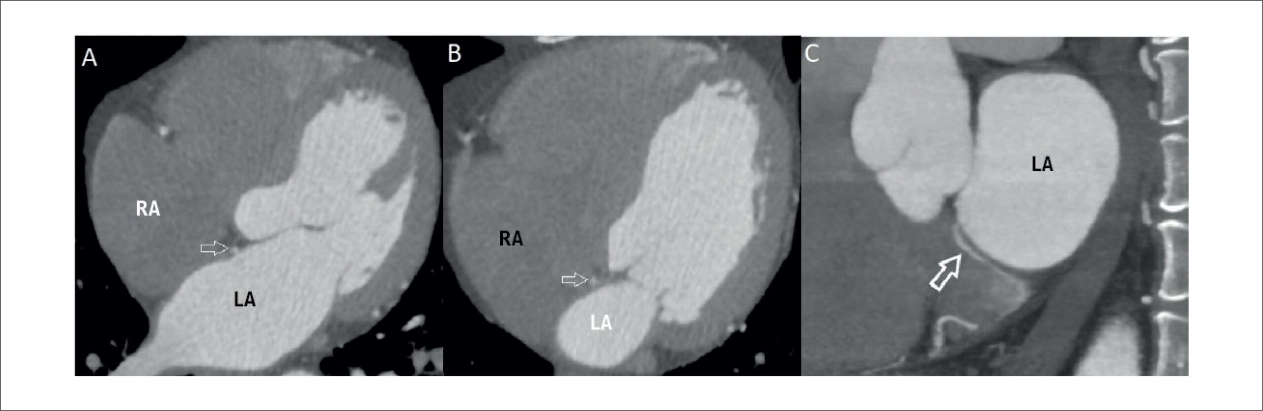
A Bridging Vein Connecting Left Atrium and Coronary Sinus: Coronary Computed Tomography Angiography Findings

Sercan Şahin et al. Nilgün Işıksalan Özbülbül

Introduction

Until recent years, coronary venous system imaging was overshadowed by coronary artery imaging. However, in recent times, the significance of coronary venous system imaging has risen markedly, particularly due to its pivotal role in diverse interventional cardiology procedures. These interventions encompass a spectrum of tasks, including left ventricular pacing, mapping and ablation of arrhythmias, and retrograde cardioplegia. These procedures are facilitated by utilizing the potential of the coronary sinus (CS). As a result, comprehending the anatomy and anomalies of the CS holds paramount importance, and coronary computed tomography (CT) angiography has proven invaluable for achieving this objective.

In this case report, we present an extraordinary cardiac venous anomaly: a bridging vein connecting the left atrium to the CS with non-specific chest pain. We believe that this case serves as an illustrative example for understanding rarely observed cardiac venous vascular anomalies, while also making a valuable contribution to the existing literature through the utilization of clear and direct coronary CT angiography images.

[…]

  • Computed Tomography Angiography
  • Coronary sinus
  • Heart Atria
Read Full Article
Creative Commons Attribution (CC BY) Creative Commons license
12/03/2024 DOI: 10.36660/abcimg.20230111i Views 1
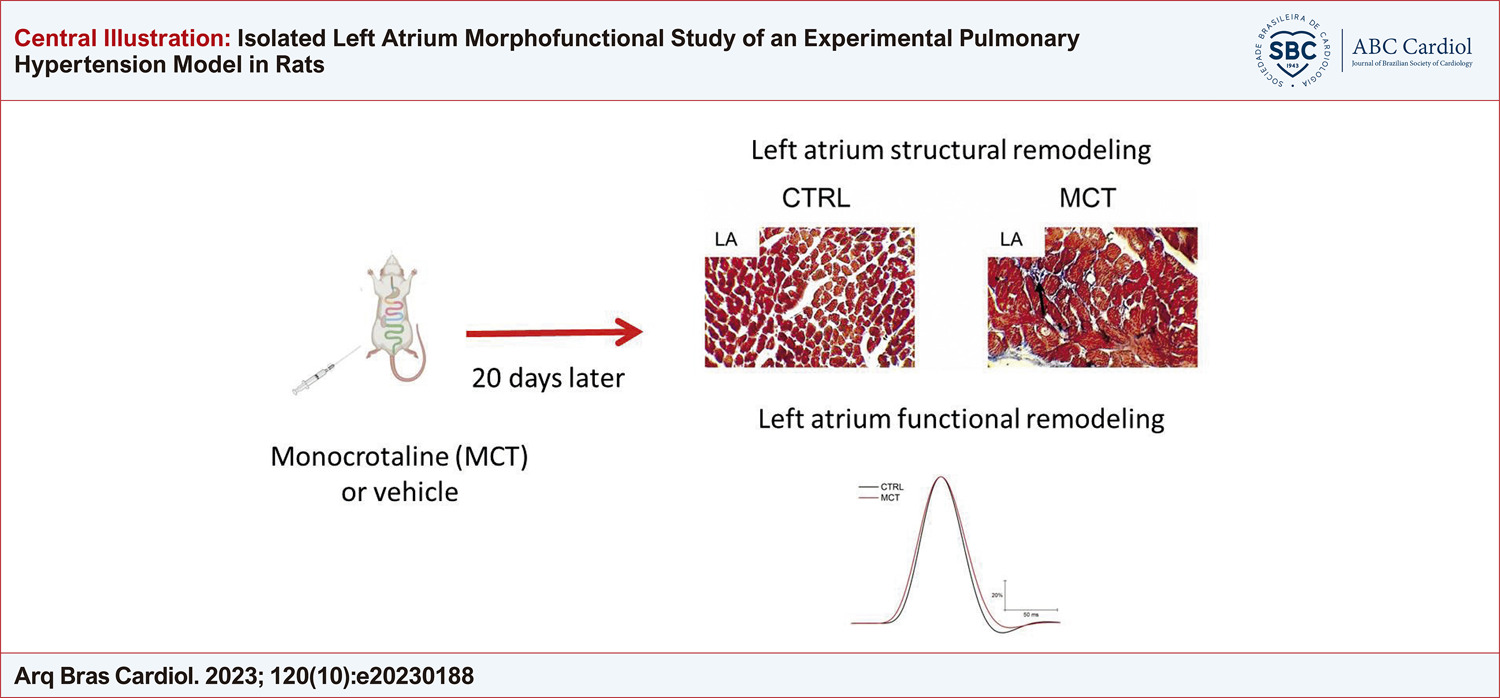
Isolated Left Atrium Morphofunctional Study of an Experimental Pulmonary Hypertension Model in Rats
ABC
Original Article

Isolated Left Atrium Morphofunctional Study of an Experimental Pulmonary Hypertension Model in Rats

Jorge Lucas Teixeira-Fonseca et al. Julliane Vasconcelos Joviano-Santos Fabiana da Silva Alcântara Polyana Leal da Silva … Michael Ramon Lima Conceição Danilo Roman-Campos

Abstract

Background

The high incidence of atrial arrhythmias in pulmonary hypertension (PH) might be associated with poor prognosis, and the left atrium (LA) may play a role in this. An important finding in PH studies is that LA remodeling is underestimated.

Objective

This study investigated LA morphology and mechanical function, as well as the susceptibility to develop arrhythmias in a monocrotaline-induced PH (MCT-PH) model.

Methods

Wistar rats aged 4 weeks received 50 mg/kg of MCT. Electrocardiography and histology analysis were performed to evaluate the establishment of the MCT-PH model. The tissue was mounted in an isolated organ bath to characterize the LA mechanical function

Results

Compared with the control group (CTRL), the MCT-PH model presented LA hypertrophy and changes in cardiac electrical activity, as evidenced by increased P wave duration, PR and QT interval in MCT-PH rats. In LA isolated from MCT-PH rats, no alteration in inotropism was observed; however, the time to peak contraction was delayed in the experimental MCT-PH group. Finally, there was no difference in arrhythmia susceptibility of LA from MCT-PH animals after the burst pacing protocol.

Conclusion

The morphofunctional remodeling of the LA did not lead to increased susceptibility to ex vivo arrhythmia after application of the burst pacing protocol.

  • Cardiac Arrhythmias
  • Fibrosis
  • Heart Atria
  • Monocrotaline
  • Pulmonary Hypertension
Read Full Article
Creative Commons Attribution (CC BY) Creative Commons license
20/10/2023 DOI: 10.36660/abc.20230188 Views 2
Figure 1
ABCIC
Case Report

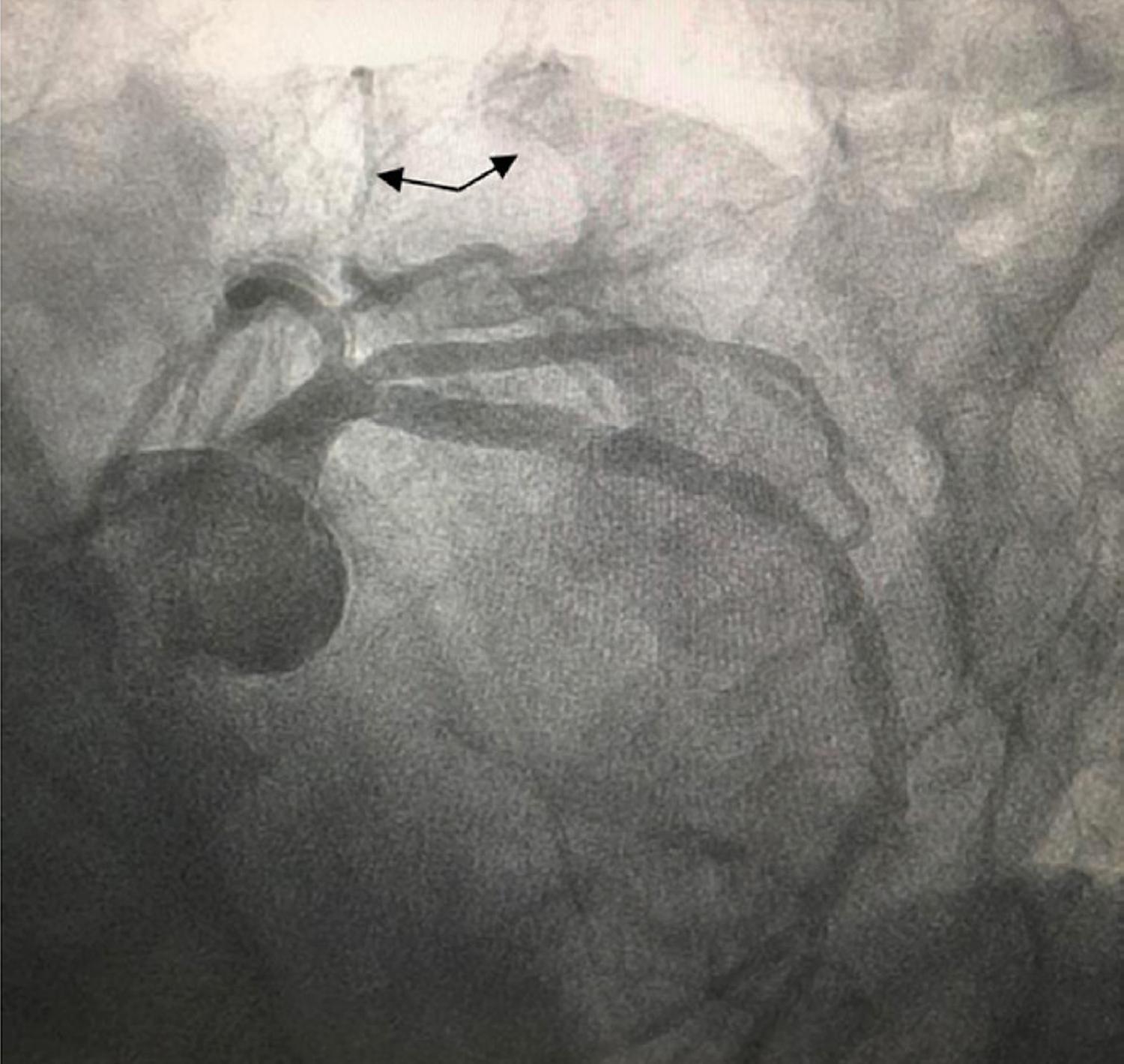
Coronary–Cavitary Fistula of the Circumflex Coronary Artery to the Left Atrium as a Probable Etiology of Heart Failure: A Case Report

Roberto Carlos Alvarez Coello et al. Vitor Coutinho Andrade Santiago Andrés Castro Vintimilla Vagner Madrini Júnior José Felix Alvarez Ramirez

Abstract

Coronary artery fistulas have a low incidence and are often diagnosed by echocardiography or coronary computed tomography angiography, although coronary angiography is the gold standard. They commonly originate in the right coronary artery, with drainage to low-pressure chambers being the most frequent finding. Treatment can be expectant, surgical, or percutaneous. This report describes a case of a rare presentation of heart failure due to a fistula of the circumflex coronary artery with drainage into the left atrium.

  • Fistula
  • Heart Atria
  • Heart Failure
Read Full Article
Creative Commons Attribution (CC BY) Creative Commons license
25/03/2022 DOI: 10.47593/2675-312X/20213404eabc220 Views 1
Figure 1
IJCS
Review Article

Transcatheter Interatrial Shunts for the Treatment of Heart Failure with Preserved Ejection Fraction

Anju Bhardwaj et al. Vishal Y. Parikh Ajith Nair

Abstract

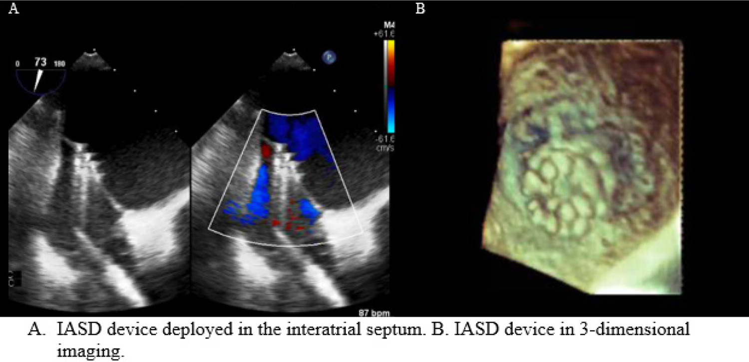
Heart failure with preserved ejection fraction (HFpEF) is a clinical syndrome, which accounts for about 50% of patients with heart failure (HF). The morbidity and mortality associated with HFpEF is similar to HFrEF. Clinical trials to date have failed to show a benefit of medical therapy for HFpEF, which may due to lack of uniform phenotypes and heterogeneous population. In addition, medical therapy proven for HFrEF may not address the pathophysiologic basis for HFpEF. Left atrial remodeling and dysfunction is central to HFpEF and accounts for secondary pulmonary hypertension and pulmonary vascular congestion that frequently occurs with exertion. Interatrial shunts represent a novel treatment modality for HFpEF. These shunts allow for left atrial decongestion and a reduction in pulmonary venous hypertension during exercise leading to improvements in hemodynamics, functional status and quality of life. Trials to date have demonstrated safety and short-term efficacy of these devices for HFpEF. The long-term benefits are currently being evaluated in ongoing trials. If effective, the use of interatrial shunts may be a new therapeutic paradigm for the treatment of HFpEF.

  • Heart Atria
  • Heart Failure
  • Hypertrophy Left Ventricular
  • Stroke Volume
  • transcatheter
Read Full Article
Creative Commons Attribution (CC BY) Creative Commons license
01/02/2021 DOI: 10.36660/ijcs.20200236 Views 2
Figure 1
ABC
Original Article

Value of left atrial diameter with CHA2DS2-VASc score in predicting left atrial/left atrial appendage thrombosis in non-valvular atrial fibrillation

Yu Zhang et al. Yi-Qiang Yuan

Abstract

Background

Atrial fibrillation is the most common persistent arrhythmia, and is the main factor that leads to thromboembolism.

Objective

To investigate the value of left atrial diameter combined with CHA2DS2-VASc score in predicting left atrial/left atrial appendage thrombosis in non-valvular atrial fibrillation.

Methods

This is a retrospective study. 238 patients with non-valvular atrial fibrillation were selected and divided into two groups: thrombosis and non-thrombosis. CHA2DS2-VASc score was determined. P<0.05 was considered statistically significant.

Results

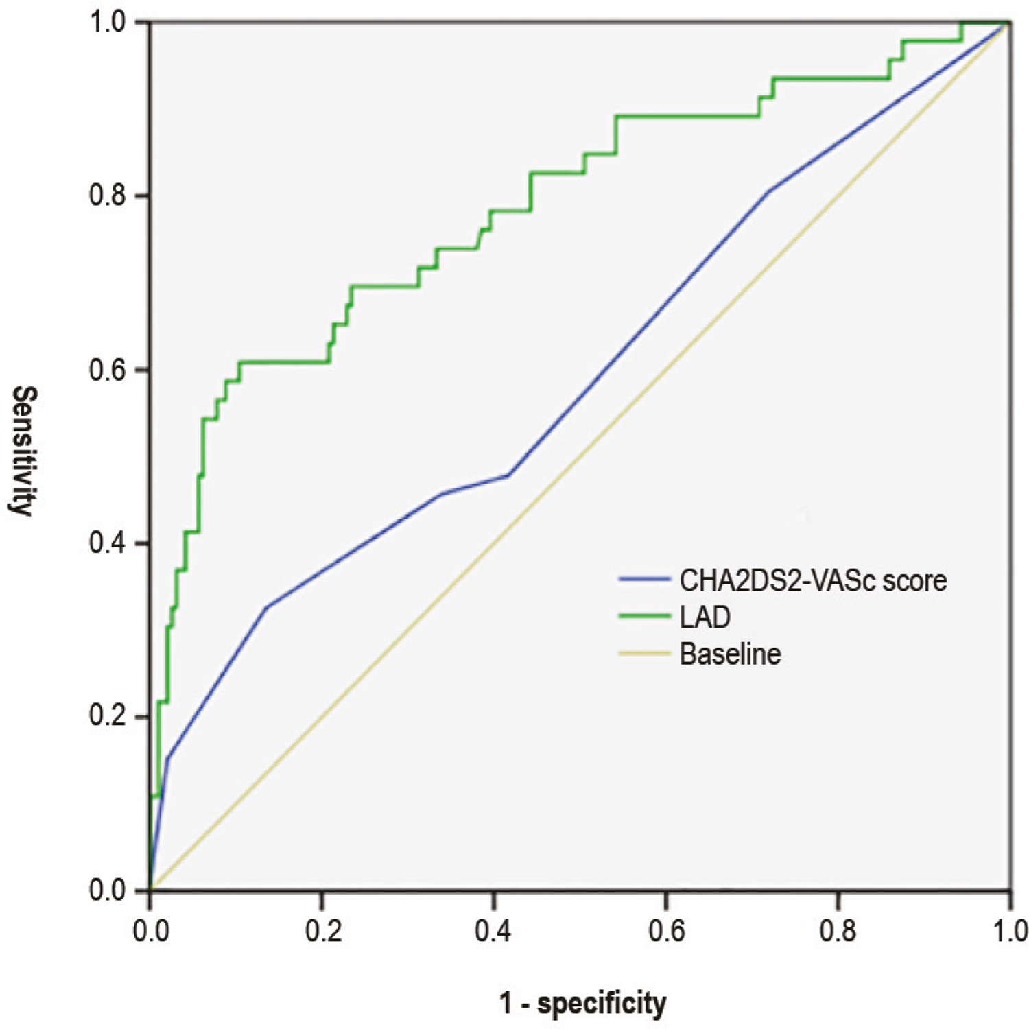
Multivariate logistic regression analysis revealed that the history of stroke/transient ischemic attack, vascular disease, CHA2DS2-VASc score, left atrial diameter (LAD), left ventricular end-diastolic dimension (LVEDD) and left ventricular ejection fraction (LVEF) were independent risk factors for left atrial/left atrial appendage thrombosis (p<0.05). Receiver operating characteristic curve analysis revealed that the area under the curve for the CHA2DS2-VASc score in predicting left atrial/left atrial appendage thrombosis was 0.593 when the CHA2DS2-VASc score was ≥3 points, and sensitivity and specificity were 86.5% and 32.6%, respectively, while the area under the curve for LAD in predicting left atrial/left atrial appendage thrombosis was 0.786 when LAD was ≥44.17 mm, and sensitivity and specificity were 89.6% and 60.9%, respectively. Among the different CHA2DS2-VASc groups, the incidence rate of left atrial/left atrial appendage thrombosis in patients with LAD ≥44.17 mm was higher than patients with LAD <44.17 mm (p<0.05).

Conclusion

CHA2DS2-VASc score and LAD are correlated with left atrial/left atrial appendage thrombosis in non-valvular atrial fibrillation. For patients with a CHA2DS2-VASc score of 0 or 1, when LAD is ≥44.17 mm, the risk for left atrial/left atrial appendage thrombosis remained high. (Arq Bras Cardiol. 2020; [online].ahead print, PP.0-0)

  • Atrial Appendage
  • Atrial Fibrillation Non Valvar
  • Heart Atria
  • Propensity Score
  • Risk Assessment
  • Stroke
Read Full Article
Creative Commons Attribution (CC BY) Creative Commons license
01/01/2021 DOI: 10.36660/abc.20190492 Views 1
Figure 1
IJCS
Original Article

Increased Left Atrial Volume and Its Relationship to Vitamin D in Primary Care

Erica de Abreu Macedo et al. Maria Luiza Garcia Rosa Antônio José Lagoeiro Jorge Adson Renato Leite … Leivys Henrique Silva Santos Juliana Salge Vieira

Abstract

Background:

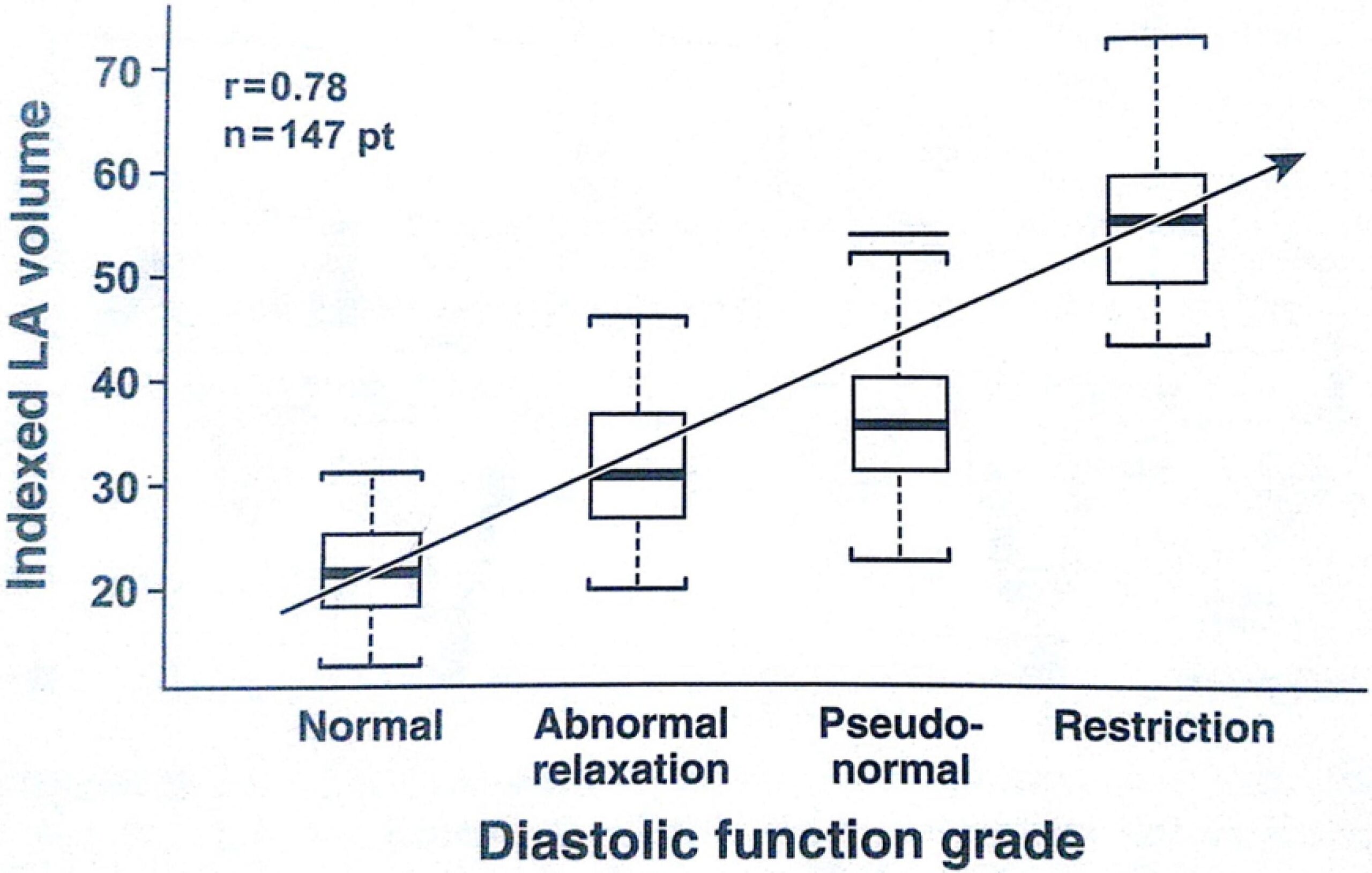
The study is based on the fact that left atrial (LA) volume measurement is a marker of the presence of diastolic dysfunction and that Vitamin D deficiency may be associated with ventricular remodeling, worsening of left ventricular (LV) diastolic and systolic function, and activation of the renin-angiotensin-aldosterone system.

Objective:

To evaluate whether LAV changes are related to vitamin D deficiency.

Method:

A cross-sectional, population-based, unicentric study in which 640 patients over 45 years of age enrolled in the Niterói’s Médico de Família program, RJ, were evaluated. Patients were submitted to tissue Doppler echocardiography to evaluate the parameters of diastolic and systolic function and vitamin D dosage. The presence or absence of hypovitaminosis D associated with structural and functional cardiac changes was compared between each group. A p < 0.05 value was considered as an indicator of statistical significance.

Results:

Of the 640 individuals analyzed, hypovitaminosis D was confirmed in 39.2% of the patients, of whom 34.8% had diastolic dysfunction. The most relevant echocardiographic parameters that were statistically significant were non-indexed AEDs and LAV, E’/A’ and E wave deceleration time, which were associated with the presence of hypovitaminosis D (P < 0.01).

Conclusion:

The study of the association of hypovitaminosis D and the appearance of structural and functional cardiac abnormalities may contribute to the discussion of the adoption of one more criterion to select individuals at risk of developing clinical cardiac insufficiency in primary care since, with the use of echocardiography, the subclinical condition of cardiac involvement, with prognostic and treatment implications for the referred patients with hypovitaminosis D, can be identified early.
  • Atrial Function
  • Cholecalciferol
  • Echocardiography
  • Heart Atria
  • Heart Failure
  • Primary Health Care
Read Full Article
Creative Commons Attribution (CC BY) Creative Commons license
08/08/2019 DOI: 10.5935/2359-4802.20190043
Figure 1
ABC
Original Article

Intra-Atrial Dyssynchrony Using Cardiac Magnetic Resonance to Quantify Tissue Remodeling in Patients with Atrial Fibrillation

Luisa Allen Ciuffo et al. João Lima Henrique Doria de Vasconcellos Muhammad Balouch … Susumu Tao Saman Nazarian David D. Spragg Joseph E. Marine Ronald D. Berger Hugh Calkins Hiroshi Ashikaga

Abstract

Background:

Recent studies suggest that left atrial (LA) late gadolinium enhancement (LGE) can quantify the underlying tissue remodeling that harbors atrial fibrillation (AF). However, quantification of LA-LGE requires labor-intensive magnetic resonance imaging acquisition and postprocessing at experienced centers. LA intra-atrial dyssynchrony assessment is an emerging imaging technique that predicts AF recurrence after catheter ablation. We hypothesized that 1) LA intra-atrial dyssynchrony is associated with LA-LGE in patients with AF and 2) LA intra-atrial dyssynchrony is greater in patients with persistent AF than in those with paroxysmal AF.

Method:

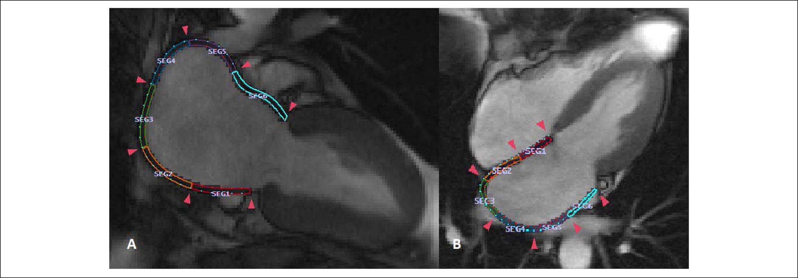
We conducted a cross-sectional study comparing LA intra-atrial dyssynchrony and LA-LGE in 146 patients with a history of AF (60.0 ± 10.0 years, 30.1% nonparoxysmal AF) who underwent pre-AF ablation cardiac magnetic resonance (CMR) in sinus rhythm. Using tissue-tracking CMR, we measured the LA longitudinal strain in two- and four-chamber views. We defined intra-atrial dyssynchrony as the standard deviation (SD) of the time to peak longitudinal strain (SD-TPS, in %) and the SD of the time to the peak pre-atrial contraction strain corrected by the cycle length (SD-TPSpreA, in %). We used the image intensity ratio (IIR) to quantify LA-LGE.

Results:

Intra-atrial dyssynchrony analysis took 5 ± 9 minutes per case. Multivariable analysis showed that LA intra-atrial dyssynchrony was independently associated with LA-LGE. In addition, LA intra-atrial dyssynchrony was significantly greater in patients with persistent AF than those with paroxysmal AF. In contrast, there was no significant difference in LA-LGE between patients with persistent and paroxysmal AF. LA intra-atrial dyssynchrony showed excellent reproducibility and its analysis was less time-consuming (5 ± 9 minutes) than the LA-LGE (60 ± 20 minutes).

Conclusion:

LA Intra-atrial dyssynchrony is a quick and reproducible index that is independently associated with LA-LGE to reflect the underlying tissue remodeling.

  • Atrial Fibrillation
  • Diagnostic Imaging
  • Echocardiography/methods
  • Heart Atria
  • Magnetic Resonance Spectroscopy
Read Full Article
Creative Commons Attribution (CC BY) Creative Commons license
15/04/2019 DOI: 10.5935/abc.20190064 Views 1
Figure 1
ABC
Original Article

Left Atrial Stiffness: A Predictor of Atrial Fibrillation Recurrence after Radiofrequency Catheter Ablation – A Systematic Review and Meta-Analysis

Eduardo Thadeu de Oliveira Correia et al. Letícia Mara dos Santos Barbetta Othon Moura Pereira da Silva Evandro Tinoco Mesquita

Abstract

Background:

Radiofrequency catheter ablation (RFCA) is a standard procedure for patients with atrial fibrillation (AF) not responsive to previous treatments, that has been increasingly considered as a first-line therapy. In this context, perioperative screening for risk factors has become important. A previous study showed that a high left atrial (LA) pressure is associated with AF recurrence after ablation, which may be secondary to a stiff left atrium.

Objective:

To investigate, through a systematic review and meta-analysis, if LA stiffness could be a predictor of AF recurrence after RFCA, and to discuss its clinical use.

Methods:

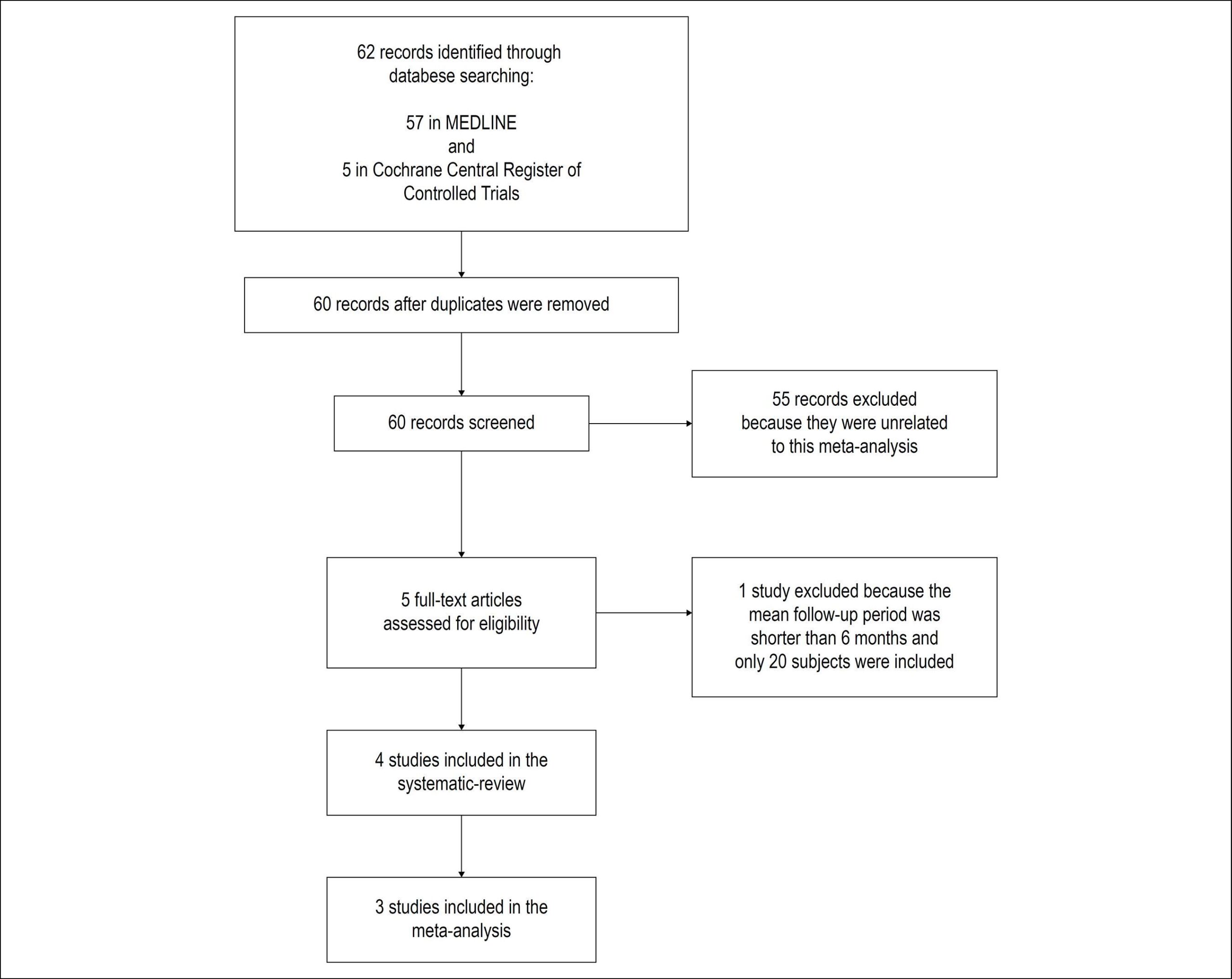
The meta-analysis followed the MOOSE recommendations. The search was performed in MEDLINE and Cochrane Central Register of Controlled Trials databases, until March 2018. Two authors performed screening, data extraction and quality assessment of the studies.

Results:

All studies were graded with good quality. A funnel plot was constructed, which did not show any publication bias. Four prospective observational studies were included in the systematic review and 3 of them in the meta-analysis. Statistical significance was defined at p value < 0.05. LA stiffness was a strong independent predictor of AF recurrence after RFCA (HR = 3.55, 95% CI 1.75-4.73, p = 0.0002).

Conclusion:

A non-invasive assessment of LA stiffness prior to ablation can be used as a potential screening factor to select or to closely follow patients with higher risks of AF recurrence and development of the stiff LA syndrome.

  • Atrial Fibrillation
  • Catheter Ablation/methods
  • Heart Atria
  • Metanalysis
  • Tachycardia, Paroxysmal
Read Full Article
Creative Commons Attribution (CC BY) Creative Commons license
07/03/2019 DOI: 10.5935/abc.20190040 Views 1
Figure 1
IJCS
Original Article

Does Percutaneous Left Atrial Appendage Closure Affect Left Atrial Performance?

Marta Madeira et al. Rogério Teixeira Liliana Reis Paulo Dinis … Luís Paiva Ana Botelho Marco Costa Lino Gonçalves

Abstract

Background:

Percutaneous left atrial appendage (LAA) occlusion may be an alternative therapy for atrial fibrillation (AF) patients with contraindication for anti-coagulation therapy. However, the influence of LAA occlusion on left atrial (LA) performance has not been studied.

Objective:

Our aim was to evaluate the influence of percutaneous LAA occlusion device on LA function by transthoracic echocardiography plus speckle-tracking echocardiography (STE).

Methods:

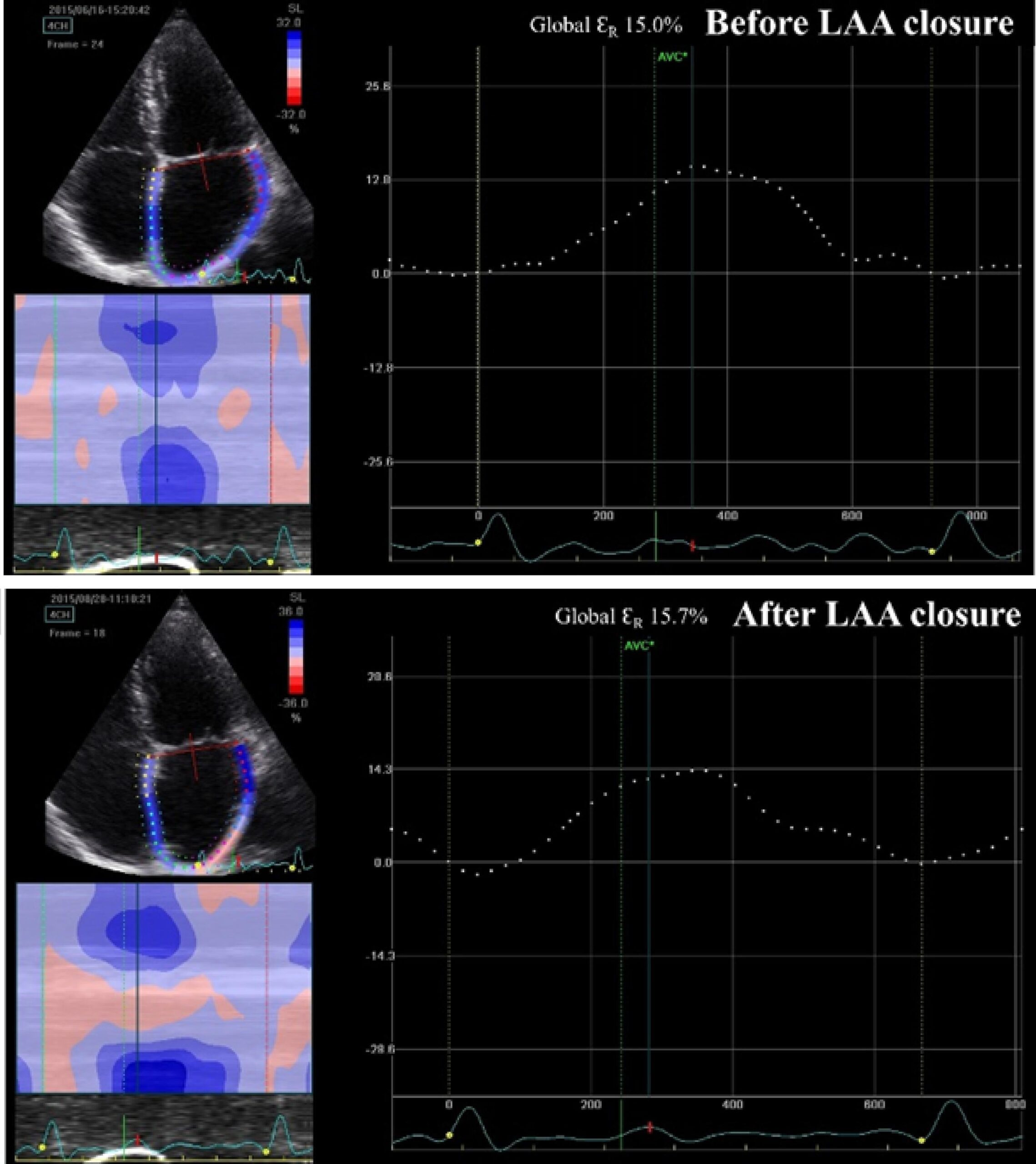
We included 16 patients undergoing percutaneous LAA closure with adequate echocardiographic window for the study of LA mechanics. Transthoracic echocardiography was performed before and after the procedure. LA volumes were calculated using the biplane method, and LA mechanics were assessed using STE. The analysis focused on the LA reservoir phase strain and strain rate.

Results:

Seventy-five percent of patients had permanent atrial fibrillation. Embolic and bleeding risk scores used were CHA2DS2-VASc [median of 4-5] and HAS-BLED [median of 2-3]. Major bleeding (62%) was the most common indication for the procedure. Percutaneous LAA closure was performed successfully in all patients, without major complications. No differences were found in maximum LA volume (44 ± 11 vs. 46 ± 13 mL/m2; p = 0.54), minimum LA volume (32 ± 8 vs. 37 ± 14 mL/m2; p = 0.09) or LA emptying fraction (26 ± 17 vs. 21 ± 14%; p = 0.33) before and after the procedure. Similarly, no differences were noted in left atrial strain (13.7 ± 11.1 vs. 13.0 ± 8.8%; p = 0.63) or strain rate (1.06 ± 0.26 vs. 1.13 ± 0.34 s-1; p = 0.38) in the reservoir phase.

Conclusions:

Our data suggest that percutaneous LAA closure does not affect LA reservoir function.

  • Atrial Appendage
  • Atrial Fibrillation
  • Echocardiography, Transthoracic
  • Heart Atria
Read Full Article
Creative Commons Attribution (CC BY) Creative Commons license
01/11/2018 DOI: 10.5935/2359-4802.20180075 Views 1
Figure 1
ABC
Case Report

Rare Complication of non-Treated Abdominal Aortic Aneurysm: Extensive Thrombus in Right Cardiac Chambers

Viviane Tiemi Hotta et al. David A. Bluemke Kamila Fernanda Staszko Ana Neri Rodrigues Epitacio Pereira Carlos Eduardo Rochitte

Abstract

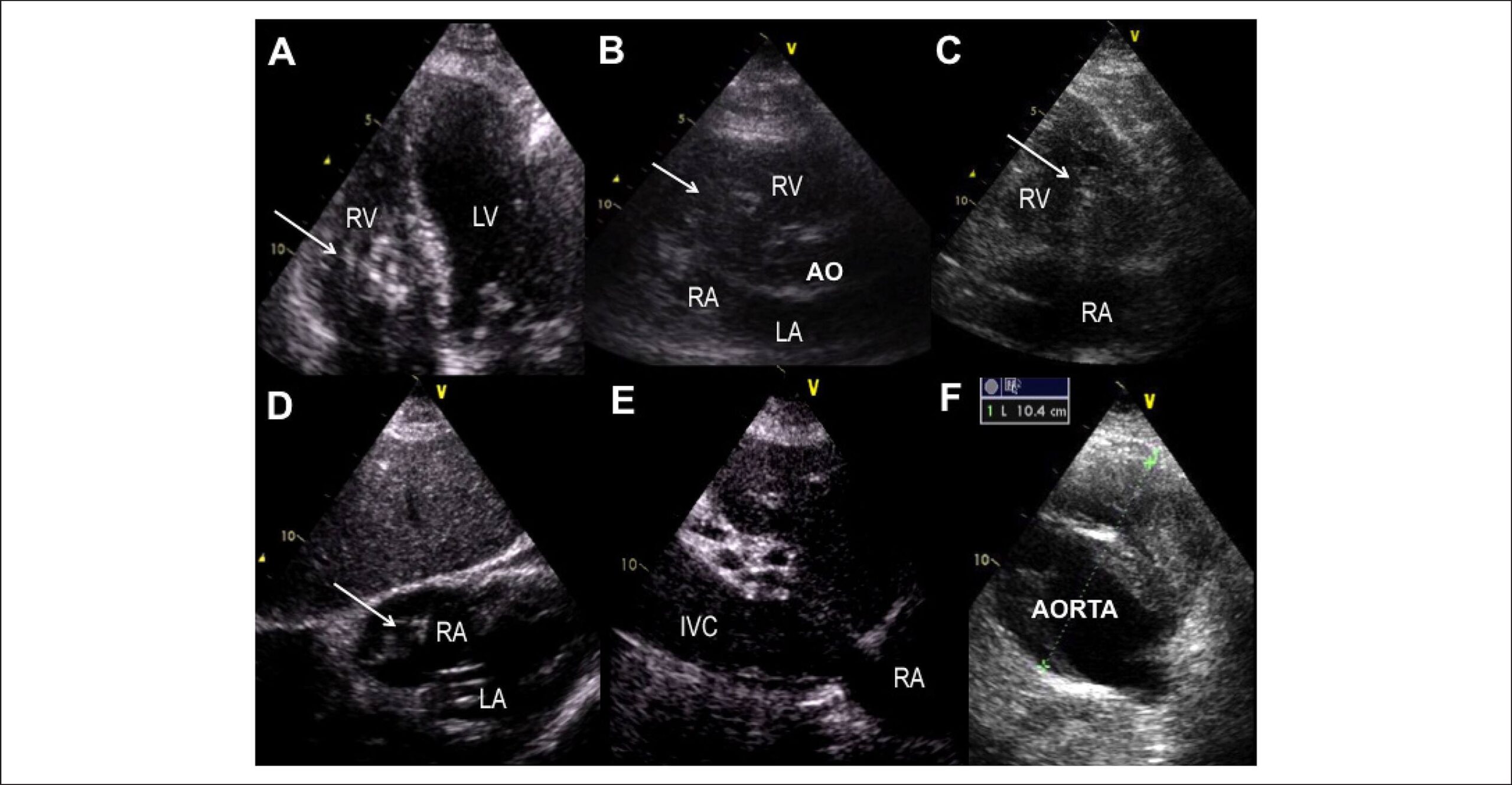
A 78-year-old patient presented with shortness of breath after falling down. Transthoracic echocardiogram showed an extensive thrombus in the right atrium (RA), extensive thrombosis of the inferior vena cava (IVC), and abdominal aortic aneurysm (AAA). A magnetic resonance confirmed the thrombosis of the RA extending to the IVC, which was apparently fused to the abdominal aortic aneurysm (compression? erosion?). This case illustrates a severe and rare complication of a non-treated AAA. There probably was IVC erosion by the aortic aneurysm, leading to blood stasis and extensive thrombosis of the IVC and right cardiac chambers.

  • Aortic Aneurysm, Abdominal / complications
  • Echocardiography
  • Heart Atria
  • Thrombosis
Read Full Article
Creative Commons Attribution (CC BY) Creative Commons license
01/10/2016 DOI: 10.5935/abc.20160143
Multiple Thrombi in the Inferior Vena Cava and Right Atrium – Recurrent Thromboembolism Due to Polycythemia Vera
ABC
Image

Multiple Thrombi in the Inferior Vena Cava and Right Atrium – Recurrent Thromboembolism Due to Polycythemia Vera

Kevin Domingues et al. Liliana Marta Marisa Peres Isabel Monteiro Margarida Leal

Thromboembolic events are a major cause of morbidity in patients with polycythemia vera (PV), accounting for a third of the deaths in this population. PV is a clonal disorder in which disturbed hematopoiesis leads to increased erythropoiesis, myelopoiesis, and/or megakaryopoiesis, characterizing this disorder as a prothrombotic state.

We report here the case of a 72-year-old man with a history of hypertension, type 2 diabetes mellitus, and implantation of a DDDR pacemaker 6 years before due to sick sinus syndrome. He also had an unclear, recent history of high hemoglobin levels and leukocytosis. He had been admitted three times in less than 1 year for acute pulmonary thromboembolism (PTE), despite being on anticoagulation therapy (one time on rivaroxaban, another on warfarin with adequate INR). A computed tomography scan revealed occlusive bilateral thrombi in the lower lobe arteries. Echocardiography was performed and showed enlargement of the right cavities, D-shaped left ventricle, severe pulmonary hypertension, and right ventricular dysfunction (tissue Doppler imaging = 8 cm/s). Multiple thrombi were present in the right atrium (some attached to the pacemaker lead) and in the inferior vena cava. Nevertheless, the patient was hemodynamically stable and comfortable with an oxygen mask.

[…]

  • Heart Atria
  • Polycythemia Vera
  • Pulmonary Embolism/mortality
  • Thrombosis
  • Tomography, X Ray Computed, Echocardiography
  • Vena cava, inferior
Read Full Article
Creative Commons Attribution (CC BY) Creative Commons license
01/08/2016 DOI: 10.5935/abc.20160088 Views 2
Figure 1
ABC
Original Article

Left Atrial Volume Determinants in Patients with Non-Ischemic Dilated Cardiomyopathy

Frederico José Neves Mancuso et al. Valdir Ambrósio Moisés Dirceu Rodrigues Almeida Dalva Poyares … Luciana Julio Storti Wércules Antonio Oliveira Flavio Souza Brito Angelo Amato Vincenzo de Paola Antonio Carlos Camargo Carvalho Orlando Campos

Background:

Left atrial volume (LAV) is a predictor of prognosis in patients with heart failure.

Objective:

We aimed to evaluate the determinants of LAV in patients with dilated cardiomyopathy (DCM).

Methods:

Ninety patients with DCM and left ventricular (LV) ejection fraction ≤ 0.50 were included. LAV was measured with real-time three-dimensional echocardiography (eco3D). The variables evaluated were heart rate, systolic blood pressure, LV end-diastolic volume and end-systolic volume and ejection fraction (eco3D), mitral inflow E wave, tissue Doppler e´ wave, E/e´ ratio, intraventricular dyssynchrony, 3D dyssynchrony index and mitral regurgitation vena contracta. Pearson´s coefficient was used to identify the correlation of the LAV with the assessed variables. A multiple linear regression model was developed that included LAV as the dependent variable and the variables correlated with it as the predictive variables.

Results:

Mean age was 52 ± 11 years-old, LV ejection fraction: 31.5 ± 8.0% (16-50%) and LAV: 39.2±15.7 ml/m2. The variables that correlated with the LAV were LV end-diastolic volume (r = 0.38; p < 0.01), LV end-systolic volume (r = 0.43; p < 0.001), LV ejection fraction (r = -0.36; p < 0.01), E wave (r = 0.50; p < 0.01), E/e´ ratio (r = 0.51; p < 0.01) and mitral regurgitation (r = 0.53; p < 0.01). A multivariate analysis identified the E/e´ ratio (p = 0.02) and mitral regurgitation (p = 0.02) as the only independent variables associated with LAV increase.

Conclusion:

The LAV is independently determined by LV filling pressures (E/e´ ratio) and mitral regurgitation in DCM.

  • Cardiomyopathy, Dilated
  • Chagas Cardiomyopathy
  • Echocardiography, Three-Dimensional
  • Heart Atria
  • Organ Size
Read Full Article
Creative Commons Attribution (CC BY) Creative Commons license
08/05/2015 DOI: 10.5935/abc.20150042 Views 1
ABC
Letter to the Editor

Left Atrial Volume Index as Predictor of Events in Acute Coronary Syndrome

Luciano Barros Pires et al. Rodrigo Pires dos Santos

Dear Editor,

The Left Atrial Volume Index (LAVi) is a measurement that has gained importance in daily clinical practice due to evidence of its capacity to predict mortality, in patients followed after an Acute Myocardial Infarction (AMI), as well as in the general population.

[…]

  • Acute Coronary Syndrome / mortality
  • Atrial Fibrillation
  • Heart Atria
  • Myocardial Reperfusion
  • Organ Size
Read Full Article
Creative Commons Attribution (CC BY) Creative Commons license
01/04/2015 DOI: 10.5935/abc.20150030
Figure 1
ABC
Case Report

Giant Left Atrial Thrombus with Double Coronary Vascularization

Ciambelli Giuliano Serafino et al. Mariana Lins Baptista Vitor Emer Egypto Rosa Antonio Sérgio de Santis Andrade Lopes … Tarso Augusto Duenhas Accorsi Flávio Tarasoutchi

Introduction

Rheumatic valvular disease, especially mitral stenosis (MS) and atrial fibrillation (AF), are the main factors related to the formation of left atrial (LA) thrombi–. Its incidence can range from 16 to 64%, and the most affected site is the left atrial appendage,.

Systemic embolism is responsible for 10 to 45% of the complications, being the most frequent clinical presentation. Despite being rare, the mechanical mitral valve obstruction caused by thrombus may be considered as potentially severe, especially among those patients with previous MS. The clinical manifestations of this condition are variable, presenting from worsened functional class (NYHA) to cardiogenic shock.

[…]

  • coronary circulation
  • Heart Atria
  • Mitral Valve Stenosis / surgery
  • Myxoma/surgery
  • Thrombosis/surgery
Read Full Article
Creative Commons Attribution (CC BY) Creative Commons license
01/02/2015 DOI: 10.5935/abc.20140128
Figure 1
ABC
Original Article

Left Atrial Volume Index and Prediction of Events in Acute Coronary Syndrome: Solar Registry

Jose Alves Secundo Junior et al. Marcos Antonio Almeida Santos Gustavo Baptista de Almeida Faro Camile Bittencourt Soares … Allyson Matos Porto Silva Paulo Fernando Carvalho Secundo Clarissa Karine Cardoso Teixeira Joselina Luzia Menezes Oliveira Jose Augusto Soares Barreto Filho Antônio Carlos Sobral Sousa

Background:

According to some international studies, patients with acute coronary syndrome (ACS) and increased left atrial volume index (LAVI) have worse long-term prognosis. However, national Brazilian studies confirming this prediction are still lacking.

Objective:

To evaluate LAVI as a predictor of major cardiovascular events (MCE) in patients with ACS during a 365-day follow-up.

Methods:

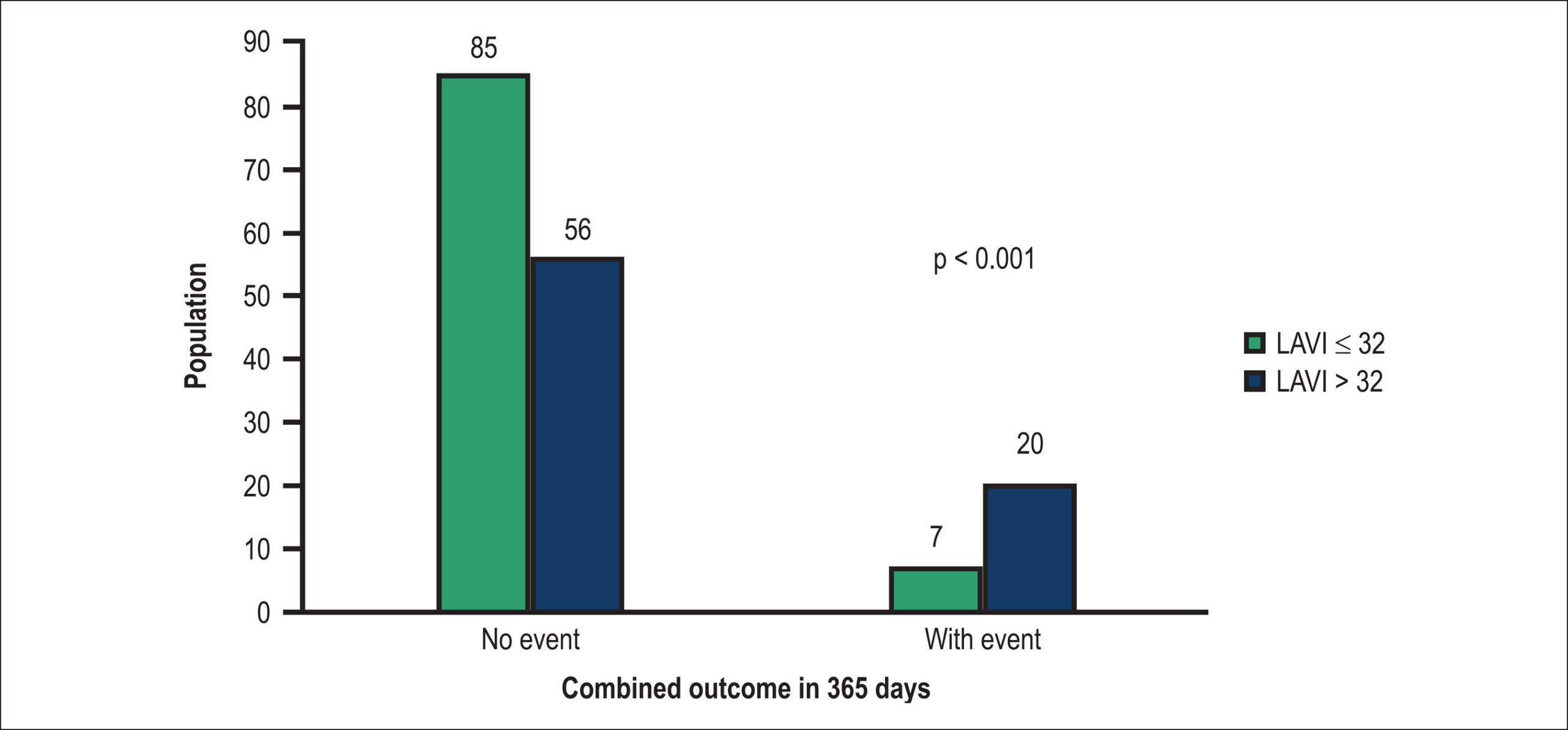
Prospective cohort of 171 patients diagnosed with ACS whose LAVI was calculated within 48 hours after hospital admission. According to LAVI, two groups were categorized: normal LAVI (≤ 32 mL/m2) and increased LAVI (> 32 mL/m2). Both groups were compared regarding clinical and echocardiographic characteristics, in- and out-of-hospital outcomes, and occurrence of ECM in up to 365 days.

Results:

Increased LAVI was observed in 78 patients (45%), and was associated with older age, higher body mass index, hypertension, history of myocardial infarction and previous angioplasty, and lower creatinine clearance and ejection fraction. During hospitalization, acute pulmonary edema was more frequent in patients with increased LAVI (14.1% vs. 4.3%, p = 0.024). After discharge, the occurrence of combined outcome for MCE was higher (p = 0.001) in the group with increased LAVI (26%) as compared to the normal LAVI group (7%) [RR (95% CI) = 3.46 (1.54-7.73) vs. 0.80 (0.69-0.92)]. After Cox regression, increased LAVI increased the probability of MCE (HR = 3.08, 95% CI = 1.28-7.40, p = 0.012).

Conclusion:

Increased LAVI is an important predictor of MCE in a one-year follow-up.

  • Acute coronary syndrome
  • Cardiac Volume
  • Heart Atria
Read Full Article
Creative Commons Attribution (CC BY) Creative Commons license
13/08/2014 DOI: 10.5935/abc.20140122 Views 1

Could not load more articles. Please try again.

How to cite this article

EndNote RIS

Electronic Document Format (Vancouver)

Saeed B, Punnanithinont N, Mansour S, Savoia P, Suksaranjit P. Recurrent Intracardiac Masses in an Orthotopic Heart Transplant Recipient. ABC Imagem Cardiovascular 2026;39(2):e20260031.

Electronic Document Format (ABNT)

Saeed, Bilal; Punnanithinont, Natdanai; Mansour, Shareef; Savoia, Paulo; Suksaranjit, Promporn. Recurrent Intracardiac Masses in an Orthotopic Heart Transplant Recipient. ABC Imagem Cardiovascular, v. 39, n. 2, e20260031, Jul. 2026.

Electronic Document Format (APA)

Saeed, B., Punnanithinont, N., Mansour, S., Savoia, P., & Suksaranjit, P. (2026). Recurrent Intracardiac Masses in an Orthotopic Heart Transplant Recipient. ABC Imagem Cardiovascular, 39(2), e20260031.

Electronic Document Format (ISO)

Saeed, Bilal and Punnanithinont, Natdanai and Mansour, Shareef and Savoia, Paulo and Suksaranjit, Promporn. Recurrent Intracardiac Masses in an Orthotopic Heart Transplant Recipient. ABC Imagem Cardiovascular [online]. 2026, vol. 39, n. 2, [cited 2026-10-14], e20260031. Available from: <https://journals.cardiol.br/en/article/recurrent-intracardiac-masses-in-an-orthotopic-heart-transplant-recipient/>. ISSN 2318-8219

EndNote RIS

Electronic Document Format (Vancouver)

Alencar JN, Barreiro DTC, Baranchuk A. The End of “Left Atrial Overload”. Arquivos Brasileiros de Cardiologia 2026;123(4):e20250814.

Electronic Document Format (ABNT)

Alencar, José Nunes de; Barreiro, Daniel Teixeira de Campos e; Baranchuk, Adrian. The End of “Left Atrial Overload”. Arquivos Brasileiros de Cardiologia, v. 123, n. 4, e20250814, May. 2026.

Electronic Document Format (APA)

Alencar, J. N. , Barreiro, D. T. C. , & Baranchuk, A. (2026). The End of “Left Atrial Overload”. Arquivos Brasileiros de Cardiologia, 123(4), e20250814.

Electronic Document Format (ISO)

Alencar, José Nunes de and Barreiro, Daniel Teixeira de Campos e and Baranchuk, Adrian. The End of “Left Atrial Overload”. Arquivos Brasileiros de Cardiologia [online]. 2026, vol. 123, n. 4, [cited 2026-10-14], e20250814. Available from: <https://journals.cardiol.br/en/article/the-end-of-left-atrial-overload/>. ISSN 0066-782X

EndNote RIS

Electronic Document Format (Vancouver)

Hortegal RA, Bihan DL, Barretto R, Mathias Jr W. Left Atrium Reservoir Strain and Cardiovascular Hemodynamics: the Hidden Truths Reveal Far More Than What is Readily Apparent. ABC Imagem Cardiovascular 2025;38(3):e20250068.

Electronic Document Format (ABNT)

Hortegal, Renato A.; Bihan, David Le; Barretto, Rodrigo; Mathias Jr, Wilson. Left Atrium Reservoir Strain and Cardiovascular Hemodynamics: the Hidden Truths Reveal Far More Than What is Readily Apparent. ABC Imagem Cardiovascular, v. 38, n. 3, e20250068, Dec. 2025.

Electronic Document Format (APA)

Hortegal, R. A., Bihan, D. L., Barretto, R., & Mathias Jr, W. (2025). Left Atrium Reservoir Strain and Cardiovascular Hemodynamics: the Hidden Truths Reveal Far More Than What is Readily Apparent. ABC Imagem Cardiovascular, 38(3), e20250068.

Electronic Document Format (ISO)

Hortegal, Renato A. and Bihan, David Le and Barretto, Rodrigo and Mathias Jr, Wilson. Left Atrium Reservoir Strain and Cardiovascular Hemodynamics: the Hidden Truths Reveal Far More Than What is Readily Apparent. ABC Imagem Cardiovascular [online]. 2025, vol. 38, n. 3, [cited 2026-10-14], e20250068. Available from: <https://journals.cardiol.br/en/article/left-atrium-reservoir-strain-and-cardiovascular-hemodynamics-the-hidden-truths-reveal-far-more-than-what-is-readily-apparent/>. ISSN 2318-8219

EndNote RIS

Electronic Document Format (Vancouver)

Oliveira ACS, Carvalho VT, Pinto-Filho MM, Brant LC, Barreto SM, Foppa M, Ribeiro ALP, et al. Diagnostic Utility of P-Wave Duration in Identifying Left Atrial Enlargement: Insights from the ELSA-Brasil Cohort. Arquivos Brasileiros de Cardiologia 2025;122(10):e20250151.

Electronic Document Format (ABNT)

Oliveira, Ana Cecília de Sena; Carvalho, Vinicius Tostes; Pinto-Filho, Marcelo Martins; Brant, Luisa Caldeira; Barreto, Sandhi Maria; Foppa, Murilo; Ribeiro, Antonio Luiz Pinho. Diagnostic Utility of P-Wave Duration in Identifying Left Atrial Enlargement: Insights from the ELSA-Brasil Cohort. Arquivos Brasileiros de Cardiologia, v. 122, n. 10, e20250151, Dec. 2025.

Electronic Document Format (APA)

Oliveira, A. C. S., Carvalho, V. T., Pinto-Filho, M. M., Brant, L. C., Barreto, S. M., Foppa, M., & Ribeiro, A. L. P. (2025). Diagnostic Utility of P-Wave Duration in Identifying Left Atrial Enlargement: Insights from the ELSA-Brasil Cohort. Arquivos Brasileiros de Cardiologia, 122(10), e20250151.

Electronic Document Format (ISO)

Oliveira, Ana Cecília de Sena and Carvalho, Vinicius Tostes and Pinto-Filho, Marcelo Martins and Brant, Luisa Caldeira and Barreto, Sandhi Maria and Foppa, Murilo and Ribeiro, Antonio Luiz Pinho. Diagnostic Utility of P-Wave Duration in Identifying Left Atrial Enlargement: Insights from the ELSA-Brasil Cohort. Arquivos Brasileiros de Cardiologia [online]. 2025, vol. 122, n. 10, [cited 2026-10-14], e20250151. Available from: <https://journals.cardiol.br/en/article/diagnostic-utility-of-p-wave-duration-in-identifying-left-atrial-enlargement-insights-from-the-elsa-brasil-cohort/>. ISSN 0066-782X

EndNote RIS

Electronic Document Format (Vancouver)

Darzé AC, Paladino Filho AT, Rassi AM, Prazeres CE, Pinto IMF, Caneschi JM, Ponte Junior JRT, et al. Giant Left Atrium in Rheumatic Mitral Valve Disease. ABC Imagem Cardiovascular 2024;37(4):e20240083.

Electronic Document Format (ABNT)

Darzé, Alice Cunha; Paladino Filho, Antonio Tito; Rassi, Amanda Marsiaj; Prazeres, Carlos Eduardo; Pinto, Ibraim Masciarelli Francisco; Caneschi, João Marcelo; Ponte Junior, Jose Roberto Tuma da; Morales, Paul Salvador; Hendges, Rafael Castro; Senra, Tiago. Giant Left Atrium in Rheumatic Mitral Valve Disease. ABC Imagem Cardiovascular, v. 37, n. 4, e20240083, Dec. 2024.

Electronic Document Format (APA)

Darzé, A. C., Paladino Filho, A. T., Rassi, A. M., Prazeres, C. E., Pinto, I. M. F., Caneschi, J. M., Ponte Junior, J. R. T. , Morales, P. S., Hendges, R. C., & Senra, T. (2024). Giant Left Atrium in Rheumatic Mitral Valve Disease. ABC Imagem Cardiovascular, 37(4), e20240083.

Electronic Document Format (ISO)

Darzé, Alice Cunha and Paladino Filho, Antonio Tito and Rassi, Amanda Marsiaj and Prazeres, Carlos Eduardo and Pinto, Ibraim Masciarelli Francisco and Caneschi, João Marcelo and Ponte Junior, Jose Roberto Tuma da and Morales, Paul Salvador and Hendges, Rafael Castro and Senra, Tiago. Giant Left Atrium in Rheumatic Mitral Valve Disease. ABC Imagem Cardiovascular [online]. 2024, vol. 37, n. 4, [cited 2026-10-14], e20240083. Available from: <https://journals.cardiol.br/en/article/giant-left-atrium-in-rheumatic-mitral-valve-disease/>. ISSN 2318-8219

EndNote RIS

Electronic Document Format (Vancouver)

Vicente MBA, Amaro T, Fernandes E. Six-Year Follow-Up of an Octogenarian With a Left Atrial Myxoma: Case Report. International Journal of Cardiovascular Sciences 2024;37:e20240135.

Electronic Document Format (ABNT)

Vicente, Miguel Bernardino Antunes; Amaro, Tânia; Fernandes, Elsa. Six-Year Follow-Up of an Octogenarian With a Left Atrial Myxoma: Case Report. International Journal of Cardiovascular Sciences, v. 37, e20240135, Dec. 2024.

Electronic Document Format (APA)

Vicente, M. B. A., Amaro, T., & Fernandes, E. (2024). Six-Year Follow-Up of an Octogenarian With a Left Atrial Myxoma: Case Report. International Journal of Cardiovascular Sciences, 37, e20240135.

Electronic Document Format (ISO)

Vicente, Miguel Bernardino Antunes and Amaro, Tânia and Fernandes, Elsa. Six-Year Follow-Up of an Octogenarian With a Left Atrial Myxoma: Case Report. International Journal of Cardiovascular Sciences [online]. 2024, vol. 37, [cited 2026-10-14], e20240135. Available from: <https://journals.cardiol.br/en/article/six-year-follow-up-of-an-octogenarian-with-a-left-atrial-myxoma-case-report/>. ISSN 2359-4802

EndNote RIS

Electronic Document Format (Vancouver)

Garbin HI, Klauberg MPM, Mazzutti G. Cardiac Tumor in the Left Atrium. ABC Heart Fail Cardiomyop 2024;4(2):e20240004.

Electronic Document Format (ABNT)

Garbin, Henrique Iahnke; Klauberg, Matheus Piccoli Machado; Mazzutti, Géris. Cardiac Tumor in the Left Atrium. ABC Heart Fail Cardiomyop, v. 4, n. 2, e20240004, Aug. 2024.

Electronic Document Format (APA)

Garbin, H. I., Klauberg, M. P. M., & Mazzutti, G. (2024). Cardiac Tumor in the Left Atrium. ABC Heart Fail Cardiomyop, 4(2), e20240004.

Electronic Document Format (ISO)

Garbin, Henrique Iahnke and Klauberg, Matheus Piccoli Machado and Mazzutti, Géris. Cardiac Tumor in the Left Atrium. ABC Heart Fail Cardiomyop [online]. 2024, vol. 4, n. 2, [cited 2026-10-14], e20240004. Available from: <https://journals.cardiol.br/en/article/cardiac-tumor-in-the-left-atrium/>

EndNote RIS

Electronic Document Format (Vancouver)

Pena JLB. Is Left Atrial Strain the Missing Part to Diagnose Diastolic Dysfunction in Children with Chronic Kidney Disease?. Arquivos Brasileiros de Cardiologia 2024;121(3):e20240257.

Electronic Document Format (ABNT)

Pena, José Luiz Barros. Is Left Atrial Strain the Missing Part to Diagnose Diastolic Dysfunction in Children with Chronic Kidney Disease?. Arquivos Brasileiros de Cardiologia, v. 121, n. 3, e20240257, Jul. 2024.

Electronic Document Format (APA)

Pena, J. L. B. (2024). Is Left Atrial Strain the Missing Part to Diagnose Diastolic Dysfunction in Children with Chronic Kidney Disease?. Arquivos Brasileiros de Cardiologia, 121(3), e20240257.

Electronic Document Format (ISO)

Pena, José Luiz Barros. Is Left Atrial Strain the Missing Part to Diagnose Diastolic Dysfunction in Children with Chronic Kidney Disease?. Arquivos Brasileiros de Cardiologia [online]. 2024, vol. 121, n. 3, [cited 2026-10-14], e20240257. Available from: <https://journals.cardiol.br/en/article/is-left-atrial-strain-the-missing-part-to-diagnose-diastolic-dysfunction-in-children-with-chronic-kidney-disease/>. ISSN 0066-782X

EndNote RIS

Electronic Document Format (Vancouver)

Penachio FM, Diniz MFR, Laurino RSP, Watanabe A, Sawamura KSS, Lianza AC, Menezes CRB, et al. Speckle-Tracking: Incremental Role in Diastolic Assessment of Pediatric Patients with Chronic Kidney Disease. Arquivos Brasileiros de Cardiologia 2024;121(3):e20230131.

Electronic Document Format (ABNT)

Penachio, Flora Maciel; Diniz, Maria de Fátima Rodrigues; Laurino, Rosana Sbruzzi Prado; Watanabe, Andreia; Sawamura, Karen Saori Shiraishi; Lianza, Alessandro Cavalcanti; Menezes, Carolina Rocha Brito; Silva, Isabela de Sousa Lobo; Leal, Gabriela Nunes. Speckle-Tracking: Incremental Role in Diastolic Assessment of Pediatric Patients with Chronic Kidney Disease. Arquivos Brasileiros de Cardiologia, v. 121, n. 3, e20230131, Apr. 2024.

Electronic Document Format (APA)

Penachio, F. M., Diniz, M. F. R., Laurino, R. S. P., Watanabe, A., Sawamura, K. S. S., Lianza, A. C., Menezes, C. R. B., Silva, I. S. L., & Leal, G. N. (2024). Speckle-Tracking: Incremental Role in Diastolic Assessment of Pediatric Patients with Chronic Kidney Disease. Arquivos Brasileiros de Cardiologia, 121(3), e20230131.

Electronic Document Format (ISO)

Penachio, Flora Maciel and Diniz, Maria de Fátima Rodrigues and Laurino, Rosana Sbruzzi Prado and Watanabe, Andreia and Sawamura, Karen Saori Shiraishi and Lianza, Alessandro Cavalcanti and Menezes, Carolina Rocha Brito and Silva, Isabela de Sousa Lobo and Leal, Gabriela Nunes. Speckle-Tracking: Incremental Role in Diastolic Assessment of Pediatric Patients with Chronic Kidney Disease. Arquivos Brasileiros de Cardiologia [online]. 2024, vol. 121, n. 3, [cited 2026-10-14], e20230131. Available from: <https://journals.cardiol.br/en/article/speckle-tracking-incremental-role-in-diastolic-assessment-of-pediatric-patients-with-chronic-kidney-disease/>. ISSN 0066-782X

EndNote RIS

Electronic Document Format (Vancouver)

Şahin S, Özbülbül NI. A Bridging Vein Connecting Left Atrium and Coronary Sinus: Coronary Computed Tomography Angiography Findings. ABC Imagem Cardiovascular 2024;37(1):e20230111.

Electronic Document Format (ABNT)

Şahin, Sercan; Özbülbül, Nilgün Işıksalan. A Bridging Vein Connecting Left Atrium and Coronary Sinus: Coronary Computed Tomography Angiography Findings. ABC Imagem Cardiovascular, v. 37, n. 1, e20230111, Mar. 2024.

Electronic Document Format (APA)

Şahin, S., & Özbülbül, N. I. (2024). A Bridging Vein Connecting Left Atrium and Coronary Sinus: Coronary Computed Tomography Angiography Findings. ABC Imagem Cardiovascular, 37(1), e20230111.

Electronic Document Format (ISO)

Şahin, Sercan and Özbülbül, Nilgün Işıksalan. A Bridging Vein Connecting Left Atrium and Coronary Sinus: Coronary Computed Tomography Angiography Findings. ABC Imagem Cardiovascular [online]. 2024, vol. 37, n. 1, [cited 2026-10-14], e20230111. Available from: <https://journals.cardiol.br/en/article/a-bridging-vein-connecting-left-atrium-and-coronary-sinus-coronary-computed-tomography-angiography-findings/>. ISSN 2318-8219

EndNote RIS

Electronic Document Format (Vancouver)

Teixeira-Fonseca JL, Joviano-Santos JV, Alcântara FS, Silva PL, Conceição MRL, Roman-Campos D. Isolated Left Atrium Morphofunctional Study of an Experimental Pulmonary Hypertension Model in Rats. Arquivos Brasileiros de Cardiologia 2023;120(10):e20230188.

Electronic Document Format (ABNT)

Teixeira-Fonseca, Jorge Lucas; Joviano-Santos, Julliane Vasconcelos; Alcântara, Fabiana da Silva; Silva, Polyana Leal da; Conceição, Michael Ramon Lima; Roman-Campos, Danilo. Isolated Left Atrium Morphofunctional Study of an Experimental Pulmonary Hypertension Model in Rats. Arquivos Brasileiros de Cardiologia, v. 120, n. 10, e20230188, Oct. 2023.

Electronic Document Format (APA)

Teixeira-Fonseca, J. L., Joviano-Santos, J. V., Alcântara, F. S., Silva, P. L. , Conceição, M. R. L., & Roman-Campos, D. (2023). Isolated Left Atrium Morphofunctional Study of an Experimental Pulmonary Hypertension Model in Rats. Arquivos Brasileiros de Cardiologia, 120(10), e20230188.

Electronic Document Format (ISO)

Teixeira-Fonseca, Jorge Lucas and Joviano-Santos, Julliane Vasconcelos and Alcântara, Fabiana da Silva and Silva, Polyana Leal da and Conceição, Michael Ramon Lima and Roman-Campos, Danilo. Isolated Left Atrium Morphofunctional Study of an Experimental Pulmonary Hypertension Model in Rats. Arquivos Brasileiros de Cardiologia [online]. 2023, vol. 120, n. 10, [cited 2026-10-14], e20230188. Available from: <https://journals.cardiol.br/en/article/isolated-left-atrium-morphofunctional-study-of-an-experimental-pulmonary-hypertension-model-in-rats/>. ISSN 0066-782X

EndNote RIS

Electronic Document Format (Vancouver)

Coello RCA, Andrade VC, Vintimilla SAC, Madrini Júnior V, Ramirez JFA. Coronary–Cavitary Fistula of the Circumflex Coronary Artery to the Left Atrium as a Probable Etiology of Heart Failure: A Case Report. Arquivos Brasileiros de Cardiologia Imagem cardiovascular 2022;34(4):eabc220.

Electronic Document Format (ABNT)

Coello, Roberto Carlos Alvarez; Andrade, Vitor Coutinho; Vintimilla, Santiago Andrés Castro; Madrini Júnior, Vagner; Ramirez, José Felix Alvarez. Coronary–Cavitary Fistula of the Circumflex Coronary Artery to the Left Atrium as a Probable Etiology of Heart Failure: A Case Report. Arquivos Brasileiros de Cardiologia Imagem cardiovascular, v. 34, n. 4, eabc220, Mar. 2022.

Electronic Document Format (APA)

Coello, R. C. A., Andrade, V. C., Vintimilla, S. A. C., Madrini Júnior, V., & Ramirez, J. F. A. (2022). Coronary–Cavitary Fistula of the Circumflex Coronary Artery to the Left Atrium as a Probable Etiology of Heart Failure: A Case Report. Arquivos Brasileiros de Cardiologia Imagem cardiovascular, 34(4), eabc220.

Electronic Document Format (ISO)

Coello, Roberto Carlos Alvarez and Andrade, Vitor Coutinho and Vintimilla, Santiago Andrés Castro and Madrini Júnior, Vagner and Ramirez, José Felix Alvarez. Coronary–Cavitary Fistula of the Circumflex Coronary Artery to the Left Atrium as a Probable Etiology of Heart Failure: A Case Report. Arquivos Brasileiros de Cardiologia Imagem cardiovascular [online]. 2022, vol. 34, n. 4, [cited 2026-10-14], eabc220. Available from: <https://journals.cardiol.br/en/article/coronary-cavitary-fistula-of-the-circumflex-coronary-artery-to-the-left-atrium-as-a-probable-etiology-of-heart-failure-a-case-report/>. ISSN 2318-8219

EndNote RIS

Electronic Document Format (Vancouver)

Bhardwaj A, Parikh VY, Nair A. Transcatheter Interatrial Shunts for the Treatment of Heart Failure with Preserved Ejection Fraction. International Journal of Cardiovascular Sciences 2021;34(1):81-8.

Electronic Document Format (ABNT)

Bhardwaj, Anju; Parikh, Vishal Y.; Nair, Ajith. Transcatheter Interatrial Shunts for the Treatment of Heart Failure with Preserved Ejection Fraction. International Journal of Cardiovascular Sciences, v. 34, n. 1, p. 81-88, Jan. 2021.

Electronic Document Format (APA)

Bhardwaj, A., Parikh, V. Y., & Nair, A. (2021). Transcatheter Interatrial Shunts for the Treatment of Heart Failure with Preserved Ejection Fraction. International Journal of Cardiovascular Sciences, 34(1), 81-88.

Electronic Document Format (ISO)

Bhardwaj, Anju and Parikh, Vishal Y. and Nair, Ajith. Transcatheter Interatrial Shunts for the Treatment of Heart Failure with Preserved Ejection Fraction. International Journal of Cardiovascular Sciences [online]. 2021, vol. 34, n. 1, [cited 2026-10-14], pp. 81-88. Available from: <https://journals.cardiol.br/en/article/transcatheter-interatrial-shunts-for-the-treatment-of-heart-failure-with-preserved-ejection-fraction/>. ISSN 2359-4802

EndNote RIS

Electronic Document Format (Vancouver)

Zhang Y, Yuan Y. Value of left atrial diameter with CHA2DS2-VASc score in predicting left atrial/left atrial appendage thrombosis in non-valvular atrial fibrillation. Arquivos Brasileiros de Cardiologia 2021;116(2):325-31.

Electronic Document Format (ABNT)

Zhang, Yu; Yuan, Yi-Qiang. Value of left atrial diameter with CHA2DS2-VASc score in predicting left atrial/left atrial appendage thrombosis in non-valvular atrial fibrillation. Arquivos Brasileiros de Cardiologia, v. 116, n. 2, p. 325-331, Jan. 2021.

Electronic Document Format (APA)

Zhang, Y., & Yuan, Y. (2021). Value of left atrial diameter with CHA2DS2-VASc score in predicting left atrial/left atrial appendage thrombosis in non-valvular atrial fibrillation. Arquivos Brasileiros de Cardiologia, 116(2), 325-331.

Electronic Document Format (ISO)

Zhang, Yu and Yuan, Yi-Qiang. Value of left atrial diameter with CHA2DS2-VASc score in predicting left atrial/left atrial appendage thrombosis in non-valvular atrial fibrillation. Arquivos Brasileiros de Cardiologia [online]. 2021, vol. 116, n. 2, [cited 2026-10-14], pp. 325-331. Available from: <https://journals.cardiol.br/en/article/value-of-left-atrial-diameter-with-cha2ds2-vasc-score-in-predicting-left-atrial-left-atrial-appendage-thrombosis-in-non-valvular-atrial-fibrillation/>. ISSN 0066-782X

EndNote RIS

Electronic Document Format (Vancouver)

Macedo EA, Rosa MLG, Jorge AJL, Leite AR, Santos LHS, Vieira JS. Increased Left Atrial Volume and Its Relationship to Vitamin D in Primary Care. International Journal of Cardiovascular Sciences 2019;32(5):508-16.

Electronic Document Format (ABNT)

Macedo, Erica de Abreu; Rosa, Maria Luiza Garcia; Jorge, Antônio José Lagoeiro; Leite, Adson Renato; Santos, Leivys Henrique Silva; Vieira, Juliana Salge. Increased Left Atrial Volume and Its Relationship to Vitamin D in Primary Care. International Journal of Cardiovascular Sciences, v. 32, n. 5, p. 508-516, Aug. 2019.

Electronic Document Format (APA)

Macedo, E. A., Rosa, M. L. G., Jorge, A. J. L., Leite, A. R., Santos, L. H. S., & Vieira, J. S. (2019). Increased Left Atrial Volume and Its Relationship to Vitamin D in Primary Care. International Journal of Cardiovascular Sciences, 32(5), 508-516.

Electronic Document Format (ISO)

Macedo, Erica de Abreu and Rosa, Maria Luiza Garcia and Jorge, Antônio José Lagoeiro and Leite, Adson Renato and Santos, Leivys Henrique Silva and Vieira, Juliana Salge. Increased Left Atrial Volume and Its Relationship to Vitamin D in Primary Care. International Journal of Cardiovascular Sciences [online]. 2019, vol. 32, n. 5, [cited 2026-10-14], pp. 508-516. Available from: <https://journals.cardiol.br/en/article/increased-left-atrial-volume-and-its-relationship-to-vitamin-d-in-primary-care/>. ISSN 2359-4802

EndNote RIS

Electronic Document Format (Vancouver)

Ciuffo LA, Lima J, Vasconcellos HD, Balouch M, Tao S, Nazarian S, Spragg DD, et al. Intra-Atrial Dyssynchrony Using Cardiac Magnetic Resonance to Quantify Tissue Remodeling in Patients with Atrial Fibrillation. Arquivos Brasileiros de Cardiologia 2019;112(4):441-50.

Electronic Document Format (ABNT)

Ciuffo, Luisa Allen; Lima, João; Vasconcellos, Henrique Doria de; Balouch, Muhammad; Tao, Susumu; Nazarian, Saman; Spragg, David D.; Marine, Joseph E.; Berger, Ronald D.; Calkins, Hugh; Ashikaga, Hiroshi. Intra-Atrial Dyssynchrony Using Cardiac Magnetic Resonance to Quantify Tissue Remodeling in Patients with Atrial Fibrillation. Arquivos Brasileiros de Cardiologia, v. 112, n. 4, p. 441-450, Apr. 2019.

Electronic Document Format (APA)

Ciuffo, L. A., Lima, J., Vasconcellos, H. D. , Balouch, M., Tao, S., Nazarian, S., Spragg, D. D., Marine, J. E., Berger, R. D., Calkins, H., & Ashikaga, H. (2019). Intra-Atrial Dyssynchrony Using Cardiac Magnetic Resonance to Quantify Tissue Remodeling in Patients with Atrial Fibrillation. Arquivos Brasileiros de Cardiologia, 112(4), 441-450.

Electronic Document Format (ISO)

Ciuffo, Luisa Allen and Lima, João and Vasconcellos, Henrique Doria de and Balouch, Muhammad and Tao, Susumu and Nazarian, Saman and Spragg, David D. and Marine, Joseph E. and Berger, Ronald D. and Calkins, Hugh and Ashikaga, Hiroshi. Intra-Atrial Dyssynchrony Using Cardiac Magnetic Resonance to Quantify Tissue Remodeling in Patients with Atrial Fibrillation. Arquivos Brasileiros de Cardiologia [online]. 2019, vol. 112, n. 4, [cited 2026-10-14], pp. 441-450. Available from: <https://journals.cardiol.br/en/article/intra-atrial-dyssynchrony-using-cardiac-magnetic-resonance-to-quantify-tissue-remodeling-in-patients-with-atrial-fibrillation/>. ISSN 0066-782X

EndNote RIS

Electronic Document Format (Vancouver)

Correia ETO, Barbetta LMS, Silva OMP, Mesquita ET. Left Atrial Stiffness: A Predictor of Atrial Fibrillation Recurrence after Radiofrequency Catheter Ablation – A Systematic Review and Meta-Analysis. Arquivos Brasileiros de Cardiologia 2019;112(5):501-8.

Electronic Document Format (ABNT)

Correia, Eduardo Thadeu de Oliveira; Barbetta, Letícia Mara dos Santos; Silva, Othon Moura Pereira da; Mesquita, Evandro Tinoco. Left Atrial Stiffness: A Predictor of Atrial Fibrillation Recurrence after Radiofrequency Catheter Ablation – A Systematic Review and Meta-Analysis. Arquivos Brasileiros de Cardiologia, v. 112, n. 5, p. 501-508, Mar. 2019.

Electronic Document Format (APA)

Correia, E. T. O., Barbetta, L. M. S., Silva, O. M. P. , & Mesquita, E. T. (2019). Left Atrial Stiffness: A Predictor of Atrial Fibrillation Recurrence after Radiofrequency Catheter Ablation – A Systematic Review and Meta-Analysis. Arquivos Brasileiros de Cardiologia, 112(5), 501-508.

Electronic Document Format (ISO)

Correia, Eduardo Thadeu de Oliveira and Barbetta, Letícia Mara dos Santos and Silva, Othon Moura Pereira da and Mesquita, Evandro Tinoco. Left Atrial Stiffness: A Predictor of Atrial Fibrillation Recurrence after Radiofrequency Catheter Ablation – A Systematic Review and Meta-Analysis. Arquivos Brasileiros de Cardiologia [online]. 2019, vol. 112, n. 5, [cited 2026-10-14], pp. 501-508. Available from: <https://journals.cardiol.br/en/article/left-atrial-stiffness-a-predictor-of-atrial-fibrillation-recurrence-after-radiofrequency-catheter-ablation-a-systematic-review-and-meta-analysis/>. ISSN 0066-782X

EndNote RIS

Electronic Document Format (Vancouver)

Madeira M, Teixeira R, Reis L, Dinis P, Paiva L, Botelho A, Costa M, et al. Does Percutaneous Left Atrial Appendage Closure Affect Left Atrial Performance?. International Journal of Cardiovascular Sciences 2018;31(6):569-77.

Electronic Document Format (ABNT)

Madeira, Marta; Teixeira, Rogério; Reis, Liliana; Dinis, Paulo; Paiva, Luís; Botelho, Ana; Costa, Marco; Gonçalves, Lino. Does Percutaneous Left Atrial Appendage Closure Affect Left Atrial Performance?. International Journal of Cardiovascular Sciences, v. 31, n. 6, p. 569-577, Oct. 2018.

Electronic Document Format (APA)

Madeira, M., Teixeira, R., Reis, L., Dinis, P., Paiva, L., Botelho, A., Costa, M., & Gonçalves, L. (2018). Does Percutaneous Left Atrial Appendage Closure Affect Left Atrial Performance?. International Journal of Cardiovascular Sciences, 31(6), 569-577.

Electronic Document Format (ISO)

Madeira, Marta and Teixeira, Rogério and Reis, Liliana and Dinis, Paulo and Paiva, Luís and Botelho, Ana and Costa, Marco and Gonçalves, Lino. Does Percutaneous Left Atrial Appendage Closure Affect Left Atrial Performance?. International Journal of Cardiovascular Sciences [online]. 2018, vol. 31, n. 6, [cited 2026-10-14], pp. 569-577. Available from: <https://journals.cardiol.br/en/article/does-percutaneous-left-atrial-appendage-closure-affect-left-atrial-performance/>. ISSN 2359-4802

EndNote RIS

Electronic Document Format (Vancouver)

Hotta VT, Bluemke DA, Staszko KF, Pereira ANRE, Rochitte CE. Rare Complication of non-Treated Abdominal Aortic Aneurysm: Extensive Thrombus in Right Cardiac Chambers. Arquivos Brasileiros de Cardiologia 2016;107(4):378-80.

Electronic Document Format (ABNT)

Hotta, Viviane Tiemi; Bluemke, David A.; Staszko, Kamila Fernanda; Pereira, Ana Neri Rodrigues Epitacio; Rochitte, Carlos Eduardo. Rare Complication of non-Treated Abdominal Aortic Aneurysm: Extensive Thrombus in Right Cardiac Chambers. Arquivos Brasileiros de Cardiologia, v. 107, n. 4, p. 378-380, Sep. 2016.

Electronic Document Format (APA)

Hotta, V. T., Bluemke, D. A., Staszko, K. F., Pereira, A. N. R. E., & Rochitte, C. E. (2016). Rare Complication of non-Treated Abdominal Aortic Aneurysm: Extensive Thrombus in Right Cardiac Chambers. Arquivos Brasileiros de Cardiologia, 107(4), 378-380.

Electronic Document Format (ISO)

Hotta, Viviane Tiemi and Bluemke, David A. and Staszko, Kamila Fernanda and Pereira, Ana Neri Rodrigues Epitacio and Rochitte, Carlos Eduardo. Rare Complication of non-Treated Abdominal Aortic Aneurysm: Extensive Thrombus in Right Cardiac Chambers. Arquivos Brasileiros de Cardiologia [online]. 2016, vol. 107, n. 4, [cited 2026-10-14], pp. 378-380. Available from: <https://journals.cardiol.br/en/article/rare-complication-of-non-treated-abdominal-aortic-aneurysm-extensive-thrombus-in-right-cardiac-chambers/>. ISSN 0066-782X

EndNote RIS

Electronic Document Format (Vancouver)

Domingues K, Marta L, Peres M, Monteiro I, Leal M. Multiple Thrombi in the Inferior Vena Cava and Right Atrium – Recurrent Thromboembolism Due to Polycythemia Vera. Arquivos Brasileiros de Cardiologia 2016;107(2):187-8.

Electronic Document Format (ABNT)

Domingues, Kevin; Marta, Liliana; Peres, Marisa; Monteiro, Isabel; Leal, Margarida. Multiple Thrombi in the Inferior Vena Cava and Right Atrium – Recurrent Thromboembolism Due to Polycythemia Vera. Arquivos Brasileiros de Cardiologia, v. 107, n. 2, p. 187-188, Jul. 2016.

Electronic Document Format (APA)

Domingues, K., Marta, L., Peres, M., Monteiro, I., & Leal, M. (2016). Multiple Thrombi in the Inferior Vena Cava and Right Atrium – Recurrent Thromboembolism Due to Polycythemia Vera. Arquivos Brasileiros de Cardiologia, 107(2), 187-188.

Electronic Document Format (ISO)

Domingues, Kevin and Marta, Liliana and Peres, Marisa and Monteiro, Isabel and Leal, Margarida. Multiple Thrombi in the Inferior Vena Cava and Right Atrium – Recurrent Thromboembolism Due to Polycythemia Vera. Arquivos Brasileiros de Cardiologia [online]. 2016, vol. 107, n. 2, [cited 2026-10-14], pp. 187-188. Available from: <https://journals.cardiol.br/en/article/multiple-thrombi-in-the-inferior-vena-cava-and-right-atrium-recurrent-thromboembolism-due-to-polycythemia-vera/>. ISSN 0066-782X

EndNote RIS

Electronic Document Format (Vancouver)

Mancuso FJN, Moisés VA, Almeida DR, Poyares D, Storti LJ, Oliveira WA, Brito FS, et al. Left Atrial Volume Determinants in Patients with Non-Ischemic Dilated Cardiomyopathy. Arquivos Brasileiros de Cardiologia 2015;105(1):65-70.

Electronic Document Format (ABNT)

Mancuso, Frederico José Neves; Moisés, Valdir Ambrósio; Almeida, Dirceu Rodrigues; Poyares, Dalva; Storti, Luciana Julio; Oliveira, Wércules Antonio; Brito, Flavio Souza; Paola, Angelo Amato Vincenzo de; Carvalho, Antonio Carlos Camargo; Campos, Orlando. Left Atrial Volume Determinants in Patients with Non-Ischemic Dilated Cardiomyopathy. Arquivos Brasileiros de Cardiologia, v. 105, n. 1, p. 65-70, May. 2015.

Electronic Document Format (APA)

Mancuso, F. J. N., Moisés, V. A., Almeida, D. R., Poyares, D., Storti, L. J., Oliveira, W. A., Brito, F. S., Paola, A. A. V. , Carvalho, A. C. C., & Campos, O. (2015). Left Atrial Volume Determinants in Patients with Non-Ischemic Dilated Cardiomyopathy. Arquivos Brasileiros de Cardiologia, 105(1), 65-70.

Electronic Document Format (ISO)

Mancuso, Frederico José Neves and Moisés, Valdir Ambrósio and Almeida, Dirceu Rodrigues and Poyares, Dalva and Storti, Luciana Julio and Oliveira, Wércules Antonio and Brito, Flavio Souza and Paola, Angelo Amato Vincenzo de and Carvalho, Antonio Carlos Camargo and Campos, Orlando. Left Atrial Volume Determinants in Patients with Non-Ischemic Dilated Cardiomyopathy. Arquivos Brasileiros de Cardiologia [online]. 2015, vol. 105, n. 1, [cited 2026-10-14], pp. 65-70. Available from: <https://journals.cardiol.br/en/article/left-atrial-volume-determinants-in-patients-with-non-ischemic-dilated-cardiomyopathy/>. ISSN 0066-782X

EndNote RIS

Electronic Document Format (Vancouver)

Pires LB, Santos RP. Left Atrial Volume Index as Predictor of Events in Acute Coronary Syndrome. Arquivos Brasileiros de Cardiologia 2015;104(4):333.

Electronic Document Format (ABNT)

Pires, Luciano Barros; Santos, Rodrigo Pires dos. Left Atrial Volume Index as Predictor of Events in Acute Coronary Syndrome. Arquivos Brasileiros de Cardiologia, v. 104, n. 4, p. 333-333, Mar. 2015.

Electronic Document Format (APA)

Pires, L. B., & Santos, R. P. (2015). Left Atrial Volume Index as Predictor of Events in Acute Coronary Syndrome. Arquivos Brasileiros de Cardiologia, 104(4), 333-333.

Electronic Document Format (ISO)

Pires, Luciano Barros and Santos, Rodrigo Pires dos. Left Atrial Volume Index as Predictor of Events in Acute Coronary Syndrome. Arquivos Brasileiros de Cardiologia [online]. 2015, vol. 104, n. 4, [cited 2026-10-14], pp. 333-333. Available from: <https://journals.cardiol.br/en/article/left-atrial-volume-index-as-predictor-of-events-in-acute-coronary-syndrome/>. ISSN 0066-782X

EndNote RIS

Electronic Document Format (Vancouver)

Serafino CG, Baptista ML, Rosa VEE, Lopes ASSA, Accorsi TAD, Tarasoutchi F. Giant Left Atrial Thrombus with Double Coronary Vascularization. Arquivos Brasileiros de Cardiologia 2015;104(2):e15-7.

Electronic Document Format (ABNT)

Serafino, Ciambelli Giuliano; Baptista, Mariana Lins; Rosa, Vitor Emer Egypto; Lopes, Antonio Sérgio de Santis Andrade; Accorsi, Tarso Augusto Duenhas; Tarasoutchi, Flávio. Giant Left Atrial Thrombus with Double Coronary Vascularization. Arquivos Brasileiros de Cardiologia, v. 104, n. 2, p. e15-e17, Jan. 2015.

Electronic Document Format (APA)

Serafino, C. G., Baptista, M. L., Rosa, V. E. E., Lopes, A. S. S. A., Accorsi, T. A. D., & Tarasoutchi, F. (2015). Giant Left Atrial Thrombus with Double Coronary Vascularization. Arquivos Brasileiros de Cardiologia, 104(2), e15-e17.

Electronic Document Format (ISO)

Serafino, Ciambelli Giuliano and Baptista, Mariana Lins and Rosa, Vitor Emer Egypto and Lopes, Antonio Sérgio de Santis Andrade and Accorsi, Tarso Augusto Duenhas and Tarasoutchi, Flávio. Giant Left Atrial Thrombus with Double Coronary Vascularization. Arquivos Brasileiros de Cardiologia [online]. 2015, vol. 104, n. 2, [cited 2026-10-14], pp. e15-e17. Available from: <https://journals.cardiol.br/en/article/giant-left-atrial-thrombus-with-double-coronary-vascularization/>. ISSN 0066-782X

EndNote RIS

Electronic Document Format (Vancouver)

Secundo Junior JA, Santos MAA, Faro GBA, Soares CB, Silva AMP, Secundo PFC, Teixeira CKC, et al. Left Atrial Volume Index and Prediction of Events in Acute Coronary Syndrome: Solar Registry. Arquivos Brasileiros de Cardiologia 2014;103(4):282-91.

Electronic Document Format (ABNT)

Secundo Junior, Jose Alves; Santos, Marcos Antonio Almeida; Faro, Gustavo Baptista de Almeida; Soares, Camile Bittencourt; Silva, Allyson Matos Porto; Secundo, Paulo Fernando Carvalho; Teixeira, Clarissa Karine Cardoso; Oliveira, Joselina Luzia Menezes; Barreto Filho, Jose Augusto Soares; Sousa, Antônio Carlos Sobral. Left Atrial Volume Index and Prediction of Events in Acute Coronary Syndrome: Solar Registry. Arquivos Brasileiros de Cardiologia, v. 103, n. 4, p. 282-291, Aug. 2014.

Electronic Document Format (APA)

Secundo Junior, J. A., Santos, M. A. A., Faro, G. B. A., Soares, C. B., Silva, A. M. P., Secundo, P. F. C., Teixeira, C. K. C., Oliveira, J. L. M., Barreto Filho, J. A. S., & Sousa, A. C. S. (2014). Left Atrial Volume Index and Prediction of Events in Acute Coronary Syndrome: Solar Registry. Arquivos Brasileiros de Cardiologia, 103(4), 282-291.

Electronic Document Format (ISO)

Secundo Junior, Jose Alves and Santos, Marcos Antonio Almeida and Faro, Gustavo Baptista de Almeida and Soares, Camile Bittencourt and Silva, Allyson Matos Porto and Secundo, Paulo Fernando Carvalho and Teixeira, Clarissa Karine Cardoso and Oliveira, Joselina Luzia Menezes and Barreto Filho, Jose Augusto Soares and Sousa, Antônio Carlos Sobral. Left Atrial Volume Index and Prediction of Events in Acute Coronary Syndrome: Solar Registry. Arquivos Brasileiros de Cardiologia [online]. 2014, vol. 103, n. 4, [cited 2026-10-14], pp. 282-291. Available from: <https://journals.cardiol.br/en/article/left-atrial-volume-index-and-prediction-of-events-in-acute-coronary-syndrome-solar-registry/>. ISSN 0066-782X

  • About ABC Family
  • Internal Regulation
  • Editorial Board
  • Brazilian Society of Cardiology (SBC)

Issues

SBC Guidelines

  • Videos
  • Podcasts
  • News