ABC Family Arquivos Brasileiros de Cardiologia International Journal of Cardiovascular Sciences ABC Heart Failure & Cardiomyopathy ABC Imagem Cardiovascular
Português English
ABC Family
Português English
  • About
    • About ABC Family
    • Internal Regulation
    • Editorial Board
    • Brazilian Society of Cardiology (SBC)
  • Issues
  • SBC Guidelines
  • Content
    • Videos
    • Podcasts
    • News
  • About
    • About ABC Family
    • Internal Regulation
    • Editorial Board
    • Brazilian Society of Cardiology (SBC)
  • Issues
  • SBC Guidelines
  • Content
    • Videos
    • Podcasts
    • News

ABC Family

  • ABC Family
  • Arquivos Brasileiros de Cardiologia
  • International Journal of Cardiovascular Sciences
  • ABC Heart Failure & Cardiomyopathy
  • ABC Imagem Cardiovascular
Search in

No content found.

Journals
Ordering
Article types
Others

  1. Home
  2. Keyword: Cardiomegaly
PRKAG2 Cardiomyopathy: A Case-Control Study on the Diagnostic Yield Of Histopathology and Ultrastructural Analysis from Endomyocardial Biopsy
ABC
Original Article

PRKAG2 Cardiomyopathy: A Case-Control Study on the Diagnostic Yield Of Histopathology and Ultrastructural Analysis from Endomyocardial Biopsy

Kinulpe Honorato-Sampaio et al. Carla de Oliveira Stanley de Almeida Araújo Frederico Soares Correa … Glauber Monteiro Dias Antônio Fernandino de Castro Bahia Neto Vinícius Lopes Cantuária Arash Yavari Maria da Consolação Vieira Moreira José Luiz Barros Pena Geraldo Brasileiro Filho Eduardo Back Sternick

Abstract

Background

The histopathological features of PRKAG2 cardiomyopathy have been reported in a fragmentary manner.

Objective

We aimed to systematically evaluate the cardiac pathological features of PRKAG2 cardiomyopathy in a large patient cohort and assess their diagnostic potential compared to genetic sequencing.

Methods

We conducted an observational, cross-sectional, case-control study including 18 patients with PRKAG2 cardiomyopathy and 11 heart transplant recipients as controls. All patients underwent percutaneous right ventricular endomyocardial biopsy. Tissue samples were analyzed using H&E staining, Periodic Acid-Schiff staining for glycogen, Masson’s trichrome for fibrosis, and ultrastructural assessment by transmission electron microscopy. Statistical significance was set at p < 0.05 for all analyses.

Results

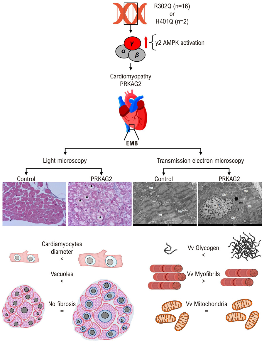
PRKAG2 cardiomyopathy hearts exhibited significant cardiomyocyte enlargement, normal-appearing mitochondria, extensive vacuolization of most myofibers, minimal interstitial fibrosis (only two patients had mild fibrosis), and no inflammatory cell infiltration. Transmission electron microscopy revealed abundant cytosolic glycogen, primarily in the perinuclear region, with additional deposits in intermyofibrillar and subsarcolemmal areas. This pronounced glycogen accumulation, consistently observed in all PRKAG2 patients, was absent in controls.

Conclusion

Histological and ultrastructural examination of right ventricular endomyocardial biopsy samples reveals a distinct set of features that strongly suggest PRKAG2 cardiomyopathy.
  • AMP-Activated Protein Kinases
  • Cardiomegaly
  • Microscopy, Electron, Transmission
Read Full Article
Creative Commons Attribution (CC BY) Creative Commons license
16/03/2026 DOI: 10.36660/abc.20240616i Views 2
Knockdown of the Long Noncoding RNA ZEB1-AS1 Accelerates Cardiac Hypertrophy via the miR-186-5p/HDAC2 Pathway
ABC
Original Article

Knockdown of the Long Noncoding RNA ZEB1-AS1 Accelerates Cardiac Hypertrophy via the miR-186-5p/HDAC2 Pathway

Bingfeng Cao et al. Qingxia Liu Xiaoyuan Zhang Yufeng Wang

Abstract

Background

The role of the long noncoding RNA ZEB1-AS1 in cardiac hypertrophy (CH) remains unclear.

Objectives

To investigate the function of ZEB1-AS1 in the development and progression of CH as well as elucidate its underlying molecular mechanism.

Methods

RNA expression levels were quantified by quantitative real-time PCR. Surface area of AC16 cells was assessed by immunofluorescence staining. Protein expression was evaluated by Western blotting. Interactions among RNAs were examined using luciferase reporter assays and RNA immunoprecipitation. Statistical significance was set at p < 0.05.

Results

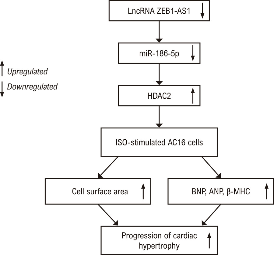
The expression of ZEB1-AS1 was upregulated in myocardial tissues and in isoproterenol (ISO)-stimulated AC16 cells. The knockdown of ZEB1-AS1 mitigated ISO-induced hypertrophic responses. Mechanistically, ZEB1-AS1 modulated histone deacetylase 2 (HDAC2) expression by acting as a molecular sponge for miR-186-5p. Consistently, the knockdown of ZEB1-AS1 reduced HDAC2 and decreased the expression of hypertrophic markers, including B-type natriuretic peptide, atrial natriuretic peptide, and β-myosin heavy chain, thereby restraining the progression of CH.

Conclusions

ZEB1-AS1 is upregulated in myocardial tissues and ISO-stimulated AC16 cells. Our findings indicate the ZEB1-AS1/miR-186-5p/HDAC2 axis contributes to CH, providing a mechanistic basis and potential therapeutic target for clinical intervention.

  • Cardiomegaly
  • Histone Deacetylase 2
  • Long Noncoding RNA
Read Full Article
Creative Commons Attribution (CC BY) Creative Commons license
16/01/2026 DOI: 10.36660/abc.20240703i Views 2
LncRNA CCAT2 Knockdown Alleviates Pressure Overload or Ang II-Induced Cardiac Hypertrophy Via Disruption of the Wnt/β-Catenin Signaling
ABC
Original Article

LncRNA CCAT2 Knockdown Alleviates Pressure Overload or Ang II-Induced Cardiac Hypertrophy Via Disruption of the Wnt/β-Catenin Signaling

Xiaojun Zhang et al. Zhen Chen Ning Zhang Bo Yu … Wei Li Mengli Zhang Xian Wu Ganzhe Liu Meizhen Dong

Abstract

Background

Sustained pathological cardiac hypertrophy (CH) is an independent risk factor for increased incidence and mortality of cardiovascular events.

Objectives

This research was designed to unravel the role of long non-coding RNA (LncRNA) CCAT2 in CH progression.

Methods

Transverse aortic constriction (TAC) procedures were conducted to construct a pressure overload-induced in vivo CH model. Angiotensin II (Ang II) treatment was utilized to induce hypertrophic rat cardiomyocyte H9c2 cells.

Results

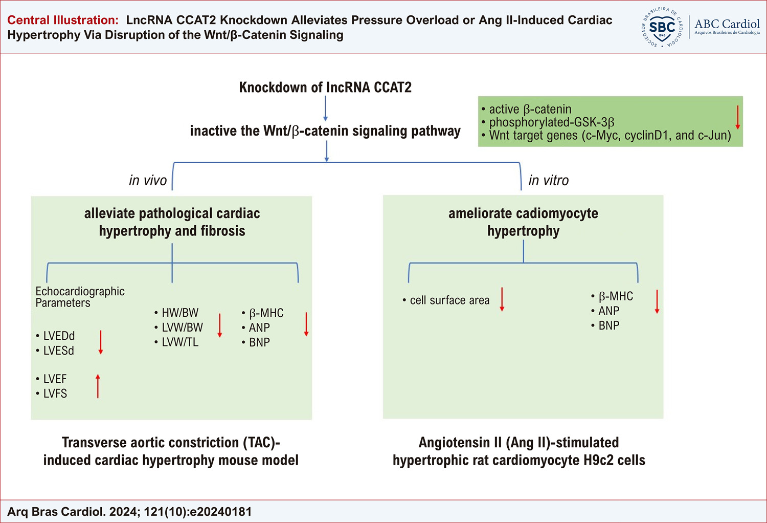
In vivo results showed that silencing of CCAT2 reduced cardiomyocyte surface area, alleviated cardiac fibrosis, and decreased β-MHC, ANP, and BNP levels in CH mouse models. In vitro results revealed that CCAT2 knockdown reduced cell surface area and attenuated β-MHC, ANP, and BNP levels in hypertrophic H9c2 cells. Besides, CCAT2 silencing decreased the levels of active β-catenin, phosphorylated-GSK-3β, and Wnt target genes (c-Myc, cyclinD1, and c-Jun) in CH mice and hypertrophic H9c2 cells. Importantly, treatment with the Wnt/β-catenin pathway activator LiCl reversed the suppression of CCAT2 knockdown on H9c2 cell surface area and MHC, ANP, and BNP levels.

Conclusions

Collectively, CCAT2 silencing plays a protective role against CH through inactivating the Wnt/β-catenin signaling, which suggests that CCAT2 might become a promising therapeutic target for CH.

  • angiotensin II
  • Cardiomegaly
  • Catenins
Read Full Article
Creative Commons Attribution (CC BY) Creative Commons license
11/11/2024 DOI: 10.36660/abc.20240181i Views 1
ABC
Editorial

PRKAG2 Syndrome: Is Screening with Early Echocardiography Effective in Children with a Positive Genotype?

Maria Elisa Lucena Sales de Melo Assunção et al. Norma Lucena Cavalcanti Licínio da Silva Mychelle Pascoaline de Miranda Silva

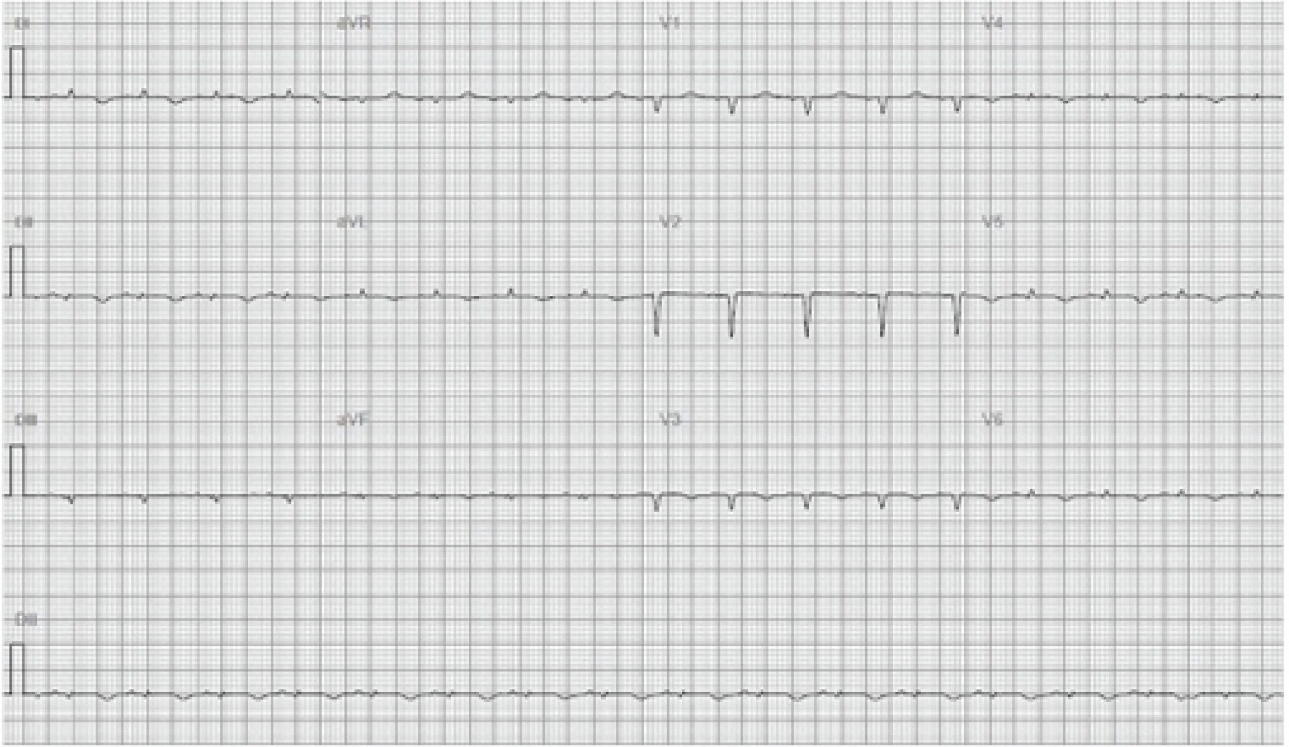
Described for the first time in 2001 by Gollob et al., the syndrome determined by the mutation of the PRKAG2 gene is rare, autosomal dominant, with high penetrance and unknown prevalence in the population. It manifests with the phenotype of myocardial hypertrophy and associated ventricular pre-excitation. Tachyarrhythmias are common in this genetic syndrome, as are disturbances in the cardiac conduction system that lead, in some cases, to pacemaker implantation at an early age.

The PRKAG2 gene mutation usually presents as a phenocopy of hypertrophic cardiomyopathy, differing from this in that there are no sarcomeric changes and, rather, accumulation of glycogen in cardiac myocytes. The definitive diagnosis is given by genetic study, but the presentation with left ventricular (LV) hypertrophy and conduction system disorders also form a differential diagnosis with Danon and Anderson-Fabry diseases.

[…]

  • Cardiac Arrhythmias
  • Cardiomegaly
  • Cardiomyopathies
  • Child
  • Genetics
Read Full Article
Creative Commons Attribution (CC BY) Creative Commons license
27/09/2024 DOI: 10.36660/abc.20240543i
Figure 1
ABC
Research Letter

Isolated Aneurysmal Dilatation of the Right Atrium, with Surgical Resolution, in a 53-Year-Old Adult

Edmar Atik et al. Alessandra Costa Barreto Maria Angélica Binotto Marcelo B. Jatene

Introduction

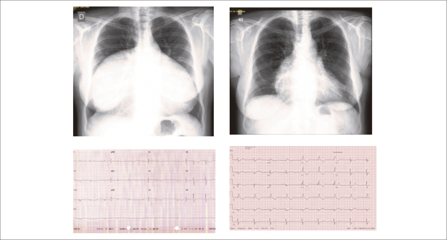
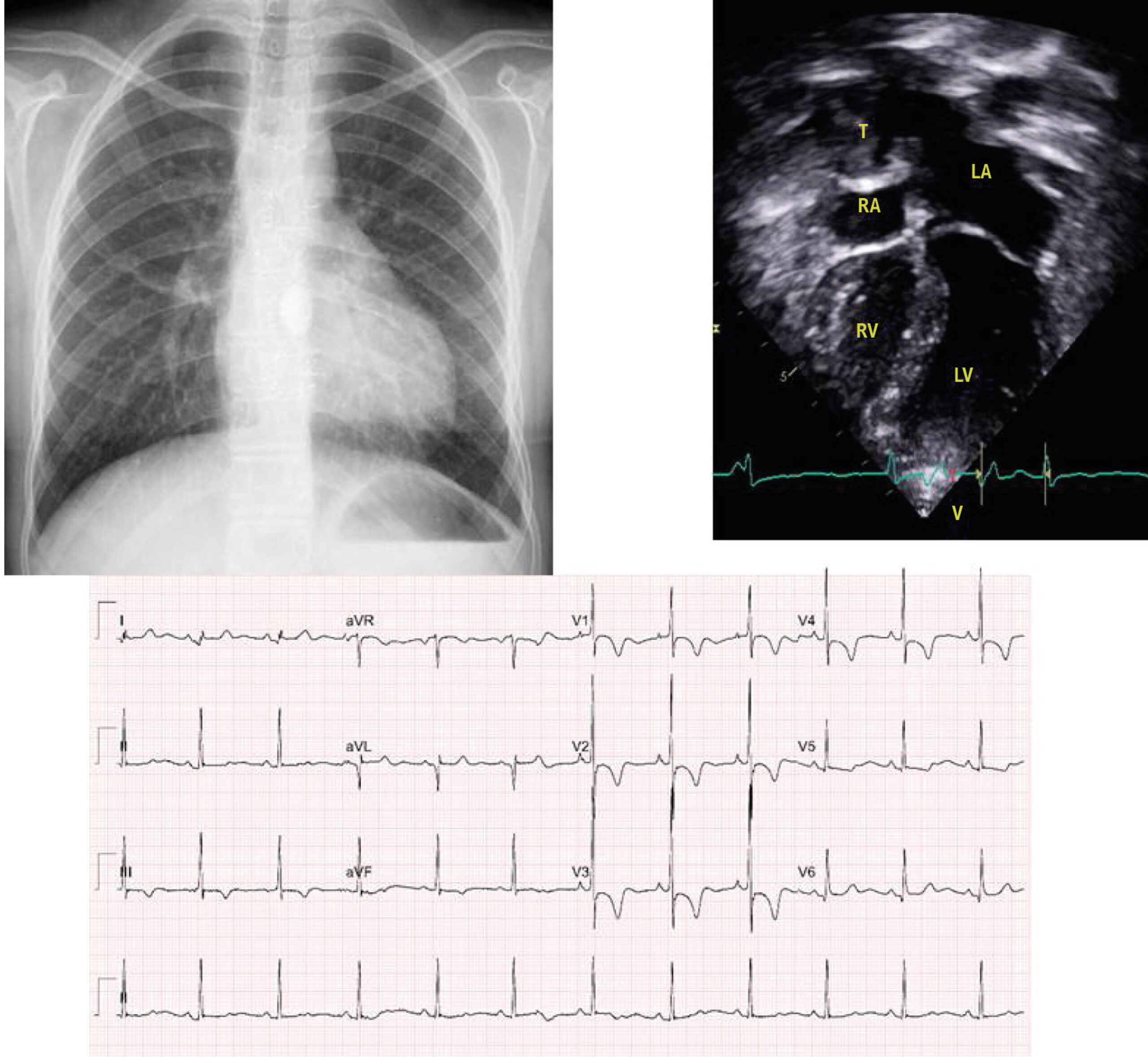
Isolated aneurysmal dilatation of the right atrium is a rare congenital anomaly. Its identification occurs in several age groups, from the fetus to adulthood. The clinical manifestation of this anomaly varies according to the magnitude of the dilation, from asymptomatic patients, usually in children and young people, to the presence of difficult-to-control supraventricular arrhythmias, right heart failure, airway obstruction, pneumonia, and thromboembolic events in adults. The diagnosis is established after verification of cardiomegaly in routine radiographic examinations and confirmed later by echocardiogram. It is noteworthy in this pathology the disproportionate size of the right atrium concerning the other cardiac cavities, with the tricuspid ring also dilated but with a preserved tricuspid valve. The tricuspid insufficiency that accompanies the condition results from the dilation of the valve ring. The recommendation for surgical correction then becomes prophylactic in childhood, given the good evolution verified later.

The differential diagnosis includes Ebstein’s anomaly, pericardial effusion, and mediastinal thoracic tumors.

[…]

  • Arrhythmias Cardiac
  • Cardiomegaly
  • Congenital Abnormalities/surgery
  • Ebstein Anomaly / surgery
  • Heart Aneurysm
  • Isolated Aneurysm of Right Atrium
  • Pericardial Effusion
Read Full Article
Creative Commons Attribution (CC BY) Creative Commons license
19/06/2023 DOI: 10.36660/abc.20220870 Views 2
ABC
Editorial

mRNA, miRNA, lncRNA, ceRNA: The Future of Cardiovascular Research?

Gustavo Augusto Ferreira Mota et al. Mariana Gatto Cristina Schmitt Gregolin Sérgio Luiz Borges de Souza Marina Politi Okoshi

Despite significant advances in cardiovascular biomedicine, heart diseases still represent a major public health problem. Thus, it is important to understand the molecular mechanisms involved in the pathophysiology of cardiovascular disease.

Cardiac injury is followed by cardiac remodeling, a process defined as genomic alterations triggering molecular, cellular, and interstitial modifications that manifest clinically as changes in the heart’s size, shape, and function., Therefore, alterations in the synthesis and degradation of cellular proteins characterize cardiac remodeling.

[…]

  • Cardiomegaly
  • Epigenesis, Genetic
  • Heart Failure
  • Nucleic Acids
  • Ventricular Remodeling
Read Full Article
Creative Commons Attribution (CC BY) Creative Commons license
12/05/2023 DOI: 10.36660/abc.20230209 Views 1
Figure 1
ABC
Original Article

SLC26A4-AS1 Aggravates AngII-induced Cardiac Hypertrophy by Enhancing SLC26A4 Expression

Xiaoliang Han et al. Chao Li Qinjiong Ji Ling Zhang … Xiaofei Xie Huijuan Shang Hong Ye

Abstract

Background

It has been reported that solute carrier family 26 members 4 antisense RNA 1 (SLC26A4-AS1) is highly related to cardiac hypertrophy.

Objective

This research aims to investigate the role and specific mechanism of SLC26A4-AS1 in cardiac hypertrophy, providing a novel marker for cardiac hypertrophy treatment.

Methods

Angiotensin II (AngII) was infused into neonatal mouse ventricular cardiomyocytes (NMVCs) to induce cardiac hypertrophy. Gene expression was detected by quantitative real-time PCR (RT-qPCR). Protein levels were evaluated via western blot. Functional assays analyzed the role of SLC26A4-AS1. The mechanism of SLC26A4-AS1 was assessed by RNA-binding protein immunoprecipitation (RIP), RNA pull-down, and luciferase reporter assays. The P value <0.05 was identified as statistical significance. Student’s t-test evaluated the two-group comparison. The difference between different groups was analyzed by one-way analysis of variance (ANOVA).

Results

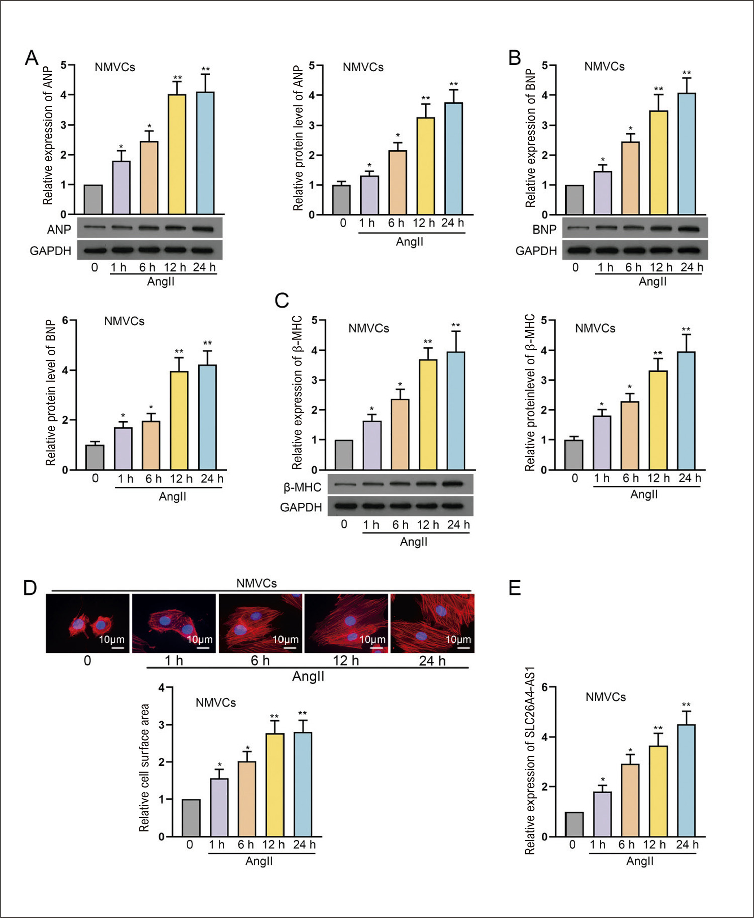
SLC26A4-AS1 is upregulated in AngII-treated NMVCs and promotes AngII-induced cardiac hypertrophy. SLC26A4-AS1 regulates its nearby gene solute carrier family 26 members 4 (SLC26A4) via functioning as a competing endogenous RNA (ceRNA) to modulate the microRNA (miR)-301a-3p and miR-301b-3p in NMVCs. SLC26A4-AS1 promotes AngII-induced cardiac hypertrophy via upregulating SLC26A4 or sponging miR-301a-3p/miR-301b-3p.

Conclusion

SLC26A4-AS1 aggravates AngII-induced cardiac hypertrophy via sponging miR-301a-3p or miR-301b-3p to enhance SLC26A4 expression.

  • Cardiomegaly
  • RNA
  • RNA, Long Noncoding
Read Full Article
Creative Commons Attribution (CC BY) Creative Commons license
21/04/2023 DOI: 10.36660/abc.20210933
ABC
Editorial

A New Therapeutic Alternative for the Treatment of Pulmonary Hypertension?

Ricardo J Gelpi

In this issue of the journal Arquivos Brasileiros de Cardiologia, important and original research work on rats is published. The work is important because pulmonary hypertension represents a serious pathology that is very difficult to treat. The only way to relieve patients is lung transplantation, but are very well known the difficulties and limitations of this treatment. The work is also original because a lot has been published about the beneficial effects of copaiba oil, such as its anti-inflammatory action, analgesic effect, and ability to prevent infections and improve the healing process. However, the beneficial effect of copaiba oil on pulmonary hypertension had not been described. In this sense, the group led by Adriane Belló Klein has been working for several years on this important issue of pulmonary hypertension in general,– and the effects of copaiba oil on this particular pathology,, which has made this research group an international benchmark on this theme.

It is important to mention that pulmonary hypertension induced in rats by monocrotaline produces vascular lesions very similar to those found in patients with this pathology., In essence, what the work published in this issue of the journal Arquivos Brasileiros de Cardiologia shows is that the administration of copaiba oil to a group of rats with pulmonary hypertension induced by monocrotaline produces a clear beneficial effect, through the decrease in peripheral blood indicators of oxidative stress, a reduction in pulmonary vascular resistance, and a favorable remodeling of the right ventricle, with a reduction in hypertrophy, which translates into a better systolic function of that ventricle. This finding is very important because it is well known that myocardial hypertrophy is one of the most important causes of heart failure and sudden death. The positive relationship between the degree of hypertrophy and the decrease in oxidative stress shown by the authors is clearly proven in this work by the high r-value obtained when these two variables were correlated. However, and beyond this important correlation, future studies should determine if the beneficial effect on the right ventricle is due to the reduction of oxidative stress or if there is also a direct effect of copaiba oil on myocardial fibers. Perhaps experiments that could be carried out on isolated hearts would make it possible to detect or not a direct impact of this natural agent. In this sense, it is important to mention that the authors of this study also published an interesting work previously where they showed that the administration of copaiba oil decreased apoptosis signals in the hypertrophic right ventricle and attributed the better remodeling to a possible decrease in the apoptosis mentioned above; obviously this same mechanism could be acting in the work that we are commenting now. Nevertheless, here it is important to make some clarifications that may allow a better understanding of what is happening in these hearts with hypertrophy secondary to pulmonary hypertension.

[…]

  • Cardiomegaly
  • Copaiba Oil
  • Cor Pulmonale
  • Fabaceae
  • Heart Failure
  • Hypertension,Pulmonary
  • Lung Transplantation
  • Monocrotaline
  • Oxidative Stress
  • phytotherapy
  • Rats
Read Full Article
Creative Commons Attribution (CC BY) Creative Commons license
03/12/2021 DOI: 10.36660/abc.20210935
Figure 1
ABCHF
Case Report

Heart Failure Due to Anomalous Pulmonary Vein Connection Associated with Atrial Septal Defect

Marcely Gimenes Bonatto et al. Ana Karyn Ehrenfried de Freitas Letícia dos Santos de Oliveira Rocha Claudinei Collatusso … Rafael de Almeida Torres Gustavo Gavazzoni Blume Lídia Zytynski Moura

Introduction

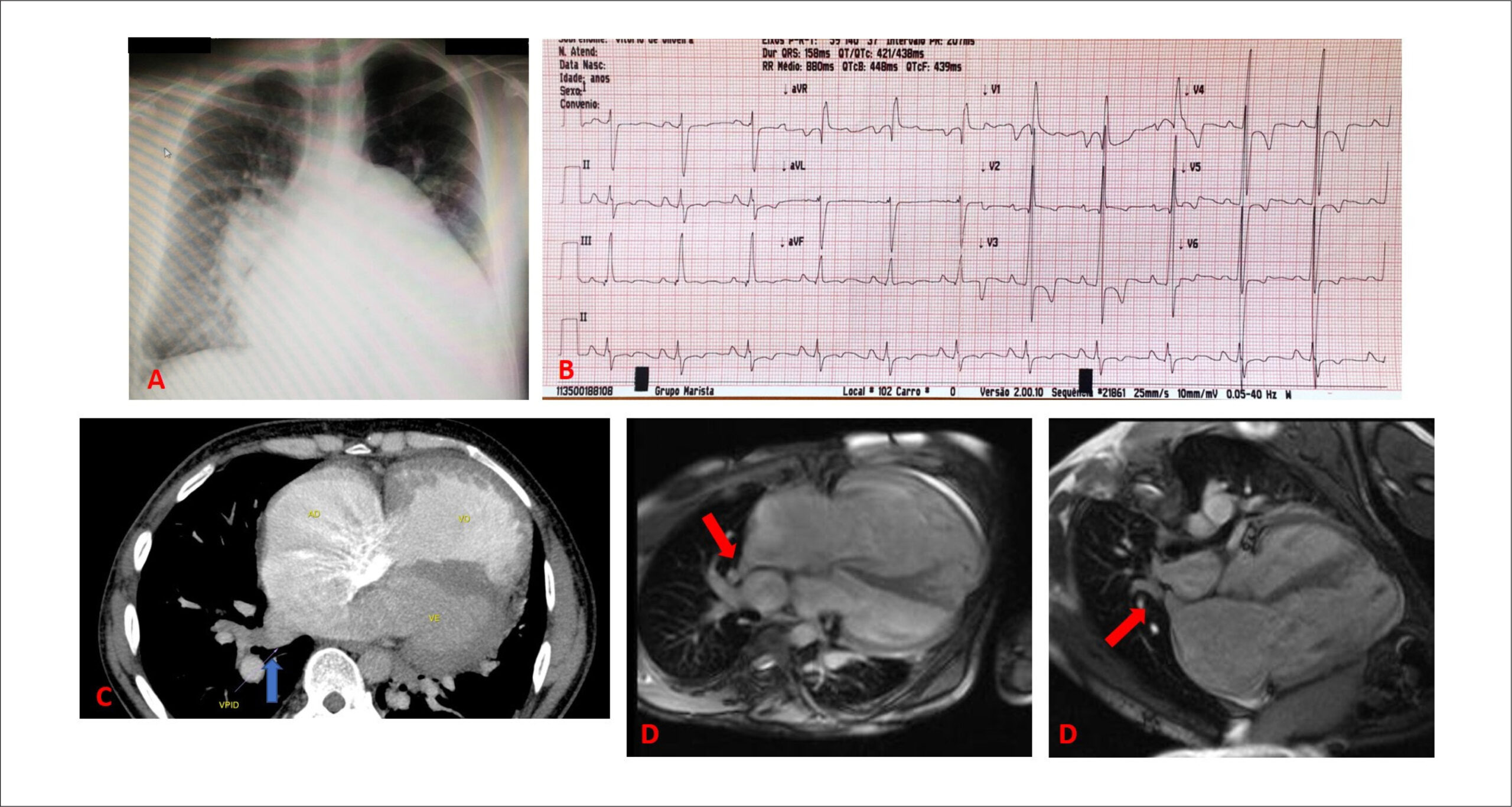
Anomalous pulmonary vein dreinage (APVD) is a rare disease, accounting for nearly 1 to 3% of congenital heart diseases. It may be total, when all PVs are involved, or partial, when only some PVs drain anomalously into one or more systemic veins or directly into the right atrium (RA)., This condition occurs alone or in association with an interatrial septal defect, more often sinus venosus interatrial communication (IAC) (which accounts for nearly 5% of all IAC cases).

Patients with this condition may remain asymptomatic until adulthood. However, overload of right chambers may trigger symptoms, especially reduced functional capacity and progressive dyspnea, in addition to the development of pulmonary hypertension (PH) and ventricular dysfunction, which is the most severe disease scenario. Recognizing these symptoms and establishing early diagnosis and treatment is crucial to avoid disease progression and its prognostic implications.

[…]

  • Cardiomegaly
  • Darinage
  • Echocardiography/methods
  • Heart Defects,Congenital
  • Heart Failure/physiopathologyu
  • Heart Septal Defects, Atrial
  • Pulmonary Veins
Read Full Article
Creative Commons Attribution (CC BY) Creative Commons license
06/07/2021 DOI: 10.36660/abchf.20210004 Views 1
Figure 1
ABCIC
Original Article

Comparative Analysis of the Coronary Arteries Flow Pattern in Secondary Myocardial Hypertrophies and by Sarcomeric Mutation

Caio Guedes de Sousa et al. José Maria Del Castillo Carlos Mazzarollo Eugenio Soares de Albuquerque … Antônia Dulcineide Medeiros Sena Djair Brindeiro Filho Carlos Antônio da Mota Silveira

Abstract

Background:

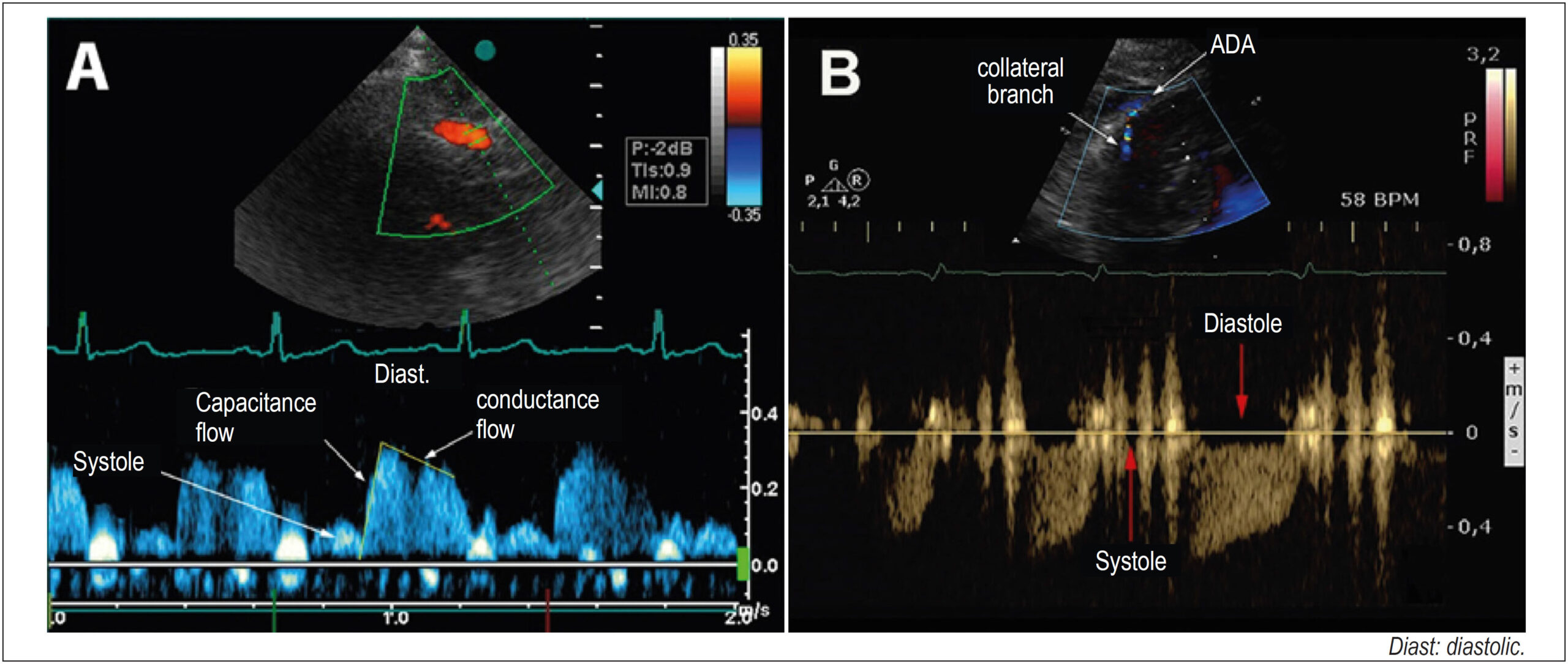
Coronary flow with a diastolic predominance increases two to five times in hyperemia, mediated by vasodilation (coronary flow reserve, CFR) and, in hypertrophy, relative ischemia may occur. In secondary hypertrophy (LVH), the flow, normal at rest, becomes ischemic due to increased demand. In hypertrophic cardiomyopathy (HCM) with perivascular fibrosis, collateral vessels appear to increase the irrigation of hypertrophied segments.

Objective:

To determine the coronary flow pattern in patients with secondary hypertrophy and hypertrophic cardiomyopathy, evaluating the coronary flow reserve.

Methods:

Coronary flow was evaluated in 34 patients with secondary hypertrophy, 24 with hypertrophic cardiomyopathy and in 16 controls. The anterior descending artery was detected with transthoracic Doppler with adequate equipment calibration. In the hypertrophic cardiomyopathy group, the flow of collaterals from the hypertrophic region was evaluated. In the control and secondary hypertrophy groups and in six patients in the hypertrophic cardiomyopathy group, the intravenous dipyridamole (0.84 mg) coronary flow reserve was calculated. The data were compared by variance with a significance of 5%.

Results:

In secondary hypertrophy there was an increase in mass index and blood pressure, and in hypertrophic cardiomyopathy an increase in relative thickness predominated. Ejection fraction and diastolic dysfunction were higher in the hypertrophic cardiomyopathy group. The coronary flow reserve was lower in the hypertrophic cardiomyopathy group, and flow of collaterals was also detected, with a reduction in the coronary flow reserve.

Conclusion:

the analysis of coronary circulation with transthoracic Doppler is possible in normal and hypertrophic individuals. Patients with secondary hypertrophy and hypertrophic cardiomyopathy have a decrease in the coronary flow reserve, and patients with hypertrophic cardiomyopathy show a hyper flow of dilated collateral vessels observed in the hypertrophic region, with a decrease in the coronary flow reserve.

  • Cardiomegaly
  • Echocardiography, Doppler
  • Fractional Flow Reserve, Myocardial
Read Full Article
Creative Commons Attribution (CC BY) Creative Commons license
16/06/2021 DOI: 10.47593/2675-312X/20213401eabc131 Views 2
Figure 1
ABC
Research Letter

17-Year-Old Man with Pulmonary Atresia and Intact Ventricular Septum Submitted to Fontan Operation, and with Persistent Coronary-Cavitary Fistula

Edmar Atik

Introduction

Pulmonary atresia with intact ventricular septum is a congenital anomaly with total interruption of blood flow between the right ventricle and the pulmonary trunk, and in general it is not accompanied by associated defects, except for interatrial septal defect, present in 20%, with predominance of patent foramen ovale. Valve atresia results from the lack of embryological development of this fibrous structure, which can be located at the valve level (fibrous membrane) but also at the infundibular level (blind fundus). As a consequence, myocardial hypertrophy and hypoplasia of the right ventricle, hypoplasia of the ring and tricuspid valve, mild tricuspid insufficiency and pulmonary flow dependent on the ductus arteriosus appear. In view of greater hypertension in right ventricle, direct connections with the coronary circulation are formed through sinusoids, with flow towards the aorta. In this situation, it is said that the coronary circulation is dependent on the right ventricle, and when these connections are present in great magnitude, they predispose to myocardial infarction, arrhythmias and right ventricular volume overload, due to retrograde flow from the aorta.

In cases where valve atresia develops later in the fetus, the right ventricular cavity may be well formed with its three portions, the entrance route, the trabecular and the exit route and, as a consequence, there is marked tricuspid insufficiency, even with Ebstein-type alteration of the redundant and myxomatous valve, ventricular wall thinning, ventricular dysfunction and right heart failure, the latter superimposed on hypoxia. In general, it is not accompanied by other associated defects and the pulmonary arteries are of adequate size. In these cases, there is no connection between the right ventricle and the coronary arteries by sinusoids.

[…]

  • Arrhythmias Cardiacs
  • Cardiomegaly
  • Coronary-Cavitary Fistula
  • Fontan Surgery
  • Heart Defects, Congenital
  • Pulmonary Atresia/surgery
  • Right Ventricle/Abnormalities
Read Full Article
Creative Commons Attribution (CC BY) Creative Commons license
14/06/2021 DOI: 10.36660/abc.20201011
Figure 1
ABC
Brief Communication

Heart Failure with Supra-normal Left Ventricular Ejection Fraction – State of the Art

Ziyin Huang et al. Yufeng Jiang Yafeng Zhou

Abstract

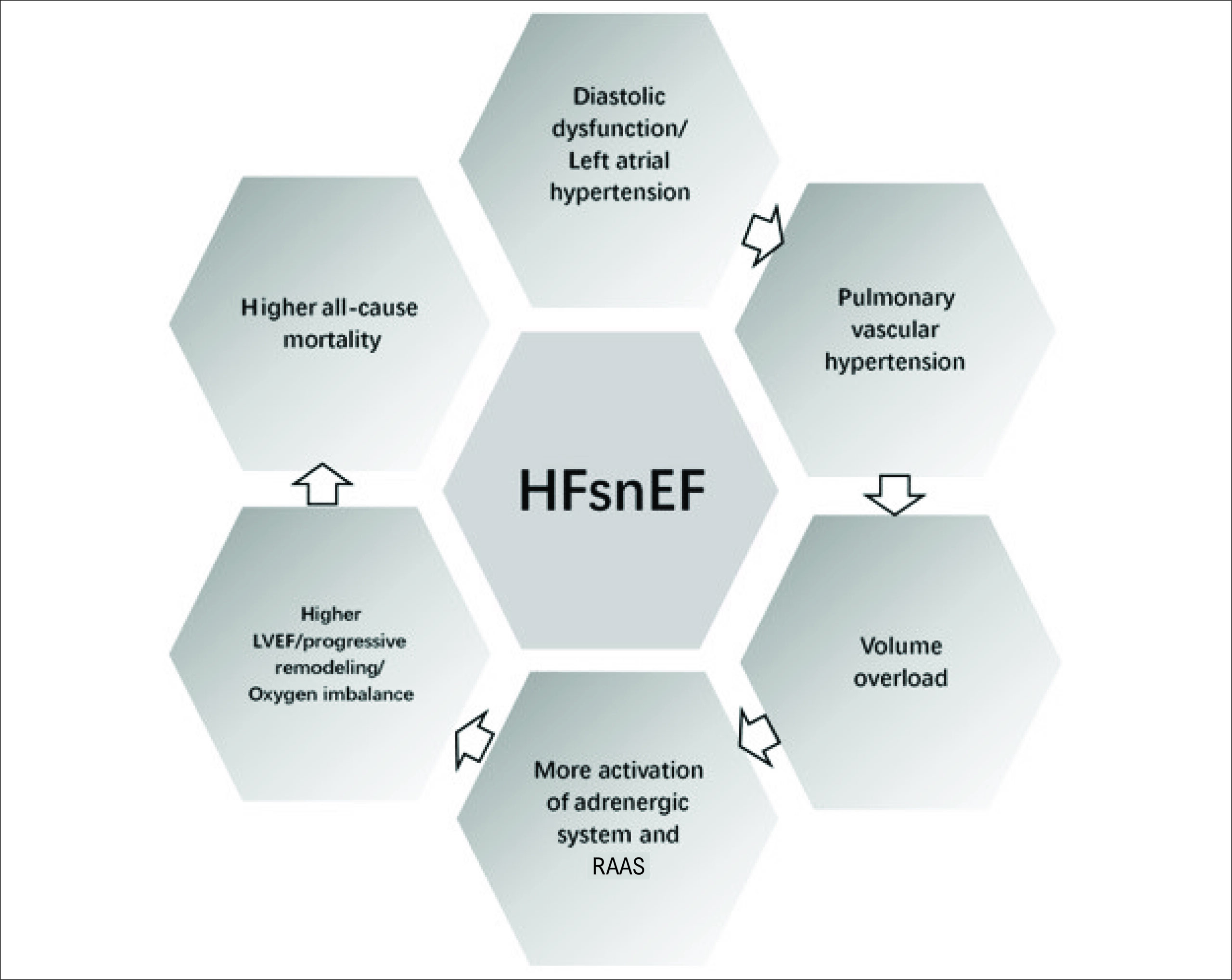
In 2019, an article published in the European Heart Journal recognized for the first time heart failure (HF) with left ventricular ejection fraction (LVEF)≥ 65% as a new HF phenotype, heart failure with supra-normal left ventricular ejection fraction (HFsnEF), with the main purpose of promoting research on this new category. They analyzed mortality in people with HF and found that there was a u-shaped relationship between mortality and LVEF. Accordingly, HFsnEF patients had a higher all-cause mortality compared with other patients diagnosed with HF with preserved ejection fraction (HFpEF). This article describes the current situation of HFsnEF and discusses future perspectives based on the preliminary results of our group. To better treat patients with HFsnEF, it is fundamental that cardiologists and physicians understand the differences and similarities of this new phenotype.

  • Cardiomegaly
  • Echocardiography/methods
  • Heart Failure
  • Heart Failure, Diastolic
  • Mortality
  • Prognosis
  • Stroke Volume
Read Full Article
Creative Commons Attribution (CC BY) Creative Commons license
17/05/2021 DOI: 10.36660/abc.20190835
Figure 1
ABC
Original Article

Correlation between Cardiomegaly on Chest X-Ray and Left Ventricular Diameter on Echocardiography in Patients with Chagas Disease

Matheus Rassi Fernandes Ramos et al. Henrique Turin Moreira Gustavo Jardim Volpe Minna Romano … Benedito Carlos Maciel André Schmidt Anis Rassi Junior Jose Antônio Marin Neto

Abstract

Background

Cardiomegaly on chest X-ray is an independent predictor of death in individuals with chronic Chagas cardiomyopathy (CCC). However, the correlation between increased cardiothoracic ratio (CTR) on chest X-ray and left ventricular end-diastolic diameter (LVEDD) on echocardiography is not well established in this population.

Objectives

To assess the relationship between chest X-ray and LVEDD on echocardiography in patients with Chagas disease and its applicability to the Rassi score.

Methods

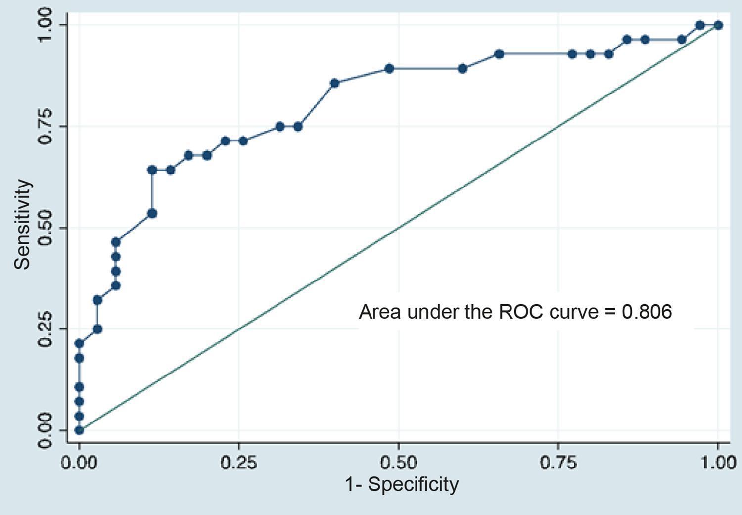
Retrospective study on 63 Chagas disease outpatients who underwent chest X-ray and echocardiography. Cardiomegaly on chest X-ray was defined as a CTR>0.5. LVEDD was analyzed as a continuous variable. ROC curve was used to evaluate the ability of LVEDD in detecting cardiomegaly by chest X-ray, with a cut-off point defined by the highest sum of sensitivity and specificity.

Results

Median age 61 years [interquartile range 48-68], 56% were women. CCC was detected in 58 patients, five patients had the indeterminate form of Chagas disease. Cardiomegaly was detected in 28 patients. The area under the ROC curve for LVEDD was 0.806 (95%CI: 0.692-0.919). The optimal cut-off for LVEDD was 60 mm (sensitivity = 64%, specificity = 89%). The use of LVEDD on echocardiography as a surrogate for CTR on chest X-ray changed the Rassi score values of 14 patients, with a reduction in the presumed risk in 10 of them.

Conclusion

LVEDD on echocardiography is an appropriate, highly specific parameter to distinguish between the presence and absence of cardiomegaly on chest X-ray in Chagas disease. (Arq Bras Cardiol. 2021; 116(1):68-74)

  • Cardiomegaly
  • Chagas Disease/physiopathology
  • chagasic cardiomyopathy
  • Heart block
  • X-Rays
Read Full Article
Creative Commons Attribution (CC BY) Creative Commons license
03/02/2021 DOI: 10.36660/abc.20190673 Views 1
ABC
Editorial

Can Transthoracic Echocardiography Replace Chest Radiography in the Evaluation of Cardiomegaly in Chagas Cardiomyopathy?

Tiago Senra

Chagas cardiomyopathy (CCM), first described in 1909 by Carlos Chagas, is still associated with high morbidity and mortality and socioeconomic impact, especially in Latin American countries.

The search for epidemiological, clinical, laboratory, electrocardiographic, and image markers associated with its prognosis plays an important role in the risk stratification of these patients.

[…]

  • Cardiomegaly
  • Chagas Cardiomyopathy
  • Echocardiography/methods
  • Epidemiology
  • Heart Failure
  • Morbimortality
  • Radiography, Thoracic/methods
  • Socioeconomic Factors
Read Full Article
Creative Commons Attribution (CC BY) Creative Commons license
03/02/2021 DOI: 10.36660/abc.20200625 Views 2
Figure 1
ABC
Case Report

A Life-threatening Combination: Indomethacin and Dabigatran

Adem Adar et al. Orhan Onalan Fahri Cakan

Introduction

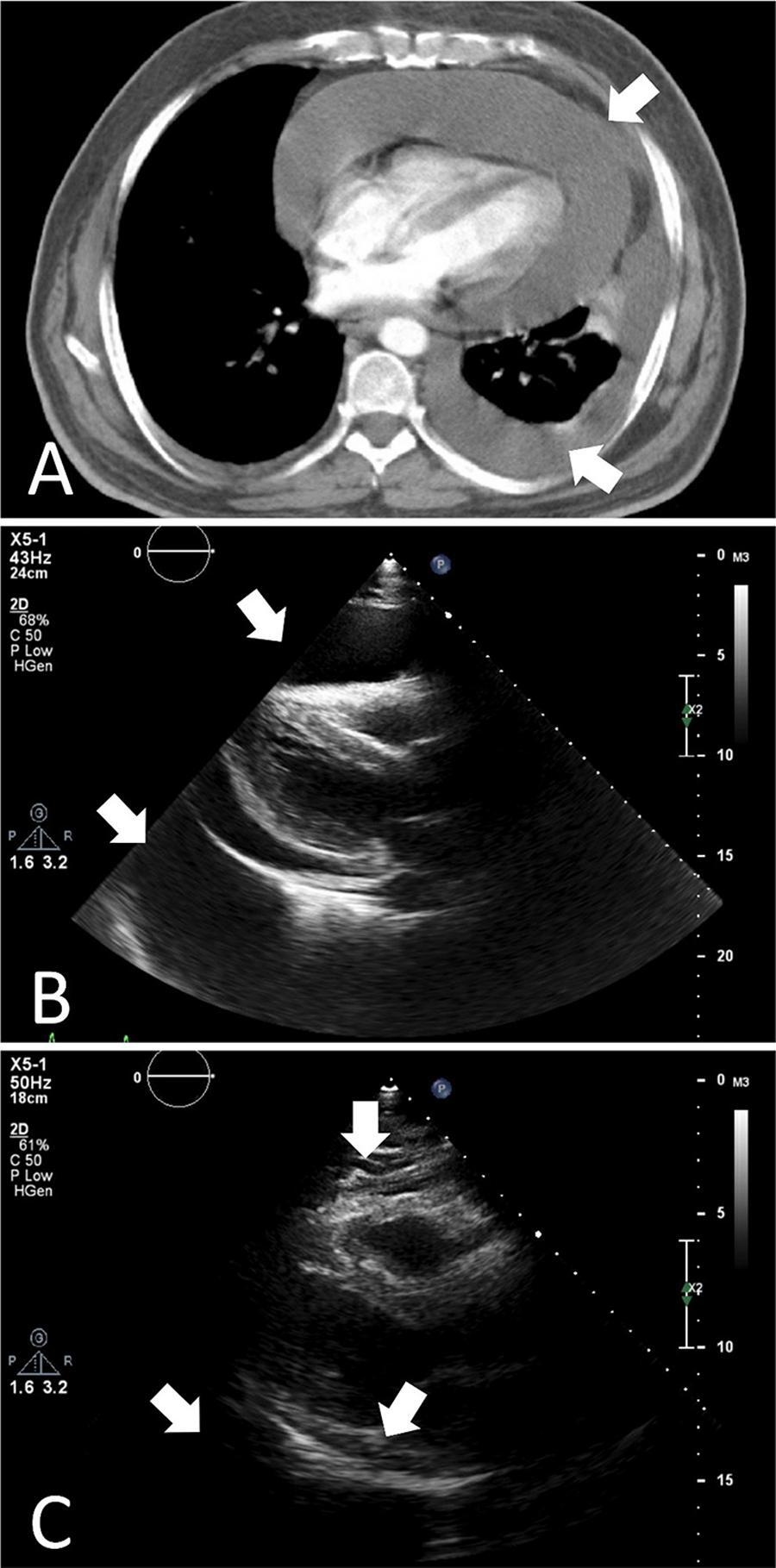
Although rare, several bleeding complications may occur in patients receiving dabigatran. The risk of bleeding is particularly high in patients with impaired kidney functions or in patients who are on concomitant nephrotoxic drugs. We report a case of massive pleuropericardial effusion developed after the initiation of indomethacin treatment in a patient who was receiving dabigatran for deep venous thrombosis.

[…]

  • Cardiomegaly
  • Dabigatran/administration & dosage
  • Echocardiography
  • Indomenthacin/administration & dosage
  • Pleural Effusion
  • Renal Insufficiency
Read Full Article
Creative Commons Attribution (CC BY) Creative Commons license
18/05/2020 DOI: 10.36660/abc.20180159 Views 1
Figure 1
ABC
Original Article

Serum Sirtuin 1, 3 and 6 Levels in Acute Myocardial Infarction Patients

Emrullah Kızıltunç et al. Arzu Kösem Can Özkan Burcu Uğurlu Ilgın … Harun Kundi Mustafa Çetin Ender Ornek

Abstract

Background:

Sirtuins may act in many cellular processes like apoptosis, DNA repair and lipid/glucose metabolism. Experimental studies suggested some sirtuin types may have protective effects against endothelial dysfunction, atherosclerosis, cardiac hypertrophy and reperfusion injury. Data about sirtuins in acute myocardial infarction (AMI) patients are scarce.

Objectives:

To investigate temporal changes of serum sirtuin 1,3 and 6 levels in AMI patients; to compare the serum sirtuin 1,3 and 6 levels between AMI patients and control subjects; and to investigate the association of serum sirtuin 1,3 and 6 levels with prognostic markers of AMI.

Methods:

Forty patients with AMI and 40 patients with normal coronary arteries were included. Left ventricular ejection fraction (LVEF), serum proBNP, CRP, sirtuin1, sirtuin 3 and sirtuin 6 levels were processed. Peak troponin T levels, GRACE score, first day / second day sirtuin levels were recorded of AMI patients. A p value < 0.05 was considered statistically significant.

Results:

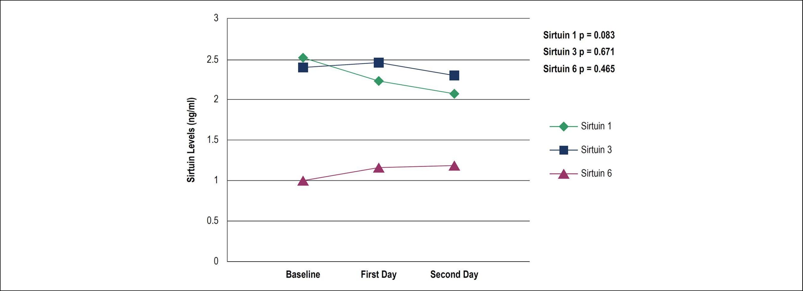
Serum sirtuin 1,3 and 6 levels in AMI patients were similar to those in normal coronary patients. No temporal change in serum sirtuin 1,3 and 6 levels were found in AMI course. No correlation was evident between the sirtuin levels and the following parameters: proBNP, CRP, peak troponin and LVEF. Baseline sirtuin 1 and 6 levels were positively correlated with reperfusion duration. Baseline sirtuin 3 levels were negatively correlated with GRACE score.

Conclusion:

Serum sirtuin 1,3 and 6 levels in AMI patients were similar to those in normal coronary patients. This study does not represent evidence of the possible protective effects of sirtuin1, 3 and 6 in AMI patients.

  • Atherosclerosis
  • Carcinogenesis
  • Cardiomegaly
  • Cellular Senescence
  • Endothelium / dysfunction
  • Lipid Metabolism
  • Sirtuins/drug effects
Read Full Article
Creative Commons Attribution (CC BY) Creative Commons license
10/07/2019 DOI: 10.5935/abc.20190114
Figure 1
IJCS
Original Article

Hypotensive Response to Continuous Aerobic and High-Intensity Interval Exercise Matched by Volume in Sedentary Subjects

Francesco Pinto Boeno et al. Thiago Rozales Ramis Juliano Boufleur Farinha Cesar Moritz … Vagner Pereira dos Santos Alvaro Reischak de Oliveira Bruno Costa Teixeira

Abstract

Background:

Systemic arterial hypertension (SAH) is one of the main risk factors for heart disease. Among the benefits linked to different modalities of physical exercise, post-exercise hypotension (PEH) is a key point for exercise prescription in this condition.

Objective:

To investigate and compare PEH in response to continuous aerobic exercise (CONT) and high-intensity interval exercise (HIIE), matched by volume, in sedentary individuals.

Methods:

A randomized cross-over study, composed of sedentary, healthy male subjects submitted to two acute physical exercise protocols matched by volume, HIIE and CONT, on a treadmill. Hemodynamic measures for the evaluation of PEH were performed pre, immediately after exercise and every five minutes thereafter, during one hour of recovery. Two-way ANOVA with repeated measurements was used for comparisons between groups and Bonferroni post hoc test as appropriate. P < 0.05 was considered significant.

Results:

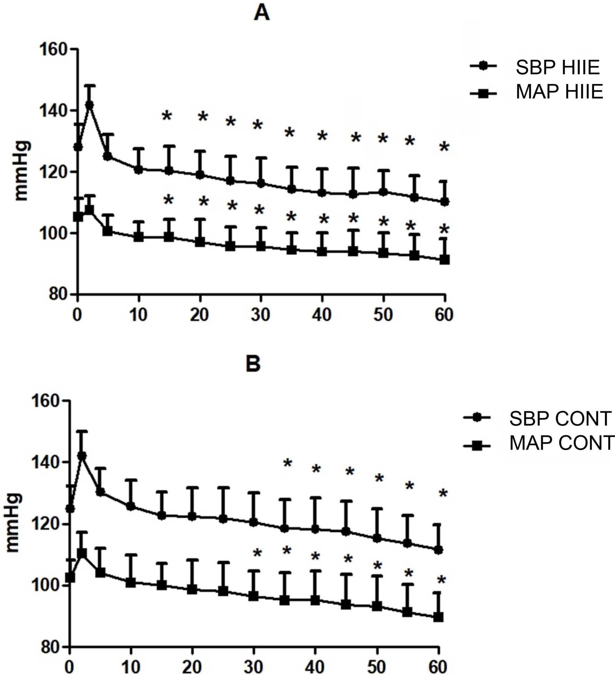
Both exercise protocols promoted significant PEH, with reductions in systolic blood pressure (SBP) and mean arterial pressure (MAP). HIIE promoted a reduction of SBP and MAP at the 15th minute, whereas the same effect was observed at the 30th following CONT.

Conclusion:

Both HIIE and CONT, matched by volume, promote PEH of similar magnitude. However, PEH occurs earlier following HIIE, suggesting a better time / effectiveness ratio, and an additional beneficial effect of this modality.

  • Adherence Guidelines
  • Blood Pressure
  • Cardiomegaly
  • Exercise
  • High-Intensity Interval Exercise
  • Hypertensio/physiopathology
  • Post-Exercise Hypotension
  • Sedentarsm
Read Full Article
Creative Commons Attribution (CC BY) Creative Commons license
01/01/2019 DOI: 10.5935/2359-4802.20180084 Views 1
Figure 1
ABC
Original Article

The Effects of Trimetazidine on QT-interval Prolongation and Cardiac Hypertrophy in Diabetic Rats

Fatemeh Ramezani-Aliakbari et al. Mohammad Badavi Mahin Dianat Seyed Ali Mard Akram Ahangarpour

Abstract

Background:

Trimetazidine (TMZ) is an anti-ischemic drug. In spite of its protective effects on cardiovascular system, there is no scientific study on the usefulness of TMZ treatment for prolonged QT interval and cardiac hypertrophy induced by diabetes.

Objectives:

To evaluate the effects of TMZ on QT interval prolongation and cardiac hypertrophy in the diabetic rats.

Methods:

Twenty-four male Sprague-Dawley rats (200-250 g) were randomly assigned into three groups (n = 8) by simple random sampling method. Control (C), diabetic (D), and diabetic administrated with TMZ at 10 mg/kg (T10). TMZ was administrated for 8 weeks. The echocardiogram was recorded before isolating the hearts and transfer to a Langendorff apparatus. Hemodynamic parameters, QT and corrected QT interval (QTc) intervals, heart rate and antioxidant enzymes were measured. The hypertrophy index was calculated. The results were evaluated by one-way ANOVA and paired t-test using SPSS (version 16) and p < 0.05 was regarded as significant.

Results:

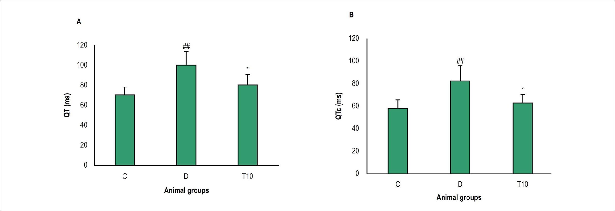
The diabetic rats significantly indicated increased hypertrophy, QT and QTc intervals and decreased Left ventricular systolic pressure (LVSP), Left ventricular developed pressure (LVDP), rate pressure product (RPP), Max dp/dt, and min dp/dt (±dp/dt max), heart rate, superoxide dismutase (SOD), glutathione peroxidase (GPx) and catalase in the heart. Treatment with TMZ in the diabetic animals was significantly improved these parameters in comparison to the untreated diabetic group.

Conclusions:

TMZ improves QTc interval prolongation and cardiac hypertrophy in diabetes.

  • Cardiomegaly
  • Diabetes Mellitus
  • Electrocardiology
  • Oxidative Stress
  • Rats
  • Trimetadizine
Read Full Article
Creative Commons Attribution (CC BY) Creative Commons license
17/12/2018 DOI: 10.5935/abc.20180248 Views 1
Figure 1
ABC
Original Article

Pioglitazone Induces Cardiomyocyte Apoptosis and Inhibits Cardiomyocyte Hypertrophy Via VEGFR-2 Signaling Pathway

Wenliang Zhong et al. Wen Jin Shanghua Xu Yanqing Wu … Shunxiang Luo Minlie Liang Lianglong Chen

Abstract

Background:

Pioglitazone has been widely used as an insulin-sensitizing agent for improving glycemic control in patients with type 2 diabetes mellitus. However, cardiovascular risk and protective effects of pioglitazone remain controversial.

Objectives:

In this study, we investigated whether pioglitazone affects cardiomyocyte apoptosis and hypertrophy by regulating the VEGFR-2 signaling pathway.

Methods:

Cardiomyocytes were enzymatically isolated from 1- to 3-day-old Sprague-Dawley rat ventricles. Effects of pioglitazone and the VEGFR-2-selective inhibitor apatinib on cardiomyocyte apoptotic rate was determined using flow cytometry, and hypertrophy was evaluated using [3H]-leucine incorporation. The protein expressions of unphosphorylated and phosphorylated VEGFR-2, Akt, P53, and mTOR were determined by Western-Blotting. Analysis of variance (ANOVA) was used to assess the differences between groups.

Results:

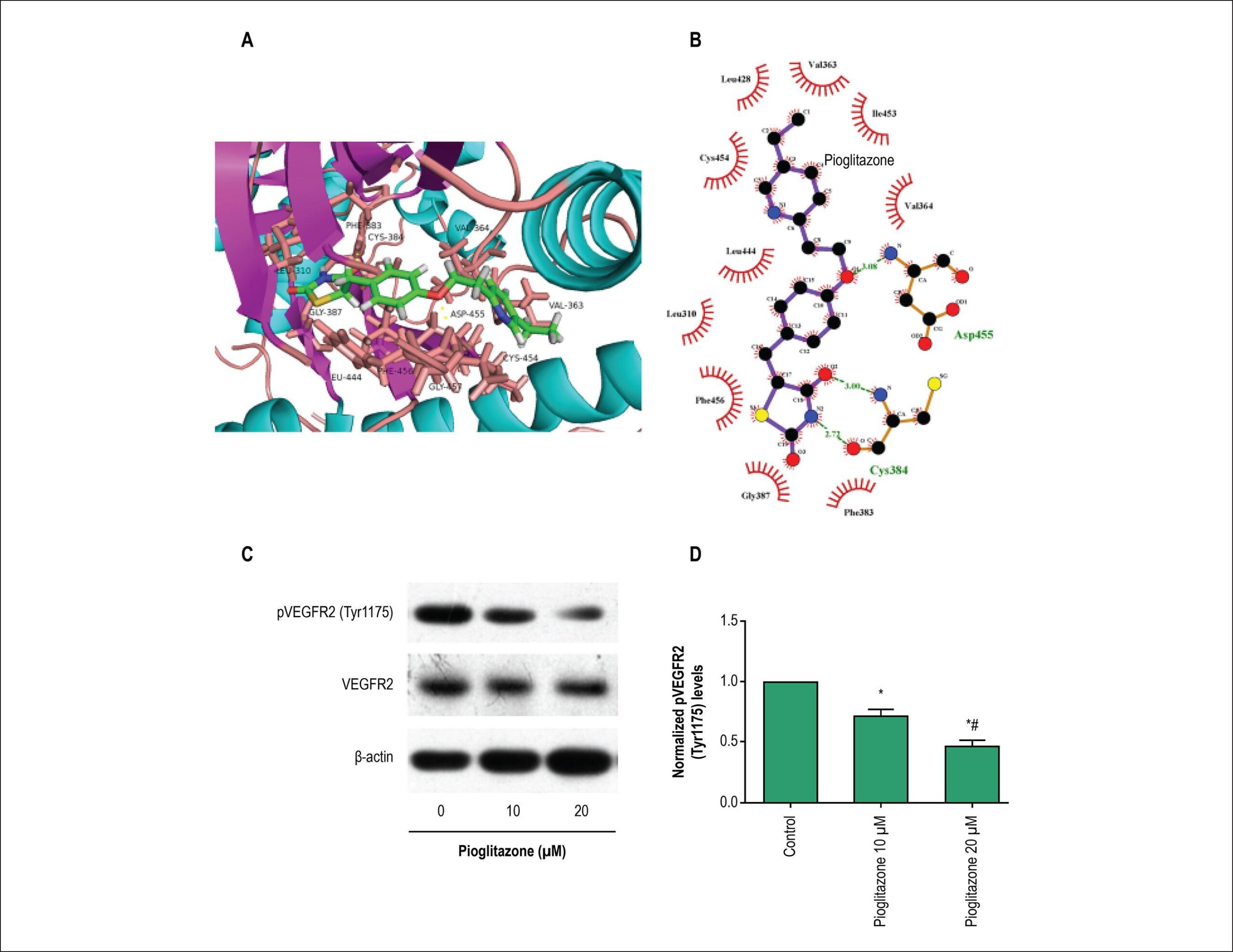
Pioglitazone and VEGFR-2-selective inhibitor apatinib reduced rat cardiomyocyte viability and cardiomyocyte hypertrophy induced by angiotensin II in vitro. Furthermore, in the same in vitro model, pioglitazone and apatinib significantly increased the expression of Bax and phosphorylated P53 and decreased the expression of phosphorylated VEGFR-2, Akt, and mTOR, which promote cardiomyocyte hypertrophy.

Conclusions:

These findings indicate that pioglitazone induces cardiomyocyte apoptosis and inhibits cardiomyocyte hypertrophy by modulating the VEGFR-2 signaling pathway.

  • antihypertensive agents
  • Apoptosis
  • Cardiomegaly
  • Heart Failure/physiopathology
  • Insulin Resistance
  • Myocytes, Cardiac
  • Thiazolidinediones
Read Full Article
Creative Commons Attribution (CC BY) Creative Commons license
02/07/2018 DOI: 10.5935/abc.20180108
Figure 1
ABC
Original Article

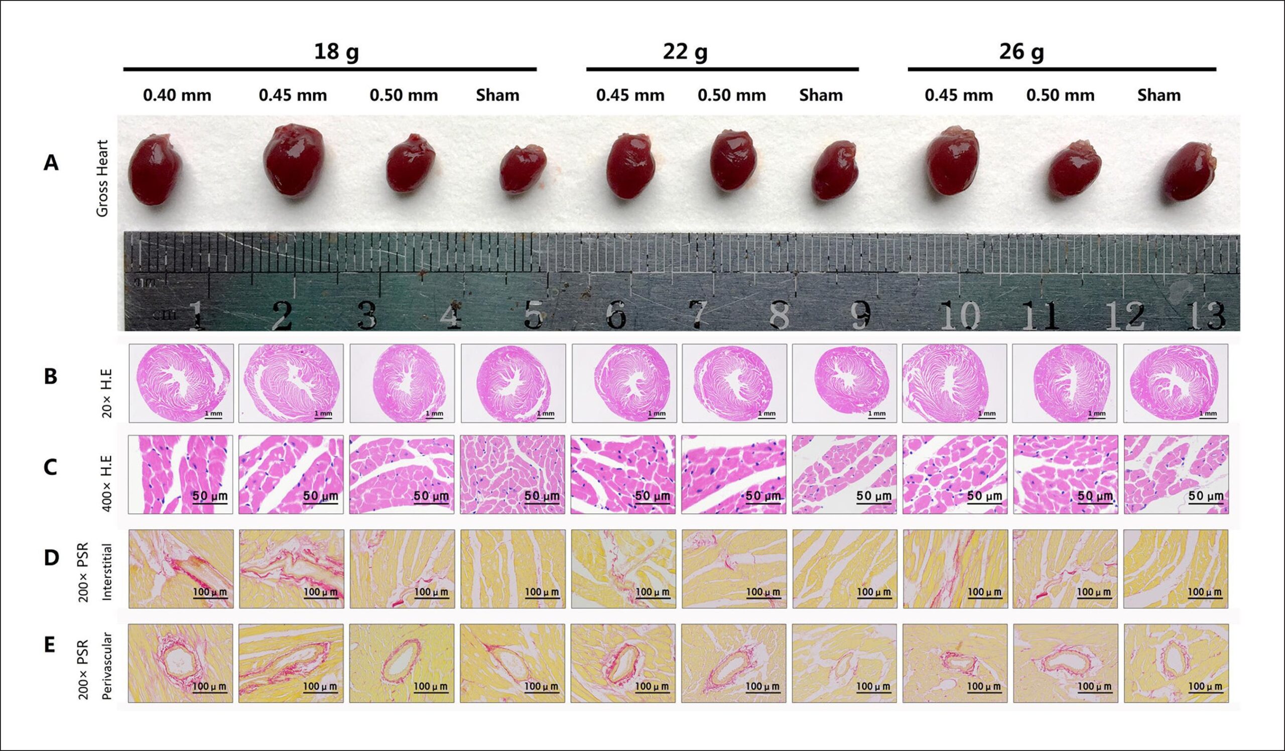
Pressure Overload-induced Cardiac Hypertrophy Varies According to Different Ligation Needle Sizes and Body Weights in Mice

Zhen Jia et al. Zhijun Chen Hongfei Xu Malik Akuffu Armah … Peng Teng Weidong Li Dongdong Jian Liang Ma Yiming Ni

Abstract

Background:

The cardiac hypertrophy (CH) model for mice has been widely used, thereby providing an effective research foundation for CH exploration.

Objective:

To research the effects of CH modeling under abdominal aortic constriction (AAC) using different needles and weights in mice.

Methods:

Four needles with different external diameters (0.35, 0.40, 0.45, and 0.50 mm) were used for AAC. 150 male C57BL/6 mice were selected according to body weight (BW) and divided into 3 weight levels: 18 g, 22 g, and 26 g (n = 50 in each group). All weight levels were divided into 5 groups: a sham group (n = 10) and 4 AAC groups using 4 ligation intensities (n = 10 per group). After surgery, survival rates were recorded, echocardiography was performed, hearts were dissected and used for histological detection, and data were statistically analyzed, P < 0.05 was considered statistically significant.

Results:

All mice died in the following AAC groups: 18g/0.35 mm, 22 g/0.35 mm, 26 g/0.35 mm, 22 g/0.40 mm, and 26 g/0.40 mm. All mice with AAC, those ligated with a 0.50-mm needle, and those that underwent sham operation survived. Different death rates occurred in the following AAC groups: 18 g/0.40 mm, 18 g/0.45 mm, 18 g/0.50 mm, 22 g/45 mm, 22 g/0.50 mm, 26 g/0.45 mm, and 26 g/0.50 mm. The heart weight/body weight ratios (5.39 ± 0.85, 6.41 ± 0.68, 4.67 ± 0.37, 5.22 ± 0.42, 4.23 ± 0.28, 5.41 ± 0.14, and 4.02 ± 0.13) were significantly increased compared with those of the sham groups for mice with the same weight levels.

Conclusion:

A 0.45-mm needle led to more obvious CH than did 0.40-mm and 0.50-mm needles and caused extraordinary CH in 18-g mice.

  • Body weight
  • Cardiomegaly
  • Heart Failure
  • Needles/utilization
  • Rats
Read Full Article
Creative Commons Attribution (CC BY) Creative Commons license
01/06/2018 DOI: 10.5935/abc.20180088
Figure 1
IJCS
Case Report

Hypertrophic Cardiomyopathy, All Phenotypes in one

Aníbal M Arias et al. Diego Perez de Arenaza Rodolfo Pizarro Ricardo G Marenchino … Fernando Garagoli Hernán Garcia Rivello César Belziti

Introduction

Hypertrophic cardiomyopathy (HCM) is an intrinsic myocardial disorder characterized by cardiac hypertrophy (wall thickness ≥ 15 mm), that is not explained by conditions of pressure overload (eg, hypertension, severe aortic stenosis). HCM is the most common genetic primary cardiomyopathy, with a prevalence estimated to be about one in 500 adults in the general population.

More than 450 mutations have been identified in the 20 genes that cause different phenotypes. In most cases, HCM is associated with sarcomere protein gene mutations, and exhibits multiple phenotypic expressions. We present a case that combines all phenotypes.

[…]

  • Cardiomegaly
  • Cardiomyopathy, Hypertrophic
  • Heart Failure
  • Heart Transplantation
Read Full Article
Creative Commons Attribution (CC BY) Creative Commons license
01/01/2018 DOI: 10.5935/2359-4802.20180016 Views 1
Figure 1
ABC
Original Article

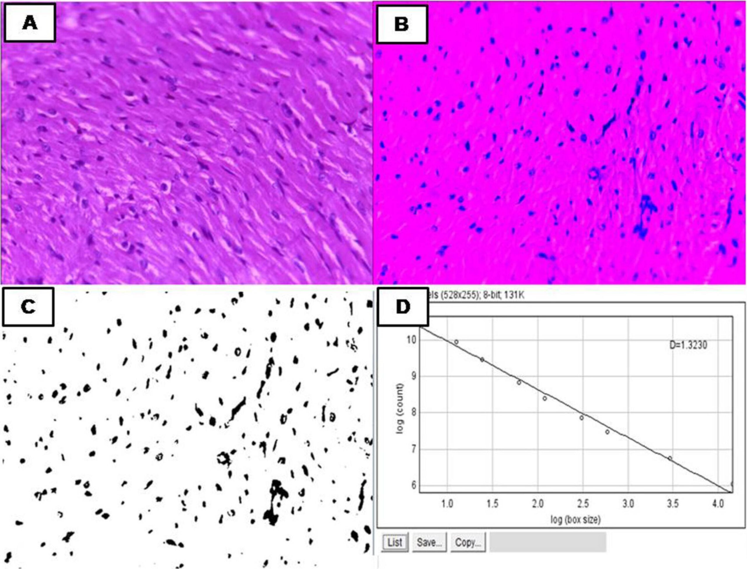
Fractal Dimension in Quantifying Experimental-Pulmonary-Hypertension-Induced Cardiac Dysfunction in Rats

Francis Lopes Pacagnelli et al. Ana Karênina Dias de Almeida Sabela Thaoan Bruno Mariano Guilherme Akio Tamura Ozaki … Robson Chacon Castoldi Edna Maria do Carmo Robson Francisco Carvalho Loreta Casquel Tomasi Katashi Okoshi Luiz Carlos Marques Vanderlei

Abstract

Background:

Right-sided heart failure has high morbidity and mortality, and may be caused by pulmonary arterial hypertension. Fractal dimension is a differentiated and innovative method used in histological evaluations that allows the characterization of irregular and complex structures and the quantification of structural tissue changes.

Objective:

To assess the use of fractal dimension in cardiomyocytes of rats with monocrotaline-induced pulmonary arterial hypertension, in addition to providing histological and functional analysis.

Methods:

Male Wistar rats were divided into 2 groups: control (C; n = 8) and monocrotaline-induced pulmonary arterial hypertension (M; n = 8). Five weeks after pulmonary arterial hypertension induction with monocrotaline, echocardiography was performed and the animals were euthanized. The heart was dissected, the ventricles weighed to assess anatomical parameters, and histological slides were prepared and stained with hematoxylin/eosin for fractal dimension analysis, performed using box-counting method. Data normality was tested (Shapiro-Wilk test), and the groups were compared with non-paired Student t test or Mann Whitney test (p < 0.05).

Results:

Higher fractal dimension values were observed in group M as compared to group C (1.39 ± 0.05 vs. 1.37 ± 0.04; p < 0.05). Echocardiography showed lower pulmonary artery flow velocity, pulmonary acceleration time and ejection time values in group M, suggesting function worsening in those animals.

Conclusion:

The changes observed confirm pulmonary-arterial-hypertension-induced cardiac dysfunction, and point to fractal dimension as an effective method to evaluate cardiac morphological changes induced by ventricular dysfunction.

  • Cardiomegaly
  • Echocardiography/methods
  • Heart Failure / mortality
  • Hypertension Pulmonary
  • Monocrotaline
  • Rats
Read Full Article
Creative Commons Attribution (CC BY) Creative Commons license
24/05/2016 DOI: 10.5935/abc.20160083
Figure 1
ABC
Anatomopathological Session

Case 4 – A 79-Year-Old Man with Congestive Heart Failure Due to Restrictive Cardiomyopathy

Sumaia Mustafa et al. Alice Tatsuko Yamada Fabio Mitsuo Lima Valdemir Melechco Carvalho … Vera Demarchi Aiello Jussara Bianchi Castelli

Clinical Aspects

The patient JAB, a 79-year-old previous smoker and alcoholic man residing in São Paulo, attended an outpatient clinic at InCor due to heart failure which worsened progressively since 2012, requiring hospitalization for treatment.

Heart failure is a systemic and complex clinical syndrome, defined as a cardiac dysfunction that causes the blood supply to be insufficient to meet tissular metabolic demands, in the presence of a normal venous return, or which only meets the demands with high filling pressure.

[…]

  • ascites
  • Cardiomegaly
  • Cardiomyopathy, Restrictive
  • Heart Arrest
  • Heart Failure
Read Full Article
Creative Commons Attribution (CC BY) Creative Commons license
01/10/2015 DOI: 10.5935/abc.20150135 Views 1
MicroRNAs: a new paradigm in the treatment and diagnosis of Heart failure?
ABC
Review Article

MicroRNAs: a new paradigm in the treatment and diagnosis of Heart failure?

Vagner Oliveira-Carvalho et al. Vitor Oliveira Carvalho Miguel Morita Silva Guilherme Veiga Guimarães Edimar Alcides Bocchi

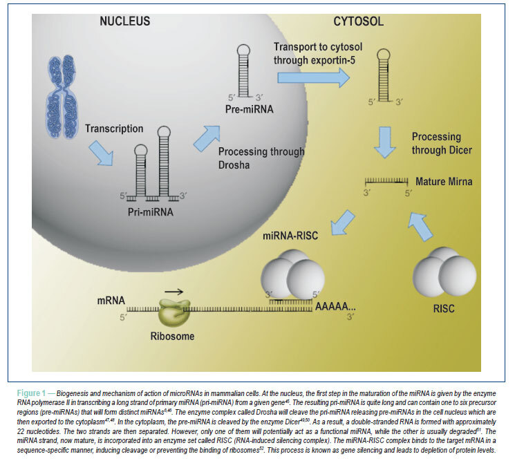
MicroRNAs (miRNAs) are a group of newly discovered small RNAs, non-coding, which represent one of the most exciting areas of modern medical science as they modulate a huge and complex regulatory network of gene expression. Lines of evidence have recently suggested that miRNAs play a key role in the pathogenesis of heart failure. Some miRNAs highly expressed in the heart, such as miR-1, miR-133 and miR-208, are strongly associated with the development of cardiac hypertrophy, while the exact role of miR-21 in the cardiovascular system remains controversial. Serum levels of circulating miRNAs such as miR-423-5p are being evaluated as potential biomarkers in the diagnosis and prognosis of heart failure. On the other hand, the manipulation of levels of miRNAs using techniques such as mimicking the miRNAs (miRmimics) and antagonistic miRNAs (antagomiRs) is making increasingly evident the enormous potential of miRNAs as promising therapeutic strategies in heart failure.

  • Cardiomegaly
  • Heart Failure
  • MicroRNAs
Read Full Article
Creative Commons Attribution (CC BY) Creative Commons license
21/06/2012 DOI: 10.1590/S0066-782X2012000400011

Could not load more articles. Please try again.

How to cite this article

EndNote RIS

Electronic Document Format (Vancouver)

Honorato-Sampaio K, Oliveira C, Araújo SA, Correa FS, Dias GM, Bahia Neto AFC, Cantuária VL, et al. PRKAG2 Cardiomyopathy: A Case-Control Study on the Diagnostic Yield Of Histopathology and Ultrastructural Analysis from Endomyocardial Biopsy. Arquivos Brasileiros de Cardiologia 2026;123(2):e20240616.

Electronic Document Format (ABNT)

Honorato-Sampaio, Kinulpe; Oliveira, Carla de; Araújo, Stanley de Almeida; Correa, Frederico Soares; Dias, Glauber Monteiro; Bahia Neto, Antônio Fernandino de Castro; Cantuária, Vinícius Lopes; Yavari, Arash; Moreira, Maria da Consolação Vieira; Pena, José Luiz Barros; Brasileiro Filho, Geraldo; Sternick, Eduardo Back. PRKAG2 Cardiomyopathy: A Case-Control Study on the Diagnostic Yield Of Histopathology and Ultrastructural Analysis from Endomyocardial Biopsy. Arquivos Brasileiros de Cardiologia, v. 123, n. 2, e20240616, Mar. 2026.

Electronic Document Format (APA)

Honorato-Sampaio, K., Oliveira, C. , Araújo, S. A., Correa, F. S., Dias, G. M., Bahia Neto, A. F. C., Cantuária, V. L., Yavari, A., Moreira, M. C. V., Pena, J. L. B., Brasileiro Filho, G., & Sternick, E. B. (2026). PRKAG2 Cardiomyopathy: A Case-Control Study on the Diagnostic Yield Of Histopathology and Ultrastructural Analysis from Endomyocardial Biopsy. Arquivos Brasileiros de Cardiologia, 123(2), e20240616.

Electronic Document Format (ISO)

Honorato-Sampaio, Kinulpe and Oliveira, Carla de and Araújo, Stanley de Almeida and Correa, Frederico Soares and Dias, Glauber Monteiro and Bahia Neto, Antônio Fernandino de Castro and Cantuária, Vinícius Lopes and Yavari, Arash and Moreira, Maria da Consolação Vieira and Pena, José Luiz Barros and Brasileiro Filho, Geraldo and Sternick, Eduardo Back. PRKAG2 Cardiomyopathy: A Case-Control Study on the Diagnostic Yield Of Histopathology and Ultrastructural Analysis from Endomyocardial Biopsy. Arquivos Brasileiros de Cardiologia [online]. 2026, vol. 123, n. 2, [cited 2026-10-14], e20240616. Available from: <https://journals.cardiol.br/en/article/prkag2-cardiomyopathy-a-case-control-study-on-the-diagnostic-yield-of-histopathology-and-ultrastructural-analysis-from-endomyocardial-biopsy/>. ISSN 0066-782X

EndNote RIS

Electronic Document Format (Vancouver)

Cao B, Liu Q, Zhang X, Wang Y. Knockdown of the Long Noncoding RNA ZEB1-AS1 Accelerates Cardiac Hypertrophy via the miR-186-5p/HDAC2 Pathway. Arquivos Brasileiros de Cardiologia 2026;122(11):e20240703.

Electronic Document Format (ABNT)

Cao, Bingfeng; Liu, Qingxia; Zhang, Xiaoyuan; Wang, Yufeng. Knockdown of the Long Noncoding RNA ZEB1-AS1 Accelerates Cardiac Hypertrophy via the miR-186-5p/HDAC2 Pathway. Arquivos Brasileiros de Cardiologia, v. 122, n. 11, e20240703, Jan. 2026.

Electronic Document Format (APA)

Cao, B., Liu, Q., Zhang, X., & Wang, Y. (2026). Knockdown of the Long Noncoding RNA ZEB1-AS1 Accelerates Cardiac Hypertrophy via the miR-186-5p/HDAC2 Pathway. Arquivos Brasileiros de Cardiologia, 122(11), e20240703.

Electronic Document Format (ISO)

Cao, Bingfeng and Liu, Qingxia and Zhang, Xiaoyuan and Wang, Yufeng. Knockdown of the Long Noncoding RNA ZEB1-AS1 Accelerates Cardiac Hypertrophy via the miR-186-5p/HDAC2 Pathway. Arquivos Brasileiros de Cardiologia [online]. 2026, vol. 122, n. 11, [cited 2026-10-14], e20240703. Available from: <https://journals.cardiol.br/en/article/knockdown-of-the-long-noncoding-rna-zeb1-as1-accelerates-cardiac-hypertrophy-via-the-mir-186-5p-hdac2-pathway/>. ISSN 0066-782X

EndNote RIS

Electronic Document Format (Vancouver)

Zhang X, Chen Z, Zhang N, Yu B, Li W, Zhang M, Wu X, et al. LncRNA CCAT2 Knockdown Alleviates Pressure Overload or Ang II-Induced Cardiac Hypertrophy Via Disruption of the Wnt/β-Catenin Signaling. Arquivos Brasileiros de Cardiologia 2024;121(10):e20240181.

Electronic Document Format (ABNT)

Zhang, Xiaojun; Chen, Zhen; Zhang, Ning; Yu, Bo; Li, Wei; Zhang, Mengli; Wu, Xian; Liu, Ganzhe; Dong, Meizhen. LncRNA CCAT2 Knockdown Alleviates Pressure Overload or Ang II-Induced Cardiac Hypertrophy Via Disruption of the Wnt/β-Catenin Signaling. Arquivos Brasileiros de Cardiologia, v. 121, n. 10, e20240181, Nov. 2024.

Electronic Document Format (APA)

Zhang, X., Chen, Z., Zhang, N., Yu, B., Li, W., Zhang, M., Wu, X., Liu, G., & Dong, M. (2024). LncRNA CCAT2 Knockdown Alleviates Pressure Overload or Ang II-Induced Cardiac Hypertrophy Via Disruption of the Wnt/β-Catenin Signaling. Arquivos Brasileiros de Cardiologia, 121(10), e20240181.

Electronic Document Format (ISO)

Zhang, Xiaojun and Chen, Zhen and Zhang, Ning and Yu, Bo and Li, Wei and Zhang, Mengli and Wu, Xian and Liu, Ganzhe and Dong, Meizhen. LncRNA CCAT2 Knockdown Alleviates Pressure Overload or Ang II-Induced Cardiac Hypertrophy Via Disruption of the Wnt/β-Catenin Signaling. Arquivos Brasileiros de Cardiologia [online]. 2024, vol. 121, n. 10, [cited 2026-10-14], e20240181. Available from: <https://journals.cardiol.br/en/article/lncrna-ccat2-knockdown-alleviates-pressure-overload-or-ang-ii-induced-cardiac-hypertrophy-via-disruption-of-the-wnt-%ce%b2-catenin-signaling/>. ISSN 0066-782X

EndNote RIS

Electronic Document Format (Vancouver)

Assunção MELSM, Silva NLCL, Silva MPM. PRKAG2 Syndrome: Is Screening with Early Echocardiography Effective in Children with a Positive Genotype?. Arquivos Brasileiros de Cardiologia 2024;121(8):e20240543.

Electronic Document Format (ABNT)

Assunção, Maria Elisa Lucena Sales de Melo; Silva, Norma Lucena Cavalcanti Licínio da; Silva, Mychelle Pascoaline de Miranda. PRKAG2 Syndrome: Is Screening with Early Echocardiography Effective in Children with a Positive Genotype?. Arquivos Brasileiros de Cardiologia, v. 121, n. 8, e20240543, Sep. 2024.

Electronic Document Format (APA)

Assunção, M. E. L. S. M., Silva, N. L. C. L. , & Silva, M. P. M. (2024). PRKAG2 Syndrome: Is Screening with Early Echocardiography Effective in Children with a Positive Genotype?. Arquivos Brasileiros de Cardiologia, 121(8), e20240543.

Electronic Document Format (ISO)

Assunção, Maria Elisa Lucena Sales de Melo and Silva, Norma Lucena Cavalcanti Licínio da and Silva, Mychelle Pascoaline de Miranda. PRKAG2 Syndrome: Is Screening with Early Echocardiography Effective in Children with a Positive Genotype?. Arquivos Brasileiros de Cardiologia [online]. 2024, vol. 121, n. 8, [cited 2026-10-14], e20240543. Available from: <https://journals.cardiol.br/en/article/prkag2-syndrome-is-screening-with-early-echocardiography-effective-in-children-with-a-positive-genotype/>. ISSN 0066-782X

EndNote RIS

Electronic Document Format (Vancouver)

Atik E, Barreto AC, Binotto MA, Jatene MB. Isolated Aneurysmal Dilatation of the Right Atrium, with Surgical Resolution, in a 53-Year-Old Adult. Arquivos Brasileiros de Cardiologia 2023;120(6):e20220870.

Electronic Document Format (ABNT)

Atik, Edmar; Barreto, Alessandra Costa; Binotto, Maria Angélica; Jatene, Marcelo B.. Isolated Aneurysmal Dilatation of the Right Atrium, with Surgical Resolution, in a 53-Year-Old Adult. Arquivos Brasileiros de Cardiologia, v. 120, n. 6, e20220870, Jun. 2023.

Electronic Document Format (APA)

Atik, E., Barreto, A. C., Binotto, M. A., & Jatene, M. B. (2023). Isolated Aneurysmal Dilatation of the Right Atrium, with Surgical Resolution, in a 53-Year-Old Adult. Arquivos Brasileiros de Cardiologia, 120(6), e20220870.

Electronic Document Format (ISO)

Atik, Edmar and Barreto, Alessandra Costa and Binotto, Maria Angélica and Jatene, Marcelo B.. Isolated Aneurysmal Dilatation of the Right Atrium, with Surgical Resolution, in a 53-Year-Old Adult. Arquivos Brasileiros de Cardiologia [online]. 2023, vol. 120, n. 6, [cited 2026-10-14], e20220870. Available from: <https://journals.cardiol.br/en/article/isolated-aneurysmal-dilatation-of-the-right-atrium-with-surgical-resolution-in-a-53-year-old-adult/>. ISSN 0066-782X

EndNote RIS

Electronic Document Format (Vancouver)

Mota GAF, Gatto M, Gregolin CS, Souza SLB, Okoshi MP. mRNA, miRNA, lncRNA, ceRNA: The Future of Cardiovascular Research?. Arquivos Brasileiros de Cardiologia 2023;120(4):e20230209.

Electronic Document Format (ABNT)

Mota, Gustavo Augusto Ferreira; Gatto, Mariana; Gregolin, Cristina Schmitt; Souza, Sérgio Luiz Borges de; Okoshi, Marina Politi. mRNA, miRNA, lncRNA, ceRNA: The Future of Cardiovascular Research?. Arquivos Brasileiros de Cardiologia, v. 120, n. 4, e20230209, May. 2023.

Electronic Document Format (APA)

Mota, G. A. F., Gatto, M., Gregolin, C. S., Souza, S. L. B. , & Okoshi, M. P. (2023). mRNA, miRNA, lncRNA, ceRNA: The Future of Cardiovascular Research?. Arquivos Brasileiros de Cardiologia, 120(4), e20230209.

Electronic Document Format (ISO)

Mota, Gustavo Augusto Ferreira and Gatto, Mariana and Gregolin, Cristina Schmitt and Souza, Sérgio Luiz Borges de and Okoshi, Marina Politi. mRNA, miRNA, lncRNA, ceRNA: The Future of Cardiovascular Research?. Arquivos Brasileiros de Cardiologia [online]. 2023, vol. 120, n. 4, [cited 2026-10-14], e20230209. Available from: <https://journals.cardiol.br/en/article/mrna-mirna-lncrna-cerna-the-future-of-cardiovascular-research/>. ISSN 0066-782X

EndNote RIS

Electronic Document Format (Vancouver)

Han X, Li C, Ji Q, Zhang L, Xie X, Shang H, Ye H, et al. SLC26A4-AS1 Aggravates AngII-induced Cardiac Hypertrophy by Enhancing SLC26A4 Expression. Arquivos Brasileiros de Cardiologia 2023;120(4):e20210933.

Electronic Document Format (ABNT)

Han, Xiaoliang; Li, Chao; Ji, Qinjiong; Zhang, Ling; Xie, Xiaofei; Shang, Huijuan; Ye, Hong. SLC26A4-AS1 Aggravates AngII-induced Cardiac Hypertrophy by Enhancing SLC26A4 Expression. Arquivos Brasileiros de Cardiologia, v. 120, n. 4, e20210933, Apr. 2023.

Electronic Document Format (APA)

Han, X., Li, C., Ji, Q., Zhang, L., Xie, X., Shang, H., & Ye, H. (2023). SLC26A4-AS1 Aggravates AngII-induced Cardiac Hypertrophy by Enhancing SLC26A4 Expression. Arquivos Brasileiros de Cardiologia, 120(4), e20210933.

Electronic Document Format (ISO)

Han, Xiaoliang and Li, Chao and Ji, Qinjiong and Zhang, Ling and Xie, Xiaofei and Shang, Huijuan and Ye, Hong. SLC26A4-AS1 Aggravates AngII-induced Cardiac Hypertrophy by Enhancing SLC26A4 Expression. Arquivos Brasileiros de Cardiologia [online]. 2023, vol. 120, n. 4, [cited 2026-10-14], e20210933. Available from: <https://journals.cardiol.br/en/article/slc26a4-as1-aggravates-angii-induced-cardiac-hypertrophy-by-enhancing-slc26a4-expression/>. ISSN 0066-782X

EndNote RIS

Electronic Document Format (Vancouver)

Gelpi RJ. A New Therapeutic Alternative for the Treatment of Pulmonary Hypertension?. Arquivos Brasileiros de Cardiologia 2021;117(6):1113-4.

Electronic Document Format (ABNT)

Gelpi, Ricardo J. A New Therapeutic Alternative for the Treatment of Pulmonary Hypertension?. Arquivos Brasileiros de Cardiologia, v. 117, n. 6, p. 1113-1114, Dec. 2021.

Electronic Document Format (APA)

Gelpi, R. J. (2021). A New Therapeutic Alternative for the Treatment of Pulmonary Hypertension?. Arquivos Brasileiros de Cardiologia, 117(6), 1113-1114.

Electronic Document Format (ISO)

Gelpi, Ricardo J. A New Therapeutic Alternative for the Treatment of Pulmonary Hypertension?. Arquivos Brasileiros de Cardiologia [online]. 2021, vol. 117, n. 6, [cited 2026-10-14], pp. 1113-1114. Available from: <https://journals.cardiol.br/en/article/a-new-therapeutic-alternative-for-the-treatment-of-pulmonary-hypertension/>. ISSN 0066-782X

EndNote RIS

Electronic Document Format (Vancouver)

Bonatto MG, Freitas AKE, Rocha LSO, Collatusso C, Torres RA, Blume GG, Moura LZ, et al. Heart Failure Due to Anomalous Pulmonary Vein Connection Associated with Atrial Septal Defect. ABC: Heart Failure & Cardiomyopathy 2021;1(1):70-3.

Electronic Document Format (ABNT)

Bonatto, Marcely Gimenes; Freitas, Ana Karyn Ehrenfried de; Rocha, Letícia dos Santos de Oliveira; Collatusso, Claudinei; Torres, Rafael de Almeida; Blume, Gustavo Gavazzoni; Moura, Lídia Zytynski. Heart Failure Due to Anomalous Pulmonary Vein Connection Associated with Atrial Septal Defect. ABC: Heart Failure & Cardiomyopathy, v. 1, n. 1, p. 70-73, Jul. 2021.

Electronic Document Format (APA)

Bonatto, M. G., Freitas, A. K. E. , Rocha, L. S. O., Collatusso, C., Torres, R. A., Blume, G. G., & Moura, L. Z. (2021). Heart Failure Due to Anomalous Pulmonary Vein Connection Associated with Atrial Septal Defect. ABC: Heart Failure & Cardiomyopathy, 1(1), 70-73.

Electronic Document Format (ISO)

Bonatto, Marcely Gimenes and Freitas, Ana Karyn Ehrenfried de and Rocha, Letícia dos Santos de Oliveira and Collatusso, Claudinei and Torres, Rafael de Almeida and Blume, Gustavo Gavazzoni and Moura, Lídia Zytynski. Heart Failure Due to Anomalous Pulmonary Vein Connection Associated with Atrial Septal Defect. ABC: Heart Failure & Cardiomyopathy [online]. 2021, vol. 1, n. 1, [cited 2026-10-14], pp. 70-73. Available from: <https://journals.cardiol.br/en/article/heart-failure-due-to-anomalous-pulmonary-vein-connection-associated-with-atrial-septal-defect/>

EndNote RIS

Electronic Document Format (Vancouver)

Sousa CG, Castillo JMD, Mazzarollo C, Albuquerque ES, Sena ADM, Brindeiro Filho D, Silveira CAM, et al. Comparative Analysis of the Coronary Arteries Flow Pattern in Secondary Myocardial Hypertrophies and by Sarcomeric Mutation. ABC Imagem Cardiovascular 2021;34(1):eabc131.

Electronic Document Format (ABNT)

Sousa, Caio Guedes de; Castillo, José Maria Del; Mazzarollo, Carlos; Albuquerque, Eugenio Soares de; Sena, Antônia Dulcineide Medeiros; Brindeiro Filho, Djair; Silveira, Carlos Antônio da Mota. Comparative Analysis of the Coronary Arteries Flow Pattern in Secondary Myocardial Hypertrophies and by Sarcomeric Mutation. ABC Imagem Cardiovascular, v. 34, n. 1, eabc131, Jun. 2021.

Electronic Document Format (APA)

Sousa, C. G. , Castillo, J. M. D., Mazzarollo, C., Albuquerque, E. S. , Sena, A. D. M., Brindeiro Filho, D., & Silveira, C. A. M. (2021). Comparative Analysis of the Coronary Arteries Flow Pattern in Secondary Myocardial Hypertrophies and by Sarcomeric Mutation. ABC Imagem Cardiovascular, 34(1), eabc131.

Electronic Document Format (ISO)

Sousa, Caio Guedes de and Castillo, José Maria Del and Mazzarollo, Carlos and Albuquerque, Eugenio Soares de and Sena, Antônia Dulcineide Medeiros and Brindeiro Filho, Djair and Silveira, Carlos Antônio da Mota. Comparative Analysis of the Coronary Arteries Flow Pattern in Secondary Myocardial Hypertrophies and by Sarcomeric Mutation. ABC Imagem Cardiovascular [online]. 2021, vol. 34, n. 1, [cited 2026-10-14], eabc131. Available from: <https://journals.cardiol.br/en/article/comparative-analysis-of-the-coronary-arteries-flow-pattern-in-secondary-myocardial-hypertrophies-and-by-sarcomeric-mutation/>. ISSN 2318-8219

EndNote RIS

Electronic Document Format (Vancouver)

Atik E. 17-Year-Old Man with Pulmonary Atresia and Intact Ventricular Septum Submitted to Fontan Operation, and with Persistent Coronary-Cavitary Fistula. Arquivos Brasileiros de Cardiologia 2021;116(6):1161-4.

Electronic Document Format (ABNT)

Atik, Edmar. 17-Year-Old Man with Pulmonary Atresia and Intact Ventricular Septum Submitted to Fontan Operation, and with Persistent Coronary-Cavitary Fistula. Arquivos Brasileiros de Cardiologia, v. 116, n. 6, p. 1161-1164, Jun. 2021.

Electronic Document Format (APA)

Atik, E. (2021). 17-Year-Old Man with Pulmonary Atresia and Intact Ventricular Septum Submitted to Fontan Operation, and with Persistent Coronary-Cavitary Fistula. Arquivos Brasileiros de Cardiologia, 116(6), 1161-1164.

Electronic Document Format (ISO)

Atik, Edmar. 17-Year-Old Man with Pulmonary Atresia and Intact Ventricular Septum Submitted to Fontan Operation, and with Persistent Coronary-Cavitary Fistula. Arquivos Brasileiros de Cardiologia [online]. 2021, vol. 116, n. 6, [cited 2026-10-14], pp. 1161-1164. Available from: <https://journals.cardiol.br/en/article/17-year-old-man-with-pulmonary-atresia-and-intact-ventricular-septum-submitted-to-fontan-operation-and-with-persistent-coronary-cavitary-fistula/>. ISSN 0066-782X

EndNote RIS

Electronic Document Format (Vancouver)

Huang Z, Jiang Y, Zhou Y. Heart Failure with Supra-normal Left Ventricular Ejection Fraction – State of the Art. Arquivos Brasileiros de Cardiologia 2021;116(5):1019-22.

Electronic Document Format (ABNT)

Huang, Ziyin; Jiang, Yufeng; Zhou, Yafeng. Heart Failure with Supra-normal Left Ventricular Ejection Fraction – State of the Art. Arquivos Brasileiros de Cardiologia, v. 116, n. 5, p. 1019-1022, May. 2021.

Electronic Document Format (APA)

Huang, Z., Jiang, Y., & Zhou, Y. (2021). Heart Failure with Supra-normal Left Ventricular Ejection Fraction – State of the Art. Arquivos Brasileiros de Cardiologia, 116(5), 1019-1022.

Electronic Document Format (ISO)

Huang, Ziyin and Jiang, Yufeng and Zhou, Yafeng. Heart Failure with Supra-normal Left Ventricular Ejection Fraction – State of the Art. Arquivos Brasileiros de Cardiologia [online]. 2021, vol. 116, n. 5, [cited 2026-10-14], pp. 1019-1022. Available from: <https://journals.cardiol.br/en/article/heart-failure-with-supra-normal-left-ventricular-ejection-fraction-state-of-the-art/>. ISSN 0066-782X

EndNote RIS

Electronic Document Format (Vancouver)

Ramos MRF, Moreira HT, Volpe GJ, Romano M, Maciel BC, Schmidt A, Rassi Junior A, et al. Correlation between Cardiomegaly on Chest X-Ray and Left Ventricular Diameter on Echocardiography in Patients with Chagas Disease. Arquivos Brasileiros de Cardiologia 2021;116(1):68-74.

Electronic Document Format (ABNT)

Ramos, Matheus Rassi Fernandes; Moreira, Henrique Turin; Volpe, Gustavo Jardim; Romano, Minna; Maciel, Benedito Carlos; Schmidt, André; Rassi Junior, Anis; Marin Neto, Jose Antônio. Correlation between Cardiomegaly on Chest X-Ray and Left Ventricular Diameter on Echocardiography in Patients with Chagas Disease. Arquivos Brasileiros de Cardiologia, v. 116, n. 1, p. 68-74, Feb. 2021.

Electronic Document Format (APA)

Ramos, M. R. F., Moreira, H. T., Volpe, G. J., Romano, M., Maciel, B. C., Schmidt, A., Rassi Junior, A., & Marin Neto, J. A. (2021). Correlation between Cardiomegaly on Chest X-Ray and Left Ventricular Diameter on Echocardiography in Patients with Chagas Disease. Arquivos Brasileiros de Cardiologia, 116(1), 68-74.

Electronic Document Format (ISO)

Ramos, Matheus Rassi Fernandes and Moreira, Henrique Turin and Volpe, Gustavo Jardim and Romano, Minna and Maciel, Benedito Carlos and Schmidt, André and Rassi Junior, Anis and Marin Neto, Jose Antônio. Correlation between Cardiomegaly on Chest X-Ray and Left Ventricular Diameter on Echocardiography in Patients with Chagas Disease. Arquivos Brasileiros de Cardiologia [online]. 2021, vol. 116, n. 1, [cited 2026-10-14], pp. 68-74. Available from: <https://journals.cardiol.br/en/article/correlation-between-cardiomegaly-on-chest-x-ray-and-left-ventricular-diameter-on-echocardiography-in-patients-with-chagas-disease/>. ISSN 0066-782X

EndNote RIS

Electronic Document Format (Vancouver)

Senra T. Can Transthoracic Echocardiography Replace Chest Radiography in the Evaluation of Cardiomegaly in Chagas Cardiomyopathy?. Arquivos Brasileiros de Cardiologia 2021;116(1):75-6.

Electronic Document Format (ABNT)

Senra, Tiago. Can Transthoracic Echocardiography Replace Chest Radiography in the Evaluation of Cardiomegaly in Chagas Cardiomyopathy?. Arquivos Brasileiros de Cardiologia, v. 116, n. 1, p. 75-76, Feb. 2021.

Electronic Document Format (APA)

Senra, T. (2021). Can Transthoracic Echocardiography Replace Chest Radiography in the Evaluation of Cardiomegaly in Chagas Cardiomyopathy?. Arquivos Brasileiros de Cardiologia, 116(1), 75-76.

Electronic Document Format (ISO)

Senra, Tiago. Can Transthoracic Echocardiography Replace Chest Radiography in the Evaluation of Cardiomegaly in Chagas Cardiomyopathy?. Arquivos Brasileiros de Cardiologia [online]. 2021, vol. 116, n. 1, [cited 2026-10-14], pp. 75-76. Available from: <https://journals.cardiol.br/en/article/can-transthoracic-echocardiography-replace-chest-radiography-in-the-evaluation-of-cardiomegaly-in-chagas-cardiomyopathy/>. ISSN 0066-782X

EndNote RIS

Electronic Document Format (Vancouver)

Adar A, Onalan O, Cakan F. A Life-threatening Combination: Indomethacin and Dabigatran. Arquivos Brasileiros de Cardiologia 2020;114(4 suppl 1):13-5.

Electronic Document Format (ABNT)

Adar, Adem; Onalan, Orhan; Cakan, Fahri. A Life-threatening Combination: Indomethacin and Dabigatran. Arquivos Brasileiros de Cardiologia, v. 114, n. 4 suppl 1, p. 13-15, May. 2020.

Electronic Document Format (APA)

Adar, A., Onalan, O., & Cakan, F. (2020). A Life-threatening Combination: Indomethacin and Dabigatran. Arquivos Brasileiros de Cardiologia, 114(4 suppl 1), 13-15.

Electronic Document Format (ISO)

Adar, Adem and Onalan, Orhan and Cakan, Fahri. A Life-threatening Combination: Indomethacin and Dabigatran. Arquivos Brasileiros de Cardiologia [online]. 2020, vol. 114, n. 4 suppl 1, [cited 2026-10-14], pp. 13-15. Available from: <https://journals.cardiol.br/en/article/a-life-threatening-combination-indomethacin-and-dabigatran/>. ISSN 0066-782X

EndNote RIS

Electronic Document Format (Vancouver)

Kızıltunç E, Kösem A, Özkan C, Ilgın BU, Kundi H, Çetin M, Ornek E, et al. Serum Sirtuin 1, 3 and 6 Levels in Acute Myocardial Infarction Patients. Arquivos Brasileiros de Cardiologia 2019;113(1):33-9.

Electronic Document Format (ABNT)

Kızıltunç, Emrullah; Kösem, Arzu; Özkan, Can; Ilgın, Burcu Uğurlu; Kundi, Harun; Çetin, Mustafa; Ornek, Ender. Serum Sirtuin 1, 3 and 6 Levels in Acute Myocardial Infarction Patients. Arquivos Brasileiros de Cardiologia, v. 113, n. 1, p. 33-39, Jul. 2019.

Electronic Document Format (APA)

Kızıltunç, E., Kösem, A., Özkan, C., Ilgın, B. U., Kundi, H., Çetin, M., & Ornek, E. (2019). Serum Sirtuin 1, 3 and 6 Levels in Acute Myocardial Infarction Patients. Arquivos Brasileiros de Cardiologia, 113(1), 33-39.

Electronic Document Format (ISO)

Kızıltunç, Emrullah and Kösem, Arzu and Özkan, Can and Ilgın, Burcu Uğurlu and Kundi, Harun and Çetin, Mustafa and Ornek, Ender. Serum Sirtuin 1, 3 and 6 Levels in Acute Myocardial Infarction Patients. Arquivos Brasileiros de Cardiologia [online]. 2019, vol. 113, n. 1, [cited 2026-10-14], pp. 33-39. Available from: <https://journals.cardiol.br/en/article/serum-sirtuin-1-3-and-6-levels-in-acute-myocardial-infarction-patients/>. ISSN 0066-782X

EndNote RIS

Electronic Document Format (Vancouver)

Boeno FP, Ramis TR, Farinha JB, Moritz C, Santos VP, Oliveira AR, Teixeira BC, et al. Hypotensive Response to Continuous Aerobic and High-Intensity Interval Exercise Matched by Volume in Sedentary Subjects. International Journal of Cardiovascular Sciences 2019;32(1):48-54.

Electronic Document Format (ABNT)

Boeno, Francesco Pinto; Ramis, Thiago Rozales; Farinha, Juliano Boufleur; Moritz, Cesar; Santos, Vagner Pereira dos; Oliveira, Alvaro Reischak de; Teixeira, Bruno Costa. Hypotensive Response to Continuous Aerobic and High-Intensity Interval Exercise Matched by Volume in Sedentary Subjects. International Journal of Cardiovascular Sciences, v. 32, n. 1, p. 48-54, Dec. 2018.

Electronic Document Format (APA)

Boeno, F. P., Ramis, T. R., Farinha, J. B., Moritz, C., Santos, V. P. , Oliveira, A. R. , & Teixeira, B. C. (2019). Hypotensive Response to Continuous Aerobic and High-Intensity Interval Exercise Matched by Volume in Sedentary Subjects. International Journal of Cardiovascular Sciences, 32(1), 48-54.

Electronic Document Format (ISO)

Boeno, Francesco Pinto and Ramis, Thiago Rozales and Farinha, Juliano Boufleur and Moritz, Cesar and Santos, Vagner Pereira dos and Oliveira, Alvaro Reischak de and Teixeira, Bruno Costa. Hypotensive Response to Continuous Aerobic and High-Intensity Interval Exercise Matched by Volume in Sedentary Subjects. International Journal of Cardiovascular Sciences [online]. 2019, vol. 32, n. 1, [cited 2026-10-14], pp. 48-54. Available from: <https://journals.cardiol.br/en/article/hypotensive-response-to-continuous-aerobic-and-high-intensity-interval-exercise-matched-by-volume-in-sedentary-subjects/>. ISSN 2359-4802

EndNote RIS

Electronic Document Format (Vancouver)

Ramezani-Aliakbari F, Badavi M, Dianat M, Mard SA, Ahangarpour A. The Effects of Trimetazidine on QT-interval Prolongation and Cardiac Hypertrophy in Diabetic Rats. Arquivos Brasileiros de Cardiologia 2018;112(2):173-8.

Electronic Document Format (ABNT)

Ramezani-Aliakbari, Fatemeh; Badavi, Mohammad; Dianat, Mahin; Mard, Seyed Ali; Ahangarpour, Akram. The Effects of Trimetazidine on QT-interval Prolongation and Cardiac Hypertrophy in Diabetic Rats. Arquivos Brasileiros de Cardiologia, v. 112, n. 2, p. 173-178, Dec. 2018.

Electronic Document Format (APA)

Ramezani-Aliakbari, F., Badavi, M., Dianat, M., Mard, S. A., & Ahangarpour, A. (2018). The Effects of Trimetazidine on QT-interval Prolongation and Cardiac Hypertrophy in Diabetic Rats. Arquivos Brasileiros de Cardiologia, 112(2), 173-178.

Electronic Document Format (ISO)

Ramezani-Aliakbari, Fatemeh and Badavi, Mohammad and Dianat, Mahin and Mard, Seyed Ali and Ahangarpour, Akram. The Effects of Trimetazidine on QT-interval Prolongation and Cardiac Hypertrophy in Diabetic Rats. Arquivos Brasileiros de Cardiologia [online]. 2018, vol. 112, n. 2, [cited 2026-10-14], pp. 173-178. Available from: <https://journals.cardiol.br/en/article/the-effects-of-trimetazidine-on-qt-interval-prolongation-and-cardiac-hypertrophy-in-diabetic-rats/>. ISSN 0066-782X

EndNote RIS

Electronic Document Format (Vancouver)

Zhong W, Jin W, Xu S, Wu Y, Luo S, Liang M, Chen L, et al. Pioglitazone Induces Cardiomyocyte Apoptosis and Inhibits Cardiomyocyte Hypertrophy Via VEGFR-2 Signaling Pathway. Arquivos Brasileiros de Cardiologia 2018;111(2):162-9.

Electronic Document Format (ABNT)

Zhong, Wenliang; Jin, Wen; Xu, Shanghua; Wu, Yanqing; Luo, Shunxiang; Liang, Minlie; Chen, Lianglong. Pioglitazone Induces Cardiomyocyte Apoptosis and Inhibits Cardiomyocyte Hypertrophy Via VEGFR-2 Signaling Pathway. Arquivos Brasileiros de Cardiologia, v. 111, n. 2, p. 162-169, Jul. 2018.

Electronic Document Format (APA)

Zhong, W., Jin, W., Xu, S., Wu, Y., Luo, S., Liang, M., & Chen, L. (2018). Pioglitazone Induces Cardiomyocyte Apoptosis and Inhibits Cardiomyocyte Hypertrophy Via VEGFR-2 Signaling Pathway. Arquivos Brasileiros de Cardiologia, 111(2), 162-169.

Electronic Document Format (ISO)

Zhong, Wenliang and Jin, Wen and Xu, Shanghua and Wu, Yanqing and Luo, Shunxiang and Liang, Minlie and Chen, Lianglong. Pioglitazone Induces Cardiomyocyte Apoptosis and Inhibits Cardiomyocyte Hypertrophy Via VEGFR-2 Signaling Pathway. Arquivos Brasileiros de Cardiologia [online]. 2018, vol. 111, n. 2, [cited 2026-10-14], pp. 162-169. Available from: <https://journals.cardiol.br/en/article/pioglitazone-induces-cardiomyocyte-apoptosis-and-inhibits-cardiomyocyte-hypertrophy-via-vegfr-2-signaling-pathway/>. ISSN 0066-782X

EndNote RIS

Electronic Document Format (Vancouver)

Jia Z, Chen Z, Xu H, Armah MA, Teng P, Li W, Jian D, et al. Pressure Overload-induced Cardiac Hypertrophy Varies According to Different Ligation Needle Sizes and Body Weights in Mice. Arquivos Brasileiros de Cardiologia 2018;110(6):568-76.

Electronic Document Format (ABNT)

Jia, Zhen; Chen, Zhijun; Xu, Hongfei; Armah, Malik Akuffu; Teng, Peng; Li, Weidong; Jian, Dongdong; Ma, Liang; Ni, Yiming. Pressure Overload-induced Cardiac Hypertrophy Varies According to Different Ligation Needle Sizes and Body Weights in Mice. Arquivos Brasileiros de Cardiologia, v. 110, n. 6, p. 568-576, May. 2018.

Electronic Document Format (APA)

Jia, Z., Chen, Z., Xu, H., Armah, M. A., Teng, P., Li, W., Jian, D., Ma, L., & Ni, Y. (2018). Pressure Overload-induced Cardiac Hypertrophy Varies According to Different Ligation Needle Sizes and Body Weights in Mice. Arquivos Brasileiros de Cardiologia, 110(6), 568-576.

Electronic Document Format (ISO)

Jia, Zhen and Chen, Zhijun and Xu, Hongfei and Armah, Malik Akuffu and Teng, Peng and Li, Weidong and Jian, Dongdong and Ma, Liang and Ni, Yiming. Pressure Overload-induced Cardiac Hypertrophy Varies According to Different Ligation Needle Sizes and Body Weights in Mice. Arquivos Brasileiros de Cardiologia [online]. 2018, vol. 110, n. 6, [cited 2026-10-14], pp. 568-576. Available from: <https://journals.cardiol.br/en/article/pressure-overload-induced-cardiac-hypertrophy-varies-according-to-different-ligation-needle-sizes-and-body-weights-in-mice/>. ISSN 0066-782X

EndNote RIS

Electronic Document Format (Vancouver)

Arias AM, Arenaza DP, Pizarro R, Marenchino RG, Garagoli F, Rivello HG, Belziti C, et al. Hypertrophic Cardiomyopathy, All Phenotypes in one. International Journal of Cardiovascular Sciences 2018;31(3):312-5.

Electronic Document Format (ABNT)

Arias, Aníbal M; Arenaza, Diego Perez de; Pizarro, Rodolfo; Marenchino, Ricardo G; Garagoli, Fernando; Rivello, Hernán Garcia; Belziti, César. Hypertrophic Cardiomyopathy, All Phenotypes in one. International Journal of Cardiovascular Sciences, v. 31, n. 3, p. 312-315, Dec. 2017.

Electronic Document Format (APA)

Arias, A. M., Arenaza, D. P. , Pizarro, R., Marenchino, R. G., Garagoli, F., Rivello, H. G., & Belziti, C. (2018). Hypertrophic Cardiomyopathy, All Phenotypes in one. International Journal of Cardiovascular Sciences, 31(3), 312-315.

Electronic Document Format (ISO)

Arias, Aníbal M and Arenaza, Diego Perez de and Pizarro, Rodolfo and Marenchino, Ricardo G and Garagoli, Fernando and Rivello, Hernán Garcia and Belziti, César. Hypertrophic Cardiomyopathy, All Phenotypes in one. International Journal of Cardiovascular Sciences [online]. 2018, vol. 31, n. 3, [cited 2026-10-14], pp. 312-315. Available from: <https://journals.cardiol.br/en/article/hypertrophic-cardiomyopathy-all-phenotypes-in-one/>. ISSN 2359-4802

EndNote RIS

Electronic Document Format (Vancouver)

Pacagnelli FL, Sabela AKDA, Mariano TB, Ozaki GAT, Castoldi RC, Carmo EM, Carvalho RF, et al. Fractal Dimension in Quantifying Experimental-Pulmonary-Hypertension-Induced Cardiac Dysfunction in Rats. Arquivos Brasileiros de Cardiologia 2016;107(1):33-9.

Electronic Document Format (ABNT)

Pacagnelli, Francis Lopes; Sabela, Ana Karênina Dias de Almeida; Mariano, Thaoan Bruno; Ozaki, Guilherme Akio Tamura; Castoldi, Robson Chacon; Carmo, Edna Maria do; Carvalho, Robson Francisco; Tomasi, Loreta Casquel; Okoshi, Katashi; Vanderlei, Luiz Carlos Marques. Fractal Dimension in Quantifying Experimental-Pulmonary-Hypertension-Induced Cardiac Dysfunction in Rats. Arquivos Brasileiros de Cardiologia, v. 107, n. 1, p. 33-39, May. 2016.

Electronic Document Format (APA)

Pacagnelli, F. L., Sabela, A. K. D. A., Mariano, T. B., Ozaki, G. A. T., Castoldi, R. C., Carmo, E. M. , Carvalho, R. F., Tomasi, L. C., Okoshi, K., & Vanderlei, L. C. M. (2016). Fractal Dimension in Quantifying Experimental-Pulmonary-Hypertension-Induced Cardiac Dysfunction in Rats. Arquivos Brasileiros de Cardiologia, 107(1), 33-39.

Electronic Document Format (ISO)

Pacagnelli, Francis Lopes and Sabela, Ana Karênina Dias de Almeida and Mariano, Thaoan Bruno and Ozaki, Guilherme Akio Tamura and Castoldi, Robson Chacon and Carmo, Edna Maria do and Carvalho, Robson Francisco and Tomasi, Loreta Casquel and Okoshi, Katashi and Vanderlei, Luiz Carlos Marques. Fractal Dimension in Quantifying Experimental-Pulmonary-Hypertension-Induced Cardiac Dysfunction in Rats. Arquivos Brasileiros de Cardiologia [online]. 2016, vol. 107, n. 1, [cited 2026-10-14], pp. 33-39. Available from: <https://journals.cardiol.br/en/article/fractal-dimension-in-quantifying-experimental-pulmonary-hypertension-induced-cardiac-dysfunction-in-rats/>. ISSN 0066-782X

EndNote RIS

Electronic Document Format (Vancouver)

Mustafa S, Yamada AT, Lima FM, Carvalho VM, Aiello VD, Castelli JB. Case 4 – A 79-Year-Old Man with Congestive Heart Failure Due to Restrictive Cardiomyopathy. Arquivos Brasileiros de Cardiologia 2015;105(4):430-9.

Electronic Document Format (ABNT)

Mustafa, Sumaia; Yamada, Alice Tatsuko; Lima, Fabio Mitsuo; Carvalho, Valdemir Melechco; Aiello, Vera Demarchi; Castelli, Jussara Bianchi. Case 4 – A 79-Year-Old Man with Congestive Heart Failure Due to Restrictive Cardiomyopathy. Arquivos Brasileiros de Cardiologia, v. 105, n. 4, p. 430-439, Sep. 2015.

Electronic Document Format (APA)

Mustafa, S., Yamada, A. T., Lima, F. M., Carvalho, V. M., Aiello, V. D., & Castelli, J. B. (2015). Case 4 – A 79-Year-Old Man with Congestive Heart Failure Due to Restrictive Cardiomyopathy. Arquivos Brasileiros de Cardiologia, 105(4), 430-439.

Electronic Document Format (ISO)

Mustafa, Sumaia and Yamada, Alice Tatsuko and Lima, Fabio Mitsuo and Carvalho, Valdemir Melechco and Aiello, Vera Demarchi and Castelli, Jussara Bianchi. Case 4 – A 79-Year-Old Man with Congestive Heart Failure Due to Restrictive Cardiomyopathy. Arquivos Brasileiros de Cardiologia [online]. 2015, vol. 105, n. 4, [cited 2026-10-14], pp. 430-439. Available from: <https://journals.cardiol.br/en/article/case-4-a-79-year-old-man-with-congestive-heart-failure-due-to-restrictive-cardiomyopathy/>. ISSN 0066-782X

EndNote RIS

Electronic Document Format (Vancouver)

Oliveira-Carvalho V, Carvalho VO, Silva MM, Guimarães GV, Bocchi EA. MicroRNAs: a new paradigm in the treatment and diagnosis of Heart failure?. Arquivos Brasileiros de Cardiologia 2012;98(4):362-70.

Electronic Document Format (ABNT)

Oliveira-Carvalho, Vagner; Carvalho, Vitor Oliveira; Silva, Miguel Morita; Guimarães, Guilherme Veiga; Bocchi, Edimar Alcides. MicroRNAs: a new paradigm in the treatment and diagnosis of Heart failure?. Arquivos Brasileiros de Cardiologia, v. 98, n. 4, p. 362-370, Jun. 2012.

Electronic Document Format (APA)

Oliveira-Carvalho, V., Carvalho, V. O., Silva, M. M., Guimarães, G. V., & Bocchi, E. A. (2012). MicroRNAs: a new paradigm in the treatment and diagnosis of Heart failure?. Arquivos Brasileiros de Cardiologia, 98(4), 362-370.

Electronic Document Format (ISO)

Oliveira-Carvalho, Vagner and Carvalho, Vitor Oliveira and Silva, Miguel Morita and Guimarães, Guilherme Veiga and Bocchi, Edimar Alcides. MicroRNAs: a new paradigm in the treatment and diagnosis of Heart failure?. Arquivos Brasileiros de Cardiologia [online]. 2012, vol. 98, n. 4, [cited 2026-10-14], pp. 362-370. Available from: <https://journals.cardiol.br/en/article/micrornas-a-new-paradigm-in-the-treatment-and-diagnosis-of-heart-failure/>. ISSN 0066-782X

  • About ABC Family
  • Internal Regulation
  • Editorial Board
  • Brazilian Society of Cardiology (SBC)

Issues

SBC Guidelines

  • Videos
  • Podcasts
  • News