| ABCIC
ABC Family Arquivos Brasileiros de Cardiologia International Journal of Cardiovascular Sciences ABC Heart Failure & Cardiomyopathy ABC Imagem Cardiovascular
Português English
ABC Family | ABCIC
Português English
  • About
    • About the Journal
    • Internal Regulation
    • Editors Section
    • Editorial Board
    • SBC Award
  • Instructions to Authors
    • Guidelines for Publication
    • Editorial Policy
    • Ethics Policy
    • Policy on the Use of Artificial Intelligence
    • Mandatory Publication Form
  • Issues
  • SBC Guidelines
  • About
    • About the Journal
    • Internal Regulation
    • Editors Section
    • Editorial Board
    • SBC Award
  • Instructions to Authors
    • Guidelines for Publication
    • Editorial Policy
    • Ethics Policy
    • Policy on the Use of Artificial Intelligence
    • Mandatory Publication Form
  • Issues
  • SBC Guidelines

ABC Family

  • ABC Family
  • Arquivos Brasileiros de Cardiologia
  • International Journal of Cardiovascular Sciences
  • ABC Heart Failure & Cardiomyopathy
  • ABC Imagem Cardiovascular
Search in

No content found.

Journals
Ordering
Article types
Others

  1. Home
  2. ABCIC
  3. Issues
  4. 2024, Volume 37, Number 4
Figure 1
Review Article

Left Ventricular Strain in Heart Failure with Preserved Ejection Fraction: A Fast Dive into Diagnosis, Response to Exercise and Afterload Challenge

Renato A. Hortegal et al. David Le Bihan Mariana Mendes Mathias Rodrigo Barretto Wilson Mathias Jr

Abstract

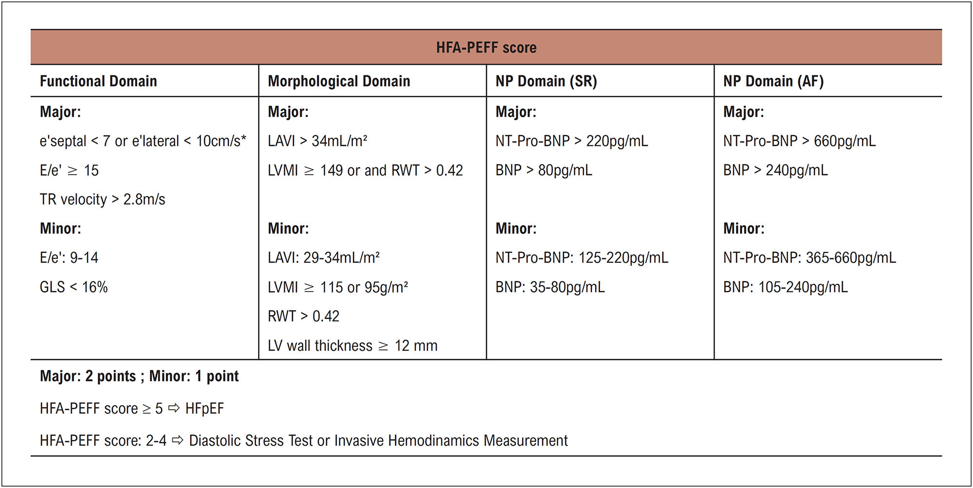
Heart Failure with Preserved Ejection Fraction (HFpEF) is an increasing challenge in cardiology, marked by impairments in systolic and/or diastolic function. HFpEF patients often present with exercise intolerance, yet diagnosis is complicated by overlapping comorbidities, such as obesity and hypertension. Left ventricular Global Longitudinal Strain (GLS) enables the detection of subtle systolic dysfunction, with studies indicating a stronger correlation between GLS and exercise capacity than ejection fraction (EF). In exercise echocardiography, GLS displays a characteristic bimodal pattern due to hemodynamic adaptations, with deviations suggesting HFpEF. When exercise testing is unfeasible or when a specific evaluation of ventricular-arterial coupling is desired, an afterload challenge offers a controlled alternative by increasing systemic resistance. Beyond GLS, mechanical dispersion (MD) measures contractile heterogeneity, while left atrial strain (LAS) has shown promise in identifying atrial dysfunction linked to HFpEF. These parameters, evaluated alongside E/e’ ratios and pulmonary systolic pressure during exercise, enhance HFpEF diagnosis, provide a more comprehensive view of the syndrome’s pathophysiology, and offer insights to support treatment.
  • Echocardiography
  • Exercise
  • Heart Failure
Read Full Article
Creative Commons Attribution (CC BY) Creative Commons license
16/12/2024 DOI: 10.36660/abcimg.20240104i Views 1
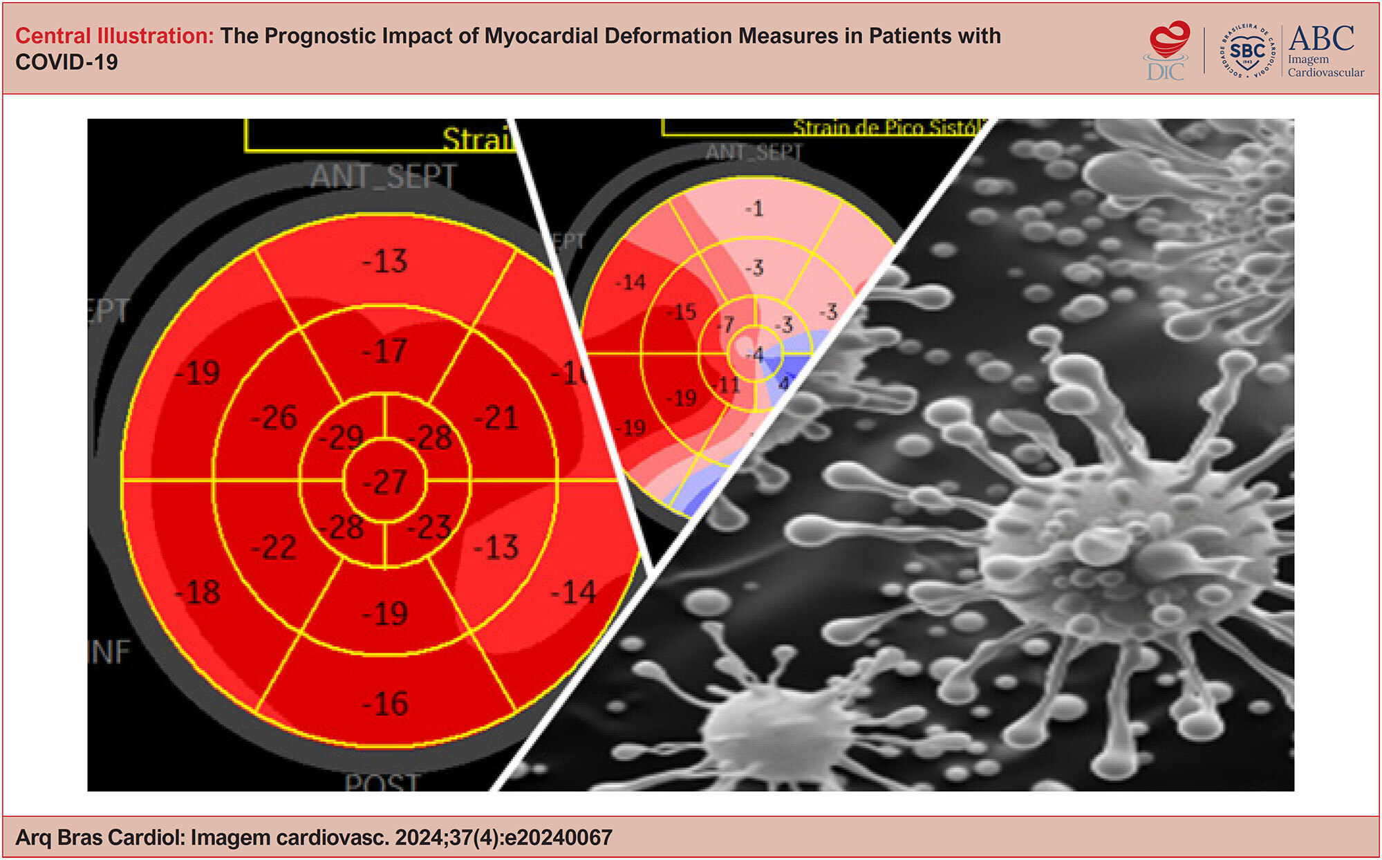
The Prognostic Impact of Myocardial Deformation Measures in Patients with COVID-19
Original Article

The Prognostic Impact of Myocardial Deformation Measures in Patients with COVID-19

João Giffoni Silveira Neto et al. Marcello de Brito Campos Luiz Mello Marcela de Castro Giffoni … Natanael Arnor da Silva Barros Goveia Antonio Claudio Lucas da Nóbrega Renata R. T. Castro

Abstract

Background:

Cardiovascular involvement in COVID-19 has been extensively studied. Echocardiography is an important technique in identifying cardiac involvement, serving as an excellent tool for prognostic variables.

Objectives:

Analyze the prognostic impact of speckle tracking in analyzing the left ventricle in patients with COVID-19 admitted to intensive care units (ICUs).

Methods:

Patients diagnosed with COVID-19 admitted to the ICU underwent an echocardiographic examination within the first 48 hours of hospitalization and were divided into two groups according to hospital outcome (discharge or death). Conventional echocardiographic data, as well as global longitudinal strain of the left ventricle (LVGLS) and right ventricle (RVGLS) of both groups, were compared using Student’s t test (for continuous variables) or the chi-square test (for categorical variables), considering p < 0.05 as statistically significant.

Results:

The average age was 56 ± 14 years, and the proportion of men and women was similar. The mortality rate was 64%, more frequent in elderly patients and in patients with a higher number of comorbidities. LVGLS revealed lower values in patients who progressed to death, with a cutoff point below −18.1% (sensitivity = 90.4%, specificity = 96.6%) for this outcome, and, after multivariate statistical analysis, it was found to be the only statistically significant echocardiographic variable.

Conclusion:

LVGLS is an important tool in the prognostic analysis of critically patients with COVID-19, offering a new window of possibilities for evaluating these patients.

  • COVID-19
  • Echocardiography
  • Global Longitudinal Strain
Read Full Article
Creative Commons Attribution (CC BY) Creative Commons license
13/12/2024 DOI: 10.36660/abcimg.20240067i Views 1
Figure 1
Review Article

My Approach to Functional Analysis in Computed Tomography Angiography: Myocardial Perfusion and FFRct

Gustavo Corrêa de Almeida Teixeira et al. Bruno Vicente Gomes de Castro Douglas Carli Silva Tiago Magalhães

Abstract

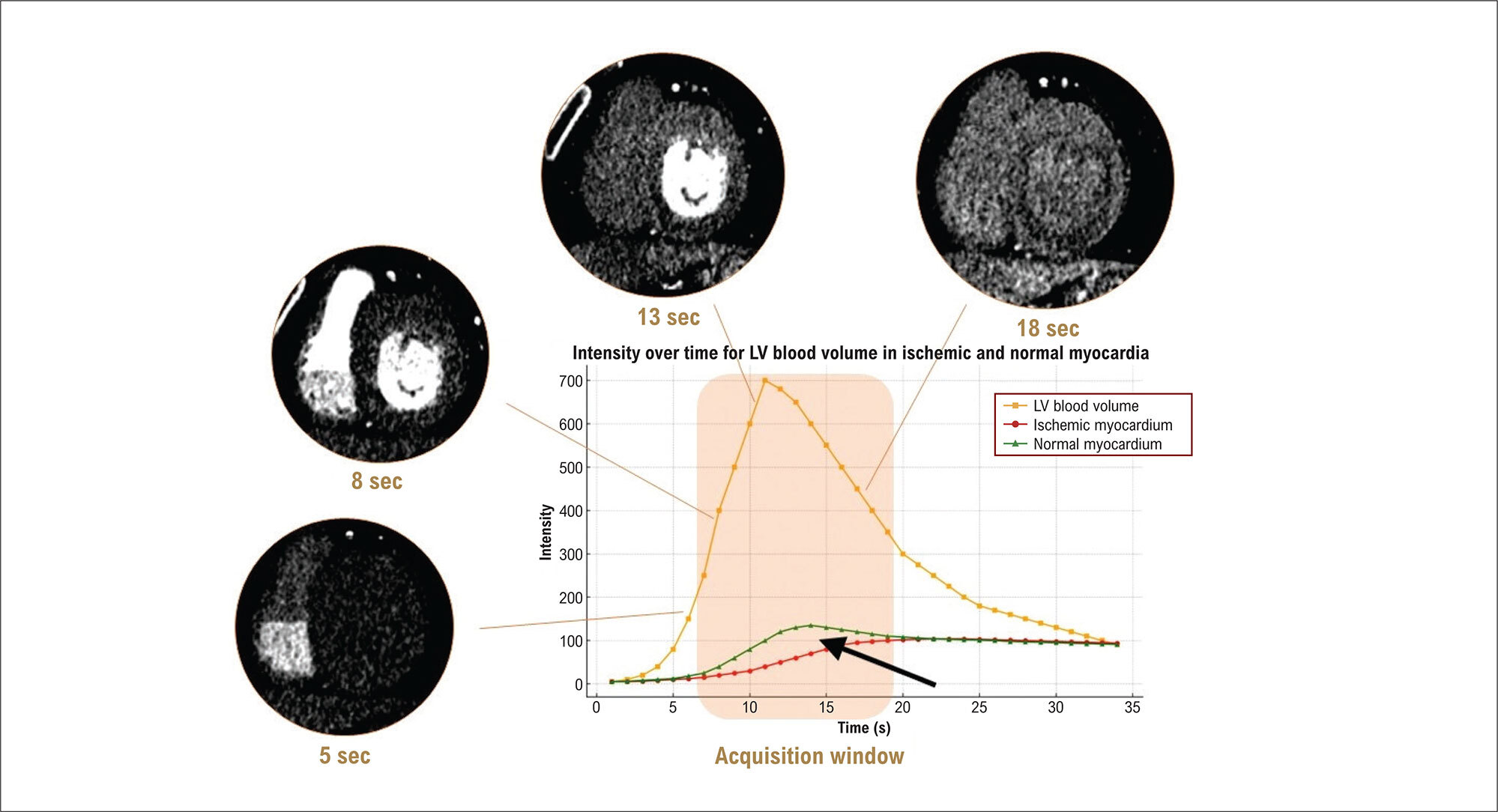
Coronary computed tomography angiography (CCTA) is an effective noninvasive method for detecting atherosclerotic disease, especially in symptomatic patients with low-to-moderate disease probability. Despite good accuracy in detecting obstructions, its relationship with functional lesion severity is limited. For a more accurate assessment, anatomical evaluation can be associated with myocardial computed tomography perfusion and fractional flow reserve derived from CCTA (FFRct). Myocardial perfusion computed tomography identifies areas of ischemia. FFRct, which assesses the hemodynamic significance of stenoses, has shown high accuracy and can reduce the need for invasive catheterizations. The image acquisition and interpretation protocols are essential to ensure reliable results and appropriate management of patients with coronary artery disease.

  • Computed Tomography Angiography
  • Myocardial Fractional Flow Reserve
  • Myocardial Perfusion Imaging
Read Full Article
Creative Commons Attribution (CC BY) Creative Commons license
13/12/2024 DOI: 10.36660/abcimg.20240113i Views 1
Viewpoint

A New Horizon in Nuclear Cardiology in Brazil: Impact and Barriers of F-18 Flurpiridaz

Simone Cristina Soares Brandão

Introduction

Advances have been made in positron emission tomography (PET) myocardial perfusion since the Food and Drug Administration (FDA) approved the use of flurpiridaz, which was shown effective in the detection of coronary artery disease (CAD) in two phase III trials, with a high diagnostic precision as compared with Single Photon Emission Computed Tomography (SPECT) scintigraphy.– Flurpiridaz is an injectable radiotracer, labeled with fluorine-18 (F-18), developed by GE HealthCare, that will be sold as “Flyrcado” in the United States from 2025 onwards.

With a half-life of 110 minutes, F-18-flurpiridaz requires neither a generator nor a local production of tracers, allowing a more efficient distribution among hospitals and imaging centers, similarly to Fluorodeoxyglucose labeled with Fluorine-18 (F-18 FDG). This feature may favor the adoption of myocardial perfusion PET, benefiting more patients with cardiovascular diseases. Besides, the longer half-life of F-18-flurpiridaz allows the combination of a physical stress test, optimizing the evaluation of ischemia and improving patients’ experience.,

[…]

  • Coronary Artery Disease
  • Positron Emission Tomography-Computed Tomography
  • Radiopharmaceuticals
Read Full Article
Creative Commons Attribution (CC BY) Creative Commons license
13/12/2024 DOI: 10.36660/abcimg.20240118i Views 1
Editorial

Climate Change and Cardiovascular Imaging: Challenges and Opportunities

Eduardo Galvão Freire et al. José Arimateia Batista Araújo-Filho Cesar Higa Nomura

Climate change directly and indirectly affects human health. The increasing incidence and intensity of extreme weather events are directly associated with adverse health outcomes, including cardiovascular events, as well as increased demand and expenditure on health systems globally. Vulnerable populations, such as the elderly and people with pre-existing cardiovascular and respiratory conditions, face increased risks due to temperature variations and poor air quality. Moreover, high temperatures also limit outdoor exercise, with potential long-term detrimental effects on cardiovascular health. In a country like Brazil, where economic disparities often expose low-income populations to higher levels of pollution and less access to health care, the impacts of climate change on cardiovascular health are a matter of growing concern among medical and scientific communities.

On the other hand, the healthcare sector is responsible for almost 5% of the planet’s greenhouse gas (GHG) emissions, with a significant contribution coming from general imaging and cardiovascular diagnostic tests. We thus face a double challenge: to improve cardiovascular health outcomes for the population while reducing the environmental footprint of our tests. Although advances in cardiovascular imaging technologies have undoubtedly improved our diagnostic accuracy and patient outcomes, these benefits have come at considerable financial and environmental costs.

[…]

  • Cardiovascular Diseases
  • Climate Change
  • Echocardiography
Read Full Article
Creative Commons Attribution (CC BY) Creative Commons license
13/12/2024 DOI: 10.36660/abcimg.20240117i
Editorial

Quantification of Myocardial Fibrosis by Cardiac Magnetic Resonance Imaging: Advances, Impacts, and Perspectives

Isabela Bispo Santos da Silva Costa

Cardiac magnetic resonance imaging (CMR) has been established as a fundamental tool for the diagnosis and management of cardiomyopathies, especially due to its accuracy in tissue characterization, identification, and quantification of myocardial fibrosis., Late gadolinium enhancement (LGE) allows accurate assessment, identifying areas of myocardial fibrosis, and it serves to characterize, locate, and quantify them. In recent years, the role of LGE in the risk stratification of patients with hypertrophic cardiomyopathy (HCM) and other genetic cardiomyopathies has been the subject of several studies that aimed to correlate the presence and extent of myocardial fibrosis with the risk of sudden cardiac death (SCD) and major cardiovascular events.–

Different techniques are currently used to acquire, analyze, and quantify LGE by CMR. The presence of LGE is based on the differential tissue retention of gadolinium, which is preferentially absorbed in areas of fibrosis, where scar tissue is altered due to the presence of collagen. LGE acquisition techniques nowadays are fast, safe, and can be performed without requiring long respiratory pauses, as was the case in the past, with progressively better resolution and image definition. To quantify areas of fibrosis, different software programs that allow adequate quantification in a semi-automated manner are currently available.

[…]

  • Cardiomyopathies
  • Endomyocardial Fibrosis
  • Magnetic Resonance Imaging
Read Full Article
Creative Commons Attribution (CC BY) Creative Commons license
13/12/2024 DOI: 10.36660/abcimg.20240114i Views 1
Figure 1
Case Report

Pericardial Hematoma With Chest Pain and Tamponade in the Late Postoperative Period of Cardiac Surgery

Felipe Carvalho de Oliveira et al. Jun Ramos Kawaoka Bruno de Freitas Leite Diogo Freitas Cardoso de Azevedo … Rafael Modesto Fernandes Marcia Maria Noya-Rabelo

Introduction

Pericardial effusion and mediastinal bleeding are common complications in the postoperative period following cardiac surgery. These conditions typically present within the first few days after the procedure and necessitate additional surgical intervention in a minority of cases.

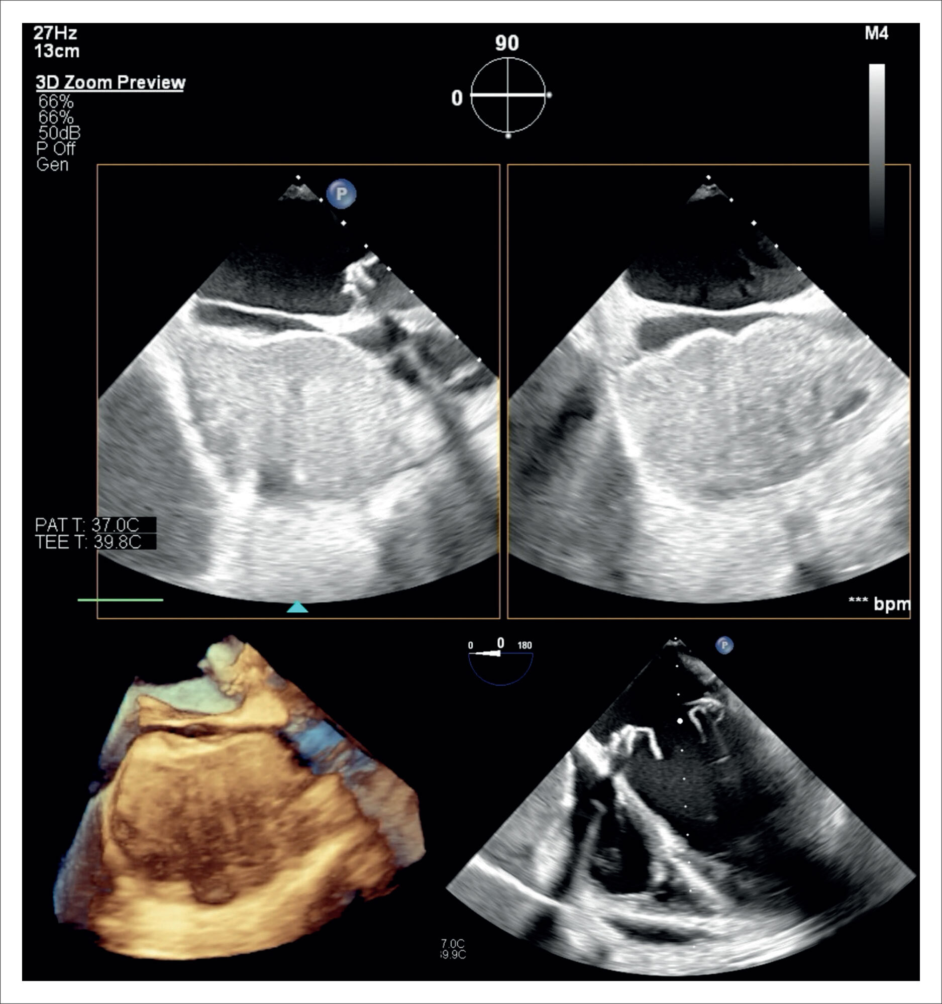
This report describes an atypical presentation of pericardial hematoma, which manifested as severe chest pain on the 23rd postoperative day and rapidly progressed to hemodynamic instability. Echocardiography was instrumental in both diagnosing and managing the case, ultimately contributing to a favorable outcome.

[…]

  • Echocardiography
  • heart surgery
  • pericardial hematoma
Read Full Article
Creative Commons Attribution (CC BY) Creative Commons license
09/12/2024 DOI: 10.36660/abcimg.20240090i Views 1
Figure 1
Original Article

Prevalence of Fetal Heart Disease in Pregnant Women Referred to a Specialized Service Due to Cardiac Abnormalities on Morphological Ultrasound

Andressa Pinto Michael et al. Marcelo França Soares Roque Antonio Foresti Marco Otilio Duarte Rodrigues Sandra Mara Witkowski

Abstract

Background:

Congenital Heart Disease (CHD) is a malformation of the heart and/or great vessels and is the third leading cause of neonatal death. It can be detected in utero by Morphological Ultrasound/Morphology Scan (MUS) and confirmed by Fetal Echocardiography (FE).

Objective:

This study aims to determine whether heart disease identified by MUS in pregnant women at a maternal referral center in a southern Brazilian city is confirmed by FE.

Methods:

This observational, descriptive, and retrospective study was conducted from March 2019 to January 2023. It focused on pregnant women in the public health system, treated through the Unified Health System (SUS) at a maternal referral center in a southern Brazilian city, who had cardiac abnormalities detected on MUS and confirmed by FE.

Results:

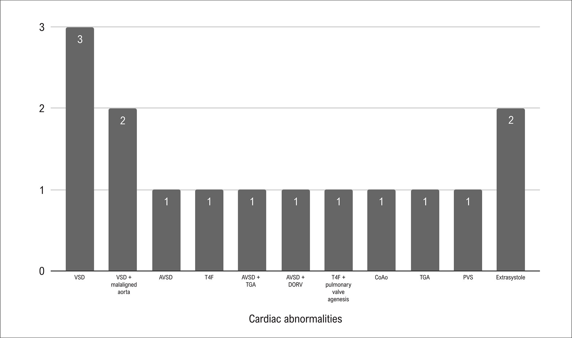
Among the 37 pregnant women with suspected cardiac abnormalities on MUS, 15 cases were confirmed by FE, yielding a diagnostic agreement rate of 21.62% (n = 8) between the two exams. Ventricular Septal Defect (VSD) was the most prevalent heart defect, accounting for 20% of isolated cases. Of the 15 cases with cardiac alterations on FE, two were transferred to a specialized hospital and five died.

Conclusion:

Fetal cardiac abnormalities were confirmed in fewer than half of the women referred following MUS, with concordance between exams in only eight cases. Continuous training in MUS reduces costs and inconvenience. Professionals qualified in FE are essential for the early diagnosis of CHD, improving care, and reducing infant mortality.

  • Congenital Heart Defects
  • Echocardiography
  • Prenatal Ultrasonography
Read Full Article
Creative Commons Attribution (CC BY) Creative Commons license
09/12/2024 DOI: 10.36660/abcimg.20240102i
Figure 1
Editorial

Top Vascular Ultrasound 2024: Carotid Plaque-RADS – A Novel Stroke Risk Classification System

Simone Nascimento dos Santos

Introduction

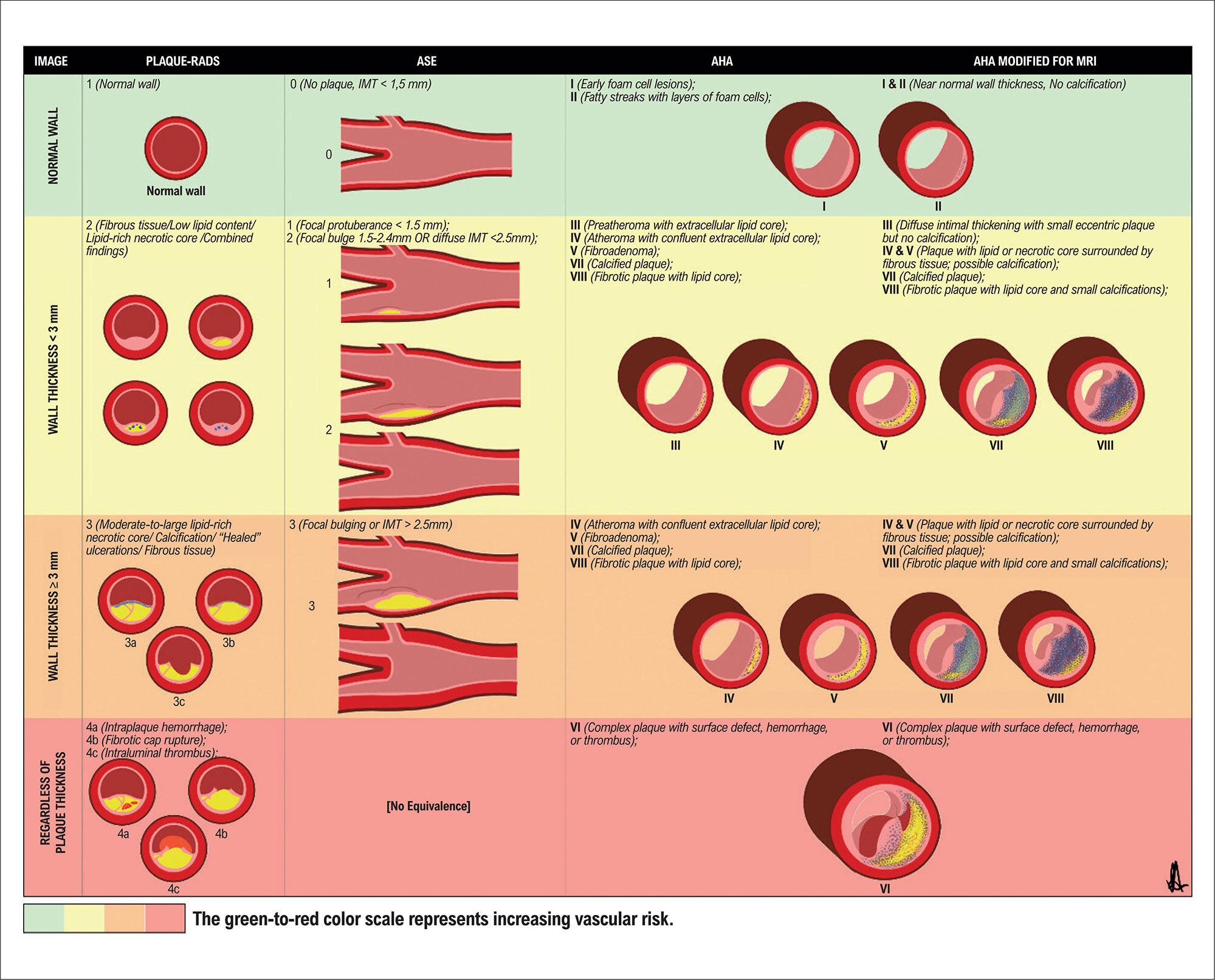
Many vascular ultrasound articles were published in 2024, but certainly the most discussed in Brazil this year was “Carotid Plaque-RADS – A Novel Stroke Risk Classification System” by Saba et al. in JACC: Cardiovascular Imaging. The project brought together 51 specialists in different vascular imaging modalities from 10 countries. This new proposal for categorizing carotid plaque attracted the attention of physicians from various specialties, such as cardiologists, neurologists, radiologists, ultrasound specialists, and vascular surgeons. In this section, we will discuss its main aspects, comparing them with previous classification systems.

[…]

  • Atherosclerosis
  • Atherosclerotic Plaque
  • Stroke
  • Ultrasonography
Read Full Article
Creative Commons Attribution (CC BY) Creative Commons license
09/12/2024 DOI: 10.36660/abcimg.20240110i
Figure 1
Editorial

The Use of X (Formerly Twitter) in Cardiovascular Imaging: Opportunities and Risks

Alex dos Santos Felix

Introduction

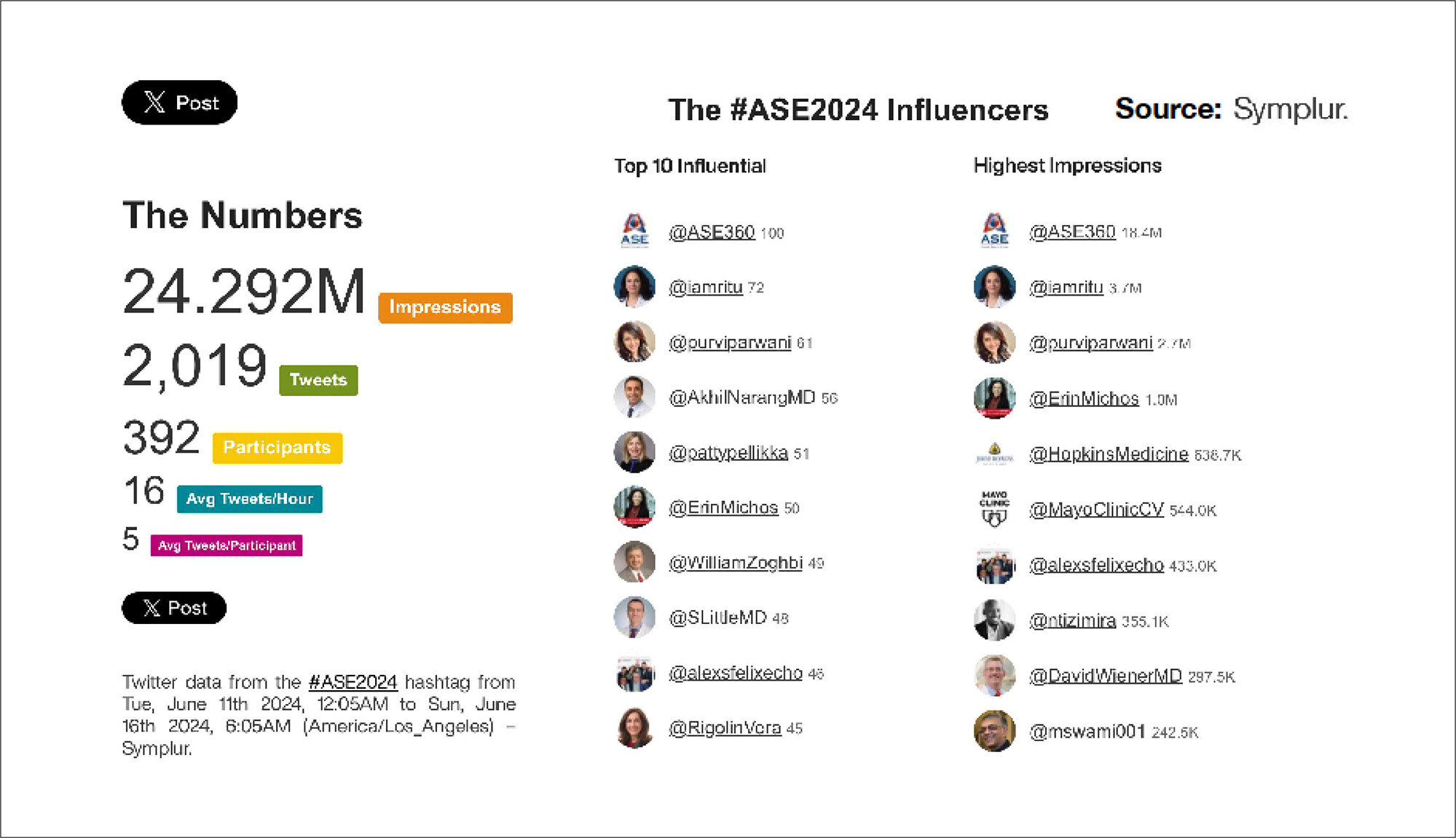
Social media has decisively transformed the way we disseminate medical information and knowledge, launching new digital tools to boost scientific debate and help redesign healthcare education. Globally, X, formerly Twitter, stands out in the medical field as a more scientific platform, with great interaction between peers, helping doctors and other healthcare professionals from all continents connect to discuss publications, debate scientific evidence, and share clinical experiences. X is also highly important as a channel for the dissemination of scientific events, courses and conferences. In a recent study involving healthcare professionals from 35 countries, Guerra et al. identified that X was the second most accessed social network (55.1%), second only to LinkedIn (60.8%). The speed of dissemination of information and interactivity are strong points of X, with the possibility of bringing together younger doctors, or those who work in institutions with lower complexity, with renowned professionals who work in large centers around the world. This valuable exchange of experiences creates a large virtual “grand round,” bringing other professionals into the debate and allowing for the discussion of controversial technical concepts and challenging clinical cases.

X was founded in 2006, still as Twitter, as an environment for the exchange of short messages, initially limited to 140 characters, having been extended to 280 characters in 2017, and recently relaxed to 25,000 characters for Premium users only. According to the Digital Report 2024, Brazil is the second country in the world where users spend the most time online. There are 245 million users on X, with more than 500 million posts per day, making Brazil the fourth largest user base of this platform in the world.

[…]

  • Cardiac Imaging Techniques
  • Echocardiography
  • Social Media
Read Full Article
Creative Commons Attribution (CC BY) Creative Commons license
09/12/2024 DOI: 10.36660/abcimg.20240109i
Impact of Valvular Event Time Assessment Methods on Myocardial Work Calculation by Echocardiography
Original Article

Impact of Valvular Event Time Assessment Methods on Myocardial Work Calculation by Echocardiography

Marcio Mendes Pereira et al. Francisco das Chagas Monteiro Júnior Sayuri Yamamura

Abstract

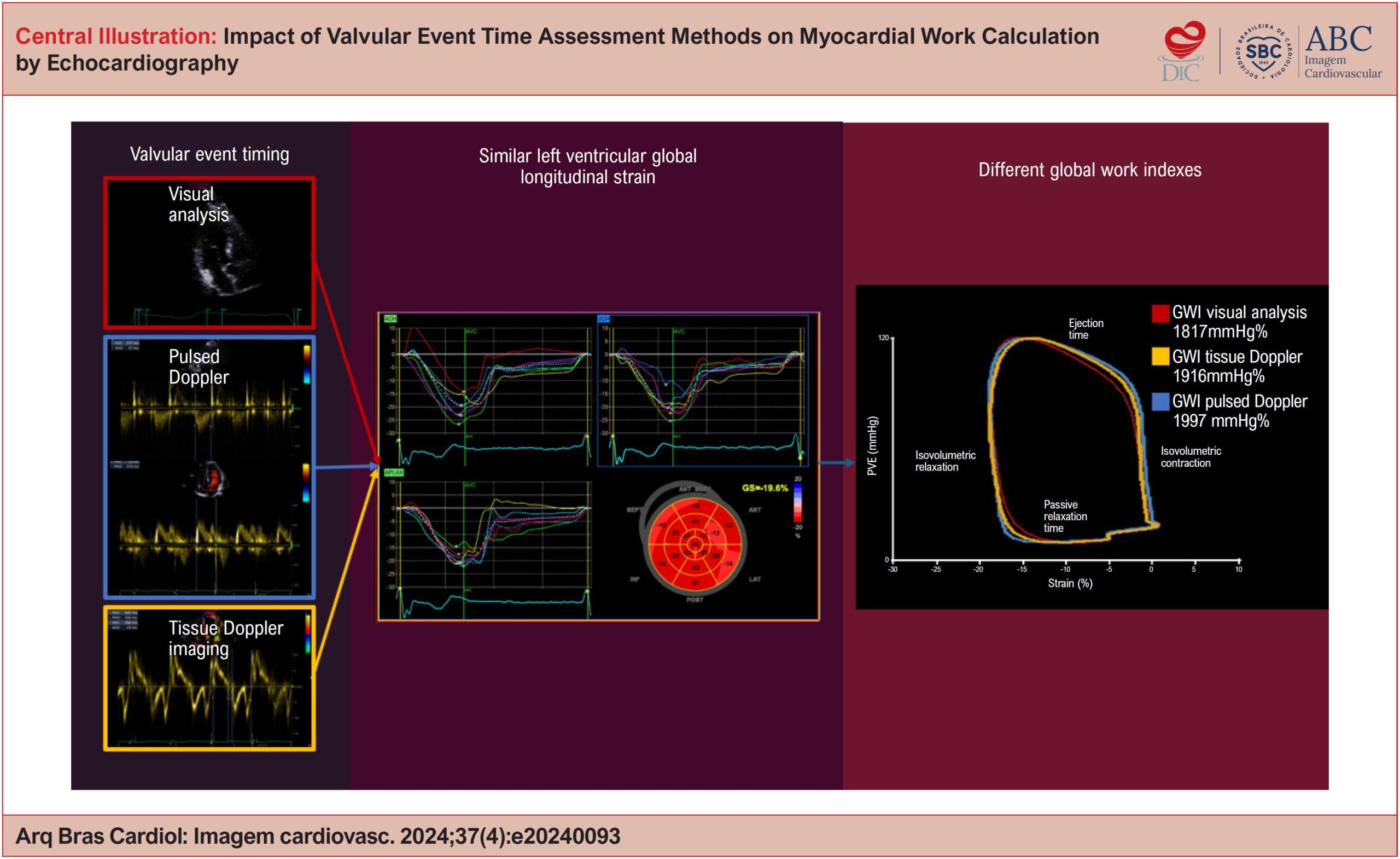
Background:

Left ventricular ejection fraction (LVEF) is the traditional parameter for assessing cardiac function, but it has limitations in the early detection of dysfunction. Global Longitudinal Strain (GLS) and myocardial work (MW) are promising alternatives, the latter being less influenced by load conditions. The calculation of MW depends on valvular event times (VET), obtained by methods that may generate divergent results.

Objectives:

To compare the impact of different methods of VET measurement – visual analysis (VA), tissue Doppler imaging (TDI) and pulsed Doppler (PD) – on global work index (GWI), global constructive work (GCW), global wasted work (GWW) and global work efficiency (GWE).

Method:

Cross-sectional clinical study with 35 healthy patients who underwent echocardiography between October 2022 and March 2023. MW was assessed using the EchoPAC software, with VET measurement by VA, TDI and DP. The results were analyzed by ANOVA and Friedman tests, followed by post-hoc analyses with a significance level of 5%.

Results:

GWI showed significant variation between the VET measurement methods, being lower by AV compared to TDI and DP (p<0.001). The GCW, GWW and GWE indexes did not demonstrate statistically significant differences between the methods.

Conclusions:

The VET measurement method significantly impacts the GWI, but does not affect the other MW indexes.

  • Echocardiography
  • Left Ventricular Dysfunction
  • Ventricular Function
Read Full Article
Creative Commons Attribution (CC BY) Creative Commons license
05/12/2024 DOI: 10.36660/abcimg.20240093i Views 1
Figura 1
Case Report

Not Everything is as it Seems at First Glance: Multimodal Imaging in Heart Cyst Evaluation

Carlos Guaman-Valdivieso et al. Julia Aramburu Natalia Lluberas Natalia Huart Gabriel Parma

Abstract

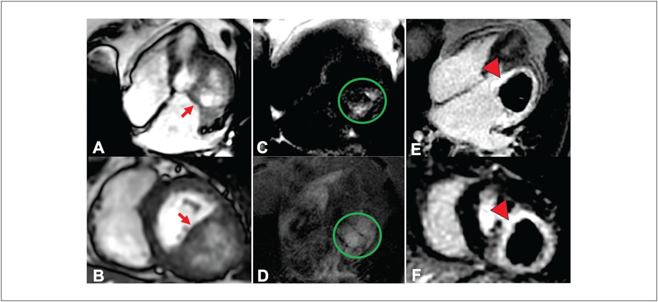
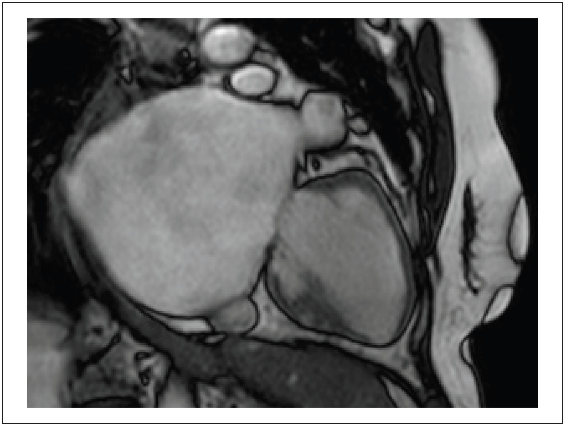
Cardiac masses, though rare, can lead to significant hemodynamic and arrhythmic issues, even if benign. Improved multimodal imaging has refined the diagnosis and understanding of these masses. We present an 85-year-old woman with New York Heart Association class II-III dyspnea for six months. Echocardiography showed a mass in the basal inferolateral wall of the left ventricle, along with severe left atrial dilation and moderate mitral regurgitation. Initially thought to be a pericardial cyst, further cardiac magnetic resonance (CMR) imaging identified an intramyocardial mass, and computed tomography (CT) suggested a hydatid cyst. Given the non-limiting symptoms and high surgical risk, the decision was made for expectant management. This case illustrates the rarity of cardiac cysts and the essential role of multimodal imaging in diagnosis and treatment planning.

  • Cardiac Imaging Techniques
  • Echinococcosis
  • Heart Neoplasms
Read Full Article
Creative Commons Attribution (CC BY) Creative Commons license
05/12/2024 DOI: 10.36660/abcimg.20240086i Views 1
Figure 1
Viewpoint

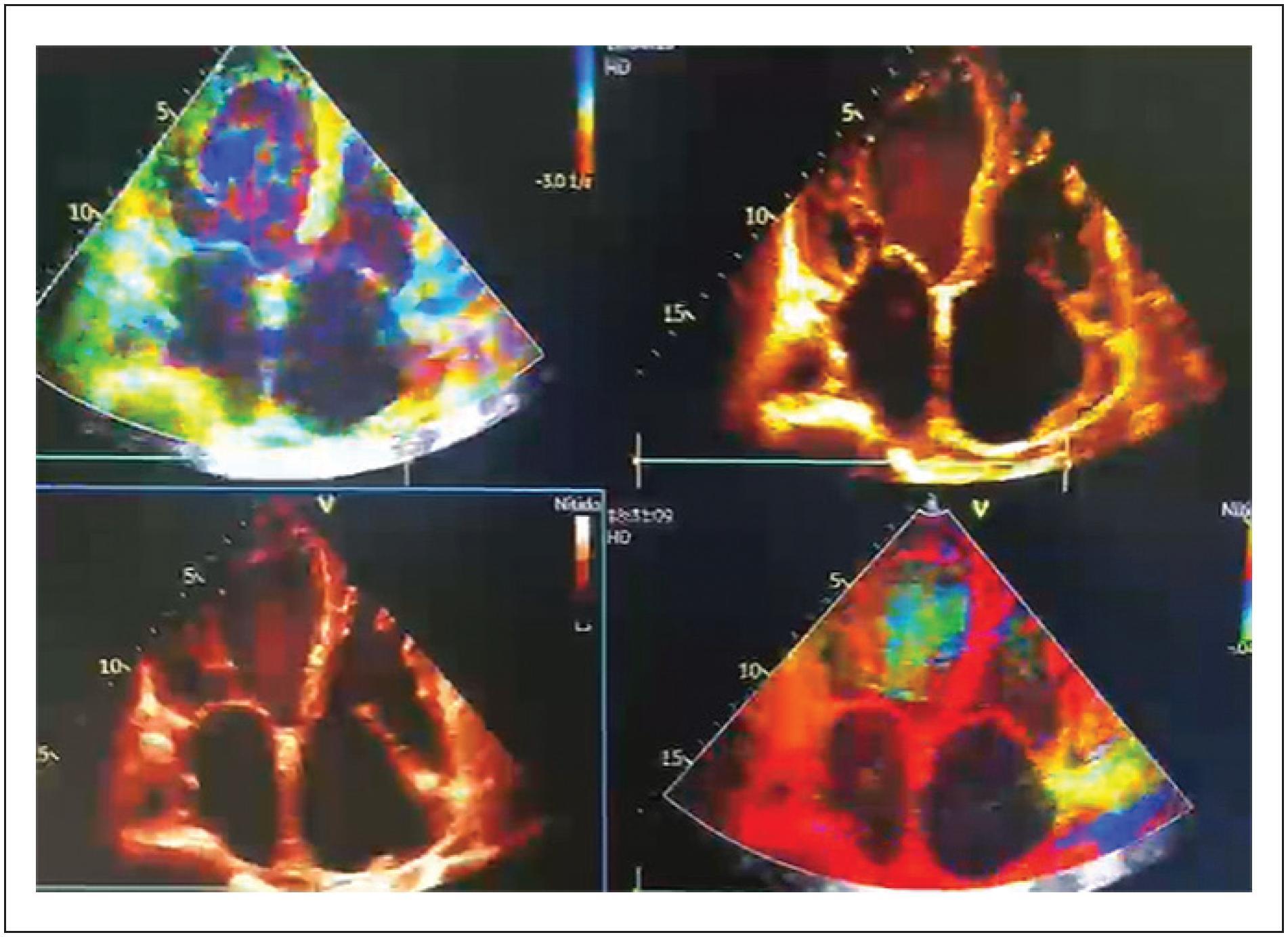
An Explosion of Colors, Fauvism in Echocardiography, a New Proposal

Jaime Andres Nieto Zarate et al. Maria Jose Amaya Mahecha

Limitations

The article offers an intriguing perspective, but further scientific validation is needed for broader acceptance in clinical practice. The connection between art and medical imaging is fascinating, yet requires more exploration to establish its practical relevance and impact on patient outcomes.

[…]

  • Cardiology
  • Cardiovascular Diagnostic Techniques
  • Echocardiography
  • Medicine in the Arts
Read Full Article
Creative Commons Attribution (CC BY) Creative Commons license
05/12/2024 DOI: 10.36660/abcimg.20240081i
Figura 1
Case Report

Unicuspid Aortic Stenosis: Case Report

Welison Gutherrez Silva e Sousa et al. Julia Lucena Domingues Adriana Teixeira Damasceno Alessandra Edna Teófilo Lemos … Antônio Luiz Júnior Araújo Nelson Lopes Evangelista

Introduction

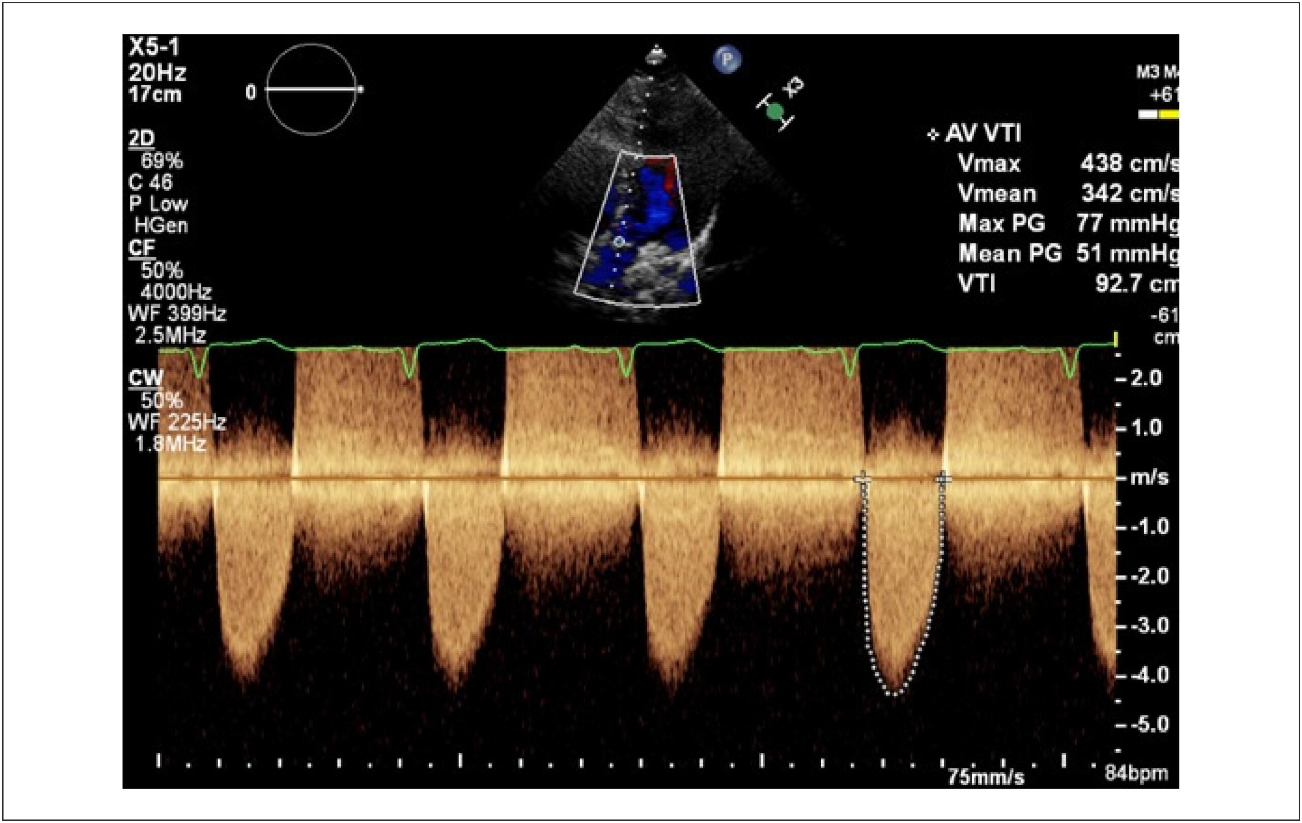
Unicuspid aortic valve (UAV) is an uncommon congenital pathology, first described by Edwards in 1958. It is a rare malformation (especially when compared to other aortic valve anomalies), with an estimated incidence of 0.02% of adults undergoing echocardiography and approximately 4% to 5% of patients undergoing surgery for aortic stenosis. Echocardiography is the first-choice method for diagnosis and definition of hemodynamic repercussions. In this study, we report the case of a patient with symptomatic aortic stenosis and describe her diagnosis and therapeutic management.

[…]

  • Aortic Valve Stenosis
  • Echocardiography
  • Heart Valve Diseases
Read Full Article
Creative Commons Attribution (CC BY) Creative Commons license
05/12/2024 DOI: 10.36660/abcimg.20240087i Views 2
Figure 1
Case Report

Giant Left Atrium in Rheumatic Mitral Valve Disease

Alice Cunha Darzé et al. Antonio Tito Paladino Filho Amanda Marsiaj Rassi Carlos Eduardo Prazeres … Ibraim Masciarelli Francisco Pinto João Marcelo Caneschi Jose Roberto Tuma da Ponte Junior Paul Salvador Morales Rafael Castro Hendges Tiago Senra

Abstract

The Giant Left Atrium (GLA) is a rare condition, more commonly related to rheumatic mitral valve disease; it can be mistaken with a few possible differential diagnoses when less accurate methods are implemented, potentially leading to particularly dangerous and unnecessary procedures in this setting. Cardiovascular imaging methods, such as Cardiac Magnetic Resonance (CMR), can be valuable for this differentiation and provide high spatial resolution, supporting accurate diagnosis and surgical planning when indicated. Selecting the most precise diagnostic method, however, requires deep knowledge of the GLA condition and clinical data, leading to a correct suspicion. Furthermore, opting for using volumetry rather than the biplanar method in cases of significant deformity ensures greater accuracy in the values obtained.

  • Heart Atria
  • Heart Valve Diseases
  • Magnetic Resonance Imaging
Read Full Article
Creative Commons Attribution (CC BY) Creative Commons license
05/12/2024 DOI: 10.36660/abcimg.20240083i Views 1
The Many Faces of Arrhythmic Mitral Valve Prolapse: Case Series
Original Article

The Many Faces of Arrhythmic Mitral Valve Prolapse: Case Series

Yasmim Evelyn Lisboa Barbosa et al. Isadora Wanderley Queiroga de Freitas Evangelista Julyana Maria Ramalho de Sousa Lorena Carvalho Leite Jales … Elisabete Louise de Medeiros Viegas Patrícia Vidal de Negreiros Nóbrega Daniel Moreira Costa Moura Gabriel Pelegrineti Targueta Lilian Amador Resende Renner Augusto Raposo Pereira Guilherme Augusto Teodoro Athayde

Abstract

Background:

Although it is frequently a benign condition, mitral valve prolapse (MVP) may be associated with an increased risk of ventricular arrhythmias (VA), a condition known as arrhythmic mitral valve prolapse (AMVP).

Objectives:

To present diverse manifestations of AMVP by means of clinical cases that illustrate these patients’ symptoms and electrocardiogram (ECG), 24-hour Holter, transthoracic echocardiogram (TTE), and cardiac magnetic resonance imaging (CMRI) findings and to discuss the decisions made in relation to different clinical outcomes.

Methods:

This retrospective, descriptive, and observational study analyzed 5 patients with AMVP treated between 2019 and 2024, investigating markers of elevated risk for severe clinical outcomes, especially sudden cardiac death (SCD).

Results:

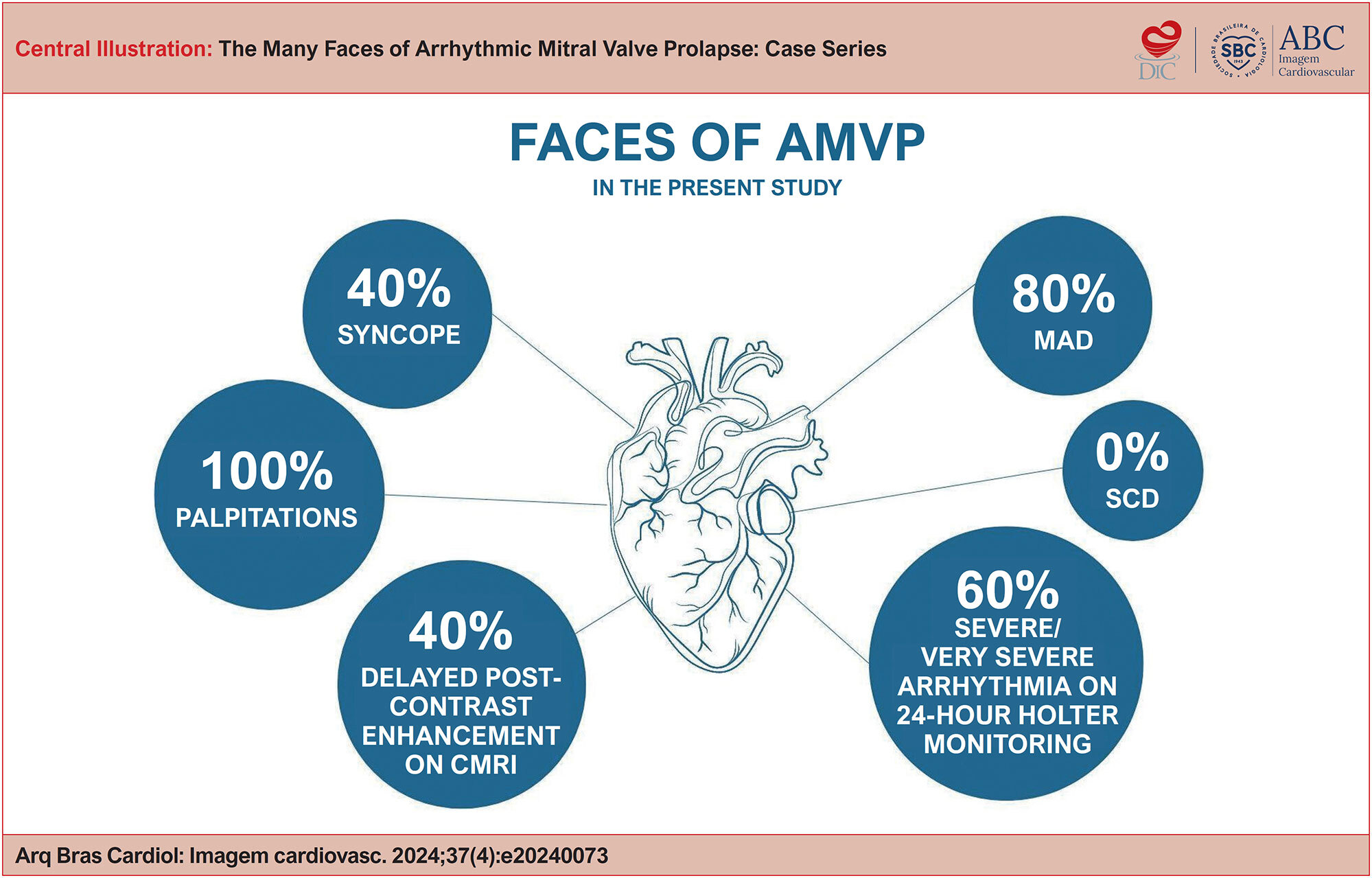
Among the 5 patients evaluated, the most common clinical presentation was palpitations (100% of cases), followed by syncope (40%). Three patients (60%) had severe or very severe arrhythmia on 24-hour Holter monitoring, while 4 (80%) had mitral annular disjunction (MAD). Positive delayed enhancement was observed in 2 (40%) patients. In 2 cases (40%), implantable cardioverter-defibrillator (ICD) was indicated. In 1 case, mitral valve replacement was chosen, but the arrhythmias were not resolved; consequently, radiofrequency ablation was indicated. Although SCD has been described in the literature, no cases were observed in our sample.

Conclusion:

AMVP may present distinct clinical manifestations, including severe outcomes. It is essential to identify risk markers for early diagnosis and treatment of this condition, with the goal of reducing SCD-related mortality in these patients.

  • Cardiac Arrhythmias
  • Cardiac Sudden Death
  • Mitral Valve Prolapse
  • Ventricular tachycardia
Read Full Article
Creative Commons Attribution (CC BY) Creative Commons license
22/11/2024 DOI: 10.36660/abcimg.20240073i
Figure 1
Case Report

Atrial Myxoma in Male Young Adult Patient: A Case Report and Literature Review

Monize Aparecida Gonçalves do Nascimento et al. Eliseu Henrique Bispo Pereira Giovanna Karine Leal Magalhães Laura Souza Mariano … Luana Mireli Carbonera Rodrigues Leonardo Alves Batista Rogério de Paula Garcia Caravante Ayná Emanuelli Alves Barreto

Introduction

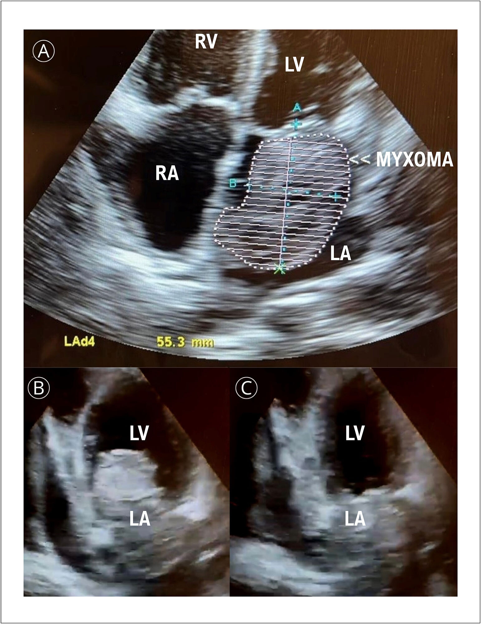
Cardiac tumors are found in 0.001% to 0.3% of autopsies, where myxoma is the most common cardiac neoplasia. Approximately 70% appear in the left atrium next to the foramen ovale and are pedunculated. Its pedunculus can be short, limiting its movement, or can be long, allowing tumor movement inside and outside the atrium during the cardiac cycle.,

Clinical conditions depend on the size, mobility, and location of the tumor. Classic symptoms are related to Goodwin’s triad, which includes unspecific systemic symptoms, embolism and intracardiac obstruction. Among systemic symptoms are dyspnea, orthopnea, paroxysmal nocturnal dyspnea, pulmonary edema, cough, hemoptysis, edema, and fatigue due to obstructive features of neoplasia.,

[…]

  • Atrial Myxoma
  • Cardiovascular Surgical Procedures
  • Heart Atriums
Read Full Article
Creative Commons Attribution (CC BY) Creative Commons license
22/11/2024 DOI: 10.36660/abcimg.20240064i Views 1
Editorial

Artificial Intelligence in Echocardiography: The Future of Precision Diagnosis

Maria Estefânia Bosco Otto

AI and the Systolic Function

Ejection fraction (EF) is an essential parameter used to assess the systolic function; however, it has been historically subjected to interobserver variability and the level of experience of the examiner. AI-based algorithms present solutions for EF, providing automated and reproducible calculations.3–5 A pioneering study published by He et al. evaluated a deep learning (DL) system comprised of convolutional neural networks (CNNs), called EchoNet-Dynamic flow, and compared EF assessments performed by AI with measurements performed by experienced sonographers. The reference cardiologists who checked these results were unable to differentiate between the sonographer and AI. Furthermore, the results demonstrated that the assessment by AI was not inferior to those performed by experts, with a difference of less than 5% in variability. Other advantages of this system were the speed of measurement and the possibility of averaging up to five heartbeats, thus improving the accuracy of the result in atrial fibrillation.

Another parameter widely used to assess systolic function in echocardiography is the Global Longitudinal Strain (GLS) analysis, which not only aids in the diagnosis of cardiac function, but also reveals characteristic patterns of certain diseases, such as amyloidosis. The GLS technique itself requires machine learning (ML) training of the equipment to identify acoustic window incidences and outline edges. However, AP techniques by CNR were able to assess GLS more quickly and with less variability when compared to a commercially established workstation used in echocardiography laboratories. Recently, Kwan et al. evaluated a CNR system used to assess GLS with images produced by equipment from two different companies, concluding that, in addition to having less variability and good reproducibility, this system was able to identify GLS patterns and differentiate between athlete’s hypertrophy, amyloidosis, and hypertrophic cardiomyopathy. However, it important to understand that the quality of the images obtained for both GLS and EF assessments must be good, a factor that cannot yet be achieved by AI.

[…]

  • Artificial Intelligence
  • Diagnosis
  • Echocardiography
Read Full Article
Creative Commons Attribution (CC BY) Creative Commons license
22/11/2024 DOI: 10.36660/abcimg.20240095i Views 2
Advances in Percutaneous Tricuspid Valve Treatment
Review Article

Advances in Percutaneous Tricuspid Valve Treatment

Marcio Mendes Pereira et al. Maria Estefânia Otto Fernando Melo Netto Fernanda Esteves

Abstract

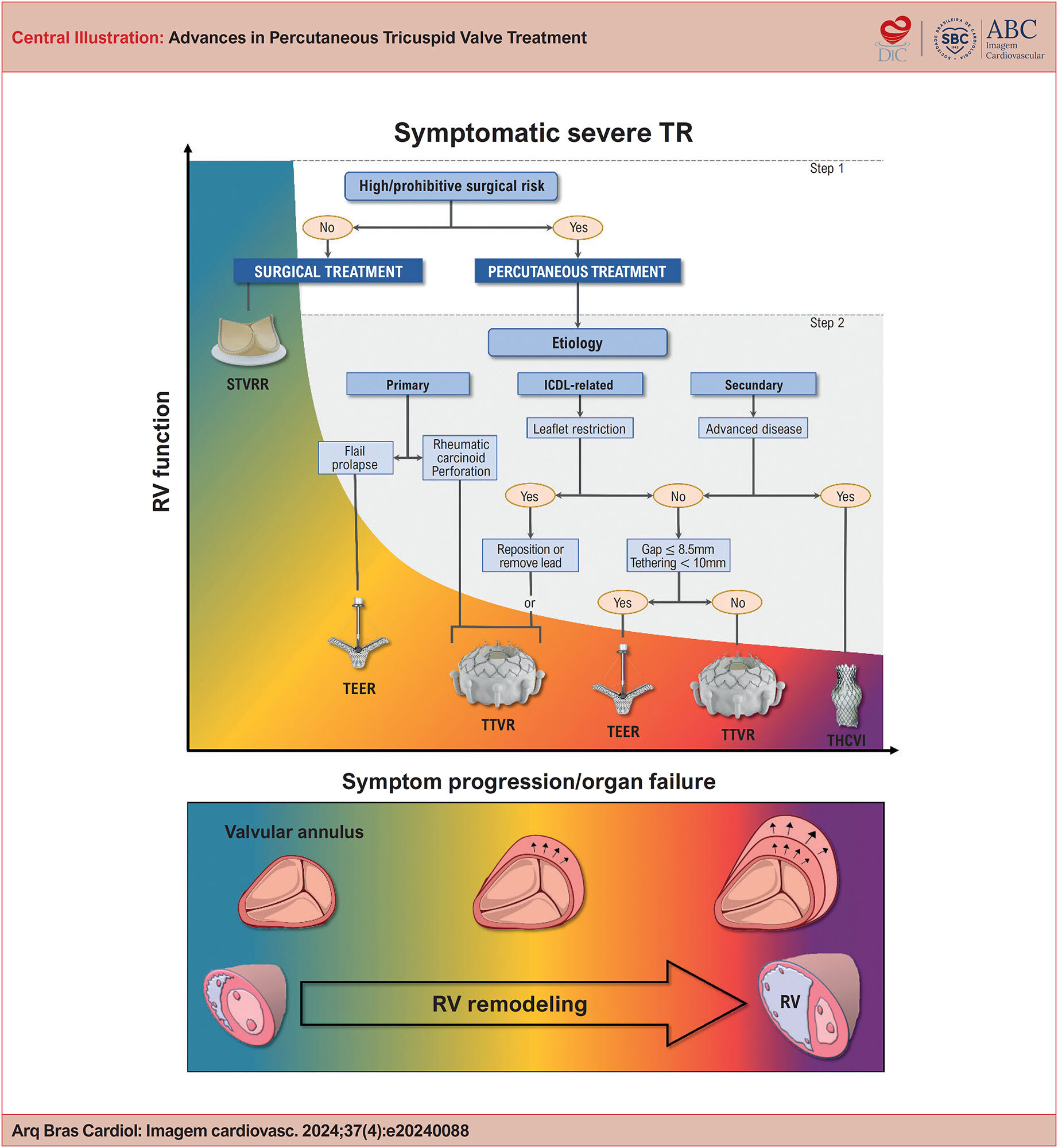
Tricuspid regurgitation (TR) is a frequently underdiagnosed and undertreated condition, often associated with poor clinical outcomes, particularly in elderly and high-risk surgical patients. As the prevalence of TR continues to rise and traditional surgical methods face limitations, percutaneous techniques have emerged as promising alternatives. This article reviews the latest advancements in the percutaneous treatment of TR, highlighting new etiological and quantitative classifications, in addition to exploring the indications and outcomes of key techniques, including percutaneous edge-to-edge repair and transcatheter valve replacement. Moreover, it discusses the anatomical challenges involved and the vital role of echocardiography in periprocedural planning and monitoring. Based on current data, percutaneous interventions have shown effectiveness in alleviating symptoms and enhancing the quality of life for patients with severe TR, making them a viable option for those who are not candidates for conventional surgery.

  • Echocardiography
  • Transcatheter Aortic Valve Replacement
  • Tricuspid Valve Insufficiency
Read Full Article
Creative Commons Attribution (CC BY) Creative Commons license
22/11/2024 DOI: 10.36660/abcimg.20240088i
Figure 1
Case Report

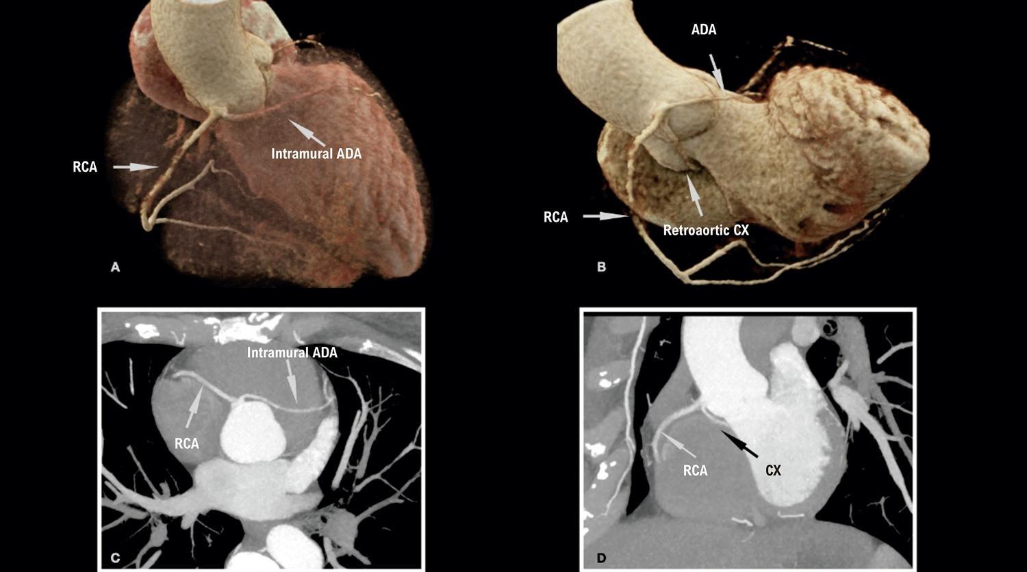
Rare Presentation of an Anatomical Congenital Anomaly of the Coronary Arteries on Computed Tomography Angiography

Marcello Zapparoli et al. Alexandre Alessi Franco Camargo Zapparoli Mateus Rodrigues Alessi

Introduction

Anomalous origin of the coronary arteries represents a heterogeneous group of alterations in the origin and course of these vessels, ranging from the most common, asymptomatic and low risk, to the rarest and highest risk, which may eventually be symptomatic or have sudden death as their first manifestation., Their prevalence corresponds to 1.3% of patients undergoing coronary angiography, but this number may be higher depending on the diagnostic method and the population selected, representing the second leading cause of sudden death in young athletes, behind only hypertrophic cardiomyopathy.,

Within this group, coronary anomalies with a single ostium are present in a smaller proportion of the population, approximately 0.004% to 0.098%. They are most often asymptomatic, but they may manifest with chest pain, dyspnea, palpitation, myocardial infarction, and sudden death., The presentation of this anomaly has worse prognosis in the presence of an interarterial vessel course (between the aorta and pulmonary trunk), an acute angle of origin, and atherosclerotic plaque; the latter change has a higher incidence in anomalous vessels on account of the greater stress on the arterial wall., In addition to presenting in isolation, they may also be associated with other alterations such as cardiac fistulas or a bicuspid aortic valve.

[…]

  • Anatomic Variation
  • Chest Pain
  • Computed Tomography Angiography
  • Coronary Vessel Anomalies
  • Coronary Vessels
Read Full Article
Creative Commons Attribution (CC BY) Creative Commons license
21/11/2024 DOI: 10.36660/abcimg.20240075i Views 1
Central Illustration
Original Article

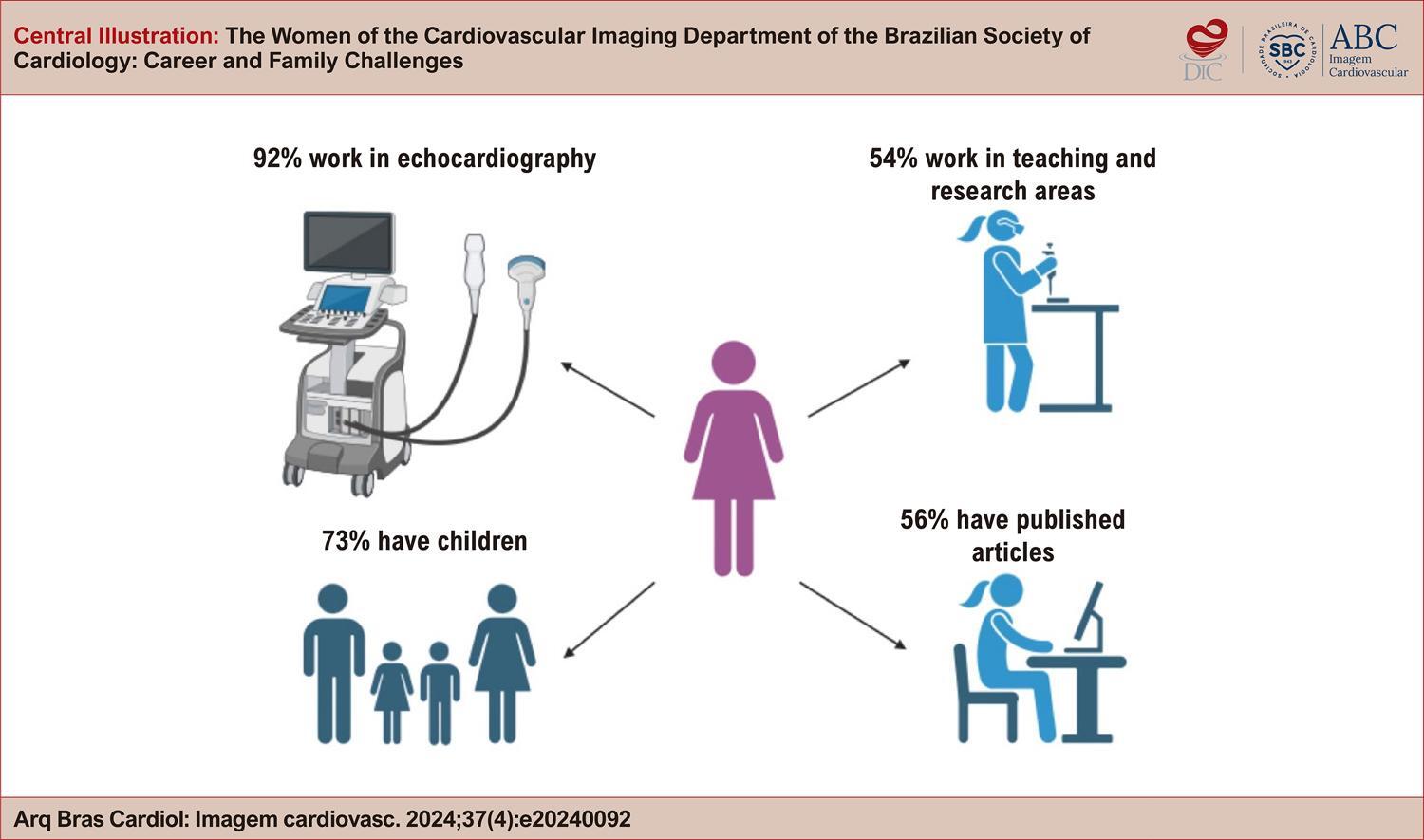
The Women of the Cardiovascular Imaging Department of the Brazilian Society of Cardiology: Career and Family Challenges

Marcia Ferreira Alves Barberato et al. Daniela do Carmo Rassi Adenalva Lima de Souza Beck Marly Uellendahl … Samira Saady Morhy Silvio Henrique Barberato

Abstract

Introduction

The “DIC Mulheres” working group was created to research and discuss the role of women working in cardiovascular imaging in Brazil. Objectives: to map the challenges faced by women in the Cardiovascular Imaging Department of the Brazilian Society of Cardiology (DIC-SBC) in managing their careers and families.

Methods

A questionnaire with 29 questions on careers and family was made available between March and April 2024, inviting women from DIC-SBC to participate anonymously. The questions addressed age, medical training, career, family relationships, and mental health status. Data obtained were described in the form of categorical variables in percentages.

Results

A total of 321 women responded to the questionnaire, representing 25% of DIC members. Most respondents are between 30 and 50 years old and work with echocardiography. Approximately 66% combine imaging activities with clinical cardiology, and 47% report a workload of more than 40 hours per week. Only 39% hold management positions, but more than 50% have published scientific production. Regarding motherhood, 73% have children and almost half of them did not stop working after the birth of their child. Regarding mental health, 63% reported symptoms, the most common being burnout (22%), anxiety (17%) and depression (17%).

Conclusion

Women face challenges in balancing their careers with family responsibilities, which can take a toll on their mental health. This study highlights the need for institutional policies that support motherhood, provide mentorship, and develop mental health programs specifically for those working in cardiovascular imaging.

  • Cardiac Imaging Techniques
  • Mental Health
  • Women
Read Full Article
Creative Commons Attribution (CC BY) Creative Commons license
21/11/2024 DOI: 10.36660/abcimg.20240092i Views 2
Central Illustration
Review Article

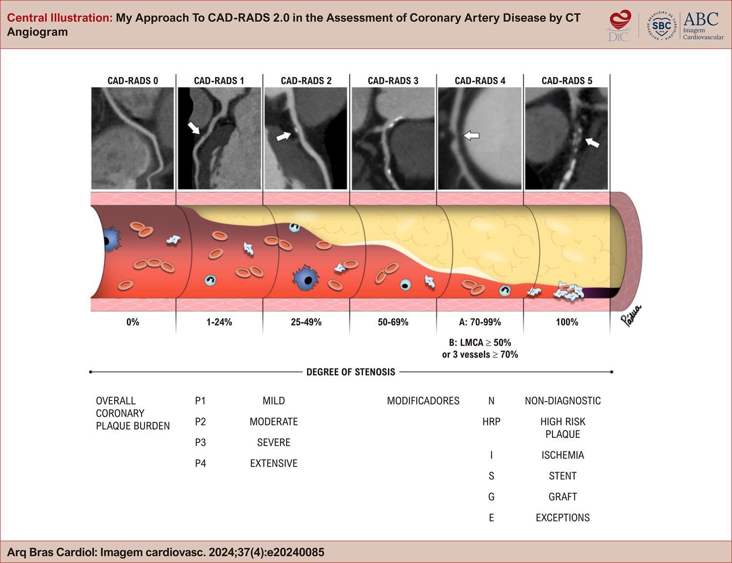
My Approach To CAD-RADS 2.0 in the Assessment of Coronary Artery Disease by CT Angiogram

Bruno Maeda Fuzissima et al. Roberto Vitor Almeida Torres Renata Muller Couto Lucas de Pádua Gomes de Farias … Bernardo Salgado Pinto Oliveira José de Arimatéia Batista Araújo Filho

Abstract

Coronary artery disease (CAD) is a common cause of chest pain, frequently seen in emergency departments and during elective consultations, and remains the leading cause of death worldwide. Coronary computed tomography angiography (CCTA) has become an established diagnostic tool for evaluating stable angina and acute chest pain in patients with low or intermediate risk. In this setting, CAD-RADS (Coronary Artery Disease-Reporting and Data System) was introduced as a CCTA reporting model, first published in 2016 and updated in 2022, with the aim of standardizing the description of imaging findings, improving communication between imaging specialists and clinicians, and providing guidance on patient management. This article reviews the CAD-RADS classification categories, which are based on the degree of stenosis, atherosclerotic plaque burden, and other modifying findings related to coronary abnormalities while also addressing certain exceptional situations. Through clinical cases, examples of structured reports, and recommendations for clinical management in different scenarios, we examine the objectives, benefits, limitations, and future potential of this classification, which remains underutilized in clinical practice.

  • Chest Pain
  • Coronary Artery Disease
  • Tomography
Read Full Article
Creative Commons Attribution (CC BY) Creative Commons license
21/11/2024 DOI: 10.36660/abcimg.20240085i

How to cite this article

EndNote RIS

Electronic Document Format (Vancouver)

Hortegal RA, Bihan DL, Mathias MM, Barretto R, Mathias Jr W. Left Ventricular Strain in Heart Failure with Preserved Ejection Fraction: A Fast Dive into Diagnosis, Response to Exercise and Afterload Challenge. ABC Imagem Cardiovascular 2024;37(4):e20240104.

Electronic Document Format (ABNT)

Hortegal, Renato A.; Bihan, David Le; Mathias, Mariana Mendes; Barretto, Rodrigo; Mathias Jr, Wilson. Left Ventricular Strain in Heart Failure with Preserved Ejection Fraction: A Fast Dive into Diagnosis, Response to Exercise and Afterload Challenge. ABC Imagem Cardiovascular, v. 37, n. 4, e20240104, Dec. 2024.

Electronic Document Format (APA)

Hortegal, R. A., Bihan, D. L., Mathias, M. M., Barretto, R., & Mathias Jr, W. (2024). Left Ventricular Strain in Heart Failure with Preserved Ejection Fraction: A Fast Dive into Diagnosis, Response to Exercise and Afterload Challenge. ABC Imagem Cardiovascular, 37(4), e20240104.

Electronic Document Format (ISO)

Hortegal, Renato A. and Bihan, David Le and Mathias, Mariana Mendes and Barretto, Rodrigo and Mathias Jr, Wilson. Left Ventricular Strain in Heart Failure with Preserved Ejection Fraction: A Fast Dive into Diagnosis, Response to Exercise and Afterload Challenge. ABC Imagem Cardiovascular [online]. 2024, vol. 37, n. 4, [cited 2026-10-14], e20240104. Available from: <https://journals.cardiol.br/en/article/left-ventricular-strain-in-heart-failure-with-preserved-ejection-fraction-a-fast-dive-into-diagnosis-response-to-exercise-and-afterload-challenge/>. ISSN 2318-8219

EndNote RIS

Electronic Document Format (Vancouver)

Silveira Neto JG, Campos MB, Mello L, Giffoni MC, Goveia NASB, Nóbrega ACL, Castro RRT, et al. The Prognostic Impact of Myocardial Deformation Measures in Patients with COVID-19. ABC Imagem Cardiovascular 2024;37(4):e20240067.

Electronic Document Format (ABNT)

Silveira Neto, João Giffoni; Campos, Marcello de Brito; Mello, Luiz; Giffoni, Marcela de Castro; Goveia, Natanael Arnor da Silva Barros; Nóbrega, Antonio Claudio Lucas da; Castro, Renata R. T.. The Prognostic Impact of Myocardial Deformation Measures in Patients with COVID-19. ABC Imagem Cardiovascular, v. 37, n. 4, e20240067, Dec. 2024.

Electronic Document Format (APA)

Silveira Neto, J. G., Campos, M. B., Mello, L., Giffoni, M. C., Goveia, N. A. S. B., Nóbrega, A. C. L. , & Castro, R. R. T. (2024). The Prognostic Impact of Myocardial Deformation Measures in Patients with COVID-19. ABC Imagem Cardiovascular, 37(4), e20240067.

Electronic Document Format (ISO)

Silveira Neto, João Giffoni and Campos, Marcello de Brito and Mello, Luiz and Giffoni, Marcela de Castro and Goveia, Natanael Arnor da Silva Barros and Nóbrega, Antonio Claudio Lucas da and Castro, Renata R. T.. The Prognostic Impact of Myocardial Deformation Measures in Patients with COVID-19. ABC Imagem Cardiovascular [online]. 2024, vol. 37, n. 4, [cited 2026-10-14], e20240067. Available from: <https://journals.cardiol.br/en/article/the-prognostic-impact-of-myocardial-deformation-measures-in-patients-with-covid-19/>. ISSN 2318-8219

EndNote RIS

Electronic Document Format (Vancouver)

Teixeira GCA, Castro BVG, Silva DC, Magalhães T. My Approach to Functional Analysis in Computed Tomography Angiography: Myocardial Perfusion and FFRct. ABC Imagem Cardiovascular 2024;37(4):e20240113.

Electronic Document Format (ABNT)

Teixeira, Gustavo Corrêa de Almeida; Castro, Bruno Vicente Gomes de; Silva, Douglas Carli; Magalhães, Tiago. My Approach to Functional Analysis in Computed Tomography Angiography: Myocardial Perfusion and FFRct. ABC Imagem Cardiovascular, v. 37, n. 4, e20240113, Dec. 2024.

Electronic Document Format (APA)

Teixeira, G. C. A., Castro, B. V. G. , Silva, D. C., & Magalhães, T. (2024). My Approach to Functional Analysis in Computed Tomography Angiography: Myocardial Perfusion and FFRct. ABC Imagem Cardiovascular, 37(4), e20240113.

Electronic Document Format (ISO)

Teixeira, Gustavo Corrêa de Almeida and Castro, Bruno Vicente Gomes de and Silva, Douglas Carli and Magalhães, Tiago. My Approach to Functional Analysis in Computed Tomography Angiography: Myocardial Perfusion and FFRct. ABC Imagem Cardiovascular [online]. 2024, vol. 37, n. 4, [cited 2026-10-14], e20240113. Available from: <https://journals.cardiol.br/en/article/my-approach-to-functional-analysis-in-computed-tomography-angiography-myocardial-perfusion-and-ffrct/>. ISSN 2318-8219

EndNote RIS

Electronic Document Format (Vancouver)

Brandão SCS. A New Horizon in Nuclear Cardiology in Brazil: Impact and Barriers of F-18 Flurpiridaz. ABC Imagem Cardiovascular 2024;37(4):e20240118.

Electronic Document Format (ABNT)

Brandão, Simone Cristina Soares. A New Horizon in Nuclear Cardiology in Brazil: Impact and Barriers of F-18 Flurpiridaz. ABC Imagem Cardiovascular, v. 37, n. 4, e20240118, Dec. 2024.

Electronic Document Format (APA)

Brandão, S. C. S. (2024). A New Horizon in Nuclear Cardiology in Brazil: Impact and Barriers of F-18 Flurpiridaz. ABC Imagem Cardiovascular, 37(4), e20240118.

Electronic Document Format (ISO)

Brandão, Simone Cristina Soares. A New Horizon in Nuclear Cardiology in Brazil: Impact and Barriers of F-18 Flurpiridaz. ABC Imagem Cardiovascular [online]. 2024, vol. 37, n. 4, [cited 2026-10-14], e20240118. Available from: <https://journals.cardiol.br/en/article/a-new-horizon-in-nuclear-cardiology-in-brazil-impact-and-barriers-of-f-18-flurpiridaz/>. ISSN 2318-8219

EndNote RIS

Electronic Document Format (Vancouver)

Freire EG, Araújo-Filho JAB, Nomura CH. Climate Change and Cardiovascular Imaging: Challenges and Opportunities. ABC Imagem Cardiovascular 2024;37(4):e20240117.

Electronic Document Format (ABNT)

Freire, Eduardo Galvão; Araújo-Filho, José Arimateia Batista; Nomura, Cesar Higa. Climate Change and Cardiovascular Imaging: Challenges and Opportunities. ABC Imagem Cardiovascular, v. 37, n. 4, e20240117, Dec. 2024.

Electronic Document Format (APA)

Freire, E. G., Araújo-Filho, J. A. B., & Nomura, C. H. (2024). Climate Change and Cardiovascular Imaging: Challenges and Opportunities. ABC Imagem Cardiovascular, 37(4), e20240117.

Electronic Document Format (ISO)

Freire, Eduardo Galvão and Araújo-Filho, José Arimateia Batista and Nomura, Cesar Higa. Climate Change and Cardiovascular Imaging: Challenges and Opportunities. ABC Imagem Cardiovascular [online]. 2024, vol. 37, n. 4, [cited 2026-10-14], e20240117. Available from: <https://journals.cardiol.br/en/article/climate-change-and-cardiovascular-imaging-challenges-and-opportunities/>. ISSN 2318-8219

EndNote RIS

Electronic Document Format (Vancouver)

Costa IBSS. Quantification of Myocardial Fibrosis by Cardiac Magnetic Resonance Imaging: Advances, Impacts, and Perspectives. ABC Imagem Cardiovascular 2024;37(4):e20240114.

Electronic Document Format (ABNT)

Costa, Isabela Bispo Santos da Silva. Quantification of Myocardial Fibrosis by Cardiac Magnetic Resonance Imaging: Advances, Impacts, and Perspectives. ABC Imagem Cardiovascular, v. 37, n. 4, e20240114, Dec. 2024.

Electronic Document Format (APA)

Costa, I. B. S. S. (2024). Quantification of Myocardial Fibrosis by Cardiac Magnetic Resonance Imaging: Advances, Impacts, and Perspectives. ABC Imagem Cardiovascular, 37(4), e20240114.

Electronic Document Format (ISO)

Costa, Isabela Bispo Santos da Silva. Quantification of Myocardial Fibrosis by Cardiac Magnetic Resonance Imaging: Advances, Impacts, and Perspectives. ABC Imagem Cardiovascular [online]. 2024, vol. 37, n. 4, [cited 2026-10-14], e20240114. Available from: <https://journals.cardiol.br/en/article/quantification-of-myocardial-fibrosis-by-cardiac-magnetic-resonance-imaging-advances-impacts-and-perspectives/>. ISSN 2318-8219

EndNote RIS

Electronic Document Format (Vancouver)

Oliveira FC, Kawaoka JR, Leite BF, Azevedo DFC, Fernandes RM, Noya-Rabelo MM. Pericardial Hematoma With Chest Pain and Tamponade in the Late Postoperative Period of Cardiac Surgery. ABC Imagem Cardiovascular 2024;37(4):e20240090.

Electronic Document Format (ABNT)

Oliveira, Felipe Carvalho de; Kawaoka, Jun Ramos; Leite, Bruno de Freitas; Azevedo, Diogo Freitas Cardoso de; Fernandes, Rafael Modesto; Noya-Rabelo, Marcia Maria. Pericardial Hematoma With Chest Pain and Tamponade in the Late Postoperative Period of Cardiac Surgery. ABC Imagem Cardiovascular, v. 37, n. 4, e20240090, Dec. 2024.

Electronic Document Format (APA)

Oliveira, F. C. , Kawaoka, J. R., Leite, B. F., Azevedo, D. F. C. , Fernandes, R. M., & Noya-Rabelo, M. M. (2024). Pericardial Hematoma With Chest Pain and Tamponade in the Late Postoperative Period of Cardiac Surgery. ABC Imagem Cardiovascular, 37(4), e20240090.

Electronic Document Format (ISO)

Oliveira, Felipe Carvalho de and Kawaoka, Jun Ramos and Leite, Bruno de Freitas and Azevedo, Diogo Freitas Cardoso de and Fernandes, Rafael Modesto and Noya-Rabelo, Marcia Maria. Pericardial Hematoma With Chest Pain and Tamponade in the Late Postoperative Period of Cardiac Surgery. ABC Imagem Cardiovascular [online]. 2024, vol. 37, n. 4, [cited 2026-10-14], e20240090. Available from: <https://journals.cardiol.br/en/article/pericardial-hematoma-with-chest-pain-and-tamponade-in-the-late-postoperative-period-of-cardiac-surgery/>. ISSN 2318-8219

EndNote RIS

Electronic Document Format (Vancouver)

Michael AP, Soares MF, Foresti RA, Rodrigues MOD, Witkowski SM. Prevalence of Fetal Heart Disease in Pregnant Women Referred to a Specialized Service Due to Cardiac Abnormalities on Morphological Ultrasound. ABC Imagem Cardiovascular 2024;37(4):e20240102.

Electronic Document Format (ABNT)

Michael, Andressa Pinto; Soares, Marcelo França; Foresti, Roque Antonio; Rodrigues, Marco Otilio Duarte; Witkowski, Sandra Mara. Prevalence of Fetal Heart Disease in Pregnant Women Referred to a Specialized Service Due to Cardiac Abnormalities on Morphological Ultrasound. ABC Imagem Cardiovascular, v. 37, n. 4, e20240102, Dec. 2024.

Electronic Document Format (APA)

Michael, A. P., Soares, M. F., Foresti, R. A., Rodrigues, M. O. D., & Witkowski, S. M. (2024). Prevalence of Fetal Heart Disease in Pregnant Women Referred to a Specialized Service Due to Cardiac Abnormalities on Morphological Ultrasound. ABC Imagem Cardiovascular, 37(4), e20240102.

Electronic Document Format (ISO)

Michael, Andressa Pinto and Soares, Marcelo França and Foresti, Roque Antonio and Rodrigues, Marco Otilio Duarte and Witkowski, Sandra Mara. Prevalence of Fetal Heart Disease in Pregnant Women Referred to a Specialized Service Due to Cardiac Abnormalities on Morphological Ultrasound. ABC Imagem Cardiovascular [online]. 2024, vol. 37, n. 4, [cited 2026-10-14], e20240102. Available from: <https://journals.cardiol.br/en/article/prevalence-of-fetal-heart-disease-in-pregnant-women-referred-to-a-specialized-service-due-to-cardiac-abnormalities-on-morphological-ultrasound/>. ISSN 2318-8219

EndNote RIS

Electronic Document Format (Vancouver)

Santos SN. Top Vascular Ultrasound 2024: Carotid Plaque-RADS – A Novel Stroke Risk Classification System. ABC Imagem Cardiovascular 2024;37(4):e20240110.

Electronic Document Format (ABNT)

Santos, Simone Nascimento dos. Top Vascular Ultrasound 2024: Carotid Plaque-RADS – A Novel Stroke Risk Classification System. ABC Imagem Cardiovascular, v. 37, n. 4, e20240110, Dec. 2024.

Electronic Document Format (APA)

Santos, S. N. (2024). Top Vascular Ultrasound 2024: Carotid Plaque-RADS – A Novel Stroke Risk Classification System. ABC Imagem Cardiovascular, 37(4), e20240110.

Electronic Document Format (ISO)

Santos, Simone Nascimento dos. Top Vascular Ultrasound 2024: Carotid Plaque-RADS – A Novel Stroke Risk Classification System. ABC Imagem Cardiovascular [online]. 2024, vol. 37, n. 4, [cited 2026-10-14], e20240110. Available from: <https://journals.cardiol.br/en/article/top-vascular-ultrasound-2024-carotid-plaque-rads-a-novel-stroke-risk-classification-system/>. ISSN 2318-8219

EndNote RIS

Electronic Document Format (Vancouver)

Felix AS. The Use of X (Formerly Twitter) in Cardiovascular Imaging: Opportunities and Risks. ABC Imagem Cardiovascular 2024;37(4):e20240109.

Electronic Document Format (ABNT)

Felix, Alex dos Santos. The Use of X (Formerly Twitter) in Cardiovascular Imaging: Opportunities and Risks. ABC Imagem Cardiovascular, v. 37, n. 4, e20240109, Dec. 2024.

Electronic Document Format (APA)

Felix, A. S. (2024). The Use of X (Formerly Twitter) in Cardiovascular Imaging: Opportunities and Risks. ABC Imagem Cardiovascular, 37(4), e20240109.

Electronic Document Format (ISO)

Felix, Alex dos Santos. The Use of X (Formerly Twitter) in Cardiovascular Imaging: Opportunities and Risks. ABC Imagem Cardiovascular [online]. 2024, vol. 37, n. 4, [cited 2026-10-14], e20240109. Available from: <https://journals.cardiol.br/en/article/the-use-of-x-formerly-twitter-in-cardiovascular-imaging-opportunities-and-risks/>. ISSN 2318-8219

EndNote RIS

Electronic Document Format (Vancouver)

Pereira MM, Monteiro Júnior FC, Yamamura S. Impact of Valvular Event Time Assessment Methods on Myocardial Work Calculation by Echocardiography. ABC Imagem Cardiovascular 2024;37(4):e20240093.

Electronic Document Format (ABNT)

Pereira, Marcio Mendes; Monteiro Júnior, Francisco das Chagas; Yamamura, Sayuri. Impact of Valvular Event Time Assessment Methods on Myocardial Work Calculation by Echocardiography. ABC Imagem Cardiovascular, v. 37, n. 4, e20240093, Dec. 2024.

Electronic Document Format (APA)

Pereira, M. M., Monteiro Júnior, F. C., & Yamamura, S. (2024). Impact of Valvular Event Time Assessment Methods on Myocardial Work Calculation by Echocardiography. ABC Imagem Cardiovascular, 37(4), e20240093.

Electronic Document Format (ISO)

Pereira, Marcio Mendes and Monteiro Júnior, Francisco das Chagas and Yamamura, Sayuri. Impact of Valvular Event Time Assessment Methods on Myocardial Work Calculation by Echocardiography. ABC Imagem Cardiovascular [online]. 2024, vol. 37, n. 4, [cited 2026-10-14], e20240093. Available from: <https://journals.cardiol.br/en/article/impact-of-valvular-event-time-assessment-methods-on-myocardial-work-calculation-by-echocardiography/>. ISSN 2318-8219

EndNote RIS

Electronic Document Format (Vancouver)

Guaman-Valdivieso C, Aramburu J, Lluberas N, Huart N, Parma G. Not Everything is as it Seems at First Glance: Multimodal Imaging in Heart Cyst Evaluation. ABC Imagem Cardiovascular 2024;37(4):e20240086.

Electronic Document Format (ABNT)

Guaman-Valdivieso, Carlos; Aramburu, Julia; Lluberas, Natalia; Huart, Natalia; Parma, Gabriel. Not Everything is as it Seems at First Glance: Multimodal Imaging in Heart Cyst Evaluation. ABC Imagem Cardiovascular, v. 37, n. 4, e20240086, Dec. 2024.

Electronic Document Format (APA)

Guaman-Valdivieso, C., Aramburu, J., Lluberas, N., Huart, N., & Parma, G. (2024). Not Everything is as it Seems at First Glance: Multimodal Imaging in Heart Cyst Evaluation. ABC Imagem Cardiovascular, 37(4), e20240086.

Electronic Document Format (ISO)

Guaman-Valdivieso, Carlos and Aramburu, Julia and Lluberas, Natalia and Huart, Natalia and Parma, Gabriel. Not Everything is as it Seems at First Glance: Multimodal Imaging in Heart Cyst Evaluation. ABC Imagem Cardiovascular [online]. 2024, vol. 37, n. 4, [cited 2026-10-14], e20240086. Available from: <https://journals.cardiol.br/en/article/not-everything-is-as-it-seems-at-first-glance-multimodal-imaging-in-heart-cyst-evaluation/>. ISSN 2318-8219

EndNote RIS

Electronic Document Format (Vancouver)

Zarate JAN, Mahecha MJA. An Explosion of Colors, Fauvism in Echocardiography, a New Proposal. ABC Imagem Cardiovascular 2024;37(4):e20240081.

Electronic Document Format (ABNT)

Zarate, Jaime Andres Nieto; Mahecha, Maria Jose Amaya. An Explosion of Colors, Fauvism in Echocardiography, a New Proposal. ABC Imagem Cardiovascular, v. 37, n. 4, e20240081, Dec. 2024.

Electronic Document Format (APA)

Zarate, J. A. N., & Mahecha, M. J. A. (2024). An Explosion of Colors, Fauvism in Echocardiography, a New Proposal. ABC Imagem Cardiovascular, 37(4), e20240081.

Electronic Document Format (ISO)

Zarate, Jaime Andres Nieto and Mahecha, Maria Jose Amaya. An Explosion of Colors, Fauvism in Echocardiography, a New Proposal. ABC Imagem Cardiovascular [online]. 2024, vol. 37, n. 4, [cited 2026-10-14], e20240081. Available from: <https://journals.cardiol.br/en/article/an-explosion-of-colors-fauvism-in-echocardiography-a-new-proposal/>. ISSN 2318-8219

EndNote RIS

Electronic Document Format (Vancouver)

Sousa WGS, Domingues JL, Damasceno AT, Lemos AET, Araújo ALJ, Evangelista NL. Unicuspid Aortic Stenosis: Case Report. ABC Imagem Cardiovascular 2024;37(4):e20240087.

Electronic Document Format (ABNT)

Sousa, Welison Gutherrez Silva e; Domingues, Julia Lucena; Damasceno, Adriana Teixeira; Lemos, Alessandra Edna Teófilo; Araújo, Antônio Luiz Júnior; Evangelista, Nelson Lopes. Unicuspid Aortic Stenosis: Case Report. ABC Imagem Cardiovascular, v. 37, n. 4, e20240087, Dec. 2024.

Electronic Document Format (APA)

Sousa, W. G. S. , Domingues, J. L., Damasceno, A. T., Lemos, A. E. T., Araújo, A. L. J., & Evangelista, N. L. (2024). Unicuspid Aortic Stenosis: Case Report. ABC Imagem Cardiovascular, 37(4), e20240087.

Electronic Document Format (ISO)

Sousa, Welison Gutherrez Silva e and Domingues, Julia Lucena and Damasceno, Adriana Teixeira and Lemos, Alessandra Edna Teófilo and Araújo, Antônio Luiz Júnior and Evangelista, Nelson Lopes. Unicuspid Aortic Stenosis: Case Report. ABC Imagem Cardiovascular [online]. 2024, vol. 37, n. 4, [cited 2026-10-14], e20240087. Available from: <https://journals.cardiol.br/en/article/unicuspid-aortic-stenosis-case-report/>. ISSN 2318-8219

EndNote RIS

Electronic Document Format (Vancouver)

Darzé AC, Paladino Filho AT, Rassi AM, Prazeres CE, Pinto IMF, Caneschi JM, Ponte Junior JRT, et al. Giant Left Atrium in Rheumatic Mitral Valve Disease. ABC Imagem Cardiovascular 2024;37(4):e20240083.

Electronic Document Format (ABNT)

Darzé, Alice Cunha; Paladino Filho, Antonio Tito; Rassi, Amanda Marsiaj; Prazeres, Carlos Eduardo; Pinto, Ibraim Masciarelli Francisco; Caneschi, João Marcelo; Ponte Junior, Jose Roberto Tuma da; Morales, Paul Salvador; Hendges, Rafael Castro; Senra, Tiago. Giant Left Atrium in Rheumatic Mitral Valve Disease. ABC Imagem Cardiovascular, v. 37, n. 4, e20240083, Dec. 2024.

Electronic Document Format (APA)

Darzé, A. C., Paladino Filho, A. T., Rassi, A. M., Prazeres, C. E., Pinto, I. M. F., Caneschi, J. M., Ponte Junior, J. R. T. , Morales, P. S., Hendges, R. C., & Senra, T. (2024). Giant Left Atrium in Rheumatic Mitral Valve Disease. ABC Imagem Cardiovascular, 37(4), e20240083.

Electronic Document Format (ISO)

Darzé, Alice Cunha and Paladino Filho, Antonio Tito and Rassi, Amanda Marsiaj and Prazeres, Carlos Eduardo and Pinto, Ibraim Masciarelli Francisco and Caneschi, João Marcelo and Ponte Junior, Jose Roberto Tuma da and Morales, Paul Salvador and Hendges, Rafael Castro and Senra, Tiago. Giant Left Atrium in Rheumatic Mitral Valve Disease. ABC Imagem Cardiovascular [online]. 2024, vol. 37, n. 4, [cited 2026-10-14], e20240083. Available from: <https://journals.cardiol.br/en/article/giant-left-atrium-in-rheumatic-mitral-valve-disease/>. ISSN 2318-8219

EndNote RIS

Electronic Document Format (Vancouver)

Barbosa YEL, Evangelista IWQF, Sousa JMR, Jales LCL, Viegas ELM, Nóbrega PVN, Moura DMC, et al. The Many Faces of Arrhythmic Mitral Valve Prolapse: Case Series. ABC Imagem Cardiovascular 2024;37(4):e20240073.

Electronic Document Format (ABNT)

Barbosa, Yasmim Evelyn Lisboa; Evangelista, Isadora Wanderley Queiroga de Freitas; Sousa, Julyana Maria Ramalho de; Jales, Lorena Carvalho Leite; Viegas, Elisabete Louise de Medeiros; Nóbrega, Patrícia Vidal de Negreiros; Moura, Daniel Moreira Costa; Targueta, Gabriel Pelegrineti; Resende, Lilian Amador; Pereira, Renner Augusto Raposo; Athayde, Guilherme Augusto Teodoro. The Many Faces of Arrhythmic Mitral Valve Prolapse: Case Series. ABC Imagem Cardiovascular, v. 37, n. 4, e20240073, Nov. 2024.

Electronic Document Format (APA)

Barbosa, Y. E. L., Evangelista, I. W. Q. F., Sousa, J. M. R. , Jales, L. C. L., Viegas, E. L. M., Nóbrega, P. V. N., Moura, D. M. C., Targueta, G. P., Resende, L. A., Pereira, R. A. R., & Athayde, G. A. T. (2024). The Many Faces of Arrhythmic Mitral Valve Prolapse: Case Series. ABC Imagem Cardiovascular, 37(4), e20240073.

Electronic Document Format (ISO)

Barbosa, Yasmim Evelyn Lisboa and Evangelista, Isadora Wanderley Queiroga de Freitas and Sousa, Julyana Maria Ramalho de and Jales, Lorena Carvalho Leite and Viegas, Elisabete Louise de Medeiros and Nóbrega, Patrícia Vidal de Negreiros and Moura, Daniel Moreira Costa and Targueta, Gabriel Pelegrineti and Resende, Lilian Amador and Pereira, Renner Augusto Raposo and Athayde, Guilherme Augusto Teodoro. The Many Faces of Arrhythmic Mitral Valve Prolapse: Case Series. ABC Imagem Cardiovascular [online]. 2024, vol. 37, n. 4, [cited 2026-10-14], e20240073. Available from: <https://journals.cardiol.br/en/article/the-many-faces-of-arrhythmic-mitral-valve-prolapse-case-series/>. ISSN 2318-8219

EndNote RIS

Electronic Document Format (Vancouver)

Nascimento MAG, Pereira EHB, Magalhães GKL, Mariano LS, Rodrigues LMC, Batista LA, Caravante RPG, et al. Atrial Myxoma in Male Young Adult Patient: A Case Report and Literature Review. ABC Imagem Cardiovascular 2024;37(4):e20240064.

Electronic Document Format (ABNT)

Nascimento, Monize Aparecida Gonçalves do; Pereira, Eliseu Henrique Bispo; Magalhães, Giovanna Karine Leal; Mariano, Laura Souza; Rodrigues, Luana Mireli Carbonera; Batista, Leonardo Alves; Caravante, Rogério de Paula Garcia; Barreto, Ayná Emanuelli Alves. Atrial Myxoma in Male Young Adult Patient: A Case Report and Literature Review. ABC Imagem Cardiovascular, v. 37, n. 4, e20240064, Nov. 2024.

Electronic Document Format (APA)

Nascimento, M. A. G. , Pereira, E. H. B., Magalhães, G. K. L., Mariano, L. S., Rodrigues, L. M. C., Batista, L. A., Caravante, R. P. G., & Barreto, A. E. A. (2024). Atrial Myxoma in Male Young Adult Patient: A Case Report and Literature Review. ABC Imagem Cardiovascular, 37(4), e20240064.

Electronic Document Format (ISO)

Nascimento, Monize Aparecida Gonçalves do and Pereira, Eliseu Henrique Bispo and Magalhães, Giovanna Karine Leal and Mariano, Laura Souza and Rodrigues, Luana Mireli Carbonera and Batista, Leonardo Alves and Caravante, Rogério de Paula Garcia and Barreto, Ayná Emanuelli Alves. Atrial Myxoma in Male Young Adult Patient: A Case Report and Literature Review. ABC Imagem Cardiovascular [online]. 2024, vol. 37, n. 4, [cited 2026-10-14], e20240064. Available from: <https://journals.cardiol.br/en/article/atrial-myxoma-in-male-young-adult-patient-a-case-report-and-literature-review/>. ISSN 2318-8219

EndNote RIS

Electronic Document Format (Vancouver)

Otto MEB. Artificial Intelligence in Echocardiography: The Future of Precision Diagnosis. ABC Imagem Cardiovascular 2024;37(4):e20240095.

Electronic Document Format (ABNT)

Otto, Maria Estefânia Bosco. Artificial Intelligence in Echocardiography: The Future of Precision Diagnosis. ABC Imagem Cardiovascular, v. 37, n. 4, e20240095, Nov. 2024.

Electronic Document Format (APA)

Otto, M. E. B. (2024). Artificial Intelligence in Echocardiography: The Future of Precision Diagnosis. ABC Imagem Cardiovascular, 37(4), e20240095.

Electronic Document Format (ISO)

Otto, Maria Estefânia Bosco. Artificial Intelligence in Echocardiography: The Future of Precision Diagnosis. ABC Imagem Cardiovascular [online]. 2024, vol. 37, n. 4, [cited 2026-10-14], e20240095. Available from: <https://journals.cardiol.br/en/article/artificial-intelligence-in-echocardiography-the-future-of-precision-diagnosis/>. ISSN 2318-8219

EndNote RIS

Electronic Document Format (Vancouver)

Pereira MM, Otto ME, Netto FM, Esteves F. Advances in Percutaneous Tricuspid Valve Treatment. ABC Imagem Cardiovascular 2024;37(4):e20240088.

Electronic Document Format (ABNT)

Pereira, Marcio Mendes; Otto, Maria Estefânia; Netto, Fernando Melo; Esteves, Fernanda. Advances in Percutaneous Tricuspid Valve Treatment. ABC Imagem Cardiovascular, v. 37, n. 4, e20240088, Nov. 2024.

Electronic Document Format (APA)

Pereira, M. M., Otto, M. E., Netto, F. M., & Esteves, F. (2024). Advances in Percutaneous Tricuspid Valve Treatment. ABC Imagem Cardiovascular, 37(4), e20240088.

Electronic Document Format (ISO)

Pereira, Marcio Mendes and Otto, Maria Estefânia and Netto, Fernando Melo and Esteves, Fernanda. Advances in Percutaneous Tricuspid Valve Treatment. ABC Imagem Cardiovascular [online]. 2024, vol. 37, n. 4, [cited 2026-10-14], e20240088. Available from: <https://journals.cardiol.br/en/article/advances-in-percutaneous-tricuspid-valve-treatment/>. ISSN 2318-8219

EndNote RIS

Electronic Document Format (Vancouver)

Zapparoli M, Alessi A, Zapparoli FC, Alessi MR. Rare Presentation of an Anatomical Congenital Anomaly of the Coronary Arteries on Computed Tomography Angiography. ABC Imagem Cardiovascular 2024;37(4):e20240075.

Electronic Document Format (ABNT)

Zapparoli, Marcello; Alessi, Alexandre; Zapparoli, Franco Camargo; Alessi, Mateus Rodrigues. Rare Presentation of an Anatomical Congenital Anomaly of the Coronary Arteries on Computed Tomography Angiography. ABC Imagem Cardiovascular, v. 37, n. 4, e20240075, Nov. 2024.

Electronic Document Format (APA)

Zapparoli, M., Alessi, A., Zapparoli, F. C., & Alessi, M. R. (2024). Rare Presentation of an Anatomical Congenital Anomaly of the Coronary Arteries on Computed Tomography Angiography. ABC Imagem Cardiovascular, 37(4), e20240075.

Electronic Document Format (ISO)

Zapparoli, Marcello and Alessi, Alexandre and Zapparoli, Franco Camargo and Alessi, Mateus Rodrigues. Rare Presentation of an Anatomical Congenital Anomaly of the Coronary Arteries on Computed Tomography Angiography. ABC Imagem Cardiovascular [online]. 2024, vol. 37, n. 4, [cited 2026-10-14], e20240075. Available from: <https://journals.cardiol.br/en/article/rare-presentation-of-an-anatomical-congenital-anomaly-of-the-coronary-arteries-on-computed-tomography-angiography/>. ISSN 2318-8219

EndNote RIS

Electronic Document Format (Vancouver)

Barberato MFA, Rassi DC, Beck ALS, Uellendahl M, Morhy SS, Barberato SH. The Women of the Cardiovascular Imaging Department of the Brazilian Society of Cardiology: Career and Family Challenges. ABC Imagem Cardiovascular 2024;37(4):e20240092.

Electronic Document Format (ABNT)

Barberato, Marcia Ferreira Alves; Rassi, Daniela do Carmo; Beck, Adenalva Lima de Souza; Uellendahl, Marly; Morhy, Samira Saady; Barberato, Silvio Henrique. The Women of the Cardiovascular Imaging Department of the Brazilian Society of Cardiology: Career and Family Challenges. ABC Imagem Cardiovascular, v. 37, n. 4, e20240092, Nov. 2024.

Electronic Document Format (APA)

Barberato, M. F. A., Rassi, D. C., Beck, A. L. S., Uellendahl, M., Morhy, S. S., & Barberato, S. H. (2024). The Women of the Cardiovascular Imaging Department of the Brazilian Society of Cardiology: Career and Family Challenges. ABC Imagem Cardiovascular, 37(4), e20240092.

Electronic Document Format (ISO)

Barberato, Marcia Ferreira Alves and Rassi, Daniela do Carmo and Beck, Adenalva Lima de Souza and Uellendahl, Marly and Morhy, Samira Saady and Barberato, Silvio Henrique. The Women of the Cardiovascular Imaging Department of the Brazilian Society of Cardiology: Career and Family Challenges. ABC Imagem Cardiovascular [online]. 2024, vol. 37, n. 4, [cited 2026-10-14], e20240092. Available from: <https://journals.cardiol.br/en/article/the-women-of-the-cardiovascular-imaging-department-of-the-brazilian-society-of-cardiology-career-and-family-challenges/>. ISSN 2318-8219

EndNote RIS

Electronic Document Format (Vancouver)

Fuzissima BM, Torres RVA, Couto RM, Farias LPG, Oliveira BSP, Araújo Filho JAB. My Approach To CAD-RADS 2.0 in the Assessment of Coronary Artery Disease by CT Angiogram. ABC Imagem Cardiovascular 2024;37(4):e20240085.

Electronic Document Format (ABNT)

Fuzissima, Bruno Maeda; Torres, Roberto Vitor Almeida; Couto, Renata Muller; Farias, Lucas de Pádua Gomes de; Oliveira, Bernardo Salgado Pinto; Araújo Filho, José de Arimatéia Batista. My Approach To CAD-RADS 2.0 in the Assessment of Coronary Artery Disease by CT Angiogram. ABC Imagem Cardiovascular, v. 37, n. 4, e20240085, Nov. 2024.

Electronic Document Format (APA)

Fuzissima, B. M., Torres, R. V. A., Couto, R. M., Farias, L. P. G. , Oliveira, B. S. P., & Araújo Filho, J. A. B. (2024). My Approach To CAD-RADS 2.0 in the Assessment of Coronary Artery Disease by CT Angiogram. ABC Imagem Cardiovascular, 37(4), e20240085.

Electronic Document Format (ISO)

Fuzissima, Bruno Maeda and Torres, Roberto Vitor Almeida and Couto, Renata Muller and Farias, Lucas de Pádua Gomes de and Oliveira, Bernardo Salgado Pinto and Araújo Filho, José de Arimatéia Batista. My Approach To CAD-RADS 2.0 in the Assessment of Coronary Artery Disease by CT Angiogram. ABC Imagem Cardiovascular [online]. 2024, vol. 37, n. 4, [cited 2026-10-14], e20240085. Available from: <https://journals.cardiol.br/en/article/my-approach-to-cad-rads-2-0-in-the-assessment-of-coronary-artery-disease-by-ct-angiogram/>. ISSN 2318-8219

Sede – São Paulo
Rua Barata Ribeiro, 380 cj 54 Bela Vista
São Paulo, Brasil – CEP: 01308-000
Tel.: +55 (11) 3120-3363
Telefax: +55 (11) 3259-2988
E-mail: depeco@cardiol.br

Sede – Rio de Janeiro
Av. Marechal Câmara, 160
3º andar – Sala: 330 – Centro
Rio de Janeiro – CEP: 20020-907
Tel.: +55 (21) 3478-2700
ou: +55 (21) 99386-3961
E-mail: abcimaging@cardiol.br

  • About the Journal
  • Internal Regulation
  • Editors Section
  • Editorial Board
  • SBC Award

  • Guidelines for Publication
  • Editorial Policy
  • Ethics Policy
  • Policy on the Use of Artificial Intelligence
  • Mandatory Publication Form

Issues

SBC Guidelines