ABC Family Arquivos Brasileiros de Cardiologia International Journal of Cardiovascular Sciences ABC Heart Failure & Cardiomyopathy ABC Imagem Cardiovascular
Português English
ABC Family
Português English
  • About
    • About ABC Family
    • Internal Regulation
    • Editorial Board
    • Brazilian Society of Cardiology (SBC)
  • Issues
  • SBC Guidelines
  • Content
    • Videos
    • Podcasts
    • News
  • About
    • About ABC Family
    • Internal Regulation
    • Editorial Board
    • Brazilian Society of Cardiology (SBC)
  • Issues
  • SBC Guidelines
  • Content
    • Videos
    • Podcasts
    • News

ABC Family

  • ABC Family
  • Arquivos Brasileiros de Cardiologia
  • International Journal of Cardiovascular Sciences
  • ABC Heart Failure & Cardiomyopathy
  • ABC Imagem Cardiovascular
Search in

No content found.

Journals
Ordering
Article types
Others

  1. Home
  2. Keyword: Cardiovascular Diagnostic Techniques
Figure 1
ABCIC
Editorial

Photon-Counting Computed Tomography in Cardiovascular Imaging: Where We Are and What Lies Ahead

Paulo Savoia et al. Rodrigo Bello

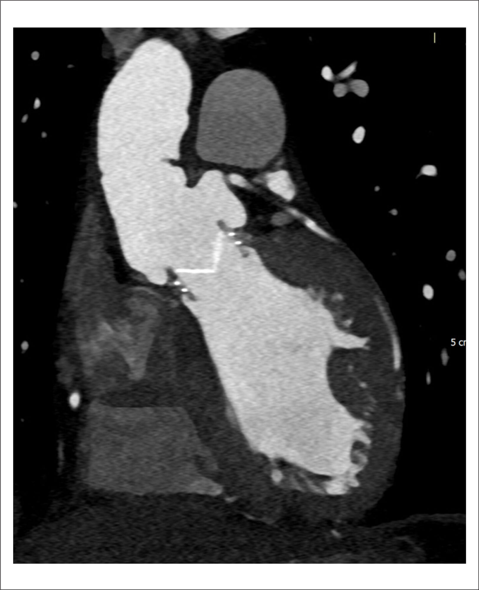
Computed Tomography Angiography (CTA) has been an important asset in cardiovascular diagnosis for decades. The continuous improvement of rapid data acquisition with often higher spatial resolution has established the method as indispensable for diagnosing and managing multiple cardiovascular entities such as aneurysms, aortic dissection, and pulmonary embolism., The use of CTA in heart and coronary imaging has also gained important territory and relevance., Computed Tomography (CT) techniques have been advancing significantly since its introduction in the medical field. Starting with single detectors, then helicoidal scanners, multidetector scanners with high pitch, improvement of reconstruction algorithms, the appearance of dual-energy technology, and lately the ability to acquire images of the whole heart in just one heartbeat with information from the entire cardiac cycle, are incredible milestones. Fractional Flow Reserve CT (FFR-CT) is also a very promising technique. Progression does not come without downsides: ionizing radiation is still concerning, and despite ways to reduce it such as dose modulation, some cardiovascular CTA exams still show relatively high levels of absorbed radiation, especially exams with multiple series, extremely thin slices, and retrospective reconstruction of larger parts of the cardiac cycle. Following this trend of improving image quality with less ionizing radiation as possible, a new complex technique emerged: Photon-counting CT (PCCT).

The main distinction between conventional CT and PCCT is in the detectors of X-rays. The conventional CT detector is based on receiving the X-ray photons that interact with the patient, converting them into light photons (it is an indirect system), and then converting this light into electrical signals that ultimately are going to be converted into digital signals. This system is called an Energy-Integrating Detector (EID). Basically, more light means more X-rays arriving and less interaction / more penetration in the patient’s tissues. It is not possible to differentiate exactly how many X-rays are arriving, nor the different energy levels of each X-ray photon. Moreover, the septa between each of the detectors also limit the reception of X-rays., On the other hand, PCCT detectors can detect separately each X-ray photon that is arriving and measure the energy of each one of the X-ray photons directly. This is possible because the PCCT detector has a high-voltage crystal layer that relocates electrons for each X-ray photon received, and the number of electrons relocated is proportional to the energy of each X-ray. The relocated electrons generate the electrical pulse that is going to be finally converted into the digital signal. And even smaller energy X-rays can be detected, improving the image contrast-to-noise ratio. It is a direct system, and there is no septa between the detectors. In the end, less X-rays are necessary (therefore less radiation) and it is possible to separate the X-rays according to the energy of each one, leading to applications of tissue recognition (different known energy X-rays are likely to have interacted with different known tissues) functioning as a dual energy CT.,

[…]
  • Cardiology
  • Cardiovascular Diagnostic Techniques
  • Computed Tomography Angiography
  • Radiology
Read Full Article
Creative Commons Attribution (CC BY) Creative Commons license
27/06/2025 DOI: 10.36660/abcimg.20250035i Views 1
Figure 1
ABCIC
Viewpoint

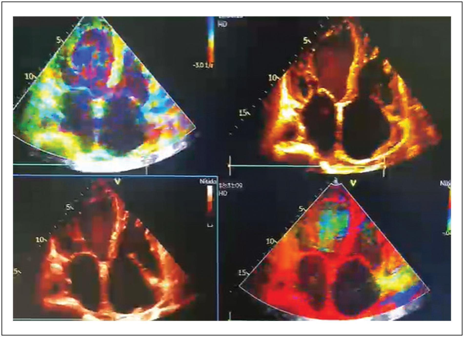
An Explosion of Colors, Fauvism in Echocardiography, a New Proposal

Jaime Andres Nieto Zarate et al. Maria Jose Amaya Mahecha

Limitations

The article offers an intriguing perspective, but further scientific validation is needed for broader acceptance in clinical practice. The connection between art and medical imaging is fascinating, yet requires more exploration to establish its practical relevance and impact on patient outcomes.

[…]

  • Cardiology
  • Cardiovascular Diagnostic Techniques
  • Echocardiography
  • Medicine in the Arts
Read Full Article
Creative Commons Attribution (CC BY) Creative Commons license
05/12/2024 DOI: 10.36660/abcimg.20240081i

How to cite this article

EndNote RIS

Electronic Document Format (Vancouver)

Savoia P, Bello R. Photon-Counting Computed Tomography in Cardiovascular Imaging: Where We Are and What Lies Ahead. ABC Imagem Cardiovascular 2025;38(2):e20250035.

Electronic Document Format (ABNT)

Savoia, Paulo; Bello, Rodrigo. Photon-Counting Computed Tomography in Cardiovascular Imaging: Where We Are and What Lies Ahead. ABC Imagem Cardiovascular, v. 38, n. 2, e20250035, Jun. 2025.

Electronic Document Format (APA)

Savoia, P., & Bello, R. (2025). Photon-Counting Computed Tomography in Cardiovascular Imaging: Where We Are and What Lies Ahead. ABC Imagem Cardiovascular, 38(2), e20250035.

Electronic Document Format (ISO)

Savoia, Paulo and Bello, Rodrigo. Photon-Counting Computed Tomography in Cardiovascular Imaging: Where We Are and What Lies Ahead. ABC Imagem Cardiovascular [online]. 2025, vol. 38, n. 2, [cited 2026-10-14], e20250035. Available from: <https://journals.cardiol.br/en/article/photon-counting-computed-tomography-in-cardiovascular-imaging-where-we-are-and-what-lies-ahead/>. ISSN 2318-8219

EndNote RIS

Electronic Document Format (Vancouver)

Zarate JAN, Mahecha MJA. An Explosion of Colors, Fauvism in Echocardiography, a New Proposal. ABC Imagem Cardiovascular 2024;37(4):e20240081.

Electronic Document Format (ABNT)

Zarate, Jaime Andres Nieto; Mahecha, Maria Jose Amaya. An Explosion of Colors, Fauvism in Echocardiography, a New Proposal. ABC Imagem Cardiovascular, v. 37, n. 4, e20240081, Dec. 2024.

Electronic Document Format (APA)

Zarate, J. A. N., & Mahecha, M. J. A. (2024). An Explosion of Colors, Fauvism in Echocardiography, a New Proposal. ABC Imagem Cardiovascular, 37(4), e20240081.

Electronic Document Format (ISO)

Zarate, Jaime Andres Nieto and Mahecha, Maria Jose Amaya. An Explosion of Colors, Fauvism in Echocardiography, a New Proposal. ABC Imagem Cardiovascular [online]. 2024, vol. 37, n. 4, [cited 2026-10-14], e20240081. Available from: <https://journals.cardiol.br/en/article/an-explosion-of-colors-fauvism-in-echocardiography-a-new-proposal/>. ISSN 2318-8219

  • About ABC Family
  • Internal Regulation
  • Editorial Board
  • Brazilian Society of Cardiology (SBC)

Issues

SBC Guidelines

  • Videos
  • Podcasts
  • News