Carlos Guaman-Valdivieso et al. Julia Aramburu Natalia Lluberas Natalia Huart Gabriel Parma
Abstract
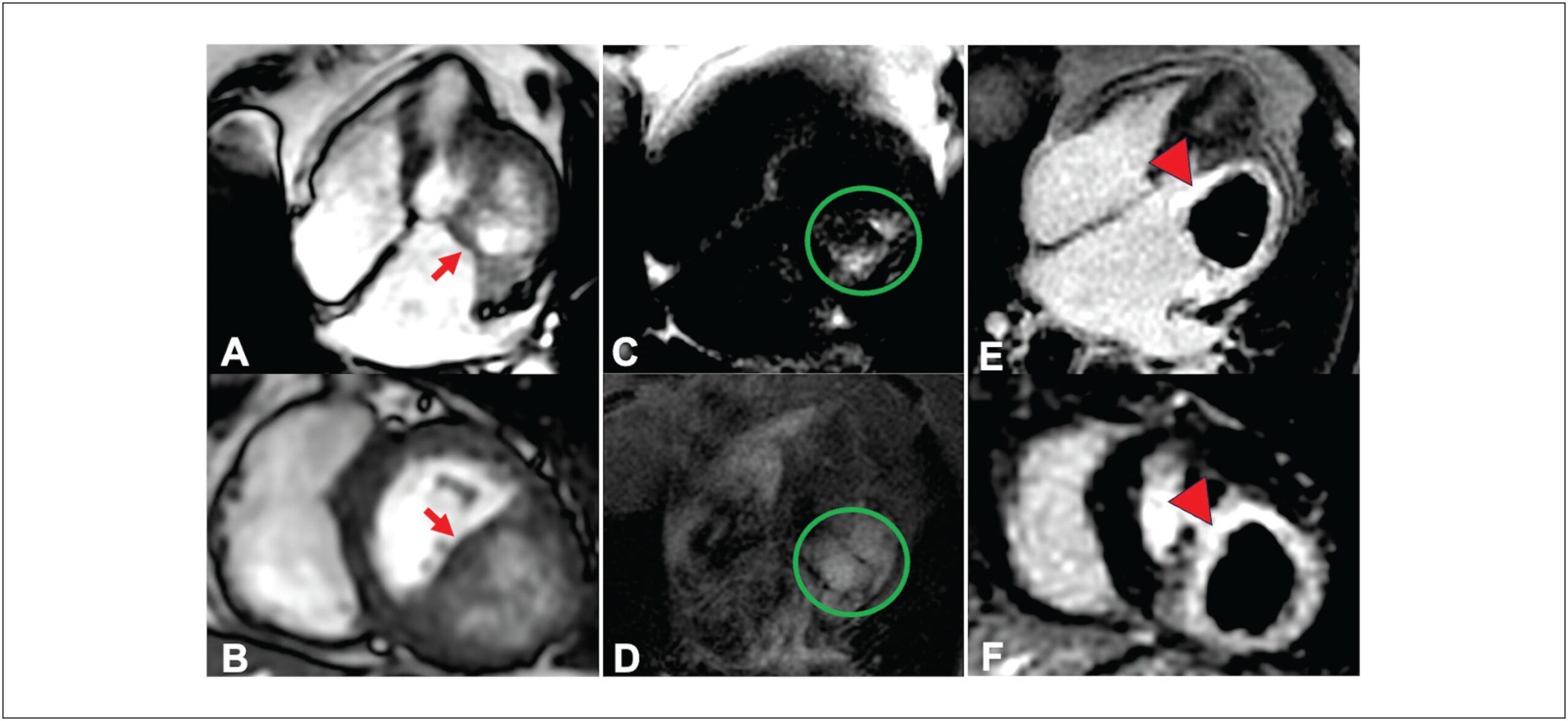
Cardiac masses, though rare, can lead to significant hemodynamic and arrhythmic issues, even if benign. Improved multimodal imaging has refined the diagnosis and understanding of these masses. We present an 85-year-old woman with New York Heart Association class II-III dyspnea for six months. Echocardiography showed a mass in the basal inferolateral wall of the left ventricle, along with severe left atrial dilation and moderate mitral regurgitation. Initially thought to be a pericardial cyst, further cardiac magnetic resonance (CMR) imaging identified an intramyocardial mass, and computed tomography (CT) suggested a hydatid cyst. Given the non-limiting symptoms and high surgical risk, the decision was made for expectant management. This case illustrates the rarity of cardiac cysts and the essential role of multimodal imaging in diagnosis and treatment planning.