
ABC Family Arquivos Brasileiros de Cardiologia International Journal of Cardiovascular Sciences ABC Heart Failure & Cardiomyopathy ABC Imagem Cardiovascular
Português English
ABC Family
Português English
  • About
    • About ABC Family
    • Internal Regulation
    • Editorial Board
    • Brazilian Society of Cardiology (SBC)
  • Issues
  • SBC Guidelines
  • Content
    • Videos
    • Podcasts
    • News
  • About
    • About ABC Family
    • Internal Regulation
    • Editorial Board
    • Brazilian Society of Cardiology (SBC)
  • Issues
  • SBC Guidelines
  • Content
    • Videos
    • Podcasts
    • News

ABC Family

  • ABC Family
  • Arquivos Brasileiros de Cardiologia
  • International Journal of Cardiovascular Sciences
  • ABC Heart Failure & Cardiomyopathy
  • ABC Imagem Cardiovascular
Search in

No content found.

Journals
Ordering
Article types
Others

  1. Home
  2. Keyword: Ultrasonography
ABC
Editorial

The New Frontier of Point-of-Care Ultrasound: Lung Ultrasound in Congenital Heart Disease

Maria Estefânia Bosco Otto

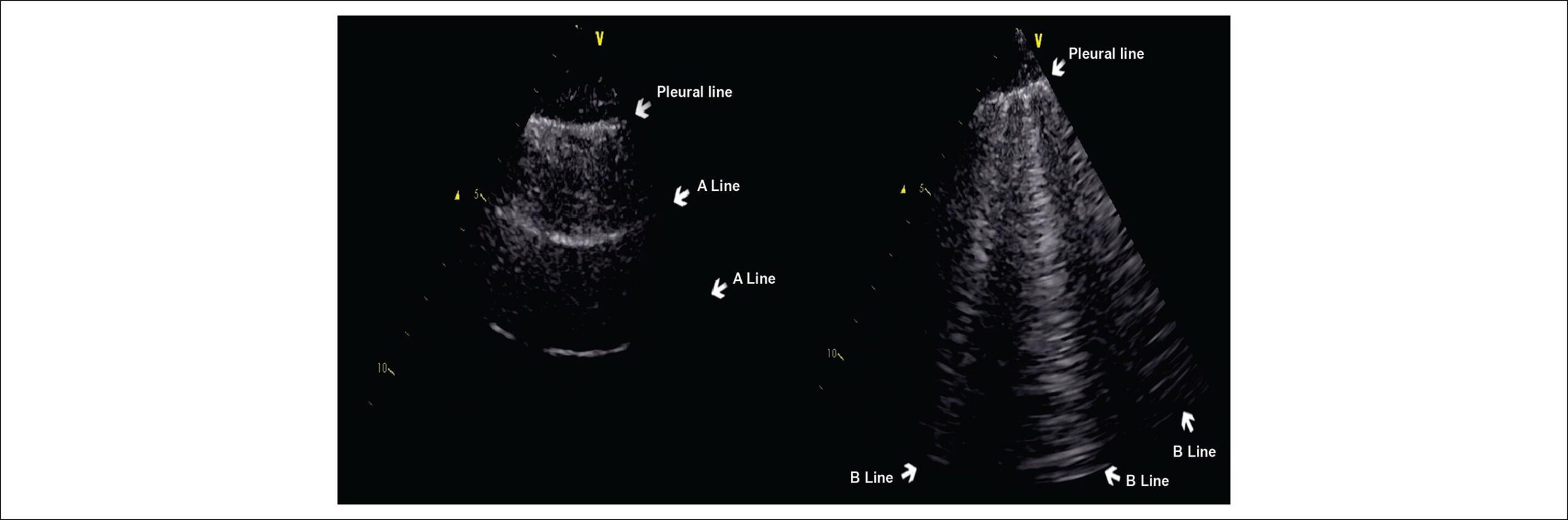
The bedside assessment of pulmonary congestion is undergoing conceptual consolidation: lung ultrasound (LUS) has evolved from being merely an “adjunct” tool to assuming a central role in the identification of extravascular fluid and aeration patterns, with important diagnostic, prognostic, and therapeutic implications. The article by Donmez et al. directly discusses the role of LUS in the assessment of pulmonary edema in children with congenital heart disease (CHD). In a retrospective cohort of 62 children, the study observed pulmonary edema in 53% by LUS and in 58% by radiography. Furthermore, the presence of edema by LUS was associated with clinically relevant variables in the clinical picture, such as recurrent respiratory infections, low weight, and elevated BNP levels. These findings reinforce that LUS adds objective and reproducible information to the care of children, in a context where clinical examination can be hampered by low cooperation and bedside radiography may have reduced accuracy due to positioning limitations and technical quality.

In adults with heart failure, the usefulness of LUS for pulmonary congestion is supported by several clinical studies demonstrating high diagnostic performance and correlation with outcomes. B-lines reflect increased extravascular fluid and, when diffusely present, reclassify the differential diagnosis of dyspnea. Comparison between LUS, biomarkers, and clinical assessment in an outpatient setting has shown that the burden of B-lines can detect subclinical congestion and anticipate the possibility of decompensation., In an acute setting, the systematic implementation of LUS improves diagnostic accuracy for decompensated heart failure, and prognostic studies associate congestion detected by LUS with worse short- and long-term outcomes. Reviews and perspective papers in cardiology have highlighted LUS as a rapid, reproducible, and low-cost diagnostic modality.

[…]

  • Congenital Heart Defects
  • Lung
  • Pediatrics
  • Point-of-Care Systems
  • Pulmonary Edema
  • Ultrasonography
Read Full Article
Creative Commons Attribution (CC BY) Creative Commons license
22/05/2026 DOI: 10.36660/abc.20260163i Views 2
ABCIC
Editorial

The Use of POCUS in Daily Practice: Are We Really Ready to Use it Efficiently?

Angelo Antunes Salgado et al. Marcos Paulo Lacerda Bernardo

Research on congestion in patients with heart failure (HF) is crucial for clinical management and prognosis of the disease. Congestion (i.e., the accumulation of fluids in the body) is the main cause of hospital admission and readmission of patients with HF, and it is associated with increased mortality. Early detection and identification of the extent of congestion are essential for choosing the most appropriate therapy, preventing complications, and improving prognosis. Clinical signs of congestion may be insufficiently sensitive and specific for the diagnosis and management of patients with HF.

The point-of-care ultrasound (POCUS) is a diagnostic tool that allows immediate and dynamic visualization of ultrasound images. This tool can be used at the bedside to obtain additional clinical information for the physical examination of a specific disease or procedure. The practicality and speed with which images are obtained result in their high use at hospitals for critically ill patients. POCUS has been used in various medical specialties, especially in pulmonary and cardiac ultrasound.

[…]

  • Clinical Diagnosis
  • Heart Failure
  • Ultrasonography
Read Full Article
Creative Commons Attribution (CC BY) Creative Commons license
01/07/2025 DOI: 10.36660/abcimg.20250026i
Figure 1
ABCIC
Editorial

New Tools for Optimizing HF Management: The Role of POCUS in the Office

Luiz Claudio Danzmann et al. Elisa Kalil Soraya Abunader Kalil

A Reflection on Clinical and Subclinical Circulatory Congestion

The management of heart failure (HF) has improved over the past 20 years, mainly due to advances in diagnostic methods and circulatory decongestion strategies. Several management strategies have demonstrated efficacy in reducing symptoms, lowering rates of HF decompensation events, decreasing mortality indices, and improving surrogate markers of clinical events, such as left ventricular ejection fraction (LVEF). In HF management, the need for detecting and monitoring circulatory congestion is justified by its strong association with neurohormonal activation and systemic inflammation, which contribute to the progressive worsening of HF and, consequently, a higher incidence of clinical events.

But what is the evidence supporting decongestion strategies with diuretics in reducing hard outcomes? In the outpatient setting, the STRONG-HF clinical trial is a good example. Comparing intensive optimization strategy versus usual HF treatment after hospital discharge, the study demonstrated that the group with more frequent visits and more intensive management achieved an absolute risk reduction of 8.1% (15.2% vs. 23.3%, P=0.0021) in the combined outcome of all-cause mortality and/or HF hospitalization within 180 days of follow-up. The statistically significant reduction in clinical signs of congestion, along with a 27% decrease in natriuretic peptide (NP) levels after three months, provided proof of concept for the relationship between decongestion and clinical benefit. This evidence reinforces the need to detect subclinical congestion, especially during the vulnerable phase of HF, which occurs between one and three months after an HF hospitalization, a period in which most potentially preventable events take place.

[…]

  • Ambulatory Care
  • Heart Failure
  • Ultrasonography
Read Full Article
Creative Commons Attribution (CC BY) Creative Commons license
30/06/2025 DOI: 10.36660/abcimg.20250039i Views 1
My Approach to Point-of-Care Ultrasound for Dyspnea Assessment
ABCIC
Review Article

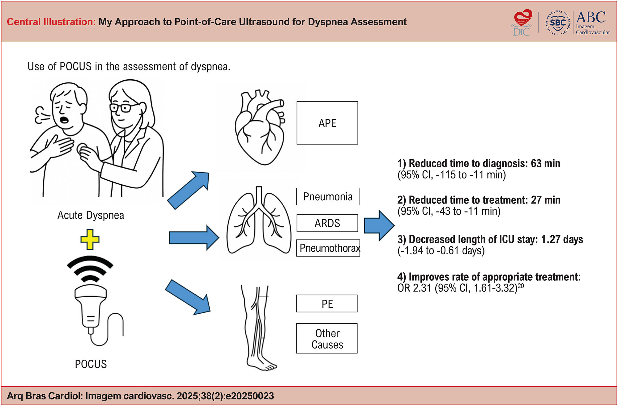
My Approach to Point-of-Care Ultrasound for Dyspnea Assessment

Adriana Brentegani et al. Fernando Arturo Effio Solis Milena de Paulis Marcelo Luiz Campos Vieira

Abstract

Dyspnea is a common and potentially serious symptom whose traditional diagnostic approach, based on physical examination and complementary tests, may present significant limitations. Point-of-care ultrasound (POCUS) has emerged as an effective tool for the rapid and accurate assessment of this symptom, both in outpatient settings and in emergency and critical care environments.

This article provides a practical, illustrated guide to the integrated application of POCUS in the evaluation of dyspnea, with an emphasis on distinguishing cardiac from pulmonary causes. It describes techniques for acquiring cardiac, pulmonary, and venous ultrasound images, highlights key sonographic findings, and outlines a step-by-step approach to clinical application. Lung POCUS has demonstrated high sensitivity in detecting differential diagnoses of dyspnea. Its integration with echocardiography and lower limb ultrasound further refines diagnosis by identifying specific clinical patterns such as acute pulmonary edema (APE), pulmonary embolism (PE), and acute respiratory distress syndrome (ARDS).

The structured use of POCUS represents a significant advancement in the modern physical exam, enabling faster and more accurate bedside clinical decision-making. Its adoption should be encouraged among cardiologists, intensivists, and healthcare professionals working in critical care settings.

  • Dyspnea
  • Echocardiography
  • Ultrasonography
Read Full Article
Creative Commons Attribution (CC BY) Creative Commons license
27/06/2025 DOI: 10.36660/abcimg.20250023i Views 1
Figure 1
ABCIC
Editorial

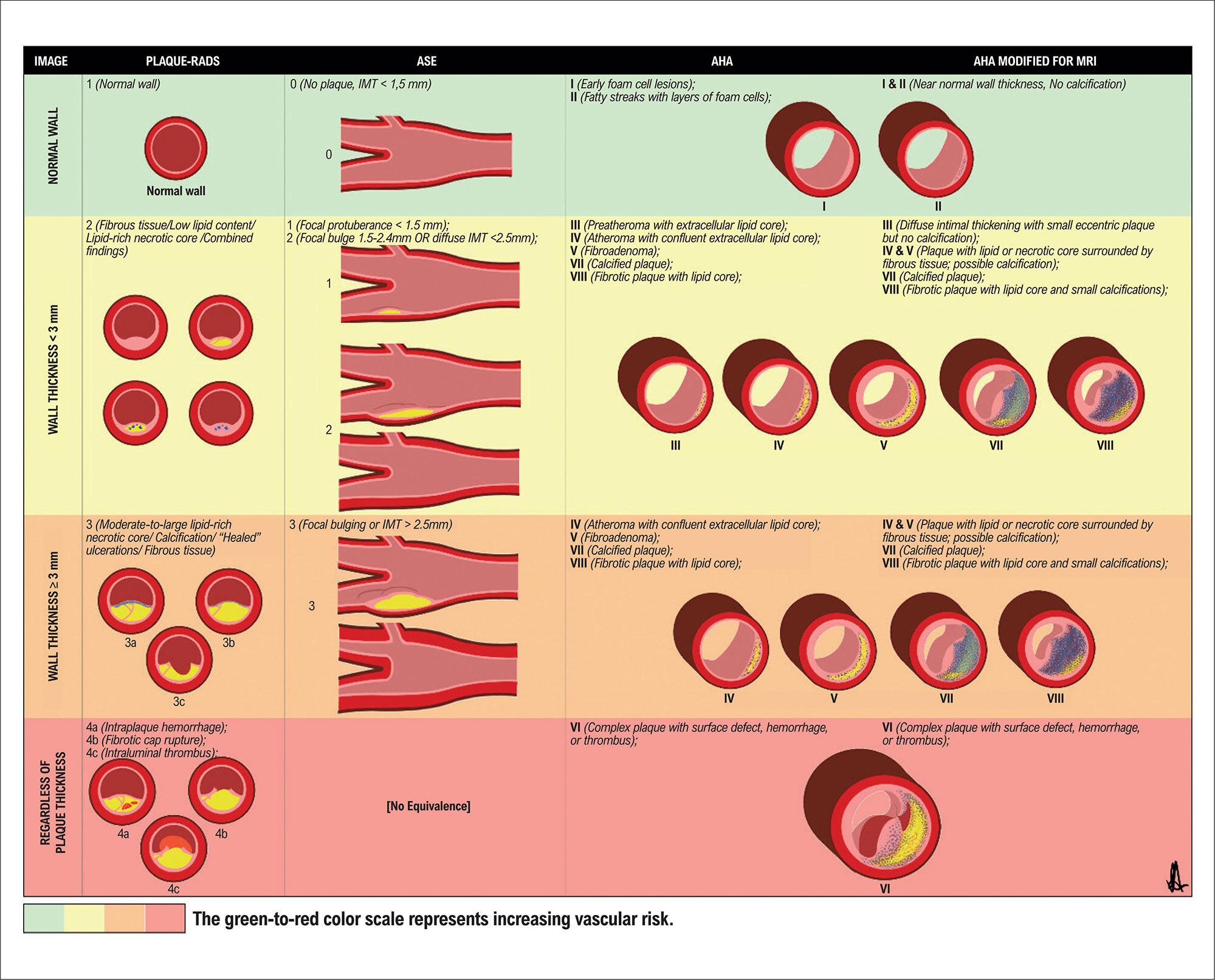
Top Vascular Ultrasound 2024: Carotid Plaque-RADS – A Novel Stroke Risk Classification System

Simone Nascimento dos Santos

Introduction

Many vascular ultrasound articles were published in 2024, but certainly the most discussed in Brazil this year was “Carotid Plaque-RADS – A Novel Stroke Risk Classification System” by Saba et al. in JACC: Cardiovascular Imaging. The project brought together 51 specialists in different vascular imaging modalities from 10 countries. This new proposal for categorizing carotid plaque attracted the attention of physicians from various specialties, such as cardiologists, neurologists, radiologists, ultrasound specialists, and vascular surgeons. In this section, we will discuss its main aspects, comparing them with previous classification systems.

[…]

  • Atherosclerosis
  • Atherosclerotic Plaque
  • Stroke
  • Ultrasonography
Read Full Article
Creative Commons Attribution (CC BY) Creative Commons license
09/12/2024 DOI: 10.36660/abcimg.20240110i
ABCHF
Review Article

Theoretical Concepts of Lung Ultrasound in the Diagnosis of Congestion

Willian Roberto Menegazzo et al. Marina Petersen Saadi Gustavo Paes Silvano Vinícius Leite Gonzalez Anderson Donelli da Silveira

Abstract

Accurate detection of pulmonary congestion remains a challenge in cardiology, despite advances in diagnostic methods. Traditional approaches, such as physical examination and chest radiography, have limitations in real-time assessment and dynamic monitoring of volume status. Lung ultrasound (LUS) has emerged as a promising tool, offering a non-invasive, bedside approach to detect pulmonary congestion with greater diagnostic accuracy, providing valuable information about volume status. In patients with heart failure, LUS has demonstrated high sensitivity and specificity in detecting pulmonary congestion, contributing to more accurate differential diagnosis and streamlining clinical decision-making. In the context of stress echocardiography, LUS improves risk stratification, identifying patients with a greater likelihood of adverse cardiovascular events. Beyond the context of heart failure, LUS has also been applied in other clinical scenarios, such as acute coronary syndrome and assessment of volume status in patients on dialysis. This expanded use reflects the potential of LUS as a versatile and valuable tool in different clinical settings, offering an earlier, more accurate, and more effective approach to cardiology care.

  • Heart Failure
  • Pulmonary Edema
  • Stress echocardiography
  • Ultrasonography
Read Full Article
Creative Commons Attribution (CC BY) Creative Commons license
28/08/2024 DOI: 10.36660/abchf.20240022i Views 1
ABCHF
Image

Cardiac Tumor in the Left Atrium

Henrique Iahnke Garbin et al. Matheus Piccoli Machado Klauberg Géris Mazzutti

A

[…]

  • Heart Atria
  • Heart Failure
  • Heart Neoplasms
  • Ultrasonography
Read Full Article
Creative Commons Attribution (CC BY) Creative Commons license
07/08/2024 DOI: 10.36660/abchf.20240004i
ABC
Editorial

Monitoring Systemic Congestion in Heart Failure: Is Clinical Evaluation Sufficient?

Silvia Moreira Ayub-Ferreira et al. Danielle Louvet Guazzelli

The management of acute decompensated heart failure (ADHF) remains a significant challenge in cardiology, particularly in preventing hospital readmissions. In this context, signs and symptoms related to congestion are among the most common causes of hospitalization for heart failure and subsequent readmissions.

In this edition of Arquivos Brasileiros de Cardiologia the authors present a cohort study that incorporates the use of the VExUS score to assess residual venous congestion before hospital discharge (VExUS Score at Hospital Discharge as a Predictor of Readmission in Patients with Acutely Decompensated Heart Failure: A Cohort Study). This study highlights the complexity of managing patients with heart failure and suggests a potentially useful tool to improve clinical outcomes.

[…]

  • Heart Failure
  • Hospitalization
  • Patient Readmission
  • Ultrasonography
Read Full Article
Creative Commons Attribution (CC BY) Creative Commons license
02/08/2024 DOI: 10.36660/abc.20240354i Views 1
Contribution of Lung Ultrasound and VExUS in the Diagnosis and Monitoring of Patients with Heart Failure
ABCHF
Review Article

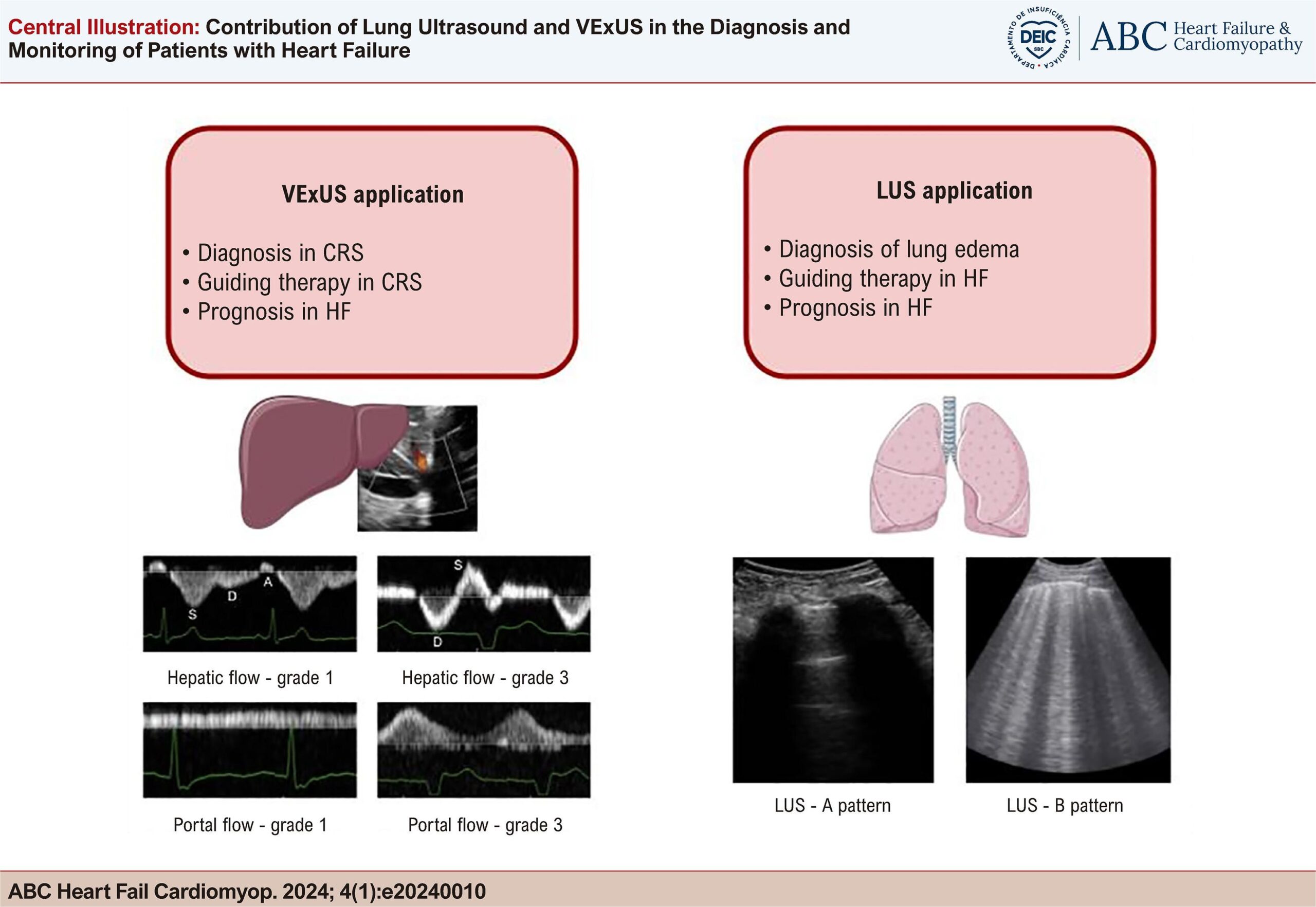
Contribution of Lung Ultrasound and VExUS in the Diagnosis and Monitoring of Patients with Heart Failure

Guilherme Heiden Telo et al. Marina Petersen Saadi Gustavo Paes Silvano Anderson Donelli da Silveira Andreia Biolo

Abstract

Despite advancements in the treatment of heart failure (HF), accurately assessing and monitoring fluid status and congestion remains a challenge. Traditional methods, including clinical evaluation, biomarker analysis, and imaging techniques, exhibit limitations in their capacity for real-time evaluation and monitoring of venous and pulmonary congestion. Lung ultrasound (LUS) detects B-lines and provides a rapid, sensitive assessment of pulmonary congestion, crucial for both acute and chronic HF management. Venous Excess Ultrasound Score (VExUS) provides a comprehensive assessment of systemic venous congestion by evaluating venous flows in hepatic, portal, and renal veins, alongside the inferior vena cava measurement. This review explores the integration of LUS and VExUS as innovative non-invasive, bedside technologies poised to enhance diagnostic accuracy, predict outcomes, and guide therapeutic decisions, potentially improving patient care. By shedding light on the underlying pathophysiological processes of HF, and offering insights into fluid dynamics, the synergistic application of LUS and VExUS, in conjunction with clinical examination, promises to significantly transform and personalize treatment strategies in both acute and chronic HF scenarios.

  • Heart Failure
  • Pulmonary Edema
  • Ultrasonography
Read Full Article
Creative Commons Attribution (CC BY) Creative Commons license
22/07/2024 DOI: 10.36660/abchf.20240010i Views 2
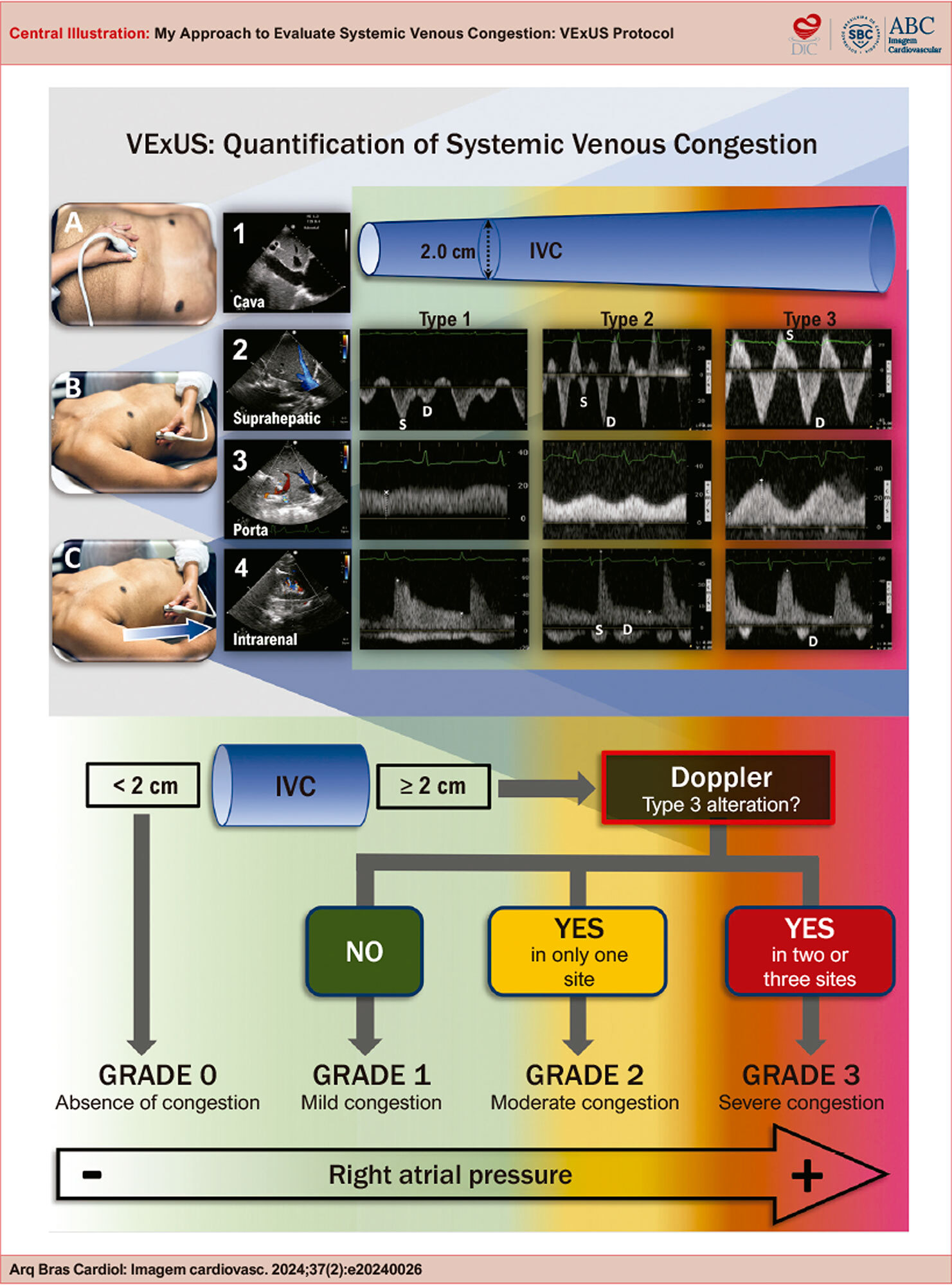
My Approach to Evaluate Systemic Venous Congestion: VExUS Protocol
ABCIC
Review Article

My Approach to Evaluate Systemic Venous Congestion: VExUS Protocol

Angelo Antunes Salgado et al. Marcos Paulo Lacerda Bernardo Fernando Melo Netto

Abstract

The use of non-invasive tools for analyzing systemic venous congestion in critically ill patients in the intensive care unit has been gaining increasing popularity for diagnosis, estimating the severity of congestion, and providing prognostic estimates. Patients with systemic congestion are more likely to develop renal dysfunction compared to those without congestion. In this review, the authors demonstrate how to perform systemic congestion analysis, its potential limitations, and its practical and objective applicability.

  • Heart Failure
  • Pathology
  • Ultrasonography
Read Full Article
Creative Commons Attribution (CC BY) Creative Commons license
16/05/2024 DOI: 10.36660/abcimg.20240026i Views 2
Figure 1
ABC
Image

Spatio-Temporal Image Correlation: Three-Dimensional Imaging for Fetal Cardiac Screening and Congenital Heart Disease Assessment

André Malho et al. Renato Silveira Ximenes Nathalie Jeanne Bravo-Valenzuela Edward Araujo Júnior

Heart defects are among the most common congenital malformations, occurring in 4 to 12 per 1,000 live births, depending on the population studied. Congenital heart disease (CHD) is an important cause of mortality related to birth defects. Therefore, prenatal diagnosis of CHD is crucial to improve the prognosis associated with this disease. In this scenario, the development of prenatal cardiac ultrasound screening programs serves as crucial tools that have increased the early detection of CHD. These programs achieve this by adding the ventricular outflow tract and superior mediastinum views (three vessels – 3V and 3V with trachea) to the standard four-chamber (4C) view when scanning the cardiac ultrasound.

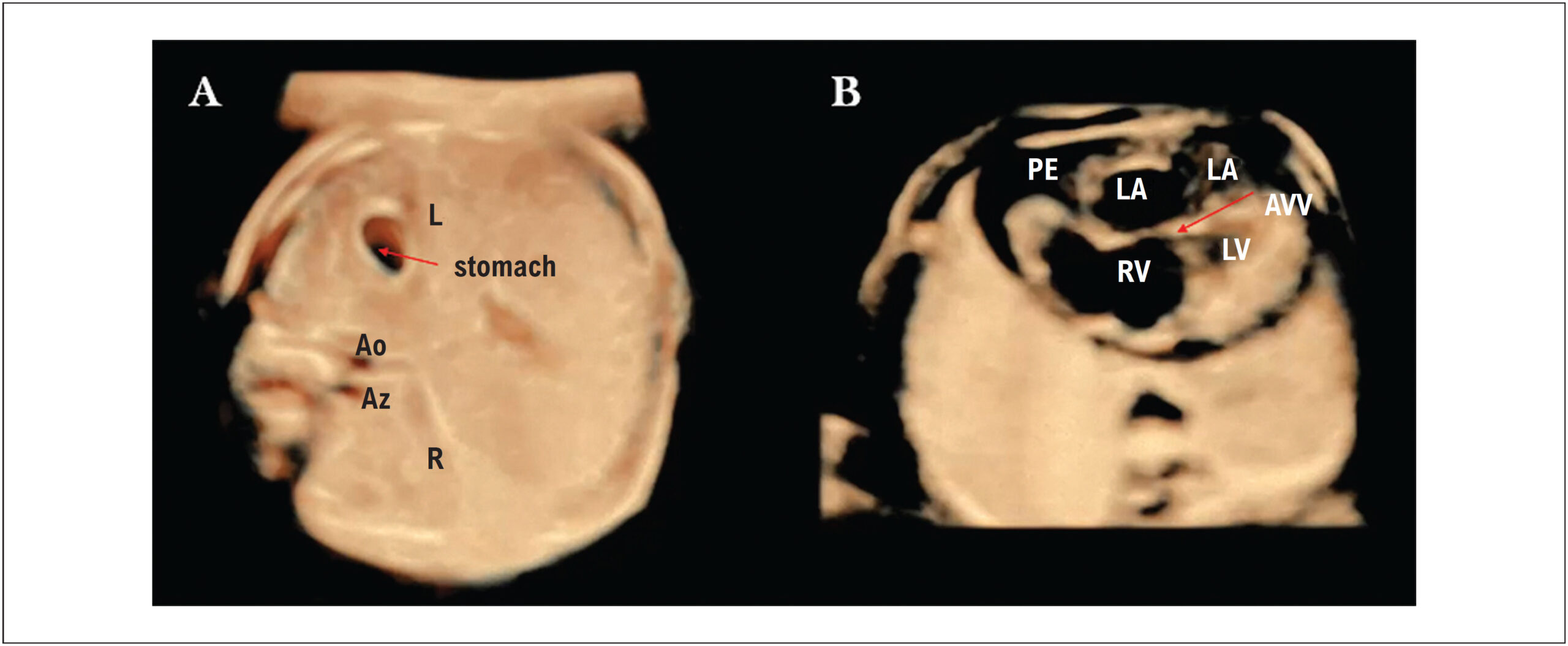
The development of advanced imaging technologies, such as three- and four-dimensional (3D/4D) ultrasound and Spatio-Temporal Image Correlation (STIC), has improved the quality of ultrasound images of the fetal heart with a positive impact on the diagnosis of cardiac malformations. The 4D-STIC technology allows the capture of thousands of cardiac images using a 3D volumetric transducer by a single 7.5 to 15-second slow-motion scan of the 4C view of the fetal heart. The acquired 3D/4D volumes contain a “block” of information of the complete cardiac cycle that can be stored for later off-line navigation of the fetal heart, allowing detailed analysis of cardiac anatomy by the sonographer, or sent via Internet link for analysis by experts in tertiary centers.

[…]

  • Congenital Heart Defects
  • fetal heart
  • Three-Dimensional Imaging
  • Ultrasonography
Read Full Article
Creative Commons Attribution (CC BY) Creative Commons license
29/04/2024 DOI: 10.36660/abc.20230580i Views 1
Figure 1
ABCIC
Brief Communication

Correlation of Telemedicine-guided POCUS Echocardiography and In Situ Data: Pilot Study in a Remote Area in Brazil

Juliane Rompkoski et al. Tarso Augusto Duenhas Accorsi Bruna Dayanne Reges Amaral Christian Barbosa de Freitas … Flavio Tocci Moreira Karen Francine Kohler Karine De Amicis Lima Renata Albaladejo Morbeck Carlos Henrique Sartorato Pedrotti

Abstract

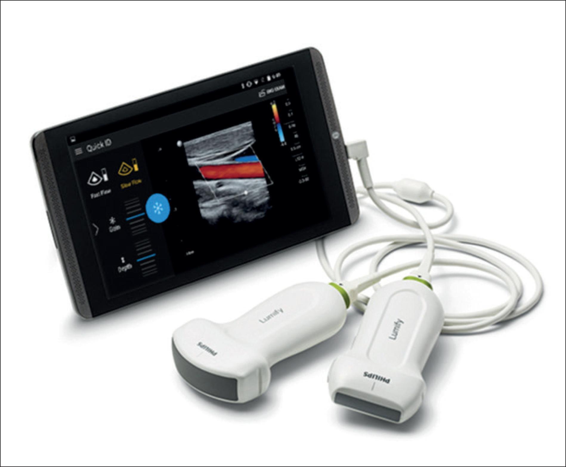
Cardiovascular diseases remain the leading cause of mortality in Brazil. Nevertheless, a significant portion of the Brazilian population still faces challenges in accessing specialized care and undergoing complementary exams, especially in remote areas. Currently, data transmission and remote specialized assessment are possible, but obtaining echocardiographic images still relies on a trained professional on-site. This pilot study aimed to assess the feasibility of Point-of-Care Ultrasound (POCUS) tele-echocardiography administered by a general physician with specialist guidance to improve accessibility to cardiovascular exams. Ten patients (mean age was 58.6+8 years, 50% female) from the North region participated, undergoing remote-guided tests using Philips Lumify™ ultrasound and Facetime for transmission. These patients had recent official echocardiographic exams. As part of the POCUS examination, seven echocardiographic views were obtained — parasternal long axis, parasternal short axis (PSAX), apical four chambers (A4C), apical five chambers (A5C), apical two chambers (A2C), subcostal (SC), and suprasternal (SSN). The procedure included linear measurements of cardiac chambers, and valve analysis was performed using color flow. The on-site examinations were carried out by a general physician who underwent brief training prior to the official exams. Despite encountering technical challenges, the study demonstrated the viability of acquiring echocardiographic images. Overall agreement in examination results was observed, except for left ventricular segmental contractility in two cases and the systolic diameter of the left ventricle. The approach, though performed by untrained professionals, showed promise in screening cardiovascular diseases in remote locations, focusing on ventricular function and valvular diseases. Nevertheless, further research is necessary to improve the quality of this process.

  • Echocardiography
  • Telecardiology
  • Telediagnostics
  • Telemedicine
  • Ultrasonography
Read Full Article
Creative Commons Attribution (CC BY) Creative Commons license
12/03/2024 DOI: 10.36660/abcimg.20230110i Views 1
ABC
Editorial

Point-of-Care in Clinical Practice: Consolidated Reality

Miguel José Francisco Neto et al. Edgar Bezerra de Lira Filho Marcos Roberto Gomes de Queiroz

Ultrasound and echocardiography with Doppler studies have represented, over the last few decades, highly noteworthy advances in medical practice in general. Particularly in internal medicine, they have promoted a true revolution in daily work and are currently considered a fundamental tool in clinical practice. This application of the method includes both urgent care and outpatient settings.,

In emergency scenarios, the initial establishment of the FAST (Focused Assessment with Sonography in Trauma) protocol for systematized trauma care made it possible to popularize the use of the method and served as a basis for multiple applications. In expanded application in emergency medicine, the sequence has included implementation of the RUSH (Rapid Ultrasound for Shock and Hypotension) and C.A.U.S.E. (Cardiac Arrest Ultrasound Exam) protocols,, progressively gaining ground as a multipurpose tool for use in medical education, research, and outpatient medical practice.

[…]

  • Echocardiography
  • emergency medicine
  • Medical Schools
  • Point-of-Care Systems
  • Ultrasonography
Read Full Article
Creative Commons Attribution (CC BY) Creative Commons license
04/03/2024 DOI: 10.36660/abc.20230688
Figure 1
ABCHF
Review Article

Point-of-Care Ultrasound in Acute Heart Failure: Basic Concepts for Clinical Practice

Leticia Kunst et al. Luiz Claudio Danzmann Keslly Krauspenhar Cuchinski João Ricardo Cambruzzi Zimmer

Introduction

Heart failure (HF) is a complex clinical syndrome that promotes a system of poor tissue perfusion in conditions of low cardiac output and elevated filling pressures, resulting in circulatory congestion and hypoperfusion. The exacerbation of this condition is responsible for 40% of consultations due to dyspnea in emergency rooms, and the main cause is worsening congestion due to poor medication adherence. In in-hospital clinical assessment, the most prevalent HF profiles are B (warm and wet) and C (cold and wet), both of which are characterized by circulatory congestion.

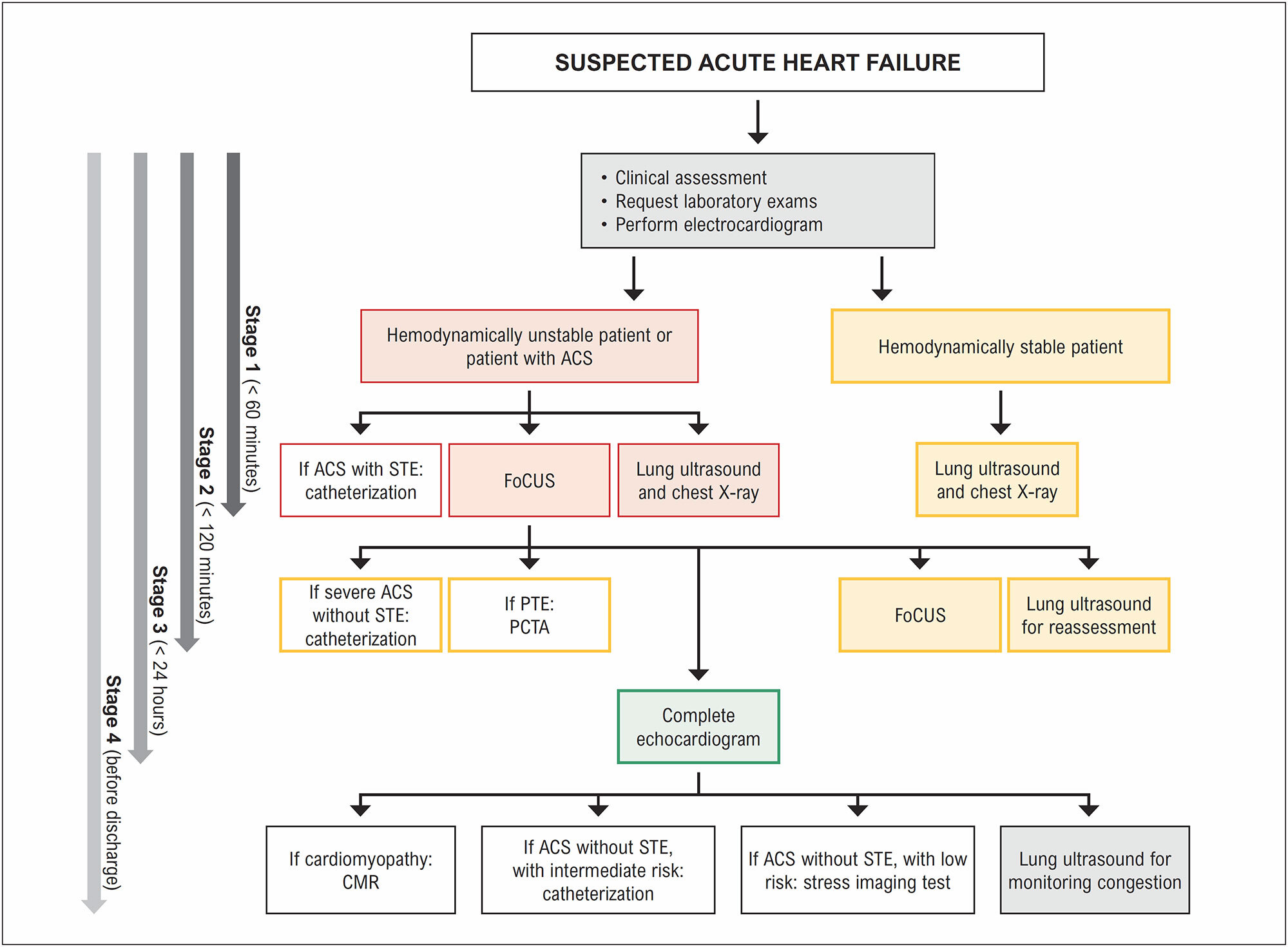
Although it causes great demand in the emergency room, it is sometimes difficult to identify signs of hypervolemia in acute decompensated HF in the context of differential diagnosis with other determining syndromes of dyspnea and hypoperfusion. Clinical findings are fundamental for the initial investigation, but objective and rapid confirmation of diagnosis reduces door-to-furosemide time, which is directly related to in-hospital mortality. Natriuretic peptides are tools with high negative predictive value for congestion; however, moderately elevated serum levels represent a diagnostic gray area, as the increase in this marker is related to several diseases that are independent of HF. The use of bedside point-of-care ultrasound (POCUS) has emerged as an excellent tool for comprehensive assessment of this condition, with analysis of cardiac structure and function, the pulmonary interstitium, and some venous vessels. It also offers a rapid, non-invasive, and low-cost opportunity, with greater accuracy than conventional assessment methods for detecting circulatory congestion.

[…]

  • Heart Failure
  • Practice Guideline
  • Ultrasonography
Read Full Article
Creative Commons Attribution (CC BY) Creative Commons license
31/12/2023 DOI: 10.36660/abchf.20230073 Views 1
Figure 1
ABCIC
Original Article

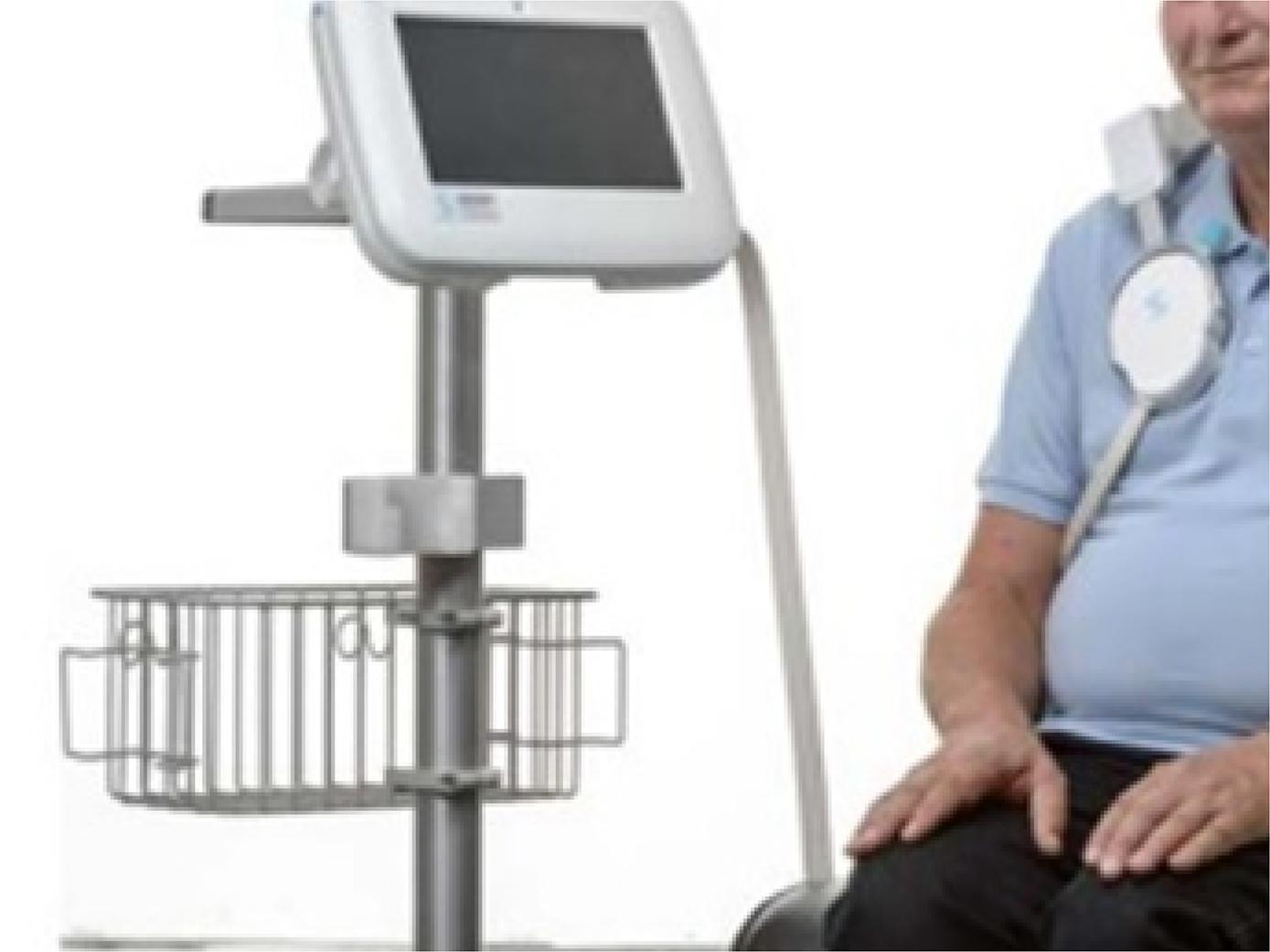
Pulmonary Congestion in Heart Failure With Reduced Ejection Fraction: Comparison Between Lung Ultrasound and Remote Dielectric Sensing

Maria Estefânia Bosco Otto et al. Vanessa Andreoli Esmanhoto Edileide de Barros Correia Ana Cristina de Souza Murta … Larissa Ventura Ribeiro Bruscky Andrea de Andrade Vilela Antonio Tito Paladino Filho Jorge Eduardo Assef

Abstract

Backgound

Outpatient assessment of pulmonary congestion in patients with heart failure with reduced ejection fraction (HFREF) can minimize hospitalizations due to decompensation and optimize the use of diuretics.

Objective

To compare the remote dielectric sensor (REDS), a validated device for detecting pulmonary extravascular fluid, with clinical parameters, transthoracic echocardiogram (TTE) and lung ultrasound (ULSP).

Methods

We included 38 patients from heart failure clinic (63±12 years; 21 men). All were submitted within 24 hours to clinical evaluation with description of paroxysmal nocturnal dyspnea, leg edema (LE), presence of dizziness; laboratory evaluation of NT-ProBNP; evaluation by REDS and TTE with analysis of parameters of systemic congestion by the inferior vena cava evaluation, function of the right ventricle and of pulmonary congestion, such as evaluation of filling pressures by average of E/e’ and indexed left atrial volume. The ULSP was performed using the 8 points anterior quadrants protocol, counting B lines in one respiratory cycle.

Results

22 patients had REDS ≥ 35% (indicative of pulmonary congestion) and 16 patients REDS<35%. Clinical and echocardiographic parameters were compared with REDS ≥ 35%. In the multivariate analysis, the variables body surface area, B lines and LE were associated with REDS ≥ 35%. NT-ProBNP was similar and elevated in both groups.

Conclusions

Outpatient monitoring of HFREF for volume control can be sensitized by the presence of B lines on the ULSP with good correlation to REDS ≥ 35%. NT pro BNP was not able to differentiate patients with congestion detected by REDS.

  • Heart Failure
  • Lung
  • Ultrasonography
Read Full Article
Creative Commons Attribution (CC BY) Creative Commons license
24/05/2023 DOI: 10.36660/abcimg.20230027i
My Approach to Lung Ultrasound to Evaluate Extravascular Lung Water
ABCIC
My Approach To

My Approach to Lung Ultrasound to Evaluate Extravascular Lung Water

Maria Estefânia Bosco Otto et al. Vanessa Andreoli Esmanhoto

Introduction

A clinical history, physical examination, and focused ultrasound can accelerate the appropriate diagnosis and treatment of patients with progressive or acute dyspnea. Extravascular lung water (EVLW) is easily detected with lung ultrasound (LUS). Pivetta et al. reported excellent accuracy detecting EVLW by clinical evaluation and by the presence of “B” lines on LUS heigher than B natriuretic peptide dosage.

The usefulness of LUS for intensive care unit patients was described in the 1980s, later than that of other ultrasound modalities. LUS is useful not only for diagnosing EVLW but also for risk stratification in myocardial infarction and acute congestive heart failure (CHF).

[…]

  • Dyspneia
  • Lung
  • Ultrasonography
Read Full Article
Creative Commons Attribution (CC BY) Creative Commons license
17/08/2022 DOI: 10.47593/2675-312X/20223503ecom22
ABCIC
Original Article

Association between Insufficiency of Anterior Perforator Vein of the Knee and Great Saphenous Vein Insufficiency in Patients with Primary Varicose Veins of the Lower Limbs

Marcio Vinícius Lins Barros et al. Gabriela Silva Lauar Jéssica Monteiro Machado Maria Cláudia Freitas Silva Taynara Andrade Capanema

Abstract

Background

The accurate identification of reflux points in the study of primary varicose veins of the lower limbs is important in the therapeutic approach of these patients.

Objective

To evaluate the association between insufficiency of the anterior perforator vein of the knee and great saphenous vein insufficiency in patients with primary varicose veins of the lower limbs.

Methods

The study included 886 patients, corresponding to 1,772 lower limbs, in patients older than 18, of both sexes, undergoing superficial venous mapping using vascular ultrasound.

Results

The mean age of the patients was 46.1 ± 14.5 and 81.7% were females. Great saphenous vein insufficiency was found in 38.8% of the cases. Insufficiency of the anterior perforator vein of the knee was found in 146 lower limbs (8.2%), with a mean diameter of 1.7 ± 0.15 mm, prevailing in the infrapatellar area (86.4%). The anterior tributary vein of the great saphenous vein originated leg reflux in 34%, with a direct relation of this tributary vein with the anterior perforator vein of the knee in 79.4%. An association between the presence of anterior perforator vein of the knee and great saphenous vein insufficiency (p = 0.0001) and male gender (p = 0.001).

Conclusion

There was an association between insufficiency of the anterior perforator vein of the knee and great saphenous vein insufficiency in patients with primary varicose veins of the lower limbs at vascular ultrasound, and the accurate identification of this perforator vein may be important in the therapeutic approach of these patients. (Arq Bras Cardiol: Imagem cardiovasc. 2019;32(1):14-18)

  • Saphenous Vein/physiopathology
  • Ultrasonography
  • Varicose Veins/diagnostic Imaging Lower Extremity/surgery
  • Venous Insufficiency/surgery
Read Full Article
Creative Commons Attribution (CC BY) Creative Commons license
20/01/2022 DOI: 10.5935/2318-8219.20190006 Views 1
ABCIC
Case Report

Isolated Infundibular Pulmonary Stenosis with Infective Endocarditis: a Rare Ecocardiographic Finding

Leonardo de Souza Carneiro et al. Nelson Lopes Evangelista Carlos José Mota de Lima Maria Suely Bezerra Diógenes … Ana Carolina Brito de Alcantara Maria Luiza Calixto de Lima

[…]

  • Endocarditis
  • Stenosis, Pulmonary
  • Ultrasonography
Read Full Article
Creative Commons Attribution (CC BY) Creative Commons license
14/01/2022 DOI: 10.5935/2318-8219.20190040 Views 1
Figure 1
ABCIC
Systematic Review

Inferior Vena Cava Ultrasound for Assessing Volume Status and Fluid Responsiveness in Critically ill Patients: A Systematic Review

Djalma Gomes Xavier Filho et al. Akeme Laissa Novais Coutinho Romero Henrique de Almeida Barbosa Matheus Rodrigues Lopes Adirlene Pontes de Oliveira Tenório

Abstract

Background:

The assessment of intravascular volume and fluid responsiveness is challenging in the management of critically ill patients. Diagnostic methods of hemodynamic monitoring must be safe, reproducible, and practical.

Objective:

To describe the applicability of ultrasound indices of the inferior vena cava in the assessment of intravascular volume and prediction of fluid responsiveness in critically ill patients.

Method:

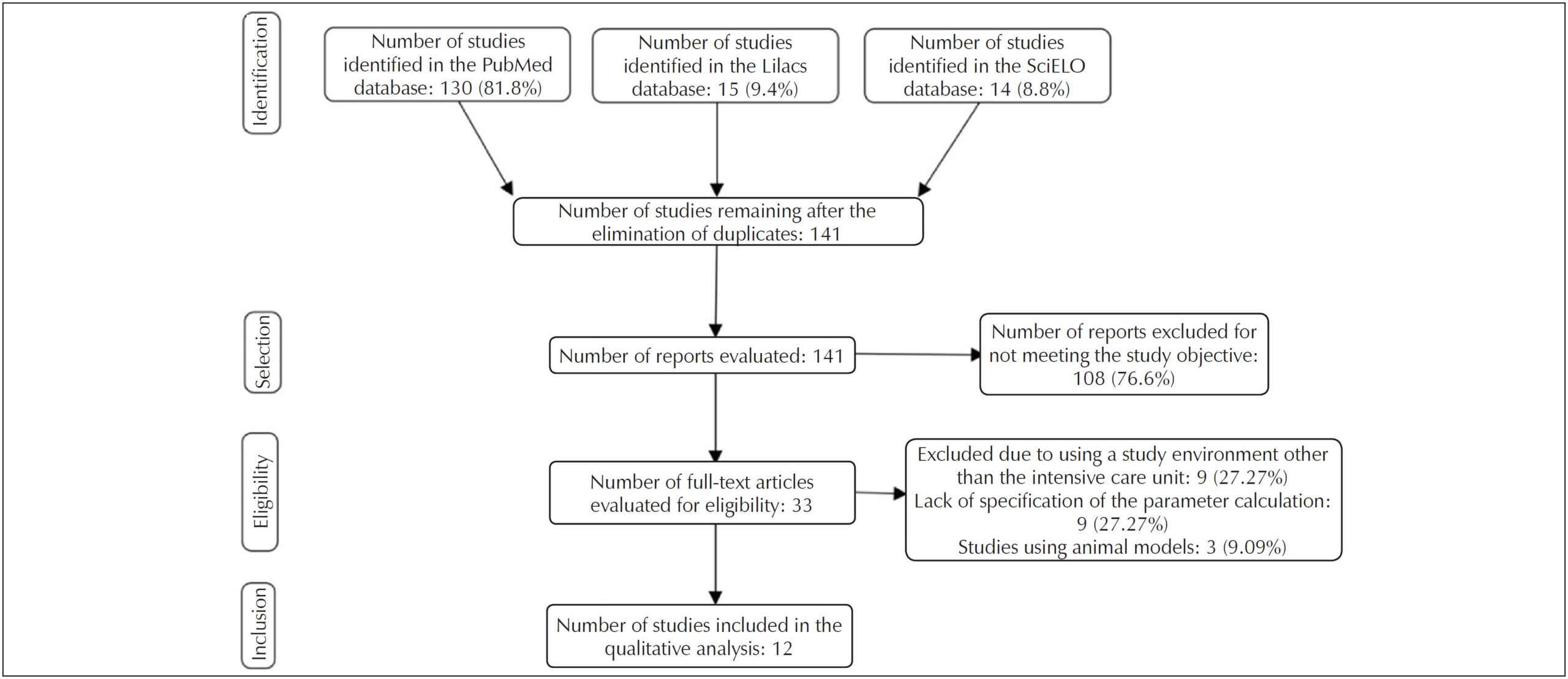
A systematic review performed of the PubMed®, Latin American and Caribbean Health Sciences Literature, and Scientific Electronic Library Online databases for articles published in the previous five years. The descriptors used were “inferior vena cava,” “ultrasonography,” “fluid responsiveness,” and “volume status.”

Results:

The search identified 13 relevant articles. The collapsibility index of the inferior vena cava was 25%–50% as the cutoff point for defining hypovolemia and showed applicability in predicting fluid responsiveness in patients breathing spontaneously with cutoff points of 25%–57%. In mechanical ventilation scenarios, the distensibility index of the inferior vena cava was more effective at predicting fluid responsiveness than the other measurements with variation of 10.2%–20.5%. The inferior vena cava/aorta diameter index was especially useful in the pediatric population for defining intravascular volume; however, in adults, there were many divergences in its applicability.

Conclusion:

The assessment of intravascular volume and fluid responsiveness through ultrasound indices of the inferior vena cava is applicable and safe for diagnosing and monitoring hemodynamic instability. However, studies of value standardization are necessary due to divergences in the cutoff points used in each index.

  • Hemodynamics
  • Inferior vena cava
  • Intensive Care Unit
  • Organism fluid status
  • Ultrasonography
Read Full Article
Creative Commons Attribution (CC BY) Creative Commons license
26/11/2021 DOI: 10.47593/2675-312X/20213403eabc193 Views 2
Figure 1
ABC
Original Article

Genetic Testing and Pregnancy Outcome Analysis of 362 Fetuses with Congenital Heart Disease Identified by Prenatal Ultrasound

Shiyu Luo et al. Dahua Meng Qifei Li Xuehua Hu … Yuhua Chen Chun He Bobo Xie Shangyang She Yingfeng Li Chunyun Fu

Abstract

Background:

Congenital heart defects (CHD), as the most common congenital anomaly, have been reported to be associated with chromosomal abnormalities. Currently, patients with CHD are routinely offered karyotyping and chromosomal microarray (CMA) testing, but the genotype-phenotype relationship has not yet been fully established.

Objective:

To determine the type and frequency of chromosomal abnormalities in fetuses with CHD and to analyze pregnancy outcomes of fetuses with heart abnormalities caused by different genetic factors.

Methods:

A total of 362 cases of CHD were enrolled from 2009 to 2016. Detailed ultrasound and laboratory examinations, including karyotyping and CMA, were performed. Outcome was obtained from discharge summaries.

Results:

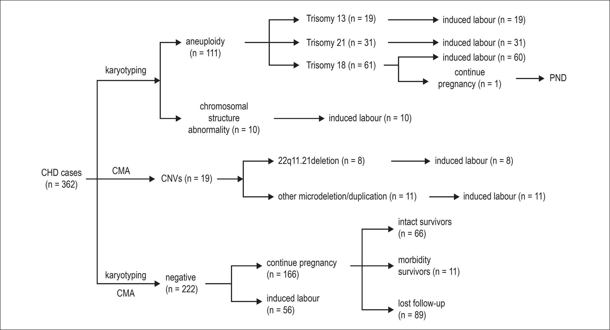
Of the 362 fetuses, 220 were found with an isolated CHD, and 142 had CHD with extracardiac anomaly. Among these 362 fetuses, 140 were identified with a genetic cause, including 111 cases with aneuploidy, 10 cases with abnormality of chromosomal structure by karyotyping and 19 cases with pathogenic or likely pathogenic copy-number variations (CNVs) by CMA. The detection rate is close to 38.7%. Only one (identified as trisomy 18 syndrome) in 140 positive cases resulted in perinatal death, with the others being induced. The remaining 222 cases had negative results for both genetic testing and of these cases, 56 resulted in induced labor, and 77 had natural childbirth or caesarean births. The pregnancy outcome of the remaining 89 cases was uncertain.

Conclusions:

Karyotyping and CMA are effective and accurate prenatal genetic techniques for identifying fetal chromosomal abnormalities associated with cardiac defects, and this can assist clinical doctors to perform appropriate genetic counselling with regard to the etiology and outcome of CHD.

  • Chromosome Disorders
  • Fetus
  • Heart Defects, Congenital
  • Pregnancy
  • Spectral Karyotyping
  • Ultrasonography
Read Full Article
Creative Commons Attribution (CC BY) Creative Commons license
20/08/2018 DOI: 10.5935/abc.20180144 Views 1
Figure 1
ABC
Viewpoint

Lung Ultrasound: The Cardiologists' New Friend

Marcelo Haertel Miglioranza et al. Antonio Carlos Sobral Sousa Caroline de Souza Costa Araujo Marcos Antonio Almeida-Santos Luna Gargani

The role of pulmonary congestion in heart failure and the limits of the traditional clinical examination

Pulmonary congestion, such as low cardiac output, is a preponderant element in patients with heart failure (HF), which is considered an important cause of hospital admissions and death., Thus, the identification of pulmonary extravascular fluid in patients with HF can be used as an aid in strategies to optimize clinical therapy.

Traditionally, assessment of pulmonary congestion has been based on the patient’s clinical status and physical examination. However, this evaluation presents limitations even for skilled professionals, showing high specificity, but low sensitivity for the detection of pulmonary congestion., Thus, cases of decompensation are often recognized at a very late stage of clinical congestion, so frequent hospitalizations are not avoided. In the cascade of congestion, the clinical manifestation represents a final stage, different from hemodynamic congestion (increase in left ventricular filling pressure), which is pulmonary and systemic. Pulmonary congestion corresponds specifically to the presence of extravascular pulmonary fluid, which can be evaluated by lung ultrasound.

[…]

  • Diagnosis, Differential
  • Heart Failure
  • Pulmonary Edema
  • Ultrasonography
Read Full Article
Creative Commons Attribution (CC BY) Creative Commons license
01/12/2017 DOI: 10.5935/abc.20170169
Figure 1
ABC
Original Article

Assessment of Intima-Media Thickness in Healthy Children Aged 1 to 15 Years

Liz Andréa Villela Baroncini et al. Lucimary de Castro Sylvestre Roberto Pecoits Filho

Abstract

Background:

Carotid intima-media thickness (CIMT) has been shown to be increased in children and adolescents with traditional cardiovascular risk factors such as obesity, hypertension, and chronic kidney disease, compared with those of healthy children.

Objective:

To assess the influence of sex, age and body mass index (BMI) on the CIMT in healthy children and adolescents aged 1 to 15 years.

Methods:

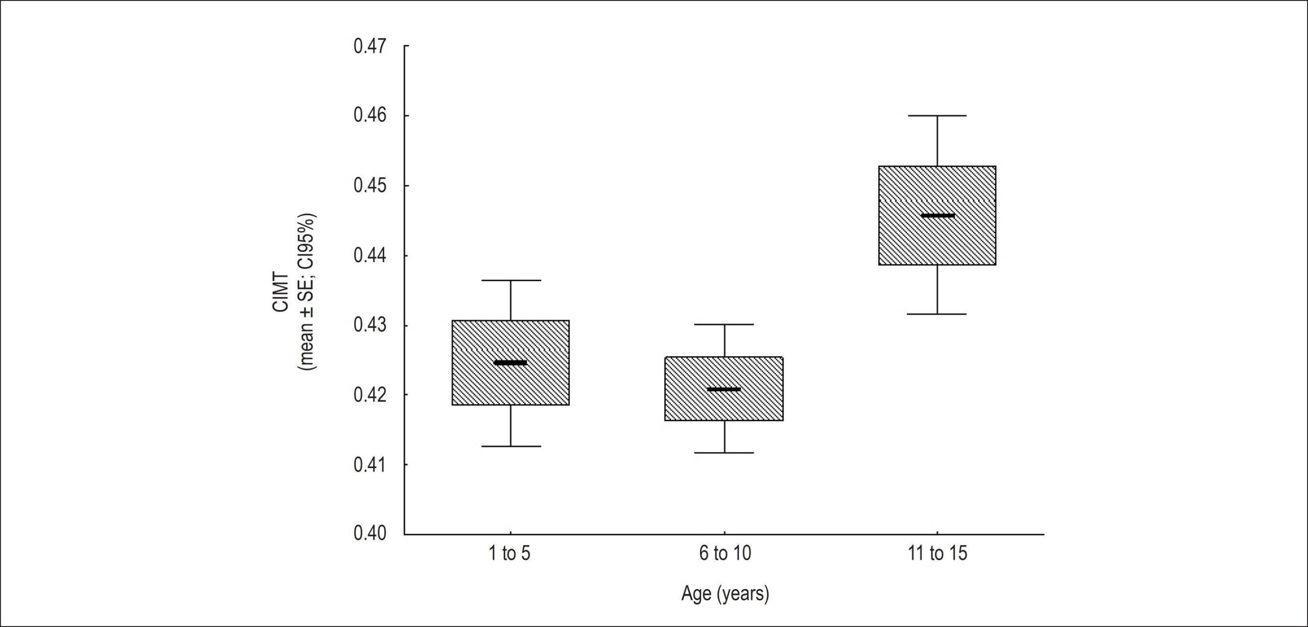
A total of 280 healthy children and adolescents (males, n=175; mean age, 7.49±3.57 years; mean BMI, 17.94±4.1 kg/m2) were screened for CIMT assessment. They were divided into 3 groups according to age: GI, 1 to 5 years [n=93 (33.2%); males, 57; mean BMI, 16±3 kg/m2]; GII, 6 to 10 years [n=127 (45.4%); males, 78; mean BMI, 17.9±3.7 kg/m2], and GIII, 11 to 15 years [n=60 (21.4%); males, 40; mean BMI, 20.9±4.5 kg/m2].

Results:

There was no significant difference in CIMT values between male and female children and adolescents (0.43±0.06 mm vs. 0.42±0.05 mm, respectively; p=0.243). CIMT correlated with BMI neither in the total population nor in the 3 age groups according to Pearson correlation coefficient. Subjects aged 11 to 15 years had the highest CIMT values (GI vs. GII, p=0.615; GI vs. GIII, p=0.02; GII vs. GIII, p=0.004).

Conclusions:

CIMT is constant in healthy children younger than 10 years, regardless of sex or BMI. CIMT increases after the age of 10 years.

  • Atherosclerosis
  • Carotid artery
  • Carotid Intima-Media Thickness
  • Child
  • Ultrasonography
Read Full Article
Creative Commons Attribution (CC BY) Creative Commons license
08/03/2016 DOI: 10.5935/abc.20160030
Figure 1
ABC
Original Article

A Novel Algorithm to Quantify Coronary Remodeling Using Inferred Normal Dimensions

Breno A. A. Falcão et al. João Luiz A. A. Falcão Gustavo R. Morais Rafael C. Silva … Augusto C. Lopes Paulo R. Soares José Mariani Jr Roberto Kalil-Filho Elazer R. Edelman Pedro A. Lemos

Background:

Vascular remodeling, the dynamic dimensional change in face of stress, can assume different directions as well as magnitudes in atherosclerotic disease. Classical measurements rely on reference to segments at a distance, risking inappropriate comparison between dislike vessel portions.

Objective:

to explore a new method for quantifying vessel remodeling, based on the comparison between a given target segment and its inferred normal dimensions.

Methods:

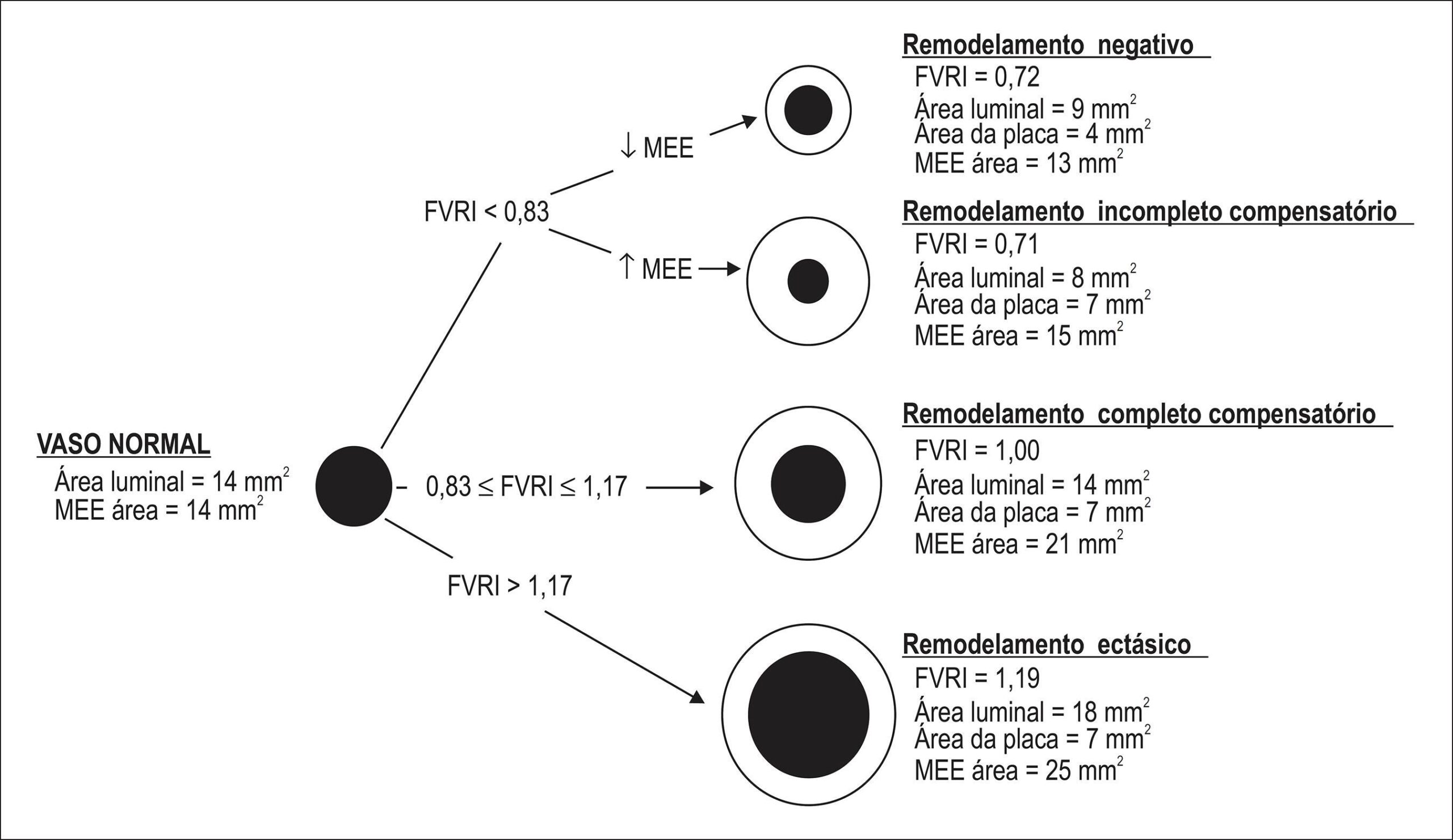
Geometric parameters and plaque composition were determined in 67 patients using three-vessel intravascular ultrasound with virtual histology (IVUS-VH). Coronary vessel remodeling at cross-section (n = 27.639) and lesion (n = 618) levels was assessed using classical metrics and a novel analytic algorithm based on the fractional vessel remodeling index (FVRI), which quantifies the total change in arterial wall dimensions related to the estimated normal dimension of the vessel. A prediction model was built to estimate the normal dimension of the vessel for calculation of FVRI.

Results:

According to the new algorithm, “Ectatic” remodeling pattern was least common, “Complete compensatory” remodeling was present in approximately half of the instances, and “Negative” and “Incomplete compensatory” remodeling types were detected in the remaining. Compared to a traditional diagnostic scheme, FVRI-based classification seemed to better discriminate plaque composition by IVUS-VH.

Conclusion:

Quantitative assessment of coronary remodeling using target segment dimensions offers a promising approach to evaluate the vessel response to plaque growth/regression.

  • Atherosclerosis / physiopathology
  • Coronary Artery Diseases
  • Neovascularization, Pathologic
  • Ultrasonography
  • Vascular Remodeling
Read Full Article
Creative Commons Attribution (CC BY) Creative Commons license
18/08/2015 DOI: 10.5935/abc.20150098 Views 4
Association between ankle-brachial index and carotid atherosclerotic disease
ABC
Artigo Original

Association between ankle-brachial index and carotid atherosclerotic disease

Augusto Cezar Lacerda Brasileiro et al. Dinaldo Cavalcanti de Oliveira Edgar Guimarães Victor Danielle A. G. Cavalcanti Oliveira Laecio Leitão Batista

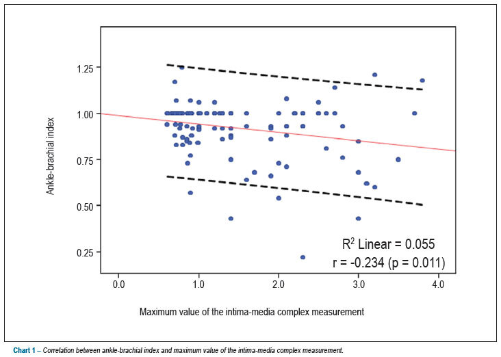
BACKGROUND: The association between the ankle brachial index (ABI) and the measurement of intimal medial thickness (IMT) has not been fully studied. OBJECTIVE: We aimed to evaluate whether the prevalence of carotid atherosclerosis was higher in patients with ABI 0.9. METHODS: From January 2011 to December 2011, 118 patients (48 men and 70 women) were enrolled. ABI and IMT Measurements were performed in all patients. Patients were divided in Group 1 (ABI 0.9) according to ABI values. Mann-Whitney, Chi-square and Fischer tests were used for comparison among the groups. Pearson’s correlation was used to assess correlation between ABI and IMT. RESULTS: The prevalence of ABI 1.5 mm was 34.7 %. Clinical characteristics were similar between groups 1 and 2: mean age (64 ± 9 vs. 62 ± 7.2 years, p = 0.1), male gender (40% vs. 41%, p = 0.9), hypertension (74% vs. 59%, p = 0.1), diabetes mellitus (54% vs. 35%, p = 0.051), dyslipidemia (26% vs. 24%, p = 0.8), smoking (57% vs. 65%, p = 0.4). The prevalence of carotid atherosclerosis was higher in group 1 (48.6% vs. 28.9%, p = 0.04). Pearson’s correlation between ABI and IMT was -0.235, with a p value = 0.01. CONCLUSION: Patients with ABI < 0.9 showed a higher prevalence of carotid atherosclerosis. There was a negative correlation between ABI and IMT.

  • Ankle Brachial Index
  • Carotid Artery Diseases
  • Ultrasonography
Read Full Article
Creative Commons Attribution (CC BY) Creative Commons license
09/04/2013 DOI: 10.5935/abc.20130057

Could not load more articles. Please try again.

How to cite this article

EndNote RIS

Electronic Document Format (Vancouver)

Otto MEB. The New Frontier of Point-of-Care Ultrasound: Lung Ultrasound in Congenital Heart Disease. Arquivos Brasileiros de Cardiologia 2026;123(3):e20260163.

Electronic Document Format (ABNT)

Otto, Maria Estefânia Bosco. The New Frontier of Point-of-Care Ultrasound: Lung Ultrasound in Congenital Heart Disease. Arquivos Brasileiros de Cardiologia, v. 123, n. 3, e20260163, May. 2026.

Electronic Document Format (APA)

Otto, M. E. B. (2026). The New Frontier of Point-of-Care Ultrasound: Lung Ultrasound in Congenital Heart Disease. Arquivos Brasileiros de Cardiologia, 123(3), e20260163.

Electronic Document Format (ISO)

Otto, Maria Estefânia Bosco. The New Frontier of Point-of-Care Ultrasound: Lung Ultrasound in Congenital Heart Disease. Arquivos Brasileiros de Cardiologia [online]. 2026, vol. 123, n. 3, [cited 2026-10-14], e20260163. Available from: <https://journals.cardiol.br/en/article/the-new-frontier-of-point-of-care-ultrasound-lung-ultrasound-in-congenital-heart-disease/>. ISSN 0066-782X

EndNote RIS

Electronic Document Format (Vancouver)

Salgado AA, Bernardo MPL. The Use of POCUS in Daily Practice: Are We Really Ready to Use it Efficiently?. ABC Imagem Cardiovascular 2025;38(2):e20250026.

Electronic Document Format (ABNT)

Salgado, Angelo Antunes; Bernardo, Marcos Paulo Lacerda. The Use of POCUS in Daily Practice: Are We Really Ready to Use it Efficiently?. ABC Imagem Cardiovascular, v. 38, n. 2, e20250026, Jun. 2025.

Electronic Document Format (APA)

Salgado, A. A., & Bernardo, M. P. L. (2025). The Use of POCUS in Daily Practice: Are We Really Ready to Use it Efficiently?. ABC Imagem Cardiovascular, 38(2), e20250026.

Electronic Document Format (ISO)

Salgado, Angelo Antunes and Bernardo, Marcos Paulo Lacerda. The Use of POCUS in Daily Practice: Are We Really Ready to Use it Efficiently?. ABC Imagem Cardiovascular [online]. 2025, vol. 38, n. 2, [cited 2026-10-14], e20250026. Available from: <https://journals.cardiol.br/en/article/the-use-of-pocus-in-daily-practice-are-we-really-ready-to-use-it-efficiently/>. ISSN 2318-8219

EndNote RIS

Electronic Document Format (Vancouver)

Danzmann LC, Kalil E, Kalil SA. New Tools for Optimizing HF Management: The Role of POCUS in the Office. ABC Imagem Cardiovascular 2025;38(2):e20250039.

Electronic Document Format (ABNT)

Danzmann, Luiz Claudio; Kalil, Elisa; Kalil, Soraya Abunader. New Tools for Optimizing HF Management: The Role of POCUS in the Office. ABC Imagem Cardiovascular, v. 38, n. 2, e20250039, Jun. 2025.

Electronic Document Format (APA)

Danzmann, L. C., Kalil, E., & Kalil, S. A. (2025). New Tools for Optimizing HF Management: The Role of POCUS in the Office. ABC Imagem Cardiovascular, 38(2), e20250039.

Electronic Document Format (ISO)

Danzmann, Luiz Claudio and Kalil, Elisa and Kalil, Soraya Abunader. New Tools for Optimizing HF Management: The Role of POCUS in the Office. ABC Imagem Cardiovascular [online]. 2025, vol. 38, n. 2, [cited 2026-10-14], e20250039. Available from: <https://journals.cardiol.br/en/article/new-tools-for-optimizing-hf-management-the-role-of-pocus-in-the-office/>. ISSN 2318-8219

EndNote RIS

Electronic Document Format (Vancouver)

Brentegani A, Solis FAE, Paulis M, Vieira MLC. My Approach to Point-of-Care Ultrasound for Dyspnea Assessment. ABC Imagem Cardiovascular 2025;38(2):e20250023.

Electronic Document Format (ABNT)

Brentegani, Adriana; Solis, Fernando Arturo Effio; Paulis, Milena de; Vieira, Marcelo Luiz Campos. My Approach to Point-of-Care Ultrasound for Dyspnea Assessment. ABC Imagem Cardiovascular, v. 38, n. 2, e20250023, Jun. 2025.

Electronic Document Format (APA)

Brentegani, A., Solis, F. A. E., Paulis, M. , & Vieira, M. L. C. (2025). My Approach to Point-of-Care Ultrasound for Dyspnea Assessment. ABC Imagem Cardiovascular, 38(2), e20250023.

Electronic Document Format (ISO)

Brentegani, Adriana and Solis, Fernando Arturo Effio and Paulis, Milena de and Vieira, Marcelo Luiz Campos. My Approach to Point-of-Care Ultrasound for Dyspnea Assessment. ABC Imagem Cardiovascular [online]. 2025, vol. 38, n. 2, [cited 2026-10-14], e20250023. Available from: <https://journals.cardiol.br/en/article/my-approach-to-point-of-care-ultrasound-for-dyspnea-assessment/>. ISSN 2318-8219

EndNote RIS

Electronic Document Format (Vancouver)

Santos SN. Top Vascular Ultrasound 2024: Carotid Plaque-RADS – A Novel Stroke Risk Classification System. ABC Imagem Cardiovascular 2024;37(4):e20240110.

Electronic Document Format (ABNT)

Santos, Simone Nascimento dos. Top Vascular Ultrasound 2024: Carotid Plaque-RADS – A Novel Stroke Risk Classification System. ABC Imagem Cardiovascular, v. 37, n. 4, e20240110, Dec. 2024.

Electronic Document Format (APA)

Santos, S. N. (2024). Top Vascular Ultrasound 2024: Carotid Plaque-RADS – A Novel Stroke Risk Classification System. ABC Imagem Cardiovascular, 37(4), e20240110.

Electronic Document Format (ISO)

Santos, Simone Nascimento dos. Top Vascular Ultrasound 2024: Carotid Plaque-RADS – A Novel Stroke Risk Classification System. ABC Imagem Cardiovascular [online]. 2024, vol. 37, n. 4, [cited 2026-10-14], e20240110. Available from: <https://journals.cardiol.br/en/article/top-vascular-ultrasound-2024-carotid-plaque-rads-a-novel-stroke-risk-classification-system/>. ISSN 2318-8219

EndNote RIS

Electronic Document Format (Vancouver)

Menegazzo WR, Saadi MP, Silvano GP, Gonzalez VL, Silveira AD. Theoretical Concepts of Lung Ultrasound in the Diagnosis of Congestion. ABC Heart Fail Cardiomyop 2024;4(1):e20240022.

Electronic Document Format (ABNT)

Menegazzo, Willian Roberto; Saadi, Marina Petersen; Silvano, Gustavo Paes; Gonzalez, Vinícius Leite; Silveira, Anderson Donelli da. Theoretical Concepts of Lung Ultrasound in the Diagnosis of Congestion. ABC Heart Fail Cardiomyop, v. 4, n. 1, e20240022, Aug. 2024.

Electronic Document Format (APA)

Menegazzo, W. R., Saadi, M. P., Silvano, G. P., Gonzalez, V. L., & Silveira, A. D. (2024). Theoretical Concepts of Lung Ultrasound in the Diagnosis of Congestion. ABC Heart Fail Cardiomyop, 4(1), e20240022.

Electronic Document Format (ISO)

Menegazzo, Willian Roberto and Saadi, Marina Petersen and Silvano, Gustavo Paes and Gonzalez, Vinícius Leite and Silveira, Anderson Donelli da. Theoretical Concepts of Lung Ultrasound in the Diagnosis of Congestion. ABC Heart Fail Cardiomyop [online]. 2024, vol. 4, n. 1, [cited 2026-10-14], e20240022. Available from: <https://journals.cardiol.br/en/article/theoretical-concepts-of-lung-ultrasound-in-the-diagnosis-of-congestion/>

EndNote RIS

Electronic Document Format (Vancouver)

Garbin HI, Klauberg MPM, Mazzutti G. Cardiac Tumor in the Left Atrium. ABC Heart Fail Cardiomyop 2024;4(2):e20240004.

Electronic Document Format (ABNT)

Garbin, Henrique Iahnke; Klauberg, Matheus Piccoli Machado; Mazzutti, Géris. Cardiac Tumor in the Left Atrium. ABC Heart Fail Cardiomyop, v. 4, n. 2, e20240004, Aug. 2024.

Electronic Document Format (APA)

Garbin, H. I., Klauberg, M. P. M., & Mazzutti, G. (2024). Cardiac Tumor in the Left Atrium. ABC Heart Fail Cardiomyop, 4(2), e20240004.

Electronic Document Format (ISO)

Garbin, Henrique Iahnke and Klauberg, Matheus Piccoli Machado and Mazzutti, Géris. Cardiac Tumor in the Left Atrium. ABC Heart Fail Cardiomyop [online]. 2024, vol. 4, n. 2, [cited 2026-10-14], e20240004. Available from: <https://journals.cardiol.br/en/article/cardiac-tumor-in-the-left-atrium/>

EndNote RIS

Electronic Document Format (Vancouver)

Ayub-Ferreira SM, Guazzelli DL. Monitoring Systemic Congestion in Heart Failure: Is Clinical Evaluation Sufficient?. Arquivos Brasileiros de Cardiologia 2024;121(5):e20240354.

Electronic Document Format (ABNT)

Ayub-Ferreira, Silvia Moreira; Guazzelli, Danielle Louvet. Monitoring Systemic Congestion in Heart Failure: Is Clinical Evaluation Sufficient?. Arquivos Brasileiros de Cardiologia, v. 121, n. 5, e20240354, Aug. 2024.

Electronic Document Format (APA)

Ayub-Ferreira, S. M., & Guazzelli, D. L. (2024). Monitoring Systemic Congestion in Heart Failure: Is Clinical Evaluation Sufficient?. Arquivos Brasileiros de Cardiologia, 121(5), e20240354.

Electronic Document Format (ISO)

Ayub-Ferreira, Silvia Moreira and Guazzelli, Danielle Louvet. Monitoring Systemic Congestion in Heart Failure: Is Clinical Evaluation Sufficient?. Arquivos Brasileiros de Cardiologia [online]. 2024, vol. 121, n. 5, [cited 2026-10-14], e20240354. Available from: <https://journals.cardiol.br/en/article/monitoring-systemic-congestion-in-heart-failure-is-clinical-evaluation-sufficient/>. ISSN 0066-782X

EndNote RIS

Electronic Document Format (Vancouver)

Telo GH, Saadi MP, Silvano GP, Silveira AD, Biolo A. Contribution of Lung Ultrasound and VExUS in the Diagnosis and Monitoring of Patients with Heart Failure. ABC Heart Fail Cardiomyop 2024;4(1):e20240010.

Electronic Document Format (ABNT)

Telo, Guilherme Heiden; Saadi, Marina Petersen; Silvano, Gustavo Paes; Silveira, Anderson Donelli da; Biolo, Andreia. Contribution of Lung Ultrasound and VExUS in the Diagnosis and Monitoring of Patients with Heart Failure. ABC Heart Fail Cardiomyop, v. 4, n. 1, e20240010, Jul. 2024.

Electronic Document Format (APA)

Telo, G. H., Saadi, M. P., Silvano, G. P., Silveira, A. D. , & Biolo, A. (2024). Contribution of Lung Ultrasound and VExUS in the Diagnosis and Monitoring of Patients with Heart Failure. ABC Heart Fail Cardiomyop, 4(1), e20240010.

Electronic Document Format (ISO)

Telo, Guilherme Heiden and Saadi, Marina Petersen and Silvano, Gustavo Paes and Silveira, Anderson Donelli da and Biolo, Andreia. Contribution of Lung Ultrasound and VExUS in the Diagnosis and Monitoring of Patients with Heart Failure. ABC Heart Fail Cardiomyop [online]. 2024, vol. 4, n. 1, [cited 2026-10-14], e20240010. Available from: <https://journals.cardiol.br/en/article/contribution-of-lung-ultrasound-and-vexus-in-the-diagnosis-and-monitoring-of-patients-with-heart-failure/>

EndNote RIS

Electronic Document Format (Vancouver)

Salgado AA, Bernardo MPL, Netto FM. My Approach to Evaluate Systemic Venous Congestion: VExUS Protocol. ABC Imagem Cardiovascular 2024;37(2):e20240026.

Electronic Document Format (ABNT)

Salgado, Angelo Antunes; Bernardo, Marcos Paulo Lacerda; Netto, Fernando Melo. My Approach to Evaluate Systemic Venous Congestion: VExUS Protocol. ABC Imagem Cardiovascular, v. 37, n. 2, e20240026, May. 2024.

Electronic Document Format (APA)

Salgado, A. A., Bernardo, M. P. L., & Netto, F. M. (2024). My Approach to Evaluate Systemic Venous Congestion: VExUS Protocol. ABC Imagem Cardiovascular, 37(2), e20240026.

Electronic Document Format (ISO)

Salgado, Angelo Antunes and Bernardo, Marcos Paulo Lacerda and Netto, Fernando Melo. My Approach to Evaluate Systemic Venous Congestion: VExUS Protocol. ABC Imagem Cardiovascular [online]. 2024, vol. 37, n. 2, [cited 2026-10-14], e20240026. Available from: <https://journals.cardiol.br/en/article/my-approach-to-evaluate-systemic-venous-congestion-vexus-protocol/>. ISSN 2318-8219

EndNote RIS

Electronic Document Format (Vancouver)

Malho A, Ximenes RS, Bravo-Valenzuela NJ, Araujo Júnior E. Spatio-Temporal Image Correlation: Three-Dimensional Imaging for Fetal Cardiac Screening and Congenital Heart Disease Assessment. Arquivos Brasileiros de Cardiologia 2024;121(4):e20230580.

Electronic Document Format (ABNT)

Malho, André; Ximenes, Renato Silveira; Bravo-Valenzuela, Nathalie Jeanne; Araujo Júnior, Edward. Spatio-Temporal Image Correlation: Three-Dimensional Imaging for Fetal Cardiac Screening and Congenital Heart Disease Assessment. Arquivos Brasileiros de Cardiologia, v. 121, n. 4, e20230580, Apr. 2024.

Electronic Document Format (APA)

Malho, A., Ximenes, R. S., Bravo-Valenzuela, N. J., & Araujo Júnior, E. (2024). Spatio-Temporal Image Correlation: Three-Dimensional Imaging for Fetal Cardiac Screening and Congenital Heart Disease Assessment. Arquivos Brasileiros de Cardiologia, 121(4), e20230580.

Electronic Document Format (ISO)

Malho, André and Ximenes, Renato Silveira and Bravo-Valenzuela, Nathalie Jeanne and Araujo Júnior, Edward. Spatio-Temporal Image Correlation: Three-Dimensional Imaging for Fetal Cardiac Screening and Congenital Heart Disease Assessment. Arquivos Brasileiros de Cardiologia [online]. 2024, vol. 121, n. 4, [cited 2026-10-14], e20230580. Available from: <https://journals.cardiol.br/en/article/spatio-temporal-image-correlation-three-dimensional-imaging-for-fetal-cardiac-screening-and-congenital-heart-disease-assessment/>. ISSN 0066-782X

EndNote RIS

Electronic Document Format (Vancouver)

Rompkoski J, Accorsi TAD, Amaral BDR, Freitas CB, Moreira FT, Kohler KF, Lima KDA, et al. Correlation of Telemedicine-guided POCUS Echocardiography and In Situ Data: Pilot Study in a Remote Area in Brazil. ABC Imagem Cardiovascular 2024;37(1):e20230110.

Electronic Document Format (ABNT)

Rompkoski, Juliane; Accorsi, Tarso Augusto Duenhas; Amaral, Bruna Dayanne Reges; Freitas, Christian Barbosa de; Moreira, Flavio Tocci; Kohler, Karen Francine; Lima, Karine De Amicis; Morbeck, Renata Albaladejo; Pedrotti, Carlos Henrique Sartorato. Correlation of Telemedicine-guided POCUS Echocardiography and In Situ Data: Pilot Study in a Remote Area in Brazil. ABC Imagem Cardiovascular, v. 37, n. 1, e20230110, Mar. 2024.

Electronic Document Format (APA)

Rompkoski, J., Accorsi, T. A. D., Amaral, B. D. R., Freitas, C. B. , Moreira, F. T., Kohler, K. F., Lima, K. D. A., Morbeck, R. A., & Pedrotti, C. H. S. (2024). Correlation of Telemedicine-guided POCUS Echocardiography and In Situ Data: Pilot Study in a Remote Area in Brazil. ABC Imagem Cardiovascular, 37(1), e20230110.

Electronic Document Format (ISO)

Rompkoski, Juliane and Accorsi, Tarso Augusto Duenhas and Amaral, Bruna Dayanne Reges and Freitas, Christian Barbosa de and Moreira, Flavio Tocci and Kohler, Karen Francine and Lima, Karine De Amicis and Morbeck, Renata Albaladejo and Pedrotti, Carlos Henrique Sartorato. Correlation of Telemedicine-guided POCUS Echocardiography and In Situ Data: Pilot Study in a Remote Area in Brazil. ABC Imagem Cardiovascular [online]. 2024, vol. 37, n. 1, [cited 2026-10-14], e20230110. Available from: <https://journals.cardiol.br/en/article/correlation-of-telemedicine-guided-pocus-echocardiography-and-in-situ-data-pilot-study-in-a-remote-area-in-brazil/>. ISSN 2318-8219

EndNote RIS

Electronic Document Format (Vancouver)

Francisco Neto MJ, Lira Filho EB, Queiroz MRG. Point-of-Care in Clinical Practice: Consolidated Reality. Arquivos Brasileiros de Cardiologia 2024;121(1):e20230688.

Electronic Document Format (ABNT)

Francisco Neto, Miguel José; Lira Filho, Edgar Bezerra de; Queiroz, Marcos Roberto Gomes de. Point-of-Care in Clinical Practice: Consolidated Reality. Arquivos Brasileiros de Cardiologia, v. 121, n. 1, e20230688, Mar. 2024.

Electronic Document Format (APA)

Francisco Neto, M. J., Lira Filho, E. B. , & Queiroz, M. R. G. (2024). Point-of-Care in Clinical Practice: Consolidated Reality. Arquivos Brasileiros de Cardiologia, 121(1), e20230688.

Electronic Document Format (ISO)

Francisco Neto, Miguel José and Lira Filho, Edgar Bezerra de and Queiroz, Marcos Roberto Gomes de. Point-of-Care in Clinical Practice: Consolidated Reality. Arquivos Brasileiros de Cardiologia [online]. 2024, vol. 121, n. 1, [cited 2026-10-14], e20230688. Available from: <https://journals.cardiol.br/en/article/point-of-care-in-clinical-practice-consolidated-reality/>. ISSN 0066-782X

EndNote RIS

Electronic Document Format (Vancouver)

Kunst L, Danzmann LC, Cuchinski KK, Zimmer JRC. Point-of-Care Ultrasound in Acute Heart Failure: Basic Concepts for Clinical Practice. ABC: Heart Failure & Cardiomyopathy 2023;3(2):e20230073.

Electronic Document Format (ABNT)

Kunst, Leticia; Danzmann, Luiz Claudio; Cuchinski, Keslly Krauspenhar; Zimmer, João Ricardo Cambruzzi. Point-of-Care Ultrasound in Acute Heart Failure: Basic Concepts for Clinical Practice. ABC: Heart Failure & Cardiomyopathy, v. 3, n. 2, e20230073, Dec. 2023.

Electronic Document Format (APA)

Kunst, L., Danzmann, L. C., Cuchinski, K. K., & Zimmer, J. R. C. (2023). Point-of-Care Ultrasound in Acute Heart Failure: Basic Concepts for Clinical Practice. ABC: Heart Failure & Cardiomyopathy, 3(2), e20230073.

Electronic Document Format (ISO)

Kunst, Leticia and Danzmann, Luiz Claudio and Cuchinski, Keslly Krauspenhar and Zimmer, João Ricardo Cambruzzi. Point-of-Care Ultrasound in Acute Heart Failure: Basic Concepts for Clinical Practice. ABC: Heart Failure & Cardiomyopathy [online]. 2023, vol. 3, n. 2, [cited 2026-10-14], e20230073. Available from: <https://journals.cardiol.br/en/article/point-of-care-ultrasound-in-acute-heart-failure-basic-concepts-for-clinical-practice/>

EndNote RIS

Electronic Document Format (Vancouver)

Otto MEB, Esmanhoto VA, Correia EB, Murta ACS, Bruscky LVR, Vilela AA, Paladino Filho AT, et al. Pulmonary Congestion in Heart Failure With Reduced Ejection Fraction: Comparison Between Lung Ultrasound and Remote Dielectric Sensing. ABC Imagem Cardiovascular 2023;36(2):e20230027.

Electronic Document Format (ABNT)

Otto, Maria Estefânia Bosco; Esmanhoto, Vanessa Andreoli; Correia, Edileide de Barros; Murta, Ana Cristina de Souza; Bruscky, Larissa Ventura Ribeiro; Vilela, Andrea de Andrade; Paladino Filho, Antonio Tito; Assef, Jorge Eduardo. Pulmonary Congestion in Heart Failure With Reduced Ejection Fraction: Comparison Between Lung Ultrasound and Remote Dielectric Sensing. ABC Imagem Cardiovascular, v. 36, n. 2, e20230027, May. 2023.

Electronic Document Format (APA)

Otto, M. E. B., Esmanhoto, V. A., Correia, E. B., Murta, A. C. S., Bruscky, L. V. R., Vilela, A. A., Paladino Filho, A. T., & Assef, J. E. (2023). Pulmonary Congestion in Heart Failure With Reduced Ejection Fraction: Comparison Between Lung Ultrasound and Remote Dielectric Sensing. ABC Imagem Cardiovascular, 36(2), e20230027.

Electronic Document Format (ISO)

Otto, Maria Estefânia Bosco and Esmanhoto, Vanessa Andreoli and Correia, Edileide de Barros and Murta, Ana Cristina de Souza and Bruscky, Larissa Ventura Ribeiro and Vilela, Andrea de Andrade and Paladino Filho, Antonio Tito and Assef, Jorge Eduardo. Pulmonary Congestion in Heart Failure With Reduced Ejection Fraction: Comparison Between Lung Ultrasound and Remote Dielectric Sensing. ABC Imagem Cardiovascular [online]. 2023, vol. 36, n. 2, [cited 2026-10-14], e20230027. Available from: <https://journals.cardiol.br/en/article/pulmonary-congestion-in-heart-failure-with-reduced-ejection-fraction-comparison-between-lung-ultrasound-and-remote-dielectric-sensing/>. ISSN 2318-8219

EndNote RIS

Electronic Document Format (Vancouver)

Otto MEB, Esmanhoto VA. My Approach to Lung Ultrasound to Evaluate Extravascular Lung Water. ABC Imagem Cardiovascular 2022;35(3):ecom22.

Electronic Document Format (ABNT)

Otto, Maria Estefânia Bosco; Esmanhoto, Vanessa Andreoli. My Approach to Lung Ultrasound to Evaluate Extravascular Lung Water. ABC Imagem Cardiovascular, v. 35, n. 3, ecom22, Aug. 2022.

Electronic Document Format (APA)

Otto, M. E. B., & Esmanhoto, V. A. (2022). My Approach to Lung Ultrasound to Evaluate Extravascular Lung Water. ABC Imagem Cardiovascular, 35(3), ecom22.

Electronic Document Format (ISO)

Otto, Maria Estefânia Bosco and Esmanhoto, Vanessa Andreoli. My Approach to Lung Ultrasound to Evaluate Extravascular Lung Water. ABC Imagem Cardiovascular [online]. 2022, vol. 35, n. 3, [cited 2026-10-14], ecom22. Available from: <https://journals.cardiol.br/en/article/my-approach-to-lung-ultrasound-to-evaluate-extravascular-lung-water/>. ISSN 2318-8219

EndNote RIS

Electronic Document Format (Vancouver)

Barros MVL, Lauar GS, Machado JM, Silva MCF, Capanema TA. Association between Insufficiency of Anterior Perforator Vein of the Knee and Great Saphenous Vein Insufficiency in Patients with Primary Varicose Veins of the Lower Limbs. Arquivos Brasileiros de Cardiologia Imagem cardiovascular 2022;32(1):14-8.

Electronic Document Format (ABNT)

Barros, Marcio Vinícius Lins; Lauar, Gabriela Silva; Machado, Jéssica Monteiro; Silva, Maria Cláudia Freitas; Capanema, Taynara Andrade. Association between Insufficiency of Anterior Perforator Vein of the Knee and Great Saphenous Vein Insufficiency in Patients with Primary Varicose Veins of the Lower Limbs. Arquivos Brasileiros de Cardiologia Imagem cardiovascular, v. 32, n. 1, p. 14-18, Jan. 2022.

Electronic Document Format (APA)

Barros, M. V. L., Lauar, G. S., Machado, J. M., Silva, M. C. F., & Capanema, T. A. (2022). Association between Insufficiency of Anterior Perforator Vein of the Knee and Great Saphenous Vein Insufficiency in Patients with Primary Varicose Veins of the Lower Limbs. Arquivos Brasileiros de Cardiologia Imagem cardiovascular, 32(1), 14-18.

Electronic Document Format (ISO)

Barros, Marcio Vinícius Lins and Lauar, Gabriela Silva and Machado, Jéssica Monteiro and Silva, Maria Cláudia Freitas and Capanema, Taynara Andrade. Association between Insufficiency of Anterior Perforator Vein of the Knee and Great Saphenous Vein Insufficiency in Patients with Primary Varicose Veins of the Lower Limbs. Arquivos Brasileiros de Cardiologia Imagem cardiovascular [online]. 2022, vol. 32, n. 1, [cited 2026-10-14], pp. 14-18. Available from: <https://journals.cardiol.br/en/article/association-between-insufficiency-of-anterior-perforator-vein-of-the-knee-and-great-saphenous-vein-insufficiency-in-patients-with-primary-varicose-veins-of-the-lower-limbs/>. ISSN 2318-8219

EndNote RIS

Electronic Document Format (Vancouver)

Carneiro LS, Evangelista NL, Lima CJM, Diógenes MSB, Alcantara ACB, Lima MLC. Isolated Infundibular Pulmonary Stenosis with Infective Endocarditis: a Rare Ecocardiographic Finding. Arquivos Brasileiros de Cardiologia Imagem cardiovascular 2022;32(3):222-5.

Electronic Document Format (ABNT)

Carneiro, Leonardo de Souza; Evangelista, Nelson Lopes; Lima, Carlos José Mota de; Diógenes, Maria Suely Bezerra; Alcantara, Ana Carolina Brito de; Lima, Maria Luiza Calixto de. Isolated Infundibular Pulmonary Stenosis with Infective Endocarditis: a Rare Ecocardiographic Finding. Arquivos Brasileiros de Cardiologia Imagem cardiovascular, v. 32, n. 3, p. 222-225, Jan. 2022.

Electronic Document Format (APA)

Carneiro, L. S., Evangelista, N. L., Lima, C. J. M. , Diógenes, M. S. B., Alcantara, A. C. B. , & Lima, M. L. C. (2022). Isolated Infundibular Pulmonary Stenosis with Infective Endocarditis: a Rare Ecocardiographic Finding. Arquivos Brasileiros de Cardiologia Imagem cardiovascular, 32(3), 222-225.

Electronic Document Format (ISO)

Carneiro, Leonardo de Souza and Evangelista, Nelson Lopes and Lima, Carlos José Mota de and Diógenes, Maria Suely Bezerra and Alcantara, Ana Carolina Brito de and Lima, Maria Luiza Calixto de. Isolated Infundibular Pulmonary Stenosis with Infective Endocarditis: a Rare Ecocardiographic Finding. Arquivos Brasileiros de Cardiologia Imagem cardiovascular [online]. 2022, vol. 32, n. 3, [cited 2026-10-14], pp. 222-225. Available from: <https://journals.cardiol.br/en/article/isolated-infundibular-pulmonary-stenosis-with-infective-endocarditis-a-rare-ecocardiographic-finding/>. ISSN 2318-8219

EndNote RIS

Electronic Document Format (Vancouver)

Xavier Filho DG, Coutinho ALN, Barbosa RHA, Lopes MR, Tenório APO. Inferior Vena Cava Ultrasound for Assessing Volume Status and Fluid Responsiveness in Critically ill Patients: A Systematic Review. ABC Imagem Cardiovascular 2021;34(3):eabc193.

Electronic Document Format (ABNT)

Xavier Filho, Djalma Gomes; Coutinho, Akeme Laissa Novais; Barbosa, Romero Henrique de Almeida; Lopes, Matheus Rodrigues; Tenório, Adirlene Pontes de Oliveira. Inferior Vena Cava Ultrasound for Assessing Volume Status and Fluid Responsiveness in Critically ill Patients: A Systematic Review. ABC Imagem Cardiovascular, v. 34, n. 3, eabc193, Nov. 2021.

Electronic Document Format (APA)

Xavier Filho, D. G., Coutinho, A. L. N., Barbosa, R. H. A., Lopes, M. R., & Tenório, A. P. O. (2021). Inferior Vena Cava Ultrasound for Assessing Volume Status and Fluid Responsiveness in Critically ill Patients: A Systematic Review. ABC Imagem Cardiovascular, 34(3), eabc193.

Electronic Document Format (ISO)

Xavier Filho, Djalma Gomes and Coutinho, Akeme Laissa Novais and Barbosa, Romero Henrique de Almeida and Lopes, Matheus Rodrigues and Tenório, Adirlene Pontes de Oliveira. Inferior Vena Cava Ultrasound for Assessing Volume Status and Fluid Responsiveness in Critically ill Patients: A Systematic Review. ABC Imagem Cardiovascular [online]. 2021, vol. 34, n. 3, [cited 2026-10-14], eabc193. Available from: <https://journals.cardiol.br/en/article/inferior-vena-cava-ultrasound-for-assessing-volume-status-and-fluid-responsiveness-in-critically-ill-patients-a-systematic-review/>. ISSN 2318-8219

EndNote RIS

Electronic Document Format (Vancouver)

Luo S, Meng D, Li Q, Hu X, Chen Y, He C, Xie B, et al. Genetic Testing and Pregnancy Outcome Analysis of 362 Fetuses with Congenital Heart Disease Identified by Prenatal Ultrasound. Arquivos Brasileiros de Cardiologia 2018;111(4):571-7.

Electronic Document Format (ABNT)

Luo, Shiyu; Meng, Dahua; Li, Qifei; Hu, Xuehua; Chen, Yuhua; He, Chun; Xie, Bobo; She, Shangyang; Li, Yingfeng; Fu, Chunyun. Genetic Testing and Pregnancy Outcome Analysis of 362 Fetuses with Congenital Heart Disease Identified by Prenatal Ultrasound. Arquivos Brasileiros de Cardiologia, v. 111, n. 4, p. 571-577, Aug. 2018.

Electronic Document Format (APA)

Luo, S., Meng, D., Li, Q., Hu, X., Chen, Y., He, C., Xie, B., She, S., Li, Y., & Fu, C. (2018). Genetic Testing and Pregnancy Outcome Analysis of 362 Fetuses with Congenital Heart Disease Identified by Prenatal Ultrasound. Arquivos Brasileiros de Cardiologia, 111(4), 571-577.

Electronic Document Format (ISO)

Luo, Shiyu and Meng, Dahua and Li, Qifei and Hu, Xuehua and Chen, Yuhua and He, Chun and Xie, Bobo and She, Shangyang and Li, Yingfeng and Fu, Chunyun. Genetic Testing and Pregnancy Outcome Analysis of 362 Fetuses with Congenital Heart Disease Identified by Prenatal Ultrasound. Arquivos Brasileiros de Cardiologia [online]. 2018, vol. 111, n. 4, [cited 2026-10-14], pp. 571-577. Available from: <https://journals.cardiol.br/en/article/genetic-testing-and-pregnancy-outcome-analysis-of-362-fetuses-with-congenital-heart-disease-identified-by-prenatal-ultrasound/>. ISSN 0066-782X

EndNote RIS

Electronic Document Format (Vancouver)

Miglioranza MH, Sousa ACS, Araujo CSC, Almeida-Santos MA, Gargani L. Lung Ultrasound: The Cardiologists’ New Friend. Arquivos Brasileiros de Cardiologia 2017;109(6):606-8.

Electronic Document Format (ABNT)

Miglioranza, Marcelo Haertel; Sousa, Antonio Carlos Sobral; Araujo, Caroline de Souza Costa; Almeida-Santos, Marcos Antonio; Gargani, Luna. Lung Ultrasound: The Cardiologists’ New Friend. Arquivos Brasileiros de Cardiologia, v. 109, n. 6, p. 606-608, Nov. 2017.

Electronic Document Format (APA)

Miglioranza, M. H., Sousa, A. C. S., Araujo, C. S. C., Almeida-Santos, M. A., & Gargani, L. (2017). Lung Ultrasound: The Cardiologists’ New Friend. Arquivos Brasileiros de Cardiologia, 109(6), 606-608.

Electronic Document Format (ISO)

Miglioranza, Marcelo Haertel and Sousa, Antonio Carlos Sobral and Araujo, Caroline de Souza Costa and Almeida-Santos, Marcos Antonio and Gargani, Luna. Lung Ultrasound: The Cardiologists’ New Friend. Arquivos Brasileiros de Cardiologia [online]. 2017, vol. 109, n. 6, [cited 2026-10-14], pp. 606-608. Available from: <https://journals.cardiol.br/en/article/lung-ultrasound-the-cardiologists-new-friend/>. ISSN 0066-782X

EndNote RIS

Electronic Document Format (Vancouver)

Baroncini LAV, Sylvestre LC, Pecoits Filho R. Assessment of Intima-Media Thickness in Healthy Children Aged 1 to 15 Years. Arquivos Brasileiros de Cardiologia 2016;106(4):327-32.

Electronic Document Format (ABNT)

Baroncini, Liz Andréa Villela; Sylvestre, Lucimary de Castro; Pecoits Filho, Roberto. Assessment of Intima-Media Thickness in Healthy Children Aged 1 to 15 Years. Arquivos Brasileiros de Cardiologia, v. 106, n. 4, p. 327-332, Mar. 2016.

Electronic Document Format (APA)

Baroncini, L. A. V., Sylvestre, L. C., & Pecoits Filho, R. (2016). Assessment of Intima-Media Thickness in Healthy Children Aged 1 to 15 Years. Arquivos Brasileiros de Cardiologia, 106(4), 327-332.

Electronic Document Format (ISO)

Baroncini, Liz Andréa Villela and Sylvestre, Lucimary de Castro and Pecoits Filho, Roberto. Assessment of Intima-Media Thickness in Healthy Children Aged 1 to 15 Years. Arquivos Brasileiros de Cardiologia [online]. 2016, vol. 106, n. 4, [cited 2026-10-14], pp. 327-332. Available from: <https://journals.cardiol.br/en/article/assessment-of-intima-media-thickness-in-healthy-children-aged-1-to-15-years/>. ISSN 0066-782X

EndNote RIS

Electronic Document Format (Vancouver)

Falcão BAA, Falcão JLAA, Morais GR, Silva RC, Lopes AC, Soares PR, Mariani Jr J, et al. A Novel Algorithm to Quantify Coronary Remodeling Using Inferred Normal Dimensions. Arquivos Brasileiros de Cardiologia 2015;105(4):390-8.

Electronic Document Format (ABNT)

Falcão, Breno A. A.; Falcão, João Luiz A. A.; Morais, Gustavo R.; Silva, Rafael C.; Lopes, Augusto C.; Soares, Paulo R.; Mariani Jr, José; Kalil-Filho, Roberto; Edelman, Elazer R.; Lemos, Pedro A.. A Novel Algorithm to Quantify Coronary Remodeling Using Inferred Normal Dimensions. Arquivos Brasileiros de Cardiologia, v. 105, n. 4, p. 390-398, Aug. 2015.

Electronic Document Format (APA)

Falcão, B. A. A., Falcão, J. L. A. A., Morais, G. R., Silva, R. C., Lopes, A. C., Soares, P. R., Mariani Jr, J., Kalil-Filho, R., Edelman, E. R., & Lemos, P. A. (2015). A Novel Algorithm to Quantify Coronary Remodeling Using Inferred Normal Dimensions. Arquivos Brasileiros de Cardiologia, 105(4), 390-398.

Electronic Document Format (ISO)

Falcão, Breno A. A. and Falcão, João Luiz A. A. and Morais, Gustavo R. and Silva, Rafael C. and Lopes, Augusto C. and Soares, Paulo R. and Mariani Jr, José and Kalil-Filho, Roberto and Edelman, Elazer R. and Lemos, Pedro A.. A Novel Algorithm to Quantify Coronary Remodeling Using Inferred Normal Dimensions. Arquivos Brasileiros de Cardiologia [online]. 2015, vol. 105, n. 4, [cited 2026-10-14], pp. 390-398. Available from: <https://journals.cardiol.br/en/article/a-novel-algorithm-to-quantify-coronary-remodeling-using-inferred-normal-dimensions/>. ISSN 0066-782X

EndNote RIS

Electronic Document Format (Vancouver)

Brasileiro ACL, Oliveira DC, Victor EG, Oliveira DAGC, Batista LL. Association between ankle-brachial index and carotid atherosclerotic disease. Arquivos Brasileiros de Cardiologia 2013;100(5):422-8.

Electronic Document Format (ABNT)

Brasileiro, Augusto Cezar Lacerda; Oliveira, Dinaldo Cavalcanti de; Victor, Edgar Guimarães; Oliveira, Danielle A. G. Cavalcanti; Batista, Laecio Leitão. Association between ankle-brachial index and carotid atherosclerotic disease. Arquivos Brasileiros de Cardiologia, v. 100, n. 5, p. 422-428, Apr. 2013.

Electronic Document Format (APA)

Brasileiro, A. C. L., Oliveira, D. C. , Victor, E. G., Oliveira, D. A. G. C., & Batista, L. L. (2013). Association between ankle-brachial index and carotid atherosclerotic disease. Arquivos Brasileiros de Cardiologia, 100(5), 422-428.

Electronic Document Format (ISO)

Brasileiro, Augusto Cezar Lacerda and Oliveira, Dinaldo Cavalcanti de and Victor, Edgar Guimarães and Oliveira, Danielle A. G. Cavalcanti and Batista, Laecio Leitão. Association between ankle-brachial index and carotid atherosclerotic disease. Arquivos Brasileiros de Cardiologia [online]. 2013, vol. 100, n. 5, [cited 2026-10-14], pp. 422-428. Available from: <https://journals.cardiol.br/en/article/association-between-ankle-brachial-index-and-carotid-atherosclerotic-disease/>. ISSN 0066-782X

  • About ABC Family
  • Internal Regulation
  • Editorial Board
  • Brazilian Society of Cardiology (SBC)

Issues

SBC Guidelines

  • Videos
  • Podcasts
  • News