| ABCIC
ABC Family Arquivos Brasileiros de Cardiologia International Journal of Cardiovascular Sciences ABC Heart Failure & Cardiomyopathy ABC Imagem Cardiovascular
Português English
ABC Family | ABCIC
Português English
  • About
    • About the Journal
    • Internal Regulation
    • Editors Section
    • Editorial Board
    • SBC Award
  • Instructions to Authors
    • Guidelines for Publication
    • Editorial Policy
    • Ethics Policy
    • Policy on the Use of Artificial Intelligence
    • Mandatory Publication Form
  • Issues
  • SBC Guidelines
  • About
    • About the Journal
    • Internal Regulation
    • Editors Section
    • Editorial Board
    • SBC Award
  • Instructions to Authors
    • Guidelines for Publication
    • Editorial Policy
    • Ethics Policy
    • Policy on the Use of Artificial Intelligence
    • Mandatory Publication Form
  • Issues
  • SBC Guidelines

ABC Family

  • ABC Family
  • Arquivos Brasileiros de Cardiologia
  • International Journal of Cardiovascular Sciences
  • ABC Heart Failure & Cardiomyopathy
  • ABC Imagem Cardiovascular
Search in

No content found.

Journals
Ordering
Article types
Others

  1. Home
  2. ABCIC
  3. Issues
  4. 2024, Volume 37, Number 2
Figure 1
Review Article

What do Cardiologists Expect Regarding Echocardiogram in Aortopathies

Gustavo de Azevedo Martinhago et al. Alan Rodrigues Andrade Vinicius Machado Correia Fabio Fernandes … Jose Augusto Duncan Santiago Ricardo Ribeiro Dias Vagner Madrini Junior

Abstract

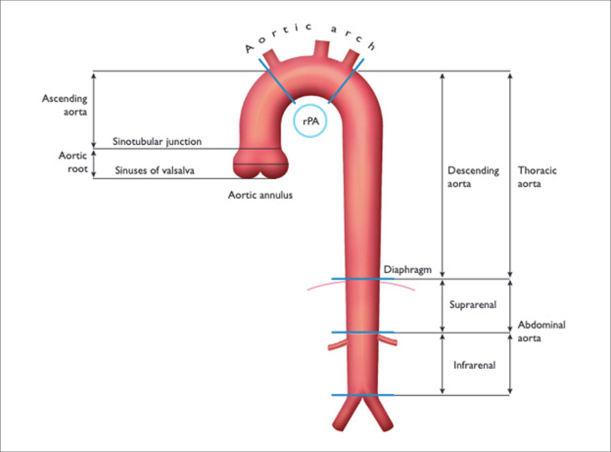
The echocardiogram is an available and essential imaging tool for the evaluation of thoracic aortic diseases. This examination plays a significant role in the identification and diagnosis, as well as the follow-up of chronic cases. Its availability, portability, cost-effectiveness, and the absence of exposure to radiation or contrast make it an important tool for monitoring patients with known aortopathy, who usually require serial examinations for follow-up. Although it is inferior in the total and detailed characterization of the aorta in all its sections compared to aortic angiotomography or angioresonance, the echocardiogram allows for functional assessment of the aortic valve and valve apparatus. In acute aortopathies, it also enables the evaluation of complications and can be performed on hemodynamically unstable patients. This article will discuss the theoretical and practical aspects of the applicability of the echocardiogram in the context of aortopathies.
  • Aorta
  • Aortic Diseases
  • Echocardiography
Read Full Article
Creative Commons Attribution (CC BY) Creative Commons license
28/06/2024 DOI: 10.36660/abcimg.20240022i Views 1
Figure 1
Review Article

My Approach to Perform Echocardiographic Assessment during Cardiopulmonary Arrest

Fátima Elizabeth Fonseca de Oliveira Negri et al. Alexandre Jorge de Andrade Negri Alexandre Jorge de Andrade Negri Júnior Taciana Assis Bezerra Negri

Abstract

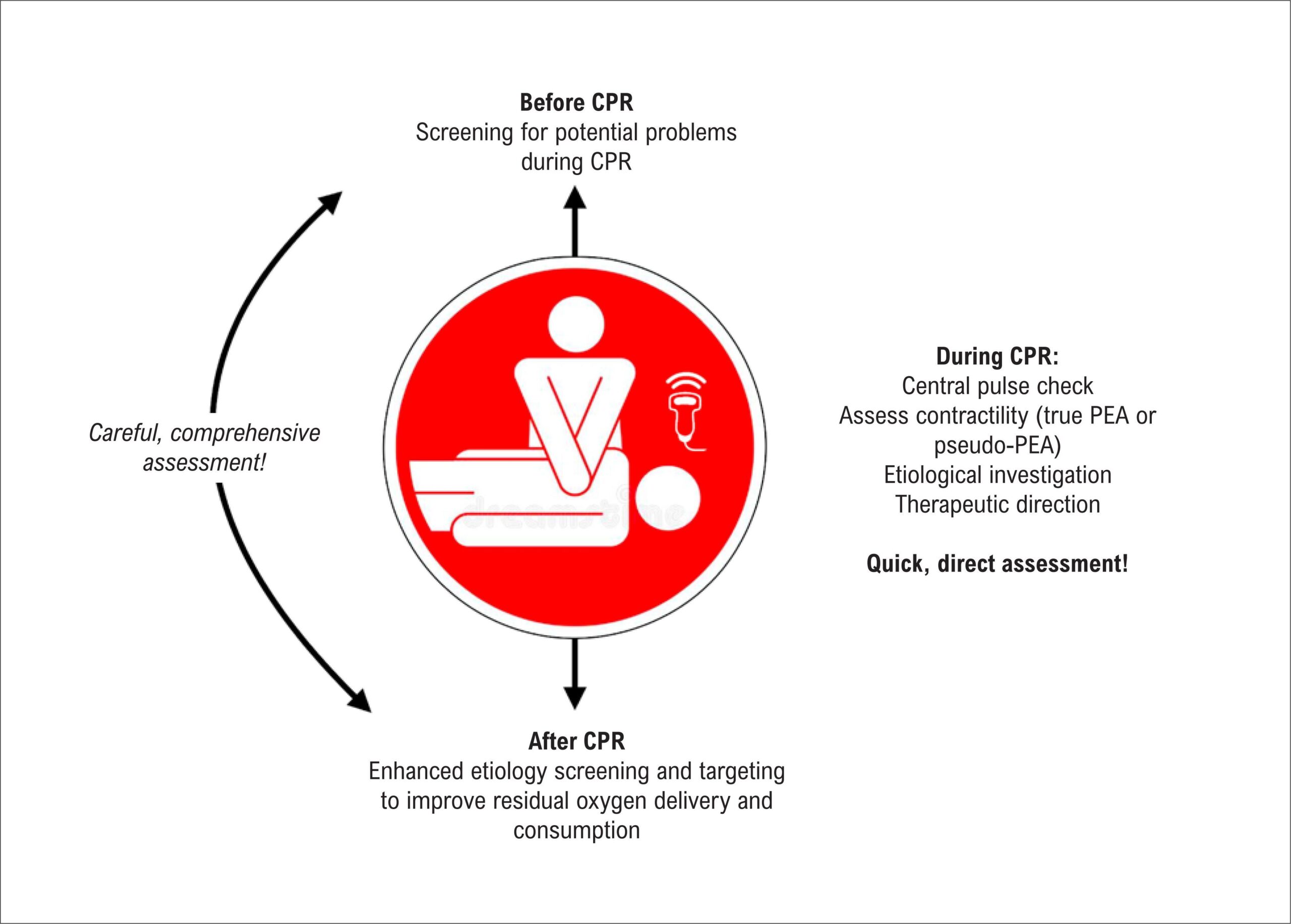
Cardiopulmonary arrest (CPA) is one of the most common events encountered by health care professionals. Attempts to reverse it by performing cardiopulmonary resuscitation (CPR) maneuvers still have very low success rates. Pulseless electrical activity (PEA) and asystole are associated with the worst outcomes. Major CPR guidelines emphasize the importance of identifying potentially treatable causes of CPA. Point-of-care ultrasound (PoCUS), when performed in a systematic, orderly fashion (i.e., without delaying or interfering with other CPR maneuvers), may be important in this change in outcome because it is a diagnostic and prognostic tool used in a scenario where the physical examination is not always conclusive. In this context, the main applications of PoCUS include diagnosis of CPA, differential diagnosis between PEA and pseudo-PEA, and etiologic diagnosis. Several protocols have been developed to optimize the performance of cardiac PoCUS simultaneously with each central pulse check. Pulmonary, abdominal and vascular PoCUS should be performed during resuscitation maneuvers, as they do not interfere with these maneuvers. In addition, PoCUS should preferably be performed by a trained physician who is not actively involved in the resuscitation maneuvers so that the dynamics of CPR are maintained. PoCUS contributes to the diagnosis, prognosis, and therapeutic care of patients suffering from CPA, especially in cases of PEA and asystole; however, there is still no robust evidence that the use of PoCUS reduces mortality among these patients.

  • Cardiorespiratory Arrest
  • Diagnosis
  • Echocardiography
Read Full Article
Creative Commons Attribution (CC BY) Creative Commons license
28/06/2024 DOI: 10.36660/abcimg.20240035i Views 1
Figure 1
Review Article

My Approach to Evaluation of Cardiac Masses Using FDG PET/CT

Simone Cristina Soares Brandão et al. João Marcelo Duarte Ribeiro Sobrinho Emanuel Davi Lima de Matos Leão Lara Cristiane Terra Ferreira Carreira Adriana Soares Xavier de Brito

Abstract

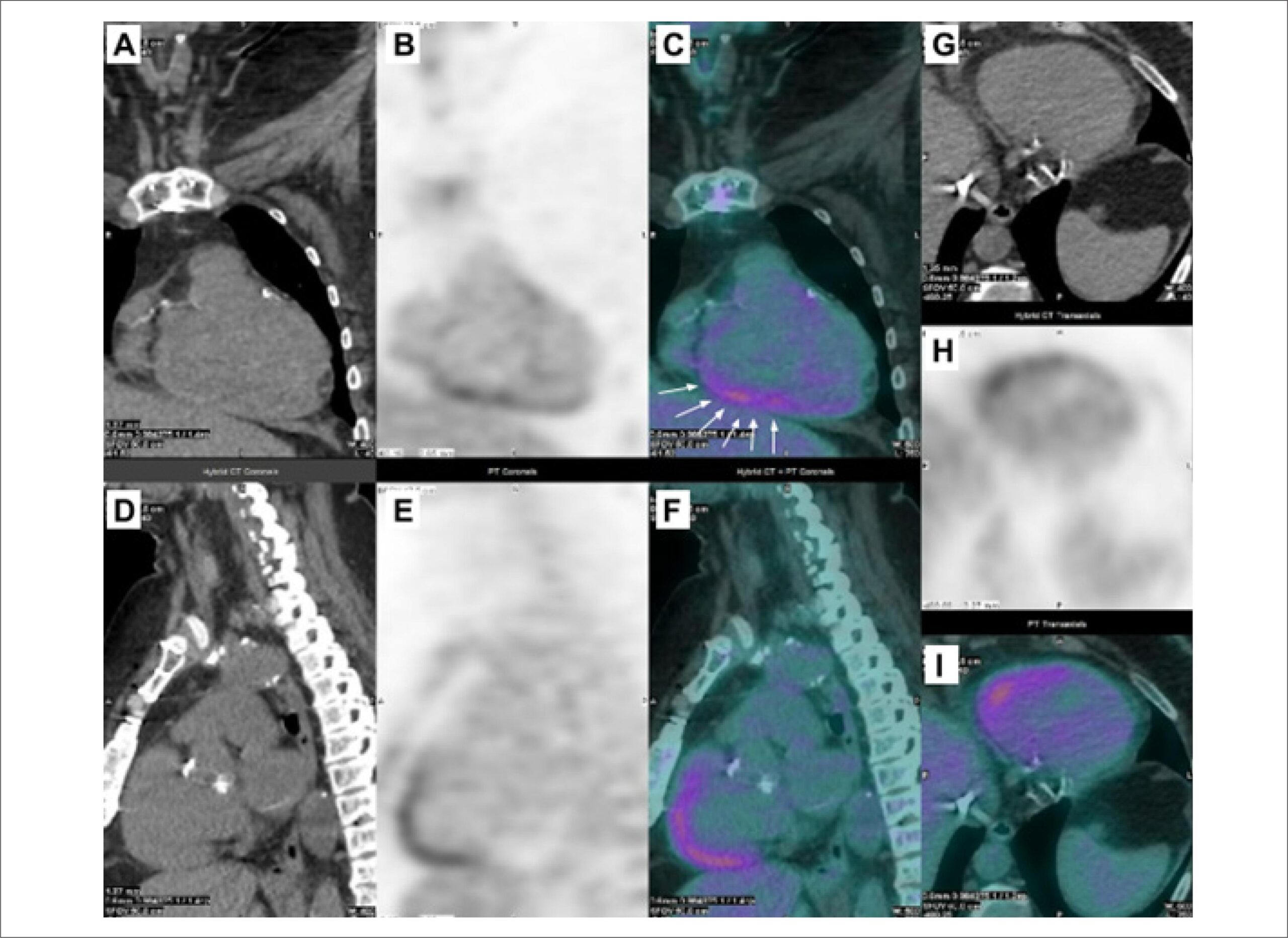
Cardiac masses represent solid formations of various origins, with accurate diagnosis being crucial, particularly for those of a malignant nature, which are typically associated with a worse prognosis. Given this diversity, a comprehensive approach often involves the integration of multiple imaging modalities. Conventional cardiovascular imaging techniques such as transthoracic echocardiography, magnetic resonance imaging (MRI), and cardiac tomography offer insights into anatomical features and hemodynamic implications associated with these masses. However, due to its limitations, greater precision in the etiological investigation of these masses can be achieved by combining Nuclear Medicine with complementary methods, notably positron emission tomography combined with computed tomography using fluorodeoxyglucose-Fluorine-18 (FDG PET/CT). This approach aids in effectively assessing cardiac masses’ metabolic activity, particularly when distinguishing between benign and malignant masses, guiding optimal biopsy site selection, and facilitating comprehensive staging. The Standardized Uptake Value (SUVmax) serves as a quantitative measure of FDG uptake by the mass, with elevated SUVmax values indicating malignancy. Clinical and conventional imaging data should complement the interpretation of FDG PET/CT findings for a more conclusive diagnosis. Thus, a multimodal strategy, particularly incorporating FDG PET/CT, enables a more precise differentiation between benign and malignant cardiac masses, ultimately optimizing patient management.

  • Fluorodeoxyglucose F18
  • Nuclear Medicine
  • Positron Emission Tomography-Computed Tomography
Read Full Article
Creative Commons Attribution (CC BY) Creative Commons license
28/06/2024 DOI: 10.36660/abcimg.20240040i Views 2
Editorial

One-Dimensional Echocardiography: Still Needed or Already Outdated?

Fernando Antonio de Portugal Morcerf

There is a widespread lack of knowledge about one-dimensional echocardiography (M-mode) by the current generation of echocardiographers. Has two-dimensional echocardiography (B-mode) and Doppler made M-mode outdated? M-mode provides information that cannot be obtained by using B-mode or even Doppler—be it spectral, color, or tissue Doppler—for two reasons:

Now let us consider some conditions that can only be detected by M-mode and not by B-mode or Doppler, which can aid in the diagnosis via B-mode or in the understanding of pathologies.

[…]

  • Cardiac Imaging Techniques
  • Cardiovascular Diseases
  • Echocardiography
Read Full Article
Creative Commons Attribution (CC BY) Creative Commons license
28/06/2024 DOI: 10.36660/abcimg.20240038i
Figure 1
Review Article

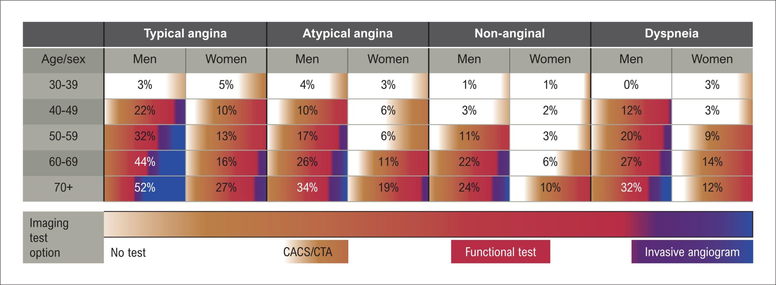
Assessment of Myocardial Ischemia with the Integrated Use of Multimodality Imaging

Thiago Nunes Pereira Leite et al. Luís Henrique Wolff Gowdak

Abstract

Myocardial ischemia, which is characterized by reduced oxygen supply to the myocardium, is a common manifestation of coronary artery disease (CAD), one of the leading causes of morbidity and mortality worldwide. Early and accurate detection of myocardial ischemia is essential for risk stratification, treatment planning, and improved clinical outcomes. In recent years, advances in cardiac imaging modalities have provided a more comprehensive assessment of myocardial ischemia. This article reviews recent advances in the assessment and detection of myocardial ischemia with a focus on the integrated use of multimodality imaging. The article discusses the main techniques available, their advantages and limitations, and their role in current clinical practice, in addition to future perspectives in this area. Cardiac imaging modalities for assessment of myocardial ischemia include exercise electrocardiography, stress echocardiography, single-photon emission computed tomography, positron emission tomography, cardiac magnetic resonance imaging, and coronary computed tomography angiography. When selected imaging tests, it is necessary to consider the pre-test probability (PTP) of CAD and patients’ clinical characteristics. Non-invasive functional modalities are preferable for patients with intermediate probability of CAD. The integration of these modalities can improve sensitivity, specificity, and diagnostic accuracy, offering a more complete assessment of the patient’s cardiovascular condition and allowing for more personalized therapeutic planning.

In conclusion, the multimodal approach to myocardial ischemia assessment improves early detection, risk stratification, and therapeutic planning. The combination of several imaging modalities allows a holistic view of the cardiovascular condition, improving diagnostic accuracy and clinical outcomes in patients with suspected or established diagnosis of CAD.

  • Coronary Artery Disease
  • Echocardiography
  • Ischemia
Read Full Article
Creative Commons Attribution (CC BY) Creative Commons license
28/06/2024 DOI: 10.36660/abcimg.20240037i Views 1
Review Article

Cardiac Tumors: Recognition Is Essential!

Eliza de Almeida Gripp et al. Bruna Zanforlin Jácome Anna Esther Araujo e Silva Fernanda Baptista Ribeiro Dias … Bruna Barbosa Chagas de Almeida Arnaldo Rabischoffsky

Abstract

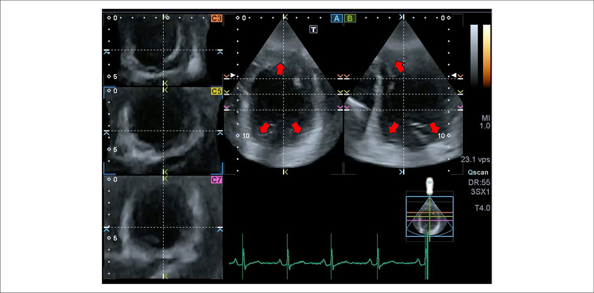
Although cardiac tumors are rare and often found accidentally, they can be associated with constitutional, cardiac, or embolic symptoms and, thus, must always be considered in diagnostic hypotheses. Three-dimensional Doppler echocardiography and contrast enhancement are additional diagnostic resources and can help determine the most appropriate surgical approach, when necessary.

  • Echocardiography
  • Heart Neoplasms
  • Three-Dimensional Echocardiography
Read Full Article
Creative Commons Attribution (CC BY) Creative Commons license
26/06/2024 DOI: 10.36660/abcimg.20240032i Views 1
Figure 1
Review Article

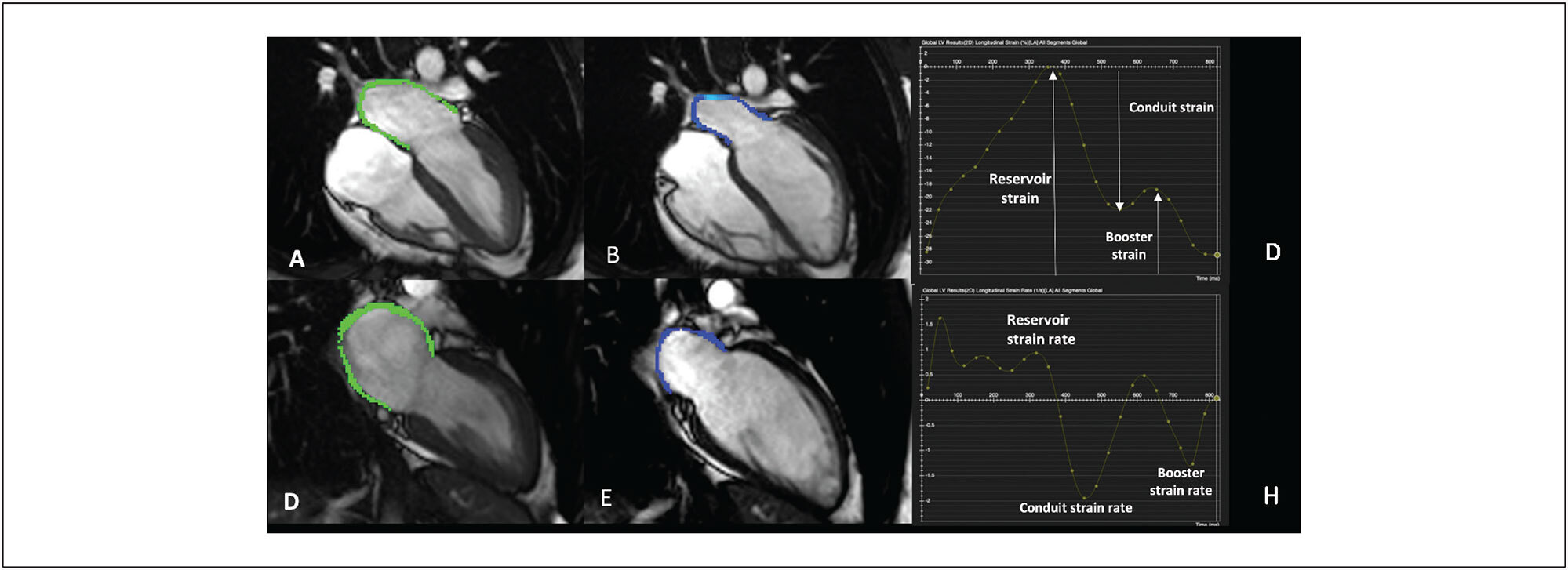
My Approach to Atrial Strain by Cardiac Magnetic Resonanc

Isabela Bispo Santos da Silva Costa et al. Rafael Almeida Fonseca Carlos Eduardo Rochitte

Abstract

Assessment of left atrial strain (LAS) using cardiac magnetic resonance (CMR) is gaining interest in cardiology as a novel parameter, as it quantifies the left atrium’s (LA) deformation throughout the cardiac cycle. Traditionally, LA function was inferred from left ventricular ejection fraction and LA volume. However, recent studies emphasize the prognostic significance of directly analyzing atrial function, particularly in atrial fibrillation (AF), heart failure, and arterial hypertension. CMR provides a thorough evaluation of cardiac anatomy and function, enabling precise and reproducible measurement of atrial tissue deformation.

  • Cardiac Magnetic Resonance Imaging
  • Left atrium
  • Right Atrium
  • Stran Imaging
Read Full Article
Creative Commons Attribution (CC BY) Creative Commons license
26/06/2024 DOI: 10.36660/abcimg.20240020i Views 1
Case Report

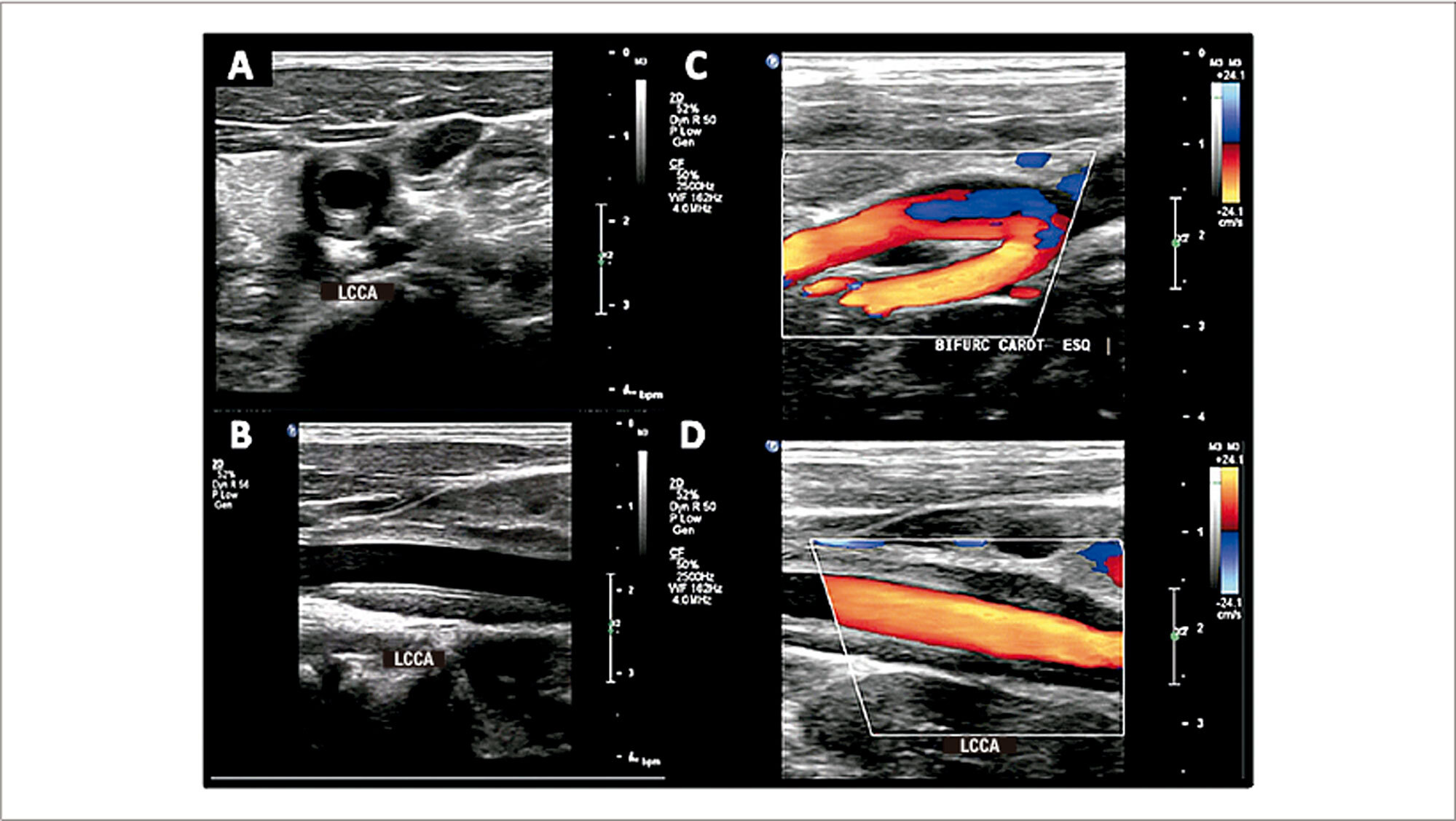
Dural Arteriovenous Fistula: A Rare Finding Identified by Vascular Ultrasonography

Halsted Alarcão Gomes Pereira da Silva et al. Fanilda Souto Barros João Luiz Sandri Simone Nascimento dos Santos

Introduction

Intracranial dural arteriovenous fistulas (AVFs) are abnormal arteriovenous communications located in the intracranial dura mater. The main supply arteries are generally the meningeal branches of the external carotid artery, internal carotid artery, and vertebral artery. Digital subtraction cerebral angiography continues to be the standard method for diagnosing dural AVFs.,

Vascular ultrasound (VUS) is highly accurate in the diagnosis of diseases that involve extracranial vessels, and the objective of this case report is to demonstrate that, by analyzing the flow in these arteries, it is possible to consider intracranial lesions that are not accessible on conventional Doppler.

[…]

  • Artéria Carótida Externa
  • Fístula Arteriovenosa
  • Ultrassonografia das Artérias Carótidas
Read Full Article
Creative Commons Attribution (CC BY) Creative Commons license
26/06/2024 DOI: 10.36660/abcimg.20240036i Views 1
Figure 1
Case Report

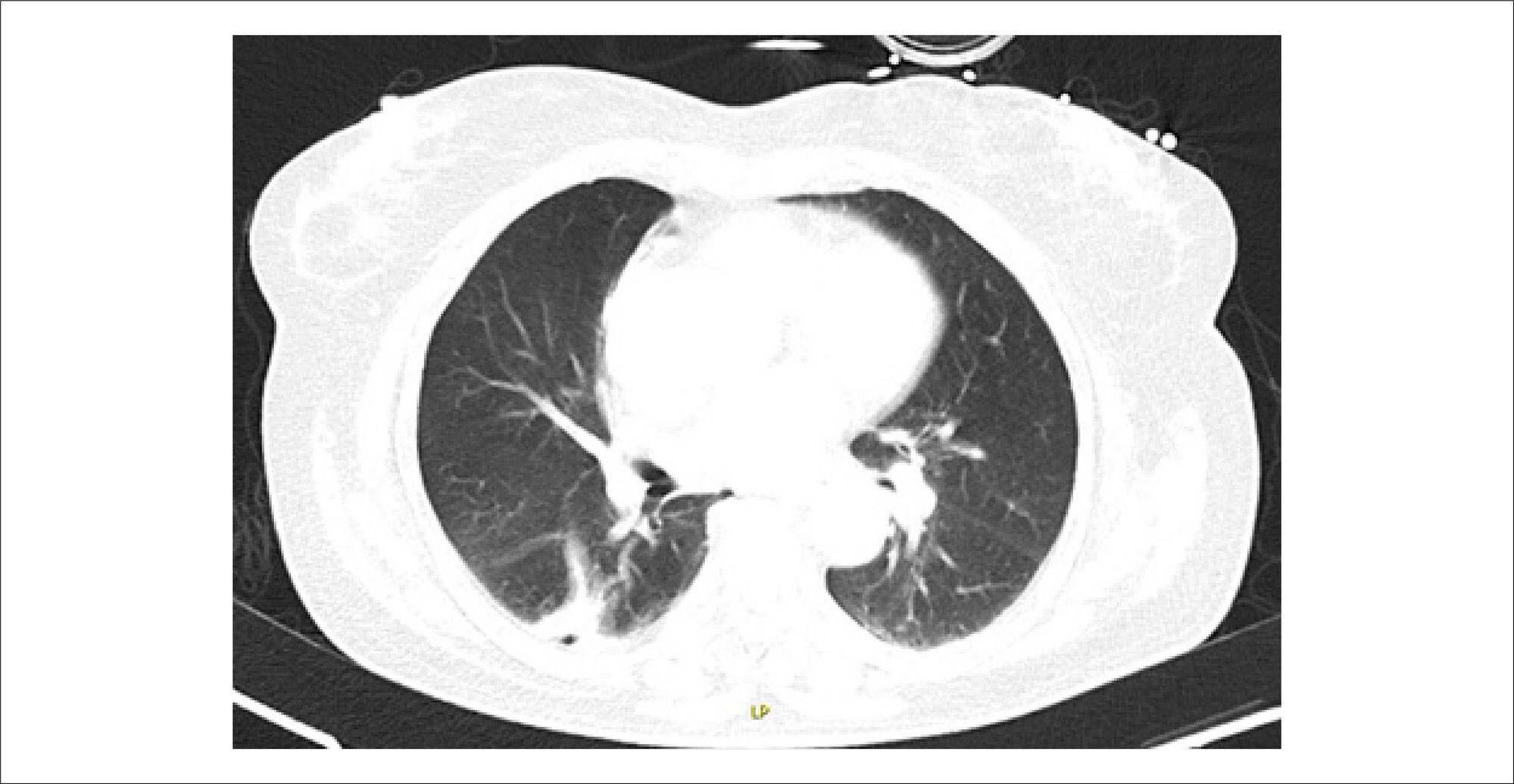
Patent Foramen Ovale: An Unusual Cause of Refractory Hypoxemia

Inês Miranda et al. Christopher Strong Pedro Póvoa

Case Report

We report the case of a 70-year-old woman with a history of cryptogenic ischemic stroke for which she underwent intravenous fibrinolysis with no neurological sequelae. She had no known history of pulmonary or heart disease.

Approximately 1.5 years later, she presented to the Emergency Department (ED) with a one-week history of cough, fatigue, and shortness of breath. She denied fever, chest pain, orthopnea, or paroxysmal nocturnal dyspnea.

[…]

  • Hypoxia
  • Patent Foramen Ovale
  • Pulmonary Artery
  • Respiratory Insufficiency
Read Full Article
Creative Commons Attribution (CC BY) Creative Commons license
26/06/2024 DOI: 10.36660/abcimg.20240030i Views 1
Figure 1
Review Article

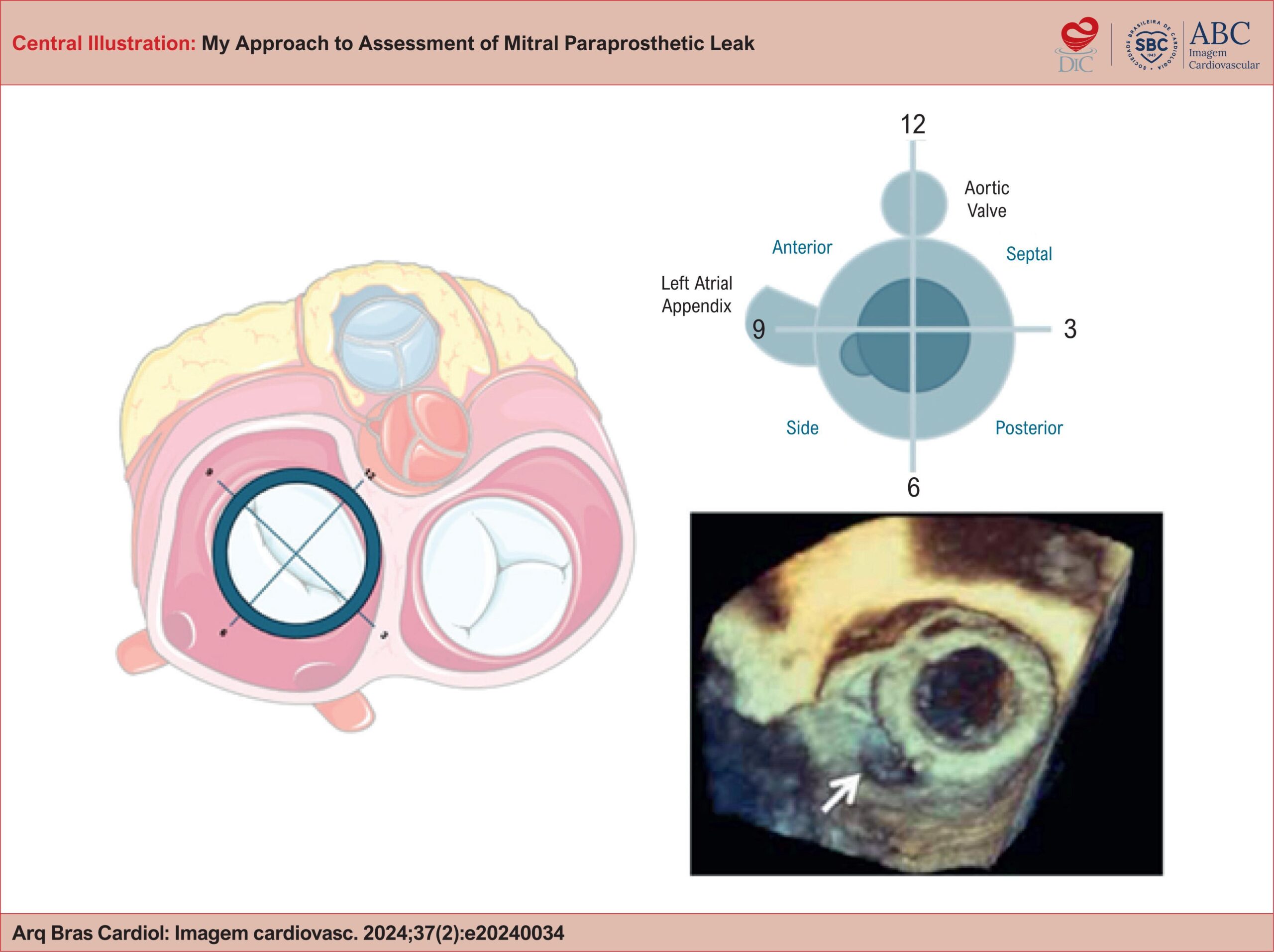
My Approach to Assessment of Mitral Paraprosthetic Leak

Tiago Bignoto

Abstract

Mitral paraprosthetic leak is a common, often asymptomatic condition. When presented with clinical manifestations such as heart failure and/or hemolysis, interventional treatment is indicated. The transcatheter approach has become increasingly popular, requiring careful planning using imaging methods, particularly echocardiography. Echocardiography is crucial for assessing the severity, location, size, shape, and number of leaks. Once these variables are determined, the treatment approach is planned, typically using the transseptal route. The optimal puncture site depends on various factors, with precise puncture location being paramount. Intraoperative echocardiographic evaluation is essential for procedure success, guiding device positioning and adjustments while also identifying any potential complications.

  • Heart Valve Diseases
  • Heart Valve Prosthesis
  • Mitral Valve
Read Full Article
Creative Commons Attribution (CC BY) Creative Commons license
06/06/2024 DOI: 10.36660/abcimg.20240034i Views 1
Figure 1
Case Report

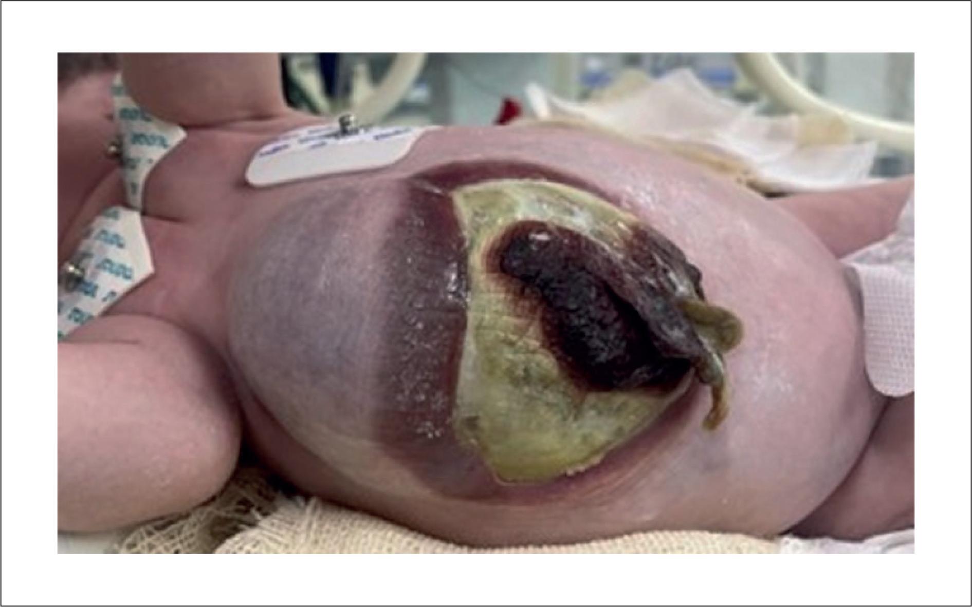
Stent Implantation in the Right Ventricular Outflow Tract to Pulmonary Artery in a Child with Pentalogy of Cantrell: Case Report

Jonathan Guimarães Lombardi et al. Paulo Correia Calamita Laura Moreira Tannus Mayra Rosana Palmeira Barreto … Luiza Emylce Pelá Rosado Giulliano Gardenghi

Introduction

Pentalogy of Cantrell is a syndrome that was first described in 1958. It is characterized by 5 main malformations: defect in the lower sternum, defect in the anterior pericardium, defect in the upper abdomen, deficiency of the anterior diaphragm, and heart defects. The syndrome is rare, with an incidence of 1 in 65,000 live births. It is characterized by midline defects resulting from defective development of the septum transversum. Other congenital heart lesions associated with pentalogy of Cantrell may include atrial septal defect, pulmonary valve stenosis, tetralogy of Fallot, dextrocardia, anomalous pulmonary venous connection, tricuspid atresia, and truncus arteriosus. Children with pentalogy of Cantrell constitute a population with high morbidity and mortality. In this case report, we demonstrate a hemodynamic procedure performed in a patient with this rare congenital anomaly.

[…]

  • Catheterization
  • Congenital Heart Defects
  • Treatment Outcome
Read Full Article
Creative Commons Attribution (CC BY) Creative Commons license
06/06/2024 DOI: 10.36660/abcimg.20240029i Views 2
Figure 1
Review Article

My Approach to 3-Dimensional Left Ventricular Strain

Rafael Bonafim Piveta et al. Miguel Osman Dias Aguiar Liria Lima Maria da Silva Marcelo Luiz Campos Vieira

Abstract

The role of strain analysis using 2-dimensional speckle tracking (2DST) has been well documented, and it represents an important complementary tool in the assessment of cardiac mechanics, adding value in the identification of incipient and subclinical myocardial injury in different clinical scenarios. A significant advance related to echocardiography was the development of 3-dimensional speckle tracking (3DST), which has the potential to overcome many limitations intrinsic to 2-dimensional technology, offering additional parameters of myocardial deformation (such as area strain) and a more comprehensive quantification of the geometry and function of the left ventricular myocardium, based on a single image acquisition. Among its main limitations, the low temporal and spatial resolution stands out, in addition to the dependence on high-quality images and a well-trained operator. Although it is a relatively recent technique that is still under development, several experimental studies and some clinical investigations have already demonstrated the reproducibility and potential applicability of 3DST. The objective of this article is to add information about adequate analysis of 3-dimensional left ventricular strain and explore its main points of vulnerability, discussing important variables to increase the accuracy and reproducibility of this technology.

  • Echocardiography
  • Heart Ventricles
  • Three-Dimensional Imaging
Read Full Article
Creative Commons Attribution (CC BY) Creative Commons license
28/05/2024 DOI: 10.36660/abcimg.20240028i Views 2
Editorial

Transesophageal Echocardiography in Aortic Valve Stenosis: A Pre-Intervention “Toll”?

Antonio de Santis et al. Francisco Monteiro de Almeida Magalhães

Aortic stenosis (AS) has become one of the most relevant valve diseases in cardiology care due to its increasing prevalence, resulting from the positive relationship between the preeminently degenerative etiology and global population aging. In Brazil, we highlight an epidemiological scenario characterized by an etiopathogenic coexistence: rheumatic fever and bicuspid valve disease in younger patients and calcific (degenerative) etiology in elderly patients. Regarding the natural history, AS leads to elevated cardiovascular morbidity and mortality in its symptomatic phase, with unfavorable prognosis when it is not diagnosed correctly and without timely intervention.–

Echocardiography is still a fundamental tool for diagnosis, allowing both evaluation of etiological characteristics and grading of AS severity using parameters such as aortic valve area, transvalvular pressure gradient, and peak aortic jet velocity. However, there are still questions related to the best window for acquiring echocardiographic data. Is the transesophageal window a necessary “toll” for all patients with AS for whom intervention is planned? In general, transesophageal echocardiography (TEE) has the potential to provide superior image quality to transthoracic echocardiography (TTE) in the evaluation of posterior cardiac structures close to the esophagus, for example, the mitral valve. In this case, the greater distance between these structures and the echocardiographic transducer might make evaluation through the transthoracic window difficult. However, the anterior anatomical topography of the aortic valve allows adequate analysis by TTE in most patients, thus avoiding exposure to sedation and possible complications of the transesophageal modality. Data from the literature have shown that TTE and TEE are comparable, mainly in the anatomical and etiological characterization of AS, with compatible results in estimating the valve area.– Nonetheless, there is evidence that the determination of the hemodynamic severity of AS, expressed by the transvalvular pressure gradient and aortic jet velocity, may be underestimated in TEE. Variations in blood volume and preload, conditioned by the fasting and sedation required for the exam, could justify these particularities in transesophageal evaluation.

[…]

  • Aortic Valve Stenosis
  • Heart Valve Diseases
  • Transesophageal echocardiography
Read Full Article
Creative Commons Attribution (CC BY) Creative Commons license
28/05/2024 DOI: 10.36660/abcimg.20240023i Views 1
Figure 1
Original Article

Use of a 3D Printer in the Prototyping of an Anatomical Model of the Human Heart with Morphophysiological Alterations

Kaio Gomes de Freitas et al. Ana Laura Barros Lana Vinícius Lana Ferreira Melissa Araújo Ulhôa Quintão

Abstract

Background:

Three-dimensional (3D) printing has a wide range of applications, including medicine and medical education. It presents itself as an alternative to traditional teaching-learning methods of human morphophysiology.

Objectives:

To report the initial experience in prototyping a 3D-printed anatomical model of the human heart with valvular heart disease, as well as its potential use in the active teaching-learning process in human morphophysiology.

Methods:

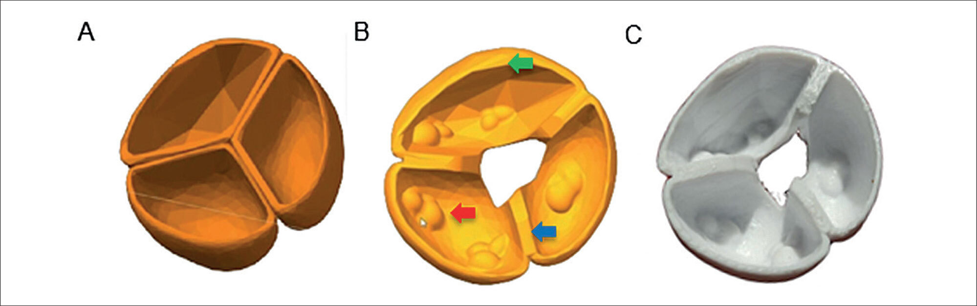
This is an experimental, descriptive study developed in three distinct stages: 1) Selection of the morphofunctional structure for manufacturing the 3D part; 2) design and adaptation of the object in 3D computer-aided design (CAD) software; and 3) printing and application of the prototype. Models extracted from the Thingiverse website, simulating cardiac structures, were used.

Results:

A cardiac prototype was 3D-printed to illustrate morphophysiological changes, including aortic stenosis and mitral insufficiency, modeled on the aortic and mitral valves, respectively.

Conclusion:

The valve diseases represented in the prototype accurately reflect the pathological changes used as the basis for the study, providing a detailed and precise view of the anatomy, which shows significant potential for enhancing the teaching-learning approaches in morphophysiology and medical pathology.

  • Anatomy
  • Medical Education
  • Proof of Concept Study
  • Three-Dimensional Printing
Read Full Article
Creative Commons Attribution (CC BY) Creative Commons license
28/05/2024 DOI: 10.36660/abcimg.20240012i Views 1
My Approach to Evaluate Systemic Venous Congestion: VExUS Protocol
Review Article

My Approach to Evaluate Systemic Venous Congestion: VExUS Protocol

Angelo Antunes Salgado et al. Marcos Paulo Lacerda Bernardo Fernando Melo Netto

Abstract

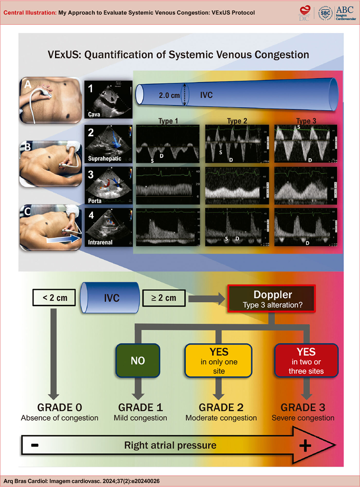
The use of non-invasive tools for analyzing systemic venous congestion in critically ill patients in the intensive care unit has been gaining increasing popularity for diagnosis, estimating the severity of congestion, and providing prognostic estimates. Patients with systemic congestion are more likely to develop renal dysfunction compared to those without congestion. In this review, the authors demonstrate how to perform systemic congestion analysis, its potential limitations, and its practical and objective applicability.

  • Heart Failure
  • Pathology
  • Ultrasonography
Read Full Article
Creative Commons Attribution (CC BY) Creative Commons license
16/05/2024 DOI: 10.36660/abcimg.20240026i Views 2
Figure 1
Case Report

A Rare Case of Pulmonary Sarcoidosis With Extracranial Carotid System Involvement

Fanilda Souto Barros et al. Halsted Alarcão Gomes Pereira da Silva Daniela Souto Barros Simone Nascimento dos Santos

Introduction

Sarcoidosis is a disease of unknown etiology, whose pathogenesis appears to involve genetic, immunologic, infectious, and environmental factors. It was first described in 1889 by Ernest Besnier and is also called Besnier-Boeck-Schaumann disease.,

In sarcoidosis, a noninfectious inflammatory process leads to the formation of granulomas, which predominantly affect the lungs and intrathoracic lymph nodes in approximately 90% to 95% of cases. Close to 30% of patients with sarcoidosis present with extrapulmonary involvement, which may be the primary manifestation of the disease or occur in isolation.–

[…]

  • Carotid Arteries
  • Endosonography
  • Sarcoidosis
Read Full Article
Creative Commons Attribution (CC BY) Creative Commons license
16/05/2024 DOI: 10.36660/abcimg.20240027i Views 1
Figure 1
Case Report

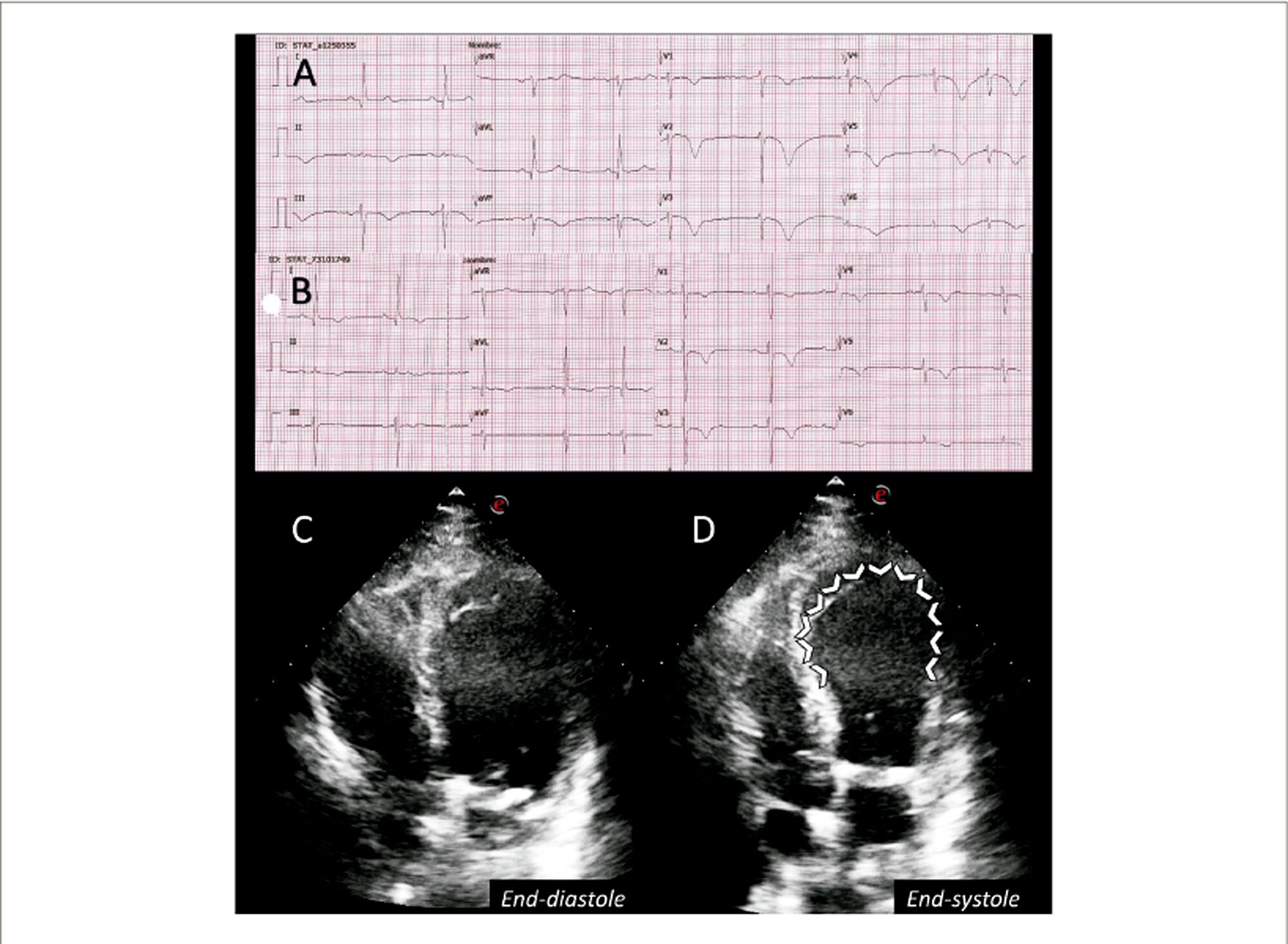
Takotsubo Syndrome in a Critical COVID-19 Patient: Electrocardiographic and Echocardiographic Evolution

Cuitláhuac Arroyo-Rodríguez et al. Alejandra Hernández-Parra

Background

By resembling a Japanese traditional octopus trap, i.e. a takotsubo, Hikaru Sato described the first case of “Takotsubo cardiomyopathy” in a patient without coronary stenosis and end-systolic left ventricular apical ballooning on a cardiac left ventriculography.Takotsubo syndrome (TTS) is a clinical syndrome characterized by an acute and transient left ventricular systolic dysfunction related to an emotional or physical stressful event. COVID-19 is a recognized trigger for TTS, with the possible explanations being the enormous emotional stress caused by the pandemic and the entire spectrum of myocardial damage associated with SARS-COV-2.

[…]

  • COVID-19
  • Echocardiography
  • Takotsubo Cardiomyopathy
Read Full Article
Creative Commons Attribution (CC BY) Creative Commons license
16/05/2024 DOI: 10.36660/abcimg.20240014i Views 2
Figure 1
Case Report

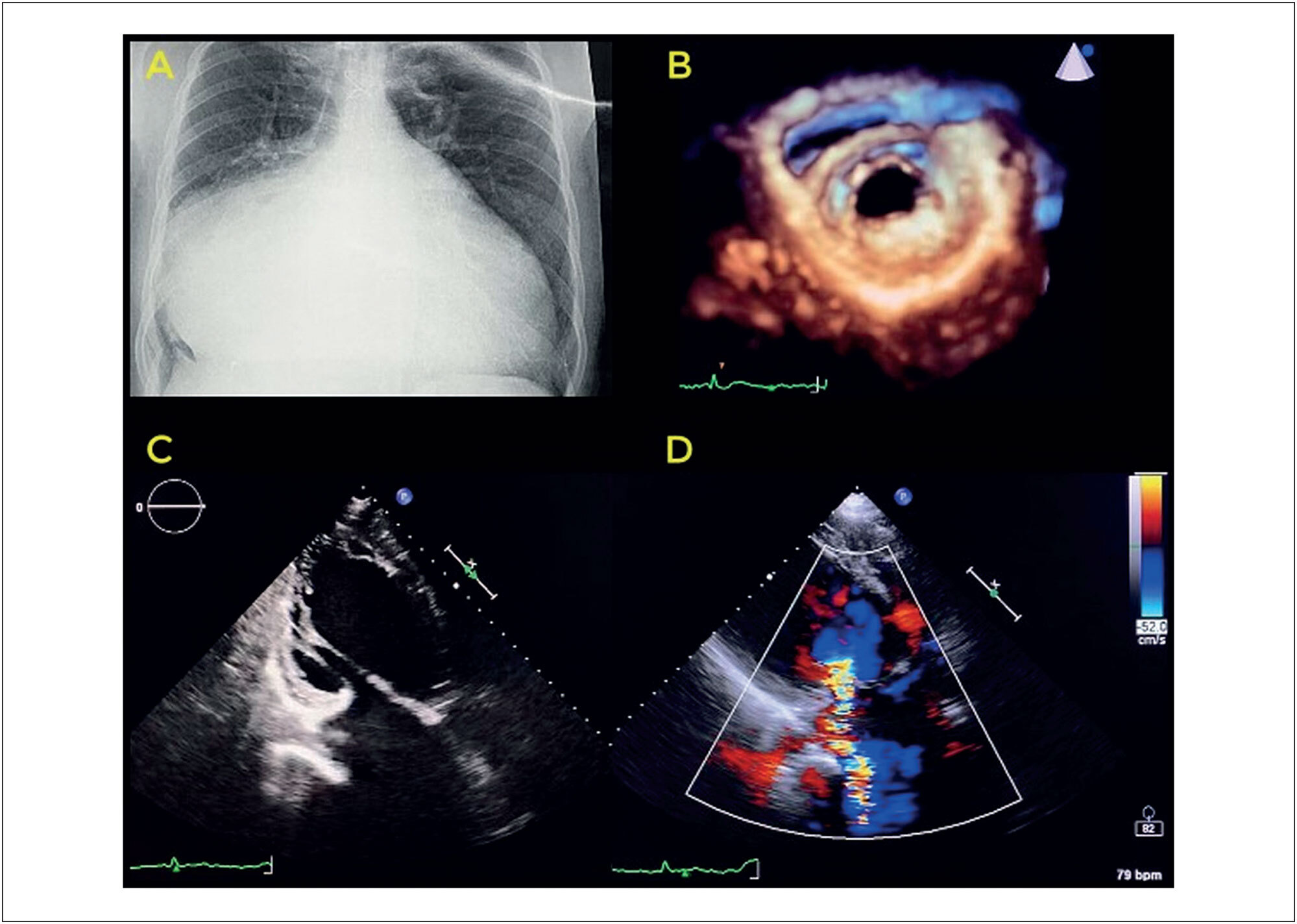
Giant Left Atrium due to Probable Rheumatic Disease and Atrial Functional Mitral Insufficiency

Israel Nilton de Almeida Feitosa et al. Isaac Newton Guimarães Andrade

Introduction

Giant left atrium is a rare condition, and most cases described in the literature are associated with rheumatic disease. However, some clinical descriptions suggest a nonrheumatic etiology for this condition, in which characteristic findings of rheumatic disease are not identified or anatomopathological findings do not suggest a specific etiology.

Atrial fibrillation is present in almost all cases and contributes to the maintenance of left atrial remodeling and persistent changes in structure and function.

[…]

  • Echocardiography
  • Left atrium
  • rheumatic heart disease
Read Full Article
Creative Commons Attribution (CC BY) Creative Commons license
25/04/2024 DOI: 10.36660/abcimg.20240018i
Figure 1
Case Report

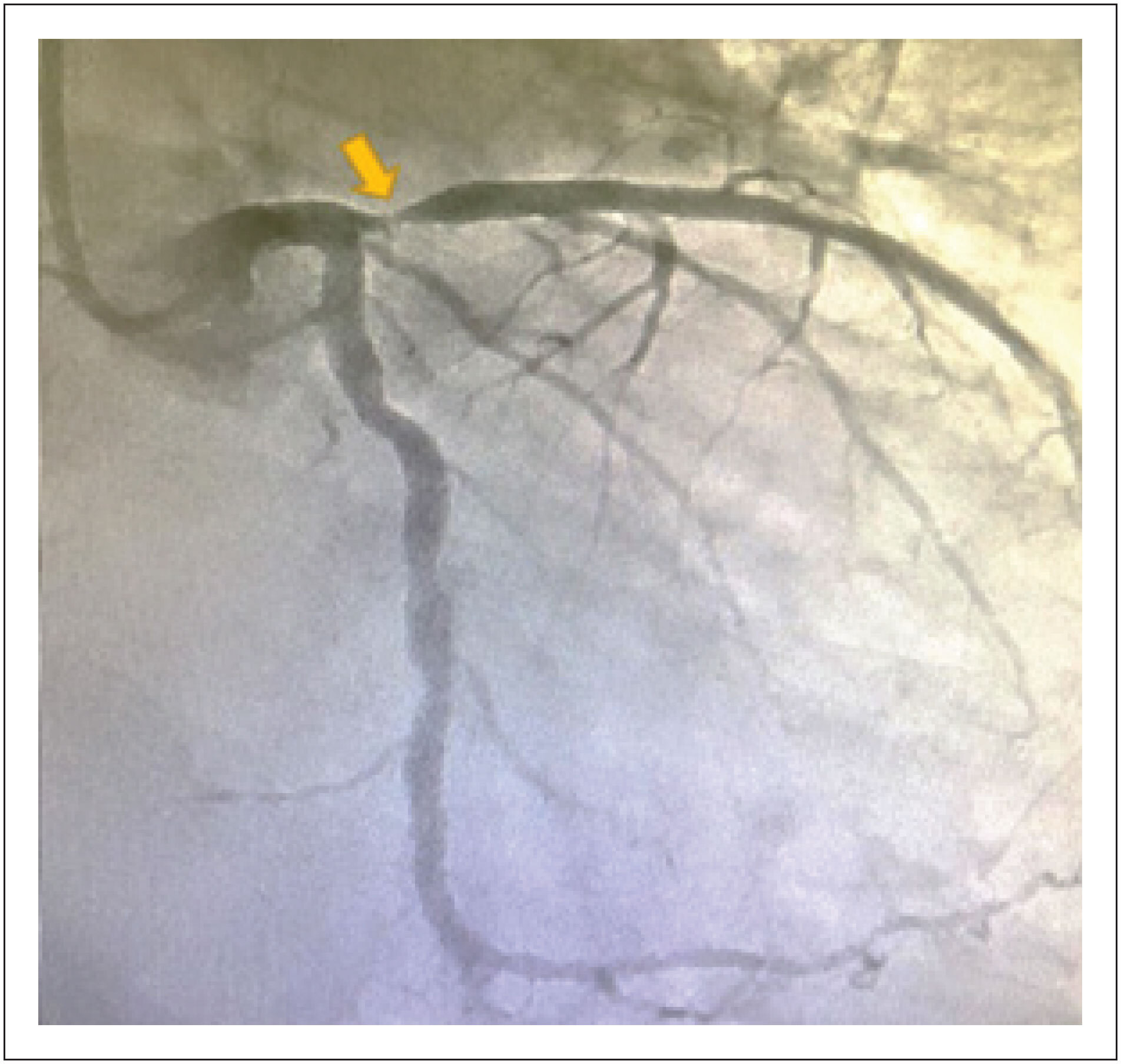
Post-infarction Left Ventricular Pseudoaneurysm Treated with Percutaneous Transcatheter Occlusion: A Case Report

Isabela Galizzi Fae et al. Carolina Kuchenbecker Soares Ricardo Wang Gustavo Brandão de Oliveira … Maria Carmo Pereira Nunes Glaucia Cristina da Silva Rochelle Coppo Militão Rausch Luiz Ricardo de Ataide Castro Anderson Ferreira Leite

Abstract

Left ventricular pseudoaneurysm (LVPSA) is a rare complication after myocardial infarction (MI) with a significant risk of rupture if untreated. Given the elevated surgical risk in this context, alternative treatments are emerging. This case describes a percutaneous closure of an LVPSA after extensive MI with delayed revascularization.

  • False Aneurysm
  • Myocardial Infarction
  • Percutaneous Coronary Intervention
Read Full Article
Creative Commons Attribution (CC BY) Creative Commons license
25/04/2024 DOI: 10.36660/abcimg.20240024i
Figure 1
Case Report

Idiopathic Mitral and Aortic Subvalvular Aneurysm in a Young Patient

João Pedro Laydner et al. Ana Paula Siciliano Rafael Castro da Silva Alex dos Santos Felix Maximiliano Freire Dutra

Abstract

Left ventricular aneurysms are primarily caused by coronary artery disease. Among them, there are also less common types, such as those located in the subvalvular regions of idiopathic origin. This article describes the case of a patient with a history of aortic subvalvular and Sinus of Valsalva aneurysm repairs who developed a new mitral subvalvular aneurysm.

  • Aortico-Ventricular Tunnel
  • Heart Aneurysm
  • Mitral Valve Insufficiency
Read Full Article
Creative Commons Attribution (CC BY) Creative Commons license
23/04/2024 DOI: 10.36660/abcimg.20240021i Views 2

How to cite this article

EndNote RIS

Electronic Document Format (Vancouver)

Martinhago GA, Andrade AR, Correia VM, Fernandes F, Santiago JAD, Dias RR, Madrini Junior V, et al. What do Cardiologists Expect Regarding Echocardiogram in Aortopathies. ABC Imagem Cardiovascular 2024;37(2):e20240022.

Electronic Document Format (ABNT)

Martinhago, Gustavo de Azevedo; Andrade, Alan Rodrigues; Correia, Vinicius Machado; Fernandes, Fabio; Santiago, Jose Augusto Duncan; Dias, Ricardo Ribeiro; Madrini Junior, Vagner. What do Cardiologists Expect Regarding Echocardiogram in Aortopathies. ABC Imagem Cardiovascular, v. 37, n. 2, e20240022, Jun. 2024.

Electronic Document Format (APA)

Martinhago, G. A., Andrade, A. R., Correia, V. M., Fernandes, F., Santiago, J. A. D., Dias, R. R., & Madrini Junior, V. (2024). What do Cardiologists Expect Regarding Echocardiogram in Aortopathies. ABC Imagem Cardiovascular, 37(2), e20240022.

Electronic Document Format (ISO)

Martinhago, Gustavo de Azevedo and Andrade, Alan Rodrigues and Correia, Vinicius Machado and Fernandes, Fabio and Santiago, Jose Augusto Duncan and Dias, Ricardo Ribeiro and Madrini Junior, Vagner. What do Cardiologists Expect Regarding Echocardiogram in Aortopathies. ABC Imagem Cardiovascular [online]. 2024, vol. 37, n. 2, [cited 2026-10-14], e20240022. Available from: <https://journals.cardiol.br/en/article/what-do-cardiologists-expect-regarding-echocardiogram-in-aortopathies/>. ISSN 2318-8219

EndNote RIS

Electronic Document Format (Vancouver)

Negri FEFO, Negri AJA, Negri Júnior AJA, Negri TAB. My Approach to Perform Echocardiographic Assessment during Cardiopulmonary Arrest. ABC Imagem Cardiovascular 2024;37(2):e20240035.

Electronic Document Format (ABNT)

Negri, Fátima Elizabeth Fonseca de Oliveira; Negri, Alexandre Jorge de Andrade; Negri Júnior, Alexandre Jorge de Andrade; Negri, Taciana Assis Bezerra. My Approach to Perform Echocardiographic Assessment during Cardiopulmonary Arrest. ABC Imagem Cardiovascular, v. 37, n. 2, e20240035, Jun. 2024.

Electronic Document Format (APA)

Negri, F. E. F. O., Negri, A. J. A., Negri Júnior, A. J. A., & Negri, T. A. B. (2024). My Approach to Perform Echocardiographic Assessment during Cardiopulmonary Arrest. ABC Imagem Cardiovascular, 37(2), e20240035.

Electronic Document Format (ISO)

Negri, Fátima Elizabeth Fonseca de Oliveira and Negri, Alexandre Jorge de Andrade and Negri Júnior, Alexandre Jorge de Andrade and Negri, Taciana Assis Bezerra. My Approach to Perform Echocardiographic Assessment during Cardiopulmonary Arrest. ABC Imagem Cardiovascular [online]. 2024, vol. 37, n. 2, [cited 2026-10-14], e20240035. Available from: <https://journals.cardiol.br/en/article/my-approach-to-perform-echocardiographic-assessment-during-cardiopulmonary-arrest/>. ISSN 2318-8219

EndNote RIS

Electronic Document Format (Vancouver)

Brandão SCS, Sobrinho JMDR, Leão EDLM, Carreira LCTF, Brito ASX. My Approach to Evaluation of Cardiac Masses Using FDG PET/CT. ABC Imagem Cardiovascular 2024;37(2):e20240040.

Electronic Document Format (ABNT)

Brandão, Simone Cristina Soares; Sobrinho, João Marcelo Duarte Ribeiro; Leão, Emanuel Davi Lima de Matos; Carreira, Lara Cristiane Terra Ferreira; Brito, Adriana Soares Xavier de. My Approach to Evaluation of Cardiac Masses Using FDG PET/CT. ABC Imagem Cardiovascular, v. 37, n. 2, e20240040, Jun. 2024.

Electronic Document Format (APA)

Brandão, S. C. S., Sobrinho, J. M. D. R., Leão, E. D. L. M., Carreira, L. C. T. F., & Brito, A. S. X. (2024). My Approach to Evaluation of Cardiac Masses Using FDG PET/CT. ABC Imagem Cardiovascular, 37(2), e20240040.

Electronic Document Format (ISO)

Brandão, Simone Cristina Soares and Sobrinho, João Marcelo Duarte Ribeiro and Leão, Emanuel Davi Lima de Matos and Carreira, Lara Cristiane Terra Ferreira and Brito, Adriana Soares Xavier de. My Approach to Evaluation of Cardiac Masses Using FDG PET/CT. ABC Imagem Cardiovascular [online]. 2024, vol. 37, n. 2, [cited 2026-10-14], e20240040. Available from: <https://journals.cardiol.br/en/article/my-approach-to-evaluation-of-cardiac-masses-using-fdg-pet-ct/>. ISSN 2318-8219

EndNote RIS

Electronic Document Format (Vancouver)

Morcerf FAP. One-Dimensional Echocardiography: Still Needed or Already Outdated?. ABC Imagem Cardiovascular 2024;37(2):e20240038.

Electronic Document Format (ABNT)

Morcerf, Fernando Antonio de Portugal. One-Dimensional Echocardiography: Still Needed or Already Outdated?. ABC Imagem Cardiovascular, v. 37, n. 2, e20240038, Jun. 2024.

Electronic Document Format (APA)

Morcerf, F. A. P. (2024). One-Dimensional Echocardiography: Still Needed or Already Outdated?. ABC Imagem Cardiovascular, 37(2), e20240038.

Electronic Document Format (ISO)

Morcerf, Fernando Antonio de Portugal. One-Dimensional Echocardiography: Still Needed or Already Outdated?. ABC Imagem Cardiovascular [online]. 2024, vol. 37, n. 2, [cited 2026-10-14], e20240038. Available from: <https://journals.cardiol.br/en/article/one-dimensional-echocardiography-still-needed-or-already-outdated/>. ISSN 2318-8219

EndNote RIS

Electronic Document Format (Vancouver)

Leite TNP, Gowdak LHW. Assessment of Myocardial Ischemia with the Integrated Use of Multimodality Imaging. ABC Imagem Cardiovascular 2024;37(2):e20240037.

Electronic Document Format (ABNT)

Leite, Thiago Nunes Pereira; Gowdak, Luís Henrique Wolff. Assessment of Myocardial Ischemia with the Integrated Use of Multimodality Imaging. ABC Imagem Cardiovascular, v. 37, n. 2, e20240037, Jun. 2024.

Electronic Document Format (APA)

Leite, T. N. P., & Gowdak, L. H. W. (2024). Assessment of Myocardial Ischemia with the Integrated Use of Multimodality Imaging. ABC Imagem Cardiovascular, 37(2), e20240037.

Electronic Document Format (ISO)

Leite, Thiago Nunes Pereira and Gowdak, Luís Henrique Wolff. Assessment of Myocardial Ischemia with the Integrated Use of Multimodality Imaging. ABC Imagem Cardiovascular [online]. 2024, vol. 37, n. 2, [cited 2026-10-14], e20240037. Available from: <https://journals.cardiol.br/en/article/assessment-of-myocardial-ischemia-with-the-integrated-use-of-multimodality-imaging/>. ISSN 2318-8219

EndNote RIS

Electronic Document Format (Vancouver)

Gripp EA, Jácome BZ, Silva AEA, Dias FBR, Almeida BBC, Rabischoffsky A. Cardiac Tumors: Recognition Is Essential!. ABC Imagem Cardiovascular 2024;37(2):e20240032.

Electronic Document Format (ABNT)

Gripp, Eliza de Almeida; Jácome, Bruna Zanforlin; Silva, Anna Esther Araujo e; Dias, Fernanda Baptista Ribeiro; Almeida, Bruna Barbosa Chagas de; Rabischoffsky, Arnaldo. Cardiac Tumors: Recognition Is Essential!. ABC Imagem Cardiovascular, v. 37, n. 2, e20240032, Jun. 2024.

Electronic Document Format (APA)

Gripp, E. A., Jácome, B. Z., Silva, A. E. A. , Dias, F. B. R., Almeida, B. B. C. , & Rabischoffsky, A. (2024). Cardiac Tumors: Recognition Is Essential!. ABC Imagem Cardiovascular, 37(2), e20240032.

Electronic Document Format (ISO)

Gripp, Eliza de Almeida and Jácome, Bruna Zanforlin and Silva, Anna Esther Araujo e and Dias, Fernanda Baptista Ribeiro and Almeida, Bruna Barbosa Chagas de and Rabischoffsky, Arnaldo. Cardiac Tumors: Recognition Is Essential!. ABC Imagem Cardiovascular [online]. 2024, vol. 37, n. 2, [cited 2026-10-14], e20240032. Available from: <https://journals.cardiol.br/en/article/cardiac-tumors-recognition-is-essential/>. ISSN 2318-8219

EndNote RIS

Electronic Document Format (Vancouver)

Costa IBSS, Fonseca RA, Rochitte CE. My Approach to Atrial Strain by Cardiac Magnetic Resonanc. ABC Imagem Cardiovascular 2024;37(2):e20240020.

Electronic Document Format (ABNT)

Costa, Isabela Bispo Santos da Silva; Fonseca, Rafael Almeida; Rochitte, Carlos Eduardo. My Approach to Atrial Strain by Cardiac Magnetic Resonanc. ABC Imagem Cardiovascular, v. 37, n. 2, e20240020, Jun. 2024.

Electronic Document Format (APA)

Costa, I. B. S. S., Fonseca, R. A., & Rochitte, C. E. (2024). My Approach to Atrial Strain by Cardiac Magnetic Resonanc. ABC Imagem Cardiovascular, 37(2), e20240020.

Electronic Document Format (ISO)

Costa, Isabela Bispo Santos da Silva and Fonseca, Rafael Almeida and Rochitte, Carlos Eduardo. My Approach to Atrial Strain by Cardiac Magnetic Resonanc. ABC Imagem Cardiovascular [online]. 2024, vol. 37, n. 2, [cited 2026-10-14], e20240020. Available from: <https://journals.cardiol.br/en/article/my-approach-to-atrial-strain-by-cardiac-magnetic-resonanc/>. ISSN 2318-8219

EndNote RIS

Electronic Document Format (Vancouver)

Silva HAGP, Barros FS, Sandri JL, Santos SN. Dural Arteriovenous Fistula: A Rare Finding Identified by Vascular Ultrasonography. ABC Imagem Cardiovascular 2024;37(2):e20240036.

Electronic Document Format (ABNT)

Silva, Halsted Alarcão Gomes Pereira da; Barros, Fanilda Souto; Sandri, João Luiz; Santos, Simone Nascimento dos. Dural Arteriovenous Fistula: A Rare Finding Identified by Vascular Ultrasonography. ABC Imagem Cardiovascular, v. 37, n. 2, e20240036, Jun. 2024.

Electronic Document Format (APA)

Silva, H. A. G. P. , Barros, F. S., Sandri, J. L., & Santos, S. N. (2024). Dural Arteriovenous Fistula: A Rare Finding Identified by Vascular Ultrasonography. ABC Imagem Cardiovascular, 37(2), e20240036.

Electronic Document Format (ISO)

Silva, Halsted Alarcão Gomes Pereira da and Barros, Fanilda Souto and Sandri, João Luiz and Santos, Simone Nascimento dos. Dural Arteriovenous Fistula: A Rare Finding Identified by Vascular Ultrasonography. ABC Imagem Cardiovascular [online]. 2024, vol. 37, n. 2, [cited 2026-10-14], e20240036. Available from: <https://journals.cardiol.br/en/article/dural-arteriovenous-fistula-a-rare-finding-identified-by-vascular-ultrasonography/>. ISSN 2318-8219

EndNote RIS

Electronic Document Format (Vancouver)

Miranda I, Strong C, Póvoa P. Patent Foramen Ovale: An Unusual Cause of Refractory Hypoxemia. ABC Imagem Cardiovascular 2024;37(2):e20240030.

Electronic Document Format (ABNT)

Miranda, Inês; Strong, Christopher; Póvoa, Pedro. Patent Foramen Ovale: An Unusual Cause of Refractory Hypoxemia. ABC Imagem Cardiovascular, v. 37, n. 2, e20240030, Jun. 2024.

Electronic Document Format (APA)

Miranda, I., Strong, C., & Póvoa, P. (2024). Patent Foramen Ovale: An Unusual Cause of Refractory Hypoxemia. ABC Imagem Cardiovascular, 37(2), e20240030.

Electronic Document Format (ISO)

Miranda, Inês and Strong, Christopher and Póvoa, Pedro. Patent Foramen Ovale: An Unusual Cause of Refractory Hypoxemia. ABC Imagem Cardiovascular [online]. 2024, vol. 37, n. 2, [cited 2026-10-14], e20240030. Available from: <https://journals.cardiol.br/en/article/patent-foramen-ovale-an-unusual-cause-of-refractory-hypoxemia/>. ISSN 2318-8219

EndNote RIS

Electronic Document Format (Vancouver)

Bignoto T. My Approach to Assessment of Mitral Paraprosthetic Leak. ABC Imagem Cardiovascular 2024;37(2):e20240034.

Electronic Document Format (ABNT)

Bignoto, Tiago. My Approach to Assessment of Mitral Paraprosthetic Leak. ABC Imagem Cardiovascular, v. 37, n. 2, e20240034, Jun. 2024.

Electronic Document Format (APA)

Bignoto, T. (2024). My Approach to Assessment of Mitral Paraprosthetic Leak. ABC Imagem Cardiovascular, 37(2), e20240034.

Electronic Document Format (ISO)

Bignoto, Tiago. My Approach to Assessment of Mitral Paraprosthetic Leak. ABC Imagem Cardiovascular [online]. 2024, vol. 37, n. 2, [cited 2026-10-14], e20240034. Available from: <https://journals.cardiol.br/en/article/my-approach-to-assessment-of-mitral-paraprosthetic-leak/>. ISSN 2318-8219

EndNote RIS

Electronic Document Format (Vancouver)

Lombardi JG, Calamita PC, Tannus LM, Barreto MRP, Rosado LEP, Gardenghi G. Stent Implantation in the Right Ventricular Outflow Tract to Pulmonary Artery in a Child with Pentalogy of Cantrell: Case Report. ABC Imagem Cardiovascular 2024;37(2):e20240029.

Electronic Document Format (ABNT)

Lombardi, Jonathan Guimarães; Calamita, Paulo Correia; Tannus, Laura Moreira; Barreto, Mayra Rosana Palmeira; Rosado, Luiza Emylce Pelá; Gardenghi, Giulliano. Stent Implantation in the Right Ventricular Outflow Tract to Pulmonary Artery in a Child with Pentalogy of Cantrell: Case Report. ABC Imagem Cardiovascular, v. 37, n. 2, e20240029, Jun. 2024.

Electronic Document Format (APA)

Lombardi, J. G., Calamita, P. C., Tannus, L. M., Barreto, M. R. P., Rosado, L. E. P., & Gardenghi, G. (2024). Stent Implantation in the Right Ventricular Outflow Tract to Pulmonary Artery in a Child with Pentalogy of Cantrell: Case Report. ABC Imagem Cardiovascular, 37(2), e20240029.

Electronic Document Format (ISO)

Lombardi, Jonathan Guimarães and Calamita, Paulo Correia and Tannus, Laura Moreira and Barreto, Mayra Rosana Palmeira and Rosado, Luiza Emylce Pelá and Gardenghi, Giulliano. Stent Implantation in the Right Ventricular Outflow Tract to Pulmonary Artery in a Child with Pentalogy of Cantrell: Case Report. ABC Imagem Cardiovascular [online]. 2024, vol. 37, n. 2, [cited 2026-10-14], e20240029. Available from: <https://journals.cardiol.br/en/article/stent-implantation-in-the-right-ventricular-outflow-tract-to-pulmonary-artery-in-a-child-with-pentalogy-of-cantrell-case-report/>. ISSN 2318-8219

EndNote RIS

Electronic Document Format (Vancouver)

Piveta RB, Aguiar MOD, Silva LLM, Vieira MLC. My Approach to 3-Dimensional Left Ventricular Strain. ABC Imagem Cardiovascular 2024;37(2):e20240028.

Electronic Document Format (ABNT)

Piveta, Rafael Bonafim; Aguiar, Miguel Osman Dias; Silva, Liria Lima Maria da; Vieira, Marcelo Luiz Campos. My Approach to 3-Dimensional Left Ventricular Strain. ABC Imagem Cardiovascular, v. 37, n. 2, e20240028, May. 2024.

Electronic Document Format (APA)

Piveta, R. B., Aguiar, M. O. D., Silva, L. L. M. , & Vieira, M. L. C. (2024). My Approach to 3-Dimensional Left Ventricular Strain. ABC Imagem Cardiovascular, 37(2), e20240028.

Electronic Document Format (ISO)

Piveta, Rafael Bonafim and Aguiar, Miguel Osman Dias and Silva, Liria Lima Maria da and Vieira, Marcelo Luiz Campos. My Approach to 3-Dimensional Left Ventricular Strain. ABC Imagem Cardiovascular [online]. 2024, vol. 37, n. 2, [cited 2026-10-14], e20240028. Available from: <https://journals.cardiol.br/en/article/my-approach-to-3-dimensional-left-ventricular-strain/>. ISSN 2318-8219

EndNote RIS

Electronic Document Format (Vancouver)

Santis A, Magalhães FMA. Transesophageal Echocardiography in Aortic Valve Stenosis: A Pre-Intervention “Toll”?. ABC Imagem Cardiovascular 2024;37(2):e20240023.

Electronic Document Format (ABNT)

Santis, Antonio de; Magalhães, Francisco Monteiro de Almeida. Transesophageal Echocardiography in Aortic Valve Stenosis: A Pre-Intervention “Toll”?. ABC Imagem Cardiovascular, v. 37, n. 2, e20240023, May. 2024.

Electronic Document Format (APA)

Santis, A. , & Magalhães, F. M. A. (2024). Transesophageal Echocardiography in Aortic Valve Stenosis: A Pre-Intervention “Toll”?. ABC Imagem Cardiovascular, 37(2), e20240023.

Electronic Document Format (ISO)

Santis, Antonio de and Magalhães, Francisco Monteiro de Almeida. Transesophageal Echocardiography in Aortic Valve Stenosis: A Pre-Intervention “Toll”?. ABC Imagem Cardiovascular [online]. 2024, vol. 37, n. 2, [cited 2026-10-14], e20240023. Available from: <https://journals.cardiol.br/en/article/transesophageal-echocardiography-in-aortic-valve-stenosis-a-pre-intervention-toll/>. ISSN 2318-8219

EndNote RIS

Electronic Document Format (Vancouver)

Freitas KG, Lana ALB, Ferreira VL, Quintão MAU. Use of a 3D Printer in the Prototyping of an Anatomical Model of the Human Heart with Morphophysiological Alterations. ABC Imagem Cardiovascular 2024;37(2):e20240012.

Electronic Document Format (ABNT)

Freitas, Kaio Gomes de; Lana, Ana Laura Barros; Ferreira, Vinícius Lana; Quintão, Melissa Araújo Ulhôa. Use of a 3D Printer in the Prototyping of an Anatomical Model of the Human Heart with Morphophysiological Alterations. ABC Imagem Cardiovascular, v. 37, n. 2, e20240012, May. 2024.

Electronic Document Format (APA)

Freitas, K. G. , Lana, A. L. B., Ferreira, V. L., & Quintão, M. A. U. (2024). Use of a 3D Printer in the Prototyping of an Anatomical Model of the Human Heart with Morphophysiological Alterations. ABC Imagem Cardiovascular, 37(2), e20240012.

Electronic Document Format (ISO)

Freitas, Kaio Gomes de and Lana, Ana Laura Barros and Ferreira, Vinícius Lana and Quintão, Melissa Araújo Ulhôa. Use of a 3D Printer in the Prototyping of an Anatomical Model of the Human Heart with Morphophysiological Alterations. ABC Imagem Cardiovascular [online]. 2024, vol. 37, n. 2, [cited 2026-10-14], e20240012. Available from: <https://journals.cardiol.br/en/article/use-of-a-3d-printer-in-the-prototyping-of-an-anatomical-model-of-the-human-heart-with-morphophysiological-alterations/>. ISSN 2318-8219

EndNote RIS

Electronic Document Format (Vancouver)

Salgado AA, Bernardo MPL, Netto FM. My Approach to Evaluate Systemic Venous Congestion: VExUS Protocol. ABC Imagem Cardiovascular 2024;37(2):e20240026.

Electronic Document Format (ABNT)

Salgado, Angelo Antunes; Bernardo, Marcos Paulo Lacerda; Netto, Fernando Melo. My Approach to Evaluate Systemic Venous Congestion: VExUS Protocol. ABC Imagem Cardiovascular, v. 37, n. 2, e20240026, May. 2024.

Electronic Document Format (APA)

Salgado, A. A., Bernardo, M. P. L., & Netto, F. M. (2024). My Approach to Evaluate Systemic Venous Congestion: VExUS Protocol. ABC Imagem Cardiovascular, 37(2), e20240026.

Electronic Document Format (ISO)

Salgado, Angelo Antunes and Bernardo, Marcos Paulo Lacerda and Netto, Fernando Melo. My Approach to Evaluate Systemic Venous Congestion: VExUS Protocol. ABC Imagem Cardiovascular [online]. 2024, vol. 37, n. 2, [cited 2026-10-14], e20240026. Available from: <https://journals.cardiol.br/en/article/my-approach-to-evaluate-systemic-venous-congestion-vexus-protocol/>. ISSN 2318-8219

EndNote RIS

Electronic Document Format (Vancouver)

Barros FS, Silva HAGP, Barros DS, Santos SN. A Rare Case of Pulmonary Sarcoidosis With Extracranial Carotid System Involvement. ABC Imagem Cardiovascular 2024;37(2):e20240027.

Electronic Document Format (ABNT)

Barros, Fanilda Souto; Silva, Halsted Alarcão Gomes Pereira da; Barros, Daniela Souto; Santos, Simone Nascimento dos. A Rare Case of Pulmonary Sarcoidosis With Extracranial Carotid System Involvement. ABC Imagem Cardiovascular, v. 37, n. 2, e20240027, May. 2024.

Electronic Document Format (APA)

Barros, F. S., Silva, H. A. G. P. , Barros, D. S., & Santos, S. N. (2024). A Rare Case of Pulmonary Sarcoidosis With Extracranial Carotid System Involvement. ABC Imagem Cardiovascular, 37(2), e20240027.

Electronic Document Format (ISO)

Barros, Fanilda Souto and Silva, Halsted Alarcão Gomes Pereira da and Barros, Daniela Souto and Santos, Simone Nascimento dos. A Rare Case of Pulmonary Sarcoidosis With Extracranial Carotid System Involvement. ABC Imagem Cardiovascular [online]. 2024, vol. 37, n. 2, [cited 2026-10-14], e20240027. Available from: <https://journals.cardiol.br/en/article/a-rare-case-of-pulmonary-sarcoidosis-with-extracranial-carotid-system-involvement/>. ISSN 2318-8219

EndNote RIS

Electronic Document Format (Vancouver)

Arroyo-Rodríguez C, Hernández-Parra A. Takotsubo Syndrome in a Critical COVID-19 Patient: Electrocardiographic and Echocardiographic Evolution. ABC Imagem Cardiovascular 2024;37(2):e20240014.

Electronic Document Format (ABNT)

Arroyo-Rodríguez, Cuitláhuac; Hernández-Parra, Alejandra. Takotsubo Syndrome in a Critical COVID-19 Patient: Electrocardiographic and Echocardiographic Evolution. ABC Imagem Cardiovascular, v. 37, n. 2, e20240014, May. 2024.

Electronic Document Format (APA)

Arroyo-Rodríguez, C., & Hernández-Parra, A. (2024). Takotsubo Syndrome in a Critical COVID-19 Patient: Electrocardiographic and Echocardiographic Evolution. ABC Imagem Cardiovascular, 37(2), e20240014.

Electronic Document Format (ISO)

Arroyo-Rodríguez, Cuitláhuac and Hernández-Parra, Alejandra. Takotsubo Syndrome in a Critical COVID-19 Patient: Electrocardiographic and Echocardiographic Evolution. ABC Imagem Cardiovascular [online]. 2024, vol. 37, n. 2, [cited 2026-10-14], e20240014. Available from: <https://journals.cardiol.br/en/article/takotsubo-syndrome-in-a-critical-covid-19-patient-electrocardiographic-and-echocardiographic-evolution/>. ISSN 2318-8219

EndNote RIS

Electronic Document Format (Vancouver)

Feitosa INA, Andrade ING. Giant Left Atrium due to Probable Rheumatic Disease and Atrial Functional Mitral Insufficiency. ABC Imagem Cardiovascular 2024;37(2):e20240018.

Electronic Document Format (ABNT)

Feitosa, Israel Nilton de Almeida; Andrade, Isaac Newton Guimarães. Giant Left Atrium due to Probable Rheumatic Disease and Atrial Functional Mitral Insufficiency. ABC Imagem Cardiovascular, v. 37, n. 2, e20240018, Apr. 2024.

Electronic Document Format (APA)

Feitosa, I. N. A., & Andrade, I. N. G. (2024). Giant Left Atrium due to Probable Rheumatic Disease and Atrial Functional Mitral Insufficiency. ABC Imagem Cardiovascular, 37(2), e20240018.

Electronic Document Format (ISO)

Feitosa, Israel Nilton de Almeida and Andrade, Isaac Newton Guimarães. Giant Left Atrium due to Probable Rheumatic Disease and Atrial Functional Mitral Insufficiency. ABC Imagem Cardiovascular [online]. 2024, vol. 37, n. 2, [cited 2026-10-14], e20240018. Available from: <https://journals.cardiol.br/en/article/giant-left-atrium-due-to-probable-rheumatic-disease-and-atrial-functional-mitral-insufficiency/>. ISSN 2318-8219

EndNote RIS

Electronic Document Format (Vancouver)

Fae IG, Soares CK, Wang R, Oliveira GB, Nunes MCP, Silva GC, Rausch RCM, et al. Post-infarction Left Ventricular Pseudoaneurysm Treated with Percutaneous Transcatheter Occlusion: A Case Report. ABC Imagem Cardiovascular 2024;37(2):e20240024.

Electronic Document Format (ABNT)

Fae, Isabela Galizzi; Soares, Carolina Kuchenbecker; Wang, Ricardo; Oliveira, Gustavo Brandão de; Nunes, Maria Carmo Pereira; Silva, Glaucia Cristina da; Rausch, Rochelle Coppo Militão; Castro, Luiz Ricardo de Ataide; Leite, Anderson Ferreira. Post-infarction Left Ventricular Pseudoaneurysm Treated with Percutaneous Transcatheter Occlusion: A Case Report. ABC Imagem Cardiovascular, v. 37, n. 2, e20240024, Apr. 2024.

Electronic Document Format (APA)

Fae, I. G., Soares, C. K., Wang, R., Oliveira, G. B. , Nunes, M. C. P., Silva, G. C. , Rausch, R. C. M., Castro, L. R. A., & Leite, A. F. (2024). Post-infarction Left Ventricular Pseudoaneurysm Treated with Percutaneous Transcatheter Occlusion: A Case Report. ABC Imagem Cardiovascular, 37(2), e20240024.

Electronic Document Format (ISO)

Fae, Isabela Galizzi and Soares, Carolina Kuchenbecker and Wang, Ricardo and Oliveira, Gustavo Brandão de and Nunes, Maria Carmo Pereira and Silva, Glaucia Cristina da and Rausch, Rochelle Coppo Militão and Castro, Luiz Ricardo de Ataide and Leite, Anderson Ferreira. Post-infarction Left Ventricular Pseudoaneurysm Treated with Percutaneous Transcatheter Occlusion: A Case Report. ABC Imagem Cardiovascular [online]. 2024, vol. 37, n. 2, [cited 2026-10-14], e20240024. Available from: <https://journals.cardiol.br/en/article/post-infarction-left-ventricular-pseudoaneurysm-treated-with-percutaneous-transcatheter-occlusion-a-case-report/>. ISSN 2318-8219

EndNote RIS

Electronic Document Format (Vancouver)

Laydner JP, Siciliano AP, Silva RC, Felix AS, Dutra MF. Idiopathic Mitral and Aortic Subvalvular Aneurysm in a Young Patient. ABC Imagem Cardiovascular 2024;37(2):e20240021.

Electronic Document Format (ABNT)

Laydner, João Pedro; Siciliano, Ana Paula; Silva, Rafael Castro da; Felix, Alex dos Santos; Dutra, Maximiliano Freire. Idiopathic Mitral and Aortic Subvalvular Aneurysm in a Young Patient. ABC Imagem Cardiovascular, v. 37, n. 2, e20240021, Apr. 2024.

Electronic Document Format (APA)

Laydner, J. P., Siciliano, A. P., Silva, R. C. , Felix, A. S., & Dutra, M. F. (2024). Idiopathic Mitral and Aortic Subvalvular Aneurysm in a Young Patient. ABC Imagem Cardiovascular, 37(2), e20240021.

Electronic Document Format (ISO)

Laydner, João Pedro and Siciliano, Ana Paula and Silva, Rafael Castro da and Felix, Alex dos Santos and Dutra, Maximiliano Freire. Idiopathic Mitral and Aortic Subvalvular Aneurysm in a Young Patient. ABC Imagem Cardiovascular [online]. 2024, vol. 37, n. 2, [cited 2026-10-14], e20240021. Available from: <https://journals.cardiol.br/en/article/idiopathic-mitral-and-aortic-subvalvular-aneurysm-in-a-young-patient/>. ISSN 2318-8219

Sede – São Paulo
Rua Barata Ribeiro, 380 cj 54 Bela Vista
São Paulo, Brasil – CEP: 01308-000
Tel.: +55 (11) 3120-3363
Telefax: +55 (11) 3259-2988
E-mail: depeco@cardiol.br

Sede – Rio de Janeiro
Av. Marechal Câmara, 160
3º andar – Sala: 330 – Centro
Rio de Janeiro – CEP: 20020-907
Tel.: +55 (21) 3478-2700
ou: +55 (21) 99386-3961
E-mail: abcimaging@cardiol.br

  • About the Journal
  • Internal Regulation
  • Editors Section
  • Editorial Board
  • SBC Award

  • Guidelines for Publication
  • Editorial Policy
  • Ethics Policy
  • Policy on the Use of Artificial Intelligence
  • Mandatory Publication Form

Issues

SBC Guidelines