ABC Family Arquivos Brasileiros de Cardiologia International Journal of Cardiovascular Sciences ABC Heart Failure & Cardiomyopathy ABC Imagem Cardiovascular
Português English
ABC Family
Português English
  • About
    • About ABC Family
    • Internal Regulation
    • Editorial Board
    • Brazilian Society of Cardiology (SBC)
  • Issues
  • SBC Guidelines
  • Content
    • Videos
    • Podcasts
    • News
  • About
    • About ABC Family
    • Internal Regulation
    • Editorial Board
    • Brazilian Society of Cardiology (SBC)
  • Issues
  • SBC Guidelines
  • Content
    • Videos
    • Podcasts
    • News

ABC Family

  • ABC Family
  • Arquivos Brasileiros de Cardiologia
  • International Journal of Cardiovascular Sciences
  • ABC Heart Failure & Cardiomyopathy
  • ABC Imagem Cardiovascular
Search in

No content found.

Journals
Ordering
Article types
Others

  1. Home
  2. Keyword: Left atrium
Figure 1
ABCIC
Review Article

My Approach to Atrial Strain by Cardiac Magnetic Resonanc

Isabela Bispo Santos da Silva Costa et al. Rafael Almeida Fonseca Carlos Eduardo Rochitte

Abstract

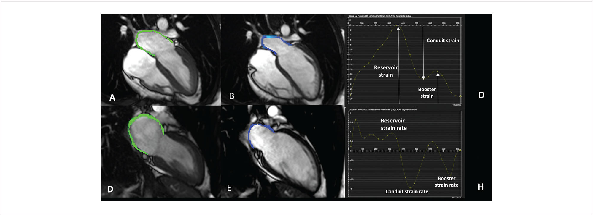
Assessment of left atrial strain (LAS) using cardiac magnetic resonance (CMR) is gaining interest in cardiology as a novel parameter, as it quantifies the left atrium’s (LA) deformation throughout the cardiac cycle. Traditionally, LA function was inferred from left ventricular ejection fraction and LA volume. However, recent studies emphasize the prognostic significance of directly analyzing atrial function, particularly in atrial fibrillation (AF), heart failure, and arterial hypertension. CMR provides a thorough evaluation of cardiac anatomy and function, enabling precise and reproducible measurement of atrial tissue deformation.

  • Cardiac Magnetic Resonance Imaging
  • Left atrium
  • Right Atrium
  • Stran Imaging
Read Full Article
Creative Commons Attribution (CC BY) Creative Commons license
26/06/2024 DOI: 10.36660/abcimg.20240020i Views 1
Figure 1
ABCIC
Case Report

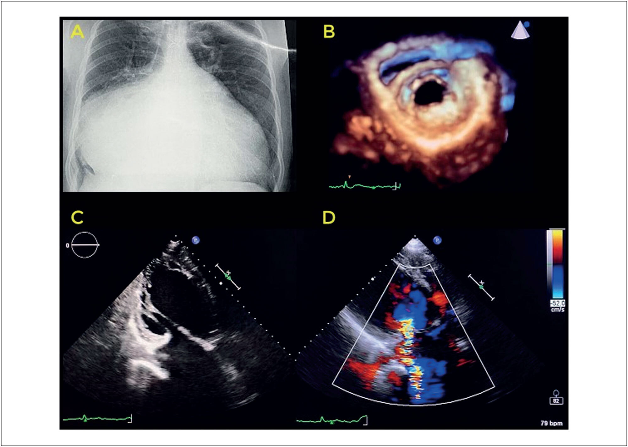
Giant Left Atrium due to Probable Rheumatic Disease and Atrial Functional Mitral Insufficiency

Israel Nilton de Almeida Feitosa et al. Isaac Newton Guimarães Andrade

Introduction

Giant left atrium is a rare condition, and most cases described in the literature are associated with rheumatic disease. However, some clinical descriptions suggest a nonrheumatic etiology for this condition, in which characteristic findings of rheumatic disease are not identified or anatomopathological findings do not suggest a specific etiology.

Atrial fibrillation is present in almost all cases and contributes to the maintenance of left atrial remodeling and persistent changes in structure and function.

[…]

  • Echocardiography
  • Left atrium
  • rheumatic heart disease
Read Full Article
Creative Commons Attribution (CC BY) Creative Commons license
25/04/2024 DOI: 10.36660/abcimg.20240018i
ABCIC
Image

Giant Left Atrium Due to Severe Rheumatic Double Mitral Valve Dysfunction

Glenda Alves de Sá et al. Danilo Bora Moleta Rogério Simões Miranda Geórgia C. S. Souza Marcos Danillo Peixoto Oliveira

[…]

  • Diagnosis
  • Echocardiography
  • Left atrium
Read Full Article
Creative Commons Attribution (CC BY) Creative Commons license
14/01/2022 DOI: 10.5935/2318-8219.20190030
Figure 1
ABCIC
Original Article

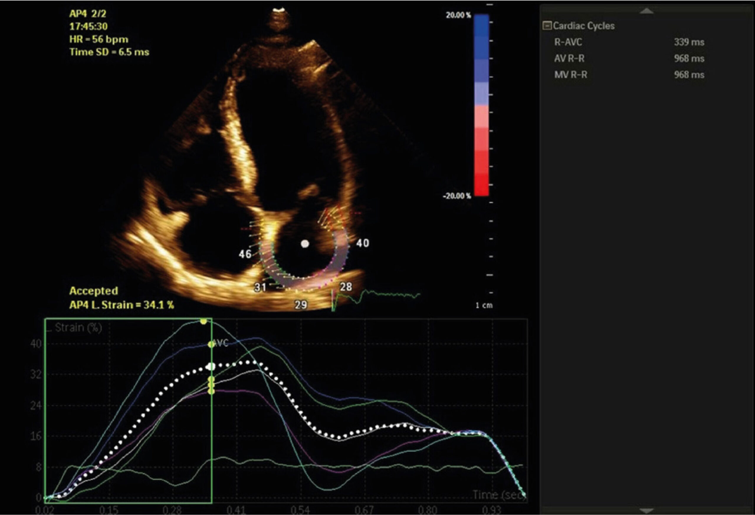
Left Atrial Reserve Function in Assessing Indeterminate Diastolic Function

Silvio Henrique Barberato et al. Rafael Borsoi

Abstract

Background

Elevation of left ventricular filling pressures secondary to diastolic dysfunction plays a central role in the pathophysiology of heart failure. However, international guidelines still fail to diagnose diastolic dysfunction in some cases.

Objective

To evaluate left atrial reservoir function in indeterminate diastolic function

Method

Observational study with individuals in sinus rhythm and preserved left ventricular ejection fraction, submitted to echocardiogram and divided into three groups according to the combined analysis of E/e´ ratio and indexed left atrium volume: Group 1, if normal left ventricular filling pressures; Group 2, if increased left ventricular filling pressures and Group 3, if indeterminate left ventricular filling pressures. Two-dimensional speckle tracking was used to measure peak left atrial strain (LAS). Analysis of variance, Student’s t test and receiver-operator curve (ROC) were used in the statistical analysis.

Results

We included 58 patients who had 61 ± 14 years old, 57% of whom were women, and had average left ventricular ejection fraction 62 ± 7%. Groups 2 and 3 had lower LAS than Group 1 (20 ± 5% versus 22 ± 6% versus 30 ± 8%, respectively, p = 0.004), but did not differ between them (p = 0.93). LAS was a good predictor of elevated left ventricular filling pressures (p = 0.026; area under the curve = 0.80), obtaining sensitivity of 60% and specificity of 80% with a cut-off value ≤ 20%.

Conclusion

The findings suggest that the left atrial reservoir function of individuals with indeterminate diastolic function is similar to that of individuals with advanced diastolic dysfunction, rendering LAS the potential to support the reclassification of indeterminate diastolic function.

  • Diastolic Function
  • Echocardiography
  • Heart Failure
  • Left atrium
Read Full Article
Creative Commons Attribution (CC BY) Creative Commons license
21/06/2021 DOI: 10.47593/2675-312X/20213402eabc196
Morphofunctional study of the junction between the left atrium and the pulmonary veins in patient with pulmonary hypertension
ABC
Original Article

Morphofunctional study of the junction between the left atrium and the pulmonary veins in patient with pulmonary hypertension

Jennecy Sales Cavalcanti et al. Laura Patrícia Ferreira Santos

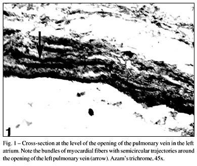
OBJECTIVE: To study the arrangement of the myocardial fiber bundles at the pulmonary venous left atrial junction in patients with pulmonary hypertension, and to discuss the pathophysiological importance of this element in the etiology of acute pulmonary edema. METHODS: We obtained 12 hearts and their pulmonary vein extremities from postmortem examinations of patients with the anatomicopathological diagnosis of acute pulmonary edema. The specimens, which had no grossly visible morphological cardiac alterations, were fixed in 10% formalin, and the muscular arrangement of the pulmonary venous left atrial junctions was analyzed. This material was then isolated, embedded in paraffin, underwent serial cutting (50 µm of thickness), and was stained with Azam’s trichrome. RESULTS: We observed in our specimens that: a) the myocardial fiber bundles that originate in the atrial wall and involve the openings of the pulmonary veins were fewer than those observed in healthy material; b) the myocardial fiber bundles that extend into the pulmonary veins were shorter than those found in material originating from individuals with no pulmonary hypertension. CONCLUSION: Anatomical changes that result in a reduction in the amount of myocardial fiber bundles in the pulmonary venous left atrial junction, isolated or associated with other factors, may be the cause of disorders in pulmonary circulation, leading to an increase in pulmonary venous pressure, and, consequently, to acute pulmonary edema.

  • Left atrium
  • Pulmonary Hypertension
  • Pulmonary Veins
Read Full Article
Creative Commons Attribution (CC BY) Creative Commons license
18/09/2001 DOI: 10.1590/S0066-782X2001000900003 Views 2

How to cite this article

EndNote RIS

Electronic Document Format (Vancouver)

Costa IBSS, Fonseca RA, Rochitte CE. My Approach to Atrial Strain by Cardiac Magnetic Resonanc. ABC Imagem Cardiovascular 2024;37(2):e20240020.

Electronic Document Format (ABNT)

Costa, Isabela Bispo Santos da Silva; Fonseca, Rafael Almeida; Rochitte, Carlos Eduardo. My Approach to Atrial Strain by Cardiac Magnetic Resonanc. ABC Imagem Cardiovascular, v. 37, n. 2, e20240020, Jun. 2024.

Electronic Document Format (APA)

Costa, I. B. S. S., Fonseca, R. A., & Rochitte, C. E. (2024). My Approach to Atrial Strain by Cardiac Magnetic Resonanc. ABC Imagem Cardiovascular, 37(2), e20240020.

Electronic Document Format (ISO)

Costa, Isabela Bispo Santos da Silva and Fonseca, Rafael Almeida and Rochitte, Carlos Eduardo. My Approach to Atrial Strain by Cardiac Magnetic Resonanc. ABC Imagem Cardiovascular [online]. 2024, vol. 37, n. 2, [cited 2026-10-14], e20240020. Available from: <https://journals.cardiol.br/en/article/my-approach-to-atrial-strain-by-cardiac-magnetic-resonanc/>. ISSN 2318-8219

EndNote RIS

Electronic Document Format (Vancouver)

Feitosa INA, Andrade ING. Giant Left Atrium due to Probable Rheumatic Disease and Atrial Functional Mitral Insufficiency. ABC Imagem Cardiovascular 2024;37(2):e20240018.

Electronic Document Format (ABNT)

Feitosa, Israel Nilton de Almeida; Andrade, Isaac Newton Guimarães. Giant Left Atrium due to Probable Rheumatic Disease and Atrial Functional Mitral Insufficiency. ABC Imagem Cardiovascular, v. 37, n. 2, e20240018, Apr. 2024.

Electronic Document Format (APA)

Feitosa, I. N. A., & Andrade, I. N. G. (2024). Giant Left Atrium due to Probable Rheumatic Disease and Atrial Functional Mitral Insufficiency. ABC Imagem Cardiovascular, 37(2), e20240018.

Electronic Document Format (ISO)

Feitosa, Israel Nilton de Almeida and Andrade, Isaac Newton Guimarães. Giant Left Atrium due to Probable Rheumatic Disease and Atrial Functional Mitral Insufficiency. ABC Imagem Cardiovascular [online]. 2024, vol. 37, n. 2, [cited 2026-10-14], e20240018. Available from: <https://journals.cardiol.br/en/article/giant-left-atrium-due-to-probable-rheumatic-disease-and-atrial-functional-mitral-insufficiency/>. ISSN 2318-8219

EndNote RIS

Electronic Document Format (Vancouver)

Sá GA, Moleta DB, Miranda RS, Souza GCS, Oliveira MDP. Giant Left Atrium Due to Severe Rheumatic Double Mitral Valve Dysfunction. Arquivos Brasileiros de Cardiologia Imagem cardiovascular 2022;32(2):141-3.

Electronic Document Format (ABNT)

Sá, Glenda Alves de; Moleta, Danilo Bora; Miranda, Rogério Simões; Souza, Geórgia C. S.; Oliveira, Marcos Danillo Peixoto. Giant Left Atrium Due to Severe Rheumatic Double Mitral Valve Dysfunction. Arquivos Brasileiros de Cardiologia Imagem cardiovascular, v. 32, n. 2, p. 141-143, Jan. 2022.

Electronic Document Format (APA)

Sá, G. A. , Moleta, D. B., Miranda, R. S., Souza, G. C. S., & Oliveira, M. D. P. (2022). Giant Left Atrium Due to Severe Rheumatic Double Mitral Valve Dysfunction. Arquivos Brasileiros de Cardiologia Imagem cardiovascular, 32(2), 141-143.

Electronic Document Format (ISO)

Sá, Glenda Alves de and Moleta, Danilo Bora and Miranda, Rogério Simões and Souza, Geórgia C. S. and Oliveira, Marcos Danillo Peixoto. Giant Left Atrium Due to Severe Rheumatic Double Mitral Valve Dysfunction. Arquivos Brasileiros de Cardiologia Imagem cardiovascular [online]. 2022, vol. 32, n. 2, [cited 2026-10-14], pp. 141-143. Available from: <https://journals.cardiol.br/en/article/giant-left-atrium-due-to-severe-rheumatic-double-mitral-valve-dysfunction/>. ISSN 2318-8219

EndNote RIS

Electronic Document Format (Vancouver)

Barberato SH, Borsoi R. Left Atrial Reserve Function in Assessing Indeterminate Diastolic Function. ABC Imagem Cardiovascular 2021;34(2):eabc196.

Electronic Document Format (ABNT)

Barberato, Silvio Henrique; Borsoi, Rafael. Left Atrial Reserve Function in Assessing Indeterminate Diastolic Function. ABC Imagem Cardiovascular, v. 34, n. 2, eabc196, Jun. 2021.

Electronic Document Format (APA)

Barberato, S. H., & Borsoi, R. (2021). Left Atrial Reserve Function in Assessing Indeterminate Diastolic Function. ABC Imagem Cardiovascular, 34(2), eabc196.

Electronic Document Format (ISO)

Barberato, Silvio Henrique and Borsoi, Rafael. Left Atrial Reserve Function in Assessing Indeterminate Diastolic Function. ABC Imagem Cardiovascular [online]. 2021, vol. 34, n. 2, [cited 2026-10-14], eabc196. Available from: <https://journals.cardiol.br/en/article/left-atrial-reserve-function-in-assessing-indeterminate-diastolic-function/>. ISSN 2318-8219

EndNote RIS

Electronic Document Format (Vancouver)

Cavalcanti JS, Santos LPF. Morphofunctional study of the junction between the left atrium and the pulmonary veins in patient with pulmonary hypertension. Arquivos Brasileiros de Cardiologia 2001;77(3):232-4.

Electronic Document Format (ABNT)

Cavalcanti, Jennecy Sales; Santos, Laura Patrícia Ferreira. Morphofunctional study of the junction between the left atrium and the pulmonary veins in patient with pulmonary hypertension. Arquivos Brasileiros de Cardiologia, v. 77, n. 3, p. 232-234, Sep. 2001.

Electronic Document Format (APA)

Cavalcanti, J. S., & Santos, L. P. F. (2001). Morphofunctional study of the junction between the left atrium and the pulmonary veins in patient with pulmonary hypertension. Arquivos Brasileiros de Cardiologia, 77(3), 232-234.

Electronic Document Format (ISO)

Cavalcanti, Jennecy Sales and Santos, Laura Patrícia Ferreira. Morphofunctional study of the junction between the left atrium and the pulmonary veins in patient with pulmonary hypertension. Arquivos Brasileiros de Cardiologia [online]. 2001, vol. 77, n. 3, [cited 2026-10-14], pp. 232-234. Available from: <https://journals.cardiol.br/en/article/morphofunctional-study-of-the-junction-between-the-left-atrium-and-the-pulmonary-veins-in-patient-with-pulmonary-hypertension/>. ISSN 0066-782X

  • About ABC Family
  • Internal Regulation
  • Editorial Board
  • Brazilian Society of Cardiology (SBC)

Issues

SBC Guidelines

  • Videos
  • Podcasts
  • News