ABC Family Arquivos Brasileiros de Cardiologia International Journal of Cardiovascular Sciences ABC Heart Failure & Cardiomyopathy ABC Imagem Cardiovascular
Português English
ABC Family
Português English
  • About
    • About ABC Family
    • Internal Regulation
    • Editorial Board
    • Brazilian Society of Cardiology (SBC)
  • Issues
  • SBC Guidelines
  • Content
    • Videos
    • Podcasts
    • News
  • About
    • About ABC Family
    • Internal Regulation
    • Editorial Board
    • Brazilian Society of Cardiology (SBC)
  • Issues
  • SBC Guidelines
  • Content
    • Videos
    • Podcasts
    • News

ABC Family

  • ABC Family
  • Arquivos Brasileiros de Cardiologia
  • International Journal of Cardiovascular Sciences
  • ABC Heart Failure & Cardiomyopathy
  • ABC Imagem Cardiovascular
Search in

No content found.

Journals
Ordering
Article types
Others

  1. Home
  2. Keyword: Transesophageal echocardiography
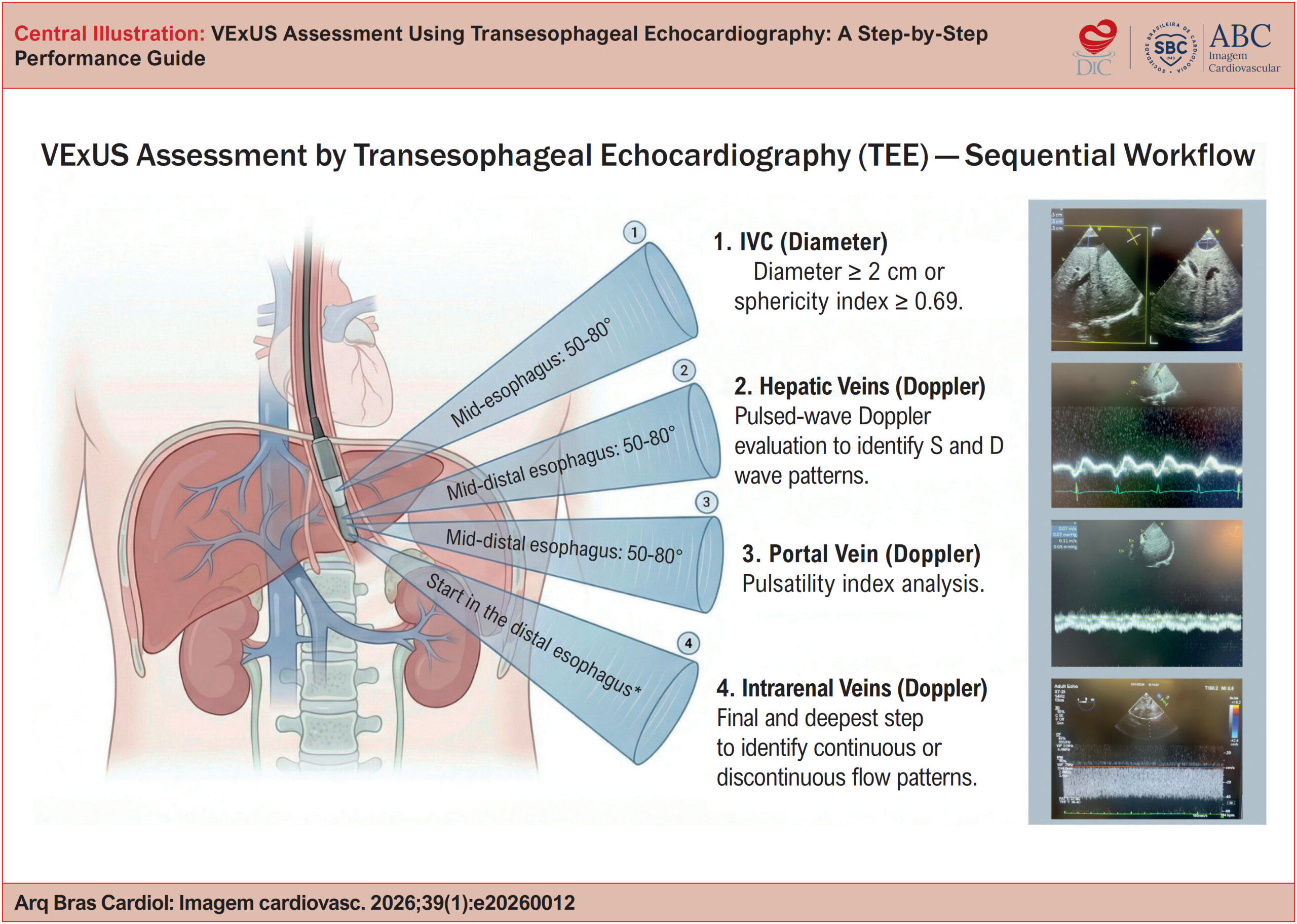
My Approach to VExUS Assessment Using Transesophageal Echocardiography: A Step-by-Step Performance Guide
ABCIC
Review Article

My Approach to VExUS Assessment Using Transesophageal Echocardiography: A Step-by-Step Performance Guide

Angelo Antunes Salgado et al. Marcos Paulo Lacerda Bernardo Marcelo Ramalho Fernandes

ABSTRACT

Monitoring systemic venous congestion has become essential in the management of critically ill patients, enabling accurate diagnosis, severity grading, and prognostic stratification. Literature shows that congestion is strongly associated with the development of acute kidney injury and increased mortality when compared with optimized volume states.

In this setting, intraoperative Transesophageal Echocardiography (TEE) has emerged as an advanced and versatile tool. In addition to allowing detailed assessment of cardiac function, TEE is effective in assessing intravascular volume status and predicting fluid responsiveness through dynamic measurement of stroke volume and the degree of systemic congestion. TEE also enables direct visualization of abdominal vessels, such as the hepatic, portal, and intrarenal veins, thereby facilitating the identification of pathological pulsatile flow patterns even in patients with limited transthoracic acoustic windows.

The integration of protocols such as VExUS (or its modified version, mVExUS) allows for a personalized approach focused on “perfusion without congestion.” This review outlines the practical application of VExUS assessment using TEE, its technical limitations, and how to use it to guide hemodynamic resuscitation, minimizing organ injury and optimizing clinical outcomes.

  • Operative Surgical Procedures
  • Prognosis
  • Transesophageal echocardiography
Read Full Article
Creative Commons Attribution (CC BY) Creative Commons license
20/04/2026 DOI: 10.36660/abcimg.20260012i Views 1
Figure 1
ABCIC
Image

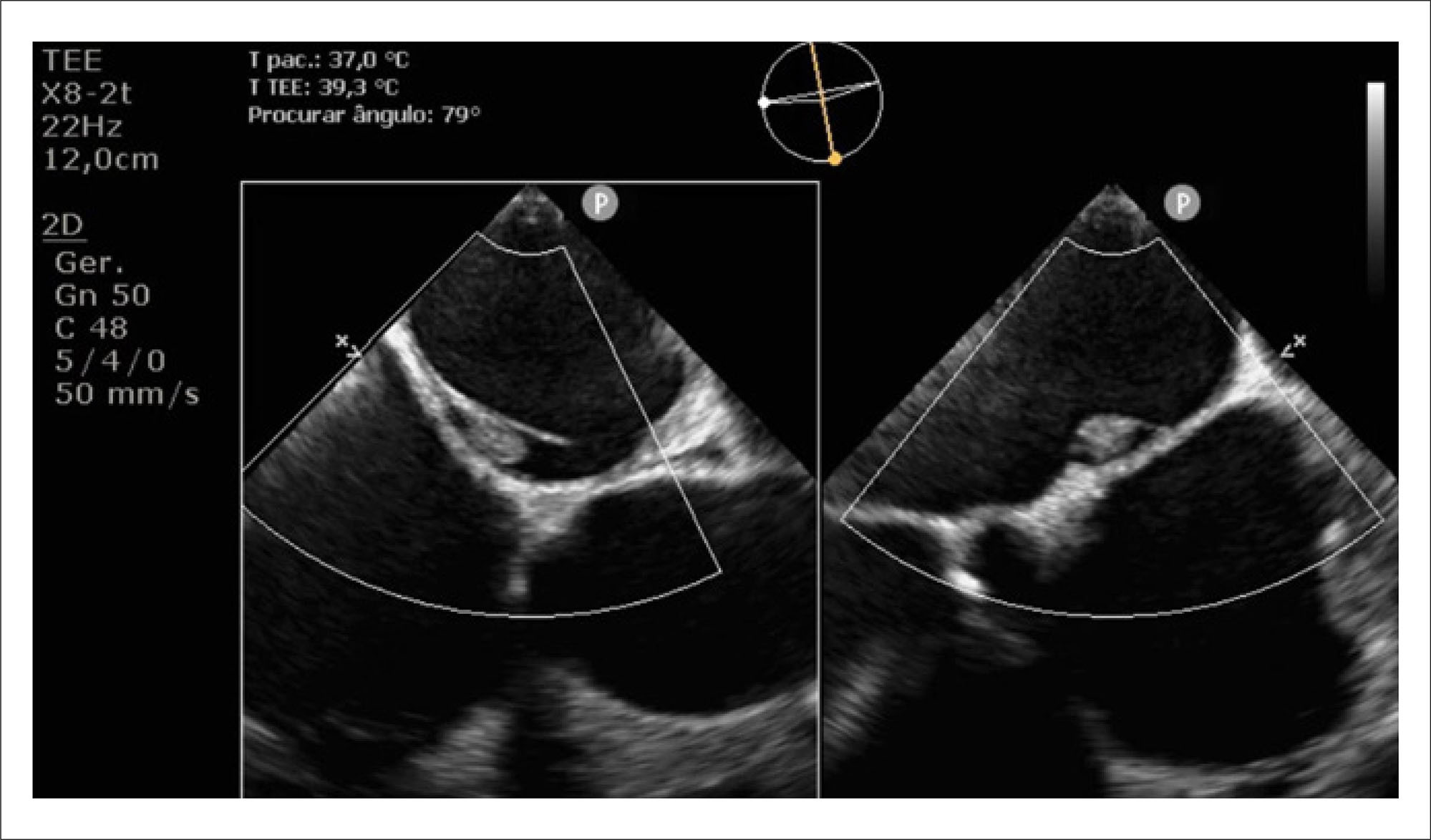
Left Atrial Septal Pouch Thrombosis Detected Before Electrical Cardioversion: a Rare Source of Embolism

Andre Barcellos Amon et al. Mariana De Castro Lopes Marina Petersen Saadi Willian R. Menegazzo Angela Barreto Santiago Santos

A 59-year-old male patient with a history of systemic arterial hypertension was admitted to the emergency department with a seven-day history of chest pain and palpitations. On examination, he was hypotensive (93/70 mmHg) and tachycardic (150 bpm). A prominent systolic–diastolic murmur was heard along the right upper sternal border.

The initial investigation included electrocardiography (ECG) and Transthoracic Echocardiography (TTE). The ECG revealed atrial flutter, and the TTE showed biventricular dilation [Left Ventricular (LV) end-diastolic/end-systolic diameters: 60/51 mm; right ventricular (RV) basal diameter: 54 mm] and severe systolic dysfunction (LV ejection fraction: 19%; RV fractional area change: 16%). Additionally, a bicuspid aortic valve with mixed disease was identified: low-flow, low-gradient aortic stenosis (peak velocity: 3.1 m/s; peak/mean gradients: 39/23 mmHg; velocity ratio: 0.15; valve area: 0.7 cm, stroke volume index: 17 ml/m) and severe aortic regurgitation (pressure half-time: 170 ms; holodiastolic reversal in the descending aorta, end-diastolic velocity: 24 cm/s).

[…]

  • Atrial Septum
  • Thrombosis
  • Transesophageal echocardiography
Read Full Article
Creative Commons Attribution (CC BY) Creative Commons license
03/02/2026 DOI: 10.36660/abcimg.20250075i
Central Illustration
ABCIC
Review Article

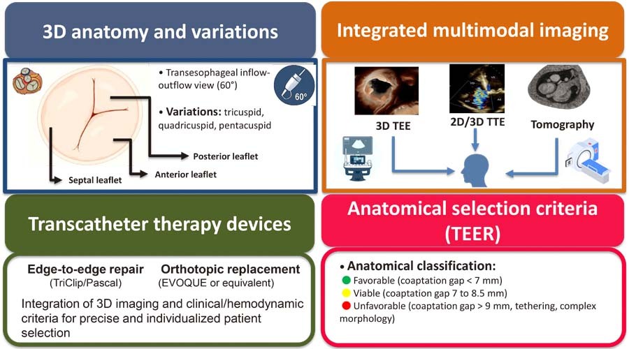
Tricuspid Valve: Anatomical Basis and Patient Selection Criteria for Transcatheter Structural Interventions

Alexandre Costa Souza et al. Halsted Alarcão Gomes Pereira da Silva Alex dos Santos Felix

Abstract

Tricuspid regurgitation has been recognized as a relevant clinical entity, with a direct impact on morbidity and mortality. Recent advances in multimodal cardiovascular imaging have promoted more detailed anatomical and functional assessment of the tricuspid apparatus, guiding the indication and planning of transcatheter interventions. To provide a review of the anatomical basis of the tricuspid valve and discuss echocardiographic and clinical criteria for selecting candidates for transcatheter therapies. This narrative review of the anatomical variability of the tricuspid valve and its subvalvular apparatus incorporates recent recommendations from echocardiography societies and clinical evidence. Special emphasis has been given to three-dimensional echocardiography, transesophageal echocardiography, and computed tomography for morphological and functional characterization of tricuspid regurgitation. Contemporary evidence has demonstrated that up to 45% of patients present with non-classical morphologies (quadricuspid or pentacuspid valves), which impact the viability and outcomes of edge-to-edge repair. The integration of three-dimensional echocardiography enhances accuracy in grading regurgitation severity, enables precise measurement of the vena contracta area, and assists in device selection. Clinical factors including right ventricular function, pulmonary artery systolic pressure, and the presence of intracardiac devices also influence patient selection. Appropriate selection of candidates for transcatheter tricuspid valve therapies requires a systematic approach with integrated application of imaging methods and clinical parameters. Detailed echocardiographic characterization of leaflet morphology, coaptation gap, and annulus geometry is crucial for planning the procedure, selecting the device, and predicting outcomes.

  • Three-Dimensional Echocardiography
  • Transesophageal echocardiography
  • Tricuspid Valve
  • Tricuspid Valve Insufficiency
Read Full Article
Creative Commons Attribution (CC BY) Creative Commons license
16/12/2025 DOI: 10.36660/abcimg.20250064i Views 2
ABCIC
Editorial

Transesophageal Echocardiogram Always Before TAVI?

Jairo Alves Pinheiro et al. João Lucas Pinheiro Lucas Velloso Dutra Luciano Holanda Renata de Sá Cassar

Cardiovascular imaging is crucial in the selection and planning of interventions for structural heart disease.

Transcatheter aortic valve implantation (TAVI) has transformed the management of severe aortic stenosis (AS) in high-risk surgical populations and is currently being expanded to intermediate- and low-risk populations.

[…]

  • Aortic Valve Stenosis
  • Magnetic Resonance Imaging
  • Transcatheter Aortic Valve Replacement
  • Transesophageal echocardiography
Read Full Article
Creative Commons Attribution (CC BY) Creative Commons license
08/12/2025 DOI: 10.36660/abcimg.20250060i
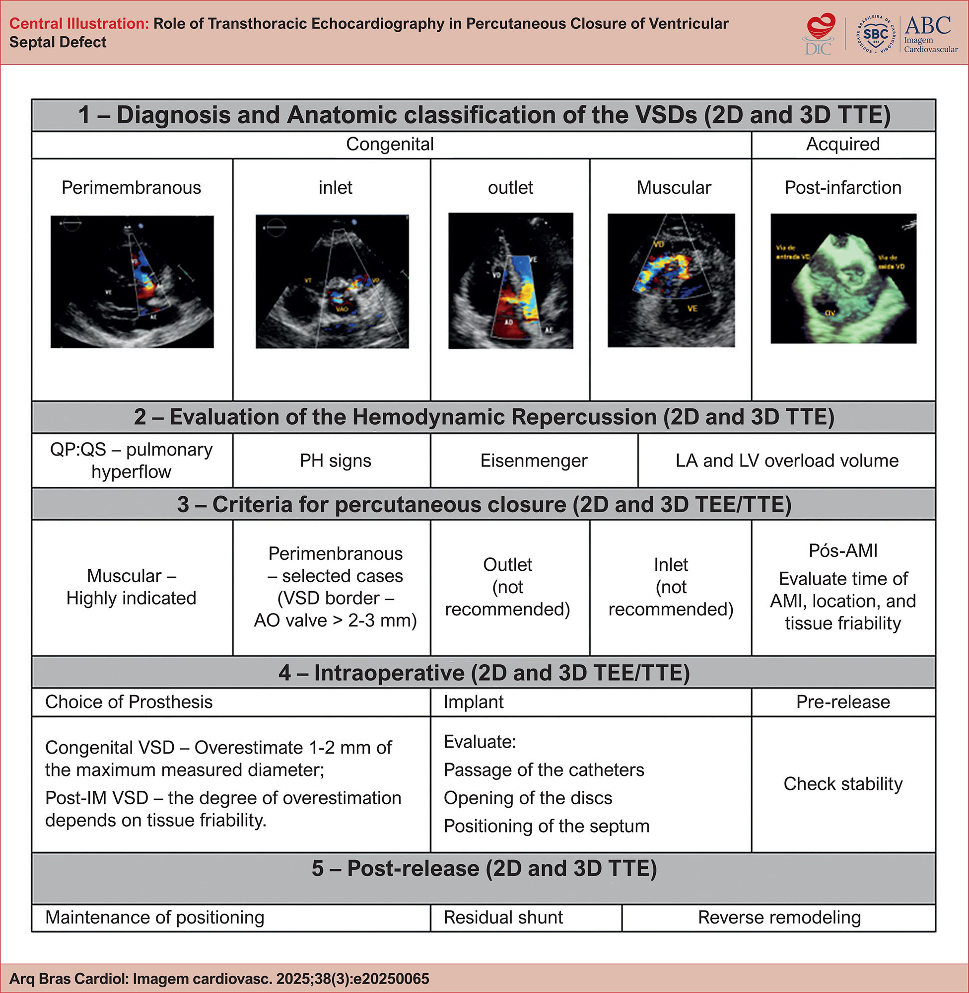
Role of Transthoracic Echocardiography in Percutaneous Closure of Ventricular Septal Defect
ABCIC
Review Article

Role of Transthoracic Echocardiography in Percutaneous Closure of Ventricular Septal Defect

João Batista Masson Silva et al. Cloves Geraldino da Silva Junior

Abstract

Ventricular septal defect (VSD) is the most common congenital heart defect, and its management has evolved with percutaneous closure, a minimally invasive alternative to surgery. In this context, echocardiography has established itself as the primary imaging tool, crucial in all phases of the procedure. Transthoracic echocardiography (TTE) is essential for diagnosis, anatomical classification (perimembranous, muscular, inlet and outlet), initial hemodynamic assessment, and long-term follow-up. Indication for closure is based on such criteria as the left ventricular (LV) volume overload, with Qp:Qs ≥ 1.5:1. For detailed planning and intraprocedural guidance, transesophageal echocardiography (TEE), especially with three-dimensional (3D) reconstruction, is the gold standard. 3D TEE offers an accurate measurement of defect diameters, a border analysis for safe device anchorage, and an assessment of the relationship with adjacent structures, such as the aortic and tricuspid valves. The success of the intervention, both in congenital VSDs and in the rare and complex post-infarction VSDs, depends directly on the quality of the echocardiographic evaluation. The multimodal approach, which integrates TTE and TEE, ensures appropriate patient selection and safe procedure management, optimizing outcomes and expanding the realms of percutaneous VSD treatment.

  • Echocardiography
  • Transesophageal echocardiography
  • Ventricular Heart Septal Defects
Read Full Article
Creative Commons Attribution (CC BY) Creative Commons license
03/12/2025 DOI: 10.36660/abcimg.20250065i
ABCIC
Editorial

Impact of Anxiety on Patients Referred for Transesophageal Echocardiography

Augusto Alberto da Costa Jr.

In this issue of ABC Cardiovascular Imaging, anxiety is highlighted not only as a factor in the subjective assessment of patients’ experiences during invasive medical procedures (transesophageal echocardiography, TEE) but also for its influence on the amount of sedative drugs administered during the procedure.

The study The Impact of Anxiety on Patients Referred for Transesophageal Echocardiography, conducted at a tertiary institution, provides a detailed analysis of multiple characteristics of patients undergoing the exam, its influence on sedation, and revisits important aspects of good medical practice. The study results indicate that anxiety, assessed using easily implemented indicators, was present in 62% of patients. Self-assessment tables show mild anxiety in 40%, moderate anxiety in 16%, and severe anxiety in 6%. It is part of the experience of echocardiographers performing transesophageal exams to deal with patient anxiety, especially among younger individuals.,

[…]

  • Anxiety
  • Patients
  • Transesophageal echocardiography
Read Full Article
Creative Commons Attribution (CC BY) Creative Commons license
27/06/2025 DOI: 10.36660/abcimg.20250036i
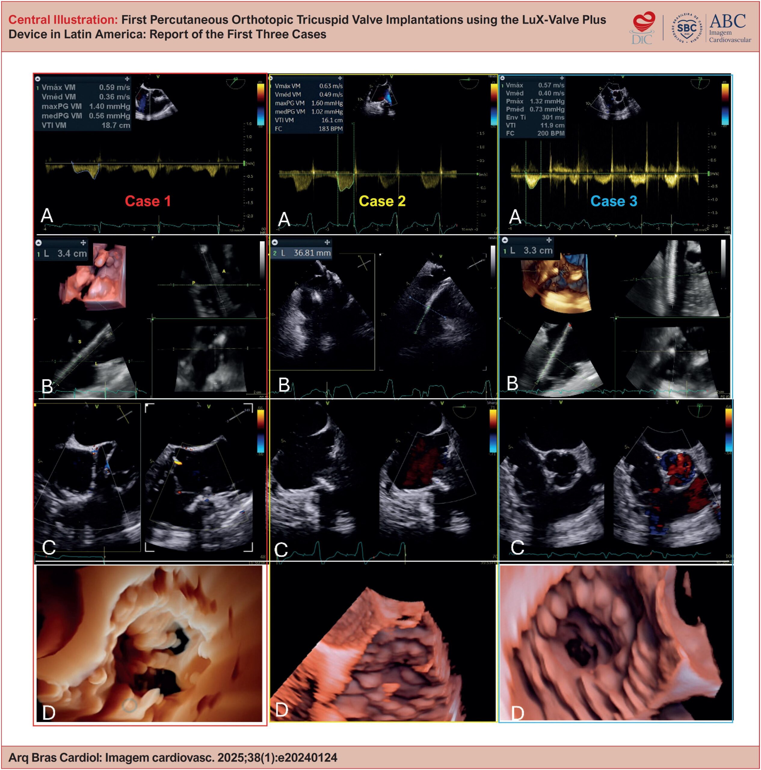
First Percutaneous Orthotopic Tricuspid Valve Implantations using the LuX-Valve Plus Device in Latin America: Report of the First Three Cases
ABCIC
Original Article

First Percutaneous Orthotopic Tricuspid Valve Implantations using the LuX-Valve Plus Device in Latin America: Report of the First Three Cases

Fernanda Esteves et al. Vinícius Esteves Marcio Mendes Pereira Frederico José Neves Mancuso … Marden Tebet Sergio Kreimer Edmur Carlos de Araújo Francisco Monteiro de Almeida Magalhães

Abstract

Background:

Severe tricuspid regurgitation (TR) is associated with high morbidity and mortality, especially in patients with contraindications for surgery. Transcatheter tricuspid valve replacement has emerged as a minimally invasive alternative for this population, with the LuX-Valve Plus device designed for orthotopic implantation via the transjugular route.

Objective:

To report the first 3 cases of transcatheter tricuspid valve replacement using the LuX-Valve Plus device in Latin America, assessing the feasibility, safety, and immediate clinical outcomes.

Methods:

The study included 3 female patients with torrential TR and high surgical risk. Pre-procedure assessment included transesophageal echocardiography, 3-dimensional transthoracic echocardiography (3D TEE), computed tomography, and cardiac catheterization. The procedure was performed via transjugular access, under general anesthesia, with 3D TEE and fluoroscopy guidance. Implant efficacy was assessed as resolution of TR, transprosthetic gradient, and post-procedure complications.

Results:

The implants were successful in all cases, with complete resolution of TR, absence of paravalvular leaks, and adequate transprosthetic gradient. Right ventricular function was preserved, and all patients were discharged 3 to 4 days after the procedure with no significant complications.

Conclusion:

Transcatheter tricuspid valve replacement using the LuX-Valve Plus was shown to be a viable and safe alternative for patients with severe TR and high surgical risk. The use of 3D TEE was essential to the planning and execution of the implant. Further studies are needed to assess durability and long-term clinical outcomes.

  • Transcatheter Aortic Valve Replacement
  • Transesophageal echocardiography
  • Tricuspid Valve
  • Tricuspid Valve Insufficiency
Read Full Article
Creative Commons Attribution (CC BY) Creative Commons license
28/03/2025 DOI: 10.36660/abcimg.20240124i Views 1
Central Illustration
ABCIC
Review Article

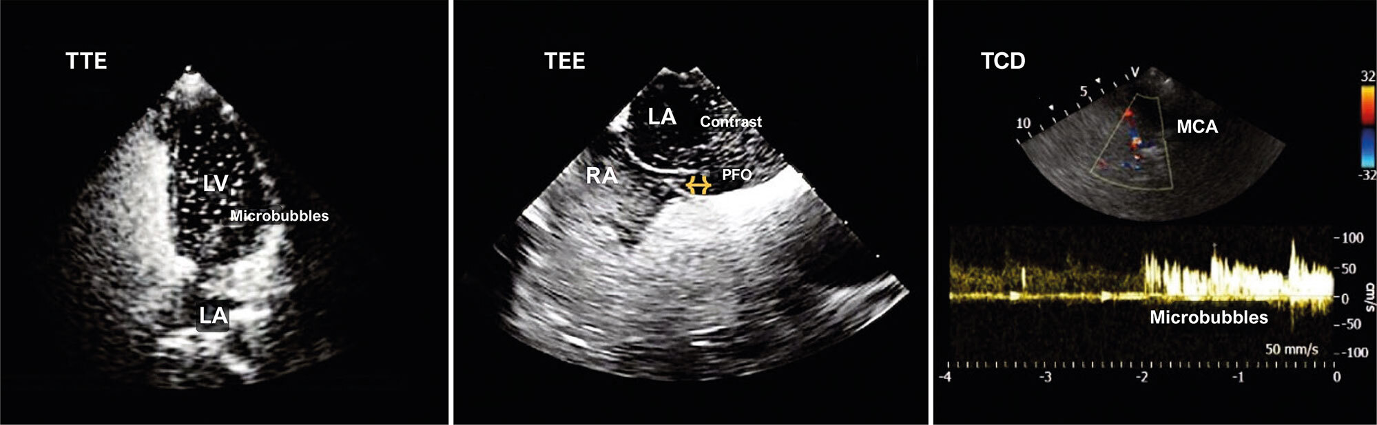
My Approach to Transcranial Doppler Combined With Transthoracic and Transesophageal Echocardiography to Investigate PFO

Gracielly Barros et al. Patrick Ventorim Costa

Abstract

This review article explores the combined use of Transcranial Doppler (TCD) with transthoracic (TTE) and transesophageal (TEE) echocardiography to investigate Patent Foramen Ovale (PFO), a condition found in approximately 25-27% of the adult population. PFO is a remnant of fetal circulation and the most common cause of interatrial shunt. It is associated with clinical complications such as platypnea-orthodeoxia syndrome, obstructive sleep apnea, migraine, and stroke.

PFO assessment is generally performed using TTE and TEE, but TCD, a highly sensitive and less used method, is also important. TCD indirectly detects shunts in awake patients and uses the microembolic signal (MES) grading score to classify shunt severity. This technique involves injecting a contrast solution into the antecubital vein during the Valsalva maneuver to detect intracardiac shunt by observing microbubble appearance in the first cardiac cycles.

The case report illustrates the effectiveness of TCD in the initial screening of a patient with suspected PFO using TTE, TCD and TEE. The conclusion highlights that TCD is a crucial diagnostic tool, particularly relevant for the therapeutic management of patients with cryptogenic stroke. Several scientific publications are referenced, providing a theoretical foundation for the combined use of TCD, TTE, and TEE in PFO investigation.

  • Foramen Ovale
  • Stroke
  • Transcranial Doppler Ultrasonography
  • Transesophageal echocardiography
Read Full Article
Creative Commons Attribution (CC BY) Creative Commons license
02/08/2024 DOI: 10.36660/abcimg.20240051i
ABCIC
Editorial

Transesophageal Echocardiography in Aortic Valve Stenosis: A Pre-Intervention “Toll”?

Antonio de Santis et al. Francisco Monteiro de Almeida Magalhães

Aortic stenosis (AS) has become one of the most relevant valve diseases in cardiology care due to its increasing prevalence, resulting from the positive relationship between the preeminently degenerative etiology and global population aging. In Brazil, we highlight an epidemiological scenario characterized by an etiopathogenic coexistence: rheumatic fever and bicuspid valve disease in younger patients and calcific (degenerative) etiology in elderly patients. Regarding the natural history, AS leads to elevated cardiovascular morbidity and mortality in its symptomatic phase, with unfavorable prognosis when it is not diagnosed correctly and without timely intervention.–

Echocardiography is still a fundamental tool for diagnosis, allowing both evaluation of etiological characteristics and grading of AS severity using parameters such as aortic valve area, transvalvular pressure gradient, and peak aortic jet velocity. However, there are still questions related to the best window for acquiring echocardiographic data. Is the transesophageal window a necessary “toll” for all patients with AS for whom intervention is planned? In general, transesophageal echocardiography (TEE) has the potential to provide superior image quality to transthoracic echocardiography (TTE) in the evaluation of posterior cardiac structures close to the esophagus, for example, the mitral valve. In this case, the greater distance between these structures and the echocardiographic transducer might make evaluation through the transthoracic window difficult. However, the anterior anatomical topography of the aortic valve allows adequate analysis by TTE in most patients, thus avoiding exposure to sedation and possible complications of the transesophageal modality. Data from the literature have shown that TTE and TEE are comparable, mainly in the anatomical and etiological characterization of AS, with compatible results in estimating the valve area.– Nonetheless, there is evidence that the determination of the hemodynamic severity of AS, expressed by the transvalvular pressure gradient and aortic jet velocity, may be underestimated in TEE. Variations in blood volume and preload, conditioned by the fasting and sedation required for the exam, could justify these particularities in transesophageal evaluation.

[…]

  • Aortic Valve Stenosis
  • Heart Valve Diseases
  • Transesophageal echocardiography
Read Full Article
Creative Commons Attribution (CC BY) Creative Commons license
28/05/2024 DOI: 10.36660/abcimg.20240023i Views 1
ABCIC
Brief Communication

Warning and Recommendations from the Department of Cardiovascular Imaging Regarding Transesophageal Echocardiography in Patients Using GLP-1 Analogs

Alex dos Santos Felix et al. Andre Luiz Cerqueira Almeida Marcelo Dantas Tavares de Melo Silvio Henrique Barberato

Transesophageal echocardiography is a fundamental diagnostic method for evaluating complex heart diseases. It provides essential data for therapeutic definition and, in some cases, assists in evaluation of percutaneous or surgical interventions.

Transesophageal echocardiography exams are performed on outpatients or inpatients, who might be in wards, intensive care units, or even in surgery, with heterogeneous scenarios that need to be individually assessed, not only regarding the exam routine, but also always following precise clinical indications, weighing the risks and benefits for the patient.

[…]

  • Diagnosis
  • Glucagon-Like Peptide 1
  • Transesophageal echocardiography
Read Full Article
Creative Commons Attribution (CC BY) Creative Commons license
13/12/2023 DOI: 10.36660/abcimg.20230092i Views 2
Fígure 1
ABCIC
Case Report

Transesophageal Echocardiogram in the Diagnosis of Superior Vena Cava Syndrome in a Patient With a Long-Term Catheter

Marcus Vinicius Silva Freire de Carvalho et al. Endy de Santana Alves Laila Caroline Gomes Carolina Thé Macêdo … Marco André Moraes Sales Alexandre Costa Souza

Introduction

The superior vena cava syndrome (SVCS) is caused by the obstruction or severe occlusion of the superior vena cava (SVC) and can result in significant morbidity or mortality., Malignancy is the most common cause of this obstruction, corresponding to approximately 70% of the cases. However, more recently, the incidence of SVCS related to devices, such as central venous catheters and electrodes of a pacemaker or defibrillator, have increased.,

These devices, together with states of hypercoagulability, can precipitate the formation of thrombi. In extreme situations, in the context of bacteremia, these can lead to the formation of a complex thrombus-vegetation association.

[…]

  • Diagnosis
  • syndrome, vein
  • Transesophageal echocardiography
Read Full Article
Creative Commons Attribution (CC BY) Creative Commons license
21/06/2023 DOI: 10.36660/abcimg.20230028i Views 2
ABC
Editorial

Rhythm Control Interventions in Patients with Atrial Fibrillation – Insights on Preprocedural Anticoagulation and Utility of Left Atrial Imaging

Mirella Facin et al. Nelson Samesima

The most common sustained arrhythmia in clinical practice is atrial fibrillation (AF), , affecting 2-4% of the adult population worldwide. It is even more frequent with aging, with almost 10% prevalence in individuals older than 80. Current estimates state that one in every three adults aged 55 years will develop AF during their lifetime, leading to substantial healthcare and economic burden. Clinical issues relate primarily to thromboembolic events (TE) and arrhythmic symptoms, both central targets while managing patients with AF. ,

Overall, atrial fibrillation confers a 2-5-fold escalated risk of TE, which is not evenly distributed, depending on unique modifiers. Important risk factors abridged in the CHA2DS2-VASc score – Congestive heart failure, Hypertension, Age ≥75 years, Diabetes, Stroke/TIA, Vascular disease, Age 65-74 years, Sex (female) – may predict stroke risk, consistently mitigated in almost 70% by proper anticoagulation. Vitamin K antagonists (VKAs) were the only oral anticoagulants available for over half a century. From 2009 to 2013, pivotal randomized controlled trials acquainted the scientific community with the new/direct oral anticoagulants (DOACs). – These drugs not only held similar efficacy to VKA in preventing thromboembolic events but also had a better safety profile against major bleeding – notably intracranial hemorrhage – and a more predictable pharmacokinetic and pharmacodynamic profile, ruling out the need for routine laboratory monitoring. However, the applicability of DOACs in off-label backgrounds, including stroke prevention during rhythm control interventions, remained unclear for many years.

[…]

  • Atrial Fibrillation
  • Electric Countershock
  • Thrombosis
  • Transesophageal echocardiography
Read Full Article
Creative Commons Attribution (CC BY) Creative Commons license
21/10/2022 DOI: 10.36660/abc.20220663
ABC
Original Article

Left Atrial Thrombus and Dense Spontaneous Contrast in Direct Oral Anticoagulant Therapy of Atrial Fibrillation: Insights from a Reference Center

Thiago Marques et al. Francisco Darrieux Fábio Gouvêa Leandro Garambone … Ana Paula Lindoso João Lage Luciana Sacilotto Ana Lúcia Coimbra Martina Pinheiro Natália Olivetti Sissy Lara Carina Hardy Guilherme Athayde Denise Hachul Cristiano Pisani Tan Chen Wu Maurício Scanavacca

Abstract

Background

In the treatment of atrial fibrillation (AF), the most frequently sustained arrhythmia, with catheter ablation (CA) or electrical cardioversion (ECV), the periprocedural period is one of the most critical phases. Currently, the use of new direct action oral anticoagulants (DOAC) is increasingly frequent; however, in the real world, there are still few data on studies on the thrombus incidence in the left atrium (TrLA) or dense spontaneous contrast (DSC) on transesophageal echocardiogram (TEE).

Objective

To evaluate the prevalence of events and association with risk factors in patients using DOACs. Primary objective: to analyze the prevalence of thrombus in the LA by TEE in patients using DOAC undergoing ECV/CA. Second, evaluate the association of comorbidities with the presence of thrombi and DSC.

Methods

Retrospective cohort, single-center study with patients followed at the Arrhythmia Outpatient Unit (InCor-HCFMUSP). Patients indicated for procedures and using DOACs were selected, and their clinical/echocardiographic data were analyzed. A significance level of 5% was considered.

Results

354 patients were included, a total of 400 procedures, from March 2012-March 2018. Thrombus in the LA was found in 11 patients (2.8%), associated with advanced age (p=0.007) and higher CHA2DS2-VASc (p<0.001) score. DSC in the LA before TEE was found in 29 patients (7.3%), with lower LVEF (p<0.038) and greater LA dimension (p<0.0001).

Conclusion

The incidence of LA thrombus and DSC in patients using DOC in the context of AF ECV/CA, although small, is not negligible. Patients with higher CHA2DS2-VASc scores, especially older and with larger LA diameter, are more prone to these echocardiographic findings.

  • Atrial Fibrillation
  • Electric Countershock
  • Transesophageal echocardiography
Read Full Article
Creative Commons Attribution (CC BY) Creative Commons license
23/09/2022 DOI: 10.36660/abc.20210658 Views 2
ABCIC
Review Article

Risk of Infection Associated with Transesophageal Echocardiography and Prevention Measures: Literature Review

Juliana Barros Becker et al. Fernanda Crosera Parreira Cláudio Henrique Fischer Valdir Ambrósio Moisés

Abstract

Background

Transesophageal echocardiography is an exam widely used in clinical practice for investigation and diagnosis of cardiac and noncardiac diseases. Although safe, it is a semi-invasive and non-risk-free examination. Cases of infection associated with transesophageal echocardiography were described and, due to the potential risk of transmission of infection during its implementation, the objective of this work was to review literature data regarding the transmission of infection during the examination, as well as prevention methods.

Methods

Review of literature on the subject between December 2017 and January 2018, through research in public domain scientific portals, in the different health science databases, including original articles, guidelines, simple and systematic reviews, case reports, published in periodicals indexed in the last 20 years.

Results

Thirteen articles fulfilled the established criteria: a systematic review of transesophageal echocardiography-related complications, six articles describing transesophageal echocardiography-related bacterial outbreaks, the British guideline on cleaning and disinfection for transesophageal echocardiography probes, four articles on adverse reactions to orthophthaldehyde residues in transesophageal echocardiography probes and an article regarding the use of protective covers for the probes.

Conclusion

The risk of infection associated with transesophageal echocardiography exists, although poorly described in the literature. It is recommended to establish specific protocols for disinfection of transesophageal echocardiography probes and routine inspection of probes. The strengthening of infection control teams is also essential for the detection and resolution of transesophageal echocardiography-related outbreaks.

  • Diagnosis
  • Disinfection
  • Transesophageal echocardiography
Read Full Article
Creative Commons Attribution (CC BY) Creative Commons license
14/01/2022 DOI: 10.5935/2318-8219.20190022 Views 1
Effective dose of sedation in transesophageal echocardiography: relation to age, body surface area and left ventricle function
ABC
Artigo Original

Effective dose of sedation in transesophageal echocardiography: relation to age, body surface area and left ventricle function

Guilherme Moreira José et al. Carlos Eduardo Suaide Silva Luiz Darcy Cortez Ferreira Yara Prosdossini Soares de Novaes … Claudia Gianini Monaco Manuel Adán Gil Renata Rejane Linhares Marcelo Sgrott Rodrigues Antônio S. S. de Moraes Juarez Ortiz

BACKGROUND: Sedation with midazolam and meperidine is widely used in transesophageal echocardiography. However, no mean dose is established for each individual case. OBJECTIVE: To correlate the mean midazolam and meperidine doses for proper sedation for transesophageal echocardiography with age range, body surface area, and left ventricular ejection fraction. METHODS: Retrospective study comprising 1,841 patients undergoing sedation based on the Ramsay scale, with a solution containing midazolam 1.5 mg (1.5 ml), meperidine 1 mg (1 ml) and distilled water (7.5 ml). Four age groups were analyzed: G1: 65 years. Body surface area was calculated using the formula {[(height x 100)0.725] x (weight0.425) x 0.0071}. As regards the left ventricular ejection fraction, two groups were studied: GA: 55%. The statistical analysis was carried out using the Kruskal-Wallis test for the correlation with age and left ventricular ejection fraction, and simple linear correlation for body surface area. RESULTS: As regards age, the mean doses of sedation required were significantly lower in G3 and G4 (p<0.01). The analysis of left ventricular ejection fraction showed that this was significantly lower in GA (p<0.01). The linear correlation coefficient between dose of sedation and body surface area was 0.09 (null). CONCLUSION: The mean dose of sedatives required was lower in older individuals and in those with left ventricular systolic dysfunction. No correlation with body surface area was found.

  • Conscious Sedation
  • meperidine
  • midazolam
  • Transesophageal echocardiography
Read Full Article
Creative Commons Attribution (CC BY) Creative Commons license
05/05/2010 DOI: 10.1590/S0066-782X2009001200011 Views 1
Patent foramen ovale and ischemic stroke in young people: statistical association or causal relation?
ABC
Artigo Original

Patent foramen ovale and ischemic stroke in young people: statistical association or causal relation?

Edson Marcio Negrão et al. Ivar Viana Brandi Simone Vilela Nunes Daniel Gurgel Fernandes Távora … Mauro Nakayama Paulo Sergio Siebra Beraldo

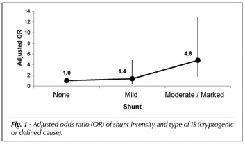
OBJECTIVES: To determine if there are evidences of a causal relation between patent foramen ovale (PFO) x cryptogenic ischemic stroke (IS) in the young population and to analyze this relation in terms of causal criteria. METHODS: A total of 168 young patients with IS was retrospectively evaluated and divided into two groups: cryptogenic and with a defined cause. As a routine procedure, the patients underwent investigation of the PFO by means of transesophageal echocardiogram and/or transcranial Doppler sonography, both of them associated with the bubble test. Multivariate analysis was performed after demonstration of univariate statistical association between PFO x IS. RESULTS: After multivariate analysis, the association between PFO x cryptogenic IS was still statistically significant with odds ratio (adjusted OR = 3.3; 95% CI: 1.5-7.4). The total number of cerebral lesions also presented a significant association with cryptogenic IS (adjusted OR = 0.4; 95% CI: 0.2-0.9). The association between PFO and cryptogenic IS met all the causality criteria. CONCLUSION: The causal relation between PFO and cryptogenic IS in the young population is highly probable. This fact should be considered in the therapeutic decision.

  • atrial septal defects
  • cerebrovascular accident
  • Patent Foramen Ovale
  • Transesophageal echocardiography
Read Full Article
Creative Commons Attribution (CC BY) Creative Commons license
15/06/2007 DOI: 10.1590/S0066-782X2007000500003
Texture of mitral bioprosthesis, ventricular function and formation of thrombus: analysis through transesophageal echocardiography and use of bioscore
ABC
Artigo Original

Texture of mitral bioprosthesis, ventricular function and formation of thrombus: analysis through transesophageal echocardiography and use of bioscore

Henry Abensur et al. Max Grinberg José A. F. Ramires

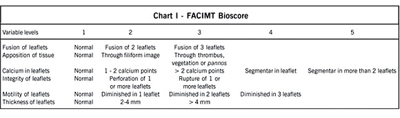
OBJECTIVE: Facing the hypothesis of participation of mechanical stress as a cause of mitral bioprosthesis dysfunction, we decided to assess the relation of preservation of the texture of the mitral bioprosthesis leaflets with left ventricular function, in addition to the casual formation of thrombus in left atrium in patients with left ventricular dysfunction from the implant of mitral bioprosthesis. METHODS: Forty 40 patients with mitral bioprosthesis through multiplane transesophageal echocardiogram were studied and divided in two groups: with left ventricular dysfunction (FE=0.40±0.09) since the bioprosthesis implant (20 patients: age 47.75±11.10 years old and surgery time 5.3±2.6 years) and with normal left ventricular function (FE=0.73±0.06) since the implant (20 patients: age 49.75±13.59 years old and surgery time 5.7±3 years). The texture of bioprosthesis leaflets was analyzed through a transesophageal echocardiographic score (FACIMT Bioscore): 1) Fusion of leaflets (score 1 to 3); 2) Apposition of tissues (score 1 to 3); 3) Calcium in leaflets (score 1 to 5); 4) Integrity of leaflets (score 1 to 3); 5) Motility of leaflets (score 1 to 4) and 6) Thickness of leaflets (score 1 to 3). The presence of thrombi in left atrium was assessed through multiplane scanning of the left atrium and left atrial appendage in the transesophageal study. RESULTS: There was no significant difference in the texture of bioprosthesis mitral position between the groups, for the total score (8.7±2.4 vs. 7.9±2.1, p=0.259), and for each analyzed item. A greater incidence of thrombi in left atrium and left atrial appendage was detected in patients with ventricular dysfunction (65% vs. 20%, p=0.004). CONCLUSION: The left ventricular dysfunction was not a protecting factor of the texture of bioprosthesis leaflets in mitral position in the tardive post-surgery period. The patients with left ventricular dysfunction showed a more favorable environment for the formation of thrombi in left atrium.

  • formation of thrombi
  • Left Ventricular Function
  • mitral bioprosthesis
  • Transesophageal echocardiography
Read Full Article
Creative Commons Attribution (CC BY) Creative Commons license
24/05/2005 DOI: 10.1590/S0066-782X2005000500011 Views 1
Bioprosthetic mitral valve thrombosis: importance of transesophageal echocardiography in the diagnosis and follow-up after treatment
ABC
Artigo Original

Bioprosthetic mitral valve thrombosis: importance of transesophageal echocardiography in the diagnosis and follow-up after treatment

Adelino Parro Jr et al. Marta Lancia Carramona Caio Augusto Ferreira Amaral José Luiz Balthazar Jacob José Carlos Nicolau

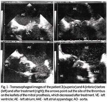
OBJECTIVE: To report the clinical and echocardiographic findings of bioprosthetic mitral valve thrombosis and the value of transesophageal echocardiography (TEE) in its diagnosis and monitoring of thrombolysis. METHODS: One hundred and eleven patients with mitral bioprostheses underwent TEE, and 4 out of 7 suspected of having a thrombus on these prostheses were included in the study (mean age = 60.2±10.2 years; 2 men). The diagnosis was confirmed with serial TEE and clinical evolution. The morphologic features of the prosthetic leaflets, as well as the presence and characteristics of attached echogenic masses were investigated. The mean gradient through the prosthesis and the valvular area were obtained. RESULTS: The diagnosis of bioprosthetic mitral valve thrombosis was established 48.7±55.2 months after surgery. Two patients had ischemic stroke in the early postoperative period. The mean overall gradient was high (11.4±3 mmHg) and the valvular area reduced (1.24±0.3 cm²). On TEE, echogenic masses on the left ventricular face of the mitral bioprosthesis suggestive of thrombus were evidenced in all patients. On serial TEE (136±233 days), in 2 patients the thrombus had disappeared and in 2 others it was smaller after treatment, the mean gradient dropped to 6.2±3 mmHg (P = 0.004; 95% CI), and the valvular area increased to 2.07±0.4 (P = NS). CONCLUSION: TEE proved to be useful for detecting bioprosthetic mitral valve thrombosis and was effective in monitoring the treatment in all patients.

  • Bioprosthesis
  • Thrombosis
  • Transesophageal echocardiography
Read Full Article
Creative Commons Attribution (CC BY) Creative Commons license
14/05/2004 DOI: 10.1590/S0066-782X2004000400006
ABC
Artigo Original

Association between the effect of spontaneous contrast in the thoracic aorta and recent ischemic stroke determined by transesophageal echocardiography

Flavio José Petersen Velho et al. Fernanda Dotta Leonora Scherer Eduardo Bartholomay … Daniela Augusta da Silva Jefferson Gomes Fernandes Marco Antonio Rodrigues Torres

OBJECTIVE: To assess the independent association between the presence of spontaneous echo contrast in the aorta and recent stroke events. METHODS: Two hundred and twenty-four individuals with a diagnosis of recent stroke and 85 control individuals who were examined due to various present/suspected heart diseases were studied through transesophageal echocardiography. The effects of spontaneous contrast in the aorta and the presence of other potential sources of cardiac embolism associated with them were researched and a questionnaire was completed about patients’ clinical risk factors at the time of examination. RESULTS: The effects of contrast in the aorta was associated with stroke (OR=2.83; CI = 95%, 1.65-4.46; P 60 years, a history of systemic blood hypertension and smoking, and dyslipidemia were risk factors independently associated with the effects of contrast in the aorta. The presence of a spontaneous contrast effect in the left atrium and Lambl’s excrescences were echocardiographic factors independently associated with the effects of contrast in the aorta. CONCLUSION: The effect of contrast in the aorta was independently associated with recent stroke and with its clinical risk factors. These results reinforce the hypothesis that the phenomenon is a predictor of several risk factors.

  • spontaneous contrast effects on the aorta
  • Stroke
  • Transesophageal echocardiography
Read Full Article
Creative Commons Attribution (CC BY) Creative Commons license
12/02/2004 DOI: 10.1590/S0066-782X2004000100005
ABC
Artigo Original

Flail mitral valve leaflet diagnosed by transesophageal echocardiography. Report and evolution of 35 cases

Mônica M. C. Caldas et al. Lúcia M. A. Fenelon William A. M. Esteves Tamara Katina … Nilson Maia Márcia M. Barbosa

PURPOSE: To describe transesophageal echocardiographic findings in patients presenting with flail mitral valve and to obtain their clinical follow-up. METHODS: From January/93 to March/97, 1675 patients were submitted to transesophageal echocardiogram at our institution; 35 of them were diagnosed as having flail mitral valve and their clinical follow-up was obtained. RESULTS: Thirty-five patients with the diagnosis of flail mitral valve were studied. Mean age was 65±15 years (12-87) and 27 (77%) were male. The posterior leaflet was involved in 25 (71%) patients. Ruptured chordae tendineae was present in all but one patient in whom flail mitral valve was due to a very elongated and redundant chordae. The etiology was prolapse and/or mixomatous degeneration in 16 patients, degenerative in 10, ischemic in 5, rheumatic in 4 and endocarditis in 3. Mitral regurgitation was severe in 25 (71%) patients and moderate in 10 (29%). The mean follow-up was 375±395 days (1 to 1380). Nineteen patients were followed clinically and 16 were submitted to surgery (transesophageal echocardiogram findings were confirmed in all 16). Overall mortality was high (34%). Among the survivors, 17 are in NYHA class I and 6 in class II. CONCLUSION: The diagnosis of flail mitral valve by transesophageal echocardiography is accurate, allowing for the definition of its mechanism and etiology, as well as the evaluation of hemodynamic compromise. The observed high mortality at the time of diagnosis may be related to the severity of the disease causing the flail mitral valve. Although non-operated survivors are doing well, the observed low functional class in these patients may be related to the short period of follow-up.

  • flail mitral valve
  • mitral regurgitation
  • Transesophageal echocardiography
Read Full Article
Creative Commons Attribution (CC BY) Creative Commons license
08/01/2002 DOI: 10.1590/S0066-782X1998001200005 Views 2
ABC
Artigo Original

Quantification of mitral prosthesis regurgitation by transesophageal echocardiography. Evaluation of different methods

Cláudio Henrique Fischer et al. Orlando Campos Fº Valdir Ambrósio Moisés José Marconi Almeida Sousa … Cristiano Vieira Machado Edgar Bezerra de Lira Fº José Lázaro de Andrade Antonio Carlos de Camargo Carvalho Dirceu Vieira Santos Fº

PURPOSE: To evaluate the different methods for grading mitral regurgitation (MR) by transesophageal echocardiography (TEE) in patients with clinical suspicion of mitral prosthesis dysfunction. METHODS: Cardiac catheterization (Cath) was performed in 15 patients for grading the severity of prosthetic MR, divided in two groups based on the presence or absence of severe MR. Prosthetic MR was quantified by TEE using methods commonly used for MR of native valves: subjective assessment by color Doppler, objective assessment based on absolute jet area and on its relative area (jet area/left atrial area) and assessment based on the presence of systolic flow reversal in pulmonary vein. RESULTS: Prosthetic MR was mostly transprosthetic (14 patients) and eccentric (11 patients). There was significant correlation (p7cm²) and relative (jet area >35% of left atrial area) jet areas did not reveal significant correlation with the angiographic grade and showed clear underestimation by TEE when the last method was used. However, there was good correlation (p30% were considered as cut point. CONCLUSION: TEE correctly identified angiographic severe mitral prosthesis regurgitation, mainly by the presence of systolic flow reversal in pulmonary vein and subjective assessment. The estimation of severity of the prosthetic MR by absolute or relative jet area seems to be limited and should be used with caution due to eccentricity of the regurgitant jet, frequently seen in mitral prosthesis dysfunction.

  • mitral regurgitation
  • prosthesis dysfunction
  • Transesophageal echocardiography
Read Full Article
Creative Commons Attribution (CC BY) Creative Commons license
08/01/2002 DOI: 10.1590/S0066-782X1998001200002 Views 2
Use of transesophageal echocardiography during implantation of aortic endoprosthesis (stent). Initial experience
ABC
Original Article

Use of transesophageal echocardiography during implantation of aortic endoprosthesis (stent). Initial experience

Cláudio Henrique Fischer et al. Orlando Campos Fº José Honório de Almeida Palma da Fonseca Cláudia Maria Rodrigues Alves … José Augusto Marcondes Sousa Edgar Bezerra de Lira Fº Antonio Carlos de Camargo Carvalho Ângelo Amato Vicenzo de Paola Ênio Buffolo

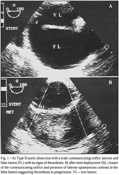
OBJECTIVE: To report the role played by transesophageal echocardiography during implantation of self-expanding aortic endoprostheses (stent) at a hemodynamics laboratory. METHODS: Thirteen patients underwent stent implantation in the descending thoracic aorta with the aid of transesophageal echocardiography during the entire procedure. Indications for stenting were as follows: 8 aortic dissections, 2 true aneurysms, 2 penetrating atherosclerotic ulcers, and 1 traumatic pseudoaneurysm. RESULTS: No complications resulting from the use of transesophageal echocardiography were observed. In 12 patients, the initial result was considered appropriate, with total or partial resolution of the major lesion confirmed by a posterior examination. In 1 patient, the procedure was suspended after transesophageal echocardiography and angiography showed that the proximal aortic diameter was inappropriate. Transesophageal echocardiography contributed to clarifying relevant points, such as aortic diameter, anatomic detail of the intimal lesion, and location and size of the communicating orifice. In addition, it facilitated placing the stent in the target lesion, reduced the time of exposure to radiation and the use of contrast medium, and provided rapid identification of intercurrent events, possibly reducing the total duration of the procedure. CONCLUSION: The use of transesophageal echocardiography during placement of aortic stents seems appropriate. The actual advantages of the procedure will be defined in a comparative prospective study.

  • aortopathies
  • Stents
  • Transesophageal echocardiography
Read Full Article
Creative Commons Attribution (CC BY) Creative Commons license
30/07/2001 DOI: 10.1590/S0066-782X2001000700001

How to cite this article

EndNote RIS

Electronic Document Format (Vancouver)

Salgado AA, Bernardo MPL, Fernandes MR. My Approach to VExUS Assessment Using Transesophageal Echocardiography: A Step-by-Step Performance Guide. ABC Imagem Cardiovascular 2026;39(1):e20260012.

Electronic Document Format (ABNT)

Salgado, Angelo Antunes; Bernardo, Marcos Paulo Lacerda; Fernandes, Marcelo Ramalho. My Approach to VExUS Assessment Using Transesophageal Echocardiography: A Step-by-Step Performance Guide. ABC Imagem Cardiovascular, v. 39, n. 1, e20260012, Apr. 2026.

Electronic Document Format (APA)

Salgado, A. A., Bernardo, M. P. L., & Fernandes, M. R. (2026). My Approach to VExUS Assessment Using Transesophageal Echocardiography: A Step-by-Step Performance Guide. ABC Imagem Cardiovascular, 39(1), e20260012.

Electronic Document Format (ISO)

Salgado, Angelo Antunes and Bernardo, Marcos Paulo Lacerda and Fernandes, Marcelo Ramalho. My Approach to VExUS Assessment Using Transesophageal Echocardiography: A Step-by-Step Performance Guide. ABC Imagem Cardiovascular [online]. 2026, vol. 39, n. 1, [cited 2026-10-14], e20260012. Available from: <https://journals.cardiol.br/en/article/my-approach-to-vexus-assessment-using-transesophageal-echocardiography-a-step-by-step-performance-guide/>. ISSN 2318-8219

EndNote RIS

Electronic Document Format (Vancouver)

Amon AB, Lopes MDC, Saadi MP, Menegazzo WR, Santos ABS. Left Atrial Septal Pouch Thrombosis Detected Before Electrical Cardioversion: a Rare Source of Embolism. ABC Imagem Cardiovascular 2026;38(4):e20250075.

Electronic Document Format (ABNT)

Amon, Andre Barcellos; Lopes, Mariana De Castro; Saadi, Marina Petersen; Menegazzo, Willian R.; Santos, Angela Barreto Santiago. Left Atrial Septal Pouch Thrombosis Detected Before Electrical Cardioversion: a Rare Source of Embolism. ABC Imagem Cardiovascular, v. 38, n. 4, e20250075, Feb. 2026.

Electronic Document Format (APA)

Amon, A. B., Lopes, M. D. C., Saadi, M. P., Menegazzo, W. R., & Santos, A. B. S. (2026). Left Atrial Septal Pouch Thrombosis Detected Before Electrical Cardioversion: a Rare Source of Embolism. ABC Imagem Cardiovascular, 38(4), e20250075.

Electronic Document Format (ISO)

Amon, Andre Barcellos and Lopes, Mariana De Castro and Saadi, Marina Petersen and Menegazzo, Willian R. and Santos, Angela Barreto Santiago. Left Atrial Septal Pouch Thrombosis Detected Before Electrical Cardioversion: a Rare Source of Embolism. ABC Imagem Cardiovascular [online]. 2026, vol. 38, n. 4, [cited 2026-10-14], e20250075. Available from: <https://journals.cardiol.br/en/article/left-atrial-septal-pouch-thrombosis-detected-before-electrical-cardioversion-a-rare-source-of-embolism/>. ISSN 2318-8219

EndNote RIS

Electronic Document Format (Vancouver)

Souza AC, Silva HAGP, Felix AS. Tricuspid Valve: Anatomical Basis and Patient Selection Criteria for Transcatheter Structural Interventions. ABC Imagem Cardiovascular 2025;38(3):e20250064.

Electronic Document Format (ABNT)

Souza, Alexandre Costa; Silva, Halsted Alarcão Gomes Pereira da; Felix, Alex dos Santos. Tricuspid Valve: Anatomical Basis and Patient Selection Criteria for Transcatheter Structural Interventions. ABC Imagem Cardiovascular, v. 38, n. 3, e20250064, Dec. 2025.

Electronic Document Format (APA)

Souza, A. C., Silva, H. A. G. P. , & Felix, A. S. (2025). Tricuspid Valve: Anatomical Basis and Patient Selection Criteria for Transcatheter Structural Interventions. ABC Imagem Cardiovascular, 38(3), e20250064.

Electronic Document Format (ISO)

Souza, Alexandre Costa and Silva, Halsted Alarcão Gomes Pereira da and Felix, Alex dos Santos. Tricuspid Valve: Anatomical Basis and Patient Selection Criteria for Transcatheter Structural Interventions. ABC Imagem Cardiovascular [online]. 2025, vol. 38, n. 3, [cited 2026-10-14], e20250064. Available from: <https://journals.cardiol.br/en/article/tricuspid-valve-anatomical-basis-and-patient-selection-criteria-for-transcatheter-structural-interventions/>. ISSN 2318-8219

EndNote RIS

Electronic Document Format (Vancouver)

Pinheiro JA, Pinheiro JL, Dutra LV, Holanda L, Cassar RS. Transesophageal Echocardiogram Always Before TAVI?. ABC Imagem Cardiovascular 2025;38(3):e20250060.

Electronic Document Format (ABNT)

Pinheiro, Jairo Alves; Pinheiro, João Lucas; Dutra, Lucas Velloso; Holanda, Luciano; Cassar, Renata de Sá. Transesophageal Echocardiogram Always Before TAVI?. ABC Imagem Cardiovascular, v. 38, n. 3, e20250060, Dec. 2025.

Electronic Document Format (APA)

Pinheiro, J. A., Pinheiro, J. L., Dutra, L. V., Holanda, L., & Cassar, R. S. (2025). Transesophageal Echocardiogram Always Before TAVI?. ABC Imagem Cardiovascular, 38(3), e20250060.

Electronic Document Format (ISO)

Pinheiro, Jairo Alves and Pinheiro, João Lucas and Dutra, Lucas Velloso and Holanda, Luciano and Cassar, Renata de Sá. Transesophageal Echocardiogram Always Before TAVI?. ABC Imagem Cardiovascular [online]. 2025, vol. 38, n. 3, [cited 2026-10-14], e20250060. Available from: <https://journals.cardiol.br/en/article/transesophageal-echocardiogram-always-before-tavi/>. ISSN 2318-8219

EndNote RIS

Electronic Document Format (Vancouver)

Silva JBM, Silva Junior CG. Role of Transthoracic Echocardiography in Percutaneous Closure of Ventricular Septal Defect. ABC Imagem Cardiovascular 2025;38(3):e20250065.

Electronic Document Format (ABNT)

Silva, João Batista Masson; Silva Junior, Cloves Geraldino da. Role of Transthoracic Echocardiography in Percutaneous Closure of Ventricular Septal Defect. ABC Imagem Cardiovascular, v. 38, n. 3, e20250065, Dec. 2025.

Electronic Document Format (APA)

Silva, J. B. M., & Silva Junior, C. G. (2025). Role of Transthoracic Echocardiography in Percutaneous Closure of Ventricular Septal Defect. ABC Imagem Cardiovascular, 38(3), e20250065.

Electronic Document Format (ISO)

Silva, João Batista Masson and Silva Junior, Cloves Geraldino da. Role of Transthoracic Echocardiography in Percutaneous Closure of Ventricular Septal Defect. ABC Imagem Cardiovascular [online]. 2025, vol. 38, n. 3, [cited 2026-10-14], e20250065. Available from: <https://journals.cardiol.br/en/article/role-of-transthoracic-echocardiography-in-percutaneous-closure-of-ventricular-septal-defect/>. ISSN 2318-8219

EndNote RIS

Electronic Document Format (Vancouver)

Costa Jr. AA. Impact of Anxiety on Patients Referred for Transesophageal Echocardiography. ABC Imagem Cardiovascular 2025;38(2):e20250036.

Electronic Document Format (ABNT)

Costa Jr., Augusto Alberto da. Impact of Anxiety on Patients Referred for Transesophageal Echocardiography. ABC Imagem Cardiovascular, v. 38, n. 2, e20250036, Jun. 2025.

Electronic Document Format (APA)

Costa Jr., A. A. (2025). Impact of Anxiety on Patients Referred for Transesophageal Echocardiography. ABC Imagem Cardiovascular, 38(2), e20250036.

Electronic Document Format (ISO)

Costa Jr., Augusto Alberto da. Impact of Anxiety on Patients Referred for Transesophageal Echocardiography. ABC Imagem Cardiovascular [online]. 2025, vol. 38, n. 2, [cited 2026-10-14], e20250036. Available from: <https://journals.cardiol.br/article/impact-of-anxiety-on-patients-referred-for-transesophageal-echocardiography/>. ISSN 2318-8219

EndNote RIS

Electronic Document Format (Vancouver)

Esteves F, Esteves V, Pereira MM, Mancuso FJN, Tebet M, Kreimer S, Araújo EC, et al. First Percutaneous Orthotopic Tricuspid Valve Implantations using the LuX-Valve Plus Device in Latin America: Report of the First Three Cases. ABC Imagem Cardiovascular 2025;38(1):e20240124.

Electronic Document Format (ABNT)

Esteves, Fernanda; Esteves, Vinícius; Pereira, Marcio Mendes; Mancuso, Frederico José Neves; Tebet, Marden; Kreimer, Sergio; Araújo, Edmur Carlos de; Magalhães, Francisco Monteiro de Almeida. First Percutaneous Orthotopic Tricuspid Valve Implantations using the LuX-Valve Plus Device in Latin America: Report of the First Three Cases. ABC Imagem Cardiovascular, v. 38, n. 1, e20240124, Mar. 2025.

Electronic Document Format (APA)

Esteves, F., Esteves, V., Pereira, M. M., Mancuso, F. J. N., Tebet, M., Kreimer, S., Araújo, E. C. , & Magalhães, F. M. A. (2025). First Percutaneous Orthotopic Tricuspid Valve Implantations using the LuX-Valve Plus Device in Latin America: Report of the First Three Cases. ABC Imagem Cardiovascular, 38(1), e20240124.

Electronic Document Format (ISO)

Esteves, Fernanda and Esteves, Vinícius and Pereira, Marcio Mendes and Mancuso, Frederico José Neves and Tebet, Marden and Kreimer, Sergio and Araújo, Edmur Carlos de and Magalhães, Francisco Monteiro de Almeida. First Percutaneous Orthotopic Tricuspid Valve Implantations using the LuX-Valve Plus Device in Latin America: Report of the First Three Cases. ABC Imagem Cardiovascular [online]. 2025, vol. 38, n. 1, [cited 2026-10-14], e20240124. Available from: <https://journals.cardiol.br/en/article/first-percutaneous-orthotopic-tricuspid-valve-implantations-using-the-lux-valve-plus-device-in-latin-america-report-of-the-first-three-cases/>. ISSN 2318-8219

EndNote RIS

Electronic Document Format (Vancouver)

Barros G, Costa PV. My Approach to Transcranial Doppler Combined With Transthoracic and Transesophageal Echocardiography to Investigate PFO. ABC Imagem Cardiovascular 2024;37(3):e20240051.

Electronic Document Format (ABNT)

Barros, Gracielly; Costa, Patrick Ventorim. My Approach to Transcranial Doppler Combined With Transthoracic and Transesophageal Echocardiography to Investigate PFO. ABC Imagem Cardiovascular, v. 37, n. 3, e20240051, Aug. 2024.

Electronic Document Format (APA)

Barros, G., & Costa, P. V. (2024). My Approach to Transcranial Doppler Combined With Transthoracic and Transesophageal Echocardiography to Investigate PFO. ABC Imagem Cardiovascular, 37(3), e20240051.

Electronic Document Format (ISO)

Barros, Gracielly and Costa, Patrick Ventorim. My Approach to Transcranial Doppler Combined With Transthoracic and Transesophageal Echocardiography to Investigate PFO. ABC Imagem Cardiovascular [online]. 2024, vol. 37, n. 3, [cited 2026-10-14], e20240051. Available from: <https://journals.cardiol.br/en/article/my-approach-to-transcranial-doppler-combined-with-transthoracic-and-transesophageal-echocardiography-to-investigate-pfo/>. ISSN 2318-8219

EndNote RIS

Electronic Document Format (Vancouver)

Santis A, Magalhães FMA. Transesophageal Echocardiography in Aortic Valve Stenosis: A Pre-Intervention “Toll”?. ABC Imagem Cardiovascular 2024;37(2):e20240023.

Electronic Document Format (ABNT)

Santis, Antonio de; Magalhães, Francisco Monteiro de Almeida. Transesophageal Echocardiography in Aortic Valve Stenosis: A Pre-Intervention “Toll”?. ABC Imagem Cardiovascular, v. 37, n. 2, e20240023, May. 2024.

Electronic Document Format (APA)

Santis, A. , & Magalhães, F. M. A. (2024). Transesophageal Echocardiography in Aortic Valve Stenosis: A Pre-Intervention “Toll”?. ABC Imagem Cardiovascular, 37(2), e20240023.

Electronic Document Format (ISO)

Santis, Antonio de and Magalhães, Francisco Monteiro de Almeida. Transesophageal Echocardiography in Aortic Valve Stenosis: A Pre-Intervention “Toll”?. ABC Imagem Cardiovascular [online]. 2024, vol. 37, n. 2, [cited 2026-10-14], e20240023. Available from: <https://journals.cardiol.br/en/article/transesophageal-echocardiography-in-aortic-valve-stenosis-a-pre-intervention-toll/>. ISSN 2318-8219

EndNote RIS

Electronic Document Format (Vancouver)

Felix AS, Almeida ALC, Melo MDT, Barberato SH. Warning and Recommendations from the Department of Cardiovascular Imaging Regarding Transesophageal Echocardiography in Patients Using GLP-1 Analogs. ABC Imagem Cardiovascular 2023;36(4):e20230092.

Electronic Document Format (ABNT)

Felix, Alex dos Santos; Almeida, Andre Luiz Cerqueira; Melo, Marcelo Dantas Tavares de; Barberato, Silvio Henrique. Warning and Recommendations from the Department of Cardiovascular Imaging Regarding Transesophageal Echocardiography in Patients Using GLP-1 Analogs. ABC Imagem Cardiovascular, v. 36, n. 4, e20230092, Dec. 2023.

Electronic Document Format (APA)

Felix, A. S., Almeida, A. L. C., Melo, M. D. T. , & Barberato, S. H. (2023). Warning and Recommendations from the Department of Cardiovascular Imaging Regarding Transesophageal Echocardiography in Patients Using GLP-1 Analogs. ABC Imagem Cardiovascular, 36(4), e20230092.

Electronic Document Format (ISO)

Felix, Alex dos Santos and Almeida, Andre Luiz Cerqueira and Melo, Marcelo Dantas Tavares de and Barberato, Silvio Henrique. Warning and Recommendations from the Department of Cardiovascular Imaging Regarding Transesophageal Echocardiography in Patients Using GLP-1 Analogs. ABC Imagem Cardiovascular [online]. 2023, vol. 36, n. 4, [cited 2026-10-14], e20230092. Available from: <https://journals.cardiol.br/en/article/warning-and-recommendations-from-the-department-of-cardiovascular-imaging-regarding-transesophageal-echocardiography-in-patients-using-glp-1-analogs/>. ISSN 2318-8219

EndNote RIS

Electronic Document Format (Vancouver)

Carvalho MVSF, Alves ES, Gomes LC, Macêdo CT, Sales MAM, Souza AC. Transesophageal Echocardiogram in the Diagnosis of Superior Vena Cava Syndrome in a Patient With a Long-Term Catheter. ABC Imagem Cardiovascular 2023;36(2):e20230028.

Electronic Document Format (ABNT)

Carvalho, Marcus Vinicius Silva Freire de; Alves, Endy de Santana; Gomes, Laila Caroline; Macêdo, Carolina Thé; Sales, Marco André Moraes; Souza, Alexandre Costa. Transesophageal Echocardiogram in the Diagnosis of Superior Vena Cava Syndrome in a Patient With a Long-Term Catheter. ABC Imagem Cardiovascular, v. 36, n. 2, e20230028, Jun. 2023.

Electronic Document Format (APA)

Carvalho, M. V. S. F. , Alves, E. S., Gomes, L. C., Macêdo, C. T., Sales, M. A. M., & Souza, A. C. (2023). Transesophageal Echocardiogram in the Diagnosis of Superior Vena Cava Syndrome in a Patient With a Long-Term Catheter. ABC Imagem Cardiovascular, 36(2), e20230028.

Electronic Document Format (ISO)

Carvalho, Marcus Vinicius Silva Freire de and Alves, Endy de Santana and Gomes, Laila Caroline and Macêdo, Carolina Thé and Sales, Marco André Moraes and Souza, Alexandre Costa. Transesophageal Echocardiogram in the Diagnosis of Superior Vena Cava Syndrome in a Patient With a Long-Term Catheter. ABC Imagem Cardiovascular [online]. 2023, vol. 36, n. 2, [cited 2026-10-14], e20230028. Available from: <https://journals.cardiol.br/en/article/transesophageal-echocardiogram-in-the-diagnosis-of-superior-vena-cava-syndrome-in-a-patient-with-a-long-term-catheter/>. ISSN 2318-8219

EndNote RIS

Electronic Document Format (Vancouver)

Facin M, Samesima N. Rhythm Control Interventions in Patients with Atrial Fibrillation – Insights on Preprocedural Anticoagulation and Utility of Left Atrial Imaging. Arquivos Brasileiros de Cardiologia 2022;119(4):520-1.

Electronic Document Format (ABNT)

Facin, Mirella; Samesima, Nelson. Rhythm Control Interventions in Patients with Atrial Fibrillation – Insights on Preprocedural Anticoagulation and Utility of Left Atrial Imaging. Arquivos Brasileiros de Cardiologia, v. 119, n. 4, p. 520-521, Oct. 2022.

Electronic Document Format (APA)

Facin, M., & Samesima, N. (2022). Rhythm Control Interventions in Patients with Atrial Fibrillation – Insights on Preprocedural Anticoagulation and Utility of Left Atrial Imaging. Arquivos Brasileiros de Cardiologia, 119(4), 520-521.

Electronic Document Format (ISO)

Facin, Mirella and Samesima, Nelson. Rhythm Control Interventions in Patients with Atrial Fibrillation – Insights on Preprocedural Anticoagulation and Utility of Left Atrial Imaging. Arquivos Brasileiros de Cardiologia [online]. 2022, vol. 119, n. 4, [cited 2026-10-14], pp. 520-521. Available from: <https://journals.cardiol.br/en/article/rhythm-control-interventions-in-patients-with-atrial-fibrillation-insights-on-preprocedural-anticoagulation-and-utility-of-left-atrial-imaging/>. ISSN 0066-782X

EndNote RIS

Electronic Document Format (Vancouver)

Marques T, Darrieux F, Gouvêa F, Garambone L, Lindoso AP, Lage J, Sacilotto L, et al. Left Atrial Thrombus and Dense Spontaneous Contrast in Direct Oral Anticoagulant Therapy of Atrial Fibrillation: Insights from a Reference Center. Arquivos Brasileiros de Cardiologia 2022;119(4):514-9.

Electronic Document Format (ABNT)

Marques, Thiago; Darrieux, Francisco; Gouvêa, Fábio; Garambone, Leandro; Lindoso, Ana Paula; Lage, João; Sacilotto, Luciana; Coimbra, Ana Lúcia; Pinheiro, Martina; Olivetti, Natália; Lara, Sissy; Hardy, Carina; Athayde, Guilherme; Hachul, Denise; Pisani, Cristiano; Wu, Tan Chen; Scanavacca, Maurício. Left Atrial Thrombus and Dense Spontaneous Contrast in Direct Oral Anticoagulant Therapy of Atrial Fibrillation: Insights from a Reference Center. Arquivos Brasileiros de Cardiologia, v. 119, n. 4, p. 514-519, Sep. 2022.

Electronic Document Format (APA)

Marques, T., Darrieux, F., Gouvêa, F., Garambone, L., Lindoso, A. P., Lage, J., Sacilotto, L., Coimbra, A. L., Pinheiro, M., Olivetti, N., Lara, S., Hardy, C., Athayde, G., Hachul, D., Pisani, C., Wu, T. C., & Scanavacca, M. (2022). Left Atrial Thrombus and Dense Spontaneous Contrast in Direct Oral Anticoagulant Therapy of Atrial Fibrillation: Insights from a Reference Center. Arquivos Brasileiros de Cardiologia, 119(4), 514-519.

Electronic Document Format (ISO)

Marques, Thiago and Darrieux, Francisco and Gouvêa, Fábio and Garambone, Leandro and Lindoso, Ana Paula and Lage, João and Sacilotto, Luciana and Coimbra, Ana Lúcia and Pinheiro, Martina and Olivetti, Natália and Lara, Sissy and Hardy, Carina and Athayde, Guilherme and Hachul, Denise and Pisani, Cristiano and Wu, Tan Chen and Scanavacca, Maurício. Left Atrial Thrombus and Dense Spontaneous Contrast in Direct Oral Anticoagulant Therapy of Atrial Fibrillation: Insights from a Reference Center. Arquivos Brasileiros de Cardiologia [online]. 2022, vol. 119, n. 4, [cited 2026-10-14], pp. 514-519. Available from: <https://journals.cardiol.br/en/article/left-atrial-thrombus-and-dense-spontaneous-contrast-in-direct-oral-anticoagulant-therapy-of-atrial-fibrillation-insights-from-a-reference-center/>. ISSN 0066-782X

EndNote RIS

Electronic Document Format (Vancouver)

Becker JB, Parreira FC, Fischer CH, Moisés VA. Risk of Infection Associated with Transesophageal Echocardiography and Prevention Measures: Literature Review. Arquivos Brasileiros de Cardiologia Imagem cardiovascular 2022;32(2):103-8.

Electronic Document Format (ABNT)

Becker, Juliana Barros; Parreira, Fernanda Crosera; Fischer, Cláudio Henrique; Moisés, Valdir Ambrósio. Risk of Infection Associated with Transesophageal Echocardiography and Prevention Measures: Literature Review. Arquivos Brasileiros de Cardiologia Imagem cardiovascular, v. 32, n. 2, p. 103-108, Jan. 2022.

Electronic Document Format (APA)

Becker, J. B., Parreira, F. C., Fischer, C. H., & Moisés, V. A. (2022). Risk of Infection Associated with Transesophageal Echocardiography and Prevention Measures: Literature Review. Arquivos Brasileiros de Cardiologia Imagem cardiovascular, 32(2), 103-108.

Electronic Document Format (ISO)

Becker, Juliana Barros and Parreira, Fernanda Crosera and Fischer, Cláudio Henrique and Moisés, Valdir Ambrósio. Risk of Infection Associated with Transesophageal Echocardiography and Prevention Measures: Literature Review. Arquivos Brasileiros de Cardiologia Imagem cardiovascular [online]. 2022, vol. 32, n. 2, [cited 2026-10-14], pp. 103-108. Available from: <https://journals.cardiol.br/en/article/risk-of-infection-associated-with-transesophageal-echocardiography-and-prevention-measures-literature-review/>. ISSN 2318-8219

EndNote RIS

Electronic Document Format (Vancouver)

José GM, Silva CES, Ferreira LDC, Novaes YPS, Monaco CG, Gil MA, Linhares RR, et al. Effective dose of sedation in transesophageal echocardiography: relation to age, body surface area and left ventricle function. Arquivos Brasileiros de Cardiologia 2010;93(6):623-9.

Electronic Document Format (ABNT)

José, Guilherme Moreira; Silva, Carlos Eduardo Suaide; Ferreira, Luiz Darcy Cortez; Novaes, Yara Prosdossini Soares de; Monaco, Claudia Gianini; Gil, Manuel Adán; Linhares, Renata Rejane; Rodrigues, Marcelo Sgrott; Moraes, Antônio S. S. de; Ortiz, Juarez. Effective dose of sedation in transesophageal echocardiography: relation to age, body surface area and left ventricle function. Arquivos Brasileiros de Cardiologia, v. 93, n. 6, p. 623-629, May. 2010.

Electronic Document Format (APA)

José, G. M., Silva, C. E. S., Ferreira, L. D. C., Novaes, Y. P. S. , Monaco, C. G., Gil, M. A., Linhares, R. R., Rodrigues, M. S., Moraes, A. S. S. , & Ortiz, J. (2010). Effective dose of sedation in transesophageal echocardiography: relation to age, body surface area and left ventricle function. Arquivos Brasileiros de Cardiologia, 93(6), 623-629.

Electronic Document Format (ISO)

José, Guilherme Moreira and Silva, Carlos Eduardo Suaide and Ferreira, Luiz Darcy Cortez and Novaes, Yara Prosdossini Soares de and Monaco, Claudia Gianini and Gil, Manuel Adán and Linhares, Renata Rejane and Rodrigues, Marcelo Sgrott and Moraes, Antônio S. S. de and Ortiz, Juarez. Effective dose of sedation in transesophageal echocardiography: relation to age, body surface area and left ventricle function. Arquivos Brasileiros de Cardiologia [online]. 2010, vol. 93, n. 6, [cited 2026-10-14], pp. 623-629. Available from: <https://journals.cardiol.br/en/article/effective-dose-of-sedation-in-transesophageal-echocardiography-relation-to-age-body-surface-area-and-left-ventricle-function/>. ISSN 0066-782X

EndNote RIS

Electronic Document Format (Vancouver)

Negrão EM, Brandi IV, Nunes SV, Távora DGF, Nakayama M, Beraldo PSS. Patent foramen ovale and ischemic stroke in young people: statistical association or causal relation?. Arquivos Brasileiros de Cardiologia 2007;88(5):514-20.

Electronic Document Format (ABNT)

Negrão, Edson Marcio; Brandi, Ivar Viana; Nunes, Simone Vilela; Távora, Daniel Gurgel Fernandes; Nakayama, Mauro; Beraldo, Paulo Sergio Siebra. Patent foramen ovale and ischemic stroke in young people: statistical association or causal relation?. Arquivos Brasileiros de Cardiologia, v. 88, n. 5, p. 514-520, Jun. 2007.

Electronic Document Format (APA)

Negrão, E. M., Brandi, I. V., Nunes, S. V., Távora, D. G. F., Nakayama, M., & Beraldo, P. S. S. (2007). Patent foramen ovale and ischemic stroke in young people: statistical association or causal relation?. Arquivos Brasileiros de Cardiologia, 88(5), 514-520.

Electronic Document Format (ISO)

Negrão, Edson Marcio and Brandi, Ivar Viana and Nunes, Simone Vilela and Távora, Daniel Gurgel Fernandes and Nakayama, Mauro and Beraldo, Paulo Sergio Siebra. Patent foramen ovale and ischemic stroke in young people: statistical association or causal relation?. Arquivos Brasileiros de Cardiologia [online]. 2007, vol. 88, n. 5, [cited 2026-10-14], pp. 514-520. Available from: <https://journals.cardiol.br/en/article/patent-foramen-ovale-and-ischemic-stroke-in-young-people-statistical-association-or-causal-relation/>. ISSN 0066-782X

EndNote RIS

Electronic Document Format (Vancouver)

Abensur H, Grinberg M, Ramires JAF. Texture of mitral bioprosthesis, ventricular function and formation of thrombus: analysis through transesophageal echocardiography and use of bioscore. Arquivos Brasileiros de Cardiologia 2005;84(5):410-5.

Electronic Document Format (ABNT)

Abensur, Henry; Grinberg, Max; Ramires, José A. F.. Texture of mitral bioprosthesis, ventricular function and formation of thrombus: analysis through transesophageal echocardiography and use of bioscore. Arquivos Brasileiros de Cardiologia, v. 84, n. 5, p. 410-415, May. 2005.

Electronic Document Format (APA)

Abensur, H., Grinberg, M., & Ramires, J. A. F. (2005). Texture of mitral bioprosthesis, ventricular function and formation of thrombus: analysis through transesophageal echocardiography and use of bioscore. Arquivos Brasileiros de Cardiologia, 84(5), 410-415.

Electronic Document Format (ISO)

Abensur, Henry and Grinberg, Max and Ramires, José A. F.. Texture of mitral bioprosthesis, ventricular function and formation of thrombus: analysis through transesophageal echocardiography and use of bioscore. Arquivos Brasileiros de Cardiologia [online]. 2005, vol. 84, n. 5, [cited 2026-10-14], pp. 410-415. Available from: <https://journals.cardiol.br/en/article/texture-of-mitral-bioprosthesis-ventricular-function-and-formation-of-thrombus-analysis-through-transesophageal-echocardiography-and-use-of-bioscore/>. ISSN 0066-782X

EndNote RIS

Electronic Document Format (Vancouver)

Parro Jr A, Carramona ML, Amaral CAF, Jacob JLB, Nicolau JC. Bioprosthetic mitral valve thrombosis: importance of transesophageal echocardiography in the diagnosis and follow-up after treatment. Arquivos Brasileiros de Cardiologia 2004;82(4):346-52.

Electronic Document Format (ABNT)

Parro Jr, Adelino; Carramona, Marta Lancia; Amaral, Caio Augusto Ferreira; Jacob, José Luiz Balthazar; Nicolau, José Carlos. Bioprosthetic mitral valve thrombosis: importance of transesophageal echocardiography in the diagnosis and follow-up after treatment. Arquivos Brasileiros de Cardiologia, v. 82, n. 4, p. 346-352, May. 2004.

Electronic Document Format (APA)

Parro Jr, A., Carramona, M. L., Amaral, C. A. F., Jacob, J. L. B., & Nicolau, J. C. (2004). Bioprosthetic mitral valve thrombosis: importance of transesophageal echocardiography in the diagnosis and follow-up after treatment. Arquivos Brasileiros de Cardiologia, 82(4), 346-352.

Electronic Document Format (ISO)

Parro Jr, Adelino and Carramona, Marta Lancia and Amaral, Caio Augusto Ferreira and Jacob, José Luiz Balthazar and Nicolau, José Carlos. Bioprosthetic mitral valve thrombosis: importance of transesophageal echocardiography in the diagnosis and follow-up after treatment. Arquivos Brasileiros de Cardiologia [online]. 2004, vol. 82, n. 4, [cited 2026-10-14], pp. 346-352. Available from: <https://journals.cardiol.br/en/article/bioprosthetic-mitral-valve-thrombosis-importance-of-transesophageal-echocardiography-in-the-diagnosis-and-follow-up-after-treatment/>. ISSN 0066-782X

EndNote RIS

Electronic Document Format (Vancouver)

Velho FJP, Dotta F, Scherer L, Bartholomay E, Silva DA, Fernandes JG, Torres MAR, et al. Association between the effect of spontaneous contrast in the thoracic aorta and recent ischemic stroke determined by transesophageal echocardiography. Arquivos Brasileiros de Cardiologia 2004;82(1):47-51.

Electronic Document Format (ABNT)

Velho, Flavio José Petersen; Dotta, Fernanda; Scherer, Leonora; Bartholomay, Eduardo; Silva, Daniela Augusta da; Fernandes, Jefferson Gomes; Torres, Marco Antonio Rodrigues. Association between the effect of spontaneous contrast in the thoracic aorta and recent ischemic stroke determined by transesophageal echocardiography. Arquivos Brasileiros de Cardiologia, v. 82, n. 1, p. 47-51, Feb. 2004.

Electronic Document Format (APA)

Velho, F. J. P., Dotta, F., Scherer, L., Bartholomay, E., Silva, D. A. , Fernandes, J. G., & Torres, M. A. R. (2004). Association between the effect of spontaneous contrast in the thoracic aorta and recent ischemic stroke determined by transesophageal echocardiography. Arquivos Brasileiros de Cardiologia, 82(1), 47-51.

Electronic Document Format (ISO)

Velho, Flavio José Petersen and Dotta, Fernanda and Scherer, Leonora and Bartholomay, Eduardo and Silva, Daniela Augusta da and Fernandes, Jefferson Gomes and Torres, Marco Antonio Rodrigues. Association between the effect of spontaneous contrast in the thoracic aorta and recent ischemic stroke determined by transesophageal echocardiography. Arquivos Brasileiros de Cardiologia [online]. 2004, vol. 82, n. 1, [cited 2026-10-14], pp. 47-51. Available from: <https://journals.cardiol.br/en/article/association-between-the-effect-of-spontaneous-contrast-in-the-thoracic-aorta-and-recent-ischemic-stroke-determined-by-transesophageal-echocardiography/>. ISSN 0066-782X

EndNote RIS

Electronic Document Format (Vancouver)

Caldas MMC, Fenelon LMA, Esteves WAM, Katina T, Maia N, Barbosa MM. Flail mitral valve leaflet diagnosed by transesophageal echocardiography. Report and evolution of 35 cases. Arquivos Brasileiros de Cardiologia 2002;71(6):763-7.

Electronic Document Format (ABNT)

Caldas, Mônica M. C.; Fenelon, Lúcia M. A.; Esteves, William A. M.; Katina, Tamara; Maia, Nilson; Barbosa, Márcia M.. Flail mitral valve leaflet diagnosed by transesophageal echocardiography. Report and evolution of 35 cases. Arquivos Brasileiros de Cardiologia, v. 71, n. 6, p. 763-767, Jan. 2002.

Electronic Document Format (APA)

Caldas, M. M. C., Fenelon, L. M. A., Esteves, W. A. M., Katina, T., Maia, N., & Barbosa, M. M. (2002). Flail mitral valve leaflet diagnosed by transesophageal echocardiography. Report and evolution of 35 cases. Arquivos Brasileiros de Cardiologia, 71(6), 763-767.

Electronic Document Format (ISO)

Caldas, Mônica M. C. and Fenelon, Lúcia M. A. and Esteves, William A. M. and Katina, Tamara and Maia, Nilson and Barbosa, Márcia M.. Flail mitral valve leaflet diagnosed by transesophageal echocardiography. Report and evolution of 35 cases. Arquivos Brasileiros de Cardiologia [online]. 2002, vol. 71, n. 6, [cited 2026-10-14], pp. 763-767. Available from: <https://journals.cardiol.br/en/article/flail-mitral-valve-leaflet-diagnosed-by-transesophageal-echocardiography-report-and-evolution-of-35-cases/>. ISSN 0066-782X

EndNote RIS

Electronic Document Format (Vancouver)

Fischer CH, Fº OC, Moisés VA, Sousa JMA, Machado CV, Fº EBL, Andrade JL, et al. Quantification of mitral prosthesis regurgitation by transesophageal echocardiography. Evaluation of different methods. Arquivos Brasileiros de Cardiologia 2002;71(6):741-50.

Electronic Document Format (ABNT)

Fischer, Cláudio Henrique; Fº, Orlando Campos; Moisés, Valdir Ambrósio; Sousa, José Marconi Almeida; Machado, Cristiano Vieira; Fº, Edgar Bezerra de Lira; Andrade, José Lázaro de; Carvalho, Antonio Carlos de Camargo; Fº, Dirceu Vieira Santos. Quantification of mitral prosthesis regurgitation by transesophageal echocardiography. Evaluation of different methods. Arquivos Brasileiros de Cardiologia, v. 71, n. 6, p. 741-750, Jan. 2002.

Electronic Document Format (APA)

Fischer, C. H., Fº, O. C., Moisés, V. A., Sousa, J. M. A., Machado, C. V., Fº, E. B. L., Andrade, J. L. , Carvalho, A. C. C., & Fº, D. V. S. (2002). Quantification of mitral prosthesis regurgitation by transesophageal echocardiography. Evaluation of different methods. Arquivos Brasileiros de Cardiologia, 71(6), 741-750.

Electronic Document Format (ISO)

Fischer, Cláudio Henrique and Fº, Orlando Campos and Moisés, Valdir Ambrósio and Sousa, José Marconi Almeida and Machado, Cristiano Vieira and Fº, Edgar Bezerra de Lira and Andrade, José Lázaro de and Carvalho, Antonio Carlos de Camargo and Fº, Dirceu Vieira Santos. Quantification of mitral prosthesis regurgitation by transesophageal echocardiography. Evaluation of different methods. Arquivos Brasileiros de Cardiologia [online]. 2002, vol. 71, n. 6, [cited 2026-10-14], pp. 741-750. Available from: <https://journals.cardiol.br/en/article/quantification-of-mitral-prosthesis-regurgitation-by-transesophageal-echocardiography-evaluation-of-different-methods/>. ISSN 0066-782X

EndNote RIS

Electronic Document Format (Vancouver)

Fischer CH, Fº OC, Fonseca JHAP, Alves CMR, Sousa JAM, Fº EBL, Carvalho ACC, et al. Use of transesophageal echocardiography during implantation of aortic endoprosthesis (stent). Initial experience. Arquivos Brasileiros de Cardiologia 2001;77(1):05-8.

Electronic Document Format (ABNT)

Fischer, Cláudio Henrique; Fº, Orlando Campos; Fonseca, José Honório de Almeida Palma da; Alves, Cláudia Maria Rodrigues; Sousa, José Augusto Marcondes; Fº, Edgar Bezerra de Lira; Carvalho, Antonio Carlos de Camargo; Paola, Ângelo Amato Vicenzo de; Buffolo, Ênio. Use of transesophageal echocardiography during implantation of aortic endoprosthesis (stent). Initial experience. Arquivos Brasileiros de Cardiologia, v. 77, n. 1, p. 05-08, Jul. 2001.

Electronic Document Format (APA)

Fischer, C. H., Fº, O. C., Fonseca, J. H. A. P. , Alves, C. M. R., Sousa, J. A. M., Fº, E. B. L., Carvalho, A. C. C., Paola, A. V. , & Buffolo, (2001). Use of transesophageal echocardiography during implantation of aortic endoprosthesis (stent). Initial experience. Arquivos Brasileiros de Cardiologia, 77(1), 05-08.

Electronic Document Format (ISO)

Fischer, Cláudio Henrique and Fº, Orlando Campos and Fonseca, José Honório de Almeida Palma da and Alves, Cláudia Maria Rodrigues and Sousa, José Augusto Marcondes and Fº, Edgar Bezerra de Lira and Carvalho, Antonio Carlos de Camargo and Paola, Ângelo Amato Vicenzo de and Buffolo, Ênio. Use of transesophageal echocardiography during implantation of aortic endoprosthesis (stent). Initial experience. Arquivos Brasileiros de Cardiologia [online]. 2001, vol. 77, n. 1, [cited 2026-10-14], pp. 05-08. Available from: <https://journals.cardiol.br/en/article/use-of-transesophageal-echocardiography-during-implantation-of-aortic-endoprosthesis-stent-initial-experience/>. ISSN 0066-782X

  • About ABC Family
  • Internal Regulation
  • Editorial Board
  • Brazilian Society of Cardiology (SBC)

Issues

SBC Guidelines

  • Videos
  • Podcasts
  • News